Review

Marine Alkylpurines: A Promising Group of Bioactive Marine Natural Products †

1
Department of Pharmaceutical Sciences, Medicinal Chemistry Section, Pharmacy Faculty, CIETUS, IBSAL, University of Salamanca, E-37007 Salamanca, Spain
2
Department of Organic Chemistry, Faculty of Chemical Sciences, University of Salamanca, E-37008 Salamanca, Spain
*
Author to whom correspondence should be addressed.
Dedicated to Arturo San Feliciano on the occasion of his 70th birthday for his contribution to Natural Product Chemistry.
Mar. Drugs 2018, 16(1), 6; https://doi.org/10.3390/md16010006
Submission received: 30 November 2017 / Revised: 16 December 2017 / Accepted: 19 December 2017 / Published: 1 January 2018

Abstract

:
Marine secondary metabolites with a purine motif in their structure are presented in this review. The alkylpurines are grouped according to the size of the alkyl substituents and their location on the purine ring. Aspects related to the marine source, chemical structure and biological properties are considered together with synthetic approaches towards the natural products and bioactive analogues. This review contributes to studies of structure–activity relationships for these metabolites and highlights the potential of the sea as a source of new lead compounds in diverse therapeutic fields.

1. Introduction

Natural products play a highly significant role in drug discovery and development processes. It is well known that a high proportion of commercial drugs came from natural compounds isolated from plants and terrestrial organisms [1]. In this sense, the sea is a relatively unexplored source of natural products, which offers a significant potential for the discovery of molecules with relevance in different therapeutic areas. These natural products are isolated from many different organisms and show a wide structural variety [2,3].
Among all the natural products that can be isolated from the marine environment, this article focussed solely on marine secondary metabolites containing a purine moiety. Purines are the most widely naturally occurring heterocycles and exist as free bases, nucleosides, nucleotides, polynucleotides, or structural fragments of vitamins [4], but we will focus our attention on those formed by a purine unit substituted with different alkyl chains at any nitrogen atom of the purine. These compounds have been organized according to the size and nature of the alkyl chain, ranging from simple methyl groups to larger side chains.
In this article, aspects related to the marine source, chemical structure and biological properties of marine alkylpurines are considered together with some synthetic approaches towards the natural products and/or their bioactive analogues.

2. Purines Alkylated Only with Methyl Groups

The simplest alkyl substituent that can be found on the purine ring is the methyl group. Usually, the purines isolated from marine organisms contain two or three methyl groups attached to any nitrogen atoms in the heterocycle core, including the amino groups that can be present at C2 or C6, being less frequent the monomethylated derivatives. Most are derived from adenine, guanine, isoguanine or 8-oxopurine, with a few xanthine derivatives.

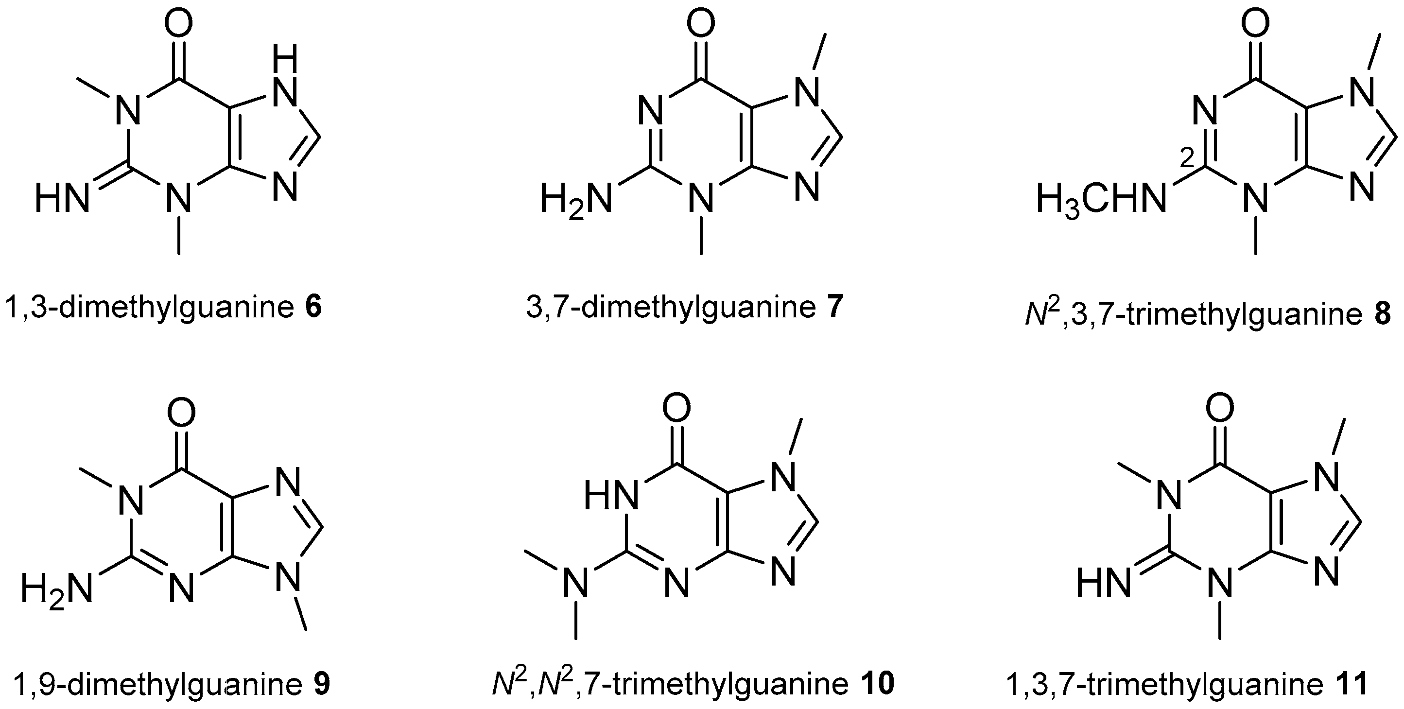
2.1. Adenine and Guanine Derivatives

Since the initial isolation of 1-methyladenine, 1, and herbipoline, 2 (Figure 1), from the sponge Geodia gigas by Ackermann et al. in the early 1960s [5,6], a few more examples of methyl-guanines have been isolated from marine organisms.
From Jaspis sp. sponges collected off Hachijo-jima, Japan, Yagi et al. isolated 1-methylherbipoline, 3, as a salt of an unidentified anion that showed a moderate collagenase inhibitory activity [7]. A few years later, Kimura et al. identified 1-methylherbipoline salts of halisulphate-1, 4, and suvanine, 5, from the sponge Coscinoderma mathewsi collected at Pohnpei, Micronesia (Figure 1). Both salts showed moderate antithrombin and antitrypsin activity, which was ascribed to the sesterterpene sulphate counterions rather than to the methylated purine [8]. 1-Methylherbipoline, 3, was also isolated from a Papua New Guinea ascidian Eudistoma sp. [9].
1,3-Dimethylguanine, 6 (Figure 2), was isolated from the marine ascidian Botrylloides leachi in relatively high proportion, although its physiological role in ascidian was not determined. This marine organism was collected at Marlborough Sound, New Zealand [10].
3,7-Dimethylguanine, 7, was isolated from the marine sponge Zyzzya fuliginosa collected in Batanes, Philippines. It was evaluated for cytotoxic activity, although it did not demonstrated bioactivity on the two cell lines tested, human T-cell leukemia CCRF CEM and human colon carcinoma HCT-116 [11]. A derivative of 3,7-dimethylguanine with an additional methyl at N2, i.e., N2,3,7-trimethylguanine, 8, was isolated from the marine sponge Petrosia sp. collected at Jeddah, Saudi Arabia, on the Red Sea coast. It showed moderate to low cytotoxicity against human hepatocellular liver carcinoma (HepG2) and human breast adenocarcinoma (MCF-7) [12]. 1,9-Dimethylguanine, 9, previously known as a synthetic compound, was isolated as a natural product from the Antarctic sponge Isodictya erinacea together with 7-methyladenine [13]. The trimethyl derivative N2,N2,7-trimethylguanine, 10, has been identified from the New Zealand ascidian Lissoclinum notti [14].
1,3,7-Trimethylguanine, 11 (Figure 2), was first isolated from the sponge Latrunculia brevis, collected at the Otago Peninsula, New Zealand [15], and also detected in the methanol extract of an Indonesian ascidian Eudistoma sp. [16] and later from the Australian Eudistoma maculosum [17]. It was inactive against the P-388 murine leukaemia cell line in vitro (EC50 > 10 μg/mL) and had no detectable antiviral or antimicrobial activity.

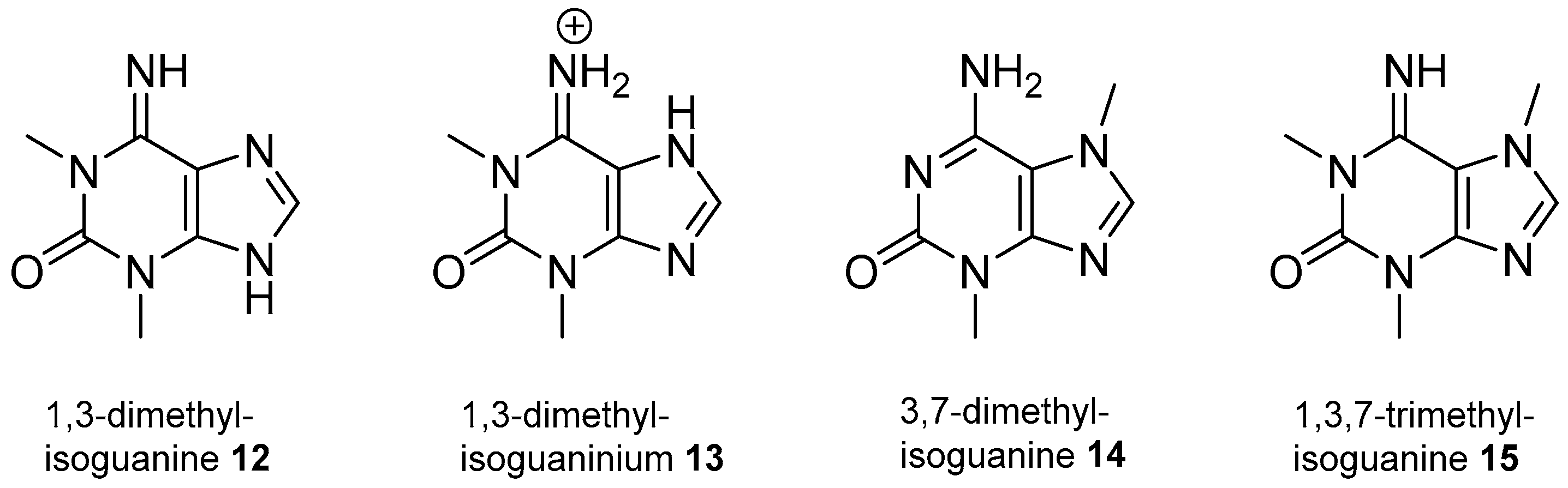
2.2. Isoguanine Derivatives

Mitchell et al. [18] and Chehade et al. [19] reported almost simultaneously the isolation of 1,3-dimethylisoguanine, 12 (Figure 3), as a natural product from the same sponge species Amphimedon viridis (formerly known as Haliclona viridis), collected in Harrington Sound, Bermuda, and in the Sao Sebastiao channel, on the southeastern coast of Brazil, respectively. Its hydrochloric salt, in the form of 1,3-dimethylisoguaninium, 13, was also reported from the sponge Amphimedon paraviridis, collected off Okinawa, Japan [20]. The same 1,3-dimethylisoguanine, 12, was isolated from the New Zealand ascidians Cnemidocarpa bicornuta [21] and Pseudodistoma cereum [22]. It was tested on 26 human cancer cell lines showing the highest cytotoxicity on an ovarian cancer cell line (IC50 2.1 μg/mL) [18]. Additionally, it was essentially inactive in haemolytic and antimitotic bioassays but stimulated mammalian gut motility in a dose-dependent manner [19] and showed a promising antiangiogenic activity [20].
3,7-Dimethylisoguanine, 14 (Figure 3), has been isolated from several marine organisms such as the sponges Agelas longissima [23] from the Caribbean Sea, Zyzzya fuliginosa (named as Z. fuliginosus) collected at Chuuk Atoll, Micronesia [24] and the gorgonian Paramuricea clavata [25], collected in the Western Mediterranean Sea. From the latter, the trimethylated derivative 1,3,7-trimethylisoguanine, 15, was also isolated showing a potential antifouling activity with low toxicity [25]. The same 1,3,7-trimethylisoguanine, 15, was previously isolated, for the first time as a natural product, from the New Zealand ascidian Pseudodistoma cereum [22].
Purines methylated at the C2 or C6 groups have also been described as natural products. Thus, mucronatine, 16 (Figure 4), a rare example of purine with a methoxy group at C2, was identified from the sponge Stryphnus mucronatus collected in the Mediterranean Sea at La Ciotat (France) [26]. Mucronatine, 16, can be considered as an O-methylisoguanine derivative that showed no cytotoxicity on KB cells, weak toxicity on the brine shrimp assay and promising antifouling properties. The trimethyl derivative N6-methylmucronatine, 17 (Figure 4), was more recently isolated from an unidentified dictyoceratid sponge collected in South East Queensland, Australia, and its structure was confirmed by X-ray crystallography [27].
Among the examples of methylisoguanines alkylated at the C6-amino group are nigricines 1–4, 1821 (Figure 5), isolated from the sponge Petrosia nigricans collected in Pulau Baranglompo, Indonesia [28]. Nigricines 1–3 are 3,9-dimethylpurines, while nigricine 4 is a 3,7,9-trimethylated derivative. All of them bear a propanoate moiety at N6 position and were not cytotoxic against the murine lymphoma cell line L5178Y.

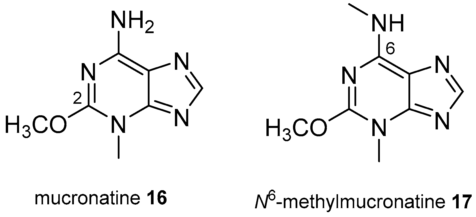
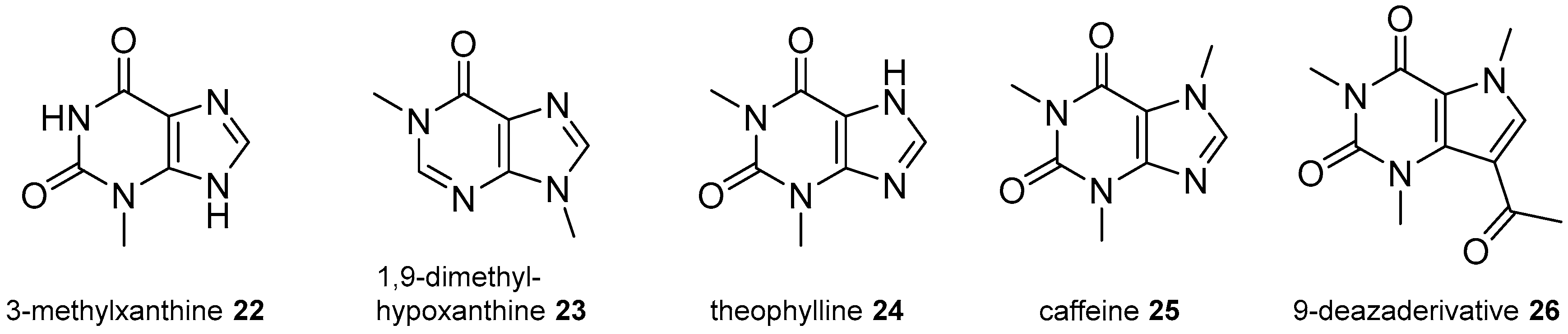
2.3. Xanthine Derivatives

Few examples of xanthine derivatives have been identified from marine organisms. 3-Methylxanthine, 22 (Figure 6), was isolated from the ascidian Symplegma rubra, collected on the southeastern coast of Brazil [29]. Other analogues are 1,9-dimethylhypoxanthine, 23, isolated from a southern Australian sponge Spongosorites sp. [30], theophylline, 24, from Amphimedon viridis [18] and caffeine, 25, from the gorgonian coral Verrucella umbraculum collected off the coast of Hainan Island, South China Sea, along with the 9-deazaderivative 26 [31].

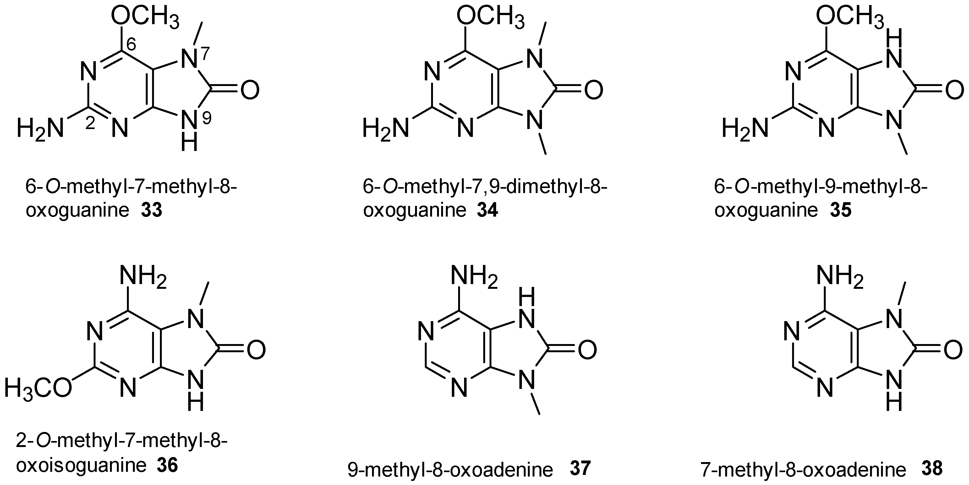
2.4. 8-Oxopurine Derivatives

8-Oxopurines are uncommon in nature, although several analogues, derived from adenine, guanine or isoguanine, and bearing methyl substituents at various positions, have been isolated from marine organisms. Although they could have been considered in the previous sections as derivatives of the more common nucleic bases, we decided to group them in this section because of the unusual 8-oxo function.
Thus, 1,9-dimethyl-6-imino-8-oxopurine, 27 (Figure 7), was isolated from the English Channel sponge Hymeniacidon sanguinea [32]. The unusual methylimino group at C6 is also present in caissarone, 28 (3,9-dimethyl-6-methylimino-8-oxopurine, Figure 7), which is a quaternary derivative isolated as the hydrochloride salt from Bunodosoma caissarum, a sea anemone abundant in the reefs of the Brazilian coast. This structure was confirmed by X-ray crystallography [33] and later by synthesis [34] (see Section 5). Caissarone was inactive against the P-388 leukaemia cell line [33], although it induced morphological anomalies in sea-urchin egg development [35] and acted as a simple competitive adenosine receptor antagonist [36].
From the New Zealand ascidian Pseudodistoma cereum [37] collected at the Three Kings Islands, Appleton et al. isolated an 8-oxopurine derivative that had no detectable cytotoxic, antibacterial or antifungal activities. They identified it as 1,3-dimethyl-8-oxoisoguanine, although this structure was corrected a few years later by Sakaruda et al., who isolated a series of methylated isomers of 8-oxoisoguanines from several Palauan sponges including Cribrochalina olemda, Haliclona sp. and Amphimedon sp. [38]. These isomers were identified as 1,9-dimethyl-8-oxoisoguanine, 29, 1,3-dimethyl-8-oxoisoguanine, 30, 3,9-dimethyl-8-oxoisoguanine, 31, and 9-methyl-8-oxoisoguanine, 32 (Figure 8). The thorough spectroscopic analysis of these compounds performed by Sakurada et al. suggested that the isoguanine previously isolated from the ascidian P. cereum suited better the 3,9-isomer rather than the 1,3-isomer proposed by Appleton [37]. These 8-oxoisoguanines seemed to be responsible for the convulsant effect observed for the aqueous extracts of these Palauan sponges, being potential new neuropharmacological agents. Indeed, 1,9-dimethyl-8-oxoisoguanine, 29, induced convulsions in a dose-dependent manner through inhibition of neuronal GABAergic transmission [38].
Other natural 8-oxopurines were isolated from two specimens of the ascidian Symplegma rubra collected on distant geographical areas. 6-O-Methyl-7-methyl-8-oxoguanine, 33 (Figure 9), was first isolated from S. rubra collected on the southeastern coast of Brazil [29] along with 8-oxoadenine and 3-methylxanthine, 22. The same three purines were identified from a specimen of S. rubra collected at the Saudi Red Sea coast [39] along with several other unknown and known purine derivatives (Figure 9): 6-O-methyl-7,9-dimethyl-8-oxoguanine, 34, 6-O-methyl-9-methyl-8-oxoguanine, 35, 2-O-methyl-7-methyl-8-oxoisoguanine, 36, 9-methyl-8-oxoadenine, 37, 7-methyl-8-oxoadenine, 38, and the nucleoside inosine. These 8-oxopurines showed moderate inhibitory activity against several protein kinases [39].

3. N-Alkylpurines with Substituents Other Than Methyl

Apart from the methylpurines shown above, derivatives with larger alkyl chains have been isolated from marine organisms. Perhaps the most common compounds are those bearing terpenic chains attached to adenine residues, for which a variety of biological properties have been described and covered by previous reviews [40,41]. However, in this section, we have decided to include all the terpenylpurines described, but grouped according the terpenoid skeleton of the chain instead the usual chronological isolation sequence, with the aim to give a more intuitive insight into the structures.

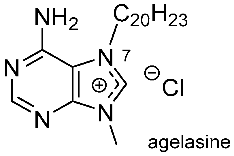
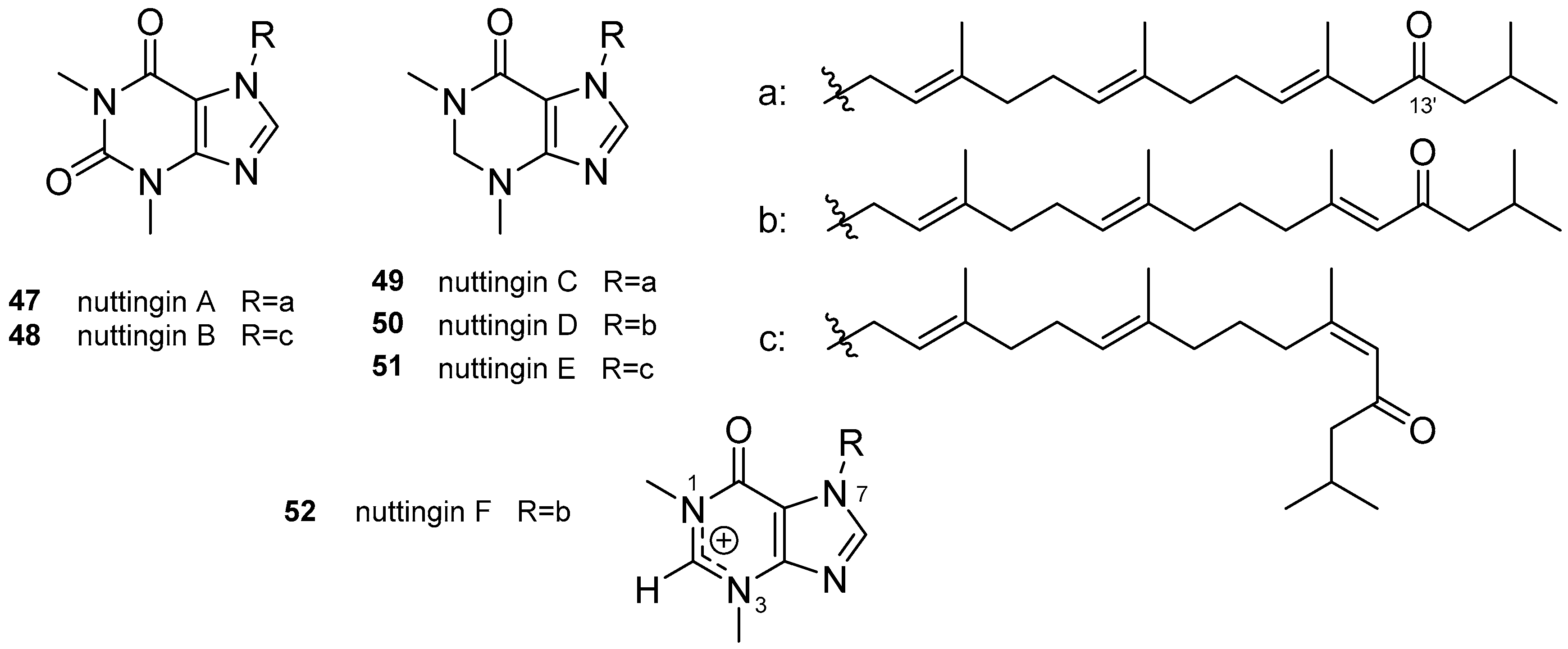
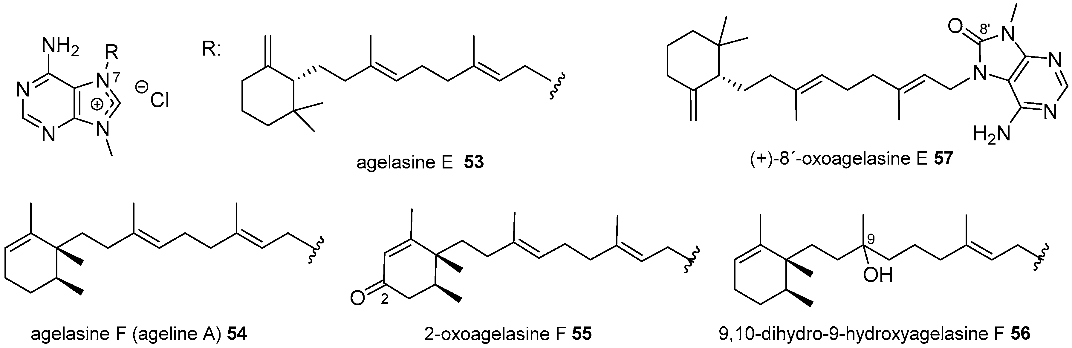
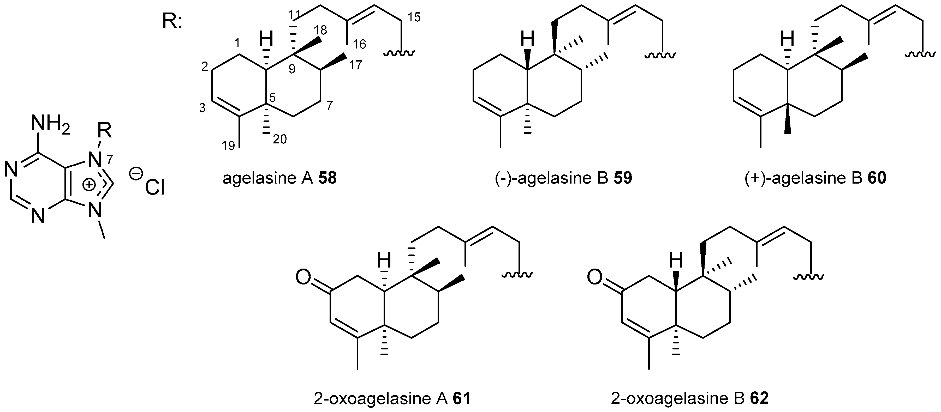
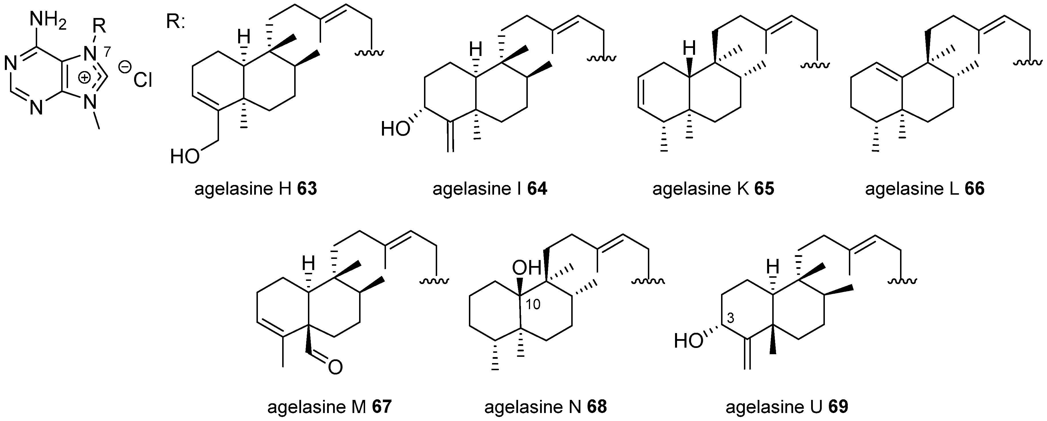
3.1. Terpenylpurines

Most of the compounds included in this group are derived from 9-methyladenine, mainly as an adeninium salt. The first compound of this type was described in 1975 by Cullen et al. [42], who described the isolation of a secondary metabolite from the sponge Agelas dispar, collected at Barbados that was called agelasine (Figure 10), in which a terpenyl moiety was attached to position 7 of a quaternary 9-methyladeninium.
The structure of the terpenyl core was undefined although it was described as a bicyclic diterpene. Since then, many other diterpenylpurines have been isolated from different marine organisms. They are derived either from adenine or from other purines with diterpenyl groups of different nature, including acyclic, monocyclic and bicyclic terpenyl moieties. They are described below starting from the acyclic diterpenes.

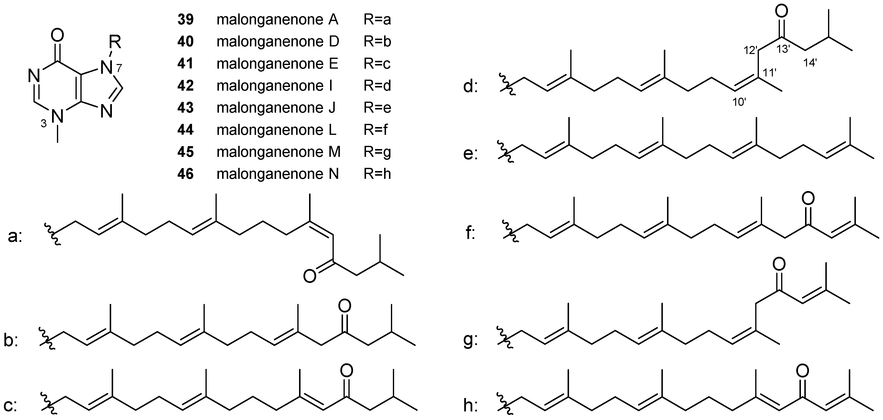
3.1.1. Terpenylpurines with an Acyclic Diterpenoid

There are two groups of marine metabolites, named malonganenones (Figure 11) and nuttingins (Figure 12), that bear an acyclic diterpenoid geranylgeranyl chain attached to the position 7 of purine heterocycles such as 3-methylhypoxanthine, 1,3-dimethylxanthine and 1,3-dimethylhypoxanthine.
Malonganenones (Figure 11) are isolated from gorgonians. Malonganenone A, 39, was first isolated from the gorgonian Leptogorgia gilchristi, collected near Ponto Malongane, Mozambique [43]. It was also isolated together with the new derivatives malonganenones D, 40, and E, 41, from the gorgonian Euplexaura nuttingi collected in Pemba Island, Tanzania [44]. Other compounds of the same family are malonganenones I, 42, and J, 43, identified from the South China Sea gorgonian Euplexaura robusta collected on Weizhou Island, China [45]. The last compounds identified of this group are malonganenones L, 44, M, 45, and N, 46 (Figure 11), isolated from the gorgonian Echinogorgia pseudossapo collected in Daya Bay, China [46]. All of them are 3-methylhypoxanthines with the diterpenyl chain attached to position 7. Malonganenone J, 43, has an unmodified geranylgeranyl side chain, while the others have a carbonyl function at C13′ and differ in the presence or absence of a double bond at C14′ and in the position and Z–E configuration of the double bond at C10′ or C11′. These compounds were evaluated for their cytotoxicity against K562 and HeLa tumour cell lines and it was found that malonganenones A, D, E and I, 3942, showed good to moderate cytotoxicities with IC50 values ranging from 0.35 to 10.82 μM while malonganenone J, 43, showed IC50 > 53 μM, suggesting that the carbonyl function at the side chain was important for the bioactivity. The inhibitory activity against the receptor tyrosinekinase (RTK) c-Met was also evaluated but only malonganenone E, 41, showed moderate inhibitory activity at a concentration of 10 μM [45]. Malonganenones L–N, 4446, were also tested as inhibitors of several phosphodiesterases showing moderate activity [46].
Others secondary metabolites isolated from these gorgonians are also named malonganenones and have the same tetraprenylated side chains but they are not purine derivatives. Malonganenones B, F, G, O, P and Q have the diterpenic chain joined to a trisubstituted imidazole fragment and could be biosynthetic precursors of the above purines and malonganenones C, H and K are N-diterpenylformamides [43,44,45,46].
During the same study on the Tanzanian gorgonian E. nuttingi, nuttingins A–F, 4752 (Figure 12), were also isolated and identified having the same 13′-oxo-prenylated side chains of malonganenones. Nuttingins A and B, 4748, are 1,3-dimethylxanthine derivatives; nutingins C–E, 4951, are 1,3-dimethyl-2,3-dihydrohypoxanthines and nuttingin F, 52, is a 1,3-dimethylhypoxanthine derivative with an unusual naturally positive charge delocalised over the two nitrogens N1 and N3. Nuttingins A–E, 4751, induce apoptosis in transformed mammalian cells and displayed inhibitory activity against both K562 and UT7 tumour cell lines; nuttingins C–E, 4951, are approximately three-fold more potent than the others [44].

3.1.2. Terpenylpurines with a Monocyclic Diterpenoid

Capon and Faulkner [47] isolated a 9-methyladeninium salt that they called ageline A (Figure 13), from Agelas sp. collected at Palau, Western Caroline Islands, for which mild ichthyotoxic and moderate antimicrobial activities were described and which contain a monocyclic diterpenoid fragment. Almost simultaneously, Nakamura et al. [48] isolated another two 9-methyladeninium salts with a monocyclic diterpenoid chain from the Okinawan Sea sponge Agelas nakamurai, named agelasine E and F, 5354 (Figure 13). Agelasine F, 54, was proven to be identical to ageline A; since then, all the diterpenoid 9-adeninuim salts are named agelasines. Agelasine F, 54, has also been isolated from an orange Agelas sp. collected at Baler, Philippines [49] and from Agelas axifera from Republic of Palau [50]. Both showed interesting antituberculosis activity [49,51].
Recent studies allowed the isolation of new analogues oxidised at the terpenic chain, i.e., 2-oxoagelasine F, 55, and 9,10-dihydro-9-hydroxyagelasine F, 56, from A. nakamurai [52], and oxidised at the purine as (+)-8′-oxoagelasine E, 57, (Figure 13), from the South China Sea sponge Agelas mauritiana [53].

3.1.3. Terpenylpurines with a Bicyclic Diterpenoid

In this section, the largest group of terpenyladenine marine metabolites are considered. They are again derived mainly from 9-methyladeninium and the diterpenoid chain attached to position 7 can be considered to be derived from labdane, clerodane and halimane type skeletons, which can be found in both enantiomeric series or epimerised at any position with a trans- or cis-fused decaline structures. Halimanes and clerodanes arise biogenetically from labdanes by one and two methyl migrations, respectively, and, sometimes, the three types are found in the same species. Although the agelasines have been given correlative letters from A to V independently of the kind of diterpene skeleton, we have grouped them according to the diterpenoid type rather than following a chronological description to better underscore the structural similarity. The relationship between the structure of the diterpene skeleton and the letter assigned to each compound is shown in Table 1.

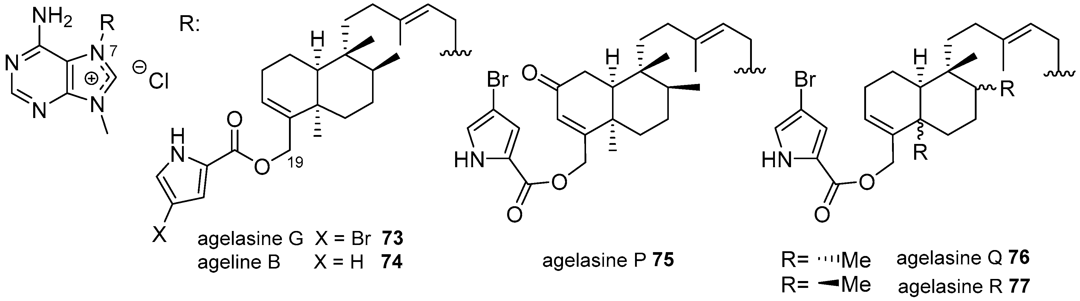
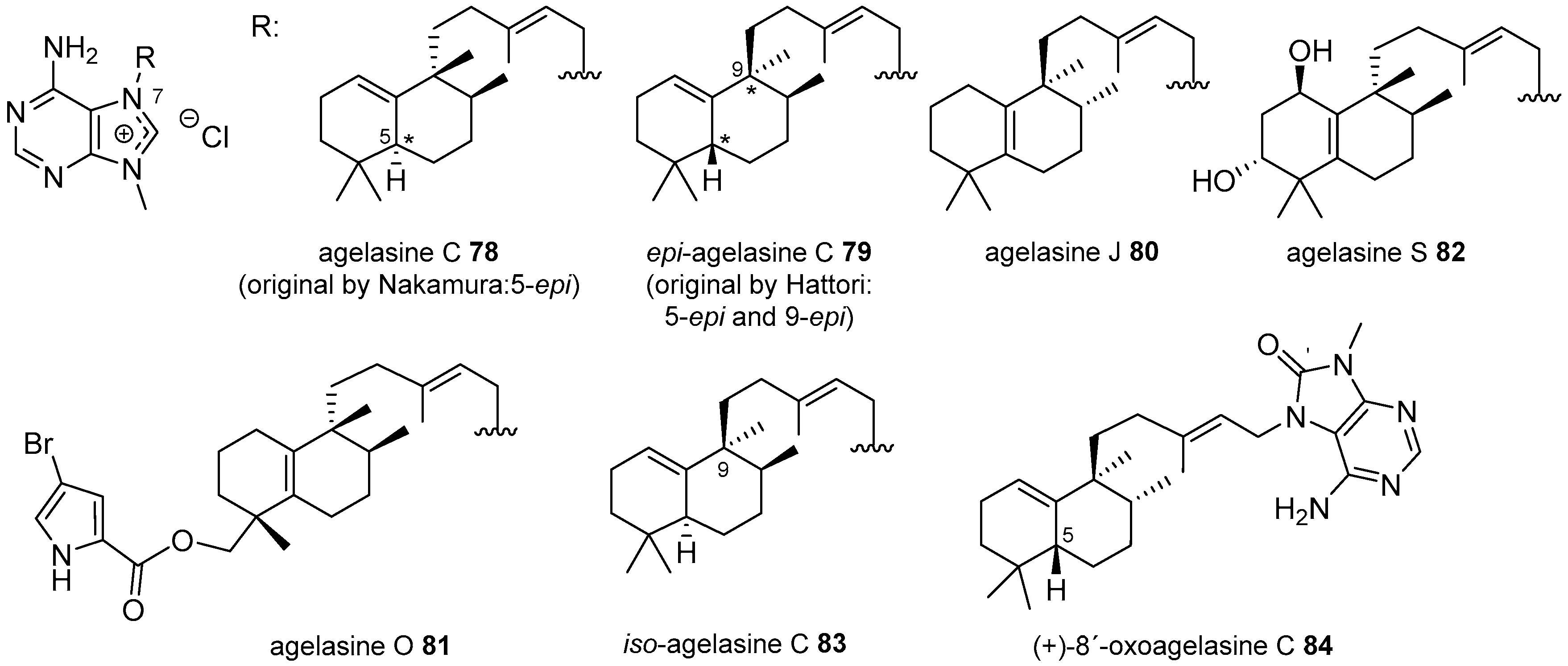
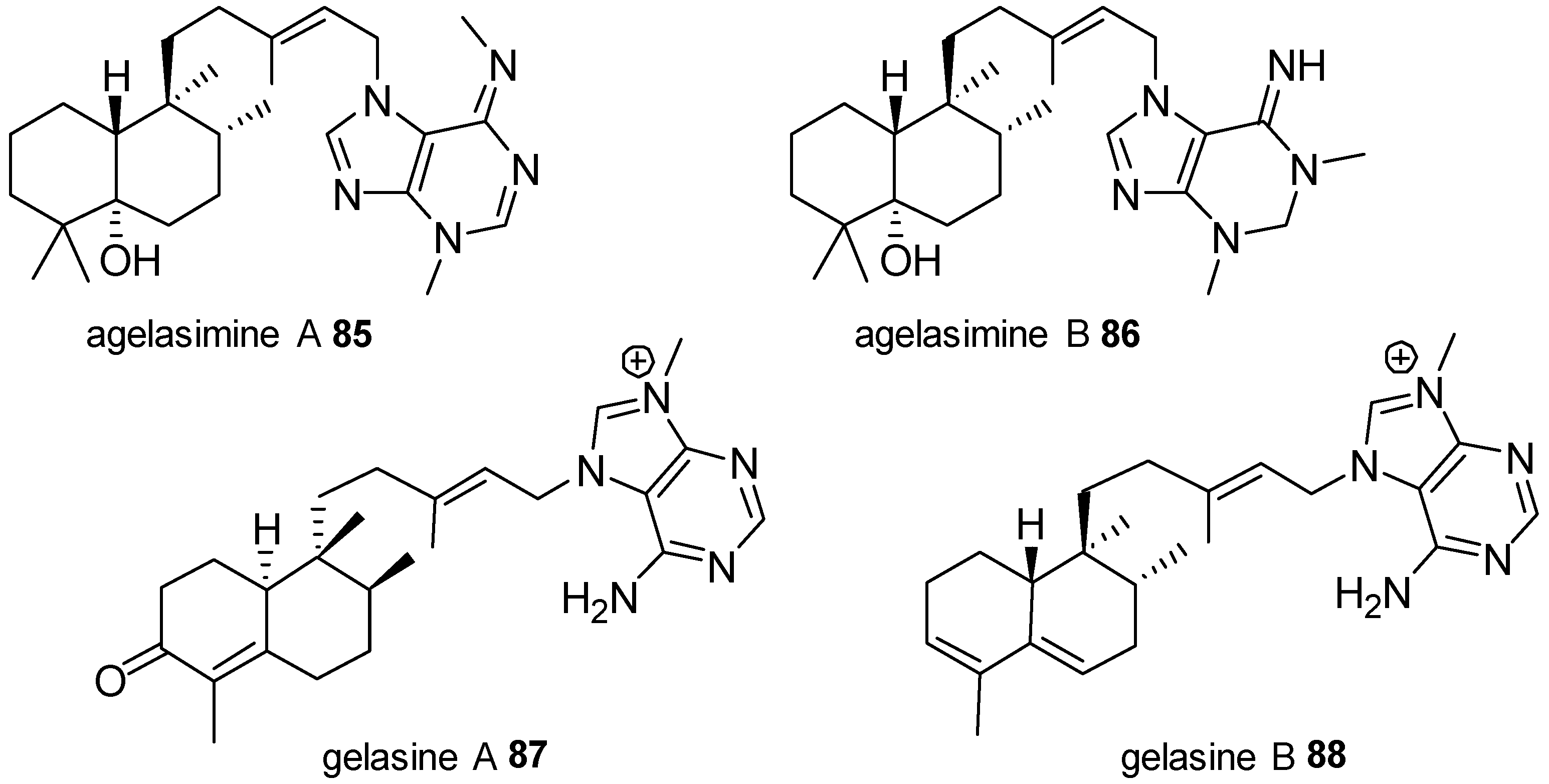
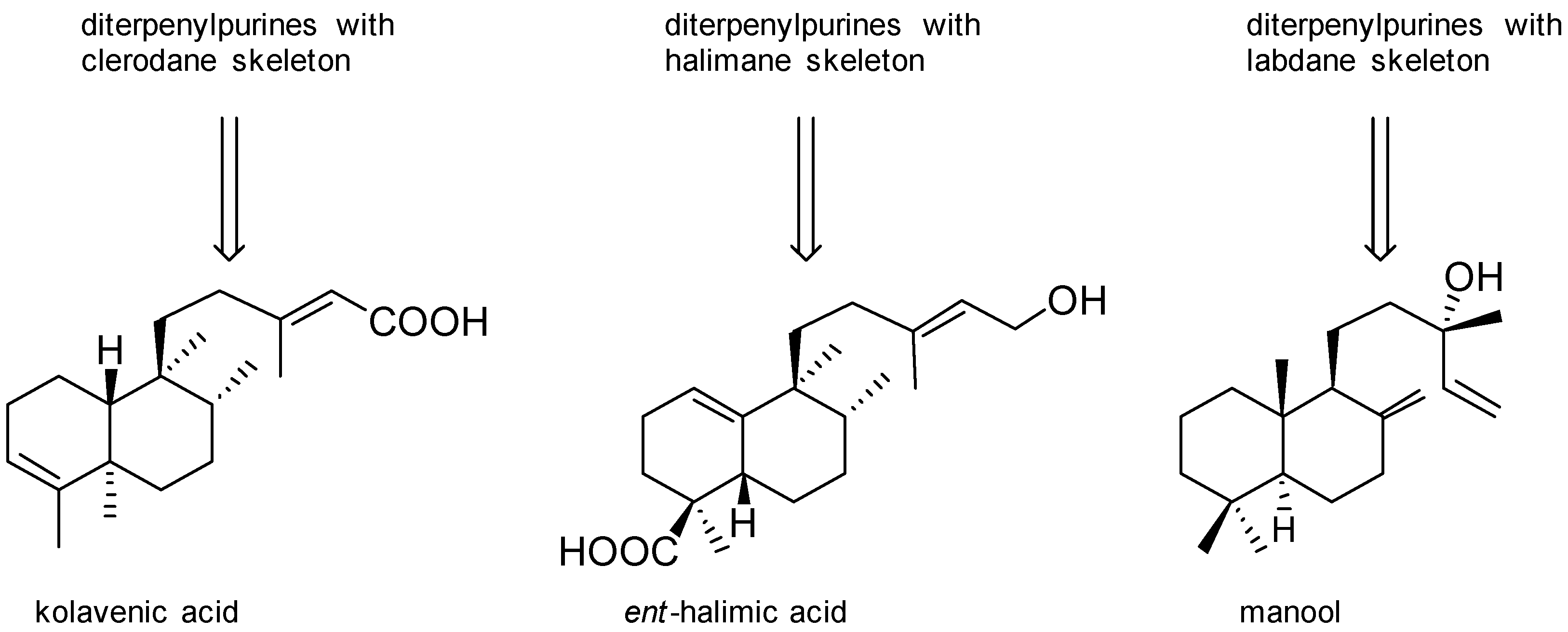
Agelasines with a Clerodane Type Diterpenoid

The first agelasines with a clerodane type diterpenoid were agelasine A, 58, and agelasine B, 59 (Figure 14), isolated by Nakamura et al. from A. nakamurai collected at Zanpa Cape, Okinawa [54,55]. Both compounds differ in the stereochemistry of the decaline core. The enantiomer (+)-agelasine B, 60, has also been isolated from A. mauritiana collected at Yongxing Island in the South China Sea [53]. The corresponding analogues with a carbonyl group at position 2 of the diterpenoid moiety, 2-oxoagelasine A, 61, and 2-oxoagelasine B, 62, were isolated several years later from the same species collected at Okinawa [52] and an Agelas sp. from Papua New Guinea, respectively [56].
The isomers agelasine H, 63, and agelasine I, 64, (Figure 15), were isolated from another Agelas sp. collected at Yap Island, Micronesia and they differed in the position of an allylic hydroxyl group [57]. They were isolated together with agelasines A, 58, agelasine B, 59, and agelasine F, 54, and some of them showed varied degrees of antimicrobial activity.
The next clerodane type derivatives are agelasine K, 65, and agelasine L, 66, isolated from A. cf. mauritiana collected at Solomon Islands [58], which differ in the position of a double bond on the decaline core (Figure 15). They showed mild activity on Plasmodium falciparum. Agelasine M, 67, and 2-oxoagelasine B, 62, identified from a Papua New Guinea Agelas sp. showed interesting activity against Trypanosoma brucei [56]. A new derivative with a hydroxyl group at position 10, agelasine N, 68, was isolated from the Caribbean sponge Agelas citrina together with some previously described agelasines [59]. Agelasine U, 69, isolated from an Okinawan Agelas sp. and belonging to the enantiomeric series as 67, had also a hydroxyl group but at C3 [60].
Derivatives with different purine moieties have been identified from A. mauritiana collected at different places, such as the 8-oxopurine analogues (−)-8′-oxoagelasine B, 70, and agelasine V, 71, (Figure 16), from the sponge collected at Yongxing Island [53] and the oxime derivative, ageloxime B, 72, from a specimen collected at Paracel Islands [61], both in the South China Sea. Antibacterial and cytotoxic assays, performed with these compounds, seem to indicate that those with the 8′-oxo function were less potent than the other analogues [53]. The agelasine 70 was almost simultaneously isolated from Agelas aff. nemoechinata collected also at Paracel Islands but it was given the name nemoechine D [62].
There are some clerodane derivatives that also bear a pyrrole or bromopyrrole attached to position 19 of the diterpenoid by an ester function. The first of this group were agelasine G, 73, and ageline B, 74, (Figure 17), isolated first from an Okinawan Agelas sp. [63] and later on from A. nakamurai [49,52]. In addition to anti-leukaemic activity, agelasine G showed selective inhibition of protein tyrosine phosphatase 1B, which suggest activation of the insulin signalling pathway, while the analogue lacking bromine was nearly inactive [64]. Other agelasines with a bromopyrrole substituent on a clerodane skeleton are agelasines P, Q and R, 7577 (Figure 17) [60].

Agelasines with an Halimane Type Diterpenoid

The first described 9-methyladeninium alkylpurines with an halimane derivative were agelasine C, 78, (Figure 18), from the Okinawan Agelas sp. [54] and later from A. mauritiana collected from Pohnpei, Micronesia, together with epi-agelasine C, 79, for which antifouling activity against macroalgae was described [65]. The structure of these compounds was later corrected to that shown in Figure 18 after the synthesis of its enantiomer by Marcos et al. [66].
Agelasine J, 80, was isolated from A. cf. mauritiana [58] together with the clerodane agelasines K and L, 6566. Other examples isolated from the Okinawan Agelas sp., are agelasines O and S, 8182, with a bromopyrrole substituent and two hydroxyl groups at the decaline core, respectively [60].
The sponge A. nakamurai, collected at Xisha Islands in the South China Sea, yielded iso-agelasine C, 83, (corresponding to 9-epi-agelasine C) together with agelasines B, 59, J, 80, and 2-oxo-agelasine B, 62, for which cytotoxic, antifungal and antibacterial activities were evaluated [67]. It was found that the presence of a carbonyl in the diterpene skeleton decreases cytotoxic and antimicrobial activities. In this group, an 8-oxopurine derivative has also been recently described, namely 8′-oxoagelasine C, 84 (Figure 18) [53].
Derived from an halimane diterpenoid are the only two non-quaternary adenine derivatives, agelasimine A, 85, and B, 86, (Figure 19), that were isolated from A. mauritiana collected at Enewetak Atoll, Marshall Islands (Pacific Ocean) [68]. Both have the same diterpenic moiety attached to position N7 of 3,N6-dimethyladenine and 1,3-dimethyladenine, respectively. They have been tested for a variety of biological activities and both were cytotoxic against L1210 leukaemia cells at nanomolar range. They also inhibited smooth muscle contractions induced by potassium chloride and reversed the effects of a variety of neurotransmitters at micromolar concentrations. It was proposed that may act as Ca2+-channel antagonists and α1 adrenergic blockers [69].
Two unusual nor-diterpenoid derivatives, gelasine A and B, 8788 (Figure 19), were isolated from the Papua New Guinea Agelas sp. in which the C19 methyl group is lost [56]. Gelasine A, 87, has an α,β-unsaturated carbonyl, and gelasine B, 88, a conjugate diene with an enantiomeric decalin core.

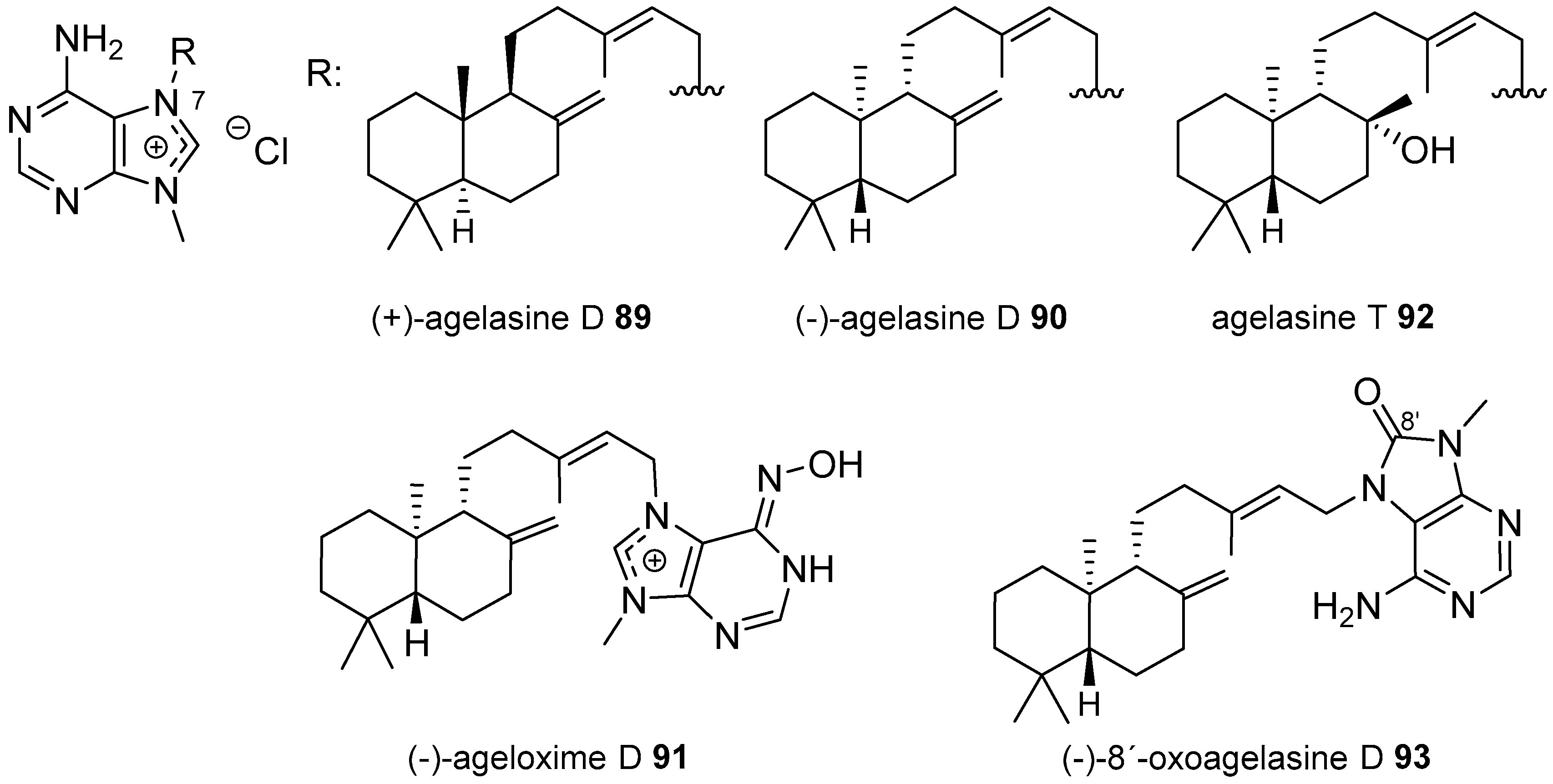
Agelasines with a Labdane Type Diterpenoid

Finally, labdane type agelasines have also been isolated from genus Agelas. Agelasine D is one of the examples in which both enantiomers have been described from different specimens of the same species. (+)-Agelasine D, 89, was described by Nakamura in 1984 together with agelasines A–C, 58, 59 and 78, from A. nakamurai showing inhibitory effects on Na+,K+-ATPase as well as antimicrobial activity and an interesting cytotoxicity against several cell lines [54,55].
The enantiomer (−)-agelasine D, 90, and its oxime derivative (−)-ageloxime D, 91, (Figure 20) were also identified from a specimen of A. nakamurai collected at Bali Island, Indonesia for which not only cytotoxicity has been described but also anti-fouling activity [70]. The results indicate that (−)-agelasine D, 90, was more potent as a cytotoxic agent and the oxime derivative showed better results on the anti-fouling assay. They were tested also for biofilm inhibition against Staphylococcus epidermidis, founding that (−)-agelasine D, 90, inhibits the growth of the planktonic bacteria but not the biofilm formation, while the oxime had the opposite behaviour [70]. A hydroxylated labdane is present in agelasine T, 92 (Figure 20), isolated from an Okinawan Agelas sp. that showed antimicrobial activity against several bacteria and fungi [60]. (−)-8′-Oxoagelasine D, 93, was identified from two species of the Paracel islands, A. mauritiana [61] and A. aff. nemoechinata [62]. It showed antifungal and anti-leishmanial activity as did the other oxime derivatives (−)-ageloxime B, 72, and (−)-ageloxime D, 91, isolated in one of the studies [61], as well as cytotoxicity against the HeLa cell line [62].
(−)-8′-Oxoagelasine D, 93, and (+)-agelasine D, 89, were also identified, from A. mauritiana collected at Yongxing Island in the South China Sea together with other 8′-oxo derivatives described above with clerodane and halimane diterpenoids [53]. Antibacterial and cytotoxic assays seem to indicate that compounds with the 8′-oxo function were less potent than the other analogues.

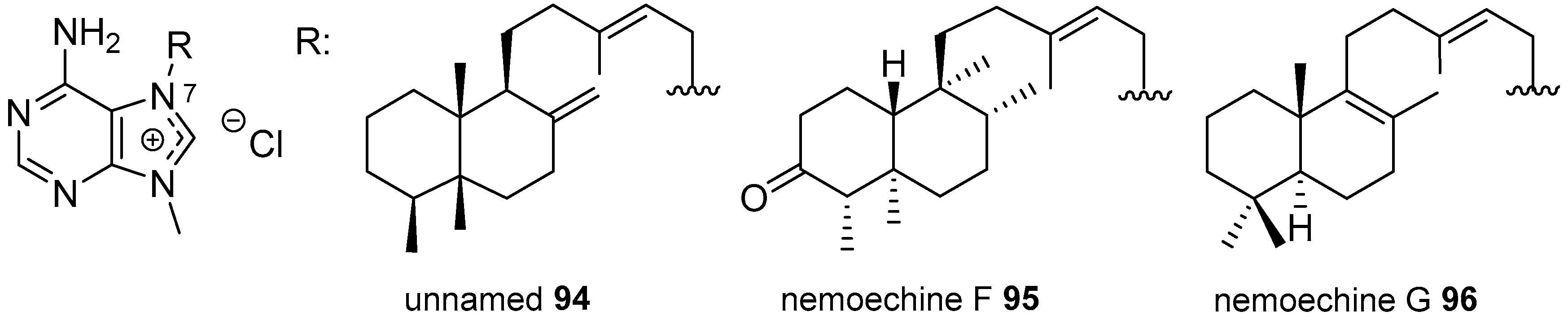
Diterpenylpurines Other Than Agelasines

The unnamed diterpenylpurine 94 (Figure 21), having an unusual telepogane skeleton which could be considered in the midway from labdane to clerodane, was isolated from A. nakamurai from Fly Islands, Papua New Guinea, together with agelasine B, 59 [71].
From A. aff. nemoechinata collected at the coral reef regions in the South China Sea, two new diterpene derivatives, nemoechine F and G, 9596, were isolated (Figure 21). They have clerodane and labdane skeletons respectively. They were tested for antimicrobial and cytotoxic activities against several tumour cell lines but only nemoechine G, 96, showed weak cytotoxicity against Jurkat cell line [72]. Nemoechine G, 96, was also identified in the South China Sea A. nakamurai [67].

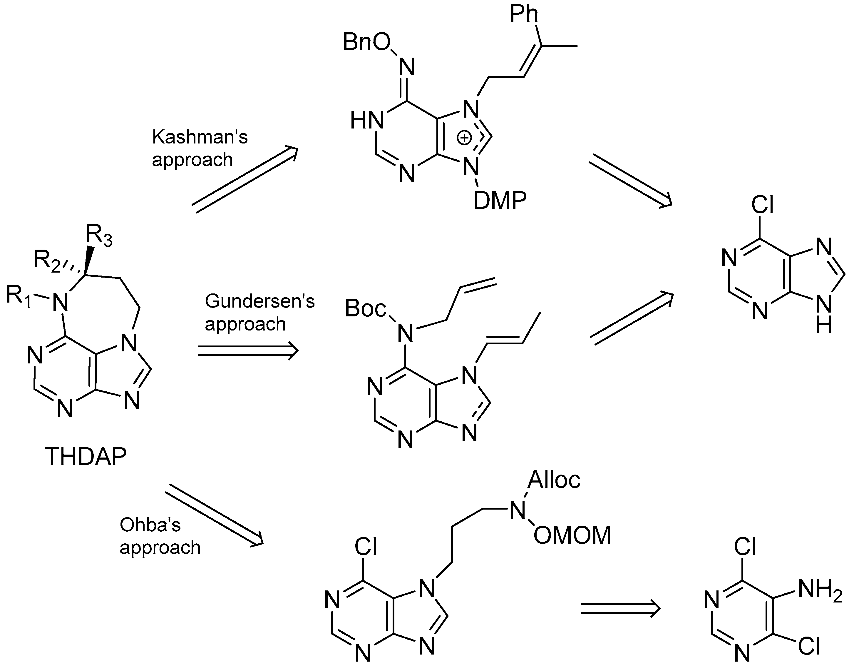
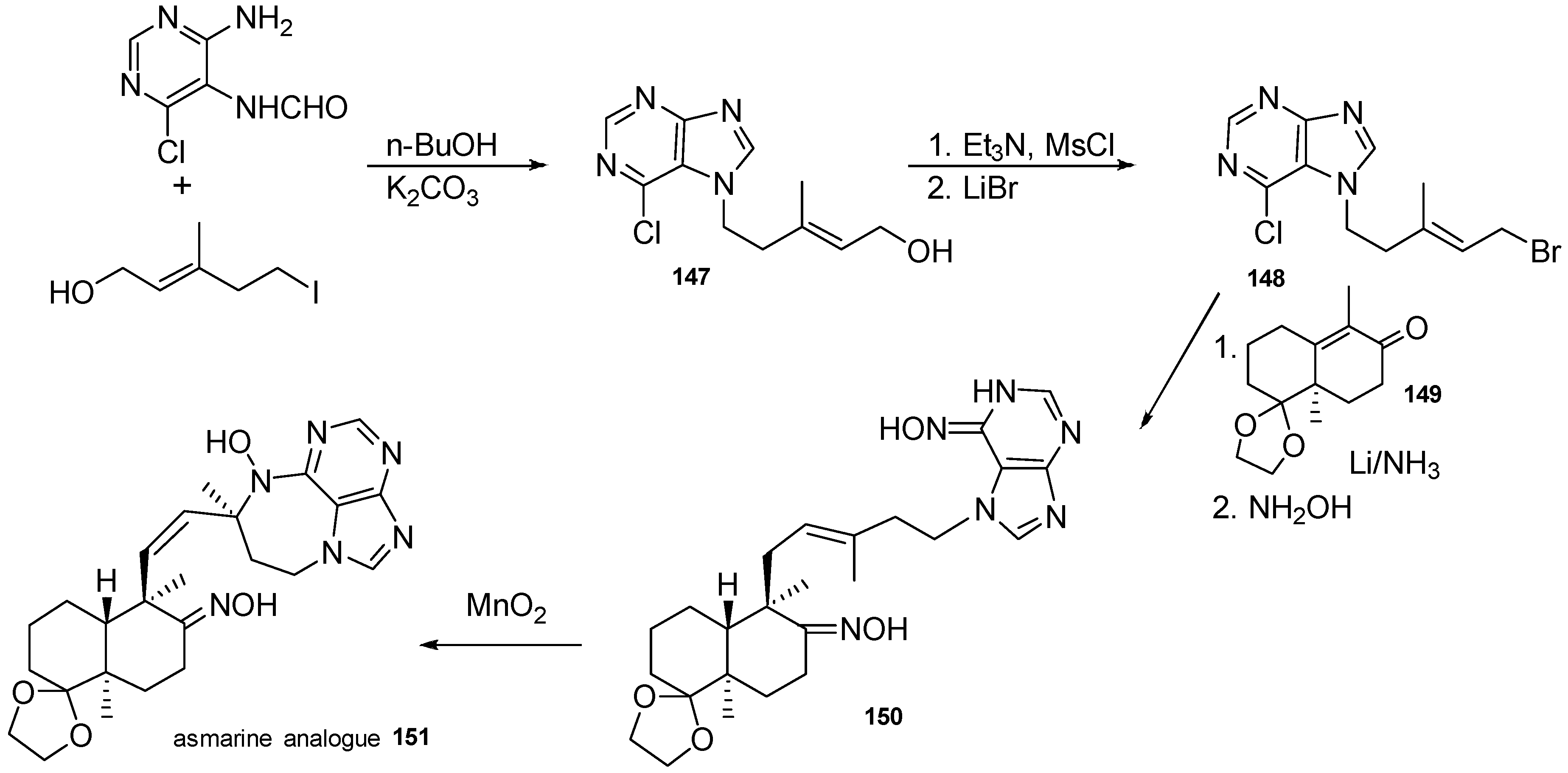
3.1.4. Terpenylpurines with a Diazepinopurine Heterocyclic System

Among the terpenylpurines with a bicyclic diterpenoid, particular attention has to be given to the asmarines, a group of 7-terpenylpurines isolated from sponges of genus Raspailia, in which the amino group of the adenine group is cyclised over the side chain of a clerodane diterpenoid forming a new diazepinopurine system. Unlike the agelasines, all of them are neutral and the differences among them lie in the oxidation degree of the heterocyclic ring and in the ring junction of the decalin core.
In 1998, Kashman et al. described the first members of this group, asmarines A and B, 9798 (Figure 22), as the major components of a Red Sea Raspailia sp. collected at Nakora, Eritrea [73]. They had a hydroxyl substituent on the diazepine nitrogen and are epimers at position 5. Two years later, and from the same sponge, four additional asmarines C–F, 99102 (Figure 22), were isolated as two inseparable pairs of compounds that derived from 9-methyl-8-oxopurine and differ also in the cis/trans junction of the decalin [74]. Asmarines C and D, 99100, have no substitution on the nitrogen of the diazepine and asmarines E and F, 101102, had a methoxy group at that position.
From a different Raspailia sp., collected at Wasini Island, Kenya, asmarines G and H, 103104, were identified together with asmarines A, 97, and F, 102, the latter being the major component (Figure 22) [75]. Similar results were obtained with a Raspailia sp. collected at Nosy Be, Madagascar, from which asmarines A, 97, and F, 102, were the major components but three new asmarines I, J and K, 105107, were also identified: asmarine K, 107, is the 5-epi isomer of asmarine H, 104, and asmarines I and J, 105106, have the same diterpenoid moiety differing in the presence of a hydroxyl group in the nitrogen of the diazepine ring [76]; this diterpenoid moiety has the methyl C20 being part of a cyclopropane ring and also the methyl groups C17 and C18 are in trans disposition instead the cis one found in the rest of asmarines.
These compounds have proved to be cytotoxic against several tumour cell lines with asmarine B, 98, being the most potent on human lung and colon carcinomas [73,75]. Not all the asmarines were evaluated because of the small amounts isolated.
Several analogues in which the decalin core has been substituted by simple chains have been synthesised and found that they can also been even more potent as cytotoxics than the natural model compounds [77]. The work of Shenvi and other authors such as Kashman, Gundersen and Ohba on asmarine analogues will be discussed in Section 5.

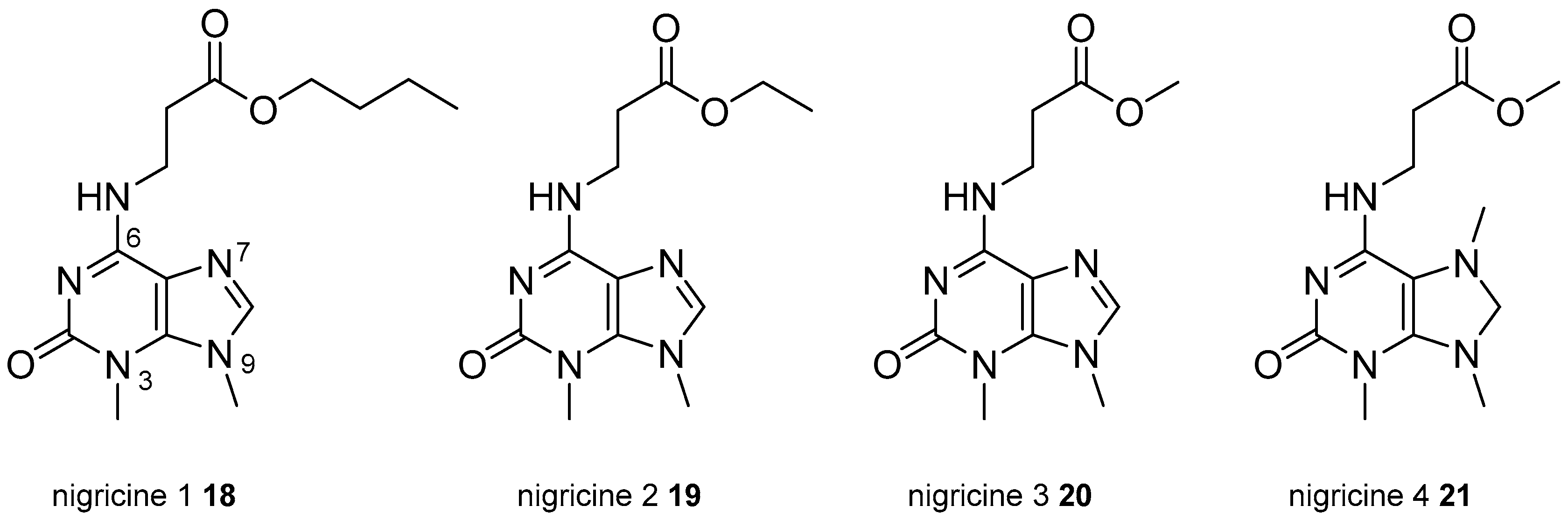
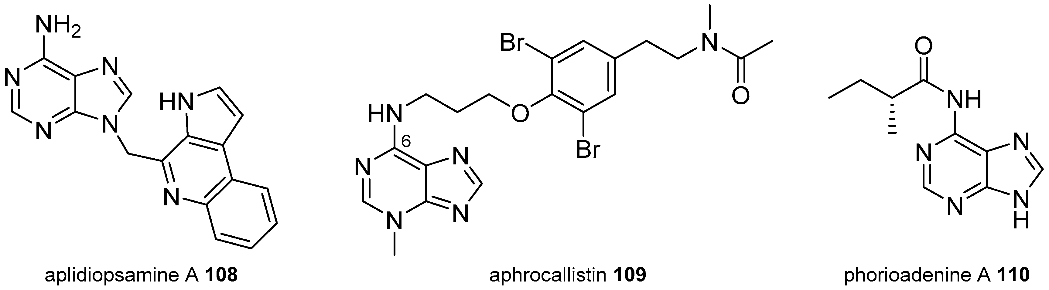
3.2. Other Alkylpurines

In this section, we consider those alkylpurines with substituents other than methyl or diterpenyl substituents at any nitrogen of the purine system.
Several adenines are included in this section, such as the unique adenine derivative with a tricyclic aromatic substituent, aplidiopsamine A, 108, (Figure 23), which was isolated from the Australian ascidian Aplidiopsis confluata collected from Tasmania and showed interesting anti-plasmodial activity on chloroquine-resistant and chloroquine-sensitive strains of Plasmodium falciparum with low toxicity towards human cells [78].
Other adenine and oxoadenine derivatives are alkylated at the N6 amino group. Aphrocallistin, 109 (Figure 23), is a 3-methyladenine bearing a bromotyrosine-derived substituent at N6 having a naturally uncommon dialkylamide; it was isolated from the sponge Aphrocallistes beatrix, collected at the Coral Reefs of Florida, near Fort Pierce [79]. Its structure was confirmed by synthesis and the compound was evaluated in a panel of human tumour cell lines with significant selectivity on colon, melanoma and breast cancer cell lines [79]. Several other pharmacological assays were performed to determine its solubility, permeability or metabolic properties. It was also tested as antimicrobial, although no significant activity was detected [79].
An N6-acyladenine derivative, phorioadenine A, 110 (Figure 23), was identified from the Australian sponge Phoriospongia sp., showing a very interesting nematocidal activity but it was non-antibacterial and non-cytotoxic on several mammalian cancer cell lines [80]. The enantiomer, the racemic and three achiral analogues, obtained by synthesis, were inactive as anthelmintics, suggesting the importance of the nature and chirality of the N-acyl group [80].
From the sponge Isodictya erinacea from McMurdo Sound, Antarctica, erinacean 111 was identified as an 8-oxoadenine alkylated at N6, (Figure 24) [13]. It had weak antibiotic activity and moderate cytotoxicity. Other related 8-oxoadenine derivatives are microxine, 112, isolated from the Australian sponge Microxina sp., which had a weak inhibitory cdc2 kinase activity [81] and aplidiamine, 113, isolated from the ascidian Aplidiopsis sp. [82,83].
From the venom of two cone snail species, Conus genuanus, collected in Sao Vicente, Cape Verde and Conus imperialis, from Hawaii, an unusual small molecule derived from guanine, genuanine, 114, was isolated (Figure 24). It has a methyl at C8 of the purine ring and a propionic acid at position 9 [84]. Its structure was confirmed by synthesis and some regioisomers were also obtained. It induced paralysis in mice at the nanomolar level mimicking the potent paralytic activity found in the venom extract [84].
Phidolopin, 115, is a relatively rare natural xanthine containing a nitro group that was isolated from the bryozoan Phidolopora pacifica, collected off Vancouver Island, Canada. It is considered largely responsible for the antifungal and anti-algae activities of the extracts [85].
From the South China Sea gorgonian Subergorgia suberosa, the dioxopurine/enol tautomer derivatives 116121, (Figure 25), were isolated. Two of them, 116 and 117, have the sesquiterpenoid suberosanone as substituent either at 1 or 9 positions; the next two compounds, 118 and 119, bear a 2-oxobutyl rest also at 1 or 9 positions; and derivatives 120 and 121 have an additional pyrimidine fused ring. All of them showed from moderate to weak cytotoxicity towards two human cancer cell lines [86,87,88].
Acremolin, 122, was obtained in 2012 by fermentation of the marine-derived fungus Acremonium strictum [89], (Figure 26). Its structure was revised from a 1H-azirine substituted purine to an imidazopurine system showed in Figure 26; this revision was done initially by Banert [90] and confirmed a year later by Januar and Molinski, after its synthesis from guanosine [91]. The synthesis that corroborated the structure will be described later in this review. Recently acremolin B, 123, has been isolated from another deep-sea derived fungus Aspergillus sp. from the Indian Ocean [92].
Several neurotoxins such as saxitoxin, 124 (Figure 26), and analogues, produced by cyanobacterias and certain marine dinoflagellates, include in their structures a perhydropurine system. They have been the subject of very good reviews, and thus they are not considered here [93,94,95].
Two pairs of alkylhypoxanthine regioisomers 125126 and 127128 were isolated from the sponge Haliclona cymaeformis, collected at Xuwen coral reef in the South China Sea [96]. They were tested for their cytotoxic, antibacterial and antifungal activities but they were found to be inactive (Figure 26).
An atypical hybrid between an 8-oxoadenine and the pyrrole-imidazole alkaloid sceptrin, that was named 15′-oxoadenosceptrin, 129, was identified from Agelas sceptrum from Plana Cays, Bahamas. It did not show cytotoxicity and was inactive as antimicrobial (Figure 26) [97].

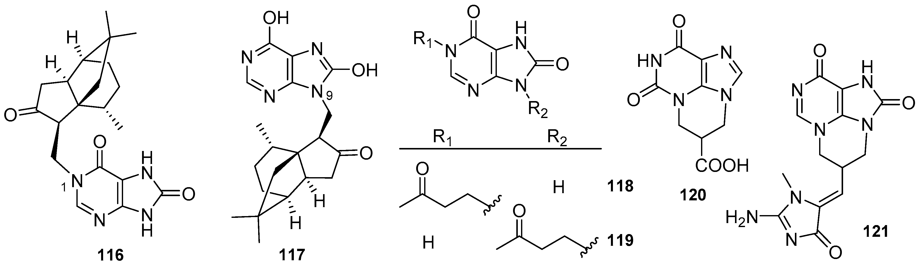
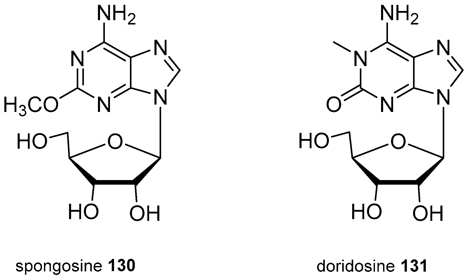
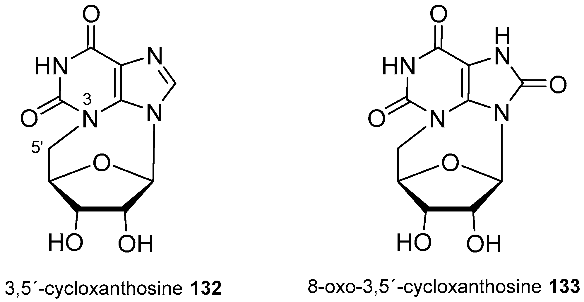
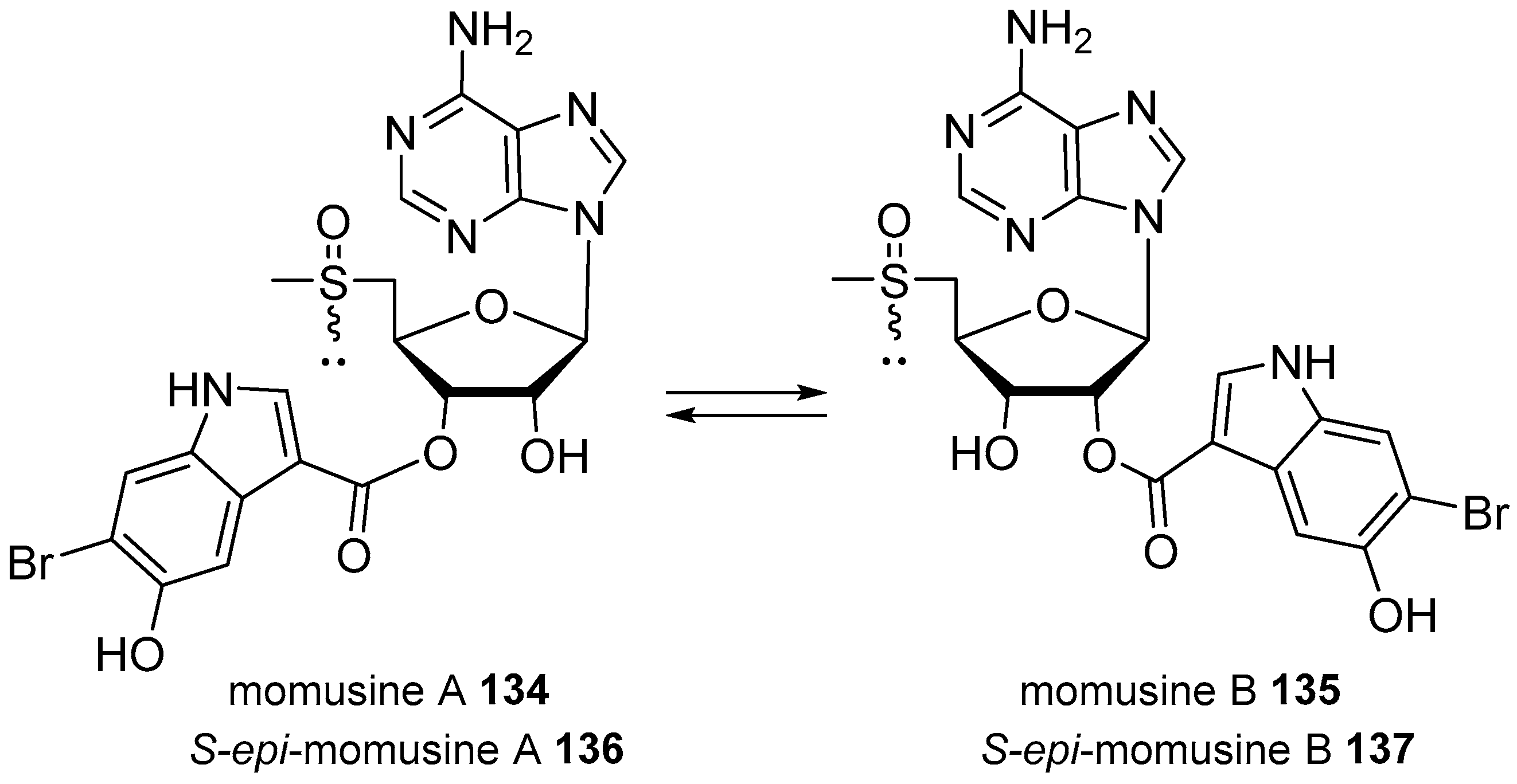
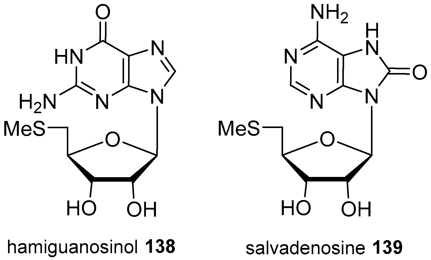
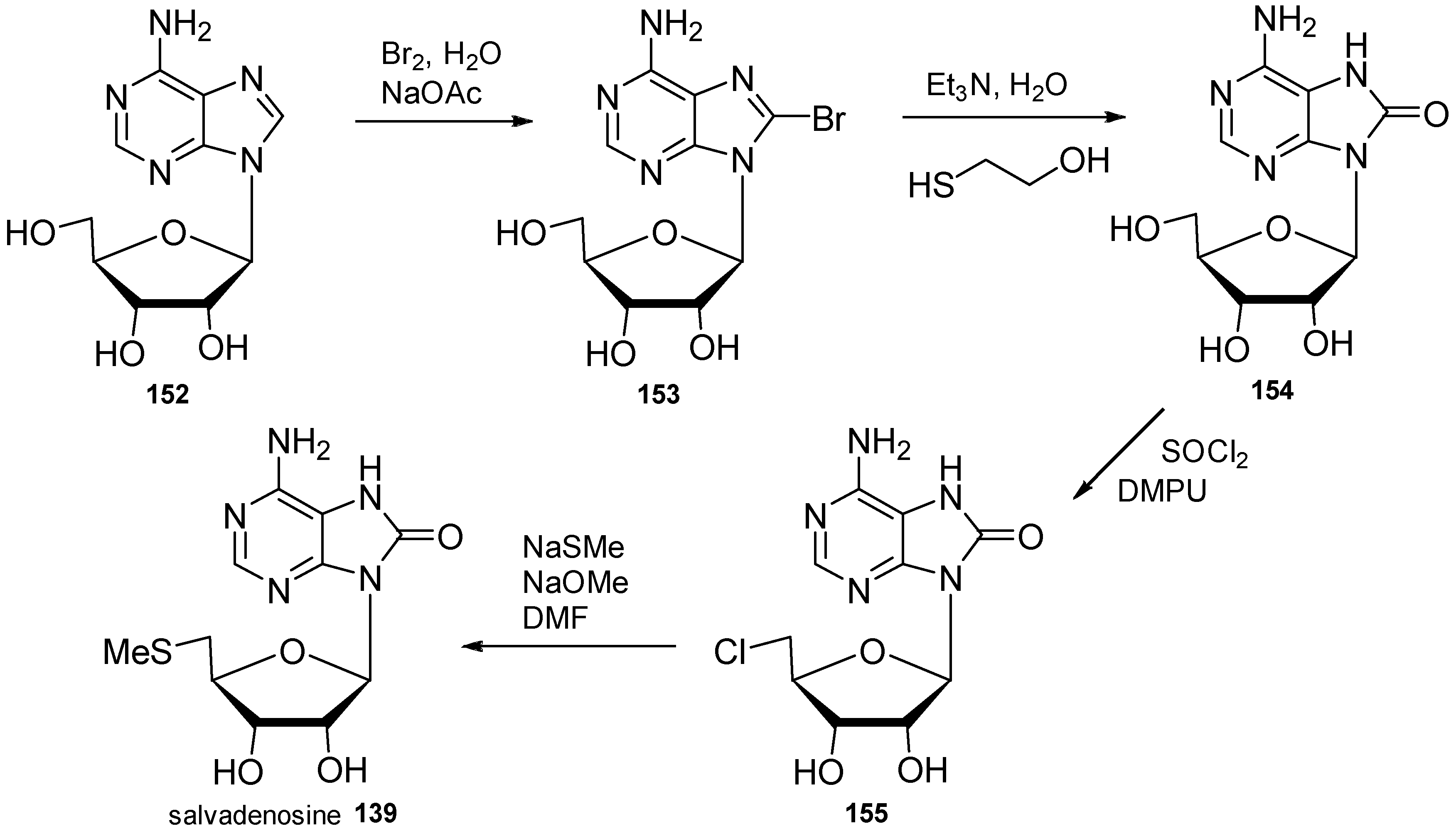
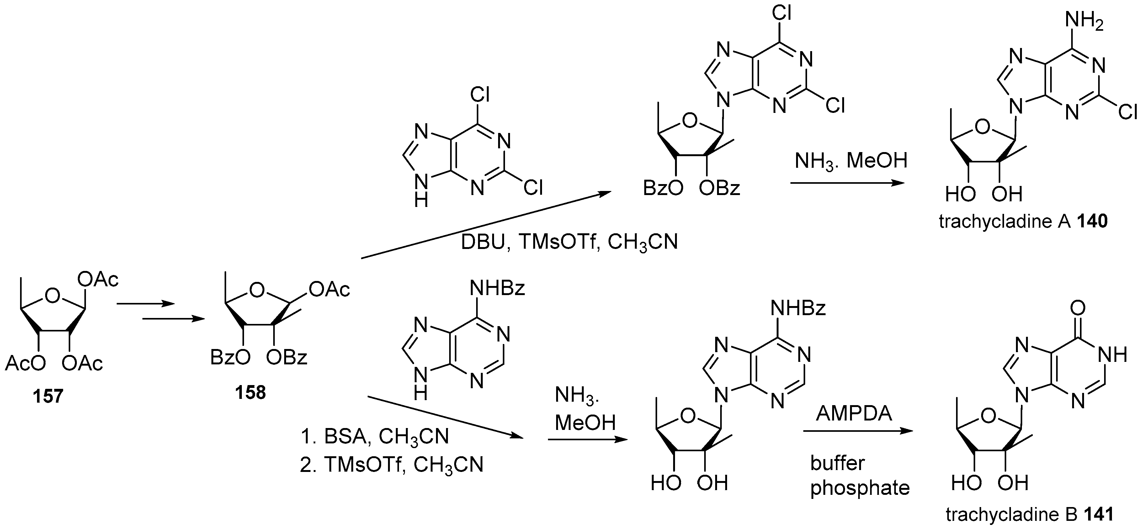
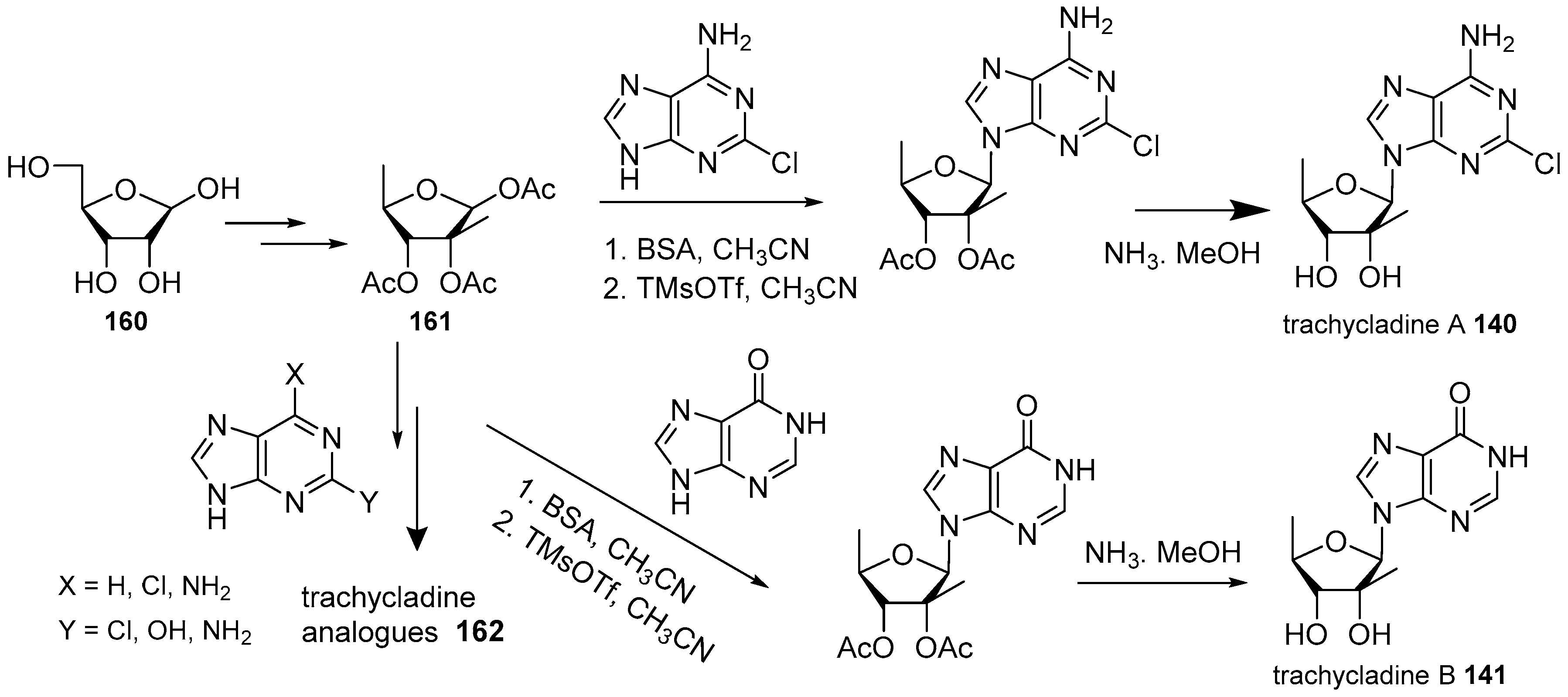
4. Nucleoside Derivatives

Modified nucleosides are relatively uncommon secondary metabolites in marine organisms, although they are known since the early work performed by Bergmann during the 1950s on different Caribbean sponges. That work led to the identification, among others, of spongosine, 130 (2-methoxyadenosine, Figure 27), which may be the first purine nucleoside of marine origin isolated from the Caribbean sponge Tectitethya crypta (named as Cryptotethya crypta) [98]. Since then, several other purine, pyrimidine and deazapurine nucleosides have been described from different marine sources and collected in previous reviews [99,100]; thus, they are not considered in this one unless they have any unusual structural feature such as additional alkylated nitrogen, uncommon substitutions either on the sugar or the purine moieties, or interesting biological activities.
Doridosine, 131, (1-methylisoguanosine, Figure 27) has been isolated from the Australian sponge Tedania anhelans (named as T. digitata) [101] and from the nudibranch Peltodoris nobilis (named as Anisodoris nobilis) [102,103]. It showed skeletal muscle relaxant, hypothermic and cardiovascular effects mediated via adenosine A1 and A2 receptors [101,103,104].
Among cyclonucleosides, 3,5′-cycloxanthosine, 132 (Figure 28), was described by the first time as a natural compound from the Australian sponge Eryus sp. [105], and later on from the Mediterranean sponge Axinella polypoides collected in the Bay of Calvi, Corsica (France), together with a new cyclonucleoside identified as 8-oxo-3,5′-cycloxanthosine, 133 [106]. Some of these cyclonucleosides were known as synthetic compounds before they were identified as natural products [105,106,107].
Four unusual methylsulphinyladenosine derivatives (Figure 29), momusine A/B, 134/135, and epi-momusine A/B, 136/137, were isolated from the ascidian Herdmania momus collected at the Korea coast; they are interconvertible transesterification isomers (ratio 56/44) and sulphinyl epimers bearing a 6-bromo-5-hydroxyindol-3-ylcarbonyl and a rare methylsulphinyl moieties on the sugar. These compounds were evaluated for antiviral activity against human pathogenic viruses but none of the isomeric mixtures showed significant antiviral effects [108].
From the Mediterranean sponge Hamigera hamigera, collected near Elba, Italy, a modified nucleoside with a 5′-deoxy-5′-methylthioribose moiety, named hamiguanosinol, 138, was isolated (Figure 30) [109]. Initially, the enol form at C6 was proposed but it was later corrected to the more usual keto form by Jamiston et al. after the isolation of salvadenosine, 139, a new 5′-methylthioribose nucleoside from the deep-water tunicate Didemnum sp. collected off Little San Salvador Island (Bahamas) [110].
The rare chloroadenosine analogue trachycladine A, 140 (Figure 31), with a 5-deoxy-2-C-methyl sugar was identified from the sponge Trachycladus laevispirulifer from Exmouth Gulf (Australia) together with trachycladine B, 141, the corresponding hypoxanthine nucleoside [111]. Trachycladine A showed cytotoxicity against several human cell lines but it was inactive against some yeasts and bacteria [111]. Simultaneously, the same adenosine nucleoside 140 was described from an Indonesian sponge, Theonella sp., and given the name kumusine [112] and later it was also identified in a specimen of Theonella cupola collected in Okinawa [113].
A recent example of new marine nucleoside, with a ribose linked to a guanine through C5′, is dragmacidoside, 142 (Figure 31), isolated from the sponge Dragmacidon coccineum (named as D. coccinea) collected from the Red Sea in Egypt [114].
Evidence of traces of the uncommon N6-acyladenosine, phorioadenosine, 143, has been reported by Farrugia et al. from a Southern Australian sponge Phoriospongia sp. [80].

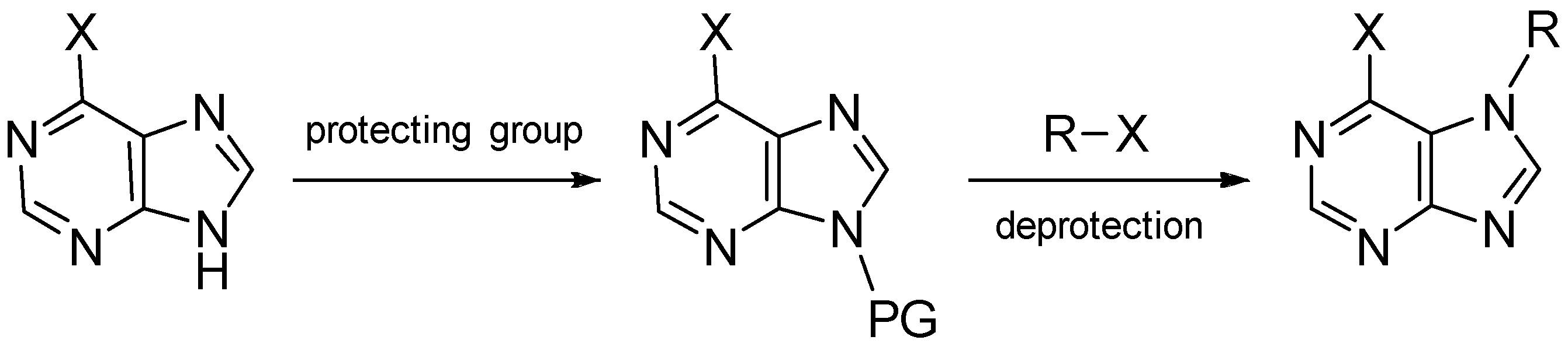
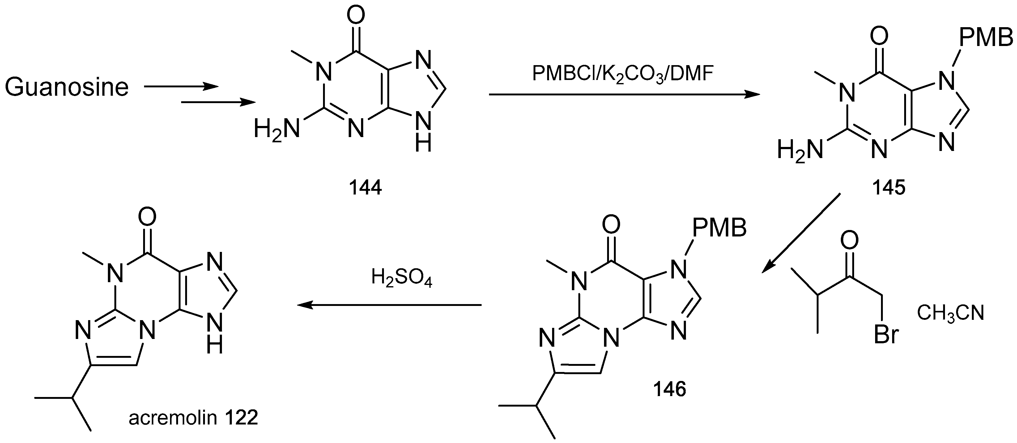
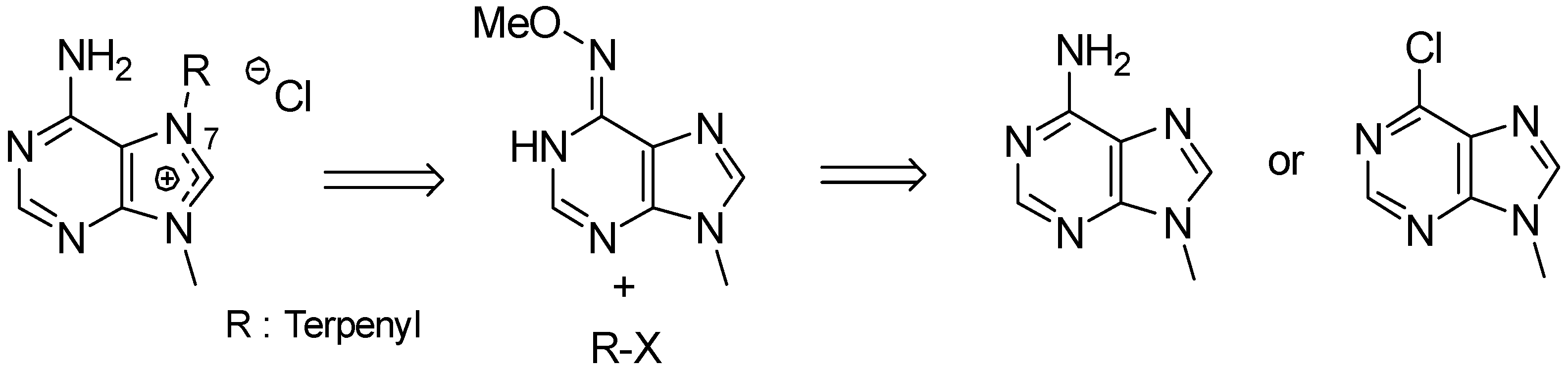
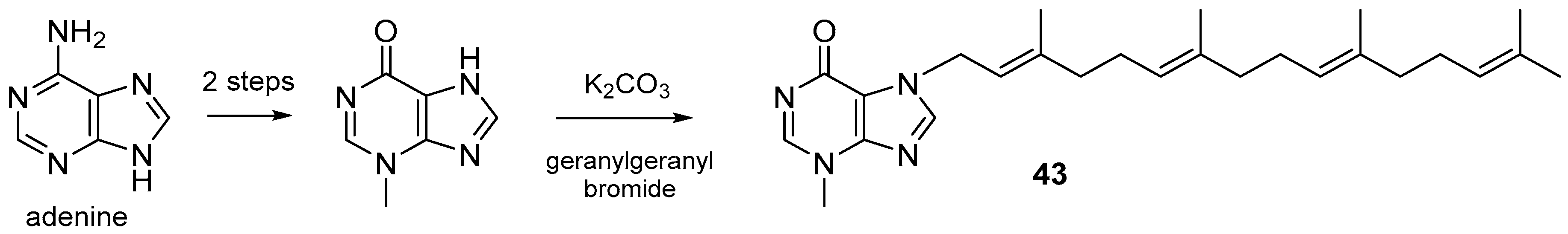
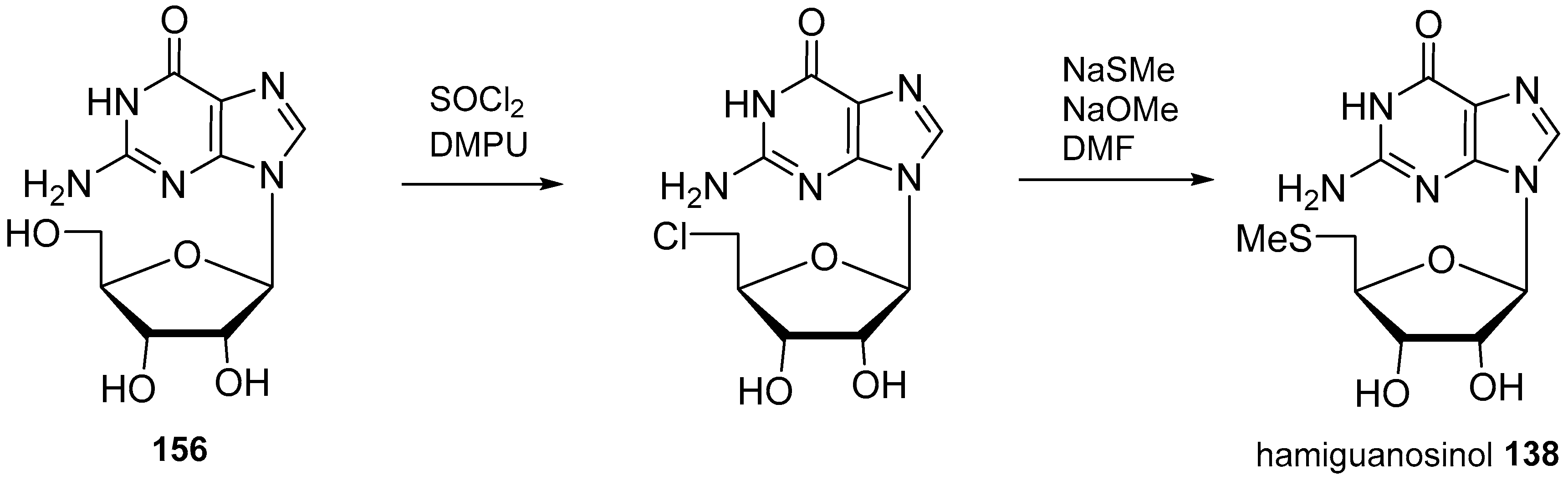
5. Synthetic Approaches towards the Natural Alkylpurines and/or Bioactive Analogues

The secondary metabolites described here show interesting biological activities that sometimes are difficult to discover due to the small amounts that are obtained from their natural sources and the difficulties often found in collecting again the same organism. This fact limits their biological evaluation and the possibility for them to become drugs. Thus, the synthesis of either the natural products themselves or their analogues has become a challenge to the scientific community to determine the main structural features for the bioactivity and to analyse structure–activity relationships in these natural products.
In this sense, much synthetic work is devoted towards the synthesis of marine alkylpurines, particularly those with a diterpenoid moiety. This section is not intended to be exhaustive but rather to give an idea of the synthetic strategies used for this type of compounds. Furthermore, because most of this work have been well compiled in previous reviews [40,77,115], only some recently published approaches are considered here.
The main synthetic difficulties used to be associated to the regioselectivity during the alkylation of starting purines such as adenine, guanine or 6-chloropurine, bearing in mind that N9 is the most nucleophilic nitrogen and these natural products are usually substituted at N7. Thus, direct alkylation leads to both regioisomers predominating the N9-substituted isomer. To obtain the corresponding N7 isomers several strategies have been followed by different authors [40,77,115,116,117,118]. One of those relatively frequent strategies is to alkylate the N9 position with a protecting group, then to introduce the substituent at N7 and finally to deprotect N9, leading to the N7-substituted purines as summarised in the general Scheme 1. Other strategies include the Traube synthesis of purines [117] or the regioselective alkylation using alkylmagnesium reagents as recently described by Chen et al. [118].
Frequently most of these secondary metabolites are polymethylated/polyalkylated at more than one nitrogen atom and in those cases, dialkyl- and trialkylpurines can also be achieved selectively starting from purines already substituted at N3 or N9, which would direct the subsequent alkylation with the corresponding halide towards the other nitrogens. This strategy has been applied by Fujii et al., whose work on preparation of N-methylpurines stands out in the 1990s [119,120]. Sometimes, the initial substituents can be easily removed to obtain the desired purine analogue as it was applied to the synthesis of acremolin, 122 (Scheme 2) [91]. Molinski et al., starting from guanosine, obtained purine 144, which was protected as its para-methoxybenzylderivative 145; the separated N7 isomer was treated with the corresponding bromide to give the imidazole ring derivative 146 that, after deprotection with sulphuric acid or trifluoroacetic acid, gave the desired target acremolin, 122 [91].
The Dimroth rearrangement has been applied to the synthesis of N6-alkyladenines as several 8-oxoadenines [121,122,123], including simple purines as caissarone, 28 [34,120,123], and aplidiamine, 113 (Scheme 3) [83]. The introduction of the carbonyl group at C8 to give 8-oxo derivatives, has been achieved by different oxidation procedures, as very well summarised recently by Resendiz et al. [124] either on simple methylpurines or directly on nucleosides.
Perhaps the diterpenylpurines, such as agelasines, agelasimines and asmarines, are the most attractive synthetic targets due to the importance and variety of their biological activities. The first two have been synthesised by many authors, but there is not known yet a total synthesis of any natural asmarine.
One of the general synthetic strategies used for 9-methyl-7-diterpenyladeninium derivatives is the regioselective alkylation of N6-methoxy-9-methyladenine, obtained from 6-chloropurine or 9-methyladenine, with the corresponding terpenyl halide as represented in Scheme 4.
Gundersen et al. have done an extensive work on the synthesis of analogues of these marine secondary metabolites [40] (and references cited therein). They synthesised many analogues of agelasine E and F with simplified side chains [125] and diterpenyl halides obtained from other natural products such as geranylgeraniol or geranyllinalool [51]. The monocyclic side chain of agelasines E and F were obtained from geraniol and β-cyclocitral [126] and pulegone [127], respectively. They modified also the substitution on the purine C2 position [128,129,130]. All those analogues were evaluated for their antimicrobial, antiprotozoal, antifouling and biofilm inhibition and cytotoxicity against a panel of human cancer cell lines and it was found that the length of the terpenyl side chain is important for bioactivity and that the presence of a methyl in the purine 2-position increased the antimicrobial and antiprotozoal activity, while amino groups increase the cytotoxicity [40,130].
Continuing in the same line, Gundersen and co-workers have recently described the first synthesis of malonganenone J, 43, starting from adenine to obtain 3-methylhypoxanthine that by alkylation with the corresponding geranylgeranyl bromide under mild conditions yielded malonganenone J, 43, (Scheme 5) [131].
The synthesis of the agelasines and agelasimines with a bicyclic diterpenoid has been achieved by similar procedures and it was also compiled previously [40,115]. The bicyclic terpenoid side chain has been obtained from available natural diterpenoids or by total synthesis in a racemic [132,133] or chiral form [134,135]. Examples of natural diterpenoids used as starting material for the three types of diterpenoid skeletons are kolavenic acid [136], ent-halimic acid available in big quantities from its natural sources [66], or manool, which is commercially available [137] (Figure 32).
A series of new neutral 9- and 7-terpenylpurines have been prepared by Castro et al. with monoterpenoid, sesquiterpenoid and diterpenoid side chains using commercial monoterpenoids such as myrtenal or geranylbromide and diterpenoids such as trans-communic acid (degradation of the side chain gave the sesquiterpenoid moiety) and cupressic acid isolated from their natural sources (Scheme 6) [138]. The cytotoxic evaluation against several human tumour cell lines indicated that the terpenyl chain induced cytotoxicity on simple purines, and this activity was higher as larger was the substituent, i.e., the best results were shown by the diterpenylpurines, in some cases with a very interesting low toxicity for non-tumour cells [138].
The other important group of diterpenylpurines are asmarines, for which no total synthesis has been reported yet; only several approaches towards the diazepinopurine system have been reported and they are summarised in Scheme 7 [40,115]. The tetrahydro[1,4]diazepino[1,2,3-gh]purine (THDAP) has been achieved mainly in three different ways. Kashman and co-workers used an aminomercuriation, iodocyclization and acid-catalyzed cyclization of an intermediate obtained from 6-chloropurine [139]. The same starting material was used by Vik and Gundersen to obtain an intermediate that by metathesis gave the required THDAP ring [140]. Finally, Ohba and Tashiro obtained the seven-membered ring by a palladium-promoted cyclization [141].
Recently, Shenvi and co-workers [77,142] have obtained a very close analogue of asmarine A, 97, by the cyclization through a nitrosopurine ene reaction as shown in Scheme 8. The starting formylpyrimidine was made to react with an alkyl iodide in basic conditions giving the alkylation and cyclization product, 147, that was transformed into the corresponding bromide 148. This bromide was made to react with the enolate of the methyl-Wieland-Miescher ketone, 149, and then, by reaction with hydroxylamine, the corresponding dihydroxylimine 150 was obtained, as the ketone and the aryl chloride react as displayed in Scheme 8. Once the agelasine scaffold was obtained, oxidation of the N6-hydroxyaminopurine into a nitroso purine was achieved by treatment with MnO2 to give the desired tert-alkylmethylallylhydroxylamine 151.
Marine nucleosides have also been attractive targets for synthesis; their biological activities, synthesis and biosynthesis have been reported in a previous review [100] and only several recently reported synthesis have been considered here. That is the case of the synthesis of the 8-oxonucleoside salvadenosine, 139, that was carried out by Molinski et al., starting with adenosine, 152. Compound 152 was brominated at C8 to give 153, transformed first into the corresponding 8-oxo-adenosine, 154, and then into the chloroderivative 155 by treatment with thionyl chloride. Finally, displacement of the halogen with NaSMe, yielded salvadenosine, 139 (Scheme 9) [110].
The same displacement of the halogen with NaSMe was used by the same authors to synthesise hamiguanosinol, 138, another marine nucleoside with a methylthio substituent in the sugar moiety from guanosine, 156, and reassign its structure to the keto tautomer 138 (Scheme 10) [110].
Another recent example is the total synthesis of the two marine nucleosides trachycladines A and B, 140141, performed by two different researchers in a very similar way as described below. The syntheses of Wu and co-workers (Scheme 11) started from a commercial triacetyl derivative of 5-deoxy-β-d-ribofuranose, 157, that was transformed into the 2-methyl-dibenzoyl derivative 158; ulterior coupling with 2,6-dichloropurine and treatment with ammonia in methanol gives trachycladine A, 140 [143]. For trachycladine B, 141, N6-benzoyladenine was used as a base and the transformation into the hypoxanthine was achieved using an adenilate deaminase (AMPDA) as a biocatalyst.
At the same time, Koumbis and co-workers [144] described the synthesis of both trachycladines A and B using similar methodology (Scheme 12), starting from d-ribose, 160, through the 2-methyl triacetate 161 but using directly the corresponding unprotected nucleobase. This procedure has been improved by the same authors [145] and applied to the synthesis of several analogues (162) modified at the nucleobase in order to evaluate their cytotoxicity, which could not be evaluated previously for trachycladine B, 141, because of the small amounts isolated from its natural source. The bioassays have shown the wide spectrum of antineoplastic activity for these compounds and the importance of the substituents in the purine ring, with trachycladine A being much more potent than trachycladine B.

6. Conclusions and Perspectives

Marine secondary metabolites containing a purine moiety alkylated at any nitrogen atom of the heterocycle are described in this review, considering the marine source and their biological properties. They have been isolated from a variety or marine organisms, mainly from sponges ascidians and gorgonians, but there are also a few examples isolated from other organisms such as a sea anemone, a nudibranch, two cone snail or one bryozoan, most of them collected at Caribbean, Okinawan, Australian and South China Sea. The purine system is derived from adenine, guanine, isoguanine and their 8-oxo-analogues and they have varied alkyl substituents ranging from methyl to diterpenoid chains. The largest group are the terpenylpurine derivatives that can bear acyclic, monocyclic or bicyclic diterpenoid side chains. The bicyclic ones are derived from clerodane, halimane or labdane skeletons. Diverse biological activities have been described for them such as anti-tumour, antibacterial, antifungal, anti-plasmodial, antifouling and biofilm inhibition, antiprotozoal, enzyme inhibition, or alteration of neuronal signalling among others, sometimes with very promising results. Although it is difficult to compare assays performed with different protocols, in general, those with a diterpenyl group are usually better cytotoxics than those with small chains while the methylpurines give better results on other bioassays.
The marine organisms still are a good source of bioactive compounds derived from purine although the tiny amounts that are sometimes isolated limit their development as future drugs. Thus, more effort is necessary to develop good synthetic procedures that allow obtaining large quantities for biological assays including mechanisms of action. These efforts should not be directed solely towards the natural products themselves, but also to the preparation of simpler derivatives because, sometimes, they are even more potent than the original natural products.

Acknowledgments

Financial support comes from the Spanish Ministerio de Economía y Competitividad (MINECO; CTQ 2015-68175-R; AGL2016-79813-C2-2-R) and Junta de Castilla y León (Ph.D. scholarship to E.V.), co-financed by the Fondo Social Europeo (FSE) of the European Union.

Author Contributions

All the authors contributed equally to the analysis of data from the references and in preparation of the manuscript. All authors approved the final text.

Conflicts of Interest

The authors declare no conflict of interest. The founding sponsors had no role in the design of the study, in the data collection, and in the writing and decision to publish the manuscript.

References

  1. Newman, D.J.; Cragg, G.M. Natural products as sources of new drugs from 1981 to 2014. J. Nat. Prod. 2016, 79, 629–661. [Google Scholar] [CrossRef] [PubMed]
  2. Blunt, J.W.; Copp, B.R.; Keyzers, R.A.; Munro, M.H.G.; Prinsep, M.R. Marine natural products. Nat. Prod. Rep. 2017, 34, 235–294. [Google Scholar] [CrossRef] [PubMed]
  3. Skropeta, D.; Wei, L. Recent advances in deep-sea natural products. Nat. Prod. Rep. 2014, 31, 999–1025. [Google Scholar] [CrossRef] [PubMed]
  4. Rosemeyer, H. The chemodiversity of purine as a constituent of natural products. Chem. Biodivers. 2004, 1, 361–401. [Google Scholar] [CrossRef] [PubMed]
  5. Ackermann, D.; List, P.H. Zur konstitution des zooanemonins und des herbipolins. Hoppe-Seyler’s Z. Physiol. Chem. 1960, 318, 281. [Google Scholar] [CrossRef] [PubMed]
  6. Ackermann, D.; List, P.H. Konstitution des spongopurins, einer in der natur neuen purinbase. Hoppe-Seyler’s Z. Physiol. Chem. 1961, 323, 192–193. [Google Scholar] [CrossRef] [PubMed]
  7. Yagi, H.; Matsunaga, S.; Fusetani, N. Isolation of 1-methylherbipoline, a purine base, from a marine sponge, Jaspis sp. J. Nat. Prod. 1994, 57, 837–838. [Google Scholar] [CrossRef]
  8. Kimura, J.; Ishizuka, E.; Nakao, Y.; Yoshida, W.Y.; Scheuer, P.J.; Kelly-Borges, M. Isolation of 1-methylherbipoline salts of halisulfate-1 and of suvanine as serine protease inhibitors from a marine sponge, Coscinoderma mathewsi. J. Nat. Prod. 1998, 61, 248–250. [Google Scholar] [CrossRef] [PubMed]
  9. Davis, R.A.; Christensen, L.V.; Richardson, A.D.; da Rocha, R.M.; Ireland, C.M. Rigidin E, a new pyrrolopyrimidine alkaloid from a Papua New Guinea tunicate Eudistoma species. Mar. Drugs 2003, 1, 27–33. [Google Scholar] [CrossRef]
  10. Lindsay, B.S.; Battershill, C.N.; Copp, B.R. 1,3-Dimethylguanine, a new purine from the New Zealand ascidian Botrylloides leachi. J. Nat. Prod. 1999, 62, 638–639. [Google Scholar] [CrossRef] [PubMed]
  11. Tasdemir, D.; Mangalindan, G.C.; Concepcion, G.P.; Harper, M.K.; Ireland, C.M. 3,7-Dimethylguanine, a new purine from a Philippine sponge Zyzzya fuliginosa. Chem. Pharm. Bull. 2001, 49, 1628–1630. [Google Scholar] [CrossRef] [PubMed]
  12. Abdel-Lateff, A.; Alarif, W.M.; Asfour, H.Z.; Ayyad, S.E.N.; Khedr, A.; Badria, F.A.; Al-lihaibi, S.S. Cytotoxic effects of three new metabolites from Red Sea marine sponge, Petrosia sp. Environ. Toxicol. Pharmacol. 2014, 37, 928–935. [Google Scholar] [CrossRef] [PubMed]
  13. Moon, B.; Baker, B.J.; McClintock, J.B. Purine and nucleoside metabolites from the Antarctic sponge Isodictya erinacea. J. Nat. Prod. 1998, 61, 116–118. [Google Scholar] [CrossRef] [PubMed]
  14. Pearce, A.N.; Babcock, R.C.; Lambert, G.; Copp, B.R. N2,N2,7-Trimethylguanine, a new trimethylated guanine natural product from the New Zealand ascidian, Lissoclinum notti. Nat. Prod. Lett. 2001, 15, 237–241. [Google Scholar] [CrossRef] [PubMed]
  15. Perry, N.B.; Blunt, J.W.; Munro, M.H.G. 1,3,7-Trimethylguanine from the sponge Latrunculia brevis. J. Nat. Prod. 1987, 50, 307–308. [Google Scholar] [CrossRef]
  16. Van Wagoner, R.M.; Jompa, J.; Tahir, A.; Ireland, C.M. Trypargine alkaloids from a previously undescribed Eudistoma sp. ascidian. J. Nat. Prod. 1999, 62, 794–797. [Google Scholar] [CrossRef] [PubMed]
  17. Berry, Y.; Bremner, J.B.; Davis, A.; Samosorn, S. Isolation and NMR spectroscopic clarification of the alkaloid 1,3,7-trimethylguanine from the ascidian Eudistoma maculosum. Nat. Prod. Res. 2006, 20, 479–483. [Google Scholar] [CrossRef] [PubMed]
  18. Mitchell, S.S.; Whitehill, A.B.; Trapido-Rosenthal, H.G.; Ireland, C.M. Isolation and characterization of 1,3-dimethylisoguanine from the Bermudian sponge Amphimedon viridis. J. Nat. Prod. 1997, 60, 727–728. [Google Scholar] [CrossRef] [PubMed]
  19. Chehade, C.C.; Dias, R.L.A.; Berlinck, R.G.S.; Ferreira, A.G.; Costa, L.V.; Rangel, M.; Malpezzi, E.L.A.; de Freitas, J.C.; Hajdu, E. 1,3-Dimethylisoguanine, a new purine from the marine sponge Amphimedon viridis. J. Nat. Prod. 1997, 60, 729–731. [Google Scholar] [CrossRef] [PubMed]
  20. Jeong, S.J.; Inagaki, M.; Higuchi, R.; Miyamoto, T.; Ono, M.; Kuwano, M.; Van Soest, R.W.M. 1,3-Dimethylisoguaninium, an antiangiogenic purine analog from the sponge Amphimedon paraviridis. Chem. Pharm. Bull. 2003, 51, 731–733. [Google Scholar] [CrossRef] [PubMed]
  21. Lindsay, B.S.; Battershill, C.N.; Copp, B.R. Isolation of 2-(3′-bromo-4′-hydroxyphenol)ethanamine from the New Zealand ascidian Cnemidocarpa bicornuta. J. Nat. Prod. 1998, 61, 857–858. [Google Scholar] [CrossRef] [PubMed]
  22. Copp, B.R.; Wassvik, C.M.; Lambert, G.; Page, M.J. Isolation and characterization of the new purine 1,3,7-trimethylisoguanine from the New Zealand ascidian Pseudodistoma cereum. J. Nat. Prod. 2000, 63, 1168–1169. [Google Scholar] [CrossRef] [PubMed]
  23. Cafieri, F.; Fattorusso, E.; Mangoni, A.; Taglialatela-Scafati, O. Longamide and 3,7-dimethylisoguanine, two novel alkaloids from the marine sponge Agelas longissima. Tetrahedron Lett. 1995, 36, 7893–7896. [Google Scholar] [CrossRef]
  24. Fu, X.; Ng, P.-L.; Schmitz, F.J.; Hossain, M.B.; van der Helm, D.; Kelly-Borges, M. Makaluvic acids A and B: Novel alkaloids from the marine sponge Zyzzya fuliginosus. J. Nat. Prod. 1996, 59, 1104–1106. [Google Scholar] [CrossRef] [PubMed]
  25. Penez, N.; Culioli, G.; Perez, T.; Briand, J.F.; Thomas, O.P.; Blache, Y. Antifouling properties of simple indole and purine alkaloids from the Mediterranean gorgonian Paramuricea clavata. J. Nat. Prod. 2011, 74, 2304–2308. [Google Scholar] [CrossRef] [PubMed]
  26. Bourguet-Kondracki, M.L.; Martin, M.T.; Vacelet, J.; Guyot, M. Mucronatine, a new N-methyl purine from the French Mediterranean marine sponge Stryphnus mucronatus. Tetrahedron Lett. 2001, 42, 7257–7259. [Google Scholar] [CrossRef]
  27. Suciati; Garson, M.J.; Bernhardt, P.V.; Pierens, G.K. A new N,N-dimethyl purine from an Australian dictyoceratid sponge. J. Chem. Crystallogr. 2011, 41, 1669–1672. [Google Scholar]
  28. Ashour, M.; Edrada-Ebel, R.; Ebel, R.; Wray, V.; van Soest, R.W.M.; Proksch, P. New purine derivatives from the marine sponge Petrosia nigricans. Nat. Prod. Commun. 2008, 3, 1889–1894. [Google Scholar]
  29. Lindsay, B.S.; Almeida, A.M.P.; Smith, C.J.; Berlinck, R.G.S.; da Rocha, R.M.; Ireland, C.M. 6-Methoxy-7-methyl-8-oxoguanine, an unusual purine from the ascidian Symplegma rubra. J. Nat. Prod. 1999, 62, 1573–1575. [Google Scholar] [CrossRef] [PubMed]
  30. Capon, R.J.; Rooney, F.; Murray, L.M. 1,9-Dimethylhypoxanthine from a Southern Australian marine sponge Spongosorites species. J. Nat. Prod. 2000, 63, 261–262. [Google Scholar] [CrossRef] [PubMed]
  31. Huang, R.; Peng, Y.; Zhou, X.; Fu, M.; Tian, S.; Liu, Y. A new pyrimidinedione derivative from the gorgonian coral Verrucella umbraculum. Nat. Prod. Res. 2013, 27, 319–322. [Google Scholar] [CrossRef] [PubMed]
  32. Cimino, G.; de Giulio, A.; de Rosa, S.; de Stefano, S.; Puliti, R.; Mattia, C.A.; Mazzarella, L. Isolation and X-ray crystal structure of a novel 8-oxopurine compound from a marine sponge. J. Nat. Prod. 1985, 48, 523–528. [Google Scholar] [CrossRef]
  33. Zelnik, R.; Haraguchi, M.; Matida, A.K.; Lavie, D.; Frolow, F.; Weis, A.L. X-ray molecular structure of caissarone, a novel purine derivative from the sea-anemone Bunodosoma caissarum Correa 1964. J. Chem. Soc. Perkin Trans. 1 1986, 2051–2053. [Google Scholar] [CrossRef]
  34. Fujii, T.; Saito, T.; Mori, S.; Chikazawa, J. Synthesis of the sea anemone purine caissarone. Tetrahedron Lett. 1991, 32, 97–100. [Google Scholar] [CrossRef]
  35. De Freitas, J.C.; Sawaya, M.I. Anomalies in sea-urchin egg development induced by a novel purine isolated from the sea-anemone Bunodosoma caissarum. Toxicon 1986, 24, 751–755. [Google Scholar] [CrossRef]
  36. Cooper, R.A.; de Freitas, J.C.; Porreca, F.; Eisenhour, C.M.; Lukas, R.; Huxtable, R.J. The sea anemone purine, caissarone: Adenosine receptor antagonism. Toxicon 1995, 33, 1025–1031. [Google Scholar] [CrossRef]
  37. Appleton, D.R.; Page, M.J.; Lambert, G.; Copp, B.R. 1,3-Dimethyl-8-oxoisoguanine, a new purine from the New Zealand ascidian Pseudodistoma cereum. Nat. Prod. Res. 2004, 18, 39–42. [Google Scholar] [CrossRef] [PubMed]
  38. Sakurada, T.; Gill, M.B.; Frausto, S.; Copits, B.; Noguchi, K.; Shimamoto, K.; Swanson, G.T.; Sakai, R. Novel N-methylated 8-oxoisoguanines from Pacific sponges with diverse neuroactivities. J. Med. Chem. 2010, 53, 6089–6099. [Google Scholar] [CrossRef] [PubMed]
  39. Youssef, D.T.A.; Mohamed, G.A.; Shaala, L.A.; Badr, J.M.; Bamanie, F.H.; Ibrahim, S.R.M. New purine alkaloids from the Red Sea marine tunicate Symplegma rubra. Phytochem. Lett. 2015, 13, 212–217. [Google Scholar] [CrossRef]
  40. Gundersen, L.L. Synthesis and biological activities of marine terpene-adenine hybrids and synthetic analogs. Phytochem. Rev. 2013, 12, 467–486. [Google Scholar] [CrossRef]
  41. Gordaliza, M. Terpenyl-purines from the sea. Mar. Drugs 2009, 7, 833–849. [Google Scholar] [CrossRef] [PubMed]
  42. Cullen, E.; Devlin, J.P. Agelasine: Novel quaternary 9-methyladenine from sponge Agelas dispar. Can. J. Chem. 1975, 53, 1690–1691. [Google Scholar] [CrossRef]
  43. Keyzers, R.A.; Gray, C.A.; Schleyer, M.H.; Whibley, C.E.; Hendricks, D.T.; Davies-Coleman, M.T. Malonganenones A–C, novel tetraprenylated alkaloids from the Mozambique gorgonian Leptogorgia gilchristi. Tetrahedron 2006, 62, 2200–2206. [Google Scholar] [CrossRef]
  44. Sorek, H.; Rudi, A.; Benayahu, Y.; Ben-Califa, N.; Neumann, D.; Kashman, Y. Nuttingins A–F and malonganenones D–H, tetraprenylated alkaloids from the Tanzanian gorgonian Euplexaura nuttingi. J. Nat. Prod. 2007, 70, 1104–1109. [Google Scholar] [CrossRef] [PubMed]
  45. Zhang, J.R.; Li, P.L.; Tang, X.L.; Qi, X.; Li, G.Q. Cytotoxic tetraprenylated alkaloids from the South China Sea gorgonian Euplexaura robusta. Chem. Biodivers. 2012, 9, 2218–2224. [Google Scholar] [CrossRef] [PubMed]
  46. Sun, Z.H.; Cai, Y.H.; Fan, C.Q.; Tang, G.H.; Luo, H.B.; Yin, S. Six new tetraprenylated alkaloids from the South China Sea gorgonian Echinogorgia pseudossapo. Mar. Drugs 2014, 12, 672–681. [Google Scholar] [CrossRef] [PubMed]
  47. Capon, R.J.; Faulkner, D.J. Antimicrobial metabolites from a Pacific sponge, Agelas sp. J. Am. Chem. Soc. 1984, 106, 1819–1822. [Google Scholar] [CrossRef]
  48. Wu, H.; Nakamura, H.; Kobayashi, J.; Ohizumi, Y.; Hirata, Y. Agelasine-E and agelasine-F, novel monocyclic diterpenoids with a 9-methyladeninium unit possessing inhibitory effects on Na,K-ATPase isolated from the Okinawan Sea sponge Agelas nakamurai Hoshino. Tetrahedron Lett. 1984, 25, 3719–3722. [Google Scholar] [CrossRef]
  49. Mangalindan, G.C.; Talaue, M.T.; Cruz, L.J.; Franzblau, S.G.; Adams, L.B.; Richardson, A.D.; Ireland, C.M.; Concepcion, G.P. Agelasine F from a Philippine Agelas sp. sponge exhibits in vitro antituberculosis activity. Planta Med. 2000, 66, 364–365. [Google Scholar] [CrossRef] [PubMed]
  50. Pettit, G.R.; Tang, Y.; Zhang, Q.; Bourne, G.T.; Arm, C.A.; Leet, J.E.; Knight, J.C.; Pettit, R.K.; Chapuis, J.C.; Doubek, D.L.; et al. Isolation and structures of axistatins 1–3 from the Republic of Palau marine sponge Agelas axifera Hentschel. J. Nat. Prod. 2013, 76, 420–424. [Google Scholar] [CrossRef] [PubMed]
  51. Bakkestuen, A.K.; Gundersen, L.L.; Petersen, D.; Utenova, B.T.; Vik, A. Synthesis and antimycobacterial activity of agelasine E and analogs. Org. Biomol. Chem. 2005, 3, 1025–1033. [Google Scholar] [CrossRef] [PubMed]
  52. Abdjul, D.B.; Yamazaki, H.; Kanno, S.I.; Takahashi, O.; Kirikoshi, R.; Ukai, K.; Namikoshi, M. Structures and biological evaluations of agelasines isolated from the Okinawan marine sponge Agelas nakamurai. J. Nat. Prod. 2015, 78, 1428–1433. [Google Scholar] [CrossRef] [PubMed]
  53. Hong, L.L.; Sun, J.B.; Yang, F.; Liu, M.; Tang, J.; Sun, F.; Jiao, W.H.; Wang, S.P.; Zhang, W.; Lin, H.W. New diterpene alkaloids from the marine sponge Agelas mauritiana. RSC Adv. 2017, 7, 23970–23976. [Google Scholar] [CrossRef]
  54. Nakamura, H.; Wu, H.M.; Ohizumi, Y.; Hirata, Y. Agelasine-A, -B, -C and -D, novel bicyclic diterpenoids with a 9-methyladeninium unit possessing inhibitory effects on Na,K-ATPase from the Okinawan Sea sponge Agelas sp. Tetrahedron Lett. 1984, 25, 2989–2992. [Google Scholar] [CrossRef]
  55. Wu, H.M.; Nakamura, H.; Kobayashi, J.; Kobayashi, M.; Ohizumi, Y.; Hirata, Y. Structures of agelasines, diterpenes having a 9-methyladeninium chromophore isolated from the Okinawan marine sponge Agelas nakamurai Hoshino. Bull. Chem. Soc. Jpn. 1986, 59, 2495–2504. [Google Scholar] [CrossRef]
  56. Calcul, L.; Tenney, K.; Ratnam, J.; McKerrow, J.H.; Crews, P. Structural variations to the 9-N-methyladeninium diterpenoid hybrid commonly isolated from Agelas sponges. Aust. J. Chem. 2010, 63, 915–921. [Google Scholar] [CrossRef]
  57. Fu, X.; Schmitz, F.J.; Tanner, R.S.; Kelly-Borges, M. Agelasines H and H, 9-methyladenine-containing diterpenoids from an Agelas sponge. J. Nat. Prod. 1998, 61, 548–550. [Google Scholar] [CrossRef] [PubMed]
  58. Appenzeller, J.; Mihci, G.; Martin, M.T.; Gallard, J.F.; Menou, J.L.; Boury-Esnault, N.; Hooper, J.; Petek, S.; Chevalley, S.; Valentin, A.; et al. Agelasines J, K, and L from the Solomon Islands marine sponge Agelas cf. mauritiana. J. Nat. Prod. 2008, 71, 1451–1454. [Google Scholar] [CrossRef] [PubMed]
  59. Stout, E.P.; Yu, L.C.; Molinski, T.F. Antifungal diterpene alkaloids from the Caribbean sponge Agelas citrina: Unified configurational assignments of agelasidines and agelasines. Eur. J. Org. Chem. 2012, 2012, 5131–5135. [Google Scholar] [CrossRef] [PubMed]
  60. Kubota, T.; Iwai, T.; Takahashi-Nakaguchi, A.; Fromont, J.; Gonoi, T.; Kobayashi, J.I. Agelasines O–U, new diterpene alkaloids with a 9-N-methyladenine unit from a marine sponge Agelas sp. Tetrahedron 2012, 68, 9738–9744. [Google Scholar] [CrossRef]
  61. Yang, F.; Hamann, M.T.; Zou, Y.; Zhang, M.Y.; Gong, X.B.; Xiao, J.R.; Chen, W.S.; Lin, H.W. Antimicrobial metabolites from the Paracel Islands sponge Agelas mauritiana. J. Nat. Prod. 2012, 75, 774–778. [Google Scholar] [CrossRef] [PubMed]
  62. An, L.; Song, W.; Tang, X.; de Voogd, N.J.; Wang, Q.; Chu, M.; Li, P.; Li, G. Alkaloids and polyketides from the South China Sea sponge Agelas aff. nemoechinata. RSC Adv. 2017, 7, 14323–14329. [Google Scholar] [CrossRef]
  63. Ishida, K.; Ishibashi, M.; Shigemori, H.; Sasaki, T.; Kobayashi, J. Agelasine G, a new antileukemic alkaloid from the Okinawan marine sponge Agelas sp. Chem. Pharm. Bull. 1992, 40, 766–767. [Google Scholar] [CrossRef] [PubMed]
  64. Yamazaki, H.; Kanno, S.I.; Abdjul, D.B.; Namikoshi, M. A bromopyrrole-containing diterpene alkaloid from the Okinawan marine sponge Agelas nakamurai activates the insulin pathway in Huh-7 human hepatoma cells by inhibiting protein tyrosine phosphatase 1B. Bioorg. Med. Chem. Lett. 2017, 27, 2207–2209. [Google Scholar] [CrossRef] [PubMed]
  65. Hattori, T.; Adachi, K.; Shizuri, Y. New agelasine compound from the marine sponge Agelas mauritiana as an antifouling substance against macroalgae. J. Nat. Prod. 1997, 60, 411–413. [Google Scholar] [CrossRef]
  66. Marcos, I.S.; García, N.; Sexmero, A.J.; Basabe, P.; Díez, D.; Urones, J.G. Synthesis of (+)-agelasine C. A structural revision. Tetrahedron 2005, 61, 11672–11678. [Google Scholar] [CrossRef]
  67. Chu, M.J.; Tang, X.L.; Qin, G.F.; Sun, Y.T.; Li, L.; de Voogd, N.J.; Li, P.L.; Li, G.Q. Pyrrole derivatives and diterpene alkaloids from the South China Sea sponge Agelas nakamurai. Chem. Biodivers. 2017, 14, e1600446. [Google Scholar] [CrossRef] [PubMed]
  68. Fathiafshar, R.; Allen, T.M. Biologically active metabolites from Agelas mauritiana. Can. J. Chem. 1988, 66, 45–50. [Google Scholar] [CrossRef]
  69. Fathiafshar, R.; Allen, T.M.; Krueger, C.A.; Cook, D.A.; Clanachan, A.S.; Vriend, R.; Baer, H.P.; Cass, C.E. Some pharmacological activities of novel adenine-related compounds isolated from a marine sponge Agelas mauritiana. Can. J. Physiol. Pharmacol. 1989, 67, 276–281. [Google Scholar] [CrossRef]
  70. Hertiani, T.; Edrada-Ebel, R.; Ortlepp, S.; van Soest, R.W.M.; de Voogd, N.J.; Wray, V.; Hentschel, U.; Kozytska, S.; Mueller, W.E.G.; Proksch, P. From anti-fouling to biofilm inhibition: New cytotoxic secondary metabolites from two Indonesian Agelas sponges. Bioorg. Med. Chem. 2010, 18, 1297–1311. [Google Scholar] [CrossRef] [PubMed]
  71. Iwagawa, T.; Kaneko, M.; Okamura, H.; Nakatani, M.; van Soest, R.W.M. New alkaloids from the Papua New Guinean sponge Agelas nakamurai. J. Nat. Prod. 1998, 61, 1310–1312. [Google Scholar] [CrossRef] [PubMed]
  72. Li, T.; Wang, B.; de Voogd, N.J.; Tang, X.L.; Wang, Q.; Chu, M.J.; Li, P.L.; Li, G.Q. Two new diterpene alkaloids from the South China Sea sponge Agelas aff. nemoechinata. Chin. Chem. Lett. 2016, 27, 1048–1051. [Google Scholar] [CrossRef]
  73. Yosief, T.; Rudi, A.; Stein, Z.; Goldberg, I.; Gravalos, G.M.D.; Schleyer, M.; Kashman, Y. Asmarines A–C; three novel cytotoxic metabolites from the marine sponge Raspailia sp. Tetrahedron Lett. 1998, 39, 3323–3326. [Google Scholar] [CrossRef]
  74. Yosief, T.; Rudi, A.; Kashman, Y. Asmarines A–F, novel cytotoxic compounds from the marine sponge Raspailia species. J. Nat. Prod. 2000, 63, 299–304. [Google Scholar] [CrossRef] [PubMed]
  75. Rudi, A.; Shalom, H.; Schleyer, M.; Benayahu, Y.; Kashman, Y. Asmarines G and H and barekol, three new compounds from the marine sponge Raspailia sp. J. Nat. Prod. 2004, 67, 106–109. [Google Scholar] [CrossRef] [PubMed]
  76. Rudi, A.; Aknin, M.; Gaydou, E.; Kashman, Y. Asmarines I, J, and K and nosyberkol: Four new compounds from the marine sponge Raspailia sp. J. Nat. Prod. 2004, 67, 1932–1935. [Google Scholar] [CrossRef] [PubMed]
  77. Wan, K.K.; Shenvi, R.A. Conjuring a supernatural product—DelMarine. Synlett 2016, 27, 1145–1164. [Google Scholar]
  78. Carroll, A.R.; Duffy, S.; Avery, V.M. Aplidiopsamine A, an antiplasmodial alkaloid from the temperate Australian ascidian, Aplidiopsis confluata. J. Org. Chem. 2010, 75, 8291–8294. [Google Scholar] [CrossRef] [PubMed]
  79. Wright, A.E.; Roth, G.P.; Hoffman, J.K.; Divlianska, D.B.; Pechter, D.; Sennett, S.H.; Guzman, E.A.; Linley, P.; McCarthy, P.J.; Pitts, T.P.; et al. Isolation, synthesis, and biological activity of aphrocallistin, an adenine-substituted bromotyramine metabolite from the hexactinellida sponge Aphrocallistes beatrix. J. Nat. Prod. 2009, 72, 1178–1183. [Google Scholar] [CrossRef] [PubMed]
  80. Farrugia, M.; Trotter, N.; Vijayasarathy, S.; Salim, A.A.; Khalil, Z.G.; Lacey, E.; Capon, R.J. Isolation and synthesis of N-acyladenine and adenosine alkaloids from a Southern Australian marine sponge, Phoriospongia sp. Tetrahedron Lett. 2014, 55, 5902–5904. [Google Scholar] [CrossRef]
  81. Killday, K.B.; Yarwood, D.; Sills, M.A.; Murphy, P.T.; Hooper, J.N.; Wright, A.E. Microxine, a new cdc2 kinase inhibitor from the Australian marine sponge Microxina species. J. Nat. Prod. 2001, 64, 525–526. [Google Scholar] [CrossRef] [PubMed]
  82. Kang, H.J.; Fenical, W. Aplidiamine, a unique zwitterionic benzyl hydroxyadenine from the Western Australian marine ascidian Aplidiopsis sp. Tetrahedron Lett. 1997, 38, 941–944. [Google Scholar] [CrossRef]
  83. Itaya, T.; Hozumi, Y.; Kanai, T.; Ohta, T. Syntheses of the marine ascidian purine aplidiamine and its 9-β-d-ribofuranoside. Tetrahedron Lett. 1998, 39, 4695–4696. [Google Scholar] [CrossRef]
  84. Neves, J.L.B.; Lin, Z.; Imperial, J.S.; Antunes, A.; Vasconcelos, V.; Olivera, B.M.; Schmidt, E.W. Small molecules in the cone snail arsenal. Org. Lett. 2015, 17, 4933–4935. [Google Scholar] [CrossRef] [PubMed]
  85. Ayer, S.W.; Andersen, R.J.; He, C.H.; Clardy, J. Phidolopin, a new purine derivative from the bryozoan Phidolopora pacifica. J. Org. Chem. 1984, 49, 3869–3870. [Google Scholar] [CrossRef]
  86. Qi, S.H.; Zhang, S.; Li, X.; Li, Q.X. A cytotoxic sesquiterpene alkaloid from the South China Sea gorgonian Subergorgia suberosa. J. Nat. Prod. 2005, 68, 1288–1289. [Google Scholar] [CrossRef] [PubMed]
  87. Qi, S.H.; Zhang, S.; Huang, H. Purine alkaloids from the South China Sea gorgonian Subergorgia suberosa. J. Nat. Prod. 2008, 71, 716–718. [Google Scholar] [CrossRef] [PubMed]
  88. Qi, S.H.; Zhang, S.; Gao, C.H.; Li, Q.X. Purine and pyrimidine derivatives from the South China Sea gorgonian Subergorgia suberosa. Chem. Pharm. Bull. 2008, 56, 993–994, retracted in Chem. Pharm. Bull. 2010, 58, 884. [Google Scholar] [CrossRef]
  89. Julianti, E.; Oh, H.; Lee, H.S.; Oh, D.C.; Oh, K.B.; Shin, J. Acremolin, a new 1H-azirine metabolite from the marine-derived fungus Acremonium strictum. Tetrahedron Lett. 2012, 53, 2885–2886. [Google Scholar] [CrossRef]
  90. Banert, K. Acremolin, a stable natural product with an antiaromatic 1H-azirine moiety? A structural reorientation. Tetrahedron Lett. 2012, 53, 6443–6445. [Google Scholar] [CrossRef]
  91. Januar, L.A.; Molinski, T.F. Acremolin from acremonium strictum is N-2,3-etheno-2′-isopropyl-1-methylguanine, not a 1H-azirine. Synthesis and structural revision. Org. Lett. 2013, 15, 2370–2373. [Google Scholar] [CrossRef] [PubMed]
  92. Tian, Y.; Qin, X.; Lin, X.; Kaliyaperumal, K.; Zhou, X.; Liu, J.; Ju, Z.; Tu, Z.; Liu, Y. Sydoxanthone C and acremolin B produced by deep-sea-derived fungus Aspergillus sp. SCSIO Ind09F01. J. Antibiot. 2015, 68, 703–706. [Google Scholar] [CrossRef] [PubMed]
  93. Namikoshi, M.; Rinehart, K.L. Bioactive compounds produced by cyanobacteria. J. Ind. Microbiol. 1996, 17, 373–384. [Google Scholar] [CrossRef]
  94. Wiese, M.; D’Agostino, P.M.; Mihali, T.K.; Moffitt, M.C.; Neilan, B.A. Neurotoxic alkaloids: Saxitoxin and its analogs. Mar. Drugs 2010, 8, 2185–2211. [Google Scholar] [CrossRef] [PubMed]
  95. Wang, D.Z.; Zhang, S.F.; Zhang, Y.; Lin, L. Paralytic shellfish toxin biosynthesis in cyanobacteria and dinoflagellates: A molecular overview. J. Proteom. 2016, 135, 132–140. [Google Scholar] [CrossRef] [PubMed]
  96. Chen, M.; Wu, X.; Shen, N.; Wang, C. Four new 6-oxy purine alkaloids from the South China Sea sponge, Haliclona cymaeformis. J. Ocean Univ. China 2017, 16, 1183–1186. [Google Scholar] [CrossRef]
  97. Munoz, J.; Koeck, M. Hybrid pyrrole-imidazole alkaloids from the sponge Agelas sceptrum. J. Nat. Prod. 2016, 79, 434–437. [Google Scholar] [CrossRef] [PubMed]
  98. Bergmann, W.; Burke, D.C. Contributions to the study of marine products. XXXIX. The nucleosides of sponges. III. Spongothymidine and spongouridine. J. Org. Chem. 1955, 20, 1501–1507. [Google Scholar] [CrossRef]
  99. McCarty, R.M.; Bandarian, V. Biosynthesis of pyrrolopyrimidines. Bioorg. Chem. 2012, 43, 15–25. [Google Scholar] [CrossRef] [PubMed]
  100. Huang, R.M.; Chen, Y.N.; Zeng, Z.; Gao, C.H.; Su, X.; Peng, Y. Marine nucleosides: Structure, bioactivity, synthesis and biosynthesis. Mar. Drugs 2014, 12, 5817–5838. [Google Scholar] [CrossRef] [PubMed]
  101. Baird-Lambert, J.; Marwood, J.F.; Davies, L.P.; Taylor, K.M. 1-Methylisoguanosine: An orally active marine natural product with skeletal-muscle and cardiovascular effects. Life Sci. 1980, 26, 1069–1077. [Google Scholar] [CrossRef]
  102. Fuhrman, F.A.; Fuhrman, G.J.; Kim, Y.H.; Pavelka, L.A.; Mosher, H.S. Doridosine: A new hypotensive N-methylpurine riboside from the nudibranch Anisodoris nobilis. Science 1980, 207, 193–195. [Google Scholar] [CrossRef] [PubMed]
  103. Kim, Y.H.; Nachman, R.J.; Pavelka, L.; Mosher, H.S.; Fuhrman, F.A.; Fuhrman, G.J. Doridosine, 1-methylisoguanosine, from Anisodoris nobilis; structure, pharmacological properties and synthesis. J. Nat. Prod. 1981, 44, 206–214. [Google Scholar] [CrossRef] [PubMed]
  104. Tao, P.L.; Yen, M.H.; Shyu, W.S.; Chern, J.W. Doridosine derivatives: Binding at adenosine receptors and in vivo effects. Eur. J. Pharmacol. 1993, 243, 135–139. [Google Scholar] [CrossRef]
  105. Capon, R.J.; Trotter, N.S. N-3,5′-Cycloxanthosine, the first natural occurrence of a cyclonucleoside. J. Nat. Prod. 2005, 68, 1689–1691. [Google Scholar] [CrossRef] [PubMed]
  106. Menna, M.; Aiello, A.; D’Aniello, F.; Fattorusso, E.; Imperatore, C.; Luciano, P.; Vitalone, R. Further investigation of the Mediterranean sponge Axinella polypoides: Isolation of a new cyclonucleoside and a new betaine. Mar. Drugs 2012, 10, 2509–2518. [Google Scholar] [CrossRef] [PubMed] [Green Version]
  107. Chen, G.S.; Chen, C.S.; Chien, T.C.; Yeh, J.Y.; Kuo, C.C.; Talekar, R.S.; Chern, J.W. Nucleosides. IX. Synthesis of purine N-3,5′-cyclonucleosides and N-3,5′-cyclo-2′,3′-seconucleosides via Mitsunobu reaction as TIBO-like derivatives. Nucleosides Nucleotides Nucleic Acids 2004, 23, 347–359. [Google Scholar] [CrossRef] [PubMed]
  108. Li, J.L.; La Kim, E.; Wang, H.; Hong, J.; Shin, S.; Lee, C.K.; Jung, J.H. Epimeric methylsulfinyladenosine derivatives from the marine ascidian Herdmania momus. Bioorg. Med. Chem. Lett. 2013, 23, 4701–4704. [Google Scholar] [CrossRef] [PubMed]
  109. Hassan, W.; Edrada, R.; Ebel, R.; Wray, V.; Proksch, P. New alkaloids from the Mediterranean sponge Hamigera hamigera. Mar. Drugs 2004, 2, 88–100. [Google Scholar] [CrossRef]
  110. Jamison, M.T.; Boddy, C.N.; Molinski, T.F. Salvadenosine, a 5′-deoxy-5′-(methylthio) nucleoside from the Bahamian tunicate Didemnum sp. J. Org. Chem. 2014, 79, 9992–9997. [Google Scholar] [CrossRef] [PubMed]
  111. Searle, P.A.; Molinski, T.F. Trachycladine A and trachycladine B: 2′-C-methyl-5′-deoxyribofuranosyl nucleosides from the marine sponge Trachycladus laevispirulifer. J. Org. Chem. 1995, 60, 4296–4298. [Google Scholar] [CrossRef]
  112. Ichiba, T.; Nakao, Y.; Scheuer, P.J.; Sata, N.U.; Kellyborges, M. Kumusine, a chloroadenine riboside from a sponge, Theonella sp. Tetrahedron Lett. 1995, 36, 3977–3980. [Google Scholar] [CrossRef]
  113. Erdogan, I.; Higa, T. A chloroadenine riboside-type nucleoside from the marine sponge Theonella cupola. J. Fac. Pharm. Ankara 2000, 29, 1–6. [Google Scholar]
  114. Abou-Hussein, D.R.; Badr, J.M.; Youssef, D.T.A. Dragmacidoside: A new nucleoside from the Red Sea sponge Dragmacidon coccinea. Nat. Prod. Res. 2014, 28, 1134–1141. [Google Scholar] [CrossRef] [PubMed]
  115. Gordaliza, M.; Baraldi, P.G. Synthetic approaches towards the marine alkyl purines. Curr. Med. Chem. 2013, 20, 2798–2811. [Google Scholar] [CrossRef] [PubMed]
  116. Kotek, V.; Tobrman, T.; Dvorak, D. Highly efficient and broad-scope protocol for the preparation of 7-substituted 6-halopurines via N-9-Boc-protected 7,8-dihydropurines. Synthesis 2012, 44, 610–618. [Google Scholar]
  117. Legraverend, M. Recent advances in the synthesis of purine derivatives and their precursors. Tetrahedron 2008, 64, 8585–8603. [Google Scholar] [CrossRef]
  118. Chen, S.; Graceffa, R.F.; Boezio, A.A. Direct, regioselective N-alkylation of 1,3-azoles. Org. Lett. 2016, 18, 16–19. [Google Scholar] [CrossRef] [PubMed]
  119. Itaya, T.; Takada, Y.; Kanai, T.; Kaneko, M.; Fujii, T. Purines. LXXVI. Alkylation of 8-oxoadenine derivatives: Syntheses of 3,7-dialkyl-, 3,9-dialkyl-, and 3,7,9-trialkyl-8-oxoadenines. Chem. Pharm. Bull. 1997, 45, 1582–1588. [Google Scholar] [CrossRef]
  120. Itaya, T.; Takada, Y.; Kanai, T.; Fujii, T. Purines. LXXVIII. An alternative synthesis of the sea anemone purine alkaloid caissarone. Chem. Pharm. Bull. 1997, 45, 1867–1869. [Google Scholar] [CrossRef]
  121. Fujii, T.; Saito, T.; Mori, S. Purines. XLIII. A total synthesis of the marine sponge base 6-imino-1,9-dimethyl-8-oxopurine. Chem. Pharm. Bull. 1990, 38, 2146–2150. [Google Scholar] [CrossRef]
  122. Fujii, T.; Saito, T.; Mori, S. Purines. XLIV.: A kinetic-study of the Dimroth rearrangement of the marine sponge purine 1,9-dimethyl-8-oxoadenine and related compounds. Chem. Pharm. Bull. 1990, 38, 2591–2594. [Google Scholar] [CrossRef]
  123. Saito, T.; Mori, S.; Chikazawa, J.; Kanai, T.; Fujii, T. Purines. LVII. Regioselective alkylation of N6,9-disubstituted 8-oxoadenines: Syntheses of the sea anemone purine caissarone and some positional isomers and analogs. Chem. Pharm. Bull. 1993, 41, 1746–1752. [Google Scholar] [CrossRef]
  124. Choi, Y.J.; Chang, S.J.; Gibala, K.S.; Resendiz, M.J.E. 8-Oxo-7,8-dihydroadenine and 8-oxo-7,8-dihydroadenosine—Chemistry, structure, and function in RNA and their presence in natural products and potential drug derivatives. Chem. Eur. J. 2017, 23, 6706–6716. [Google Scholar] [CrossRef] [PubMed]
  125. Vik, A.; Hedner, E.; Charnock, C.; Tangen, L.W.; Samuelsen, O.; Larsson, R.; Bohlin, L.; Gundersen, L.L. Antimicrobial and cytotoxic activity of agelasine and agelasimine analogs. Bioorg. Med. Chem. 2007, 15, 4016–4037. [Google Scholar] [CrossRef] [PubMed]
  126. Proszenyak, A.; Charnock, C.; Hedner, E.; Larsson, R.; Bohlin, L.; Gundersen, L.L. Synthesis, antimicrobial and antineoplastic activities for agelasine and agelasimine analogs with a β-cyclocitral derived substituent. Arch. Pharm. Chem. Life Sci. 2007, 340, 625–634. [Google Scholar] [CrossRef] [PubMed]
  127. Proszenyak, A.; Braendvang, M.; Charnock, C.; Gundersen, L.-L. The first synthesis of ent-agelasine F. Tetrahedron 2009, 65, 194–199. [Google Scholar] [CrossRef]
  128. Roggen, H.; Gundersen, L.-L. Synthetic studies directed towards agelasine analogs—Synthesis, tautomerism, and alkylation of 2-substituted N-methoxy-9-methyl-9H-purin-6-amines. Eur. J. Org. Chem. 2008, 2008, 5099–5106. [Google Scholar] [CrossRef]
  129. Roggen, H.; Bohlin, L.; Burman, R.; Charnock, C.; Felth, J.; Gorbitz, C.H.; Larsson, R.; Tamm, T.; Gundersen, L.-L. 2-Substituted agelasine analogs: Synthesis and biological activity, and structure and reactivity of synthetic intermediates. Pure Appl. Chem. 2011, 83, 645–653. [Google Scholar] [CrossRef] [Green Version]
  130. Roggen, H.; Charnock, C.; Burman, R.; Felth, J.; Larsson, R.; Bohlin, L.; Gundersen, L.-L. Antimicrobial and antineoplastic activities of agelasine analogs modified in the purine 2-position. Arch. Pharm. Chem. Life Sci. 2011, 344, 50–55. [Google Scholar] [CrossRef] [PubMed]
  131. Chamgordani, E.J.; Paulsen, J.; Gundersen, L.L. Selective N-7 alkylation of 3-methylhypoxanthine; the first synthesis of malonganenone J. Tetrahedron Lett. 2016, 57, 4926–4929. [Google Scholar] [CrossRef]
  132. Ohba, M.; Kawase, N.; Fujii, T.; Aoe, K.; Okamura, K.; Fathiafshar, R.; Allen, T.M. Racemic syntheses of agelasimine-A and agelasimine-B, bicyclic diterpenoids from the marine sponge Agelas mauritiana. Tetrahedron Lett. 1995, 36, 6101–6104. [Google Scholar] [CrossRef]
  133. Ohba, M.; Kawase, N.; Fujii, T. Total syntheses of (+/−)-agelasimine-a, (+/−)-agelasimine-B, and (+/−)-purino-diterpene and the structure of diacetylagelasimine-A. J. Am. Chem. Soc. 1996, 118, 8250–8257. [Google Scholar] [CrossRef]
  134. Piers, E.; Breau, M.L.; Han, Y.X.; Plourde, G.L.; Yeh, W.L. Total synthesis of cis-clerodane diterpenoids: (−)-agelasine-A and (+)-(3R,4S,5R,8S,9R,10S)-3,4-epoxyclerod-13-en-15,16-olide. J. Chem. Soc. Perkin Trans. 1 1995, 26, 963–966. [Google Scholar] [CrossRef]
  135. Ohba, M.; Iizuka, K.; Ishibashi, H.; Fujii, T. Syntheses and absolute configurations of the marine sponge purines (+)-agelasimine-A and (+)-agelasimine-B. Tetrahedron 1997, 53, 16977–16986. [Google Scholar] [CrossRef]
  136. Iio, H.; Asao, K.; Tokoroyama, T. Syntheses of agelasin B and its analogues. J. Chem. Soc. Chem. Commun. 1985, 12, 774–775. [Google Scholar] [CrossRef]
  137. Salazar, F.J.; Villamizar, J.E. Use of (+)-manool in the synthesis of natural products. Part II. Diterpenes and relatives. J. Chem. Res. 2013, 37, 63–70. [Google Scholar] [CrossRef]
  138. Valles, E.; García, P.A.; Miguel del Corral, J.M.; Perez, M.; Ferreira, I.C.F.R.; Calhelha, R.C.; San Feliciano, A.; Castro, M.A. Synthesis and cytotoxic evaluation of new terpenylpurines. RSC Adv. 2016, 6, 105412–105420. [Google Scholar] [CrossRef]
  139. Pappo, D.; Shimony, S.; Kashman, Y. Synthesis of 9-substituted tetrahydrodiazepinopurines: Studies toward the total synthesis of asmarines. J. Org. Chem. 2005, 70, 199–206. [Google Scholar] [CrossRef] [PubMed]
  140. Vik, A.; Gundersen, L.-L. Synthetic studies directed towards asmarines; construction of the tetrahydrodiazepinopurine moiety by ring closing metathesis. Tetrahedron Lett. 2007, 48, 1931–1934. [Google Scholar] [CrossRef]
  141. Ohba, M.; Tashiro, T. Preparatory study for the synthesis of the marine sponge alkaloids asmarines A–F: Synthesis of their heterocyclic portions. Heterocycles 2002, 57, 1235–1238. [Google Scholar] [CrossRef]
  142. Wan, K.K.; Iwasaki, K.; Umotoy, J.C.; Wolan, D.W.; Shenvi, R.A. Nitrosopurines en route to potently cytotoxic asmarines. Angew. Chem. Int. Ed. 2015, 54, 2410–2415. [Google Scholar] [CrossRef] [PubMed]
  143. Ding, H.; Li, W.; Ruan, Z.; Yang, R.; Mao, Z.; Xiao, Q.; Wu, J. Concise total synthesis of two marine natural nucleosides: Trachycladines A and B. Beilstein J. Org. Chem. 2014, 10, 1681–1685. [Google Scholar] [CrossRef] [PubMed]
  144. Peitsinis, Z.V.; Melidou, D.A.; Stefanakis, J.G.; Evgenidou, H.; Koumbis, A.E. A versatile total synthesis of trachycladines A and B and their analogues. Eur. J. Org. Chem. 2014, 2014, 8160–8166. [Google Scholar] [CrossRef]
  145. Peitsinis, Z.V.; Mitrakas, A.G.; Nakiou, E.A.; Melidou, D.A.; Kalamida, D.; Kakouratos, C.; Koukourakis, M.I.; Koumbis, A.E. Trachycladines and analogues: Synthesis and evaluation of anticancer activity. ChemMedChem 2017, 12, 448–455. [Google Scholar] [CrossRef] [PubMed]
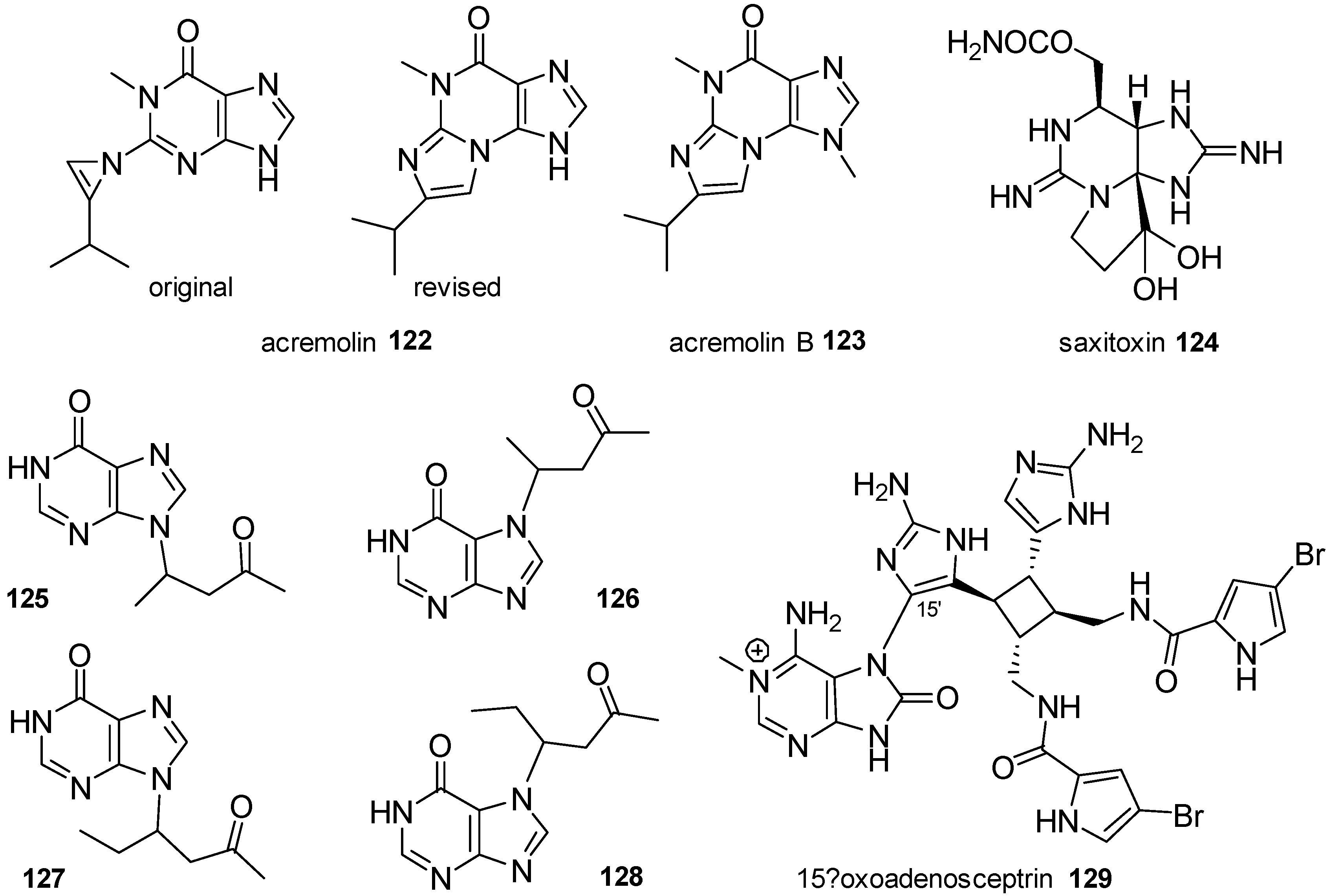
Figure 1. Structures of 1-methyladenine, herbipoline and related compounds.
Marinedrugs 16 00006 g001
Figure 2. Structure of several methylated guanines.
Marinedrugs 16 00006 g002
Figure 3. Structure of several methylated isoguanines.
Marinedrugs 16 00006 g003
Figure 4. Structure of mucronatine, 16, and methylmucronatine, 17.
Marinedrugs 16 00006 g004
Figure 5. Structure of nigricines 1821.
Marinedrugs 16 00006 g005
Figure 6. Structure of methylated xanthines 2226.
Marinedrugs 16 00006 g006
Figure 7. Structure of methylated 8-oxopurines 27 and 28.
Marinedrugs 16 00006 g007
Figure 8. Structure of methylated 8-oxopurines 2932.
Marinedrugs 16 00006 g008
Figure 9. Structure of methylated 8-oxopurines 3338.
Marinedrugs 16 00006 g009
Figure 10. Structure of agelasine.
Marinedrugs 16 00006 g010
Figure 11. Structure of malonganenones 3946.
Marinedrugs 16 00006 g011
Figure 12. Structures of nuttingins 4752.
Marinedrugs 16 00006 g012
Figure 13. Structure of agelasines E and F and related compounds 5357.
Marinedrugs 16 00006 g013
Figure 14. Structure of agelasines A and B and analogues 5862.
Marinedrugs 16 00006 g014
Figure 15. Structures of agelasines 6369.
Marinedrugs 16 00006 g015
Figure 16. Structures of agelasines 7072.
Marinedrugs 16 00006 g016
Figure 17. Structure of agelasines 7377.
Marinedrugs 16 00006 g017
Figure 18. Structures of agelasines 7884.
Marinedrugs 16 00006 g018
Figure 19. Structures of agelasimines A and B and gelasines A and B.
Marinedrugs 16 00006 g019
Figure 20. Structures of agelasines 8993.
Marinedrugs 16 00006 g020
Figure 21. Structure of terpenylpurines 9496.
Marinedrugs 16 00006 g021
Figure 22. Structures of asmarines 97107.
Marinedrugs 16 00006 g022
Figure 23. Structures of alkylpurines 108110.
Marinedrugs 16 00006 g023
Figure 24. Structures of purines 111115.
Marinedrugs 16 00006 g024
Figure 25. Structures of alkylpurines 116121.
Marinedrugs 16 00006 g025
Figure 26. Structures of alkylpurines 122129.
Marinedrugs 16 00006 g026
Figure 27. Structures of spongosine and doridosine.
Marinedrugs 16 00006 g027
Figure 28. Structures of cyclonucleosides 132 and 133.
Marinedrugs 16 00006 g028
Figure 29. Structures of momusines 134137.
Marinedrugs 16 00006 g029
Figure 30. Structures of hamiguanosinol and salvadenosine.
Marinedrugs 16 00006 g030
Figure 31. Structures of nucleosides 140143.
Marinedrugs 16 00006 g031
Scheme 1. A general synthetic procedure towards 7-alkylpurines.
Marinedrugs 16 00006 sch001
Scheme 2. Molinski’s synthesis of acremolin, 122 [91].
Marinedrugs 16 00006 sch002
Scheme 3. Fujii’s synthesis of caissarone, 28, [120] and Itaya’s synthesis of aplidiamine, 113 [83].
Marinedrugs 16 00006 sch003
Scheme 4. General retrosynthetic strategy for the synthesis of diterpenyladeninium derivatives.
Marinedrugs 16 00006 sch004
Scheme 5. Gundersen’s synthesis of malonganenone J, 43 [131].
Marinedrugs 16 00006 sch005
Figure 32. Structures of natural diterpenoids used in the synthesis of diterpenylpurines.
Marinedrugs 16 00006 g032
Scheme 6. Terpenylpurines obtained from trans-communic and cupressic acids and monoterpenes.
Marinedrugs 16 00006 sch006
Scheme 7. Retrosynthetic schemes for asmarines.
Marinedrugs 16 00006 sch007
Scheme 8. Synthesis of an advanced analogue of asmarine A by Shenvi [142].
Marinedrugs 16 00006 sch008
Scheme 9. Molinski’s synthesis of salvadenosine, 139 [110].
Marinedrugs 16 00006 sch009
Scheme 10. Molinski’s synthesis of hamiguanosinol, 138 [110].
Marinedrugs 16 00006 sch010
Scheme 11. Synthesis of trachycladines A and B by Wu [143].
Marinedrugs 16 00006 sch011
Scheme 12. Synthesis of trachycladines A and B and analogues by Koumbis [144,145].
Marinedrugs 16 00006 sch012
Table 1. Letter distribution of agelasines and related compounds according the diterpenoid skeleton.
Diterpenoid SkeletonAgelasines
Clerodane typeagelasines A, B, G, H, I, K, L, M, N, P, Q, R, U, V
ageloxime B
Halimane typeagelasines C, J, O, S
agelasimines A, B
gelasines A, B
Labdane typeagelasines D, T
ageloxime D
Monocyclic diterpenoidagelasines E, F

