


Abstract
The Conus genus includes around 500 species of marine mollusks with a peculiar production of venomous peptides known as conotoxins (CTX). Each species is able to produce up to 200 different biological active peptides. Common structure of CTX is the low number of amino acids stabilized by disulfide bridges and post-translational modifications that give rise to different isoforms. µ and µO-CTX are two isoforms that specifically target voltage-gated sodium channels. These, by inducing the entrance of sodium ions in the cell, modulate the neuronal excitability by depolarizing plasma membrane and propagating the action potential. Hyperexcitability and mutations of sodium channels are responsible for perception and transmission of inflammatory and neuropathic pain states. In this review, we describe the current knowledge of µ-CTX interacting with the different sodium channels subtypes, the mechanism of action and their potential therapeutic use as analgesic compounds in the clinical management of pain conditions.
1. Introduction
Cone snails are carnivorous and venomous molluscs belonging to the Conus genus (Figure S1) living mainly in the tropical marine areas. About 700 species of Cone snails express hundreds of peptide toxins collectively known as conotoxins (CTX) aimed to self-defense, competition and predation of other marine species by means of sting–structures that were reported to be fatal for human since from 300 years ago. CTX, however, do not exert only venomous activity but have a lot of pharmacological properties with specific bioactivity in the treatment of neurological disorders and the associated pain perception [1,2,3].
The presence of disulfide bonds is the essential characteristic for biological function of CTX that allow to divide CTX into two main categories, the disulfide-rich peptides and no-disulfide-rich ones; the first is mainly composed of a maximum of 30 amino acids and the second contains up to 80 amino acids. CTX are categorized into structural families based on the pattern of cysteine residues in terms of both number and position. Furthermore, differently from other peptides that may be subjected to poor absorption, proteolysis and biological half-lives, the presence of disulfide bonds confers to CTX a sort of stability based on the cross-linking between the cysteine side chains [4,5,6]. A further striking feature of CTX is the presence of a variety of posttranslational modifications which are, however, still to fully elucidate. CTX are used to act in a synergistic way to ensure that the venom exerts the most effective activity against the predated animals. The assemblage of CTX acting contemporarily has been named toxin cabal. Literature reports that different cabals co-exist, exerting different activities, including the modulation of different types of ion currents.
Different distribution of ions across the plasma membrane gives rise to a trans-membrane potential known as resting potential (RP), which is negative in almost all cells studied. Ion currents are due to the flux of ions through ion channels, which are specific if it is allowed predominantly the passage of one ion species and may be gated in response to a change in voltage, defined voltage-operated channels. Ion currents are associated with a change in the RP that may shift towards more positive values, giving rise to the depolarisation of the plasma membrane [7].
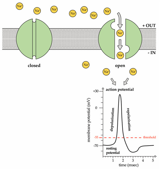
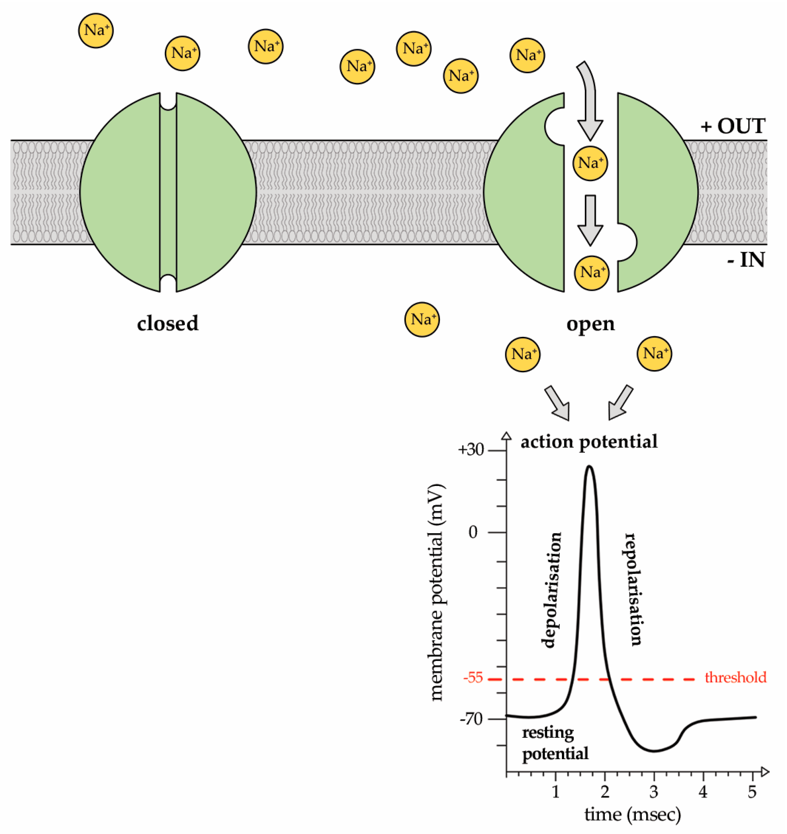
Voltage-gated sodium (Na+) channels (Nav channels) are responsible for the generation of the rapid depolarization of the membrane potential known as action potentials in excitable cells that, in turn, propagate electrical signals in muscles and nerves (Figure 1).
Figure 1.
Representative image of the voltage-gated sodium channel (Nav) state. At the resting potential, the channel is closed. In response to a voltage change impulse greater than the threshold potential of −55 mV, the channel is activated and Na+ ions enter into the cytosol down their concentration gradient, giving rise to the action potential. It is a sudden, transient depolarization of the membrane potential that reaches a peak and, then, is followed by repolarization.
Hence, Nav channel defects and mutations are associated with a wide range of neurological diseases known as channelopathies. Several CTX families have been identified to modulate Na+ current, in particular μ- and μO-CTX are antagonist of the Nav channels. This specificity has been used to discriminate different Nav channel subtypes, characterize specific binding sites on the channels and elucidate the μ-CTX-Nav channel complex interaction [8].
The aim of this review is to give an overview on the pharmacological activity that the µ-CTX superfamily exerts through the modulation of Na+ ion currents. A specific focus will be done on different physiological processes and mechanisms underlying neurological disorders and potential clinical application of these CTX in the therapeutic strategy for neuropathic pain alleviation.
2. Sodium (Na+) Ion Currents
Discovery of Nav channels dates back to the 1950s [9] in the studies on the electric conductance in squid’s giant axon. Later on, Nav channels were isolated and purified in Electrophorus electricus electroplax membrane [10]. Recent advanced studies cloned different Nav channel subtypes.
The role of Nav channels in the propagation of action potential in nerve, muscle and most of the excitable cells has stimulated intense research aimed to determine their structure and to clarify the basis of the voltage-dependent gating. The current recorded in the squid giant axon underlined by Nav channels lasted for a few milliseconds and was quickly inactivated, giving rise to a cascade of other ion currents activation aimed to restore the original potential. Following studies in the 1970s, a conceptual model of Na+ channel function was elaborated, defining also a detailed model of the selectivity of the Na+ channels (for review see [11]). Nav channel activators have been isolated from the venom of several animals, plants and bacteria, providing key insight into the pathophysiological roles of these channels [12].
Interestingly, these studies also established that drugs with anesthetic activity act on Na+ channels binding to a receptor located in the pore of the channel, through different mechanisms. Due to the crucial role of Nav currents in the transmission of electrical stimuli, their inhibitors have been largely used in clinical practice as anticonvulsant, antiarrhythmic and local anesthetic drugs. At present, the Nav channel family includes nine members encoded by Nav channel genes which share sequence homologies and that, due to their complex biochemistry, appear to be associated with many human diseases when down-regulated and/or mutated [13].
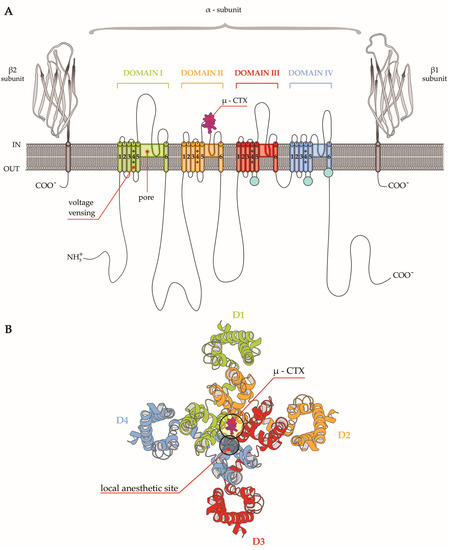
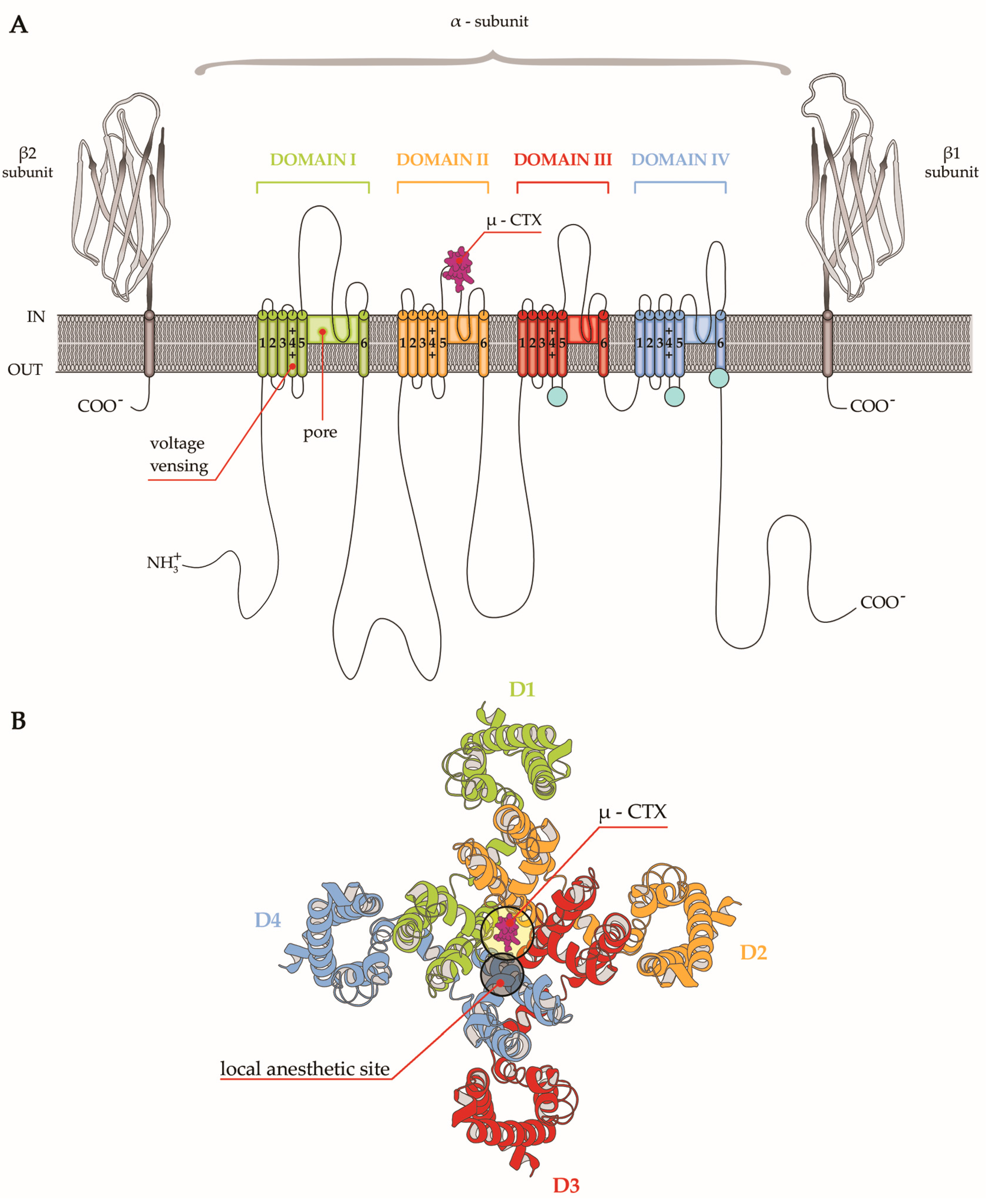
Structurally, Nav channels are heteromeric complexes consisting of an α subunit of about 260s KDa coupled to one or two β subunits with lower weight. The subunits are single-chain peptides of about 2000 amino acids, which determines the differences between subtypes, and contain the receptors for toxins targeting the channel. In mammalian subtypes, the α subunits contain transmembrane and extracellular domains with high-sequence homology. Each domain is composed of six transmembrane helical segments named S1 to S6. The S4 segment present in every domain is the voltage sensor due to the richness in arginine and lysine and is responsible for the generation of the depolarization and the following return to the steady state. Segments 5 and 6 instead represent the Na+ pore and the filters to select Na+ passage. During a resting state, the channels are closed whereas, after depolarization of the RP, the segment S4 is alerted giving rise to a brief opening of the pore and Na+ passage (the open state) to quickly shift to an inactivated state. These main states are the basis for the sensitivity to drugs and inhibitors, which show different affinity for a specific state [14]. In the past, β subunits were considered as auxiliary of the α subunit; however, recent investigations have disclosed their multifunctional signaling role in physiological processes as cell adhesion, gene regulation and brain development [15] (Figure 2).
Figure 2.
(A) Schematic representation of the sodium channel structure comprising a core α subunit and two auxiliary β subunits. The alpha subunit contains four homologue domains (Domain I-Domain IV), each consisting of six transmembrane helices (S1–S6) reported as cylinders. The pore of the channel is formed by S5 and S6 helices in DI, while the voltage sensor is formed by S1–S4 helices in DI. Auxiliary β subunits of the channels as immunoglobulin-like folds are illustrated. µ-CTX binding site is located between S5 and S6 helices in DII. (B) Schematic representation of the top view of the extracellular face of the α-subunit Nav channel. The location of the µ-CTX binding site and the close local anesthetic binding site are indicated.
Mutations in the genes encoding β subunits are linked to a number of diseases, including epilepsy, sudden death syndromes like sudden unexpected death in epilepsy, sudden infant death syndrome and cardiac arrhythmia. Although Nav channels β subunit-specific drugs have not yet been developed, this protein family is an emerging therapeutic target since it has been postulated that it may influence the kinetics of toxin block. From a pharmacological point of view, Na+ channel subtypes upon their diverse sensitivity to tetrodotoxin (TTX) can be distinguished as TTX-sensitive (the neuronal isoforms, Nav channels 1.1, 1.2, 1.3, 1.4, 1.6 and 1.7), or TTX-resistant (Nav channels 1.5, 1.8, 1.9) [16,17]. The role of Nav channels as analgesic targets has been deeply studied and highlighted with a specific focus on some specific isoforms.
3. Na+ Currents—Linked Channelopathies
Channelopathies are diseases caused and underlined by disorders in ion channel functions whose etiology may be either genetic mainly due to ion mutations or acquired in cases of autoimmune insults, drugs and toxins [18]. Channelopathies can be found in many organ systems as cardiovascular, respiratory, endocrine, urinary, immune and nervous. In the latter, several neurological disorders such as epilepsy, cerebellar ataxia, myasthenia, myotonia, erythermalgia, schizophrenia, encephalopathy, Alzheimer syndrome, Dravet syndrome, and other neuropathies are associated with channels malfunctioning. Since ion currents are the flow of ions across the plasma membranes of either the cell or organelles, they play crucial roles in several cellular activities and in mechanisms of signal transduction in organs and related systems. Several channelopathies of the nervous system are underlined by Nav channel subtypes modulation. Literature reports that mutations of Nav channels 1.1 and 1.2 are linked to either epilepsy and the alteration of other central nervous system functions, whereas other Nav channel subtypes are mainly related to cardiac dysfunctions [19]. Neurological disorders, such as paralyses and cerebellar atrophy, are also associated with mutations in Nav channel subtypes (see for review [20]). In particular, nine isoforms according to the α-subunit sequence have been found in the central and peripheral nervous systems. The α-subtypes (Nav channels 1.1–1.9) present in sensory neurons underpin electrical activity through action potential propagation and this depolarization due to the influx of Na+ ions has been suggested to play a role in pain perception and transmission [21,22]. Although α subunits possess the features for Nav channel functioning, a co-expression of the β subunit was shown to influence channel gating, trafficking, expression and the biological activities of venom-derived toxins [23].
The anomalies in Na+ conductance due to injuries of different origin may lead to hyperexcitability of neurons resulting in neuropathic pain and disorders. In fact, channel defects and mutations have been related to vascular and painful organ diseases [24], whereas in other cases Nav channel mutations in functional sites are responsible for pain insensitivity [25]. At present, four channels seem to be strictly involved in pain disorders associated with several human pathologies from multiple sclerosis to cancer [26,27].
6. Conclusions
A worldwide interest in the discovery of new analgesic compounds is due to the limited efficacy and unacceptable side effects of opioid-based pain therapies. These, in fact, causing constipation, emesis, dizziness, vomiting and seriously impacting driving and working activities, pose patients at risk of tolerance rather than mitigate their primary objective, that is, pain relief [89]. A major hurdle for this field is to identify excellent alternatives to opioids as analgesics in the costly pain therapy [90,91]. Modulators of Nav channel subtypes may represent new tools for facing pain signaling and disorders. The ample interest on Nav channels involvement for drug discovery and therapeutic treatment of pain [92,93] is supported by the findings that subunits 1.3, 1.7, 1.8 and 1.9 predominately expressed in sensory neurons are functionally involved in many different forms of pain. Thus, it is clear that µ-CTX, as inhibitors of Nav channels, are appropriate candidates to be administered to induce analgesia without undesirable side effects.
This lesson comes from the unique CTX (ω-MVIIA) approved for clinical use and marketed for treatment of chronic pain (Prialt, the trade name) which acts by inhibiting calcium channels. Prialt exerts many side effects and, being administered by direct infusion in the spinal cord (intrathecally) is invasive; hence, it has been considered the last possibility for alleviation of chronic pain in clinical practice. Although µ-CTX targeting Nav channels have a systemic way of administration [82], there is still a paucity of high selective Nav channel blockers since the action on multiple subtypes may create side effects.
This review is dealing with preclinical studies and there is a long way before a real therapeutic application. In fact, based on the advantages and the interest in CTX in pain therapies and the need for new drug design, further studies are required to investigate and demonstrate the pharmacological effectiveness of these compounds. However, new patents are currently reporting invention related to novel µ-CTX peptides, and/or biologically active fragments being possible candidates in pharmaceutical composition for the anesthetic medications [94].
In many cases, µ-CTX selectivity is still to be elucidated; hence, the hope is to discover new subtype-selective agents against Nav channels and create engineered analogues of therapeutic utility with decreased side effects, safety and the most noninvasive administration as the oral route [95].
New challenging perspective for structure-based drug discovery is at present to elucidate atomic structures of Nav channels in order to understand their function and mechanisms of action. Recent investigation by Huang [96] is, in fact, aimed to generate a homologous model of human Nav channel 1.7, to disclose disease-associated mutations. The search for new technical approaches are also in line with the fact that Conus species are threatened by increased pollution, climate change and overfishing. These conditions pose these mollusks at high risk of extinction in the years to come and their survival may be further compromised by the extraction of bioactive compounds described in this review. The important contribution of these animals in biomedicine and biotechnologies may, however, rely on new sustainable bio-molecular techniques as chemical synthesis and recombinant production in heterologous expression systems and polymerase chain reaction, sequencing of DNA fragments and transcriptomes that will allow in the future to obtain bioactive material with few or null animal sacrifice [97].
Supplementary Materials
The following are available online at www.mdpi.com/1660-3397/15/10/295/s1. Figure S1: Different species of Conus genus.
Acknowledgments
This work has been supported by Stazione Zoologica Anton Dohrn institutional funds. A.G. has been supported by a Stazione Zoologica Anton Dohrn post-doc fellowship.
Author Contributions
E.T., R.B., A.G. made equal contribution in collection and considering of the data for the review and in preparation of the text.
Conflicts of Interest
The authors declare no conflict of interest.
References
- Terlau, H.; Olivera, B.M. Conus venoms: A rich source of novel ion channel-targeted peptides. Physiol. Rev. 2004, 84, 41–68. [Google Scholar] [CrossRef] [Scilit] [PubMed]
- Stix, G. A toxin against pain. Sci. Am. 2005, 292, 88–93. [Google Scholar] [CrossRef] [Scilit] [PubMed]
- Layer, R.T.; McIntosh, J.M. Conotoxins: Therapeutic potential and application. Mar. Drugs 2006, 4, 119–142. [Google Scholar] [CrossRef] [Scilit]
- Becker, S.; Terlau, H. Toxins from cone snails: Properties, applications and biotechnological production. Appl. Microbiol. Biotechnol. 2008, 79, 1–9. [Google Scholar] [CrossRef] [Scilit] [PubMed]
- Halai, R.; Craik, D.J. Conotoxins: Natural product drug leads. Nat. Prod. Rep. 2009, 26, 526–536. [Google Scholar] [CrossRef] [Scilit] [PubMed]
- Mir, R.; Karim, S.; Amjad Kamal, M.; Wilson, M.C.; Mirza, Z. Conotoxins: Structure, therapeutic potential and pharmacological applications. Curr. Pharm. Des. 2016, 22, 582–589. [Google Scholar] [CrossRef] [Scilit] [PubMed]
- Tosti, E.; Boni, R. Electrical events during gamete maturation and fertilization in animals and humans. Hum. Reprod. Update 2004, 10, 53–65. [Google Scholar] [CrossRef] [Scilit] [PubMed]
- Green, B.; Olivera, B. Chapter three-venom peptides from cone snails: Pharmacological probes for voltage-gated sodium channels. Curr. Top. Membr. 2016, 78, 65–86. [Google Scholar] [PubMed]
- Hodgkin, A.L.; Huxley, A.F. A quantitative description of membrane current and its application to conduction and excitation in nerve. J. Physiol. 1952, 117, 500. [Google Scholar] [CrossRef] [Scilit] [PubMed]
- Agnew, W.S.; Levinson, S.R.; Brabson, J.S.; Raftery, M.A. Purification of the tetrodotoxin-binding component associated with the voltage-sensitive sodium channel from Electrophorus electricus electroplax membranes. Proc. Natl. Acad. Sci. USA 1978, 75, 2606–2610. [Google Scholar] [CrossRef] [Scilit] [PubMed]
- Catterall, W.A. Structure and function of voltage-gated sodium channels at atomic resolution. Exp. Physiol. 2014, 99, 35–51. [Google Scholar] [CrossRef] [Scilit] [PubMed]
- Deuis, J.R.; Mueller, A.; Israel, M.R.; Vetter, I. The pharmacology of voltage-gated sodium channel activators. Neuropharmacology 2017. [Google Scholar] [CrossRef] [Scilit] [PubMed]
- Clairfeuille, T.; Xu, H.; Koth, C.M.; Payandeh, J. Voltage-gated sodium channels viewed through a structural biology lens. Curr. Opin. Struct. Biol. 2017, 45, 74–84. [Google Scholar] [CrossRef] [Scilit] [PubMed]
- Bagal, S.K.; Marron, B.E.; Owen, R.M.; Storer, R.I.; Swain, N.A. Voltage gated sodium channels as drug discovery targets. Channels 2015, 9, 360–366. [Google Scholar] [CrossRef] [Scilit] [PubMed]
- O’Malley, H.A.; Isom, L.L. Sodium channel β subunits: Emerging targets in channelopathies. Annu. Rev. Physiol. 2015, 77, 481–504. [Google Scholar] [CrossRef] [Scilit] [PubMed]
- Wood, J.N.; Boorman, J.P.; Okuse, K.; Baker, M.D. Voltage-gated sodium channels and pain pathways. J. Neurobiol. 2004, 61, 55–71. [Google Scholar] [CrossRef] [Scilit] [PubMed]
- Catterall, W.A.; Perez-Reyes, E.; Snutch, T.P.; Striessnig, J. International Union of Pharmacology. XLVIII. Nomenclature and structure-function relationships of voltage-gated calcium channels. Pharmacol. Rev. 2005, 57, 411–425. [Google Scholar] [CrossRef] [Scilit] [PubMed]
- Kass, R.S. The channelopathies: Novel insights into molecular and genetic mechanisms of human disease. J. Clin. Investig. 2005, 115, 1986–1989. [Google Scholar] [CrossRef] [Scilit] [PubMed]
- Kim, J.-B. Channelopathies. Korean J. Pediatr. 2014, 57, 1–18. [Google Scholar] [CrossRef] [Scilit] [PubMed]
- Andavan, G.S.; Lemmens-Gruber, R. Voltage-gated sodium channels: Mutations, channelopathies and targets. Curr. Med. Chem. 2011, 18, 377–397. [Google Scholar] [CrossRef] [Scilit] [PubMed]
- Cummins, T.R.; Sheets, P.L.; Waxman, S.G. The roles of sodium channels in nociception: Implications for mechanisms of pain. Pain 2007, 131, 243–257. [Google Scholar] [CrossRef] [Scilit] [PubMed]
- Dib-Hajj, S.D.; Cummins, T.R.; Black, J.A.; Waxman, S.G. From genes to pain: Nav 1.7 and human pain disorders. Trends Neurosci. 2007, 30, 555–563. [Google Scholar] [CrossRef] [Scilit] [PubMed]
- Chahine, M.; O’Leary, M.E. Regulatory role of voltage-gated Na+ channel β subunits in sensory neurons. Front. Pharmacol. 2011, 2, 70. [Google Scholar] [CrossRef] [Scilit] [PubMed]
- Fertleman, C.R.; Baker, M.D.; Parker, K.A.; Moffatt, S.; Elmslie, F.V.; Abrahamsen, B.; Ostman, J.; Klugbauer, N.; Wood, J.N.; Gardiner, R.M. SCN9A mutations in paroxysmal extreme pain disorder: Allelic variants underlie distinct channel defects and phenotypes. Neuron 2006, 52, 767–774. [Google Scholar] [CrossRef] [Scilit] [PubMed]
- Cox, J.J.; Reimann, F.; Nicholas, A.K.; Thornton, G.; Roberts, E.; Springell, K.; Karbani, G.; Jafri, H.; Mannan, J.; Raashid, Y. An SCN9A channelopathy causes congenital inability to experience pain. Nature 2006, 444, 894–898. [Google Scholar] [CrossRef] [Scilit] [PubMed]
- Bhattacharya, A.; Wickenden, A.D.; Chaplan, S.R. Sodium channel blockers for the treatment of neuropathic pain. Neurotherapeutics 2009, 6, 663–678. [Google Scholar] [CrossRef] [Scilit] [PubMed]
- Luiz, A.P.; Wood, J.N. Chapter six-sodium channels in pain and cancer: New therapeutic opportunities. Adv. Pharmacol. 2016, 75, 153–178. [Google Scholar] [PubMed]
- Kao, C. Tetrodotoxin, saxitoxin and their significance in the study of excitation phenomena. Pharmacol. Rev. 1966, 18, 997–1049. [Google Scholar] [PubMed]
- Llewellyn, L.E. Sodium channel inhibiting marine toxins. In Marine Toxins as Research Tools; Springer: Berlin/Heidelberg, Germany, 2009; pp. 67–97. [Google Scholar]
- Pratheepa, V.; Vasconcelos, V. Binding and pharmacokinetics of the sodium channel blocking toxins (Saxitoxin and the Tetrodotoxins). Mini Rev. Med. Chem. 2017, 17, 320–327. [Google Scholar] [CrossRef] [Scilit] [PubMed]
- Nieto, F.R.; Cobos, E.J.; Tejada, M.Á.; Sánchez-Fernández, C.; González-Cano, R.; Cendán, C.M. Tetrodotoxin (TTX) as a therapeutic agent for pain. Mar. Drugs 2012, 10, 281–305. [Google Scholar] [CrossRef] [Scilit] [PubMed]
- Norton, R.S.; Olivera, B.M. Conotoxins down under. Toxicon 2006, 48, 780–798. [Google Scholar] [CrossRef] [Scilit] [PubMed]
- Safavi-Hemami, H.; Siero, W.A.; Gorasia, D.G.; Young, N.D.; MacMillan, D.; Williamson, N.A.; Purcell, A.W. Specialisation of the venom gland proteome in predatory cone snails reveals functional diversification of the conotoxin biosynthetic pathway. J. Proteome Res. 2011, 10, 3904–3919. [Google Scholar] [CrossRef] [Scilit] [PubMed]
- Li, R.A.; Tomaselli, G.F. Using the deadly μ-conotoxins as probes of voltage-gated sodium channels. Toxicon 2004, 44, 117–122. [Google Scholar] [CrossRef] [Scilit] [PubMed]
- Olivera, B.M.; Cruz, L.J. Conotoxins, in retrospect. Toxicon 2001, 39, 7–14. [Google Scholar] [CrossRef] [Scilit]
- Green, B.R.; Bulaj, G.; Norton, R.S. Structure and function of μ-conotoxins, peptide-based sodium channel blockers with analgesic activity. Future Med. Chem. 2014, 6, 1677–1698. [Google Scholar] [CrossRef] [Scilit] [PubMed]
- Shon, K.-J.; Olivera, B.M.; Watkins, M.; Jacobsen, R.B.; Gray, W.R.; Floresca, C.Z.; Cruz, L.J.; Hillyard, D.R.; Brink, A.; Terlau, H. μ-Conotoxin PIIIA, a new peptide for discriminating among tetrodotoxin-sensitive Na channel subtypes. J. Neurosci. 1998, 18, 4473–4481. [Google Scholar] [PubMed]
- Walewska, A.; Skalicky, J.J.; Davis, D.R.; Zhang, M.-M.; Lopez-Vera, E.; Watkins, M.; Han, T.S.; Yoshikami, D.; Olivera, B.M.; Bulaj, G. NMR-based mapping of disulfide bridges in cysteine-rich peptides: Application to the μ-conotoxin SxIIIA. J. Am. Chem. Soc. 2008, 130, 14280–14286. [Google Scholar] [CrossRef] [Scilit] [PubMed]
- Leipold, E.; DeBie, H.; Zorn, S.; Adolfo, B.; Olivera, B.M.; Terlau, H.; Heinemann, S.H. µO-conotoxins inhibit Nav channels by interfering with their voltage sensors in domain-2. Channels 2007, 1, 253–262. [Google Scholar] [CrossRef] [Scilit] [PubMed]
- Cruz, L.; Gray, W.; Olivera, B.M.; Zeikus, R.; Kerr, L.; Yoshikami, D.; Moczydlowski, E. Conus geographus toxins that discriminate between neuronal and muscle sodium channels. J. Biol. Chem. 1985, 260, 9280–9288. [Google Scholar] [PubMed]
- Li, R.A.; Ennis, I.L.; Xue, T.; Nguyen, H.M.; Tomaselli, G.F.; Goldin, A.L.; Marbán, E. Molecular basis of isoform-specific μ-conotoxin block of cardiac, skeletal muscle, and brain Na+ channels. J. Biol. Chem. 2003, 278, 8717–8724. [Google Scholar] [CrossRef] [Scilit] [PubMed]
- Safo, P.; Rosenbaum, T.; Shcherbatko, A.; Choi, D.-Y.; Han, E.; Toledo-Aral, J.J.; Olivera, B.M.; Brehm, P.; Mandel, G. Distinction among neuronal subtypes of voltage-activated sodium channels by μ-conotoxin PIIIA. J. Neurosci. 2000, 20, 76–80. [Google Scholar] [PubMed]
- Chen, R.; Robinson, A.; Chung, S.-H. Mechanism of μ-conotoxin PIIIA binding to the voltage-gated Na+ channel NaV 1.4. PLoS ONE 2014, 9, e93267. [Google Scholar]
- Keizer, D.W.; West, P.J.; Lee, E.F.; Yoshikami, D.; Olivera, B.M.; Bulaj, G.; Norton, R.S. Structural basis for tetrodotoxin-resistant sodium channel binding by μ-conotoxin SmIIIA. J. Biol. Chem. 2003, 278, 46805–46813. [Google Scholar] [CrossRef] [Scilit] [PubMed]
- Nielsen, K.J.; Watson, M.; Adams, D.J.; Hammarström, A.K.; Gage, P.W.; Hill, J.M.; Craik, D.J.; Thomas, L.; Adams, D.; Alewood, P.F. Solution structure of μ-conotoxin PIIIA, a preferential inhibitor of persistent tetrodotoxin-sensitive sodium channels. J. Biol. Chem. 2002, 277, 27247–27255. [Google Scholar] [CrossRef] [Scilit] [PubMed]
- Leipold, E.; Ullrich, F.; Thiele, M.; Tietze, A.A.; Terlau, H.; Imhof, D.; Heinemann, S.H. Subtype-specific block of voltage-gated K+ channels by μ-conopeptides. Biochem. Biophys. Res. Commun. 2017, 482, 1135–1140. [Google Scholar] [CrossRef] [Scilit] [PubMed]
- Chen, R.; Chung, S.-H. Binding modes of μ-conotoxin to the bacterial sodium channel (Na V Ab). Biophys. J. 2012, 102, 483–488. [Google Scholar] [CrossRef] [Scilit] [PubMed]
- Bulaj, G.; West, P.J.; Garrett, J.E.; Watkins, M.; Zhang, M.-M.; Norton, R.S.; Smith, B.J.; Yoshikami, D.; Olivera, B.M. Novel conotoxins from Conus striatus and Conus kinoshitai selectively block TTX-resistant sodium channels. Biochemistry 2005, 44, 7259–7265. [Google Scholar] [CrossRef] [Scilit] [PubMed]
- Zhang, M.-M.; Green, B.R.; Catlin, P.; Fiedler, B.; Azam, L.; Chadwick, A.; Terlau, H.; McArthur, J.R.; French, R.J.; Gulyas, J. Structure/function characterization of μ-conotoxin KIIIA, an analgesic, nearly irreversible blocker of mammalian neuronal sodium channels. J. Biol. Chem. 2007, 282, 30699–30706. [Google Scholar] [CrossRef] [Scilit] [PubMed]
- Yao, S.; Zhang, M.-M.; Yoshikami, D.; Azam, L.; Olivera, B.M.; Bulaj, G.; Norton, R.S. Structure, dynamics, and selectivity of the sodium channel blocker μ-conotoxin SIIIA. Biochemistry 2008, 47, 10940–10949. [Google Scholar] [CrossRef] [Scilit] [PubMed]
- Wang, C.-Z.; Zhang, H.; Jiang, H.; Lu, W.; Zhao, Z.-Q.; Chi, C.-W. A novel conotoxin from Conus striatus, μ-SIIIA, selectively blocking rat tetrodotoxin-resistant sodium channels. Toxicon 2006, 47, 122–132. [Google Scholar] [CrossRef] [Scilit] [PubMed]
- West, P.J.; Bulaj, G.; Garrett, J.E.; Olivera, B.M.; Yoshikami, D. μ-Conotoxin SmIIIA, a potent inhibitor of tetrodotoxin-resistant sodium channels in amphibian sympathetic and sensory neurons. Biochemistry 2002, 41, 15388–15393. [Google Scholar] [CrossRef] [Scilit] [PubMed]
- Lewis, R.J.; Schroeder, C.I.; Ekberg, J.; Nielsen, K.J.; Loughnan, M.; Thomas, L.; Adams, D.A.; Drinkwater, R.; Adams, D.J.; Alewood, P.F. Isolation and structure-activity of μ-conotoxin TIIIA, a potent inhibitor of tetrodotoxin-sensitive voltage-gated sodium channels. Mol. Pharmacol. 2007, 71, 676–685. [Google Scholar] [CrossRef] [Scilit] [PubMed]
- Holford, M.; Zhang, M.-M.; Gowd, K.H.; Azam, L.; Green, B.R.; Watkins, M.; Ownby, J.-P.; Yoshikami, D.; Bulaj, G.; Olivera, B.M. Pruning nature: Biodiversity-derived discovery of novel sodium channel blocking conotoxins from Conus bullatus. Toxicon 2009, 53, 90–98. [Google Scholar] [CrossRef] [Scilit] [PubMed]
- Kuang, Z.; Zhang, M.-M.; Gupta, K.; Gajewiak, J.; Gulyas, J.; Balaram, P.; Rivier, J.E.; Olivera, B.M.; Yoshikami, D.; Bulaj, G. Mammalian neuronal sodium channel blocker μ-conotoxin BuIIIB has a structured N-terminus that influences potency. ACS Chem. Biol. 2013, 8, 1344–1351. [Google Scholar] [CrossRef] [Scilit] [PubMed]
- Favreau, P.; Benoit, E.; Hocking, H.G.; Carlier, L.; D’hoedt, D.; Leipold, E.; Markgraf, R.; Schlumberger, S.; Córdova, M.A.; Gaertner, H. A novel µ-conopeptide, CnIIIC, exerts potent and preferential inhibition of NaV1. 2/1.4 channels and blocks neuronal nicotinic acetylcholine receptors. Br. J. Pharmacol. 2012, 166, 1654–1668. [Google Scholar] [CrossRef] [Scilit] [PubMed]
- Green, B.R.; Gajewiak, J.; Chhabra, S.; Skalicky, J.J.; Zhang, M.-M.; Rivier, J.E.; Bulaj, G.; Olivera, B.M.; Yoshikami, D.; Norton, R.S. Structural basis for the inhibition of voltage-gated sodium channels by conotoxin μO § -GVIIJ. J. Biol. Chem. 2016, 291, 7205–7220. [Google Scholar] [CrossRef] [Scilit] [PubMed]
- Zhang, M.-M.; Fiedler, B.; Green, B.R.; Catlin, P.; Watkins, M.; Garrett, J.E.; Smith, B.J.; Yoshikami, D.; Olivera, B.M.; Bulaj, G. Structural and functional diversities among μ-conotoxins targeting TTX-resistant sodium channels. Biochemistry 2006, 45, 3723–3732. [Google Scholar] [CrossRef] [Scilit] [PubMed]
- Yang, M.; Zhao, S.; Min, X.; Shao, M.; Chen, Y.; Chen, Z.; Zhou, M. A novel μ-conotoxin from worm-hunting Conus tessulatus that selectively inhibit rat TTX-resistant sodium currents. Toxicon 2017, 130, 11–18. [Google Scholar] [CrossRef] [Scilit] [PubMed]
- Daly, N.L.; Ekberg, J.A.; Thomas, L.; Adams, D.J.; Lewis, R.J.; Craik, D.J. Structures of μO-conotoxins from Conus marmoreus inhibitors of tetrodotoxin (TTX)-sensitive and TTX-resistant sodium channels in mammalian sensory neurons. J. Biol. Chem. 2004, 279, 25774–25782. [Google Scholar] [CrossRef] [Scilit] [PubMed]
- Zorn, S.; Leipold, E.; Hansel, A.; Bulaj, G.; Olivera, B.M.; Terlau, H.; Heinemann, S.H. The μO-conotoxin MrVIA inhibits voltage-gated sodium channels by associating with domain-3. FEBS Lett. 2006, 580, 1360–1364. [Google Scholar] [CrossRef] [Scilit] [PubMed]
- Ekberg, J.; Jayamanne, A.; Vaughan, C.W.; Aslan, S.; Thomas, L.; Mould, J.; Drinkwater, R.; Baker, M.; Abrahamsen, B.; Wood, J. μO-conotoxin MrVIB selectively blocks Nav1. 8 sensory neuron specific sodium channels and chronic pain behavior without motor deficits. Proc. Natl. Acad. Sci. USA 2006, 103, 17030–17035. [Google Scholar] [CrossRef] [Scilit] [PubMed]
- Vetter, I.; Dekan, Z.; Knapp, O.; Adams, D.J.; Alewood, P.F.; Lewis, R.J. Isolation, characterization and total regioselective synthesis of the novel μO-conotoxin MfVIA from Conus magnificus that targets voltage-gated sodium channels. Biochem. Pharmacol. 2012, 84, 540–548. [Google Scholar] [CrossRef] [Scilit] [PubMed]
- Gajewiak, J.; Azam, L.; Imperial, J.; Walewska, A.; Green, B.R.; Bandyopadhyay, P.K.; Raghuraman, S.; Ueberheide, B.; Bern, M.; Zhou, H.M. A disulfide tether stabilizes the block of sodium channels by the conotoxin μO § -GVIIJ. Proc. Natl. Acad. Sci. USA 2014, 111, 2758–2763. [Google Scholar] [CrossRef] [Scilit] [PubMed]
- DiMario, F.J. Inherited Pain Syndromes and Ion Channels; Seminars in Pediatric Neurology; Elsevier: Amsterdam, The Netherlands, 2016; pp. 248–253. [Google Scholar]
- Dubin, A.E.; Patapoutian, A. Nociceptors: The sensors of the pain pathway. J. Clin. Investig. 2010, 120, 3760–3772. [Google Scholar] [CrossRef] [Scilit] [PubMed]
- Gaskin, D.J.; Richard, P. The economic costs of pain in the United States. J. Pain 2012, 13, 715–724. [Google Scholar] [CrossRef] [Scilit] [PubMed]
- Baron, R. Neuropathic pain: A clinical perspective. In Sensory Nerves; Springer: Berlin/Heidelberg, Germany, 2009; pp. 3–30. [Google Scholar]
- Dib-Hajj, S.D.; Black, J.A.; Waxman, S.G. Voltage-gated sodium channels: Therapeutic targets for pain. Pain Med. 2009, 10, 1260–1269. [Google Scholar] [CrossRef] [Scilit] [PubMed]
- Hains, B.C.; Saab, C.Y.; Klein, J.P.; Craner, M.J.; Waxman, S.G. Altered sodium channel expression in second-order spinal sensory neurons contributes to pain after peripheral nerve injury. J. Neurosci. 2004, 24, 4832–4839. [Google Scholar] [CrossRef] [Scilit] [PubMed]
- Lampert, A.; O’Reilly, A.O.; Reeh, P.; Leffler, A. Sodium channelopathies and pain. Pflugers Arch. 2010, 460, 249–263. [Google Scholar] [CrossRef] [Scilit] [PubMed]
- Clare, J.J. Targeting voltage-gated sodium channels for pain therapy. Expert Opin. Investig. Drugs 2010, 19, 45–62. [Google Scholar] [CrossRef] [Scilit] [PubMed]
- Goldberg, Y.; MacFarlane, J.; MacDonald, M.; Thompson, J.; Dube, M.P.; Mattice, M.; Fraser, R.; Young, C.; Hossain, S.; Pape, T. Loss-of-function mutations in the Nav1. 7 gene underlie congenital indifference to pain in multiple human populations. Clin. Genet. 2007, 71, 311–319. [Google Scholar] [CrossRef] [Scilit] [PubMed]
- Akopian, A.N.; Sivilotti, L.; Wood, J.N. A tetrodotoxin-resistant voltage-gated sodium channel expressed by sensory neurons. Nature 1996, 379, 257. [Google Scholar] [CrossRef] [Scilit] [PubMed]
- Djouhri, L.; Fang, X.; Okuse, K.; Wood, J.N.; Berry, C.M.; Lawson, S.N. The TTX-resistant sodium channel Nav1. 8 (SNS/PN3): Expression and correlation with membrane properties in rat nociceptive primary afferent neurons. J. Physiol. 2003, 550, 739–752. [Google Scholar] [CrossRef] [Scilit] [PubMed]
- Renganathan, M.; Cummins, T.R.; Waxman, S.G. Contribution of Nav1. 8 sodium channels to action potential electrogenesis in DRG neurons. J. Neurophysiol. 2001, 86, 629–640. [Google Scholar] [PubMed]
- Knapp, O.; McArthur, J.R.; Adams, D.J. Conotoxins targeting neuronal voltage-gated sodium channel subtypes: Potential analgesics? Toxins 2012, 4, 1236–1260. [Google Scholar] [CrossRef] [Scilit] [PubMed]
- Bennett, D.L.; Woods, C.G. Painful and painless channelopathies. Lancet Neurol. 2014, 13, 587–599. [Google Scholar] [CrossRef] [Scilit]
- Livett, B.G.; Gayler, K.R.; Khalil, Z. Drugs from the sea: Conopeptides as potential therapeutics. Curr. Med. Chem. 2004, 11, 1715–1723. [Google Scholar] [CrossRef] [Scilit] [PubMed]
- Ahern, C.A.; Payandeh, J.; Bosmans, F.; Chanda, B. The hitchhiker’s guide to the voltage-gated sodium channel galaxy. J. Gen. Physiol. 2016, 147, 1–24. [Google Scholar] [CrossRef] [Scilit] [PubMed]
- Essack, M.; Bajic, V.B.; Archer, J.A. Conotoxins that confer therapeutic possibilities. Mar. Drugs 2012, 10, 1244–1265. [Google Scholar] [CrossRef] [Scilit] [PubMed]
- Munasinghe, N.R.; Christie, M.J. Conotoxins that could provide analgesia through voltage gated sodium channel inhibition. Toxins 2015, 7, 5386–5407. [Google Scholar] [CrossRef] [Scilit] [PubMed]
- Green, B.R.; Catlin, P.; Zhang, M.-M.; Fiedler, B.; Bayudan, W.; Morrison, A.; Norton, R.S.; Smith, B.J.; Yoshikami, D.; Olivera, B.M. Conotoxins containing nonnatural backbone spacers: Cladistic-based design, chemical synthesis, and improved analgesic activity. Chem. Biol. 2007, 14, 399–407. [Google Scholar] [CrossRef] [Scilit] [PubMed]
- Wilson, M.J.; Yoshikami, D.; Azam, L.; Gajewiak, J.; Olivera, B.M.; Bulaj, G.; Zhang, M.-M. μ-Conotoxins that differentially block sodium channels NaV1. 1 through 1.8 identify those responsible for action potentials in sciatic nerve. Proc. Natl. Acad. Sci. USA 2011, 108, 10302–10307. [Google Scholar] [CrossRef] [Scilit] [PubMed]
- Wilson, M.J.; Zhang, M.-M.; Gajewiak, J.; Azam, L.; Rivier, J.E.; Olivera, B.M.; Yoshikami, D. α- and β-subunit composition of voltage-gated sodium channels investigated with μ-conotoxins and the recently discovered μO § -conotoxin GVIIJ. J. Neurophysiol. 2015, 113, 2289–2301. [Google Scholar] [CrossRef] [Scilit] [PubMed]
- Markgraf, R.; Leipold, E.; Schirmeyer, J.; Paolini-Bertrand, M.; Hartley, O.; Heinemann, S.H. Mechanism and molecular basis for the sodium channel subtype specificity of µ-conopeptide CnIIIC. Br. J. Clin. Pharmacol. 2012, 167, 576–586. [Google Scholar] [CrossRef] [Scilit] [PubMed]
- Mahdavi, S.; Kuyucak, S. Molecular dynamics study of binding of µ-conotoxin GIIIA to the voltage-gated sodium channel Nav1. 4. PLoS ONE 2014, 9, e105300. [Google Scholar] [CrossRef] [Scilit] [PubMed]
- Patel, D.; Mahdavi, S.; Kuyucak, S. Computational study of binding of μ-conotoxin GIIIA to bacterial sodium channels NaVAb and NaVRh. Biochemistry 2016, 55, 1929–1938. [Google Scholar] [CrossRef] [Scilit] [PubMed]
- de Lera Ruiz, M.; Kraus, R.L. Voltage-gated sodium channels: Structure, function, pharmacology, and clinical indications. J. Med. Chem. 2015, 58, 7093–7118. [Google Scholar] [CrossRef] [Scilit] [PubMed]
- Norton, R.S. µ-Conotoxins as leads in the development of new analgesics. Molecules 2010, 15, 2825–2844. [Google Scholar] [CrossRef] [Scilit] [PubMed]
- O’brien, T.; Christrup, L.; Drewes, A.; Fallon, M.; Kress, H.; McQuay, H.; Mikus, G.; Morlion, B.; Perez-Cajaraville, J.; Pogatzki-Zahn, E. European Pain Federation position paper on appropriate opioid use in chronic pain management. Eur. J. Pain 2017, 21, 3–19. [Google Scholar] [CrossRef] [Scilit] [PubMed]
- Bagal, S.K.; Brown, A.D.; Cox, P.J.; Omoto, K.; Owen, R.M.; Pryde, D.C.; Sidders, B.; Skerratt, S.E.; Stevens, E.B.; Storer, R.I. Ion channels as therapeutic targets: A drug discovery perspective. J. Med. Chem. 2012, 56, 593–624. [Google Scholar] [CrossRef] [Scilit] [PubMed]
- Bagal, S.K.; Chapman, M.L.; Marron, B.E.; Prime, R.; Storer, R.I.; Swain, N.A. Recent progress in sodium channel modulators for pain. Bioorg. Med. Chem. Lett. 2014, 24, 3690–3699. [Google Scholar] [CrossRef] [Scilit] [PubMed]
- Favreau, P.; Benoit, E.; Molgo, J.; Stocklin, R. Mu-Conotoxin Peptides and Use Thereof as a Local Anesthetic. U.S. Patent 20170226166 A1, 10 August 2017. [Google Scholar]
- Woodcock, B.G. The Facies dolorosa and the Conidae. Int. J. Clin. Pharmacol. 2016, 54, 495. [Google Scholar] [CrossRef] [Scilit]
- Huang, W.; Liu, M.; Yan, S.F.; Yan, N. Structure-based assessment of disease-related mutations in human voltage-gated sodium channels. Protein Cell 2017, 8, 1–38. [Google Scholar] [CrossRef] [Scilit] [PubMed]
- Peters, H.; O’Leary, B.C.; Hawkins, J.P.; Carpenter, K.E.; Roberts, C.M. Conus: First comprehensive conservation Red List assessment of a marine gastropod mollusc genus. PLoS ONE 2013, 8, e83353. [Google Scholar] [CrossRef] [Scilit] [PubMed]
© 2017 by the authors. Licensee MDPI, Basel, Switzerland. This article is an open access article distributed under the terms and conditions of the Creative Commons Attribution (CC BY) license (http://creativecommons.org/licenses/by/4.0/).