Abstract
A new series of nortopsentin analogues, in which the imidazole ring of the natural product was replaced by thiazole and the indole unit bound to position 2 of the thiazole ring was substituted by a 7-azaindole moiety, was efficiently synthesized. Two of the new nortopsentin analogues showed good antiproliferative effect against the totality of the NCI full panel of human tumor cell lines (~60) having GI50 values ranging from low micromolar to nanomolar level. The mechanism of the antiproliferative effect of these derivatives, investigated on human hepatoma HepG2 cells, was pro-apoptotic, being associated with externalization of plasma membrane phosphatidylserine and mitochondrial dysfunction. Moreover, the compounds induced a concentration-dependent accumulation of cells in the subG0/G1phase, while confined viable cells in G2/M phase.
1. Introduction
New natural or synthetic heterocyclic scaffolds have been recently identified and developed as possible anticancer agents [1,2,3,4,5,6,7,8,9,10,11,12,13,14,15,16,17,18,19]. Among the various natural sources, viz plants, microbes, animals, and marine organisms, the latter are increasingly contributing a large number of biologically active alkaloids. An important class of deep-sea sponge metabolites with remarkable biological activities, such as anti-inflammatory, antimicrobial, antiviral, and antitumor, is constituted by bis-indolyl alkaloids, characterized by two indole units connected, through their position 3, by a spacer [20,21,22,23].
The bis-indolyl alkaloid spacer can have several structural features such as an acyclic chain or a carbocyclic ring. Thus, hyrtiosin B, isolated from Hyrtios erecta, 2,2-bis(6′-bromo-3′-indolyl)ethylamine, isolated from the tunicate Didemnum candidum and Coscinamides A–C, isolated from deep marine sponge Coscinoderma, showed antiproliferative or anti-HIV activity, as well as asterriquinone, isolated from Aspergillus fungi, and bearing a six-membered carbocyclic symmetrical structure (Chart 1) [24,25,26,27].
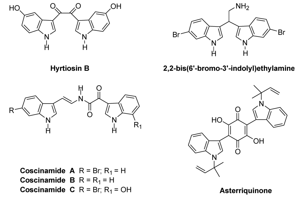
Chart 1.
bis-Indolyl alkaloids with acyclic and carbocyclic spacers.
The spacer can also be constituted by differently sized heterocycles. Thus, dragmacidin and dragmacidins A−E, isolated from a large number of deep water sponges such as Dragmacidon, Halicortex, Spongosorites, Hexadella and the tunicate Didemnum candidum, exhibit the saturated six-membered heterocyclic link piperazine or a pyrazinone spacer, and showed several biological properties (Chart 2) [25,28,29,30,31].
More recently, hyrtinadine A, isolated from the marine sponge Hyrtios and bearing a pyrimidine ring as a spacer, exhibited in vitro cytotoxicity [32].
Topsentins A, B1 and B2, which were isolated from Mediterranean sponge Topsentia genitrix, bear a 2-acyl imidazole spacer and showed antitumor and antiviral activities [33]. Later, from Spongosorites sp. and from Hexadella sp. were obtained the same natural products, which were named deoxy-topsentin, topsentin, and bromotopsentin, respectively [34,35]. More recently, topsentin C, isobromotopsentin, bromodeoxytopsentin, and its isomer isobromodeoxytopsentin, were isolated from Hexadella sp., Spongosorites sp and Spongosorites genitrix, respectively [29,36,37,38].
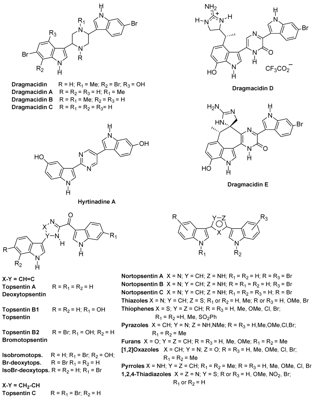
Chart 2.
bis-Indolyl alkaloids and analogues with heterocyclic spacers.
Nortopsentins A−C, isolated from Spongosorites ruetzleri, have a 2,4-disubstituted imidazole ring as a spacer and showed in vitro cytotoxicity against P388 cells (IC50, 4.5–20.7 μM). Methylation of the two-indole nitrogen atoms produced a significant improvement in cytotoxicity (IC50, 0.64–1.70 μM) [39].
Since marine organisms allow the isolation of very small amount of the biologically active substances from the natural material, several total synthesis of nortopsentins were proposed [40,41,42,43]. Moreover, due to the considerable activities shown, indolyl alkaloids have attracted remarkable attention by researchers becoming an interesting field in medicinal chemistry. Thus, several dragmacidin analogues bearing six membered heterocycles, such as pyridine, pyrazine, pyrazinone and pyrimidine, as spacer and showing antiproliferative activity were synthesized [44,45,46,47].
Also, nortopsentins were considered lead compounds and several reports dealt with the synthesis and the evaluation of the antiproliferative activity of nortopsentin analogues bearing five-membered heterocycles, which replaced the imidazole ring of the natural product, such as bis-indolyl-thiophenes [48], -pyrazoles [49], -furans [50], -isoxazoles [50], -pyrroles [51], and -1,2,4-thiadiazoles [52]. Most of these analogues showed antiproliferative activity often reaching GI50 values at submicromolar level.
Beside the eterocyclic spacer, the structural manipulation of the natural nortopsentins, was extended to one or both indole units and produced 3-[(2-indolyl)-5-phenyl]pyridine and phenylthiazolyl-7-azaindole derivatives, which showed antiproliferative activity and inhibited CDK1 [53,54].
More recently, due to the good results obtained by the aza-substitution of the indole moiety, 3-[2-(1H-indol-3-yl)-1,3-thiazol-4-yl]-1H-4-azaindole derivatives were synthesized and tested against breast cancer, prostate cancer, pancreatic carcinoma and peritoneal mesothelioma and showed cytotoxic activity, inhibited CDK1, and increased caspase-9 and caspase-3 activity (Chart 3) [55].
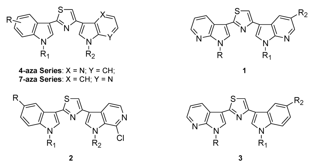
Chart 3.
Nortopsentin aza-analogues.
Contemporaneously, 3-[2-(1H-indol-3-yl)-1,3-thiazol-4-yl]-1H-7-azaindoles were synthesized and tested against the NCI full panel of human cancer cell lines and STO and MesoII cells, derived from human diffuse malignant peritoneal mesothelioma (DMPM). The most active compounds, which also act as CDK1 inhibitors, administrated with paclitaxel produced a synergistic cytotoxic effect. In the mouse model, intraperitoneal (i.p.) administration of active derivatives was effective, resulting in a significant tumor volume inhibition of DMPM xenografts (range, 58%–75%) at well-tolerated doses, and two complete responses were observed in each treatment group [56].
Lately, two new series of nortopsentin analogues, 1 and 2, were efficiently synthesized and inhibited the growth of HCT-116 colorectal cancer cells at low micromolar concentrations, whereas they did not affect the viability of normal-like intestinal cells. A compound of the series 1 induced apoptosis. Instead, a derivative of the series 2 at low concentrations (GI30) induced morphological changes characteristic of autophagic death with massive formation of cytoplasmic acid vacuoles without apparent loss of nuclear material, and with arrest of cell cycle at the G1 phase, whereas higher concentrations (GI70) induced apoptosis with arrest of cell cycle at the G1 phase [57].
In this paper, continuing our studies on indole and related azaindole systems [58,59,60,61,62,63,64], we report the synthesis of derivatives of type 3, nortopsentin analogues, in which indole and 7-azaindole units of the preceding very active 7-aza series were switched. We also report the antiproliferative activity of these new nortopsentin analogues and studies directed to elucidate their mode of action.
2. Results and Discussion
2.1. Chemistry
Substituted 3-[4-(1H-indol-3-yl)-1,3-thiazol-2-yl]-1H-pyrrolo[2,3-b]pyridines of type 3 (Scheme 1, Table 1) were obtained by Hantzsch reaction between pyrrolo[2,3-b]pyridine-carbothioamides 6a,b and 3-haloacetyl compounds 9a–d.
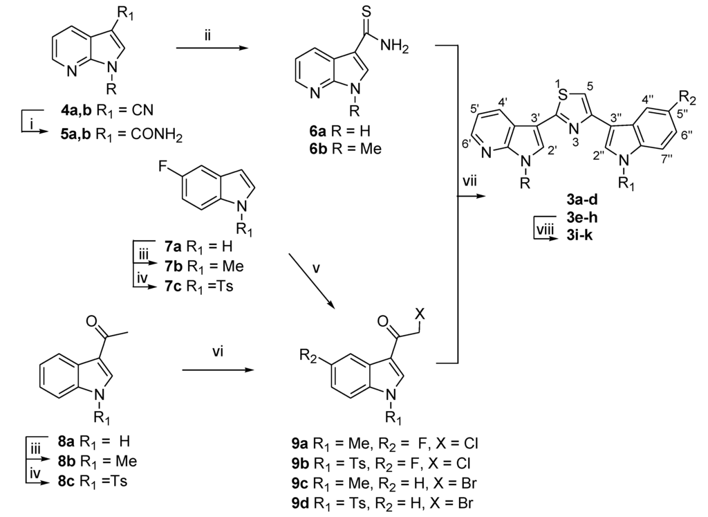
Scheme 1.
Synthesis of substituted 3-[4-(1H-indol-3-yl)-1,3-thiazol-2-yl]-1H-pyrrolo[2,3-b] pyridines 3a–k. Reagents: (i) (a) H2SO4, 25 °C, 15–60 min; (b) NaOH, 95%–99%; (ii) Lawesson’s reagent, THF, reflux, 30 min, 88%–99%; (iii) (a) t-BuOK, toluene, tris[2-(2-methoxyethoxy)ethyl]amine (TDA-1), 25 °C, 5–8 h; (b) MeI, 25 °C, 1–2 h, 80%–98%; (iv) (a) NaH, THF, 0 °C–25 °C, 1–24 h; (b) p-TsCl, 25 °C, 4–24 h, 90%–96%; (v) AlCl3, CH2Cl2, ClCOCH2Cl, 1 h, 50%–55%; (vi) Br2, MeOH, reflux, 2 h, 40%, 70%; (vii) EtOH, reflux, 30 min, 30%–97%; (viii) KOH, EtOH, reflux, 1–2 h, 40%–98%.
The key intermediates 6a,b were obtained from the corresponding pyrrolo[2,3-b]pyridine-3-carbonitriles 4a,b through the formation of carboxamides 5a,b, as previously reported [57].
N-protected 5-fluoroindoles 7b,c and 1-(1H-indol-3-yl)ethanones 8b,c were synthesized, in good yields (80%–98%), from commercially available 5-fluoroindole 7a or 1-(1H-indol-3-yl)ethanone 8a by methylation [54], or reaction with tosyl chloride using sodium hydride (NaH) as the base in tetrahydrofuran (THF).
3-Haloacetyl indoles 9a–d were prepared from the corresponding indoles 7b,c or 1-(1H-indol-3-yl)ethanones 8b,c. In particular, 3-chloroacetyl derivatives 9a,b were obtained by reaction of the corresponding N-methyl or N-tosyl 5-fluoroindole 7b,c with chloroacetyl chloride (ClCOCH2Cl) in the presence of aluminum chloride (AlCl3) in dichloromethane (CH2Cl2) in moderate yields (50%–55%); while 3-bromoacetyl compounds 9c,d were synthesized (40%–70%) from the corresponding N-methyl or N-tosyl 1-(1H-indol-3-yl)ethanone 8b,c using bromine in refluxing methanol (MeOH).
Reaction between pyrrolo[2,3-b]pyridine-carbothioamides 6a,b and 3-haloacetyl compounds 9a–d in ethanol under reflux afforded the desired 3-[4-(1H-indol-3-yl)-1,3-thiazol-2-yl]-1H-pyrrolo[2,3-b]pyridines 3a–h in moderate to excellent yields (30%–97%) (Table 1).
The subsequent deprotection of N-tosyl derivatives 3e–h using potassium hydroxide in refluxing ethanol gave the corresponding compounds 3i–k (40%–98%).

Table 1.
Substituted 3-[4-(1H-indol-3-yl)-1,3-thiazol-2-yl]-1H-pyrrolo[2,3-b]pyridine 3a–k.
| Compound | R | R1 | R2 | Yield% | Compound | R | R1 | R2 | Yield% |
|---|---|---|---|---|---|---|---|---|---|
| 3a | H | Me | F | 97 | 3g | H | Ts | H | 90 |
| 3b | Me | Me | F | 30 | 3h | Me | Ts | H | 50 |
| 3c | H | Me | H | 60 | 3i | H | H | F | 98 |
| 3d | Me | Me | H | 60 | 3l | Me | H | F | 40 |
| 3e | H | Ts | F | 97 | 3j | H | H | H | 98 |
| 3f | Me | Ts | F | 40 | 3k | Me | H | H | 98 |
2.2. Biology
All the synthesized thiazoles 3a–k were evaluated by the National Cancer Institute (Bethesda MD) for cytotoxicity against the NCI-60 cell line panel using standard protocols [65]. Initially, the selected derivatives 3c,d,g,h,j,k were tested at a single dose (10−5 M) on the full panel of approximately 60 human tumor cell lines derived from 9 human cancer cell types, that have been grouped in disease sub-panels including leukemia, non-small-cell lung, colon, central nervous system (CNS), melanoma, ovarian, renal, prostate and breast cancers (data not shown). Compounds 3d and 3k were further selected for full evaluation at five concentration levels (10−4–10−8 M).
The antitumor activity of compounds 3d and 3k was given by three parameters for each cell line: GI50 (the molar concentration of the compound that inhibits 50% net cell growth), TGI (the molar concentration of the compound leading to total inhibition of net cell growth), and LC50 (the molar concentration of the compound that induces 50% net cell death). The average values of mean graph midpoint (MG_MID) were calculated for each of these parameters.
An evaluation of the data reported in Table 2 pointed out that compounds 3d and 3k exhibited antiproliferative activity against all the human cell lines at GI50 values from micromolar to nanomolar (13.0–0.03 and 14.2–0.04 µM, respectively).

Table 2.
In vitro inhibition of cancer cell line growth by indolyl-thiazolyl-pyrrolopyridines 3d, 3k a.
| Cell Lines | GI50 (µM) | Cell Lines | GI50 (µM) | Cell Lines | GI50 (µM) | |||
|---|---|---|---|---|---|---|---|---|
| 3d | 3k | 3d | 3k | 3d | 3k | |||
| Leukemia | CNS Cancer | Renal Cancer | ||||||
| CCRF-CEM | 0.40 | 0.43 | SF-268 | 2.44 | 0.94 | 786-0 | 0.97 | 0.72 |
| HL-60(TB) | 0.29 | 0.32 | SF-295 | 0.24 | 0.31 | A498 | 0.50 | 0.37 |
| K-562 | 0.07 | 0.12 | SF-539 | 0.18 | 0.25 | ACHN | 0.72 | 0.51 |
| MOLT-4 | 0.67 | 0.66 | SNB-19 | 0.63 | 0.64 | CAKI-1 | 0.38 | 0.44 |
| RPMI-8226 | 0.45 | 0.65 | SNB-75 | 0.16 | 0.14 | RXF393 | 0.33 | 0.52 |
| SR | 0.06 | 0.14 | U251 | 0.41 | 0.43 | SN12C | 12.1 | 5.48 |
| TK-10 | 0.60 | 0.99 | ||||||
| UO-31 | 0.90 | 0.69 | ||||||
| Non-Small Cell Lung Cancer | Melanoma | Prostate Cancer | ||||||
| A549/ATCC | 0.56 | 0.71 | LOX IMVI | 0.54 | 0.51 | PC-3 | 0.46 | 0.56 |
| EKVK | 0.91 | 0.91 | MALME-3M | 1.04 | 0.74 | DU-145 | 0.39 | 0.56 |
| HOP-62 | 0.81 | 0.93 | M14 | 0.27 | 0.28 | |||
| HOP-92 | 0.35 | 0.31 | MDA-MB-435 | 0.03 | 0.04 | |||
| NCI-H226 | 0.74 | 0.65 | SK-MEL-2 | 0.58 | 0.41 | |||
| NCI-H23 | 0.72 | 0.67 | SK-MEL-28 | 1.20 | 0.76 | |||
| NCI-H322M | 0.76 | ND b | SK-MEL-5 | 0.24 | 0.27 | |||
| NCI-H460 | 0.30 | 0.37 | UACC-257 | 13.0 | 14.2 | |||
| NCI-H522 | 0.04 | 0.05 | UACC-62 | 0.30 | 0.42 | |||
| Colon Cancer | Ovarian Cancer | Breast Cancer | ||||||
| COLO-205 | 0.19 | 0.19 | IGROV1 | 1.33 | 1.18 | MCF7 | 0.33 | 0.37 |
| HCC-2998 | 1.13 | 1.18 | OVCAR-3 | 0.10 | 0.25 | MDA-MB-231/ATCC | 2.82 | 1.29 |
| HCT-116 | 0.37 | 0.39 | OVCAR-4 | 7.22 | 0.52 | HS 578T | 0.59 | 0.66 |
| HCT-15 | 0.17 | 0.29 | OVCAR-5 | 1.41 | 2.39 | BT-549 | 0.47 | 0.44 |
| HT29 | 0.25 | 0.19 | OVCAR-8 | 0.66 | 0.82 | MDA-MB-468 | 0.28 | 0.23 |
| KM12 | 0.21 | 0.40 | NCI/ADR-RES | 0.09 | 0.15 | |||
| SW-620 | 0.22 | 0.30 | SK-OV-3 | 0.57 | 0.52 | |||
a Data obtained from NCI’s in vitro disease-oriented tumor cells screen; b ND = not determined.
Indolyl-thiazolyl-pyrrolopyridine derivatives 3d and 3k were selective with respect to the leukemia cancer subpanel having all the subpanel cell lines GI50 in the range 0.06–0.67 μM and 0.12–0.66 μM, respectively. In particular, the most sensitive cell lines were K-562 (GI50 0.07 μM and 0.12 μM, respectively), and SR (GI50 0.06 μM and 0.14 μM, respectively). Derivatives 3d and 3k were also particularly sensitive against NCI-H522 (GI50 0.04 μM and 0.05 μM, respectively) of non-small cell lung cancer, NCI/ADR-RES (GI50 0.09 μM and 0.15 μM, respectively) of ovarian cancer, and MDA-MB-435 (GI50 0.03 μM and 0.04 μM, respectively) of melanoma subpanel.
Cell growth inhibitory activity of 3d and 3k was also investigated on human HepG2 hepatocarcinoma.
Cell growth inhibitory activity of compounds 3d and 3k was also investigated on human HepG2 hepatocarcinoma cells, a cell line not included in the NCI panel and of interest in the drug discovery because provided with a very active microsomal system for detoxification of xenobiotics. Monolayer cultures treated for 72 h with 0.1–10 μM concentrations of the compounds were examined by 3-(4,5-dimethylthiazol-2-yl)-2,5-diphenyltetrazolium bromide (MTT) assay for cell growth. As shown in Figure 1, both the compounds inhibited the HepG2 cells growth in dose-dependent manner. Based on the dose response curve, the GI50 values were 1.69 µM) and 0.21 µM for 3d and 3k, respectively. Under identical conditions, both the nortopsentin analogues did not substantially impaired viability of normal immortalized human liver cells (Chang), suggesting high selectivity towards tumor cells.
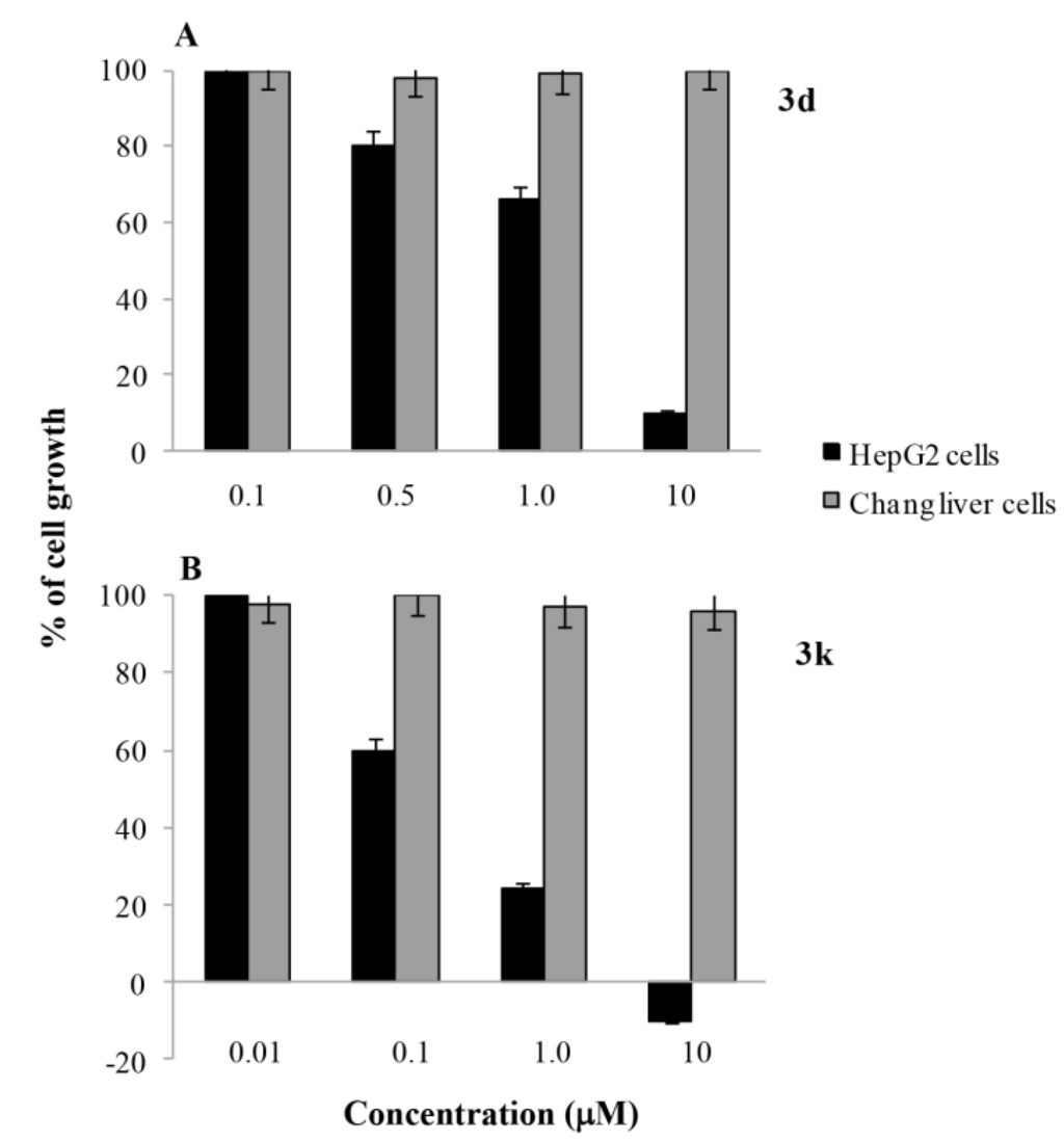
Figure 1.
Effect 3d (A) and 3k (B) on the growth of HepG2 (black bars) or Chang cells (grey bars) as assessed by 3-(4,5-dimethylthiazol-2-yl)-2,5-diphenyltetrazolium bromide (MTT). Cell monolayers were incubated for 24 h in the absence (control) or in the presence of the compounds at the indicated concentrations and cell growth was assessed by MTT test as reported Methods. Results are indicated as the percentage of viable cells with respect to untreated controls. Values are the mean ± SD of three separate experiments carried out in triplicate.
The distribution of HepG2 cells in the cell cycle phases after 24 h treatment with the two norptopsentin analogues, was assessed by flow cytometric analysis after staining of DNA with PI. Both compounds 3d and 3k caused a significant dose-dependent decrease in the percentage of cells in the G0/G1 and S phases, accompanied by a concomitant percentage increase of cells in the G2/M phase, and appearance of a subG1-cell population (Figure 2).
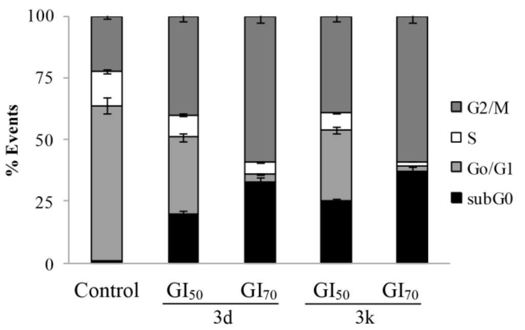
Figure 2.
Effect of 3d and 3k on the cell cycle distribution of human hepatoma HepG2 cells. Flow cytometric analysis of propidium iodide (PI)-stained cells, as determined by flow cytometry after 24 h treatment with the compounds or vehicle alone (control). The percentage of cells in the different phases of the cycle was calculated by Expo32 software. Values are the mean ± SD of three separate experiments in triplicate.
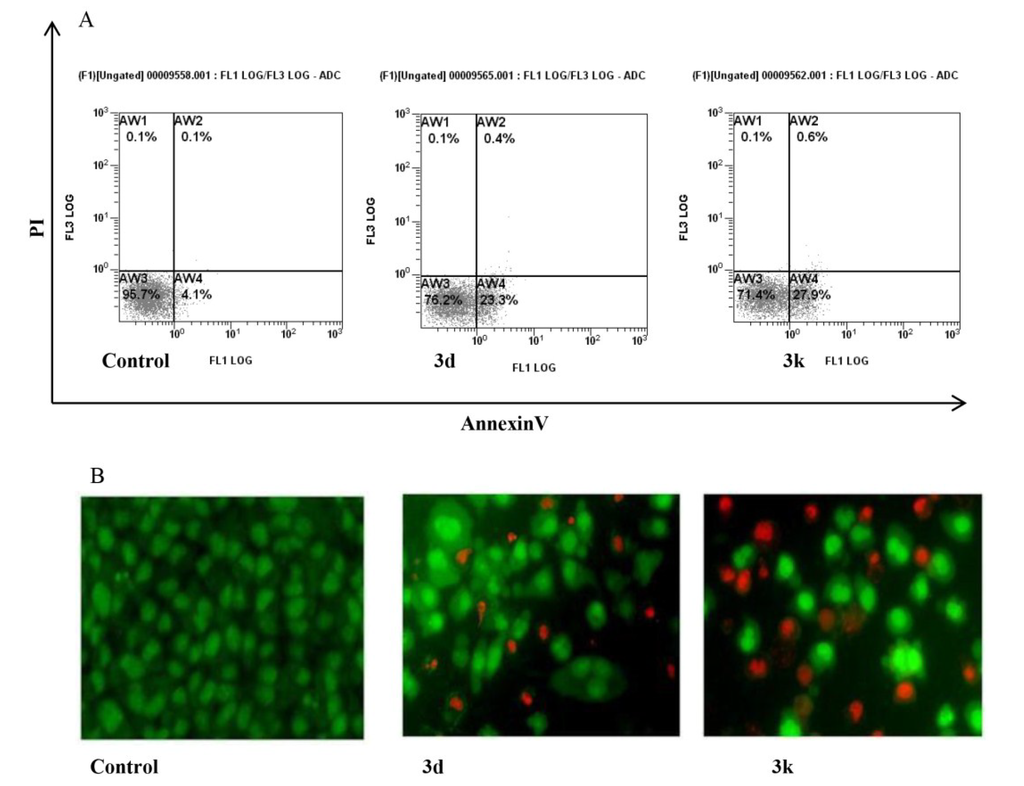
Figure 3.
Apoptosis induced by 3d an 3k nortopsentin analogues in HepG2 cells. (A) Percentage of Annexin V/propidium iodide (PI) double-stained cells, as determined by flow cytometry; (B) Fluorescence micrographs of ethidium bromide/acridine orange double-stained cells. Control, cells treated with vehicle; 3d and 3k, cells treated for 24 h with either Nortopsentin derivative at GI50 concentration. (A) Representative images of three experiments with comparable results; (B) Representative images, in 200 × magnification, of three experiments with comparable results.
Apoptosis induction by 3d and 3k in HepG2 cells was investigated by externalization of plasma membrane phosphatidylserine and changes of mitochondrial transmembrane potential. Flow cytometry analysis of Annexin V-FITC/PI-stained cells after 24 h treatment with the Norptopsentin analogues, assayed at their relevant GI50 values, indicated a high percentage of cells in early apoptosis, with externalized phosphatidylserine (Figure 3A).
To confirm apoptotic mechanism of cytotoxicity of the nortopsentin analogues, we carried out morphological evaluation of HepG2 cells using AO and EB double staining. After 24 h of treatment with 3d or 3k at GI50 concentration, fluorescent microscopy revealed the appearance of cells containing bright green patches in the nuclei as a consequence of chromatin condensation and nuclear fragmentation, which are typical features of apoptosis. Moreover, fluorescing orange cells owing to increase of cell permeability to ethidium bromide, cell shrinkage and nuclear fragmentation were also evident as cells in late apoptosis (Figure 3B). Taken together, these findings provided strong evidence that the synthesized nortopsentin analogues induced apoptosis in HepG2 cells.
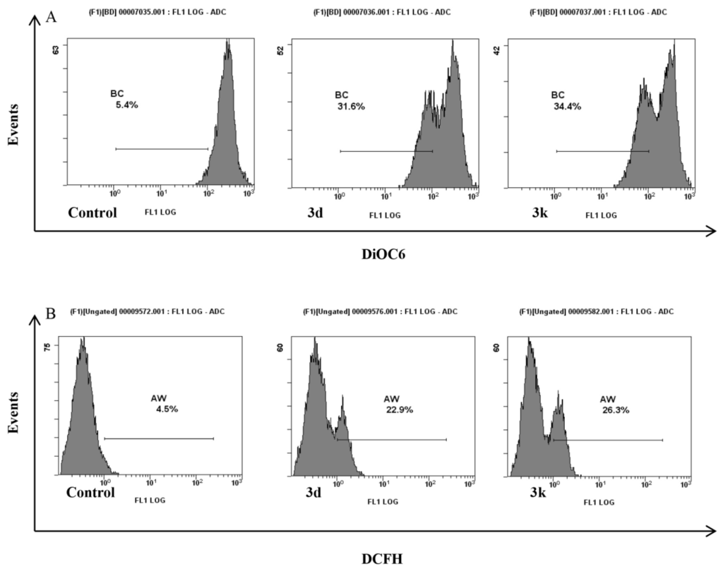
Figure 4.
Mitochondrial dysfunction induced by 3d and 3k in HepG2 cells. (A) Fluorescence intensity of 3,30-dihexyloxacarbocyanine iodide-treated cells, as determined by flow cytometry; (B) Fluorescence intensity of 2′,7′-dichlorofluorescin diacetate-stained cells, as determined by flow cytometry. Control, cells treated with vehicle; 3d and 3k, cells treated for 24 h with either Nortopsentin derivative at GI50 concentration. Representative images of three experiments with comparable results.
Involvement of mitochondria in apoptosis induced in HepG2 cells by the synthesized nortopsentin analogues, was assessed. Loss of mitochondrial trans-membrane potential was indicated by decreased mitochondrial 3,30-dihexyloxacarbocyanine iodide-red fluorescence (Figure 4A). Mitochondrial dysfunction in HepG2 cells following 24 h treatment with 3d and 3k, was also evident from the levels of intracellular ROS, revealed by cytofluorimetric analysis with 2′,7′-dichlorofluorescin diacetate, significantly higher than cell control (Figure 4B).
3. Experimental Section
3.1. Chemistry
3.1.1. General
All melting points were taken on a Büchi-Tottoly capillary apparatus. IR spectra were determined in bromoform with a Shimadzu FT/IR 8400S spectrophotometer. 1H and 13C NMR spectra were measured at 200 and 50.0 MHz, respectively, in dimethylsulfoxide (DMSO)-d6 or CDCl3 solution, using a Bruker Avance II series 200 MHz spectrometer. Compounds 3a,b were characterized only by 1H NMR spectra because of their poor solubility. Column chromatography was performed with Merk silica gel 230–400 mesh ASTM or with Büchi Sepacor chromatography module (prepacked cartridge system). Elemental analyses (C, H, N) were within ±0.4% of theoretical values and were performed with a VARIO EL III elemental analyzer. Purity of all the tested compounds was greater than 98%, determined by HPLC as described below.
3.1.2. Synthesis of 5-Fluoro-1-methyl-1H-indole (7b)
To a cold solution of 5-fluoroindole 7a (0.8 g, 5.0 mmol) in anhydrous toluene (50 mL), potassium t-butoxide (0.8 g, 6.8 mmol) and tris[2-(2-methoxyethoxy)ethyl]amine (TDA-1) (1–2 drops) were added. The reaction mixture was stirred at room temperature for 5 h and then methyl iodide (0.3 mL, 5 mmol) was added. TLC analysis (dichloromethane (DCM)/petroleum ether (9/1) revealed that methylation was complete after 1 h. The solvent was evaporated under reduced pressure and the residue, treated with water (15 mL), was extracted with DCM (3 × 15 mL), dried (Na2SO4) and evaporated to afford the pure methyl derivative 7b.
Yellow solid; yield: 98%; mp: 55–56 °C. Analytical and spectroscopic data were in agreement with those previously reported [50].
3.1.3. Synthesis of 5-Fluoro-1-[(4-methylphenyl)sulfonyl]-1H-indole (7c)
To a stirred solution of 5-fluoroindole 7a (1.0 g, 7.4 mmol) in tetrahydrofuran (THF) (5.0 mL) sodium hydride (60% dispersion in mineral oil, 0.4 g, 9.6 mmol) was added at 0 °C and the mixture was stirred at room temperature for 24 h. p-Toluenesulfonyl chloride (2.1 g, 11.1 mmol) was added to the reaction and the mixture was stirred at room temperature for 4 h. The residue was evaporated under reduced pressure, treated with water (20 mL) and extracted with ethyl acetate (EtOAc) (3 × 20 mL). The organic phase was dried (Na2SO4), evaporated under reduced pressure and purified by column chromatography using petroleum ether/DCM (9/1) as eluent. White solid; yield: 96%; mp: 111–112 °C; IR 1373, 1172 (SO2) cm−1; 1H NMR (200 MHz, DMSO-d6) δ: 2.32 (s, 3H, CH3), 6.82–6.84 (m, 1H, H-7), 7.19 (td, 1H, J = 2.6, 9.2, 11.0 Hz, H-6), 7.39 (d, 2H, J = 8.7 Hz, H-3′, H-5′), 7.45 (d, 1H, J = 2.6 Hz, H-3), 7.87 (d, 2H, J = 8.7 Hz, H-2′, H-6′), 7.88–7.97 (m, 2H, H-2, H-4); 13C NMR (50 MHz, DMSO-d6) δ: 21.0 (q), 107.0 (d, JC4–F = 24.1 Hz), 109.3 (d, JC3–F = 4.2 Hz), 112.5 (d, JC6–F = 25.8 Hz), 114.4 (d, JC7–F = 9.6 Hz), 126.7 (2xd), 128.9 (d), 130.2 (2xd), 130.6 (s), 131.6 (d, JC3a–F = 10.4 Hz), 133.9 (s), 145.6 (s), 158.9 (d, JC5–F = 237.7 Hz). Anal. Calcd for: C15H12FNO2S: C, 62.27; H, 4.18; N, 4.84. Found: C, 62.57; H, 4.38; N, 4.69.
3.1.4. Synthesis of 1-(1-Methyl-1H-indol-3-yl)ethanone (8b)
To a cold solution of 3-acetylindole 8a (1.0 g, 6.3 mmol) in anhydrous toluene (50 mL), potassium t-butoxide (1.0 g, 8.6 mmol) and TDA-1 (1–2 drops) were added. The reaction mixture was stirred at room temperature for 8 h and then methyl iodide (0.4 mL, 6.3 mmol) was added. TLC analysis (DCM/EtOAc 9/1) revealed that methylation was complete after 2 h. The solvent was evaporated under reduced pressure. The residue was treated with water (50 mL), extracted with DCM (3 × 50 mL), dried (Na2SO4), evaporated under reduced pressure, and purified by column chromatography using DCM/EtOAc (9/1) as eluent.
White solid; yield 80%; mp: 108–109 °C. Analytical and spectroscopic data were in agreement with those previously reported [48].
3.1.5. Synthesis of 1-{1-[(4-Methylphenyl)sulfonyl]-1H-indol-3-yl}ethanone (8c)
To a solution of 3-acetylindole 8a (1.0 g, 6.3 mmol) in THF (5.0 mL) sodium hydride (60% dispersion in mineral oil, 0.3 g, 6.3 mmol) was added at 0 °C and the mixture was stirred at room temperature for 1 h. p-toluenesulfonyl chloride (1.2 g, 6.3 mmol) was added and the mixture was stirred at room temperature for 24 h. The residue was evaporated under reduced pressure, treated with water (20 mL) and extracted with EtOAc (3 × 20 mL). The organic phase was dried (Na2SO4), evaporated under reduced pressure and purified by column chromatography using DCM as eluent. White solid; yield: 90%; mp: 148–149 °C; IR 1662 (CO), 1382, 1299 (SO2) cm−1; 1H NMR (200 MHz, DMSO-d6) δ: 2.33 (s, 3H, CH3), 2.60 (s, 3H, CH3), 7.32–7.39 (m, 2H, H-5, H-6), 7.43 (d, 2H, J = 7.8 Hz, H-3′, H-5′), 7.95 (d, 1H, J = 7.5 Hz, H-7), 8.04 (d, 2H, J = 7.8 Hz, H-2′, H-6′), 8.19 (d, 1H, J = 7.5 Hz, H-4), 8.81 (s, 1H, H-2); 13C NMR (50 MHz, DMSO-d6) δ:21.0 (q), 27.8 (q), 113.0 (d), 120.7 (s), 122.3 (d), 124.8 (d), 125.6 (d), 127.0 (s), 127.2 (2xd), 130.5 (2xd), 133.5 (s), 134.0 (s), 134.2 (d), 146.2 (s), 193.9 (s). Anal. Calcd for: C17H15NO3S: C, 65.16; H, 4.82; N, 4.47. Found: C, 65.28, H, 5.06, N, 4.37.
3.1.6. Synthesis of 2-Chloro-1-(5-fluoro-1H-indol-3-yl)ethanones (9a,b)
To a stirred suspension of anhydrous aluminium chloride (1.1 g, 8.5 mmol), in DCM (18.0 mL), was added dropwise at 0 °C a solution of the suitable indole 7b,c (1.2 mmol) in DCM (5.0 mL) under nitrogen atmosphere. Then, chloroacetyl chloride (0.3 mL, 3.6 mmol) was slowly added to the reaction mixture, which was stirred at room temperature for 1 h and then poured in ice and water (20 mL) and extracted with DCM (3 × 20 mL). The organic phase was dried (Na2SO4), evaporated under reduced pressure and purified by column chromatography using DCM as eluent.
3.1.6.1. 2-Chloro-1-(5-fluoro-1-methyl-1H-indol-3-yl)ethanone (9a)
White solid; yield: 50%; mp: 185–186 °C; IR 1653 (CO) cm−1; 1H NMR (200 MHz, DMSO-d6) δ: 3.89 (s, 3H, CH3), 4.84 (s, 2H, CH2), 7.20 (td, 1H, J = 2.6, 9.2, 11.0 Hz, H-6), 7.62 (dd, 1H, J = 4.5, 9.2 Hz, H-7), 7.84 (dd, 1H, J = 2.6, 11.0 Hz, H-4), 8.51 (bs, 1H, H-2);13C NMR (50 MHz, DMSO-d6) δ: 33.7 (q), 46.2 (t), 106.1 (d, JC4–F = 24.8 Hz), 111.3 (d, JC6–F = 26.1 Hz), 112.3 (s), 112.4 (d, JC7–F = 9.8 Hz), 126.4 (d, JC3a–F = 11.0 Hz), 133.9 (s), 139.5 (d), 159.0 (d, JC5–F = 236 Hz), 185.6 (s). Anal. Calcd for C11H9ClFNO: C, 58.55; H, 4.02; N, 6.21. Found: C, 58.85; H, 4.14; N, 6.01.
3.1.6.2. 2-Chloro-1-{5-fluoro-1-[(4-methylphenyl)sulfonyl]-1H-indol-3-yl}ethanone (9b)
White solid; yield: 55%; mp: 155–156 °C; IR 1683 (CO), 1375, 1171 (SO2) cm−1; 1H NMR (200 MHz, CDCl3) δ: 2.39 (s, 3H, CH3), 4.54 (s, 2H, CH2), 7.13 (td, 1H, J = 2.6, 8.9, 11.0 Hz, H-6), 7.31 (d, 2H, J = 8.0 Hz, H-3′, H-5′), 7.82 (d, 2H, J = 8.0 Hz, H-2′, H-6′), 7.90 (dd, 1H, J = 4.9, 8.9 Hz, H-7), 7.98 (dd, 2H, J = 2.6, 11.0 Hz, H-4), 8.34 (s, 1H, H-2); 13C NMR (50 MHz, CDCl3) δ: 21.7 (q), 45.8 (t), 108.9 (d, JC6–F = 25.5 Hz), 114.1 (s), 114.2 (d), 114.5 (d, JC4–F = 14.0 Hz), 117.9 (s, JC7a–F = 4.5 Hz), 127.2 (2xd), 130.2 (s), 130.4 (2xd), 133.5 (d), 134.1 (s), 146.5 (s), 160.1 (s, JC5–F = 241 Hz), 186.7 (s). Anal. Calcd for C17H13ClFNO3S: C, 55.82; H, 3.58; N, 3.83. Found: C, 55.99; H, 3.36; N, 4.10.
3.1.7. Synthesis of 2-Bromo-(1H-indol-3-yl)ethanones (9c,d)
To a cold suspension of the appropriate 3-acetylindole 8b,c (1.9 mmol) in anhydrous methanol (3.0 mL) bromine (0.1 mL, 1.9 mmol) was added dropwise. The mixture was heated at reflux for 2 h. After cooling the solvent was evaporated under reduced pressure. The residue was treated with water (20 mL), made alkaline by adding sodium hydrogen carbonate (150 mg) and extracted with EtOAc (3 × 50 mL). The organic phase was dried (Na2SO4), evaporated under reduced pressure and purified by column chromatography using DCM as eluent.
3.1.7.1. 2-Bromo-1-(1-methyl-1H-indol-3-yl)ethanone (9c)
Brown solid; yield: 70%; mp: 205–206 °C. Analytical and spectroscopic data were in agreement with those previously reported [48].
3.1.7.2. 2-Bromo-1-{1-[(4-methylphenyl)sulfonyl]-1H-indol-3-yl}ethanone (9d)
Brown solid; yield: 40%; mp: 134–135 °C; IR 1668 (CO), 1379, 1177 (SO2) cm−1; 1H NMR (200 MHz, CDCl3) δ: 2.38 (s, 3H, CH3), 4.36 (s, 2H, CH2), 7.29 (d, 2H, J = 8.2 Hz, H-3′, H-5′), 7.36–7.40 (m, 2H, H-5, H-6), 7.83–7.87 (m, 2H, H-2′, H-6′), 7.91–7.96 (m, 1H, H-7), 8.28–8.34 (m, 2H, H-2, H-4); 13C NMR (50 MHz, CDCl3) δ: 21.7 (q), 31.4 (t), 113.1 (d), 123.1 (d), 124.3 (s), 125.1 (d), 126.1 (d), 127.2 (2xd), 127.5 (s), 130.4 (2xd), 132.8 (d), 134.2 (s), 134.8 (s), 146.2 (s), 187.0 (s). Anal. Calcd. for: C17H14BrNO3S: C, 52.05; H, 3.60; N, 3.57. Found: C, 52.25; H, 3.70; N, 3.35.
3.1.8. Synthesis of 3-[4-(1H-indol-3-yl)-1,3-thiazol-2-yl]-1H-pyrrolo[2,3-b]pyridines (3a–h)
A suspension of the appropriate pyrrolo[2,3-b]pyridine-carbothioamide 6a,b (5.0 mmol) and halo-acetyl compounds 9a–d (5.0 mmol) in ethanol (3.0 mL) was heated under reflux for 30 min. The precipitate, obtained after cooling, was filtered off, dried and recrystallized from ethanol to afford derivatives 3a–h.
3.1.8.1. 3-[4-(5-Fluoro-1-methyl-1H-indol-3-yl)-1,3-thiazol-2-yl]-1H-pyrrolo[2,3-b]pyridine (3a)
Yellow solid; yield: 97%; mp: 237–238 °C; IR 3550 (NH) cm−1; 1H NMR (200 MHz, DMSO-d6) δ: 3.90 (s, 3H, CH3), 7.12 (t, 1H, J = 8.2 Hz, H-6″), 7.44 (dd, 1H, J = 5.2, 7.4 Hz, H-5′), 7.54 (dd, 1H, J = 4.6, 8.2 Hz, H-7″), 7.73 (s, 1H, H-2″), 7.94 (dd, 1H, J = 9.7, 2.5 Hz, H-4″), 8.16 (bs, 1H, H-2′), 8.38 (s, 1H, H-5), 8.45 (d, 1H, J = 3.7 Hz, H-6′), 8.89 (d, 1H, J = 7.4 Hz, H-4′), 12.74 (bs, 1H, NH). Anal. Calcd for C19H13FN4S: C, 65.50; H, 3.76; N, 16.08. Found: C, 65.76; H, 3.55; N, 16.22.
3.1.8.2. 3-[4-(5-Fluoro-1-methyl-1H-indol-3-yl)-1,3-thiazol-2-yl]-1-methyl-1H-pyrrolo[2,3-b]pyridine (3b)
Yellow solid; yield: 30%; mp: 286–287 °C; 1H NMR (200 MHz, DMSO-d6) δ: 2.56 (s, 3H, CH3), 4.03 (s, 3H, CH3), 6.02 (dd, 1H, J = 4.3, 10.7 Hz, H-7″), 6.76 (td, 1H, J = 2.3, 9.5, 10.7 Hz, H-6″), 6.98 (dd, 1H, J = 5.0, 8.4 Hz, H-5′), 7.25 (dd, 1H, J = 2.3, 9.5 Hz, H-4″), 7.72 (d, 1H, J = 8.4 Hz, H-4′), 8.19-8.81 (m, 4H, H-2’, H-2’’, H-5, H-6′). Anal. Calcd for C20H15FN4S: C, 66.28; H, 4.17; N, 15.46. Found: C, 66.53; H, 3.90; N, 15.23.
3.1.8.3. 3-[4-(1-Methyl-1H-indol-3-yl)-1,3-thiazol-2-yl]-1H-pyrrolo[2,3-b]pyridine (3c)
Yellow solid; yield: 60%; mp: 274–275 °C, IR 3427 (NH) cm−1; 1H NMR (200 MHz, DMSO-d6) δ: 3.90 (s, 3H, CH3), 7.21–7.30 (m, 2H, H-5″, H-6″), 7.36 (dd, 1H, J = 4.9, 7.7 Hz, H-5′), 7.53 (d, 1H, J = 7.1 Hz, H-7″), 7.66 (s, 1H, H-2″), 8.05 (s, 1H, H-5), 8.19 (d, 1H, J = 7.1 Hz, H-4″), 8.30 (d, 1H, J = 2.0 Hz, H-2′), 8.40 (d, 1H, J = 4.9 Hz, H-6′), 8.79 (d, 1H, J = 7.7 Hz, H-4′), 12.40 (bs, 1H, NH); 13C NMR (50 MHz, DMSO-d6) δ: 32.6 (q), 106.5 (d), 109.6 (s), 110.2 (d), 117.1 (d), 117.3 (s), 119.9 (d), 120.1 (d), 121.6 (d), 124.9 (s), 126.9 (d), 129.2 (d), 129.7 (d), 137.1 (s), 143.2 (d), 143.3 (s), 147.9 (s), 150.1 (s), 161.0 (s). Anal. Calcd for C19H14N4S: C, 69.07; H, 4.27; N, 16.96. Found: C, 68.90; H, 4.46; N, 16.75.
3.1.8.4. 1-Methyl-3-[4-(1-methyl-1H-indol-3-yl)-1,3-thiazol-2-yl]-1H-pyrrolo[2,3-b]pyridine (3d)
Yellow solid; yield: 60%; mp: 290–291 °C; 1H NMR (200 MHz, DMSO-d6) δ: 3.90 (s, 3H, CH3), 3.94 (s, 3H, CH3), 7.17–7.30 (m, 2H, H-5″, H-6″), 7.39 (dd, 1H, J = 4.7, 7.9 Hz, H-5′), 7.53 (dd, 1H, J = 1.4, 6.9 Hz, H-7″), 7.66 (s, 1H, H-2″), 8.05 (s, 1H, H-2′), 8.19 (dd, 1H, J = 1.4, 6.9 Hz, H-4″), 8.41 (s, 1H, H-5), 8.45 (dd, 1H, J = 1.3, 4.7 Hz, H-6′), 8.78 (dd, 1H, J = 1.3, 7.9 Hz, H-4′); 13C NMR (50 MHz, DMSO-d6) δ: 31.4 (q), 32.6 (q), 106.5 (d), 108.3 (s), 110.0 (s), 110.2 (d), 117.2 (d), 117.5 (s), 119.9 (d), 120.1 (d), 121.6 (d), 124.9 (s), 129.3 (d), 129.8 (d), 130.5 (d), 137.0 (s), 143.1 (d), 147.0 (s), 150.1 (s), 160.7 (s). Anal. Calcd for C20H16N4S: C, 69.74; H, 4.68; N, 16.27. Found: C, 69.50; H, 4.84; N, 16.06.
3.1.8.5. 3-(4-{5-Fluoro-1-[(4-methylphenyl)sulfonyl]-1H-indol-3-yl}-1,3-thiazol-2-yl)-1H-pyrrolo [2,3-b]pyridine (3e)
Yellow solid; yield: 97%; mp: 257–258 °C; IR 3337 (NH), 1370, 1171 (SO2); 1H NMR (200 MHz, DMSO-d6) δ: 2.31 (s, 3H, CH3), 7.28–7.32 (m, 2H, ArH), 7.39–7.42 (m, 2H, H-3‴, H-5‴), 7.94–7.98 (m, 2H, H-2‴, H-6‴), 8.03–8.15 (m, 3H, ArH), 8.42–8.49 (m, 3H, ArH), 8.74 (d, 1H, J = 7.4 Hz, H-4′), 12.67 (s, 1H, NH); 13C NMR (50 MHz, DMSO-d6) δ: 21.1 (q), 107.3 (d, JC4″–F = 24.1 Hz), 109.3 (s), 112.6 (d, JC6″–F = 26.8 Hz), 113.4 (d), 114.9 (d, JC7″–F = 9.9 Hz), 117.2 (d), 117.4 (s), 117.5 (s), 117.6 (s), 126.3 (d), 126.9 (2xd), 127.8 (d), 128.8 (s), 129.0 (s), 130.2 (d), 130.4 (2xd), 131.1 (s), 133.6 (s), 142.3 (d), 145.9 (s), 146.9 (s), 159.4 (d, JC5″–F = 245.5 Hz). Anal. Calcd. for C25H17FN4O2S2: C, 61.46; H, 3.51; N, 11.47. Found: C, 61.73; H, 3.25; N, 11.31.
3.1.8.6. 3-(4-{5-Fluoro-1-[(4-methylphenyl)sulfonyl]-1H-indol-3-yl}-1,3-thiazol-2-yl)-1-methyl-1H-pyrrolo[2,3-b]pyridine (3f)
White solid; yield: 40%; mp: 230–231 °C; IR 1380, 1299 (SO2) cm−1; 1H NMR (200 MHz, DMSO-d6) δ: 2.32 (s, 3H, CH3), 3.94 (s, 3H, CH3), 7.28–7.43 (m, 2H, ArH), 7.41 (d, 2H, J = 7.8 Hz, H-3‴, H-5‴), 7.87–7.94 (m, 2H, H-2‴, H-6‴), 7.99–8.13 (m, 3H, ArH), 8.42–8.48 (m, 3H, ArH), 8.60 (d, 1H, J = 7.3 Hz, H-4′); 13C NMR (50 MHz, DMSO-d6) δ: 21.0 (q), 31.2 (q), 107.4 (d, JC4″–F = 25.2 Hz), 107.8 (s), 112.0 (d), 113.1 (d, JC6″–F = 27.5 Hz), 114.8 (d, JC7″–F = 9.1 Hz), 116.9 (s), 117.4 (d), 117.5 (d, JC7″–a = 4.0 Hz), 126.3 (d), 126.9 (2xd), 128.6 (d), 128.8 (s), 129.0 (s), 130.4 (s), 130.8 (2xd), 131.1 (s), 133.6 (s), 143.8 (d), 145.9 (s), 146.9 (s), 147.6 (s), 159.3 (d, JC5″–F = 246.5 Hz). Anal. Calcd. for C26H19FN4O2S2: C, 62.13; H, 3.81; N, 11.15. Found: C, 61.98; H, 3.99; N, 10.90.
3.1.8.7. 3-(4-{1-[(4-Methylphenyl)sulfonyl]-1H-indol-3-yl}-1,3-thiazol-2-yl)-1H-pyrrolo[2,3-b] pyridine (3g)
Yellow solid; yield: 90%; mp: 260–261 °C; IR 3498 (NH), 1374, 1174 (SO2) cm−1; 1H NMR (200 MHz, DMSO-d6) δ: 2.30 (s, 3H, CH3), 7.31–7.41 (m, 5H, ArH), 7.94–7.98 (m, 2H, ArH), 8.01–8.07 (m, 2H, ArH), 8.36–8.39 (m, 4H, ArH), 8.64 (d, 1H, J = 7.7 Hz, H-4′), 12.39 (bs, 1H, NH);13C NMR (50 MHz, DMSO-d6) δ: 21.0 (q), 109.1 (s), 111.8 (d), 113.4 (d), 116.7 (s), 117.2 (d), 117.7 (s), 121.7 (d), 124.0 (d), 124.5 (d), 125.2 (d), 126.8 (2xd), 127.2 (d), 127.9 (s), 128.6 (d), 130.3 (2xd), 133.8 (s), 134.6 (s), 144.0 (d), 145.7 (s), 147.3 (s), 148.7 (s), 162.1 (s). Anal. Calcd. for C25H18N4O2S2: C, 63.81; H, 3.86; N, 11.91. Found: C, 63.55; H, 4.02; N, 11.76.
3.1.8.8. 1-Methyl-3-(4-{1-[(4-methylphenyl)sulfonyl]-1H-indol-3-yl}-1,3-thiazol-2-yl)-1H-pyrrolo [2,3-b]pyridine (3h)
Yellow solid; yield: 50%; mp: 250–251 °C; IR 1374, 1174 (SO2) cm−1; 1H NMR (200 MHz, DMSO-d6) δ: 2.30 (s, 3H, CH3), 3.94 (s, 3H, CH3), 7.37–7.45 (m, 5H, ArH), 7.93–7.97 (m, 2H, ArH), 8.01–8.09 (m, 2H, ArH), 8.31–8.47 (m, 4H, ArH), 8.66 (d, 1H, J = 7.5 Hz, H-4′);13C NMR (50 MHz, DMSO-d6) δ: 21.0 (q), 31.4 (q), 108.0 (s), 112.0 (d), 113.4 (d), 117.2 (s), 117.4 (d), 117.6 (s), 121.7 (d), 124.0 (d), 124.6 (d), 125.2 (d), 126.8 (2xd), 127.8 (s), 129.3 (d), 130.3 (2xd), 130.9 (d), 133.8 (s), 134.6 (s), 145.7 (s), 147.1 (s), 147.4 (s), 148.2 (d), 161.6 (s). Anal. Calcd for: C26H20N4O2S2: C, 64.44; H, 4.16, N, 11.56. Found: C, 64.19; H, 4.33; N, 11.25.
3.1.9. Synthesis of 3-[4-(1H-indol-3-yl)-1,3-thiazol-2-yl]-1H-pyrrolo[2,3-b]pyridines (3i–k)
To a suspension of derivatives 3e–h (0.6 mmol) in ethanol (2 mL), potassium hydroxide (0.1 g, 1.8 mmol) was added and the mixture was heated at reflux for 1–2 h. The precipitate, obtained after cooling, was filtered off, dried and recrystallized from ethanol to afford derivatives 3i–k.
3.1.9.1. 3-[4-(5-Fluoro-1H-indol-3-yl)-1,3-thiazol-2-yl]-1H-pyrrolo[2,3-b]pyridine (3i)
Conditions: reflux for 1.5 h. Yellow solid; yield: 98%; mp: 248–249 °C; IR 3600 and 3550(2xNH), cm−1; 1H NMR (200 MHz, DMSO-d6) δ: 7.05 (t, 1H, J = 7.8 Hz, H-6″), 7.47–7.53 (m, 2H, H-5′, H-7″), 7.78 (bs, 1H, H-2″), 7.92 (d, 1H, J = 8.9 Hz, H-4″), 8.19 (bs, 1H, H-2′), 8.47 (m, 2H, H-5, H-6′), 8.95 (d, 1H, J = 7.4 Hz, H-4′), 11.70 (bs, 1H, NH), 12.93 (bs, 1H, NH); 13C NMR (50 MHz, DMSO-d6) δ: 104.7 (d, JC4″–F = 21.6 Hz), 107.3 (d), 109.7 (d, JC6″–F = 26.1 Hz), 109.8 (s), 110.7 (s), 113.0 (d, JC7″–F = 10.0 Hz), 117.2 (d), 119.0 (s), 124.6 (d, JC3″–F = 10.5 Hz), 127.2 (d), 128.2 (d), 132.3 (d), 133.3 (s), 140.6 (d), 144.7 (s), 149.6 (s), 157.5 (d, JC5″–F = 232 Hz), 160.5 (s). Anal. Calcd. for C18H11FN4S: C, 64.66; H, 3.32; N, 16.76. Found: C, 64.37; H, 3.04; N, 16.57.
3.1.9.2. 3-[4-(5-Fluoro-1H-indol-3-yl)-1,3-thiazol-2-yl]-1-methyl-1H-pyrrolo[2,3-b]pyridine (3l)
Conditions: reflux for 1 h. Yellow solid; yield 40%; mp: 243–244 °C; IR 3557 (NH) cm−1; 1H NMR (200 MHz, DMSO-d6) δ: 3.93 (s, 3H, CH3), 7.04 (td, 1H, J = 2.1, 9.1, 10.9 Hz, H-6″), 7.35 (dd, 1H, J = 4.6, 7.8 Hz, H-5′), 7.49 (dd, 1H, J = 4.6, 9.1 Hz, H-7″), 7.70 (s, 1H, H-2″), 7.96 (dd, 1H, J = 2.1, 10.9 Hz, H-4″), 8.11 (bs, 1H, H-2′), 8.38 (s, 1H, H-5), 8.42 (d, 1H, J = 4.6 Hz, H-6′), 8.69 (d, 1H, J = 7.8 Hz, H-4′), 11.64 (bs, 1H, NH);13C NMR (50 MHz, DMSO-d6) δ: 31.2 (q), 104.9 (d, JC4″–F = 24 Hz), 106.5 (d), 108.3 (s), 109.7 (d, JC6″–F = 26 Hz), 111.3 (d, JC7″–F = 4.5 Hz), 112.9 (d, JC7″–F = 9.5 Hz), 117.0 (s), 117.2 (d), 124.8 (d, JC4″–F = 10.1 Hz), 126.8 (d), 129.0 (d), 130.2 (d), 133.3 (s), 143.7 (d), 147.6 (s), 150.2 (s), 157.4 (d, JC5″–F = 231.9 Hz), 160.8 (s). Anal. Calcd. for C19H13FN4S: C, 65.50; H, 3.76; N, 16.08. Found: C, 65.36; H, 3.47; N, 15.82.
3.1.9.3. 3-[4-(1H-Indol-3-yl)-1,3-thiazol-2-yl]-1H-pyrrolo[2,3-b]pyridine (3j)
Conditions: reflux for 2 h. Yellow solid; yield: 98%; mp: 297–298 °C; IR 3676 and 3557 (2xNH) cm−1; 1H NMR (200 MHz, DMSO-d6) δ: 7.13–7.23 (m, 2H, H-5″, H-6″), 7.39 (dd, 1H, J = 4.9, 7.9 Hz, H-5′), 7.47–7.51 (m, 1H, H-7″), 7.68 (bs, 1H, H-2″), 8.05 (d, 1H, J = 2.6 Hz, H-2′), 8.18 (dd, 1H, J = 2.7, 7.1 Hz, H-4″), 8.34 (s, 1H, H-5), 8.42 (dd, 1H, J = 1.5, 4.9 Hz, H-6′), 8.82 (dd, 1H, J = 1.5, 7.9 Hz, H-4′), 11.46 (bs, 1H, NH), 12.50 (bs, 1H, NH); 13C NMR (50 MHz, DMSO-d6) δ: 106.6 (d), 109.7 (s), 110.9 (s), 111.9 (d), 117.1 (d), 117.6 (s), 119.8 (d), 120.0 (d), 121.5 (d), 124.6 (s), 125.1 (d), 127.1 (d), 130.3 (d), 136.6 (s), 142.7 (d), 147.3 (s), 150.5 (s), 160.8 (s). Anal. Calcd for C18H12N4S: C, 68.33; H, 3.82; N, 17.71. Found: C, 68.06; H, 4.07; N, 17.45.
3.1.9.4. 3-[4-(1H-Indol-3-yl)-1,3-thiazol-2-yl]-1-methyl-1H-pyrrolo[2,3-b]pyridine (3k)
Conditions: reflux for 1.5 h. Yellow solid; yield: 98%; mp: 276–277 °C; IR 3500 (NH) cm−1; 1H NMR (200 MHz, DMSO-d6) δ: 3.95 (s, 3H, CH3), 7.14–7.25 (m, 2H, H-5″, H-6″), 7.41 (dd, 1H, J = 5.0, 7.7 Hz, H-5′), 7.50 (dd, 1H, J = 2.1, 7.4 Hz, H-7″), 7.69 (bs, 1H, H-2″), 8.05 (d, 1H, J = 5.0 Hz, H-6′), 8.16–8.21 (m, 1H, H-4″), 8.45–8.48 (m, 2H, H-5, H-2′), 8.78 (dd, 1H, J = 2.0, 7.7 Hz, H-4′), 11.49 (s, 1H, NH); 13C NMR (50 MHz, DMSO-d6) δ: 31.6 (q), 106.8 (d), 108.4 (s), 110.7 (s), 111.9 (d), 117.3 (d), 119.8 (d), 119.9 (d), 121.6 (d), 124.6 (s), 125.1 (d), 125.9 (s), 130.3 (d), 130.8 (d), 135.7 (s), 136.6 (s), 142.6 (d), 150.3 (s), 160.6 (s). Anal. Calcd for C19H14N4S: C, 69.07; H, 4.27; N, 16.96. Found: C, 69.31; H, 4.50; N, 16.70.
3.2. HPLC Analysis
Analysis of nortopsentin analogues was carried out using a Gilson modular liquid chromatography system (Gilson Inc., Middleton, WI, USA) equipped with M 302 and 305 pumps, and injector model 77–25 (Rheodyne, Berkeley, CA, USA) with a 20 μL injector loop and a M 802 manometric module. The chromatographic column was a µPorasil column (300 × 3.9 mm; Waters, Milford, MA, USA) provided with relevant guard cartridge (5 × 3.9 mm, Waters). Detection at 283 nm was by an M 118 UV-vis detector, used along with the Gilson 712 HPLC system controller software. Sensitivity was 0.05% absorbance unit (AUFS). Elution was with a 20 min linear gradient elution from solvent A (dichlomethane:ethyl acetate, 1:1) to 100% solvent B (ethyl acetate), at a flow rate of 1 mL/min. Retention times of 3d and 3k were 8.10 min and 8.33 min, respectively.
3.3. Biology
3.3.1. Viability Assay in Vitro
The tested compounds 3d and 3k were dissolved in DMSO and then diluted in culture medium so that the effective DMSO concentration did not exceed 0.1%. Human cell lines of hepatoma HepG2 and Chang liver were purchased from American Type Culture Collection, Rockville, MD, USA. Cells were grown in RPMI supplemented with 2 mM l-glutamine, 10% FBS, 100 U/mL penicillin, 100 μg/mL streptomycin and 5 μg/mL gentamicin. HepG2 culture medium also contained 1.0 mM sodium pyruvate. Cells were maintained in log phase by seeding twice a week at a density of 3 × 10⁸ cells/L in humidified 5% CO2 atmosphere, at 37 °C. In all experiments, cells were made quiescent through overnight incubation before the treatment with the compounds or vehicle alone (control cells). No differences were found between cells treated with DMSO 0.1% and untreated cells in terms of cell number and viability. Cytotoxic activity of the compounds was determined by the MTT quantitative colorimetric assay based on the reduction of the 3-(4,5-dimethyl-2-thiazolyl)-2,5-diphenyl-2H-tetrazolium bromide (MTT) into purple formazan by mitochondrial dehydrogenases of living cells. Briefly, HepG2 and Chang cells were plated at 5 × 10⁴ cells/well in 96-well plates containing 200 μL RPMI. After an overnight incubation, cells were washed with fresh medium and incubated with the compounds in RPMI. After a 72 h incubation, cells were washed, and 50 µL FBS-free medium containing 5 mg/mL MTT were added. The medium was discarded after 4 h incubation at 37 °C, and formazan blue formed in the cells was dissolved in DMSO. The absorbance at 540 nm was measured in a microplate reader (Bio-RAD, Hercules, CA, USA). The growth inhibition activity of compounds was defined as GI50value, which represents the log of the molar concentration of the compound that inhibits 50% cell growth.
3.3.2. Cell Cycle Analysis
Cell cycle stage was analyzed by flow cytometry. Aliquots of 1 × 10⁶ cells were harvested by centrifugation, washed with PBS and incubated in the dark in a PBS solution containing 20 μg/mL propidium iodide (PI) and 200 μg/mL RNase, for 30 min, at room temperature. Then samples were subjected to fluorescence-activated cell sorting (FACS) analysis by Epics XL™ flow cytometer using Expo32 software (Beckman Coulter, Fullerton, CA, USA). At least 1 × 10⁴ cells were analyzed for each sample.
3.3.3. Measurement of Phosphatidylserine Exposure
The apoptosis-induced PS externalization to the cell surface was measured by flow cytometry by double staining with Annexin V-Fluorescein isothiocyanate (Annexin V-FITC)/propidium iodide (PI). Annexin V binding to phosphatidylserine is used to identify the earliest stage of apoptosis. PI, which does not enter cells with intact membranes, is used to distinguish between early apoptotic cells (annexin V-FITC positive and PI negative), late apoptotic cells (annexin V-FITC/PI-double positive) or necrotic cells (annexin V-FITC negative and PI positive). HepG2 cells were seeded in triplicate in 24-wells culture plates at a density of 2.0 × 10⁵ cells/cm2. After an overnight incubation, cells were washed with fresh medium and incubated with the compounds in RPMI. After 24 h, cells were harvested by trypsinization and adjusted at 2.0 × 10⁵ cells/mL with combining buffer. One hundred μL of cell suspended solution was added to a new tube, and incubated with 5 µL annexin V and 10 μL of a 20 μg/mL PI solution at room temperature in the dark for 15 min. Then at least 1.0 × 10⁴ cells were immediately subjected to FACS analysis using appropriate 2-bidimensional gating method.
3.3.4. Acridine Orange and Ethidium Bromide Morphological Fluorescence Dye Staining
Acridine orange (AO) stains DNA bright green, allowing visualization of the nuclear chromatin pattern and stains both live and dead cells. Ethidium bromide (EB) stains DNA orange but is excluded by viable cells. Dual staining allows separate enumeration of populations of viable non-apoptotic, viable (early) apoptotic, nonviable (late) apoptotic, and necrotic cells. Live cells appear uniformly green. Early apoptotic cells stain green and contain bright green dots in the nuclei as a consequence of chromatin condensation and nuclear fragmentation. Late apoptotic cells incorporate EB and therefore stain orange, but, in contrast to necrotic cells, the late apoptotic cells show condensed and often fragmented nuclei. Necrotic cells stain orange, but have a nuclear morphology resembling that of viable cells, with no condensed chromatin. Briefly, after HepG2 cells were treated with 3d or 3k compounds for 24 h, the medium was discarded. Cells were washed with saline 5 mM phosphate buffer, pH 7.4 (PBS) and then incubated with 100 μL PBS containing 100 μg/mL of EB plus 100 μg/mL of AO. After 20 s, EB/AO solution was discarded and cells immediately visualized by means of fluorescent microscope equipped with an automatic photomicrograph system (Leica, Wetzlar, Germany). Multiple photos were taken at randomly selected areas of the well to ensure that the data obtained are representative.
3.3.5. Measurement of Mitochondrial Transmembrane Potential
Mitochondrial transmembrane potential was assayed by flow cytofluorometry, using the cationic lipophilic dye 3,30-dihexyloxacarbocyanine iodide (Molecular Probes, Inc., Eugene, OR, USA), which accumulates in the mitochondrial matrix. Changes in mitochondrial membrane potential are indicated by a reduction in the 3,30-dihexyloxacarbocyanine iodide-induced fluorescence intensity. Cells were incubated with 3,30-dihexyloxacarbocyanine iodide at a 40 nmol/L final concentration, for 15 min at 37 °C. After centrifugation, cells were washed with PBS and suspended in 500 µL PBS. Fluorescent intensities were analyzed in at least 1 × 10⁴ cells for each sample.
3.3.6. Measurement of Intracellular Reactive Oxygen Species
ROS level was monitored by measuring fluorescence changes that resulted from intracellular oxidation of 2′,7′-dichlorofluorescin diacetate (DCFH). DCFH, at 10 mM final concentration, was added to the cell medium 30 min before the end of the treatment. The cells were collected by centrifugation for 5 min at 2000 rpm at 4 °C, washed, suspended in PBS and immediately subjected to fluorescence-activated cell sorting analysis. At least 1 × 10⁴ cells were analyzed for each sample.
4. Conclusions
A new series of nortopsentin analogues in which the imidazole ring of the natural product was replaced by thiazole and the indole unit bound to the position 2 of thiazole was substituted by a 7-azaindole moiety was efficiently synthesized. Two of the new nortopsentin derivatives, 3d and 3k, showed good antiproliferative activity against the totality of the about 60 human tumor cell lines of NCI full panel with GI50 values ranging from low micromolar to nanomolar level (13.0–0.03 and 14.2–0.04 μM, respectively. Moreover, they have shown potent cytotoxic activity on HepG2 hepatocarcinoma cells, while under identical conditions, they did not affect normal immortalized human liver cells (Chang). Both the compounds induced a concentration-dependent accumulation of cells in the subG0/G1phase while confined viable cells in G2/M phase. The mechanism of the anti-proliferative effect of the nortopsentin derivatives was pro-apoptotic, being associated with externalization of plasma membrane phosphatidylserine and mitochondrial dysfunction.
Acknowledgments
This work was financially supported by Ministero dell’Istruzione dell’Università e della Ricerca (MIUR).
Author Contributions
Anna Carbone, Barbara Parrino, Gloria Di Vita Virginia Spanò, Alessandra Montalbano performed chemical research and analyzed the data. Alessandro Attanzio, Luisa Tesoriere, and Maria Antonia Livrea, performed biological research and analysed the data. Girolamo Cirrincione, Patrizia Diana, Paola Barraja, Luisa Tesoriere, Maria Antonia Livrea, participated in the design of the research and the writing of the manuscript. All authors read and approved the final manuscript.
Conflicts of Interest
The authors declare no conflict of interest.
References and Notes
- Barraja, P.; Spanò, V.; Diana, P.; Carbone, A.; Cirrincione, G.; Vedaldi, D.; Salvador, A.; Viola, G.; Dall’Acqua, F. Pyrano[2,3-e]isoindol-2-ones, new angelicin heteroanalogue. Bioorg. Med. Chem. Lett. 2009, 19, 1711–1714. [Google Scholar] [CrossRef] [PubMed]
- Montalbano, A.; Parrino, B.; Diana, P.; Barraja, P.; Carbone, A.; Spanò, V.; Cirrincione, G. Synthesis of the new oligopeptide pyrrole derivative isonetropsin and its one pyrrole unit analogue. Tetrahedron 2013, 69, 2550–2554. [Google Scholar] [CrossRef]
- Barraja, P.; Spanò, V.; Diana, P.; Carbone, A.; Cirrincione, G. Synthesis of the new ring system 6,8-dihydro-5H-pyrrolo[3,4-h]-quinazoline. Tetrahedron Lett. 2009, 50, 5389–5391. [Google Scholar] [CrossRef]
- Barraja, P.; Caracausi, L.; Diana, P.; Carbone, A.; Montalbano, A.; Cirrincione, G.; Brun, P.; Palù, G.; Castagliuolo, I.; Dall’Acqua, F.; et al. Synthesis of pyrrolo[3,2-h]quinolinones with good photochemotherapeutic activity and no DNA damage. Bioorg. Med. Chem. 2010, 18, 4830–4843. [Google Scholar] [CrossRef] [PubMed]
- Diana, P.; Stagno, A.; Barraja, P.; Montalbano, A.; Carbone, A.; Parrino, B.; Cirrincione, G. Synthesis of the new ring system pyrrolizino[2,3-b]indol-4(5H)-one. Tetrahedron 2011, 67, 3374–3379. [Google Scholar] [CrossRef]
- Barraja, P.; Spanò, V.; Giallombardo, D.; Diana, P.; Montalbano, A.; Carbone, A.; Parrino, B.; Cirrincione, G. Synthesis of [1,2]oxazolo[5,4-e]indazoles as antitumour agents. Tetrahedron 2013, 69, 6474–6477. [Google Scholar]
- Diana, P.; Barraja, P.; Lauria, A.; Montalbano, A.; Almerico, A.M.; Dattolo, G.; Cirrincione, G. Pyrrolo[2,1-c][1,2,4]triazines from 2-diazopyrroles: Synthesis and antiproliferative activity. Eur. J. Med. Chem. 2002, 37, 267–272. [Google Scholar] [CrossRef] [PubMed]
- Barraja, P.; Diana, P.; Spanò, V.; Montalbano, A.; Carbone, A.; Parrino, B.; Cirrincione, G. An efficient synthesis of pyrrolo[3′,2′:4,5]thiopyrano[3,2-b]pyridin-2-one: A new ring system of pharmaceutical interest. Tetrahedron 2012, 68, 5087–5094. [Google Scholar] [CrossRef]
- Barraja, P.; Caracausi, L.; Diana, P.; Montalbano, A.; Carbone, A.; Salvador, A.; Brun, P.; Castagliuolo, I.; Tisi, S.; Dall’Acqua, F.; et al. Pyrrolo[3,2-h]quinazolines as photochemotherapeutic agents. ChemMedChem 2011, 6, 1238–1248. [Google Scholar] [CrossRef] [PubMed]
- Diana, P.; Martorana, A.; Barraja, P.; Lauria, A.; Montalbano, A.; Almerico, A.M.; Dattolo, G.; Cirrincione, G. Isoindolo[2,1-c]benzo[1,2,4]triazines: A new ring system with antiproliferative activity. Bioorg. Med. Chem. 2007, 15, 343–349. [Google Scholar] [CrossRef] [PubMed]
- Barraja, P.; Diana, P.; Montalbano, A.; Carbone, A.; Viola, G.; Basso, G.; Salvador, A.; Vedaldi, D.; Dall’Acqua, F.; Cirrincione, G. Pyrrolo[3,4-h]quinolinones a new class of photochemotherapeutic agents. Bioorg. Med. Chem. 2011, 19, 2326–2341. [Google Scholar] [CrossRef] [PubMed]
- Barraja, P.; Caracausi, L.; Diana, P.; Spanò, V.; Montalbano, A.; Carbone, A.; Parrino, B.; Cirrincione, G. Synthesis and antiproliferative activity of the ring system [1,2]oxazolo[4,5-g]indole. ChemMedChem 2012, 7, 1901–1904. [Google Scholar] [CrossRef] [PubMed]
- Spanò, V.; Montalbano, A.; Carbone, A.; Parrino, B.; Diana, P.; Cirrincione, G.; Barraja, P. Convenient synthesis of pyrrolo[3,4-g]indazole. Tetrahedron 2013, 69, 9839–9847. [Google Scholar] [CrossRef]
- Ma, D.-L.; Liu, L.-J.; Leung, K.-H.; Chen, Y.-T.; Zhong, H.-J.; Chan, D.S.-H.; Wang, H.-M.D.; Leung, C.-H. Antagonizing STAT3 Dimerization with a Rhodium(III) Complex. Angew. Chem. Int. Ed. 2014, 53, 9178–9182. [Google Scholar] [CrossRef]
- Ma, D.-L.; Chan, D.S.-H.; Wei, G.; Zhong, H.-J.; Yang, H.; Leung, L.T.; Gullen, E.A.; Chiu, P.; Cheng, Y.-C.; Leung, C.-H. Virtual screening and optimization of Type II inhibitors of JAK2 from a natural product library. Chem. Commun. 2014, 50, 13885–13888. [Google Scholar] [CrossRef]
- Leung, C.-H.; Liu, L.-J.; Lu, L.; He, B.; Kwong, D.W.J.; Wong, C.-Y.; Ma, D.-L. A metal-based tumor necrosis factor-alpha converting enzyme inhibitor. Chem. Commun. 2015, 51, 3973–3976. [Google Scholar] [CrossRef]
- Ma, D.-L.; Chan, D.S.-H.; Leung, C.-H. Drug repositioning by structure-based virtual screening. Chem. Soc. Rev. 2013, 42, 2130–2141. [Google Scholar] [CrossRef] [PubMed]
- Parrino, B.; Carbone, A.; Ciancimino, C.; Spano, V.; Montalbano, A.; Barraja, P.; Cirrincione, G.; Diana, P.; Sissi, C.; Palumbo, M.; et al. Water-soluble isoindolo[2,1-a]quinoxalin-6-imines: In vitro antiproliferative activity and molecular mechanism(s) of action. Eur. J. Med. Chem. 2015, 94, 149–162. [Google Scholar] [CrossRef] [PubMed]
- Parrino, B.; Carbone, A.; Spano, V.; Montalbano, A.; Giallombardo, D.; Barraja, P.; Attanzio, A.; Tesoriere, L.; Sissi, C.; Palumbo, M.; et al. Aza-isoindolo and isoindolo-azaquinoxaline derivatives with antiproliferative activity. Eur. J. Med. Chem. 2015, 94, 367–377. [Google Scholar] [CrossRef] [PubMed]
- Sun, H.H.; Sakemi, S.; Gunasekera, S.; Kashman, Y.; Lui, M.; Burres, N.; McCarthy, P. Bisindole Compounds which are Useful Antitumor and Antimicrobial Agents. U.S. Patent 4970226, 13 November 1990. [Google Scholar]
- Casapullo, A.; Bifulco, G.; Bruno, I.; Riccio, R. New bisindole alkaloids of the topsentin and hamacanthin classes from the Mediterranean marina sponge Rhaphisia lacazei. J. Nat. Prod. 2000, 63, 447–451. [Google Scholar] [CrossRef] [PubMed]
- Bao, B.; Sun, Q.; Yao, X.; Hong, J.; Lee, C.; Sim, C.J.; Im, K.S.; Jung, J.H. Cytotoxic bisindole alkaloids from a marine sponge Spongosorites sp. J. Nat. Prod. 2005, 68, 711–715. [Google Scholar] [CrossRef] [PubMed]
- Gul, W.; Hamann, M.T. Indole alkaloid marine natural products: An established source of cancer drug leads with considerable promise for the control of parasitic, neurological and other diseases. Life Sci. 2005, 78, 442–453. [Google Scholar] [CrossRef] [PubMed]
- Kobayashi, J.; Murayama, T.; Ishibashi, M.; Kosuge, S.; Takamatsu, M.; Ohizumi, Y.; Kobayashi, H.; Ohta, T.; Nozoe, S.; Sasaki, T. Hyrtiosins A and B, new indole alkaloids from the Okinawan marine sponge Hyrtios erecta. Tetrahedron 1990, 46, 7699–7702. [Google Scholar] [CrossRef]
- Fahy, E.; Potts, B.C.M.; Faulkner, D.J.; Smith, K. 6-Bromotryptamine derivatives from the Gulf of California tunicate Didemnum candidum. J. Nat. Prod. 1991, 54, 564–569. [Google Scholar] [CrossRef]
- Bokesch, H.R.; Pannell, L.K.; McKee, T.C.; Boyd, M.R. Coscinamides A, B, and C, three new bis indole alkaloids from the marine sponge Coscinoderma sp. Tetrahedron Lett. 2000, 41, 6305–6308. [Google Scholar] [CrossRef]
- Shimizu, S.; Yamamoto, Y.; Inagaki, L.; Koshimura, S. Antitumor effect and structure-activity relationship of asterriquinone analogs. Gann 1982, 73, 642–648. [Google Scholar] [PubMed]
- Kohmoto, S.; Kashman, Y.; McConnel, O.J.; Rinehart, K.L., Jr.; Wrigh, A.; Koehn, F. Dragmacidin, a new cytotoxic bis(indole)alkaloid from a deep water marine sponge, dragmacidon sp. J. Org. Chem. 1988, 53, 3116–3118. [Google Scholar] [CrossRef]
- Morris, S.A.; Andersen, R.J. Brominated bis(indole)alkaloids from the marine sponge Hexadella sp. Tetrahedron 1990, 46, 715–720. [Google Scholar] [CrossRef]
- Wright, A.E.; Pomponi, S.A.; Cross, S.S.; McCarthy, P. A new bis-(indole)alkaloid from a deep-water marine sponge of the genus Spongosorites. J. Org. Chem. 1992, 57, 4772–4775. [Google Scholar] [CrossRef]
- Capon, R.J.; Rooney, F.; Murray, L.M.; Collins, E.; Sim, A.T.R.; Rostas, J.A.P.; Butler, M.S.; Carrol, A.R. Dragmacidins: New protein phosphatase inhibitors from a Southern Australian deep-water marine sponge Spongosorites sp. J. Nat. Prod. 1998, 61, 660–662. [Google Scholar] [CrossRef] [PubMed]
- Endo, T.; Tsuda, M.; Fromont, J.; Kobayashi, J. Hyrtinadine A, a bis-indole alkaloid from a marine sponge. J. Nat. Prod. 2007, 70, 423–424. [Google Scholar]
- Bartik, K.; Braekman, J.C.; Daloze, D.; Stoller, C.; Huysecom, J.; Vandevyver, G.; Ottinger, R. Topsentin, new toxic bis-indole alkaloids from the marine sponge Topsentia genitrix. Can. J. Chem. 1987, 65, 2118–2121. [Google Scholar] [CrossRef]
- Tsujii, S.; Rinehart, K.L.; Gunasekera, S.P.; Kashman, Y.; Cross, S.S.; Lui, M.S.; Pomponi, S.A.; Diaz, M.C. Topsentin, bromotopsentin, and dihydrodeoxybromotopsentin: Antiviral and antitumor bis(indolyl)imidazoles from Caribbean deep-sea sponge of the family halichondriidae. Structural and synthetic studies. J. Org. Chem. 1988, 53, 5446–5453. [Google Scholar] [CrossRef]
- Morris, S.A.; Andersen, R.J. Nitrogenous metabolites from the deep water sponge Hexadella sp. Can. J. Chem. 1989, 67, 677–681. [Google Scholar] [CrossRef]
- Murray, L.M.; Lim, T.K.; Hooper, J.N.A.; Capon, R.J. Isobromotopsentin: A new bis(indole) alkaloid from a deep-water marine sponge Spongosorites sp. Aust. J. Chem. 1995, 48, 2053–2058. [Google Scholar] [CrossRef]
- Shin, J.; Seo, Y.; Cho, K.W.; Rho, J.R.; Sim, C.J. New bis(indole) alkaloids of the topsentin class from the sponge Spongosorites genitrix. J. Nat. Prod. 1999, 62, 647–649. [Google Scholar] [CrossRef] [PubMed]
- Alvarez, M.; Salas, M.; Joule, J.A. Marine, nitrogen-containing heterocyclic natural products. Structures and syntheses of compounds containing indole units. Heterocycles 1991, 32, 1391–1429. [Google Scholar] [CrossRef]
- Sakemi, S.; Sun, H.H. Nortopsentins A, B and C. Cytotoxic and antifungal imidazolediylbis[indoles] from the sponge Spongosorites ruetzleri. J. Org. Chem. 1991, 56, 4304–4307. [Google Scholar] [CrossRef]
- Kawasaki, I.; Yamashita, M.; Ohta, S. Total synthesis of nortopsentins A–D marine alkaloids. Chem. Pharm. Bull. 1996, 44, 1831–1839. [Google Scholar] [CrossRef]
- Moody, C.J.; Roffey, J.R.A. Synthesis of N-protected Nortopsentins B and D. Arkivoc 2000, 1, 393–401. [Google Scholar] [CrossRef]
- Miyake, F.Y.; Yakushijin, K.; Horne, D.A. A concise synthesis of topsentin A and nortopsentin B and D. Org. Lett. 2000, 2, 2121–2123. [Google Scholar] [CrossRef] [PubMed]
- Fresneda, P.M.; Molina, P.; Sanz, M.A. Microwave-assisted regioselective synthesis of 2,4-disubstituted imidazoles: Nortopsentin D synthesized by minimal effort. Synlett 2001, 2, 218–221. [Google Scholar]
- Jiang, B.; Gu, X.-H. Syntheses and cytotoxicity evaluation of bis(indolyl)thiazole, bis(indolyl)pyrazinone and bis(indolyl)pyrazine: Analogues of cytotoxic marine bis(indole) alkaloid. Bioorg. Med. Chem. 2000, 8, 363–371. [Google Scholar] [CrossRef] [PubMed]
- Jiang, B.; Yang, C.G.; Xiong, W.N.; Wang, J. Synthesis and cytotoxicity evaluation of novel indolylpyrimidines and indolylpyrazines as potential antitumor agents. Bioorg. Med. Chem. 2001, 9, 1149–1154. [Google Scholar] [CrossRef] [PubMed]
- Jiang, B.; Xiong, X.N.; Yang, C.G. Synthesis and antitumor evaluation of novel monoindolyl-4-trifluoromethylpyridines and bisindolyl-4-trifluoromethylpyridines. Bioorg. Med. Chem. Lett. 2001, 11, 475–477. [Google Scholar] [CrossRef] [PubMed]
- Xiong, W.N.; Yang, C.G.; Jiang, B. Synthesis of novel analogues of marine indole alkaloids: Mono(indolyl)-4-trifluoromethylpyridines and bis(indolyl)-4-trifluoromethylpyridines as potential anticancer agents. Bioorg. Med. Chem. 2001, 9, 1773–1780. [Google Scholar] [CrossRef] [PubMed]
- Diana, P.; Carbone, A.; Barraja, P.; Montalbano, A.; Martorana, A.; Dattolo, G.; Gia, O.; Dalla Via, L.; Cirrincione, G. Synthesis and antitumor properties of 2,5-bis(3′-indolyl)thiophenes: Analogues of marine alkaloid nortopsentin. Bioorg. Med. Chem. Lett. 2007, 17, 2342–2346. [Google Scholar] [CrossRef] [PubMed]
- Diana, P.; Carbone, A.; Barraja, P.; Martorana, A.; Gia, O.; Dalla Via, L.; Cirrincione, G. 3,5-Bis(3′-indolyl)pyrazoles, analogues of marine alkaloid nortopsentin: Synthesis and antitumor properties. Bioorg. Med. Chem. Lett. 2007, 17, 6134–6137. [Google Scholar] [CrossRef] [PubMed]
- Diana, P.; Carbone, A.; Barraja, P.; Kelter, G.; Fiebig, H.H.; Cirrincione, G. Synthesis and antitumor activity of 2,5-bis(3′-indolyl)-furans and 3,5-bis(3′-indolyl)-isoxazoles, nortopsentin analogues. Bioorg. Med. Chem. 2010, 18, 4524–4529. [Google Scholar] [CrossRef] [PubMed]
- Carbone, A.; Parrino, B.; Barraja, P.; Spanò, V.; Cirrincione, G.; Diana, P.; Maier, A.; Kelter, G.; Fiebig, H.H. Synthesis and antiproliferative activity of 2,5-bis(3′-indolyl)pyrroles, analogues of the marine alkaloid nortopsentin. Mar. Drugs 2013, 11, 643–654. [Google Scholar] [CrossRef] [PubMed]
- Kumar, D.; Kumar, N.M.; Chang, K.H.; Gupta, R.; Shah, K. Synthesis and in vitro anticancer activity of 3,5-bis(indolyl)-1,2,4-thiadiazoles. Bioorg. Med. Chem. Lett. 2011, 21, 5897–5900. [Google Scholar] [CrossRef] [PubMed]
- Jacquemard, U.; Dias, N.; Lansiaux, A.; Bailly, C.; Logè, C.; Robert, J.M.; Lozach, O.; Meijer, L.; Merour, J.Y.; Routier, S. Synthesis of 3,5-bis(2-indolyl)pyridine and 3-[(2-indolyl)-5-phenyl]pyridine derivatives as CDK inhibitors and cytotoxic agents. Bioorg. Med. Chem. 2008, 16, 4932–4953. [Google Scholar] [CrossRef] [PubMed]
- Diana, P.; Carbone, A.; Barraja, P.; Montalbano, A.; Parrino, B.; Lopergolo, A.; Pennati, M.; Zaffaroni, N.; Cirrincione, G. Synthesis and antitumor activity of 3-(2-phenyl-1,3-thiazol-4-yl)-1H-indoles and 3-(2-phenyl-1,3-thiazol-4-yl)-1H-7-azaindoles. ChemMedChem 2011, 6, 1300–1309. [Google Scholar] [CrossRef] [PubMed]
- Carbone, A.; Pennati, M.; Barraja, P.; Montalbano, A.; Parrino, B.; Spanò, V.; Lopergolo, A.; Sbarra, S.; Doldi, V.; Zaffaroni, N.; et al. Synthesis and antiproliferative activity of substituted 3[2-(1H-indol-3-yl)-1,3-thiazol-4-yl]-1H-pyrrolo[3,2-b]pyridines, marine alkaloid nortopsentin analogues. Curr. Med. Chem. 2014, 21, 1654–1666. [Google Scholar] [CrossRef] [PubMed]
- Carbone, A.; Pennati, M.; Parrino, B.; Lopergolo, A.; Barraja, P.; Montalbano, A.; Spanò, V.; Sbarra, S.; Doldi, V.; de Cesare, M.; et al. Novel 1H-pyrrolo[2,3-b]pyridine derivatives nortopsentin analogues: Synthesis and antitumor activity in peritoneal mesothelioma experimental models. J. Med. Chem. 2013, 56, 7060–7072. [Google Scholar] [CrossRef] [PubMed]
- Carbone, A.; Parrino, B.; di Vita, G.; Attanzio, A.; Spanò, V.; Montalbano, A.; Barraja, P.; Tesoriere, L.; Livrea, M.A.; Diana, P.; et al. Synthesis and antiproliferative activity of thiazolyl-bis-pyrrolo[2,3-b]pyridines and indolyl-thiazolyl-pyrrolo[2,3-c]pyridines, nortopsentin analogues. Mar. Drugs 2015, 13, 460–492. [Google Scholar] [CrossRef] [PubMed]
- Parrino, B.; Carbone, A.; Muscarella, M.; Spanò, V.; Montalbano, A.; Barraja, P.; Salvador, A.; Vedaldi, D.; Cirrincione, G.; Diana, P. 11H-Pyrido[3′,2′:4,5]pyrrolo[3,2-c]cinnoline and pyrido[3′,2′:4,5]pyrrolo[1,2-c][1,2,3]benzotriazine: Two new ring systems with antitumor activity. J. Med. Chem. 2014, 57, 9495–9511. [Google Scholar] [CrossRef] [PubMed]
- Spanò, V.; Montalbano, A.; Carbone, A.; Parrino, B.; Diana, P.; Cirrincione, G.; Castagliuolo, I.; Brun, P.; Issinger, O.G.; Tisi, S.; et al. Synthesis of a new class of pyrrolo[3,4-h]quinazolines with antimitotic activity. Eur. J. Med. Chem. 2014, 74, 340–357. [Google Scholar] [CrossRef] [PubMed]
- Diana, P.; Stagno, A.; Barraja, P.; Carbone, A.; Parrino, B.; Dall’Acqua, F.; Vedaldi, D.; Salvador, A.; Brun, P.; Castagliuolo, I.; et al. Synthesis of triazenoazaindoles: A new class of triazenes with antitumor activity. ChemMedChem 2011, 6, 1291–1299. [Google Scholar] [CrossRef] [PubMed]
- Cirrincione, G.; Almerico, A.M.; Barraja, P.; Diana, P.; Lauria, A.; Passannanti, A.; Musiu, C.; Pani, A.; Murtas, P.; Minnei, C.; et al. Derivatives of the new ring system indolo[1,2-c]benzo[1,2,3]triazine with potent antitumor and antimicrobial activity. J. Med. Chem. 1999, 42, 2561–2568. [Google Scholar] [CrossRef] [PubMed]
- Barraja, P.; Diana, P.; Carbone, A.; Cirrincione, G. Nucleophilic reactions in the indole series: Displacement of bromine under phase transfer catalysis. Tetrahedron 2008, 64, 11625–11631. [Google Scholar] [CrossRef]
- Barraja, P.; Diana, P.; Montalbano, A.; Dattolo, G.; Cirrincione, G.; Viola, G.; Vedaldi, D.; Dall’Acqua, F. Pyrrolo[2,3-h]quinolinones: A new ring system wih potent photoantiproliferative activity. Bioorg. Med. Chem. 2006, 14, 8712–8728. [Google Scholar] [CrossRef] [PubMed]
- Barraja, P.; Diana, P.; Lauria, A.; Montalbano, A.; Almerico, A.M.; Dattolo, G.; Cirrincione, G.; Viola, G.; Dall’Acqua, F. Pyrrolo[2,3-h]quinolinones: Synthesis and photo-chemotherapic activity. Bioorg. Med. Chem. Lett. 2003, 13, 2809–2811. [Google Scholar] [CrossRef] [PubMed]
- Shoemaker, R.H. The NCI60 human tumour cell line anticancer drug screen. Nat. Rev. Cancer 2006, 6, 813–823. [Google Scholar] [CrossRef] [PubMed]
© 2015 by the authors; licensee MDPI, Basel, Switzerland. This article is an open access article distributed under the terms and conditions of the Creative Commons Attribution license (http://creativecommons.org/licenses/by/4.0/).