Article

Optimal Cleavage and Oxidative Folding of α-Conotoxin TxIB as a Therapeutic Candidate Peptide

Key Laboratory of Tropical Biological Resources, Ministry of Education, Key Lab for Marine Drug of Haikou, Hainan University, Haikou, Hainan 570228, China
*
Authors to whom correspondence should be addressed.
Mar. Drugs 2013, 11(9), 3537-3553; https://doi.org/10.3390/md11093537
Submission received: 5 August 2013 / Revised: 16 August 2013 / Accepted: 19 August 2013 / Published: 17 September 2013
(This article belongs to the Special Issue Conotoxins)

Abstract

:
Alpha6beta2 nicotinic acetylcholine receptors (nAChRs) are potential therapeutic targets for the treatment of several neuropsychiatric diseases, including addiction and Parkinson’s disease. Alpha-conotoxin (α-CTx) TxIB is a uniquely selective ligand, which blocks α6/α3β2β3 nAChRs only, but does not block the other subtypes. Therefore, α-CTx TxIB is a valuable therapeutic candidate peptide. Synthesizing enough α-CTx TxIB with high yield production is required for conducting wide-range testing of its potential medicinal applications. The current study optimized the cleavage of synthesized α-CTx TxIB resin-bounded peptide and folding of the cleaved linear peptide. Key parameters influencing cleavage and oxidative folding of α-CTx TxIB were examined, such as buffer, redox agents, pH, salt, co-solvent and temperature. Twelve conditions were used for cleavage optimization. Fifty-four kinds of one-step oxidative solution were used to assess their effects on each α-CTx TxIB isomers’ yield. The result indicated that co-solvent choices were particularly important. Completely oxidative folding of globular isomer was achieved when the NH4HCO3 or Tris-HCl folding buffer at 4 °C contained 40% of co-solvent DMSO, and GSH:GSSG (2:1) or GSH only with pH 8~8.7.

1. Introduction

Neuronal nicotinic acetylcholine receptors (Alpha6beta2 nicotinic acetylcholine receptors (nAChRs)) play a crucial role in central and peripheral nervous system and development of many neuropsychiatric diseases, such as Parkinson’s disease, nicotine addiction, etc. [1]. These pentameric receptors are members of the Cys-loop family of ligand-gated ion channels, composed of a combination of α and β subunits [2]. Depending on specific receptor composition, nAChR receptor subtypes have distinct pharmacological properties and physiological functions. For example, compared to other nAChR receptors, α4β2 nAChR has the highest affinity for nicotine [3]; α6β2*-nAChRs are involved in the pathophysiology of Parkinson’s disease, as well as addiction [4]. The α9α10 nAChR is an important target for the development of analgesics and cancer chemotherapeutics [5].
Therapeutic targeting of nAChRs shows great promise for treatment of nervous system diseases. Investigators around the world have identified conotoxins (CTxs) from different species of marine cone snails (Conus) as a possible source for developing such medications [6,7]. α-conotoxins (α-CTxs) are 12–20 amino acid disulfide-constrained peptides that target diverse nAChR subtypes [8,9,10,11,12]. Prior research has identified α-CTxs as important research tools, both for studying diverse pharmacological disorders and for developing new therapeutics for treating addiction, neuropathic pain and other neuropathological conditions [13]. All α-CTxs share a conserved cysteine pattern, CC-C-C. Amino acid residues between Cys 2 and 3 are identified as m-loop; residues between Cys3 and Cys4 are identified as n-loop. The number of residues in each loop is used for α-CTx classification. Disulfide bonds of native α-CTx isomers are usually connected in a (CysI–III, CysII–IV) “globular” arrangement. In contrast, an alternative “ribbon” arrangement has Cys1–4, Cys2–3 bonding, while a “beads” arrangement has CysI–II, CysIII–IV bonding (Figure 1). α-CTx activity against specific targets is highly dependent on cysteine bond arrangement [14].
Figure 1. Schematic representation of globular, ribbon and bead disulfide connectivities of α4/7-conotoxin TxIB.
Marinedrugs 11 03537 g001
Our laboratory has recently identified α-CTx TxIB as a highly selective blocker of α6/α3β2β3 nAChRs, with little or no activity against other receptor subtypes [15]. α6β2* nAChRs are potential targets for the treatment of several neuropsychiatric diseases, including addiction and Parkinson’s disease. Therefore, α-CTx TxIB is a valuable therapeutic candidate peptide. TxIB is classified as α4/7-conotoxin, since it has four residues in the m-loop and seven residues in the n-loop (Figure 1). High-yield production of α-CTx TxIB is an essential first step for conducting wide-range testing of its potential medicinal applications. Therefore, the current study focused on developing optimal cleavage conditions and efficient oxidation folding methods for TxIB production.
One of the most important steps in peptide synthesis is cleavage and deprotection. Unless suitable reagents and reaction conditions are chosen, newly synthesized peptide would be irreversibly modified or damaged. Most cleavage and deprotection techniques are trifluoroacetic acid (TFA)-based. However, TFA cleavage also generates several highly reactive species, often resulting in covalent modification of susceptible residues [16]. All of the TFA concentration, types of scavengers used and reaction time influence cleavage reaction efficiency [17,18]. Therefore, appropriate scavengers and reaction conditions must be chosen for optimal cleavage yield.
Oxidative folding of Cys-rich peptides in endoplasmic reticulum (ER) milieu involves numerous interactions, which cannot be reproduced in vitro. Generally, it is difficult to obtain sufficient quantities of appropriately folded isomers of small, cys-rich peptides in vitro, which preclude one from efficient production of synthetic bioactive peptides [19,20,21]. Prior studies primarily relied on two approaches for oxidative folding of conotoxins: two-step regioselective oxidation and one-step random oxidation. One-step oxidation, while in principle having lower selectivity compared to regioselective oxidation, involves fewer purification steps and can be experimentally optimized for better yield. Although such optimization requires extensive testing, overall yield might be significantly greater compared to two-step regioselective oxidation.
In this study, we investigated optimal conditions for cleavage and one-step folding of TxIB. To improve final yields of TxIB globular isomer and to determine key parameters that influence disulfide formation in the TxIB folding process, we systematically examined 12 cleavage conditions, which include four different cleavage cocktails and three different cleavage times. We also tested 54 kinds of oxidative folding solutions involving different buffers, redox reagents, pH, temperature, co-solvents and reaction times.

2. Results and Discussion

2.1. Results

2.1.1. Fmoc Synthesis and Cleavage of α-CTx TxIB Resin-Bounded Peptide

The success of solid phase peptide synthesis is affected by disadvantageous side reactions that occur during TFA peptide-resin cleavage and side-chain deprotection. The bulk of these side reactions modifies susceptible residues with TFA-liberated side-chain protecting groups and linkers. Optimizing final TFA concentration, the types of scavengers used and reaction time helps to suppress undesired side reactions and improve yields. We chose four different cleavage cocktails and three different reaction time scales to discover the best cleavage solution for the resin-bounded peptide of α-CTx TxIB by one-step oxidation synthesis (Table 1). Cleavage efficiency and yield were significantly influenced by the reaction conditions (Figure 2). Cleaved crude linear peptide was purified by HPLC. Its calculated mass was 1742.7 and observed was 1742.6. The cleavage Cocktail 4 was the most effective, and Cocktail 3 had the least yield. It seems one hour of reaction time is enough for complete cleavage with the highest yield compared with two- and three-hour reactions. Cocktails 1 and 2 showed similar efficiency (p < 0.01). Cleavage Cocktail 1 also gave a comparable yield in the two-hour reaction time at 1 h (p < 0.01). In general, longer reaction times decrease yield, which might increase side reactions. Then, Cocktail 4 and 1 h reaction was used in all the rest resin-bounded peptide cleavage, and the linear peptide was used in subsequently described studies.
Table 1. Four kinds of different Fmoc cleavage cocktails. TFA, trifluoroacetic acid; TIPS, triisopropylsilane; EDT, 1,2-ethanedithiol.
NO.Recipe
Cleavage Cocktail 1TFA/phenol/water/TIPS (264/15/15/6 μL, v⁄v⁄v⁄v)
Cleavage Cocktail 2TFA/water/TIPS (279/15/6 μL, v⁄v⁄v)
Cleavage Cocktail 3TFA/water (285/15 μL, v⁄v)
Cleavage Cocktail 4TFA/phenol/water/thioanisole/EDT (247.5/15/15/7.5 μL, v⁄v⁄v⁄v/v)
Figure 2. Yield of alpha-conotoxin (α-CTx) TxIB resin-bounded peptide undergoing different cleavage conditions. The two-way ANOVA/Tukey test and three replicates were used for analysis, and data were expressed as the mean ± SEM (** p < 0.01); the values were compared to Recipe 3.
Marinedrugs 11 03537 g002

2.1.2. Two-step Oxidative Folding of α-CTx TxIB Linear Peptide

α-CTx TxIB shares a conserved cysteine pattern, CC-C-C, with four cysteine residues. There are three possible disulfide bond connectivities for α-CTxs, which is referred to as the native “globular” form to distinguish it from the alternative “ribbon” or “bead” connectivities (Figure 1). The two-step oxidation folding was used to fold linear α-CTx TxIB selectively to produce “globular” and “ribbon” isomers, respectively. We protected the cysteine side chains with two orthogonal protecting groups that could be removed selectively under different conditions, allowing the formation of one disulfide bridge at a time. For this purpose, Cys1 and Cys3 for the “globular” isomer and Cys2 and Cys3 for the “ribbon” isomer were introduced as the acid labile S-trityl protected amino acids, whereas Cys2 and Cys4 for the “globular” isomer and Cys1 and Cys4 for the “ribbon” isomer were S-acetamidomethyl cysteine. The acid-labile groups were removed simultaneously with cleavage from the resin; ferricyanide was used to oxidize the first disulfide bond. Reverse-phase HPLC was used to purify the monocyclic peptide; subsequently, the acid-stable acetamidomethyl groups were removed from the second and fourth cysteines for the “globular” isomer and from the first and fourth cysteines for the “ribbon” isomer, by iodine oxidation, which simultaneously formed the second disulfide bond. The two fully oxidized isomers were purified by HPLC (Figure 3). The TxIB “globular” isomer is the first peak in Figure 3, with a disulfide bond connectivity linking Cys1–3, Cys2–4, and its retention time was 8.5 min. The TxIB “ribbon” isomer is the second peak in Figure 3 with a disulfide bond connectivity linking Cys1–4 and Cys2–3, and its retention time was 10.6 min. This is the first time the ribbon isomer of α-CTx TxIB has been synthesized, which would help to identify the right isomer from random one-step oxidative folding products. ESI-MS mass spectra of synthetic α-CTx TxIB “globular” isomer and “ribbon” isomer were consistent with the sequence. The two isomers of TxIB have the same mass, which was calculated as 1739.70 and observed as 1739.71 for “globular” and 1739.70 for “ribbon”.
Figure 3. HPLC and ESI-MS mass spectrometry analysis of the TxIB “globular” isomer and “ribbon” isomer by two-step oxidative folding. (A) HPLC traces of globular and ribbon TxIB. Analytical HPLC was carried out using an analytical Vydac C18 (5 μm, 4.6 mm × 250 mm). The separation method was as follows: a 10%–30% solvent B gradient over 30 min (solvent B is 0.05% TFA in 90% ACN; solvent A is 0.1% TFA in H2O). (B) ESI-MS analysis of the globular and ribbon isomers. Its calculated mass was 1739.70.
Marinedrugs 11 03537 g003

2.1.3. One-Step Oxidative Folding of α-CTx TxIB Linear Peptide

When one-step air oxidation folding in regular 0.1 M Tris-HCl buffer (pH 8.7, RT) was used for TxIB folding, there were two products presented corresponding to the two isomers of the peptide (Table 2, Run 1). Each isomer was then purified by HPLC. Then, two co-elution experiments were performed with an approximately 1:2 ratio of the first and the second product of the one-step air oxidation and the “globular” and “ribbon” isomers of the two-step directed oxidation, respectively (Figure 4). As shown in Figure 4A, the first product of the one-step oxidation showed an identical elution profile with the native “globular” peptide of the two-step directed oxidation, of which the retention time was 8.5 min. The second product of the one-step and “ribbon” isomer of two-step directed oxidation had identical elution profiles with a retention time of 10.6 min (Figure 4B). Thus, the first product of the equilibrium air-oxidation folding of the peptide has the disulfide bond configuration, Cys1–3 and Cys2–4, of native “globular” α-conotoxins. The second product of the one-step oxidation of the peptide has the disulfide bond configuration, Cys1–4 and Cys2–3, of the “ribbon” isomer. There was no “bead” isomer detected with the disulfide bond arrangement, Cys1–2 and Cys3–4, among one-step oxidation products of α-CTx TxIB. The yield of the folding was calculated from one of the isomer’s percent of total folded products generated from 500 nmol linear TxIB. The native “globular” isomer’s yield with air oxidation was 66%, and the “ribbon” isomer’s yield was 7% (Table 2, Run 1).
Table 2. Proportion of globular and ribbon isomers of α-conotoxin TxIB under 54 oxidative conditions. The folding reactions were performed for 24 h.
Oxidation ConditionTxIB
RunBufferRedox ReagentpHTempCosolvent/SaltG(%)R(%)
520.1 M NH4HCO3GSH:GSSG (2:1)84 °C40% DMSO1000
500.1 M Tris-HClGSH8.74 °C40% DMSO1000
510.1 M NH4HCO3GSH:GSSG (2:1)84 °C30% DMSO962
490.1 M Tris-HClGSH8.74 °C30% DMSO943
320.1 M NH4HCO3GSH:GSSG (2:1)84 °C-918
340.1 M NH4HCO3GSH:GSSG (2:1)94 °C-908
240.1 M Tris-HClGSH8.74 °C-8812
120.1 M NH4HCO3GSH:GSSG (2:1)8RT-8811
350.1 M NH4HCO3GSH:GSSG (2:1)9RT-8712
200.1 M Tris-HClGSH737 °C-8611
310.1 M NH4HCO3GSH:GSSG (2:1)737 °C-8611
210.1 M Tris-HClGSH84 °C-8314
330.1 M NH4HCO3GSH:GSSG (2:1)837 °C-8314
300.1 M NH4HCO3GSH:GSSG (2:1)7RT-8313
450.1 M NH4HCO3GSH8RT30% IPA837
110.1 M NH4HCO3GSH:GSSG (1:1)8RT-8211
130.1 M NH4HCO3GSH:GSSG (5:1)8RT-8213
80.1 M NH4HCO3-8RT-8213
250.1 M Tris-HClGSH8.737 °C-828
290.1 M NH4HCO3GSH:GSSG (2:1)74 °C-8115
20.1 M Tris-HClGSH8.7RT-8014
90.1 M NH4HCO3GSH8RT-8011
360.1 M NH4HCO3GSH:GSSG (2:1)937 °C-8015
30.1 M Tris-HClGSSG8.7RT-7915
40.1 M Tris-HClGSH:GSSG(1:1)8.7RT-7716
50.1 M Tris-HClGSH:GSSG (2:1)8.7RT-7717
390.1 M NH4HCO3GSH:GSSG (2:1)84 °C2 M (NH4)2SO47721
220.1 M Tris-HClGSH8RT-7617
230.1 M Tris-HClGSH837 °C-7611
100.1 M NH4HCO3GSSG8RT-7522
140.1 M NH4HCO3GSH:GSSG (10:1)8RT-7511
190.1 M Tris-HClGSH7RT-7514
180.1 M Tris-HClGSH74 °C-7213
60.1 M Tris-HClGSH:GSSG (5:1)8.7RT-7015
540.1 M NH4HCO3 84 °C30% DMSO6719
10.1 M Tris-HCl-8.7RT-667
370.1 M Tris-HClGSH8.74 °C2 M (NH4)2SO46425
70.1 M Tris-HClGSH:GSSG (10:1)8.7RT-6213
480.1 M NH4HCO3GSH:GSSG (2:1)84 °C50% IPA6023
530.1 M Tris-HCl 8.74 °C30% DMSO5446
430.1 M Tris-HClGSH8.74 °C30% IPA4052
470.1 M NH4HCO3GSH:GSSG (2:1)84 °C30% IPA3558
170.1 M Tris-HClGSH637 °C-3111
410.1 M Tris-HClGSH8.7RT30% IPA3163
380.1 M Tris-HCGSH8.74 °C2 M CaCl23161
400.1 M NH4HCO3GSH:GSSG (2:1)84 °C2 M CaCl23062
280.1 M NH4HCO3GSH:GSSG (2:1)637 °C-2317
160.1 M Tris-HClGSH6RT-165
420.1 M Tris-HClGSSG8.7RT30% IPA1673
440.1 M Tris-HClGSH8.74 °C50% IPA1684
270.1 M NH4HCO3GSH:GSSG (2:1)6RT-44
460.1 M NH4HCO3GSSG8RT30% IPA494
150.1 M Tris-HClGSH64 °C-NDND
260.1 M NH4HCO3GSH:GSSG (2:1)64 °C-NDND
G: globular isomer; R: ribbon isomer; ND: not determined; RT: room temperature; GSH: reduced glutathione; GSSG: oxidized glutathione; DMSO: dimethyl sulfoxide; IPA: isopropanol.
Figure 4. Coelution of “globular” isomer and “ribbon” isomer from one-step oxidation and two-step oxidation, respectively. (A) The first product of one-step oxidation and the native “globular” isomer of two-step oxidation were co-eluted in a 1:2 ratio and analyzed by HPLC. (B) The second product of one-step oxidation and the “ribbon” isomer of two-step oxidation were co-eluted in a 1:2 ratio and analyzed by HPLC. The retention time with 8.5 min of “globular” isomer (A) and 10.6 min of “ribbon” isomer (B) was the same for one-step oxidation (black), two-step oxidation (red) and the co-eluted peptides (blue) respectively.
Marinedrugs 11 03537 g004

2.1.4. Optimization of One-Step Oxidative Folding

To increase the yield of native “globular” α-CTx TxIB, 54 kinds of oxidative folding solution were tested on one-step oxidative folding, which involved different buffers, redox reagents, pH, temperature, co-solvents and reaction times (Table 2). Several series of experiments were conducted to find optimal conditions for TxIB one-step oxidative folding, which focused on the effect of different ratios of the redox agents, reduced glutathione (GSH) and oxidized glutathione (GSSG), the effect of co-solvents, isopropanol (IPA) and dimethyl sulfoxide (DMSO), the effect of high salt concentration, as well as the effect of reaction temperature and pH. All the results of 54 kinds of oxidative folding conditions are shown in Table 2 arranged according to “globular” yield (%) in decreasing sequence.
In initial analysis, GSH (0.5 mM), GSSG (0.5 mM) alone and different ratios of GSH and GSSG (1:1, 2:1) were used to optimize the yield of native globular isomer, which were runs 2–5 in 0.1 M Tris-HCl buffer and runs 9–12 in 0.1 M NH4HCO3 buffer at room temperature (RT). The results showed that condition 12 (0.1 M NH4HCO3, pH 8, GSH (1 mM):GSSG (0.5 mM)) was the best run for globular isomer accumulation with 88% yield. The oxidative folding required 24 h to be completed. However, folding reactions extending for more than 24 h did not increase the globular yield. To examine whether a higher GSH-GSSG ratio might favor the folding of the native isomer, other ratios of GSH and GSSG (5:1, 10:1) were tested (runs 6–7, 13–14). None of these conditions produced improvement over condition 12.
Figure 5 showed the effect of temperature and pH on folding of linear TxIB in two different buffers with redox reagents GSH (0.5 mM for Tris-HCl; 1 mM for NH4CO3) and GSSG (0.5 mM). All the experimental data showed that the buffer pH clearly influenced folding yield, but the choice of buffer was not significant on the yield. At pH 6, low or no folding yield was obtained. At pH 7–9, a much higher proportion of the correctly folded isomer “globular” was obtained. For example, in NH4HCO3 buffer (Figure 5B), no folding was observed at pH 6, and a maximum yield of 91% was obtained at pH 8. Furthermore, although in general, temperature is a minor factor in oxidative folding of α-conotoxins, we observed that temperature, in combination with pH, had a slight, but noticeable effect on TxIB folding. In Tris-HCl buffer at pH 6, no folding was observed at 4 °C; a yield of 16% was obtained at RT; and a yield of 31% was obtained at 37 °C (Figure 5A). Overall, we concluded that pH 8 and 4 °C were optimal for TxIB oxidative folding.
Figure 5. Effect of temperature and pH on globular isomer yield in Tris-HCl buffer (A) and in NH4HCO3 buffer (B). The pH clearly influenced folding yield. At higher pH, a much higher proportion of correctly folded isomer was obtained, while temperature is a minor factor in oxidative folding. The pH 8 and 4 °C were optimal for TxIB oxidative folding.
Marinedrugs 11 03537 g005
We examined the effect of salt on folding reaction yield by testing 2 M (NH4)2SO4 and 2 M CaCl2 (runs 37–40). For TxIB folding, (NH4)2SO4 addition did not significantly improve folding yield and did not change the relative yield of the globular isomer. However, CaCl2 changed the relative yields of globular and ribbon isomers, which increased the yield of the ribbon isomer. We also tested the effect of co-solvents, IPA and DMSO, on folding reactions. We observed that, in contrast to other studies [22], IPA addition significantly decreased the yield of globular isomer (runs 41–48). IPA addition improved the yield of ribbon isomer yield, suggesting that IPA is a key factor for oxidative folding of the ribbon isomer. In the presence of IPA and GSH or GSSH, a 63%–94% yield of ribbon isomer could be obtained (runs 41, 42, 44 and 46). Furthermore, we found that DMSO addition significantly increased the yield of globular isomer. In the presence of DMSO and GSH or GSH:GSSG, a 94%–100% yield of globular isomer was obtained (runs 49–52).

2.1.5. CD Spectra of α-CTx TxIB Isomers

Following peptide folding and purification, we investigated secondary structure conformation of the two TxIB isomers by circular dichroism (CD) spectra analysis. Globular isomer displays a double minimum around at 208 and 222 nm, characteristic for the α-helical structure; it also has a minimum at approximately 216 nm, characteristic of the β-sheet structure (Figure 6). In contrast, CD spectra of the ribbon isomer differed noticeably from the globular isomer. The ribbon isomer displays a minimum at 202 nm, suggesting the presence of a random coil structure; a negative maximum around 200 nm generally indicates random coil conformation [23,24]. We used the CDSSTR program to calculate secondary structures of two isomers. Compared to the globular isomer, the ribbon isomer has a lower percentage of α-helix and β-turns, while having a higher percentage of β-sheet structure (Table 3).
Figure 6. Circular Dichroism (CD) spectra of globular and ribbon isomers of α-CTx TxIB. The globular isomer (red) displayed a double minimum around at 208 and 222 nm, and the ribbon isomer (black) displayed a minimum at 202 nm.
Marinedrugs 11 03537 g006
Table 3. Secondary structures of globular and ribbon isomers of α-CTx TxIB.
IsomerSecondary structure
α-helixβ-sheetβ-turnsrandom coil
globular15%24%28%33%
ribbon12%29%26%33%

2.2. Discussion

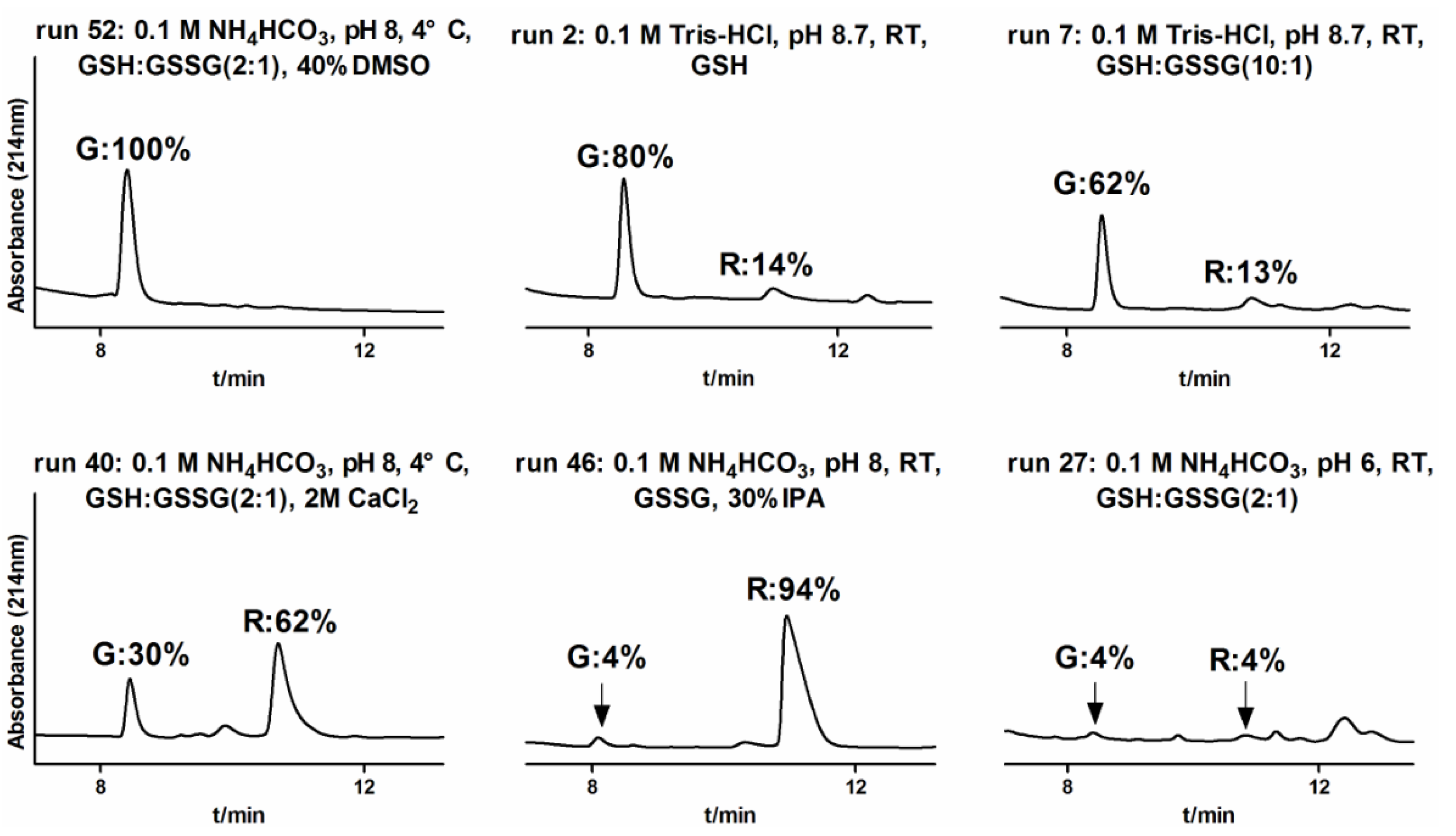
α-conotoxins are small cysteine-rich peptide toxins, which have been used as important research tools for studying numerous pharmacological disorders and for designing new drugs. Such studies require large quantities of appropriately folded, bioactive peptides. Thus, developing efficient in vitro folding methods is a necessary first step for conducting therapeutic activity tests. In this study, we optimized cleavage conditions for α-CTx TxIB and examined key parameters influencing oxidative folding of TxIB for the first time, such as buffer, redox agents, pH, salt, co-solvent and temperature. We found that different conditions had various yields (4~100%) of oxidative folding for the globular isomer (Figure 7, Table 2). We also compared our findings with prior studies on the oxidative folding of various bioactive peptides, such as cyclotides [25,26,27] and other various conotoxins [28,29,30]. Complete oxidative folding of the α-CTx TxIB globular isomer was achieved when the NH4HCO3 or Tris-HCl folding buffers at 4 °C contained 40% of the co-solvent, DMSO, and GSH:GSSG (2:1) or GSH only with pH 8~8.7.
Figure 7. Effect of several different conditions of oxidative folding on the yield of α-CTx TxIB globular (G) and ribbon (R) isomers (runs 52, 2, 7, 40, 46, 27). The globular isomer (G) can complete oxidative folding in condition 52 with 100% yield, while there was almost no folded peptide in condition 27 with 4% yield only for each isomer. Interestingly, the ribbon isomer (R) yield reached 94% in condition 46.
Marinedrugs 11 03537 g007
Initially, one-step oxidative folding of α-CTx TxIB was attempted under 0.1 M Tris-HCl, pH 8.7 (run 1) and 0.1 M NH4HCO3, pH 8 (run 8). However, we observed the accumulation of the ribbon isomer that had the same molecular mass as the native peptide, but differed significantly in retention times (Figure 3, Figure 4B). However, there was no presence of the bead isomer (Figure 3). Formation of the correct disulfide bond is extremely important in maintaining structure and activity. Generally, a disulfide bond between adjacent cysteines is energetically unfavorable, which may result in no accumulation of the bead isomer [31,32,33,34]. CD spectra showed distinct differences between the correctly folded globular isomer and the misfolded ribbon isomer of α-CTx TxIB (Figure 6). The misfolded isomer appears to be more hydrophobic, possibly due to the greater degree of exposure of hydrophobic residues compared to the compactly folded, globular isomer.
During oxidative folding of peptides, disulfide bonds are formed by multistep, reversible thiol-disulfide exchange reactions. Prior studies suggested that oxidation yield may be influenced by the GSH:GSSG ratio, which facilitates thiol-disulfide exchange during the oxidation process [35,36]. We systematically evaluated the effect of different ratios of GSH and GSSG. In these experiments, the concentration of GSH was varied, while the GSSG concentration was kept constant. The results showed that the folding rate increased by increasing the ratio of GSH and GSSG from 1:1 to 2:1, in contrast to the folding rate decreasing by increasing the ratio of GSH and GSSG to 10:1. GSH plays a key role by helping break up non-native disulfide bonds and ensuring the reversibility of the thiol-disulfide exchange. Such disulfide reshuffling is essential for the formation of the final, stably folded native product [27].
As observed by other researchers for α-conotoxin folding, temperature is not a significant factor for globular isomer yield, while the pH plays a key role [22]. Overall, our results of temperature and pH experiments are consistent with prior studies for α-conotoxins. Interestingly, at unfavorable pH conditions (pH 6), varying temperature had a slight, but noticeable effect on TxIB folding. The globular isomer is preferentially formed at lower temperature (runs 24, 32), similar to the results of Shigeru Kubo and co-workers, who have shown that ω-conotoxin MVIIC also preferentially formed the native isomer at lower temperatures [37].
pH variation, by influencing thiol-disulfide exchange reactions, frequently has a notable effect on in vitro peptide folding. Higher pH may facilitate efficient oxidative folding by regulating the degree of ionization of thiols to thiolates; disulfide bond reshuffling is also more significant at higher pH [38,39,40]. In this study, we concluded that pH 8 was optimal for oxidative folding of TxIB in NH4HCO3 buffer, while pH 9 also exhibited a higher proportion of globular isomer. At pH below 7, low or no folding yield was obtained. We hypothesized that the thiol-disulfide exchange reactions are effective at higher pH, while lower pH affects thiol ionization and hampers these reactions.
Optimizing redox agent concentration, pH and temperature was not sufficient to obtain 100% yield of the globular isomer, as under all these conditions, some amount of “ribbon” was always obtained. We further attempted to include salt or co-solvent to reduce the amount of ribbon product and increase the yield of globular isomer. Prior research about ω-conotoxin had demonstrated that salt reduced the repulsive forces between positive charges, which improve native isomer yield. Accordingly, the use of salts, such as (NH4)2SO4 and CaCl2, facilitates oxidative folding. In this work, neither (NH4)2SO4 nor CaCl2 in oxidative folding significantly improved folding yield of the globular isomer. Previously, Kubo et al. [41] had also shown that no significant improvements in refolding of HNP1 were observed by changing ammonium salts. In contrast, including CaCl2 led to a greater yield of the α-CTx TxIB ribbon isomer. It is likely that three positively charged residues (R9, K11, H12) and two negatively charged residues (D5, D14) of α-CTx TxIB are both on the globular isomer surface; the repulsive forces are weakened due to a large distance among the same kind of charged residues.
Co-solvent is a key factor reported in the literature for oxidative folding of bioactive peptides [25,32,38,42]. In order to shift the equilibrium towards the production of the native isomer, we tested IPA and DMSO to improve the yield of the globular isomer. All disulfide bonds in the ribbon isomer have to be reshuffled for globular isomer folding, an extensive rearrangement that must be combined with major conformational changes. Both co-solvents have a similar function in the reaction buffer by solvating hydrophobic side chains [43,44]; however, they had distinct effects on α-CTx TxIB folding. Notably, we found that IPA significantly decreased native isomer yield, while increasing ribbon isomer yield. These results are different from other α4/7-conotoxins, such as α-CTx MII, α-CTx PnIB and α-CTx GID, for which IPA significantly increased the accumulation of the globular isomer [22]. We hypothesize that IPA presence in the folding reaction may preferentially form more hydrophobic isomers of α-CTx TxIB.
In comparison, DMSO assisted the folding of the α-CTx TxIB globular isomer. Inclusion of DMSO as a co-solvent significantly increased the accumulation of the globular isomer (runs 49–52). Inclusion of 40% DMSO allowed for complete oxidative folding of the α-CTx TxIB globular isomer. These results are similar to the findings of Tam [38], whereby the use of DMSO can significantly increase native isomer yield for basic and hydrophobic disulfide-rich peptides, a result that cannot be obtained by varying redox agent ratios only. However, using DMSO alone, without GSH, is also not sufficient (runs 53–54). We hypothesize that while DMSO facilitates globular isomers, it cannot promote disulfide bond reshuffling and limits the accumulation of the ribbon isomer.

3. Experimental Section

3.1.Materials

Phenol, thioanisole, triisopropylsilane (TIPS), 1,2-ethanedithiol (EDT) and diethyl ether were from Tiandi (Haikou City, China). Reverse phase HPLC analytical Vydac C18 (5 μm, 4.6 mm × 250 mm) and preparative C18 Vydac columns (10 μm, 22 mm × 250 mm) were obtained from Shenyue (Shanghai City, China). Reagents for peptide synthesis were from GL Biochem (Shanghai, China). Acetonitrile was from Fisher (Fisher Scientific Company L.L.C., Pittsburgh, PA, USA), and trifluoroacetic acid was from Tedia (Fairfield, OH, USA).

3.2.Resin-Bounded Peptide Synthesis and Cleavage

The resin-bounded peptide of α-CTx TxIB was synthesized by one-step oxidation and two-step oxidation, respectively. The peptide was synthesized on an amide resin by solid-phase methodology on an ABI 433A peptide synthesizer using Fmoc (N-(9-fluorenyl) methoxycarbonyl) chemistry. For one-step oxidation, all cysteine residues were trityl protected. For two-step oxidation, Cys residues were protected in pairs with either S-trityl on Cys1 and Cys3 or S-acetamidomethyl on Cys2 and Cys4, as described previously [15]. The crude peptide was cleaved from the resin and precipitated with four different Fmoc cleavage cocktails at 3 different cleavage times, respectively (Table 1). Briefly, 25 mg of the TxIB resin-bounded peptide was placed in 10 mL tubes and was treated with four different cleavage cocktails for 1 h, 2 h and 3 h at room temperature (RT). All cleavage cocktails and scavengers were freshly prepared prior to use. The resin was removed by filtration, then the support was rinsed with TFA. Ether-peptide mixture was incubated at 4 °C overnight after the combined filtrates were added to cold (−20 °C) diethyl ether. The precipitated peptide was collected by centrifugation, which was linear TxIB peptide after being lyophilized. The linear peptide was dissolved with 2 mL HPLC-grade water and 10 μL was injected into reverse-phase HPLC (RP-HPLC) for analysis. ESI-MS mass spectrometry was utilized to confirm the identity of the products.

3.3. Two-Step Oxidation Folding

A two-step oxidation protocol was used to fold linear α-CTx TxIB selectively. The first disulfide bond between Cys1 and Cys3 (globular) or Cys2 and Cys3 (ribbon) was formed by adding peptide into an equal volume of 20 mM potassium ferricyanide, (K3[Fe(CN)6]), 0.1 M Tris, pH 7.5, and mixing for a 45 min reaction at room temperature. The monocyclic peptide was purified by reverse-phase HPLC (RP-HPLC). In order to remove S-Acm groups and simultaneously form the second disulfide bond, between Cys2 and Cys4 (globular) or Cys1 and Cys4 (ribbon), purified peptide was used for iodine oxidation as follows: peptide was dripped into an equal volume of 10 mM iodine in H2O:TFA:ACN (73:3:24 by volume) and stirred for 10 min with N2. The reaction was terminated by addition of ascorbic acid until colorless, and the final peptide was purified by RP-HPLC using a Vydac C18 column. The HPLC separation method was a linear gradient of 10%–30% solvent B gradient over 30 min (solvent B is 0.05% TFA in 90% ACN; solvent A is 0.1% TFA in H2O). ESI-MS mass spectrometry was utilized to confirm the identity of the products.

3.4. One-Step Oxidative Folding

A one-step oxidation protocol was used to fold linear α-CTx TxIB randomly. Various folding conditions were investigated to fold the peptide directly, including different buffers, redox reagents, GSH/GSSG, pH, temperatures, salt and co-solvents (Table 2). For each oxidation condition, final concentration of linear TxIB in each trial was 50 nM/mL. At 1-h, 2-h, 4-h, 24-h and 48-h intervals, samples were acidified with formic acid to terminate the folding process. Folding product profiles were analyzed by analytical RP-HPLC with a 10%–30% solvent B gradient over 30 min. For reference comparison, correctly folded globular isomer and other misfolded isomers were identified by co-injections of pure isomers obtained by two-step synthesis. A Shimadzu HPLC system was used to calculate the HPLC profile peak area. Folding yields were determined as a ratio of the steady state accumulation of native species, relative to all folding species.

3.5. Circular Dichroism (CD) Measurements

Individual isomers were purified by HPLC. CD spectra of globular and ribbon isomers were measured at the 0.35 mg/mL peptide concentration in HPLC-water (pH 7.0) using a Jasco J-810 spectropolarimeter at room temperature. Spectra were collected at 0.5 nm intervals over the wavelength range 260–190 nm in a 0.1 cm path length cuvette.

4. Conclusions

In summary, the current study focused on optimizing conditions for high-yield production of the correctly folded native globular isomer of α-CTx TxIB, which is a uniquely selective ligand blocking α6/α3β2β3 nAChRs. The results showed that inclusion of GSH, cosolvent, DMSO, basic pH conditions and lower temperature significantly improved the yield of the TxIB native isomer. The most optimized folding conditions were obtained for one-step oxidation synthesis of α-CTx TxIB, which produced the native globular isomer only and excluded the other by-products of misfolded isomers. These findings would serve as a foundation to synthesize α-CTx TxIB efficiently at a large scale for future testing of potential therapeutic properties and its biomedical applications.

Abbreviations

DMSO
dimethyl sulfoxide
GSH
reduced glutathione
GSSG
oxidized glutathione
HPLC
high performance liquid chromatography
ESI-MS
electrospray ionization mass spectrum
ACN
acetonitrile
CDSSTR
a program for estimating protein secondary structure fractions
α-CTx
α-conotoxin

Acknowledgments

This work was supported by State High-Tech Research and Development Project (863) of the Ministry of Science and Technology of China (2012AA021706), International Science & Technology Cooperation Program of China (2011DFR31210), National Natural Science Foundation of China (No. 81160503, 41366002) and Program for Changjiang Scholars and Innovative Research Team in University (PCSIRT, IRT1123).

Conflicts of Interest

The authors declare no conflict of interest.

References

  1. Albuquerque, E.X.; Pereira, E.F.; Alkondon, M.; Rogers, S.W. Mammalian nicotinic acetylcholine receptors: From structure to function. Physiol. Rev. 2009, 89, 73–120. [Google Scholar] [CrossRef]
  2. Millar, N.S.; Gotti, C. Diversity of vertebrate nicotinic acetylcholine receptors. Neuropharmacology 2009, 56, 237–246. [Google Scholar] [CrossRef]
  3. Rose, J.E. Multiple brain pathways and receptors underlying tobacco addiction. Biochem. Pharmacol. 2007, 74, 1263–1270. [Google Scholar] [CrossRef]
  4. Quik, M.; Perez, X.A.; Grady, S.R. Role of α6 nicotinic receptors in CNS dopaminergic function: Relevance to addiction and neurological disorders. Biochem. Pharmacol. 2011, 82, 873–882. [Google Scholar] [CrossRef]
  5. Luo, S.; Christensen, S.; Zhangsun, D.; Wu, Y.; Hu, Y.; Zhu, X.; Chhabra, S.; Norton, R.S.; McIntosh, J.M. A novel inhibitor of alpha9alpha10 nicotinic acetylcholine receptors from Conus vexillum delineates a new conotoxin superfamily. PLoS ONE 2013, 8, e54648. [Google Scholar]
  6. Lewis, R.J.; Garcia, M.L. Therapeutic potential of venom peptides. Nat. Rev. Drug Discov. 2003, 2, 790–802. [Google Scholar] [CrossRef]
  7. Han, T.S.; Teichert, R.W.; Olivera, B.M.; Bulaj, G. Conus venoms-a rich source of peptide-based therapeutics. Curr. Pharm. Des. 2008, 14, 2462–2479. [Google Scholar] [CrossRef]
  8. Luo, S.; Akondi, K.B.; Zhangsun, D.; Wu, Y.; Zhu, X.; Hu, Y.; Christensen, S.; Dowell, C.; Daly, N.L.; Craik, D.J.; et al. Atypical alpha-conotoxin LtIA from Conus litteratus targets a novel microsite of the alpha3beta2 nicotinic receptor. J. Biol. Chem. 2010, 285, 12355–12366. [Google Scholar] [CrossRef]
  9. Ellison, M.; Haberlandt, C.; Gomez-Casati, M.E.; Watkins, M.; Elgoyhen, A.B.; McIntosh, J.M.; Olivera, B.M. Alpha-RgIA: A novel conotoxin that specifically and potently blocks the alpha9alpha10 nAChR. Biochemistry 2006, 45, 1511–1517. [Google Scholar] [CrossRef]
  10. Luo, S.; Kulak, J.M.; Cartier, G.E.; Jacobsen, R.B.; Yoshikami, D.; Olivera, B.M.; McIntosh, J.M. alpha-conotoxin AuIB selectively blocks alpha3 beta4 nicotinic acetylcholine receptors and nicotine-evoked norepinephrine release. J. Neurosci. 1998, 18, 8571–8579. [Google Scholar]
  11. Lopez-Vera, E.; Aguilar, M.B.; Schiavon, E.; Marinzi, C.; Ortiz, E.; Restano Cassulini, R.; Batista, C.V.; Possani, L.D.; Heimer de la Cotera, E.P.; Peri, F.; et al. Novel alpha-conotoxins from Conus spurius and the alpha-conotoxin EI share high-affinity potentiation and low-affinity inhibition of nicotinic acetylcholine receptors. FEBS J. 2007, 274, 3972–3985. [Google Scholar] [CrossRef]
  12. Klink, R.; de Kerchove d’Exaerde, A.; Zoli, M.; Changeux, J.P. Molecular and physiological diversity of nicotinic acetylcholine receptors in the midbrain dopaminergic nuclei. J. Neurosci. 2001, 21, 1452–1463. [Google Scholar]
  13. Livett, B.G.; Sandall, D.W.; Keays, D.; Down, J.; Gayler, K.R.; Satkunanathan, N.; Khalil, Z. Therapeutic applications of conotoxins that target the neuronal nicotinic acetylcholine receptor. Toxicon 2006, 48, 810–829. [Google Scholar] [CrossRef]
  14. Gehrmann, J.; Alewood, P.F.; Craik, D.J. Structure determination of the three disulfide bond isomers of α-conotoxin GI: A model for the role of disulfide bonds in structural stability. J. Mol. Biol. 1998, 278, 401–415. [Google Scholar] [CrossRef]
  15. Luo, S.; Zhangsun, D.; Wu, Y.; Zhu, X.; Hu, Y.; McIntyre, M.; Christensen, S.; Akcan, M.; Craik, D.J.; McIntosh, J.M. Characterization of a Novel alpha-Conotoxin from Conus textile that Selectively Targets Alpha6/alpha3beta2betab3 Nicotinic Acetylcholine Receptors. J. Biol. Chem. 2013, 288, 894–902. [Google Scholar] [CrossRef]
  16. Guy, C.A.; Fields, G.B. Trifluoroacetic acid cleavage and deprotection of resin-bound peptides following synthesis by Fmoc chemistry. Methods Enzymol. 1997, 289, 67–83. [Google Scholar] [CrossRef]
  17. Fields, C.G.; Fields, G.B. Minimization of tryptophan alkylation following 9-fluorenylmethoxycarbonyl solid-phase peptide synthesis. Tetrahedron Lett. 1993, 34, 6661–6664. [Google Scholar] [CrossRef]
  18. King, D.S.; Fields, C.G.; Fields, G.B. A cleavage method which minimizes side reactions following Fmoc solid phase peptide synthesis. Int. J. Pept. Protein Res. 1990, 36, 255–266. [Google Scholar]
  19. Pearson, D.A.; Blanchette, M.; Baker, M.L.; Guindon, C.A. Trialkylsilanes as scavengers for the trifluoroacetic acid deblocking of protecting groups in peptide synthesis. Tetrahedron Lett. 1989, 30, 2739–2742. [Google Scholar] [CrossRef]
  20. Bulaj, G.; Olivera, B.M. Folding of conotoxins: formation of the native disulfide bridges during chemical synthesis and biosynthesis of Conus peptides. Antioxid. Redox Signal. 2008, 10, 141–155. [Google Scholar] [CrossRef]
  21. DeLa Cruz, R.; Whitby, F.G.; Buczek, O.; Bulaj, G. Detergent-assisted oxidative folding of delta-conotoxins. J. Pept. Res. 2003, 61, 202–212. [Google Scholar] [CrossRef]
  22. Gyanda, R.; Banerjee, J.; Chang, Y.P.; Phillips, A.M.; Toll, L.; Armishaw, C.J. Oxidative folding and preparation of alpha-conotoxins for use in high-throughput structure-activity relationship studies. J. Pept. Sci. 2012, 19, 16–24. [Google Scholar]
  23. Konermann, L.; Stocks, B.B.; Pan, Y.; Tong, X. Mass spectrometry combined with oxidative labeling for exploring protein structure and folding. Mass Spectrom. Rev. 2010, 29, 651–667. [Google Scholar]
  24. Salamanca, S.; Chang, J.Y. Unfolding and refolding pathways of a major kinetic trap in the oxidative folding of alpha-lactalbumin. Biochemistry 2005, 44, 744–750. [Google Scholar] [CrossRef]
  25. Wong, C.T.T.; Taichi, M.; Nishio, H.; Nishiuchi, Y.; Tam, J.P. Optimal Oxidative Folding of the Novel Antimicrobial Cyclotide from Hedyotis biflora Requires High Alcohol Concentrations. Biochemistry 2011, 50, 7275–7283. [Google Scholar] [CrossRef]
  26. Gunasekera, S.; Daly, N.L.; Clark, R.J.; Craik, D.J. Dissecting the oxidative folding of circular cystine knot miniproteins. Antioxid. Redox signal. 2009, 11, 971–980. [Google Scholar] [CrossRef] [Green Version]
  27. Leta Aboye, T.; Clark, R.J.; Craik, D.J.; Goransson, U. Ultra-stable peptide scaffolds for protein engineering-synthesis and folding of the circular cystine knotted cyclotide cycloviolacin O2. Chembiochem. 2008, 9, 103–113. [Google Scholar] [CrossRef]
  28. Steiner, A.M.; Bulaj, G. Optimization of oxidative folding methods for cysteine-rich peptides: a study of conotoxins containing three disulfide bridges. J. Pept. Sci. 2011, 17, 1–7. [Google Scholar] [CrossRef]
  29. Fuller, E.; Green, B.R.; Catlin, P.; Buczek, O.; Nielsen, J.S.; Olivera, B.M.; Bulaj, G. Oxidative folding of conotoxins sharing an identical disulfide bridging framework. FEBS J. 2005, 272, 1727–1738. [Google Scholar] [CrossRef]
  30. Bulaj, G.; Buczek, O.; Goodsell, I.; Jimenez, E.C.; Kranski, J.; Nielsen, J.S.; Garrett, J.E.; Olivera, B.M. Efficient oxidative folding of conotoxins and the radiation of venomous cone snails. Proc. Natl. Acad. Sci. USA 2003, 100 (Suppl. 2), 14562–14568. [Google Scholar] [CrossRef]
  31. Zhang, R.M.; Snyder, G.H. Factors governing selective formation of specific disulfides in synthetic variants of alpha-conotoxin. Biochemistry 1991, 30, 11343–11348. [Google Scholar] [CrossRef]
  32. Nielsen, J.S.; Buczek, P.; Bulaj, G. Cosolvent-assisted oxidative folding of a bicyclic alpha-conotoxin ImI. J. Pept. Sci. 2004, 10, 249–256. [Google Scholar] [CrossRef]
  33. Grishin, A.A.; Wang, C.I.; Muttenthaler, M.; Alewood, P.F.; Lewis, R.J.; Adams, D.J. Alpha-conotoxin AuIB isomers exhibit distinct inhibitory mechanisms and differential sensitivity to stoichiometry of alpha3beta4 nicotinic acetylcholine receptors. J. Biol. Chem. 2010, 285, 22254–22263. [Google Scholar]
  34. Clark, R.J.; Daly, N.L.; Halai, R.; Nevin, S.T.; Adams, D.J.; Craik, D.J. The three-dimensional structure of the analgesic alpha-conotoxin, RgIA. FEBS Lett. 2008, 582, 597–602. [Google Scholar] [CrossRef]
  35. Anderson, M.E.; Meister, A. Transport and direct utilization of gamma-glutamylcyst(e)ine for glutathione synthesis. Proc. Natl. Acad. Sci. USA 1983, 80, 707–711. [Google Scholar] [CrossRef]
  36. Aboye, T.L.; Clark, R.J.; Burman, R.; Roig, M.B.; Craik, D.J.; Goransson, U. Interlocking disulfides in circular proteins: toward efficient oxidative folding of cyclotides. Antioxid. Redox Signal. 2011, 14, 77–86. [Google Scholar] [CrossRef] [Green Version]
  37. Kubo, S.; Chino, N.; Kimura, T.; Sakakibara, S. Oxidative folding of omega-conotoxin MVIIC: Effects of temperature and salt. Biopolymers 1996, 38, 733–744. [Google Scholar] [CrossRef]
  38. Tam, J.P.; Wu, C.R.; Liu, W.; Zhang, J.W. Disulfide bond formation in peptides by dimethyl sulfoxide. Scope and applications. J. Am. Chem. Soc. 1991, 113, 6657–6662. [Google Scholar] [CrossRef]
  39. Bulaj, G.; Kortemme, T.; Goldenberg, D.P. Ionization-reactivity relationships for cysteine thiols in polypeptides. Biochemistry 1998, 37, 8965–8972. [Google Scholar] [CrossRef]
  40. Zhang, J.; Diamond, S.; Arvedson, T.; Sasu, B.J.; Miranda, L.P. Oxidative folding of hepcidin at acidic pH. Biopolymers 2010, 94, 257–264. [Google Scholar] [CrossRef]
  41. Kubo, S.; Tanimura, K.; Nishio, H.; Chino, N.; Teshima, T.; Kimura, T.; Nishiuchi, Y. Optimization of the Oxidative Folding Reaction and Disulfide Structure Determination of Human α-Defensin 1, 2, 3 and 5. Int. J. Pept. Res. Ther. 2008, 14, 341–349. [Google Scholar] [CrossRef]
  42. Daly, N.L.; Craik, D.J. Acyclic permutants of naturally occurring cyclic proteins Characterization of cystine knot and β-sheet formation in the macrocyclic polypeptide kalata B1. J. Biol. Chem. 2000, 275, 19068–19075. [Google Scholar] [CrossRef]
  43. Shimizu, S.; Shimizu, K. Alcohol denaturation: thermodynamic theory of peptide unit solvation. J. Am. Chem. Soc. 1999, 121, 2387–2394. [Google Scholar] [CrossRef]
  44. Welker, E.; Narayan, M.; Wedemeyer, W.J.; Scheraga, H.A. Structural determinants of oxidative folding in proteins. Proc. Natl. Acad. Sci. USA 2001, 98, 2312–2316. [Google Scholar] [CrossRef]