Review

Diversity of Secondary Metabolites from Marine Bacillus Species: Chemistry and Biological Activity

by
Muhammad Abdul Mojid Mondol
1,
Hee Jae Shin
2,* and
Mohammad Tofazzal Islam
3,*
1
School of Science and Technology, Bangladesh Open University, Board Bazar, Gazipur 1705, Bangladesh
2
Marine Natural Products Chemistry Laboratory, Korea Institute of Ocean Science & Technology, Ansan, P.O. Box 29, Seoul 425-600, Korea
3
Department of Biotechnology, Bangabandhu Sheikh Mujibur Rahman Agricultural University, Gazipur 1706, Bangladesh
*
Authors to whom correspondence should be addressed.
Mar. Drugs 2013, 11(8), 2846-2872; https://doi.org/10.3390/md11082846
Submission received: 1 May 2013 / Revised: 12 July 2013 / Accepted: 29 July 2013 / Published: 12 August 2013
(This article belongs to the Special Issue Marine Secondary Metabolites)

Abstract

:
Marine Bacillus species produce versatile secondary metabolites including lipopeptides, polypeptides, macrolactones, fatty acids, polyketides, and isocoumarins. These structurally diverse compounds exhibit a wide range of biological activities, such as antimicrobial, anticancer, and antialgal activities. Some marine Bacillus strains can detoxify heavy metals through reduction processes and have the ability to produce carotenoids. The present article reviews the chemistry and biological activities of secondary metabolites from marine isolates. Side by side, the potential for application of these novel natural products from marine Bacillus strains as drugs, pesticides, carotenoids, and tools for the bioremediation of heavy metal toxicity are also discussed.

Graphical Abstract

1. Introduction

Microorganisms especially bacteria and fungi are promising sources of structurally diverse and potent bioactive compounds [1,2,3]. Many of these bioactive compounds are used as chemotherapeutic agents for the treatment of human and animal diseases [4,5]. There is a continuing demand for novel bioactive compounds to treat drug-resistant human and animal pathogens [6,7,8], and management of devastating pathogens of crops, which are insensitive or less sensitive to existing chemical pesticides [9,10,11,12].
Marine environments including the subsurface are believed to contain a total of approximately 3.67 × 1030 microorganisms [13]. About 70% of the earth’s surface is covered by the ocean representing 80% of life on earth indicating an enormous pool of microbial biodiversity and potential discovery of new natural products [14]. Among diverse microbial species, isolates of marine Bacillus belong to phylogenetically and phenogenetically heterogeneous groups of bacteria. They are ubiquitous in the marine environment and can tolerate adverse conditions such as high temperature, pressure, salinity, and pH [15]. Generally, Bacillus strains need more nutrition and space to be the fastest growing bacteria for which they compete with other organisms. Due to the diluting effect of the ocean drives, marine organisms produce potent bioactive compounds to fight off their competitors or to escape from micropredation [16,17]. Metabolically marine strains are different from their terrestrial counterparts, and thereby, they may produce unique bioactive compounds, which are not found in their terrestrial counterparts [18,19]. The ability to produce diverse classes of antibiotics by Bacillus spp. has been evidence by several genomic studies. For example, the genome sequence of the widely distributed Bacillus strains revealed that about 8% of genome is devoted to synthesizing antibiotics [20,21]. Similarly, genome analysis of a marine B. subtilis subsp. spizisenii strain gtP20b, isolated from the Indian Ocean, indicated the presence of huge number of genes for biosynthesis of secondary metabolites [22].
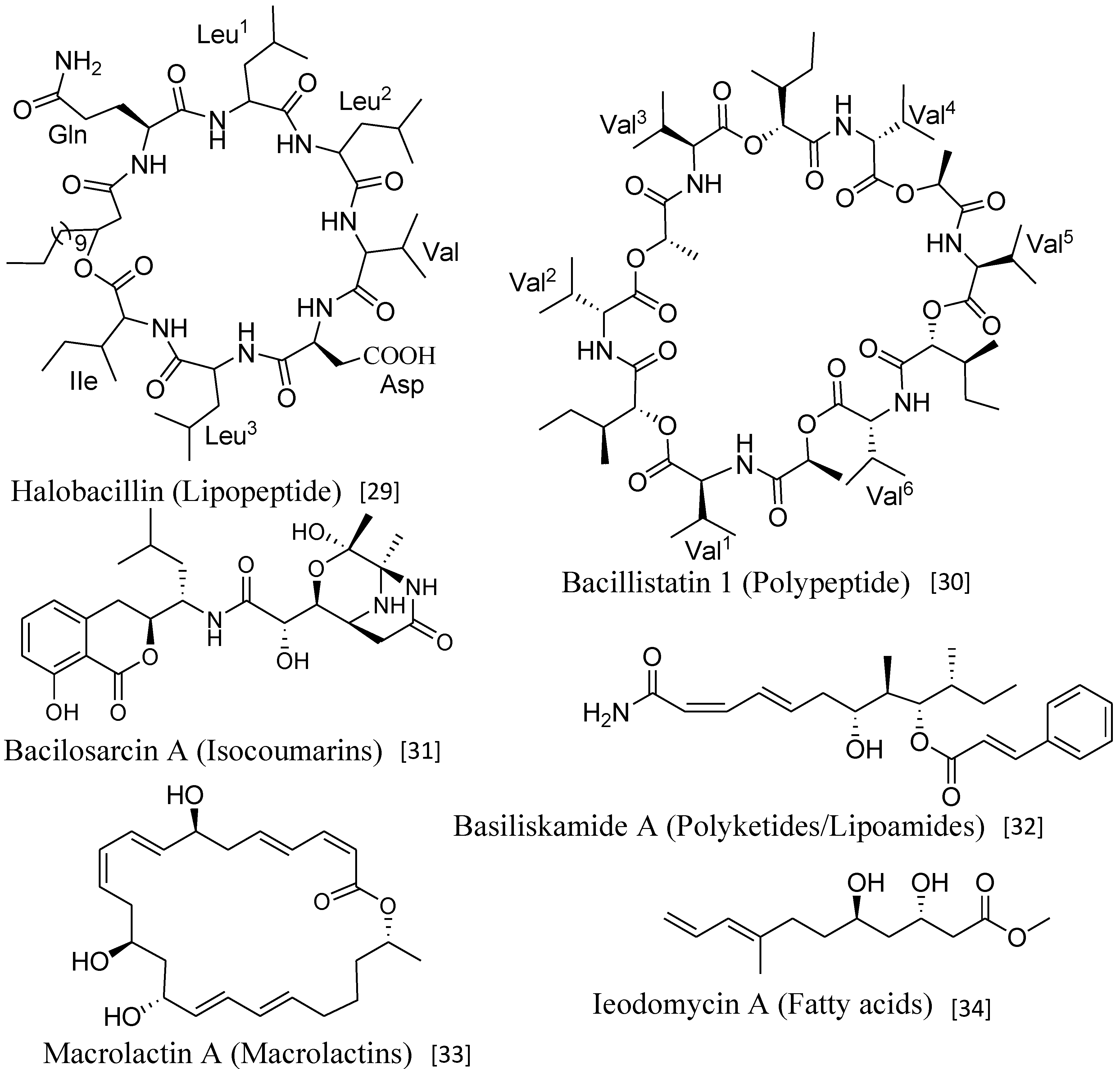
Marine Bacillus isolates produce structurally diverse classes of secondary metabolites, such as lipopeptides, polypeptides, macrolactones, fatty acids, polyketides, lipoamides, and isocoumarins [23,24] (Figure 1). These structurally versatile compounds exhibit a wide range of biological activities, such as antimicrobial, anticancer, antialgal, and antiperonosporomycetal [23,24]. As Bacillus strains rapidly grow in liquid media even under stressful conditions and readily forms resistant spores, it might be useful as an effective biocontrol agent against various phytopathogens [25]. Structures, syntheses, and specific functions of diverse antibiotics produced by B. subtilis have elaborately been reviewed [26].
Extensive use of pesticides during crop production and exposure of industrial toxic waste to the environment results in the deposition of heavy metals (Pd, Hg, Cu, Cd, Cr, Co) in soil and water bodies, and this practice can pose serious threats to crop production as well as to the health of all organisms in the aquatic environment. Although trace amount of many heavy metal ions are required for various biochemical activities of all living organisms, higher concentrations of these ions are generally toxic to their cells. Surprisingly, some bacterial species exhibit tolerance and resistance towards high concentrations of heavy metals. Marine strains, in general, thrive under harsh conditions when compared to the most of terrestrial environments, providing them with enormous tolerance, and thus, they are often considered as potential candidates for the detoxification of heavy metals [27]. Most of the toxic heavy metals are reduced rather than oxidized by native microbes, with a few exceptions, since the reduced forms are, generally, less toxic. Bacterial tolerance to heavy metals generally follows the pattern of efflux, accumulation, complexation, and reduction [28].
Figure 1. Major classes of secondary metabolites produced by Bacillus strains.
Marinedrugs 11 02846 g001
Nowadays, food growers heavily rely on chemical pesticides to prevent or control diseases in their crop plants. Deposition of these pesticide residues in food, soil, and water bodies is imposing enormous threats to human health, environment, and ecosystem [35]. Consequently, there is an increasing demand from consumers and environment conservationists to replace chemical pesticides with natural, environment-friendly antagonistic microorganisms with novel mode of action for sustainable production of crops [11,12].
As marine Bacillus isolates produce diverse bioactive secondary metabolites with novel modes of action, they may have great potential for the development of effective management strategies to combat human, animal and phytopathogens in biorational manners. Several general reviews on bioactive secondary metabolites from Bacillus isolates have been published [23,26], however, no report has so far been published on bioactive compounds from marine Bacillus species. This article comprehensively reviews the chemistry and biological activities of diverse secondary metabolites from marine Bacillus species, and discusses the potentials of these natural products for the development of effective drugs, agrochemicals, carotenoids, as well as tools for the bioremediation of environmental pollution due to heavy metal contamination.

2. Bioactive Compounds

2.1. Lipopeptides
Cyclic lipopeptides (cLPs) are versatile metabolites produced by a variety of bacterial genera. They are composed of a short cyclic oligopeptide linked to a fatty acid tail, and exhibit potent surfactant properties [36]. cLPs are produced nonribosomally on large NRPS (nonribosomal peptide synthetase)—PKS (polyketide synthetase) hybrid synthetases. Endospore-forming rhizobacterium (terrestrial) B. subtilis produces varieties of antimicrobial peptides that are either ribosomally synthesized and post-translationally modified (e.g., lantibiotics and lantibiotic-like peptides), or nonribosomally generated. Ribosomally and nonribosomally synthesized peptides by terrestrial Bacillus spp. are elaborately reviewed by Stein [26]. cLPs have received considerable attention for their antibiotic activities against a range of human- and plant-pathogenic organisms, including enveloped viruses, mycoplasmas, trypanosomes, bacteria, fungi, and peronosporomycetes [37].
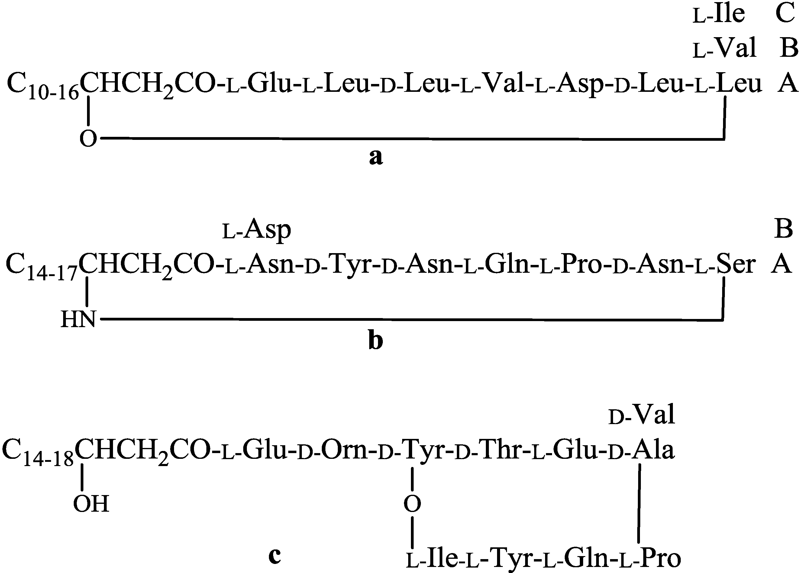
A large proportion of the secondary metabolites produced by the Bacillus isolates are cyclic lipopeptides belonging to three families: iturins, fengycins, and surfactins [38]. The chemical structure of c-LPs have a peptide backbone composed of seven (iturins and surfactins) or 10 (fengycins) amino acids connected to a β-hydroxy (fengycins and surfactins) or β-amino (iturins) fatty acid, which may vary from C-10 to C-16 for surfactins, C-14 to C-17 for iturins, and C-14 to C-18 for fengycins. Each lipopeptide family can be subdivided again based on position of specific amino acid in the peptide ring. For example, the fengycin family is subdivided into fengycin A and fengycin B, where D-alanine and D-valine is present in the sixth position, respectively. Due to difference in length, branching, and saturation of acyl chain, homologues are formed within each subdivision of cLPs (Figure 2). Based on the comparison of retention times and molecular masses with those of known compounds, all three families of lipopeptide antibiotics may be assigned using LC-MS with specific elution programs [39].
Figure 2. General structures of surfactin (a); iturin (b) and fengycin (c).
Marinedrugs 11 02846 g002
A cLPs producer, B. amyloliquefaciens strain GA1 was initially isolated from strawberry culture. To identify gene fragments responsible for synthesis of cLPs, partial DNA sequencing of strain GA1 has been done. Analysis of sequencing data revealed that fourteen gene fragments with homology toward gene clusters were involved in the synthesis of cLPs [38]. Out of fourteen, in eleven gene fragments, operons srf and fen had 80%–96% and 41%–92% amino acid identity directing the synthesis of surfactin and fengycin, respectively [38]. Further analysis of sequencing data revealed that three gene fragments have been involved in directing the synthesis of an iturin lipopeptide in the strain FZB42 and have 48%–82% amino acid similarity with the ituDABC operon encoding the iturin A synthetase in B. subtilis RB14 [40].
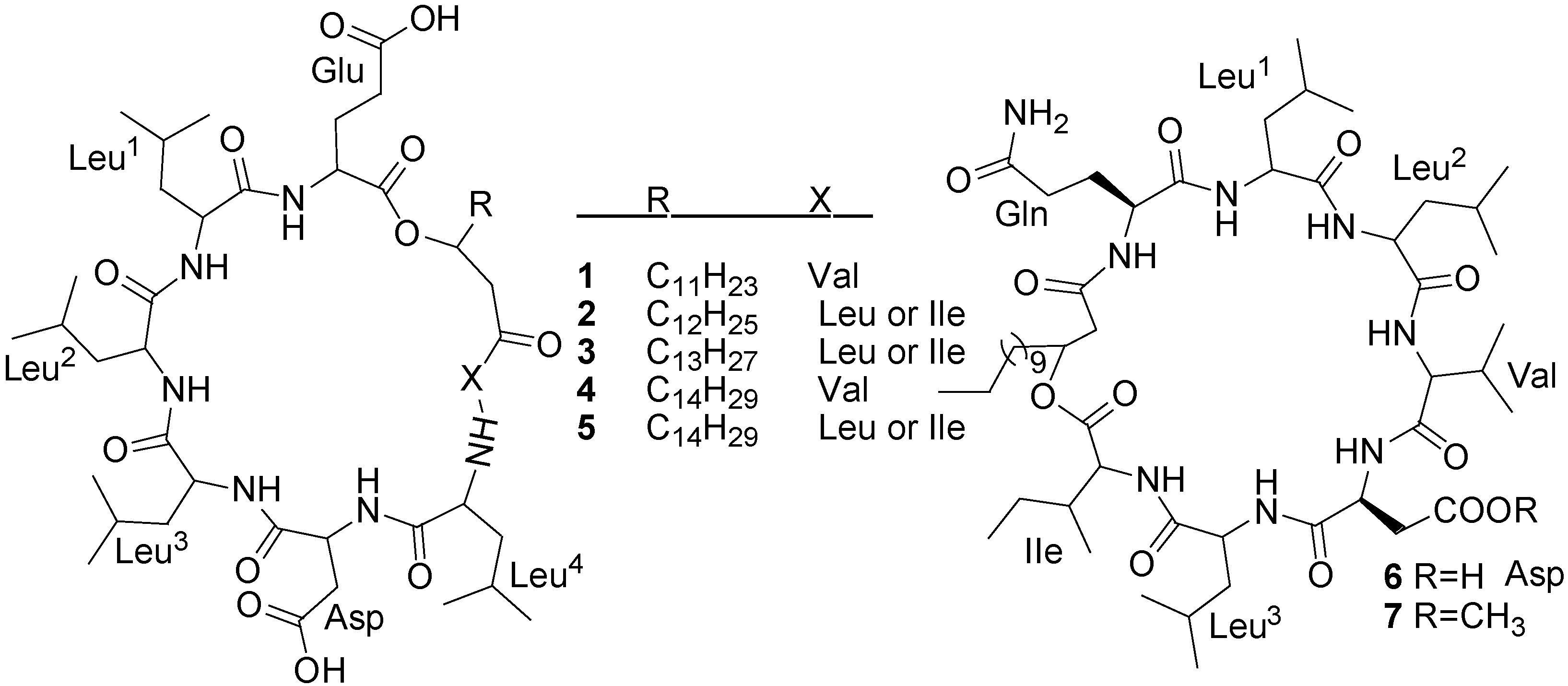
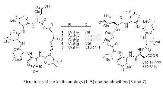
Five surfactin analogs (15) (Figure 3) have been isolated from the culture broth of a Bacillus pumilus (SP21) through bioassay-guided fractionation [41]. The producing strain was isolated from a sediment sample collected from the Bahamas. Compounds 14 showed selectively good inhibitory activity against S. aureus, P. vulgaris, and E. faecalis (6.5 to 25 µg/mL), but not against P. aeruginosa [41].
Figure 3. Structures of surfactin analogs (15) and halobacillins (6 and 7).
Marinedrugs 11 02846 g003
Halobacillin (6) and methyl halobacillin (7) (Figure 3), two cyclic acylpeptides have been isolated from the culture broth of a Bacillus sp. CND-914. The strain CND-914 was obtained from a deep-sea sediment core taken at near Guaymas Basins in Mexico [29]. Halobacillin, the first novel acylpeptide of the iturin class similar to surfactin, is one of the most effective known biosurfactants [42]. The major difference between halobacillin and surfactin is the replacement of glutamic acid of surfactin with a glutamine in halobacillin. Halobacillin inhibits the growth of human colon tumor cells (HCT-116) with an IC50 of 0.98 µg/mL but it does not exhibit antimicrobial activity like surfactin (Table 1).
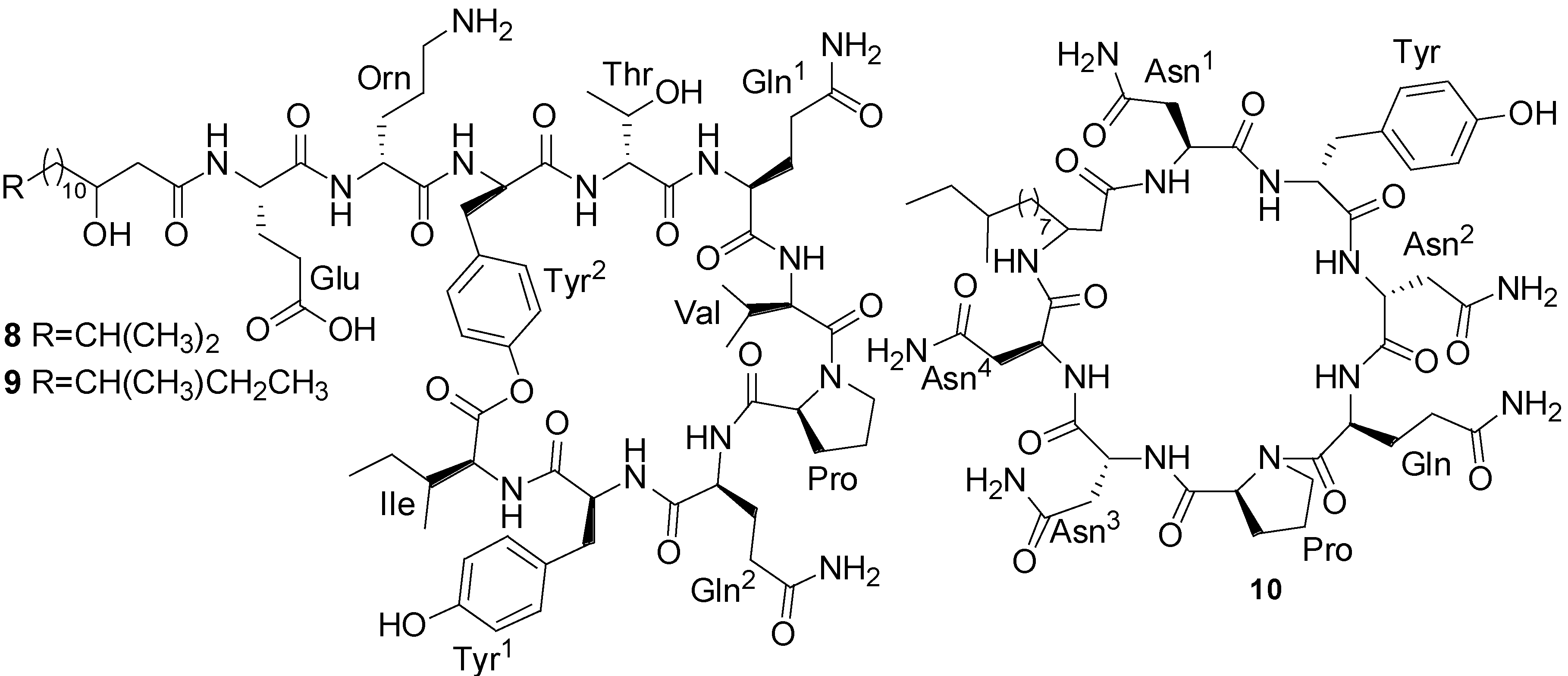
Three lipopeptides (810) (Figure 4), iso-C16 fengycin B (8), anteiso-C17 fengycin B (9) and a new iturinic lipopeptide, mojavensin A (10), were obtained by bioactivity-guided fractionation from the fermentation broth of B. mojavensis B0621A, which was isolated from the mantle of a pearl oyster P. martensii collected from Weizhou Island in the South China Sea [43]. These lipopeptides displayed dose-dependent antifungal activity against a broad spectra of phytopathogens as well as being weakly antagonistic to S. aureus. Moreover, they all displayed cytotoxic activities against the human leukemia (HL-60) cell line with IC50 values of 100, 100, and 1.6 µM, respectively [43].
Table 1. List of some important bioactive compounds isolated from marine strains.
CompoundsProducing strainsTest organisms/cell linesInhibitory concentrationsNature of bioactivitiesRef.
Halobacillin (6)Bacillus sp. CND-914Human HCT-116 cancer cells0.98 µg/mL (IC50)Anticancer[29]
Mixirin (11)Bacillus sp.Human HCT-116 cancer cells0.68 µg/mL (IC50)Anticancer[44]
Bogorol A (15)Bacillus sp.MRSA2 µg/mL (MIC)Antibacterial[45]
Loloatin B (18)Bacillus sp.MRSA, VRE1–2 µg/mL (MIC)Antibacterial[46]
Bacillistatins 1 (19) and 2 (20)B. silvestrisHuman cancer cell line10−4–10−5 µg/mL (GI50)Anticancer[30]
Bacillamide (27)Bacillus sp.C. polykrikoidesLC50 after 6 h: 3.2 µg/mLAntialgal[47]
Bacilosarcin A (35)B. subtilisBarnyard millet sprouts82% inhibition at 50 µMPlant growth regulator[31]
Macrolactin S (69)B. amyloliquefaciensE. coli and S. aureus0.3 and 0.1 µg/mL (MIC)Antibacterial[48]
Macrolactin V (86)B. amyloliquefaciensE. coli, B. subtilis and S. aureus0.1 µg/mL (MIC)Antibacterial[48]
Basiliskamides A (21) and B (22)B. laterosporusC. albicans and A. fumigatus1.0 and 3.1 µg/mL 2.5 and 5.0 µg/mLAntifungal[32]
Figure 4. Structures of fengycins (8 and 9) and mojavensin (10).
Marinedrugs 11 02846 g004
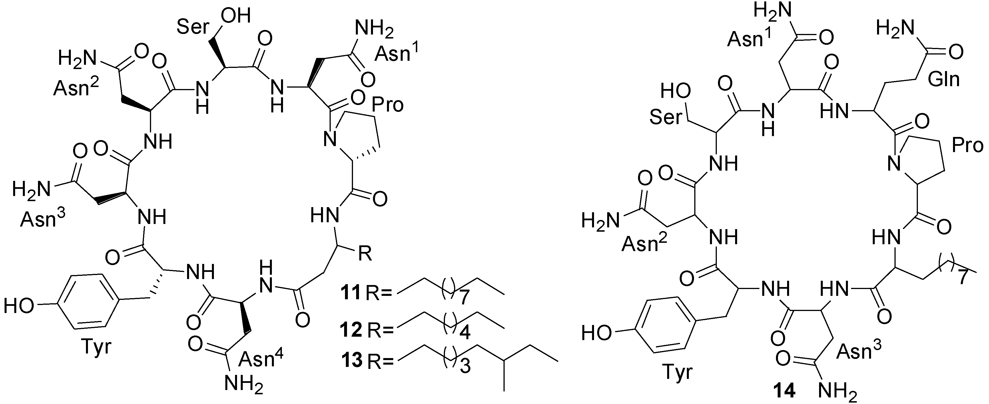
Three new cyclic acylpeptides named mixirins A–C (1113) (Figure 5) belonging to the iturin class have been isolated from marine bacterium Bacillus sp. [44]. This isolate was obtained from sea mud near the Arctic pole. Mixirins A, B, and C inhibited the growth of human colon tumor cells (HCT-116) with IC50 of 0.68, 1.6, 1.3 µg/mL, respectively (Table 1) [44].
A new cyclic lipopeptide, marihysin A (14) (Figure 5) was isolated from the fermentation broth of the marine B. marinus B-9987 isolated from the tissues of rhizophere of Suaeda salsa in the intertidal zone of the Bohai Bay, China, which exhibited a broad-spectrum activity against plant pathogens with minimum inhibitory concentrations (MICs) of 100–200 µg/mL [49].
Figure 5. Structures of mixirins A–C (1113) and marihysin A (14).
Marinedrugs 11 02846 g005

2.2. Polypeptides

Nonribosomal peptides (NRPs) are synthesized by large multimodular nonribosomal peptide-synthetase (NRPS) by elongation of activated monomers of amino acid building blocks. NRPSs are organized in modules responsible for the incorporation of a specific amino acid in polypeptide chain by three successive steps: adenylation, thiolation, and condensation [50]. Two types of polypeptides are generally produced by Bacillus strains are linear and desipeptides.
Bogorol A (15) (Figure 6), a novel peptide antibiotic has been obtained from the culture broth of a marine Bacillus sp. isolated from a tropical reef habitat in Papua New Guinea [51]. Bogorol A illustrates a new structural template for “cationic peptide antibiotics”, and it showed good activity against methicillin-resistant S. aureus (MRSA) (MIC 2 µg/mL), vancomycin-resistant enterococcus (VRE) (MIC 10 µg/mL) and moderate activity against E. coli (MIC 35 µg/mL), and no activity against S. maltophilia (>200 µg/mL).
Figure 6. Structure of bogorol A (15).
Marinedrugs 11 02846 g006
Two new cyclic depsipeptides, turnagainolides A (16) and B (17) (Figure 7), have been isolated from laboratory cultures of a marine strain RJA2194 [51]. This strain was isolated from a sediment sample collected near Turnagain Island and identified as a Bacillus species. The structures of 16 and 17 are representative of epimers at the site of macrolactonization. Turnagainolide B (17) showed activity in a SHIP1 activation assay [51].
Loloatin B (18) (Figure 7) is a novel cyclic decapeptide antibiotic with potent antimicrobial activity against Gram-positive bacteria produced by a Bacillus sp. which was isolated from the tissues of a marine worm [46]. In screening tests for antimicrobial activity, loloatin B inhibited the growth of MRSA, VRE, and penicillin resistant S. pneumoniae with MICs of 1–2 µg/mL.
Figure 7. Structures of turnagainolides A and B (16 and 17), and loloatin B (18).
Marinedrugs 11 02846 g007
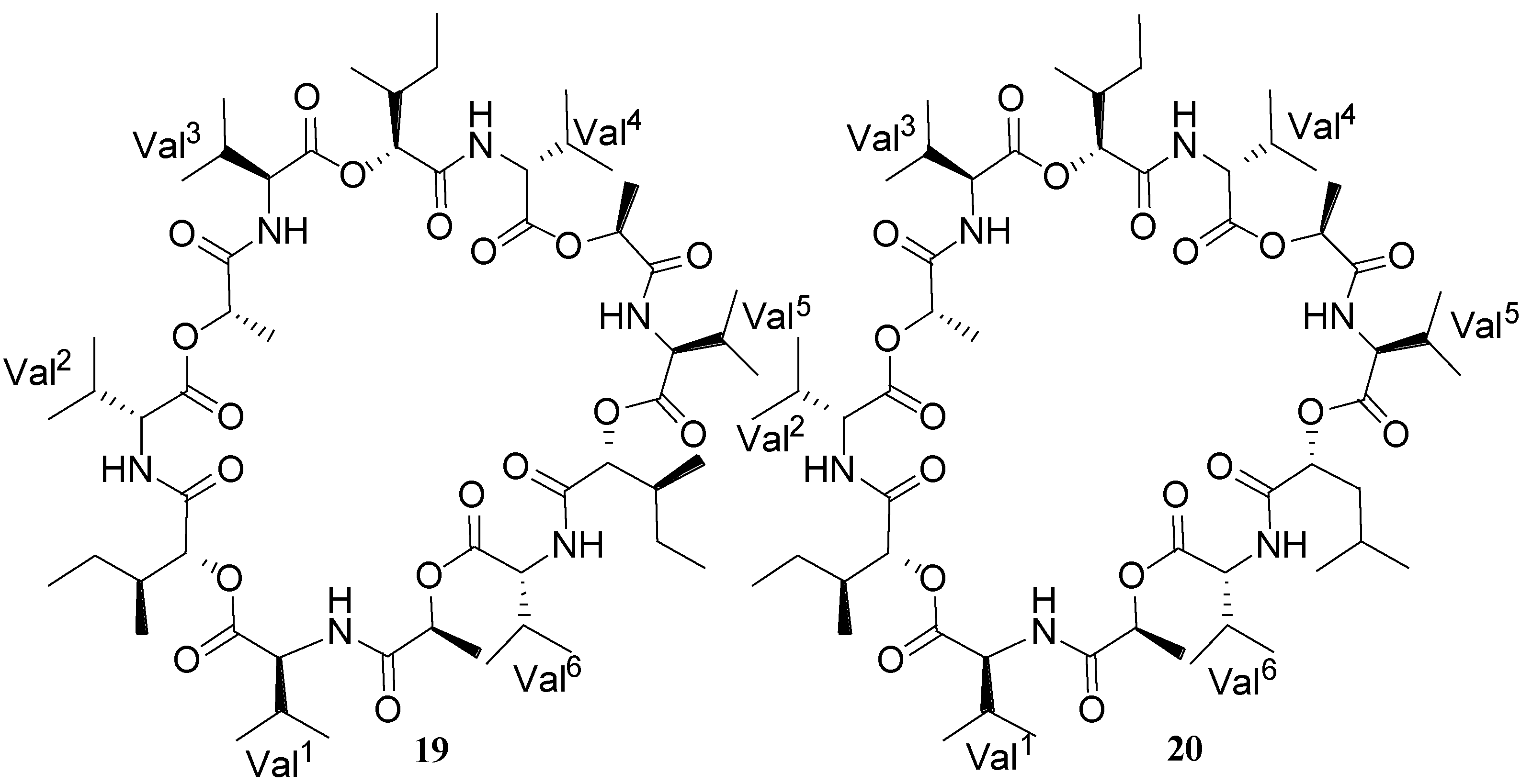
Two new cyclodepsipeptides designated as bacillistatins 1 (19) and 2 (20) (Figure 8) have been isolated from the culture broth of Bacillus silvestris that was obtained from a Pacific Ocean (southern Chile) crab [30]. Each 12-unit cyclodepsipeptide (19 and 20) strongly inhibited the growth of a human cancer cell line panel, with GI50’s of 10−4–10−5 µg/mL, and each compound was determined to be active against antibiotic-resistant S. pneumoniae.
Figure 8. Structures of bacillistatins 1 (19) and 2 (20).
Marinedrugs 11 02846 g008

2.3. Polyketides/Lipoamides

Polyketides are extremely large classes of secondary metabolites that are assembled from simple acyl-coenzyme A and form the basis of numerous pharmaceuticals, agrochemicals, and veterinary agents. Polyketides are biosynthesized by polyfunctional megasynthases organized into repeated functional units known as modules. The modular megaproteins responsible for polyketide biosynthesis are known as polyketide synthases (PKSs) [50,52]. Due to their mechanism of versatile assemblage, the polyketides exhibit remarkable diversity both in terms of structure and biological activities.
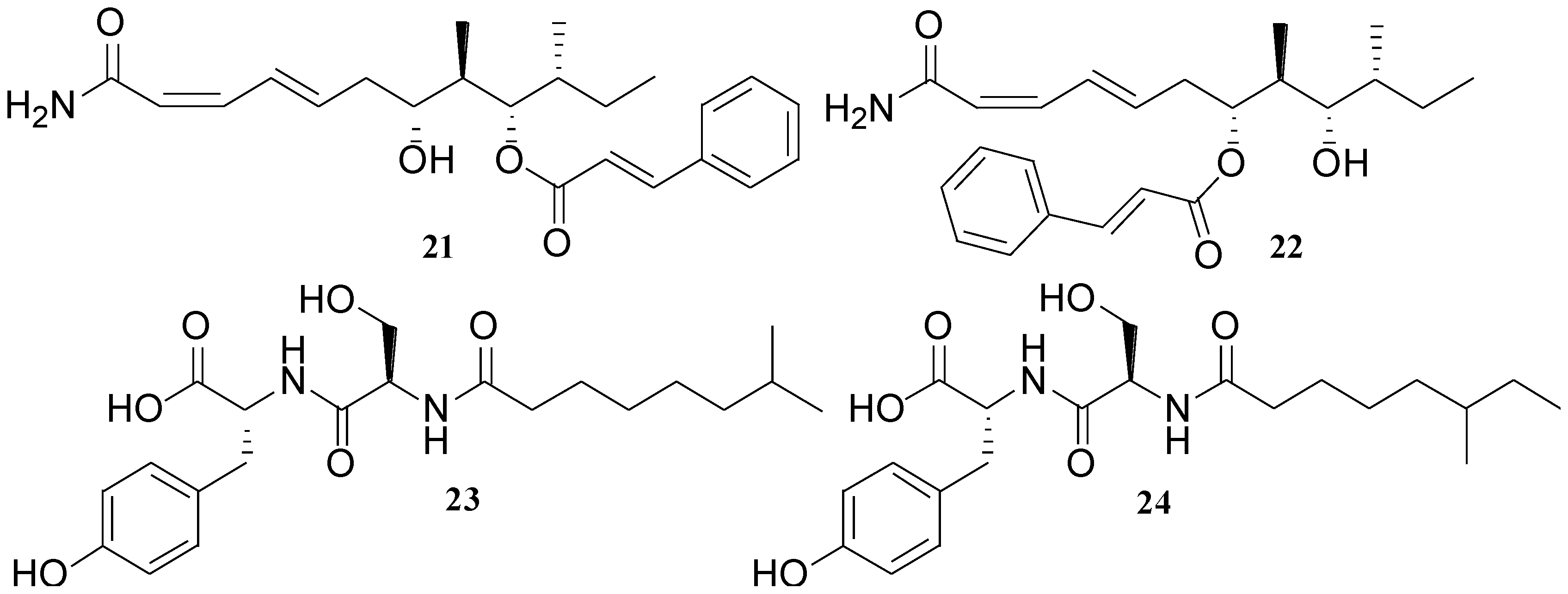
A B. laterosporus isolate, obtained from coastal waters of Papua New Guinea, has been shown to produce some novel metabolites such as basiliskamide A (21), basiliskamide B (22), tupuseleiamide A (23), and tupusleiamide B (24) [32] (Figure 9). Compound 22 was simply a regioisomer of 21, and 24 was a regioisomer of 23. The observed MIC values of 21 and 22 against C. albicans and A. fumigatus were 1.0 and 3.1 µg/mL, and 2.5 and 5.0 µg/mL, respectively.
Figure 9. Structures of basiliskamides (21 and 22) and tupuseleiamides (23 and 24).
Marinedrugs 11 02846 g009
Two unique polyketides, ieodoglucomides A (25) and B (26) (Figure 10) were isolated from a marine-derived bacterium B. licheniformis [53]. This bacterium was isolated from a sediment sample collected from Ieodo in Republic of Korea’s southern reef. Compounds 25 and 26 displayed moderate in vitro antimicrobial activity against both Gram-positive and Gram-negative pathogenic bacteria (MIC 8–32 µg/mL). Furthermore, ieodoglucomide B (26) exhibited cytotoxic activity against lung cancer and stomach cancer cell lines with GI50 values of 25.18 and 17.78 µg/mL, respectively [52].
Figure 10. Structures of ieodoglucomides A and B (25 and 26), and bacillamide (27).
Marinedrugs 11 02846 g010
Bacillamide (27) (Figure 10) was isolated from Bacillus sp. SY-1, which was obtained from seawater during the blooming period of C. polykrikoides in Masan Bay [47]. Bacillamide (27) showed significant algicidal activity against C. polykrikoides (LC50 after 6 h: 3.2 µg/mL) and selective activity against dinoflagellates and raphidophytes. However, 27 showed neither algicidal activity against microalgae of other phyla such as diatom, green algae, and cyanobacteria, nor growth inhibitory effects against bacteria, fungi, or yeast. Therefore, 27 might be considered as a useful algicidal agent for regulating the blooms of harmful dinoflagellate species such as C. polykrikoides.
A novel thiazole alkaloid, neobacillamide A (28) together with three known related bacillamides A–C (2931), were isolated from the bacterium B. atrophaeus, which was associated with the South China Sea sponge Dysidea avara [54] (Figure 11). It is interesting to note that all bacillamides A–C (2931) contain a common tryptamine moiety in their molecules while in 28 the amine portion is replaced by a phenethylamine. Neobacillamide A was the first member of Bacillus thiazole alkaloids, which has been determined to contain a phenethylamine moiety. Compounds 28 and 31 were evaluated for their inhibitory activity against HL60 human leukemia cells and A549 human lung cancer cells but both compounds were found inactive.
Figure 11. Structures of thiazole alkaloids (2831) and lipoamides A–C (3234).
Marinedrugs 11 02846 g011
Three new compounds named lipoamides A–C (3234) (Figure 11) have been isolated from the culture broth of a sediment B. pumilus (SP21) [41]. Lipoamide A (32) showed weak antibacterial activity against S. aureus and P. aeruginosa (MIC > 100 µg/mL).

2.4. Isocoumarins

Isocoumarin-type metabolites from microorganisms are characterized by the amino-containing substituent, which is presumably derived from leucine, at the 3-position in the dihydrocoumarin core. There have been several such isocoumarin compounds as baciphelacin [55], amicoumacins [56,57,58,59], and xenocoumacins [60]. These compounds possess the common chromophore, 3,4-dihydro-8-hydroxyisocoumarin in their structures and many of them are produced by the genus Bacillus.
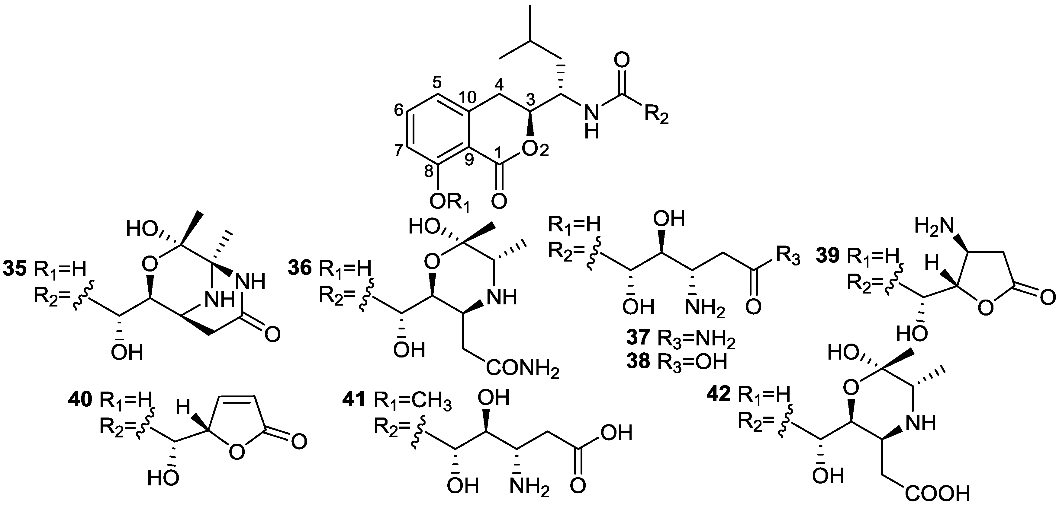
Two novel isocoumarins, bacilosarcins A (35) and B (36), and three known isocoumarins, amicoumacins A (37), B (38), and C (39), were isolated from the culture broth of a marine-derived bacterium B. subtilis TP-B0611 [31] (Figure 12). The strain B0611 was isolated from the intestine of a sardine (S. melanosticta) collected in Toyama Bay in Japan. Compound 35 possesses an unprecedented 3-oxa-6,9-diazabicyclo[3.3.1]nonane ring system, where 36 has a 2-hydroxymorpholine moiety which is rare in nature. Compound 35 showed 82% inhibition at 50 µM against growth of barnyard millet sprouts and on the contrary 36 showed very weak activity at the same concentration. Of particular interest, amicoumacin A (37) showed more potent activity than 35. On the basis of biogenetic consideration, it is reasonable to assume that compound 35 was derived from amicoumacin A (37), forming a biacetyl equivalent C4 unit and two water molecules. This may imply that 35 behaves as a prodrug of 37 in plant cells. The activity levels shown by 35 and 37 are higher than that of herbimycin A, a potent herbicidal compound from Streptomyces [61], suggesting that they may be lead molecules for plant growth regulators.
Figure 12. Structures of isocoumarins (3542).
Marinedrugs 11 02846 g012
Six known analogs (3641), one new bacisarcin C (42) (Figure 12) and four novel amicoumacins, lipoamicoumacins A–D (4346) (Figure 13) were isolated from the culture broth of a marine-derived bacterium, B. subtilis B1779 [62]. The strain B1779 was isolated from a marine sediment sample, which was collected from the Red Sea in April 2010. All isolated compounds were evaluated for cytotoxicity and antibacterial activities. Only compounds 36 and 37, which have an amide functional group (-NH2) exhibited cytotoxicity against HeLa cells with IC50 values of 33.60 and 4.32 µM, respectively, indicating that the amide group of amicoumacin plays a critical role in cytotoxicity. This was further supported by a comparison of cytotoxicity between compounds 36 and 42 and between 37 and 38. A similar antibacterial effect of amide functional group has been shown among amicoumacins [62].
Figure 13. Structures of lipoamicoumacins A–D (4346).
Marinedrugs 11 02846 g013

2.5. Fatty Acids

Bacteria are known to synthesize fatty acids via the classic fatty acid synthase (FAS) pathway with chain length ranging from C12 to C19 [63,64]. Fatty acid from acetyl-CoA and malonyl-CoA precursors through the action of enzymes called fatty acid synthases (FAS).
Methyl-branched fatty acids with methyl groups at even carbon atoms (methyl substituents on carbon 2, 4, 6) occur in several organisms, and originate from the selective incorporation of methylmalonyl-CoA by fatty acid synthases [65]. The iso-anteiso branching in the novel methyl-branched fatty acids is most probably derived from leucine and isoleucine followed by a series of elongations with malonyl-CoA. At either the last or penultimate elongation step methylmalonyl-CoA seems to be selectively incorporated by one of the fatty acid synthesizing enzymes from the bacterium resulting in the methyl-branched fatty acids. Whether this is a known or unknown, methyl-branched fatty acid synthase in bacteria is, as of yet, a matter of speculation.
A series of novel iso-anteiso fatty acids (4752) (Figure 14) with chain lengths between C11 and C19 and an interesting series of linear alkylbenzene fatty acids with chain lengths between C10 and C14 were produced by a halophilic Bacillus sp. [66]. The biological activity of these novel fatty acids has not yet been reported.
Figure 14. Structures of iso-anteiso fatty acids (4752).
Marinedrugs 11 02846 g014
Bioassay-guided isolation of the EtOAc extract of a marine Bacillus sp. 09ID194 cultured in modified Bennett’s broth medium, yielded six new unsaturated hydroxy fatty acids, named ieodomycins A–D (5356) [34] and linieodolides A and B (57 and 58) [67] (Figure 15). The producing strain was isolated from a sediment sample collected from Ieodo, Republic of Korea’s southern reef. Compounds 5358 exhibited antimicrobial activity against B. subtilis and E. coli with MICs of 32–64 µg/mL, but showed weak growth inhibition against the yeast, S. cerevisiae, with an MIC of 128–256 µg/mL.
Figure 15. Structures of unsaturated fatty acids (5358).
Marinedrugs 11 02846 g015

2.6. Macrolactins

The macrolactins are polyene cyclic macrolactones consisting of 24-membered ring lactones with modifications such as the attachment of glucose β-pyranoside, and they may also occur as linear analogs [33]. The macrolactin carbon skeleton contains three separate diene structure elements in a 24-membered lactone ring. The macrolactin class of macrolactones is mainly produced by both terrestrial and marine strains [23].
In the genome of B. amyloliquefaciens FZB42, a plant-root-colonizing environmental strain with the ability to stimulate plant growth and to suppress soil-borne plant pathogens in the rhizosphere [68,69], three PKS operons altogether about 196,340 pb were located at sites approximately 1.4 Mbp (pks2), 1.7 Mbp (pks1), and 2.3 Mbp (pks3) clockwise from the origin of replication in the genome which is 3916 kb in size [70,71]. These three-gene clusters show a modular organization which is typical for type I PKS systems, indicating that the strain FZB42 has the biosynthetic machinery for the production of at least three different kinds of polyketides. pks1 and pks3 have been attributed to the production of bacillaene and difficidin/oxydifficidin, respectively, where the pks2 is involved in macrolactins biosynthesis. The macrolactone rings of macrolactins are formed via cyclization of polyketide chains assembled by PKS type-I enzymes that perform repetitive decarboxylative condensations of carboxylic acids with an activated carboxylic acid starter unit [72].
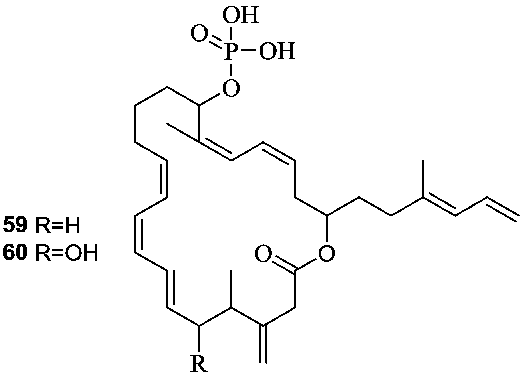
Difficidin (59) and oxidifficidin (60) (Figure 16), detected in the culture broth of B. amyloliquefaciens (FZB42) are highly unsaturated 22-membered macrocyclic polyene lactone phosphate esters with broad-spectrum of antibacterial activity [73]. Difficidin (59) has recently shown promising suppressive activity against the enterobacterium Erwinia amylovara, a devastating plant pathogen which causes necrotrophic fire blight disease affecting apple, pear, and other rosaceous plants [74].
Figure 16. Structures of difficidin (59) and oxidifficidin (60).
Marinedrugs 11 02846 g016
The primary mode of action of difficidin (59) is the inhibition of protein synthesis. It has proven to be highly bactericidal to both growing and stationary phase cultures and inhibited protein synthesis more rapidly than RNA, DNA, or cell-wall synthesis in growing cells [75].
At least 32 macrolactins have been characterized so far including macrolactins A–Z, 7-O-succinyl macrolactin A, 7-O-succinyl macrolactin F, 7-O-malonyl macrolactin A, and three ether-containing macrolactin A. Most of them were produced by marine sediment isolates and few of them were by soil isolates [76,77].
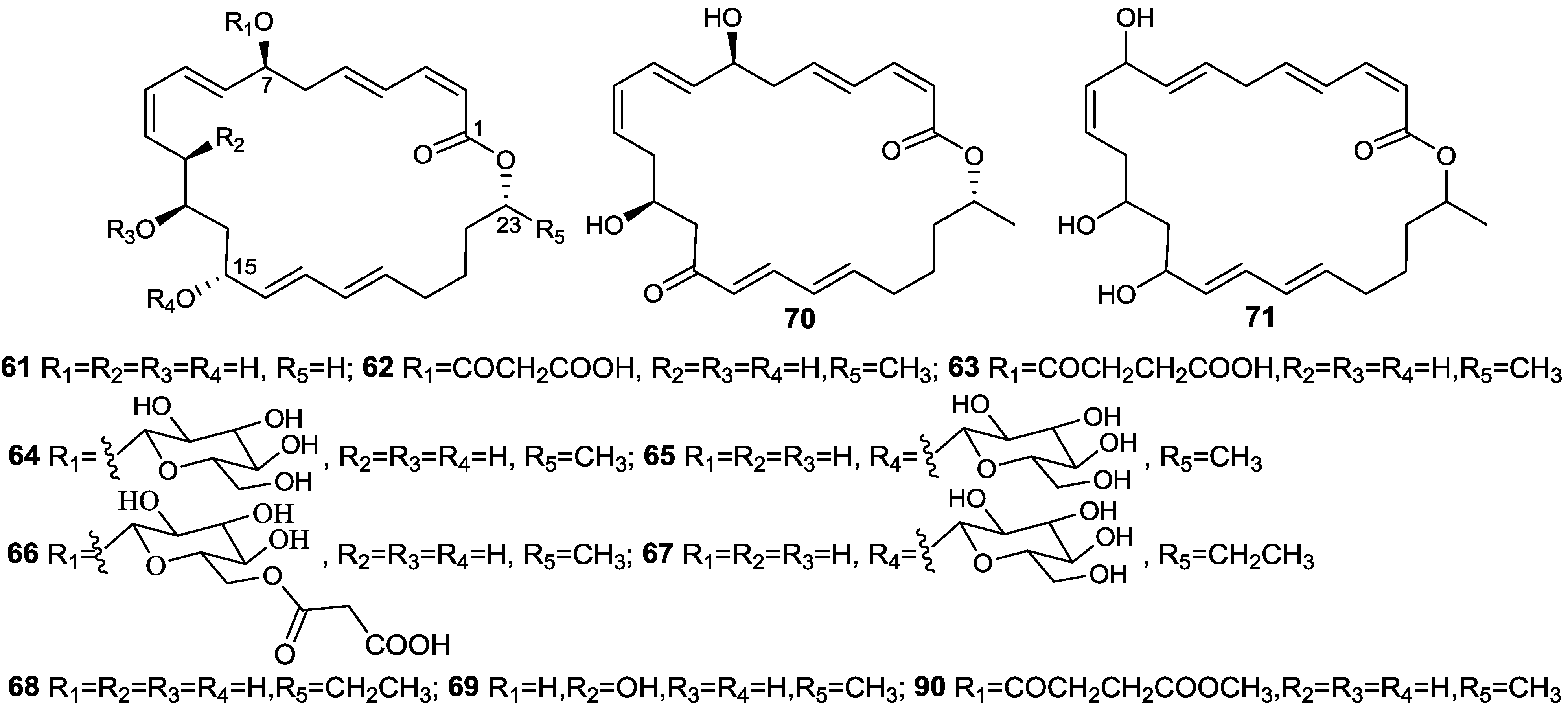
The first macrolactin family, including macrolactins A–F (61, 6466, 70, 74) (Figure 17 and Figure 18), as well as the open-chain macrolactinic and isomacrolactinic acid, were isolated from an unclassified deep-sea sediment bacterium [33]. Macrolactin A was of particular interest because of its biological activities. It shows selective antibacterial activity against S. aureus and B. subtilis at a concentration of 5 and 20 µg/disc, respectively, as well as the ability to inhibit B16-F10 murine melanoma cancer cells in in vitro assays, as well as mammalian Herpes simplex viruses. In addition, it protects lymphoblast cells against HIV by inhibiting virus replication [33].
Figure 17. Structures of 24-membered macrolactin A (61) and its derivatives (6271 and 90).
Marinedrugs 11 02846 g017
Figure 18. Structures of 22- (80) and 24-membered (81 and 82) including keto (83) macrolactins.
Marinedrugs 11 02846 g018
Figure 19. Structures of bicyclic (84) and polyene (85) macrolactins, and 12-hydroxy macrolactin A (86).
Marinedrugs 11 02846 g019
Macrolactins G–M (71, 8084, 68) (Figure 17, Figure 18, Figure 19) were produced by a Bacillus sp. PP19-H3, which was isolated from a marine macroalga, Schizymenia dubyi, collected at the Omaezaki coast of Shizuoka prefecture in Japan [78]. These macrolactins include a 22-membered ring (80) or bicyclic lactone (84) in addition to their geometric isomers of macrolactins A and F. These macrolactins were more active against S. aureus (MIC 5–10 µg/mL) than B. subtilis (MIC 60 µg/mL), whereas macrolactin K (83), containing keto group (C=O) at C-15 showed very weak antibacterial activity against both tested pathogens (MIC > 100 µg/mL), indicating that the -OH group at C-15 may play an important role in the antibacterial activity of macrolactins [78].
With a view to find out peptide deformylase inhibitors, four glycosylated macrolactins O–R (73, 67, 77, and 79) (Figure 17, Figure 20 and Figure 21) were isolated from the liquid cultures of Bacillus sp. AH159-1 [72]. Macrolactins O–R inhibited S. aureus peptide deformylase (PDF) in dose-dependent manners with IC50 (µM) values of 53.5, 57.7, 12.1, and 61.5, respectively. All these compounds also inhibited bacterial growth against E. coli with an MIC of 100 µg/mL [77]. Macrolactin N (72) (Figure 18) also inhibited S. aureus’s peptide deformylase (PDF) in a similar fashion to macrolactins O–R [79].
Figure 20. Structures of keto (7275) and ester (76) macrolactins.
Marinedrugs 11 02846 g020
Figure 21. Structures of glycosylated (77 and 79) and a 24-membered (78) macrolactins.
Marinedrugs 11 02846 g021
A new macrolactin T (78) and a new polyene δ-lactone macrolactin U (85) (Figure 19 and Figure 21), along with known macrolactins A, B, D, O and S (69) were isolated from the culture broth of the bacterium B. marinus, which was isolated from Suaeda salsa collected on the coastline of the Bohai Sea of China [80]. Macrolactins T and B exhibited MIC activity toward A. solani, P. oryzae, and S. aureus, at concentrations of 0.8, 2.8, 5.5 and 7.5, 20.1, 4.5, respectively [80].
Macrolactins V (86) and S (69) (Figure 17 and Figure 21) were isolated from the culture broth of a marine bacterium, B. amyloliquefaciens SCSIO 00856, which was isolated from a South China Sea gorgonian, Junceella juncea [48]. Macrolactin V (86) exhibited potent antibacterial activity against E. coli, B. subtilis and S. aureus with an MIC value of 0.1 µg/mL, and no activity against B. thuringiensis. Macrolactin S (69) showed strong antibacterial activity against both E. coli and S. aureus with MIC values of 0.3 and 0.1 µg/mL, respectively, but weak activity against B. subtilis (MIC 100 µg/mL), which indicated that the configuration of 7-OH may affect the antibacterial activity of the epimers 86 and 69 [48].
7-O-succinyl macrolactin A (63) and 7-O-succinyl macrolactin F (75) (Figure 17 and Figure 18) were isolated from a marine sediment Bacillus sp. Sc026 [81], and 7-O-malonyl macrolactin A (62) (Figure 17) from a B. subtilis [82]. The bacterium (Sc026) was isolated from marine sediment collected from nearby Sichang Island (at a 15-m depth), Chonburi, Thailand. 63 and 75 were tested in an agar diffusion assay at concentration of 50 and 100 µg/disk, respectively. The inhibition zones of 63 and 75 against B. subtilis and S. aureus were 10 and 9, and 24 and 8 mm, respectively. The minimum restrictive concentrations (MRCs) of 62 were between 1 and 64 µg/mL for S. aureus and MRSA strains, and between 0.06 and 4 µg/mL for E. faecalis and clinical isolates, VRAS (vancomycin-resistant/ampicillin-sensitive) E305 and VRAR (vancomycin-resistant/ampicillin-resistant) E315 [82].
Macrolactins W (76), Y (91), Z (92), X (90), macrolactinic acid (93) and three ether-containing unique macrolactins (8789) (Figure 17, Figure 18, Figure 22, and Figure 23) were isolated from a sediment Bacillus sp. 09ID194 [67,83,84]. Interestingly, these compounds were produced by this strain only in low salinity culture medium (12 g/L) but not in high salinity culture medium (32 g/L). These compounds showed MIC against pathogenic bacteria at a concentration range of 8–64 µg/mL. The position of ether group was important for the antimicrobial activity of these ether-containing macrolactins (8789) [84].
Figure 22. Structures of ether-containing macrolactins (8789).
Marinedrugs 11 02846 g022
Figure 23. Structures of ether (91) and methoxy (92) containing macrolactins and macrolactinic acid (93).
Marinedrugs 11 02846 g023

3. Detoxification of Heavy Metals

Chromium (Cr) is one of the major causes of environmental contamination by heavy metals. The toxicity of chromium (Cr) lies in its oxidative states [85]. The higher oxidation states impose more toxicity (10–100 times) than the lower oxidation states [86]. Nutritionally, small amount of Cr(III) is essential for balanced human and animal diet for preventing adverse effects caused by glucose and lipid metabolisms [87,88]. Swallowing large amounts of Cr(III) may pose threats to human health [89,90]. Hexavalent Cr (Cr6+) is extremely carcinogen and may be the cause of death after ingestion of a large dose [91]. The tolerable intake level of Cr(VI) and Cr(III) are 1 and 8 µg/L for freshwater life, 1 and 50 µg /L for marine life, and 8 and 5 µg/L for irrigation water, respectively [92,93].
A marine isolate, B. licheniformis, can reduce 10–500 mg/L of Cr(VI) to Cr(III) within 24–72 h in liquid medium [94]. When this strain was cultured in the liquid medium, it produces chromium reductase indicating that probably Cr(VI) may be reduced to Cr(III) by enzymatic activity. This strain also secretes an extracellular surface-active agent (biosurfactant) in the medium, which provides tolerance to the cells towards hexavalent chromium and protects the cells from oxidative stress.
The oxidation of soluble manganese(II) to insoluble Mn(III, IV) oxide plays an important role in removing environmental heavy metal hazards. These Mn oxides oxidize many organic and inorganic compounds, and to scavenge a variety of other metals on their highly charged surfaces. In addition to catalyzing important process, microorganisms capable of Mn(II) oxidation are potential candidates for the removal, detoxification, and recovery of metals from the environment. Mature spores of a marine Bacillus sp. strain SG1 oxidize Mn(II) to MnO2 [95]. Vegetative cells of the same strain reduce MnO2 under low-oxygen conditions. The rate of MnO2 reduction was a function of cell density.
Although Bacillus strains play a central role in biogeochemical recycling of metals in marine environment the molecular and biochemical mechanisms for most of these recycling processes are still poorly understood. It is assumed that most of the heavy metals in the environment are detoxified by reduction process. However, it is important to recognize that biochemical recycling of heavy metals is a new field and is largely limited to studies of microbial species. To remediate a range of heavy metals, it is important to select microbial strains, which carry inherently genetic machinery to reduce multiple metals. The concentration of heavy metals (Cd, Co, Cr, Hg, Pb) around hydrothermal vents is high [96]. Therefore, Bacillus strains living around hydrothermal vents may capable of recycling a wide range of heavy metals. The Bacillus strains isolated from “hydrothermal vents” should therefore be good candidates for the detoxification of environmental pollution caused by toxic heavy metals.

4. Marine Bacillus Strains as Potential Biocontrol Agents

Most of the microorganism-based biopesticides have been developed from terrestrial bacteria. Only a few fungi have been used as efficient biocontrol agents [97]. Presently, about half of the commercially available bacterial biocontrol agents are prepared from Bacillus strains [12] and among them, B. thuringiensis accounts for more than 70% of total sales. This bacterium, omnipresent in marine environment [98], produces two proteins named Cry and Cyt during its sporulation phase, each of which is highly toxic to insects, but not to mammals or to the environment. These toxic proteins kill insects by forming pores in the gut walls. Major insect families, which can be controlled by Cry/Cyt toxins include Coleoptera, Lepidoptera, and Diptera. A wide range of plant diseases (root rot, leaf spot, anthracnose, gray mold, early blight, late blight, powdery mildew, downy mildew, and bacterial spot) can also be controlled successfully by the antagonistic Bacillus isolates [99].
The genus Bacillus is present in every niche of terrestrial and marine environments, even in the hot springs [100]. Many Bacillus species can also be found in both terrestrial and marine environments. This bacterial genus could be considered as one of the major sources of potential microbial biopesticides because of its broad genetic biodiversity [22,36], its large body of literary evidence, as well as generally regarded as safe by the US Food and Drug Administration (USFDA) and capable of withstanding unfavorable conditions through the formation of resistant spores. The main specific mechanisms involved in biocontrol of plant diseases by this bacterial genus include: competition for ecological niche/substrate in the rhizosphere, production of inhibitory chemicals, and induction of so-called systemic resistance in host plants. Marine Bacilli appear to be more effective biocontrol agents, compared to their terrestrial counterparts [101].
And so, the question remains as to whether marine or terrestrial Bacillus strains will be better candidates as biocontrol agents. Marine environments (with a wide variation in temperature, pressure, salt concentration and pH) are different from the terrestrial ones. The active strain, with high resistance to salt, heat, pH and stress, can be used to prepare a biological pesticide. The marine strains carry these properties naturally [15]. Marine Bacilli forms resistant spores quickly in unfavorable conditions, and is easily converted to powder formulations, having longer shelf-life when compared to other products containing living organisms, and can be prepared commercially at a relatively low cost (relatively unspecialized culture procedures). However, these products have some limitations due to partial protection against pathogen and pests, inconsistent effects, and a lack of ecological knowledge, which warrants precaution in field applications. There are several approaches towards the improvement of biopesticide efficacy, including selection of suitable strains, combination of synergistic strains, combinations with other forms of biopesticides, chemical pesticides, plant fertilization and agricultural practices, as well as suitable formulation and application methods [11,12].

5. Marine Bacillus Isolates as a Potential Source of Natural Carotenoids
Carotenoids are yellow, orange, and red pigments, which are widely distributed in plants and microorganisms (photosynthetic organisms, bacteria, and fungi). Chemically carotenoids usually contain a polyene C40 carbon skeleton, and can be acyclic, or cyclic groups, may be present at one or both ends of the backbone and having many derivatives [102]. Plants, algae, and fungi produce carotenoids containing C40 carbon backbone, whereas bacteria can produce a diverse range of carotenoids with either C40 or C30 carbon backbone. Each double bond in the polyene chain of a carotenoid may exist in two forms: trans or cis. Carotenoids obtained from natural sources are predominantly or entirely in trans form. Carotenoids are widely used as antioxidants, pro-vitamin A, and food and feed additives. Presently, synthetic carotenoids are meeting the bulk of market demands, but due to the “green wave”, coined to represent changes in consumers, industries are now looking for natural sources of carotenoids. Currently, there are no feasible sources of natural carotenoids including widely screened terrestrial microgranisns [102]. The recent screening of marine bacteria indicated that they are a promising source of natural carotenoids [103]. So far, more than 600 different carotenoids have been identified from natural sources, of which only 24 are available in human foodstuffs. The most widely used carotenoids as food are β-carotene, β-cryptoxanthin, lycopene, lutein, and violaxanthin. Carotenoids contain isoprene skeleton and are biosynthesized by tail-to-tail linkage of two C20 geranylgeranyl diphosphate molecules.
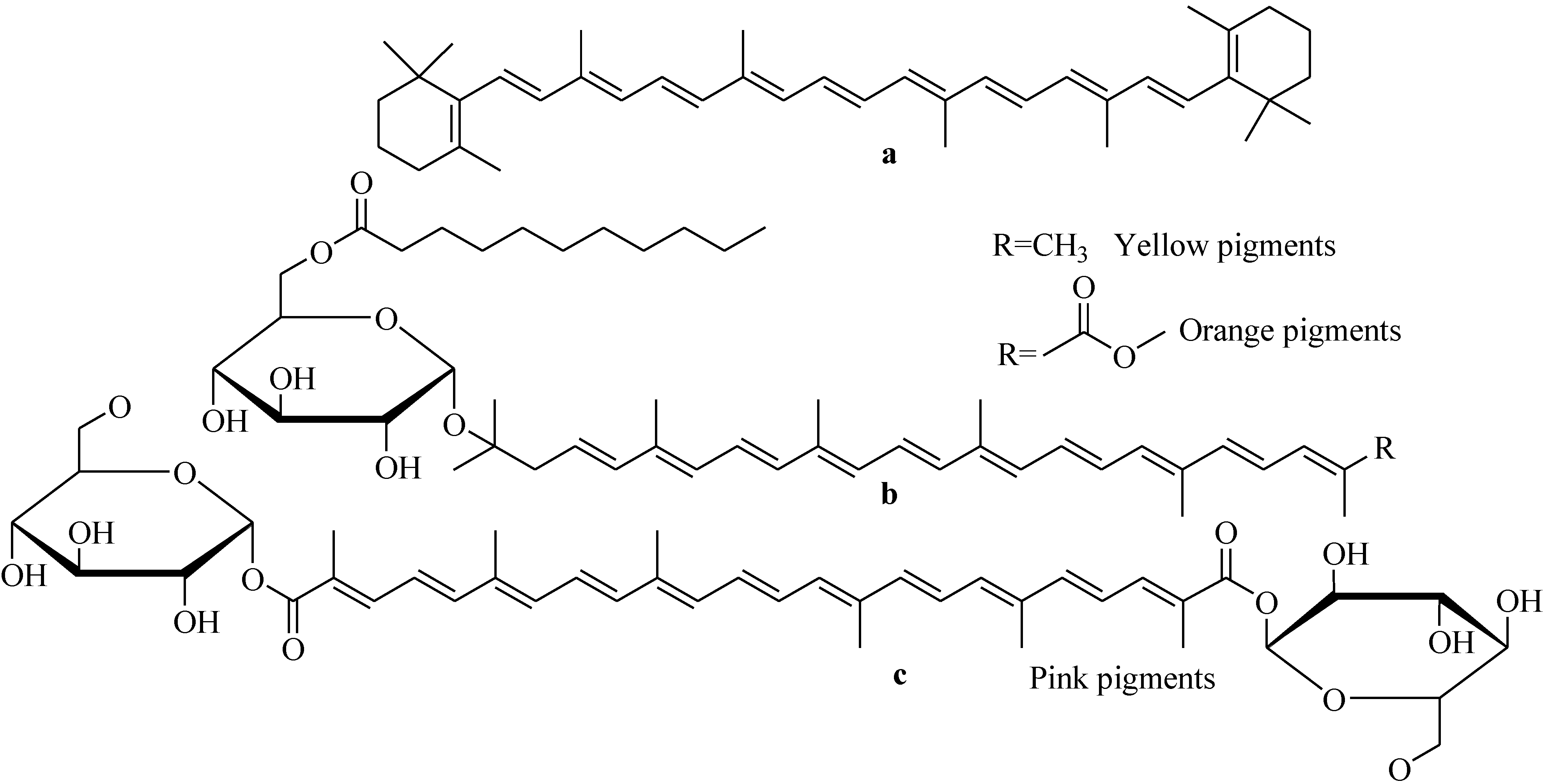
Irrespective of their sources, the effectiveness of carotenoinds largely depends on their bioavailablity. Due to unusual chemical structures of bacterial carotenoids, questions remained to be address bioavailability of these molecules in humans. Glycosylated carotenoids (Figure 24) isolated from marine spore-forming strains, B. indicus HU36 and B. firmus GB1, showed better bioavailability (about 4.5 times as high) than that of pure β-carotene in vitro digestion experiments [104]. All the marine isolates are not able to produce carotenoids. Marine yellow and orange spore-forming Bacillus strains have been shown to produce carotenoinds [105]. Photosynthetic marine organisms (algae and plants), either independently or in symbiosis with microorganisms, produce carotenoids to prevent oxidation by sunlight [106]. Marine yellow, red, and orange spore-forming Bacillus strains as well as strains living in photosynthetic organisms may be a good source of carotenoids useful for human.
Figure 24. Structures of β-carotene (a) and main carotenoids synthesised by (b) HU36 and (c) GB1 spore-forming strains.
Marinedrugs 11 02846 g024

6. Conclusions and Future Perspectives

Marine Bacillus species represent a rich source of structurally diverse classes of secondary metabolites including lipopeptides, polypeptides, macrolactones, fatty acids, polyketides, lipoamides, isocoumarins, and carotenoids. These structurally versatile natural products of marine isolates are derived from complex biosynthetic pathways. Some of these bioactive compounds have high potentials for the development of effective pharmaceutical and agrochemical products. Due to having genetic capability to adapt extreme conditions, Bacillus strains isolated from unique niches of environments (e.g., hydrothermal vent, deep sea, pH > 9.0 and salt lakes) may produce useful bioactive compounds [8]. The silent cryptic biosynthetic gene clusters of marine isolates may be activated with a view to discover new natural products by culturing them under varying stressful conditions (e.g., nutrient, pH, salinity or temperature stresses). Another important feature of the genus Bacillus is that they can detoxify heavy metals through reduction processes, which might be considered as candidates for the bioremediation of heavy metal toxicity. Bioremediation is an eco-friendly and cost-effective strategy for eliminating xenobiotic compounds from polluted environments. Next-generation sequencing is providing crucial insights in the molecular and biological mechanisms involved in bioremediation of environmental pollutants like heavy metal contaminations. These insights will improve bacterial bioremediation strategies, monitoring their progress, and determining their success [107]. Bacillus-based biopesticides can improve plant health through unique modes of action, and thus, have a high potential for commercial applications. The frequent occurrence of B. subtilis in the natural environment and the production capability of a vast array of antibiotics must be considered for application in pest management. Marine Bacillus-based biopesticides have a great potential in sustainable agricultural practices in the future. To meet the consumers demand for fully natural practices and reduced environmental hazard, marine yellow and orange spore-forming Bacillus strains, as well as those which are symbiotic with photosynthetic marine organisms may be a good source of natural carotenoids.

Acknowledgments

This research was supported in part by Korea Institute of Ocean Science and Technology (Grants PE99121 to H. J. S.). The author (MTI) is thankful to the World Bank for funding a sub-project CP # 2071 in the Department of Biotechnology of BSMR Agricultural University, Bangladesh under Higher Education Quality Enhancement Project (HEQEP) of University Grants Commission of Bangladesh.

Conflict of Interest

The authors declare no conflict of interest.

References

  1. Lebar, M.D.; Heimbegner, J.L.; Baker, B.J. Cold-water marine natural products. Nat. Prod. Rep. 2007, 24, 774–797. [Google Scholar] [CrossRef]
  2. Fenical, W. Chemical studies of marine bacteria: Developing a new resource. Chem. Rev. 1993, 93, 1673–1683. [Google Scholar] [CrossRef]
  3. Laatsch, H. Marine Bacterial Metabolites. In Frontiers in Marine Biotechnology; Proksch, P., Muller, W.E.G., Eds.; Horizon Bioscience: Norfolk, UK, 2006; pp. 225–288. [Google Scholar]
  4. Schwartsmann, G.; da Rocha, A.B.; Berlinck, R.G.S.; Jimeno, J. Marine organisms as a source of new anticancer agents. Lancet Oncol. 2001, 2, 221–225. [Google Scholar] [CrossRef]
  5. Jha, R.K.; Zi-rong, X. Biomedical compounds from marine organisms. Mar. Drugs 2004, 2, 123–146. [Google Scholar] [CrossRef]
  6. Nathan, C. Antibiotics at the crossroads. Nature 2004, 431, 899–902. [Google Scholar] [CrossRef]
  7. Von Nussbaum, F.; Brands, M.; Hinzen, B.; Weigand, S.; Häbich, D. Antibacterial natural products in medicinal chemistry—Exodus or revival. Angew. Chem. Int. Ed. 2006, 45, 5072–5129. [Google Scholar]
  8. Li, J.W.-H.; Vederas, J.C. Drug discovery and natural products: End of an era or an endless frontier? Science 2009, 325, 161–165. [Google Scholar] [CrossRef]
  9. Islam, M.T.; von Tiedemann, A.; Laatsch, H. Protein kinase C is likely to be involved in zoosporogenesis and maintenance of flagellar motility in the peronosporomycete zoospores. Mol. Plant Microbe Interact. 2011, 24, 938–947. [Google Scholar] [CrossRef]
  10. Islam, M.T.; Hashidoko, Y.; Deora, A.; Ito, T.; Tahara, S. Suppression of damping-off disease in host plants by the rhizoplane bacterium Lysobacter sp. Strain SB-K88 is linked to plant colonization and antibiosis against soilborne peronosporomycetes. Appl. Environ. Microbiol. 2005, 71, 3786–3796. [Google Scholar] [CrossRef]
  11. Islam, M.T. Potentials for Biological Control of Plant Disease by Lysobacter spp., with Special Reference to Strain SB-K88. In Bacteria in Agrobiology: Plant Growth Responses; Maheshwari, D.K., Ed.; Springer-Verlag: Berlin/Heidelberg, Germany, 2011; pp. 335–364. [Google Scholar]
  12. Islam, M.T.; Hossain, M.M. Biological Control of Peronosporomycete Phytopathogens by Antagonistic Bacteria. In Bacteria in Agrobiology: Plant Disease Management; Maheshwari, D.K., Ed.; Springer-Verlag: Berlin/Heidelberg, Germany, 2013; pp. 167–218. [Google Scholar]
  13. Whitman, W.B.; Coleman, D.C.; Wiebe, W.J. Prokaryotes: The unseen majority. Proc. Natl. Acad. Sci.USA 1998, 95, 6578–6583. [Google Scholar] [CrossRef]
  14. Kennedy, J.; Marchest, J.R.; Dobson, A.D.W. Marine metagenomics: Strategies for the discovery of novel enzymes with biotechnological applications from marine environments. Microb. Cell Fact. 2008, 7, 27–28. [Google Scholar] [CrossRef]
  15. Rampelotto, P.H. Resistance of microorganisms to extreme environmental conditions and its contribution to astrobiology. Sustainability 2010, 2, 1602–1623. [Google Scholar] [CrossRef]
  16. Sayem, S.M.A.; Manzo, E.; Ciavatta, L.; Tramice, A.; Cordone, A.; Zanfardino, A.; de Felice, M.; Varcamonti, M. Anti-biofilm activity of an exopolysaccharide from a sponge-associated strain of Bacillus licheniformis. Microb. Cell Fact. 2011, 10, 74. [Google Scholar] [CrossRef]
  17. Paul, V.J.; Arthur, K.E.; Williams, R.R.; Ross, C.; Sharp, K. Chemical defenses: From compounds to communities. Biol. Bull. 2007, 213, 226–251. [Google Scholar] [CrossRef]
  18. Jensen, P.R.; Fenical, W. Strategies for the discovery of secondary metabolites from marine bacteria: Ecological perspectives. Annu. Rev. Microbiol. 1994, 48, 559–584. [Google Scholar] [CrossRef]
  19. Feling, R.H.; Buchanan, G.O.; Mincer, T.J.; Kauffman, C.A.; Jensen, P.R.; Fenical, W. Salinosporamide A: A highly cytotoxic proteasome inhibitor from a novel microbial source, a marine bacterium of the new genus salinospora. Angew. Chem. Int. Ed. Engl. 2003, 20, 355–357. [Google Scholar]
  20. Chen, X.H.; Koumoutsi, A.; Scholz, R.; Eisenreich, A.; Schneider, K.; Heinemeyer, I.; Morgenstern, B.; Voss, B.; Hess, W.R.; Reva, O.; et al. Comparative analysis of the complete genome sequence of the plant growth—Promoting bacterium Bacillus amyloliquefaciens FZB42. Nat. Biotechnol. 2007, 25, 1007–1014. [Google Scholar] [CrossRef]
  21. Kunst, F.; Ogasawara, N.; Moszer, I.; Albertini, A.M.; Alloni, G.; Azevedo, V.; Bertero, M.G.; Bessières, P.; Bolotin, A.; Borchert, S.; et al. The complete genome sequence of the Gram-positive bacterium Bacillus subtilis. Nature 1997, 390, 249–256. [Google Scholar] [CrossRef]
  22. Fan, L.; Bo, S.; Chen, H.; Ye, W.; Kleinschmidt, K.; Baumann, H.I.; Imhoff, J.F.; Kleine, M.; Cai, D. Genome Sequence of Bacillus subtilis subsp. spizizenii gtP20b, isolated from the Indian Ocean. J. Bacteriol. 2011, 193, 1276–1277. [Google Scholar] [CrossRef]
  23. Hamdache, A.; Lamarti, A.; Aleu, J.; Collado, I.G. Non-peptide metabolites from the genus Bacillus. J. Nat. Prod. 2011, 74, 893–899. [Google Scholar] [CrossRef]
  24. Baruzzi, F.; Quintieri, L.; Morea, M.; Caputo, L. Antimicrobial Compounds Produced by Bacillus spp. and Applications in Food. In Science against Microbial Pathogens: Communicating Current Research and Technological Advances; Vilas, A.M., Ed.; Formatex: Badajoz, Spain, 2011; pp. 1102–1111. [Google Scholar]
  25. Shoda, M. Bacterial control of plant diseases. Biosci. Bioeng. 2000, 89, 515–521. [Google Scholar] [CrossRef]
  26. Stein, T. Bacillus subtilis antibiotics: Structures, syntheses and specific functions. Mol. Microbiol. 2005, 56, 845–857. [Google Scholar]
  27. Dash, H.R.; Neelam, M.; Chakraborty, J.; Kumari, S.; Das, S. Marine bacteria: Potential candidates for enhanced bioremediation. Appl. Microbiol. Biotechnol. 2013, 97, 561–571. [Google Scholar] [CrossRef]
  28. Nies, D.H. Microbial heavy metal resistance. Appl. Microbiol. Biotechnol. 1999, 51, 730–750. [Google Scholar] [CrossRef]
  29. Trischman, J.A.; Jensen, P.R.; Fenical, W. Halobacillin: A cytotoxic cyclic acylpeptide of the iturin class produced by a marine Bacillus. Tetrahedron Lett. 1994, 35, 5571–5574. [Google Scholar] [CrossRef]
  30. Pettit, G.R.; Knight, J.C.; Herald, D.L.; Pettit, R.K.; Hogan, F.; Mukku, V.J.R.V.; Hamblin, J.S.; Dodson, M.J., II; Chapuis, J.C. Antineoplastic agents. 570. Isolation and structure elucidation of bacillistatins 1 and 2 from a marine Bacillus silvestris. J. Nat. Prod. 2009, 72, 366–371. [Google Scholar] [CrossRef]
  31. Azumi, M.; Ogawa, K.; Fujita, T.; Takeshita, M.; Furumai, T.; Igarashi, Y.; Yoshida, R. Bacilosarcins A and B, novel bioactive isocoumarins with unusual heterocyclic cores from the marine-derived bacterium Bacillus subtilis. Tetrahedron 2008, 64, 6420–6425. [Google Scholar] [CrossRef]
  32. Barsby, T.; Kelly, M.T.; Andersen, R.J. Tupuseleiamides and basiliskamides, new acyldipeptides and antifungal polyketides produced in culture by a Bacillus laterosporus isolate obtained from a tropical marine habitat. J. Nat. Prod. 2002, 65, 1447–1451. [Google Scholar] [CrossRef]
  33. Gustafson, K.; Roman, M.; Fenical, W. The macrolactins, a novel class of antiviral and cytotoxic macrolides from a deep-sea marine bacterium. J. Am. Chem. Soc. 1989, 111, 7519–7524. [Google Scholar] [CrossRef]
  34. Mondol, M.A.M.; Kim, J.H.; Lee, M.A.; Tareq, F.S.; Lee, H.S.; Lee, Y.J.; Shin, H.J. Ieodomycins A–D, antimicrobial fatty acids from a marine Bacillus sp. J. Nat. Prod. 2011, 74, 1606–1612. [Google Scholar] [CrossRef]
  35. Damalas, C.A.; Eleftherohorinos, I.G. Pesticide exposure, safety issues, and risk assessment indicators. Int. J. Environ. Res. Public Health. 2011, 8, 1402–1419. [Google Scholar] [CrossRef]
  36. Ongena, M.; Jacques, P. Bacillus lipopeptides: Versatile weapons for plant disease biocontrol. Trends Microbiol. 2008, 16, 115–125. [Google Scholar]
  37. Raaijmakers, J.M.; de Bruijin, I.; de Kock, M.J. Cyclic lipopeptide production by plant-associated Pseudomonas spp.: Diversity, activity, biosynthesis, and regulation. Mol. Plant Microbe Interact. 2006, 19, 699–710. [Google Scholar] [CrossRef]
  38. Arguelles-Arias, A.; Ongena, M.; Halimi, B.; Lara, Y.; Brans, A.; Joris, B.; Fickers, P. Bacillus amyloliquefaciens GA1 as a source of potent antibiotics and other secondary metabolites for biocontrol of plant pathogens. Microb. Cell Fact. 2009, 8. [Google Scholar] [CrossRef]
  39. Malfanova, N.; Franzil, L.; Lugtenberg, B.; Chebotar, V.; Ongena, M. Cyclic lipopeptide profile of the plant-beneficial endophytic bacterium Bacillus subtilis HC8. Arch. Microbiol. 2012, 194, 893–899. [Google Scholar] [CrossRef]
  40. Tsuge, K.; Akiyama, T.; Shoda, M. Cloning, sequencing, and characterization of the iturin A operon. J. Bacterial. 2001, 183, 6265–6273. [Google Scholar] [CrossRef]
  41. Berrue, F.; Ibrahim, A.; Boland, P.; Kerr, R.G. Newly isolated marine Bacillus pumilus (SP21): A source of novel lipoamides and other antimicrobial agents. Pure Appl. Chem. 2009, 81, 1027–1031. [Google Scholar] [CrossRef]
  42. Cooper, D.G.; MacDonald, C.R.; Duff, S.J.B.; Kosaric, N. Enhanced production of surfactin from Bacillus subtilis by continuous product removal and metal cation additions. Appl. Environ. Microbiol. 1981, 42, 408–412. [Google Scholar]
  43. Ma, Z.; Wang, N.; Hu, J.; Wang, S. Isolation and characterization of a new iturinic lipopeptide, mojavensin A produced by a marine-derived bacterium Bacillus mojavensis. J. Antibiot. 2012, 65, 317–322. [Google Scholar] [CrossRef]
  44. Zhang, H.L.; Hua, H.M.; Pei, Y.H.; Yao, S. Three new cytotoxic cyclic acylpeptides from marine Bacillus sp. Chem. Pharm. Bull. 2004, 52, 1029–1030. [Google Scholar] [CrossRef]
  45. Barsby, T.; Kelly, M.T.; Gagne, S.M.; Andersen, R.J. Bogorol A produced in culture by a marine Bacillus sp. reveals a novel template for cationic peptide antibiotics. Org. Lett. 2001, 3, 437–440. [Google Scholar] [CrossRef]
  46. Gerard, J.; Haden, P.; Kelly, M.T.; Andersen, R.J. Loloatin B, cyclic decapeptide antibiotic, produced in culture by a tropical marine bacterium. Tetrahedron Lett. 1996, 37, 7201–7294. [Google Scholar] [CrossRef]
  47. Jeong, S.Y.; Ishida, K.; Ito, Y.; Okada, S.; Murakami, M. Bacillamide, a novel algicide from the marine bacterium, Bacillus sp. SY-1, against the harmful dinoflagellate, Cochlodinium polykrikoides. Tetrahedron Lett. 2003, 44, 8005–8007. [Google Scholar] [CrossRef]
  48. Gao, C.-H.; Tian, X.-P.; Qi, S.-H.; Luo, X.-M.; Wang, P.; Zhang, S. Antibacterial and antilarval compounds from marine gorgonian-associated bacterium Bacillus amyloliquefaciens SCSIO 00856. J. Antibiot. 2010, 63, 191–193. [Google Scholar] [CrossRef]
  49. Liu, R.F.; Zhang, D.J.; Li, Y.G.; Tao, L.M.; Tian, L. A new antifungal cyclic lipopeptide from Bacillus marinus B-9987. Helv. Chim. Acta 2010, 93, 2419–2425. [Google Scholar] [CrossRef]
  50. Fickers, P. Antibiotic compounds from Bacillus: Why are they so amazing? Am. J. Biochem. Biotechnol. 2012, 8, 40–46. [Google Scholar] [CrossRef]
  51. Li, D.; Carr, G.; Zhang, Y.; Williams, D.E.; Amlani, A.; Bottriell, H.; Mui, A.L.F.; Andersen, R. Turnagainolides A and B, cyclic depsipeptides produced in culture by a Bacillus sp.: Isolation, structure elucidation, and synthesis. J. Nat. Prod. 2011, 74, 1093–1099. [Google Scholar] [CrossRef]
  52. Cane, D.E.; Walsh, C.T. The parallel and convergent universes of polyketide synthases and nonribosomal peptide synthetases. Chem. Biol. 1999, 6, 319–325. [Google Scholar] [CrossRef]
  53. Tareq, F.S.; Kim, J.-H.; Lee, M.A.; Lee, H.-S.; Lee, Y.-J.; Lee, J.-S.; Shin, H.J. Ieodoglucomides A and B from a marine-derived bacterium Bacillus licheniformis. Org. Lett. 2012, 14, 1464–1467. [Google Scholar] [CrossRef]
  54. Yu, L.-L.; Li, Z.-Y.; Peng, C.-S.; Li, Z.-Y.; Guo, Y.-W. Neobacillamide A, a novel thiazole-containing alkaloid from the marine bacterium Bacillus vallismortis C89, associated with South China Sea Sponge Dysidea avar. Helv. Chim. Acta 2009, 92, 607–612. [Google Scholar] [CrossRef]
  55. Okazaki, H.; Kishi, T.; Beppu, T.; Arima, K. A new antibiotic baciphelacin. J. Antibiot. 1975, 28, 717–719. [Google Scholar] [CrossRef]
  56. Itoh, J.; Omoto, S.; Shomura, T.; Nishizawa, N.; Miyado, S.; Yuda, Y.; Shibata, U.; Inouye, S. Amicoumacin-A, a new antibiotic with strong antiinflammatory and antiulcer activity. J. Antibiot. 1981, 34, 611–613. [Google Scholar] [CrossRef]
  57. Itoh, J.; Omoto, S.; Shomura, T.; Nishizawa, N.; Miyado, S.; Yuda, Y.; Shibata, U.; Inouye, S. Chemical structures of amicoumacins produced by Bacillus pumilus. Agric. Biol. Chem. 1982, 46, 1255–1259. [Google Scholar] [CrossRef]
  58. Shimojima, Y.; Hayashi, H.; Ooka, T.; Shibukawa, M. (Studies on AI-77s, microbial products with pharmacological activity) structures and the chemical nature of AI-77s. Tetrahedron Lett. 1982, 23, 5435–5438. [Google Scholar]
  59. Shimojima, Y.; Hayashi, H.; Ooka, T.; Shibukawa, M.; Iitaka, Y. Studies on AI-77s, microbial products with gastroprotective activity. Structures and the chemical nature of AI-77s. Tetrahedron 1984, 40, 2519–2527. [Google Scholar] [CrossRef]
  60. McInerney, B.V.; Taylor, W.C.; Lacey, M.J.; Akhurst, R.J.; Gregson, R.P. Biologically active metabolites from Xenorhabdus spp., Part 2. Benzopyran-1-one derivatives with gastroprotective activity. J. Nat. Prod. 1991, 54, 785–795. [Google Scholar] [CrossRef]
  61. Omura, S.; Iwai, Y.; Takahashi, Y.; Sadakane, N.; Nakagawa, A.; Oiwa, H.; Hasegawa, Y.; Ikai, T. Herbimycin, a new antibiotic produced by a strain of Streptomyces. J. Antibiot. 1979, 32, 255–261. [Google Scholar] [CrossRef]
  62. Li, Y.; Xu, Y.; Liu, L.; Han, Z.; Lai, P.Y.; Guo, X.; Zhang, X.; Lin, W.; Qian, P.Y. Five new amicoumacins isolated from a marine-derived bacterium Bacillus subtilis. Mar. Drugs 2012, 10, 319–328. [Google Scholar] [CrossRef]
  63. Fulco, A.J. Fatty acid metabolism in bacteria. Prog. Lipid Res. 1983, 22, 133–160. [Google Scholar] [CrossRef]
  64. Fang, J.; Kato, C. FAS or PKS, lipid biosynthesis and stable carbon isotope fractionation in deep-sea piezophilic bacteria. Commun. Curr. Res. Educ. Top. Trends Appl. Microbiol. 2007, 1, 190–200. [Google Scholar]
  65. Kolattukudy, P.E.; Bohnet, S.; Sasaki, G.; Rogers, L. Developmental changes in the expression of S-acyl fatty acid synthase thioesterase gene and lipid composition in the uropygial gland of mallard ducks (Anas platyrhynchous). Arch. Biochem. Biophys. 1991, 284, 201–206. [Google Scholar] [CrossRef]
  66. Carballeira, N.M.; Ilieva, M.; Miranda, C.; Tzvetkova, I.; Lozano, C.M.; Nechev, J.T.; Ivanova, A.; Stefanov, K. Characterization of novel methyl-branched chain fatty acids from a halophilic Bacillus species. J. Nat. Prod. 2001, 64, 256–259. [Google Scholar] [CrossRef]
  67. Mondol, M.A.M.; Tareq, F.S.; Kim, J.-H.; Lee, M.A.; Lee, H.-S.; Lee, Y.-J.; Lee, J.-S.; Shin, H.J. New antimicrobial compounds from a marine-derived Bacillus sp. J. Antibiot. 2013, 66, 89–95. [Google Scholar] [CrossRef]
  68. Idriss, E.E.S.; Bochow, H.; Ross, H.; Borriss, R.Z. Use of Bacillus subtilis as biocontrol agent. VI. Phytohormone-like action of culture filtrates prepared from plant growth-promoting Bacillus amyloliquefaciens FZB24, FZB42, FZB45 and Bacillus subtilis FZB37. J. Plant Dis. Prot. 2004, 111, 583–597. [Google Scholar]
  69. Krebs, B.; Höding, B.; Kübart, S.M.; Workie, A.; Junge, H.; Schmiedeknecht, G.; Grosch, P.; Bochow, H.; Heves, M.Z. Use of Bacillus subtilis as biocontrol agent: Activities and characterization of Bacillus subtilis strains. J. Plant Dis. Prot. 1998, 105, 181–197. [Google Scholar]
  70. Koumoutsi, A.; Chen, X.-H.; Henne, A.; Liesegang, H.; Hitzeroth, G.; Franke, P.; Vater, J.; Borriss, R. Structural and functional characterization of gene clusters directing nonribosomal synthesis of bioactive cyclic lipopeptides in Bacillus amyloliquefaciens strain FZB42. J. Bacteriol. 2004, 184, 1084–1096. [Google Scholar]
  71. Chen, X.H.; Vater, J.; Piel, J.; Franke, P.; Scholz, R.; Schneider, K.; Koumoutsi, A.; Hitzeroth, G.; Grammel, N.; Strittmatter, A.W.; et al. Structural and functional characterization of three polyketide synthase gene clusters in Bacillus amyloliquefaciens FZB42. J. Bacteriol. 2006, 188, 4024–4036. [Google Scholar] [CrossRef]
  72. Schneider, K.; Chen, X.-H.; Vater, J.; Franke, P.; Nicholson, G.; Borriss, R.; Süssmuth, R.D. Macrolactin is the polyketide biosynthesis product of the pks2 cluster of Bacillus amyloliquefaciens FZB42. J. Nat. Prod. 2007, 70, 1417–1423. [Google Scholar] [CrossRef]
  73. Zimmerman, S.B.; Schwartz, C.D.; Monaghan, R.L.; Pelak, B.A.; Weissberger, B.; Gilfillan, E.C.; Mochales, S.; Hernandez, S.; Currie, S.A.; Tejera, E.; et al. Difficidin and oxydifficidin: Novel broad spectrum antibacterial antibiotics produced by Bacillus subtilis. I. Production, taxonomy and antibacterial activity. J. Antibiot. 1987, 40, 1677–1681. [Google Scholar] [CrossRef]
  74. Chen, X.H.; Scholz, R.; Borriss, M.; Junge, H.; Moegel, G.; Kunz, S.; Borriss, R. Difficidin and bacilysin produced by plant-associated Bacillus amyloliquefaciens are efficient in controlling fire blight disease. J. Biotechnol. 2009, 140, 38–44. [Google Scholar] [CrossRef]
  75. Zweerink, M.M.; Edison, A. Difficidin and oxydifficidin: Novel broad spectrum antibacterial antibiotics produced by Bacillus subtilis. III. Mode of action of difficidin. J. Antibiot. 1987, 40, 1692–1697. [Google Scholar] [CrossRef]
  76. Lu, X.L.; Xu, Q.Z.; Liu, X.Y.; Cao, X.; Ni, K.Y.; Jiao, B.H. Marine Drugs—Macrolactins. Chem. Biodivers. 2008, 5, 1669–1674. [Google Scholar]
  77. Zheng, C.-J.; Lee, S.; Lee, C.-H.; Kim, W.-G.J. Macrolactins O–R, glycosylated 24-membered lactones from Bacillus sp. AH159-1. J. Nat. Prod. 2007, 70, 1632–1635. [Google Scholar] [CrossRef]
  78. Nagao, T.; Adachi, K.; Sakai, M.; Nishijima, M.; Sano, H. Novel macrolactins as antibiotic lactones from a marine bacterium. J. Antibiot. 2001, 54, 333–339. [Google Scholar] [CrossRef]
  79. Yoo, J.; Zheng, C.; Lee, S.; Kwak, J.; Kim, W. Macrolactin N, a new peptide deformylase inhibitor produced by Bacillus subtilis. Bioorg. Med. Chem. Lett. 2006, 16, 4889–4892. [Google Scholar] [CrossRef]
  80. Xue, C.M.; Tian, L.; Xu, M.J.; Deng, Z.W.; Lin, W.H. A new 24-membered lactones and a new polyene δ-lactone from the marine bacterium Bacillus marinus. J. Antibiot. 2008, 61, 668–674. [Google Scholar] [CrossRef]
  81. Jaruchoktaweechai, C.; Suwanborirux, K.; Tanasupawatt, S.; Kittakoop, P.; Menasveta, P. New macrolactins from a marine Bacillus sp. Sc026. J. Nat. Prod. 2000, 63, 984–986. [Google Scholar] [CrossRef]
  82. Romero-Tabarez, M.; Jansen, R.; Sylla, M.; Lunsdorf, H.; Haubler, S.; Santosa, D.A.; Timmis, K.N.; Molinari, G. 7-O-malonyl macrolactin A, a new macrolactin antibiotic from Bacillus subtilis active against methicillin-resistant Staphylococcus aureus, vancomycin-resistant enterococci, and a small-colony variant of Burkholderia cepacia. Antimicrob. Agents Chemother. 2006, 50, 1701–1709. [Google Scholar] [CrossRef]
  83. Mondol, M.A.M.; Kim, J.-H.; Lee, M.A.; Lee, H.-S.; Lee, Y.-J.; Shin, H.J. Macrolactin W, a new antibacterial macrolide from a marine Bacillus sp. Bioorg. Med. Chem. Lett. 2011, 21, 3832–3835. [Google Scholar] [CrossRef]
  84. Mondol, M.A.M.; Tareq, F.S.; Kim, J.-H.; Lee, M.A.; Lee, H.-S.; Lee, Y.-J.; Lee, J.-S.; Shin, H.J. Cyclic ether-containing macrolactins, antimicrobial 24-membered isomeric macrolactones from a marine Bacillus sp. J. Nat. Prod. 2011, 74, 2582–2587. [Google Scholar] [CrossRef]
  85. Losi, E.; Amrhein, C.; Frankenberger, W.T.J. Environmental biochemistry of chromium. Rev. Environ. Contam. Toxicol. 1994, 136, 91–121. [Google Scholar] [CrossRef]
  86. Katz, S.A.; Salem, H. The Biological and Environmental Chemistry of Chromium; VCH Publishers, Inc.: New York, NY, USA, 1994. [Google Scholar]
  87. Anderson, R.A. Essentiality of Cr in humans. Sci. Total Environ. 1989, 86, 75–81. [Google Scholar] [CrossRef]
  88. Anderson, R.A. Chromium as an essential nutrient for humans. Regul. Toxicol. Pharmacol. 1997, 26, 35–41. [Google Scholar] [CrossRef]
  89. Costa, M. Toxicity and carcinogenicity of Cr(VI) in animal models and humans. Crit. Rev. Toxicol. 1997, 27, 431–442. [Google Scholar] [CrossRef]
  90. Zhitkovich, A.; Voitkun, V.; Costa, M. Formation of the aminoacid-DNA complexes by hexavalent and trivalent chromium in vitro: Importance of trivalent chromium and the phosphate group. Biochemistry 1996, 35, 7275–7282. [Google Scholar] [CrossRef]
  91. Syracuse Research Corporation, Toxicological Profile for Chromium; Prepared for U.S. Department Health and Human Services, Public Health Service, Agency for Toxic Substances and Disease Registry, under Contract No. 205-88-0608; ATSDR: Atlanta, GA, USA, 1993.
  92. Krishnamurthy, S.; Wilkens, M.M. Environmental chemistry of Cr. Northeast. Geol. 1994, 16, 14–17. [Google Scholar]
  93. Pawlisz, A.V. Canadian water quality guidelines for Cr. Environ. Toxicol. Water Qual. 1997, 12, 123–161. [Google Scholar] [CrossRef]
  94. Kavitha, V.; Radhakrishnan, N.; Gnanamani, A.; Mandal, A.B. Management of chromium induced oxidative stress by marine Bacillus licheniformis. Biol. Med. 2011, 3, 16–26. [Google Scholar]
  95. De Vrind, J.P.M.; Boogerd, F.C.; de Vrind-de Jong, E.W. Manganese reduction by a marine Bacillus species. J. Bacteriol. 1986, 167, 30–34. [Google Scholar]
  96. Jorge, R.-I.; Soto, L.A.; Federico, P.-O. Heavy-metal accumulation in the hydrothermal vent clam Vesicomya gigas from Guaymas basin, Gulf of California. Deep Sea Res. 2003, 50, 675–761. [Google Scholar] [CrossRef]
  97. Shoresh, M.; Harman, G.; Mastouri, F. Induced systemic resistance and plant responses to fungal biocontrol agents. Annu. Rev. Phytopathol. 2010, 48, 21–43. [Google Scholar] [CrossRef]
  98. Baig, D.N.; Mehnaz, S. Determination and distribution of cry-type genes in halophilc Bacillus thuringiensis isolates of Arabian Sea sedimentary rocks. Microbiol. Res. 2010, 165, 376–383. [Google Scholar] [CrossRef]
  99. Cawoy, H.; Bettiol, W.; Fickers, P.; Ongena, M. Bacillus-Based Biological Control of Plant Diseases; InTech: New York, NY, USA, 2011 October 21; ISBN 978-953-307-459-7. [Google Scholar]
  100. Hoch, J.; Sonenshein, A.; Losick, A. Bacillus subtilis and Other Gram-Positive Bacteria: Biochemistry, Physiology and Molecular Genetics; American Society for Microbiology: Washington, DC, USA, 1993. [Google Scholar]
  101. Devaraja, T.; Banerjee, S.; Yusoff, F. A holistic approach for selection of Bacillus spp. as a bioremediator for shrimp postlarvae culture. Turk. J. Biol. 2013, 37, 92–100. [Google Scholar]
  102. Nells, H.J.; de Leenheen, A.P. Microbial source of carotenoid pigments used in foods and feeds. J. Appl. Bacteriol. 1991, 70, 181–191. [Google Scholar] [CrossRef]
  103. Stafsnes, M.H.; Josefsen, K.D.; Kildahl-Andersen, G.; Valla, S.; Ellingsen, T.E.; Bruheim, P. Isolation and characterization of marine pigmented bacteria from Norwegian coastal waters and screening for carotenoids with UVA-blue light absorbing properties. J. Microbiol. 2010, 48, 16–23. [Google Scholar] [CrossRef]
  104. Sy, C.; Gleize, B.; Chamot, S.; Dangles, O.; Carlin, F.; Veyrat, C.C.; Borel, P. Glycosyl carotenoids from marine spore-forming Bacillus sp. strains are readily bioaccessible and bioavailable. Food Res. Int. 2013, 51, 914–923. [Google Scholar] [CrossRef]
  105. Duc, L.H.; Fraser, P.D.; Tam, N.K.M.; Cutting, S.M. Carotenoids present in halotolerant Bacillus spore formers. FEMS Microbiol. Lett. 2006, 255, 215–224. [Google Scholar]
  106. Nugraheni, S.A.; Khoeri, M.M.; Kusmita, L.; Widyastut, Y.; Radjasa, O.K. Characterization of carotenoid pigments from bacterial symbionts of seagrass Thalassia hemprichii. J. Coast. Dev. 2010, 14, 51–60. [Google Scholar]
  107. Desal, C.; Pathak, H.; Madamwar, D. Advances in molecular and “omics” technologies to gauge microbial communities and bioremediation at xenobiotic/anthropogen contaminated sites. Bioresour. Technol. 2010, 101, 1558–1569. [Google Scholar] [CrossRef]