Article

Tetrodotoxin Blockade on Canine Cardiac L-Type Ca2+ Channels Depends on pH and Redox Potential

1
Department of Physiology, University of Debrecen, P.O. Box 22, H-4012 Debrecen, Hungary
2
Clinical Research Center and Thrombosis Haemostasis and Vascular Biology Research Group of the Hungarian Academy of Sciences, University of Debrecen, P.O. Box 40, H-4012 Debrecen, Hungary
3
Department of Dental Physiology and Pharmacology, Medical and Health Science Center, University of Debrecen, P.O. Box 22, H-4012 Debrecen, Hungary
*
Author to whom correspondence should be addressed.
Mar. Drugs 2013, 11(6), 2140-2153; https://doi.org/10.3390/md11062140
Submission received: 17 February 2013 / Revised: 22 April 2013 / Accepted: 20 May 2013 / Published: 14 June 2013

Abstract

:
Tetrodotoxin (TTX) is believed to be one of the most selective inhibitors of voltage-gated fast Na+ channels in excitable tissues. Recently, however, TTX has been shown to block L-type Ca2+ current (ICa) in canine cardiac cells. In the present study, the TTX-sensitivity of ICa was studied in isolated canine ventricular myocytes as a function of (1) channel phosphorylation, (2) extracellular pH and (3) the redox potential of the bathing medium using the whole cell voltage clamp technique. Fifty-five micromoles of TTX (IC50 value obtained under physiological conditions) caused 60% ± 2% inhibition of ICa in acidic (pH = 6.4), while only a 26% ± 2% block in alkaline (pH = 8.4) milieu. Similarly, the same concentration of TTX induced 62% ± 6% suppression of ICa in a reductant milieu (containing glutathione + ascorbic acid + dithiothreitol, 1 mM each), in contrast to the 31% ± 3% blockade obtained in the presence of a strong oxidant (100 μM H2O2). Phosphorylation of the channel protein (induced by 3 μM forskolin) failed to modify the inhibiting potency of TTX; an IC50 value of 50 ± 4 μM was found in forskolin. The results are in a good accordance with the predictions of our model, indicating that TTX binds, in fact, to the selectivity filter of cardiac L-type Ca channels.

1. Introduction

It is generally believed that the marine guanidine toxin, tetrodotoxin (TTX), is a highly selective inhibitor of voltage-gated Na+ channels in various excitable tissues. This is really the case in skeletal muscle and neural tissues, where INa is blocked by TTX in the nanomolar range. In mammalian cardiac muscle, however, micromolar concentrations of TTX are required to suppress Na+ current effectively [1,2,3]; consequently, several tens of micromoles of TTX has to be applied in voltage clamp experiments when cardiac INa has to be eliminated.
TTX-sensitive Ca2+ current components have been identified in cardiac tissues under pathological conditions, such as in hypertrophied guinea pig or infarcted rat hearts [4,5]. TTX was shown to block L-type Ca2+ current in ventricular cardiomyocytes isolated from healthy dogs [6], i.e., in a preparation having electrophysiological properties most similar to those of human ventricular myocardium regarding the distribution and kinetic properties of transmembrane ion currents [7,8].
In order to understand in detail how TTX blocks voltage gated Cav1.2 channels, structural information on their selectivity filter, derived from experimental atomic resolution 3D structures, is required. In absence of this information, a theoretical model to explain TTX binding to Cav1.2 channels was developed [6]. This model was based on the selectivity filter region of the Nav1.4 channel in complex with the TTX molecule and on the known homology between the Nav1.4, Nav1.5 and Cav1.2 channels [9,10]. Recently, the crystal structures of wild-type and mutated NavAb channels, including their most critical selectivity filter regions, have been published [11,12]. In their paper based on sequence alignment, Tikhonov and Zhorov concluded that the resolved X-ray structure of the point mutated bacterial NavAb channel can be a promising basis for modeling calcium channels [13].
In the present study, the TTX-sensitivity of ICa was re-examined in isolated canine ventricular myocytes as a function of changes in the extracellular pH, redox potential and channel phosphorylation in order to test our model experimentally. The results are congruent with the predictions of the model, suggesting that TTX binds, in fact, to the selectivity filter of the Cav1.2 channel.

2. Results and Discussion

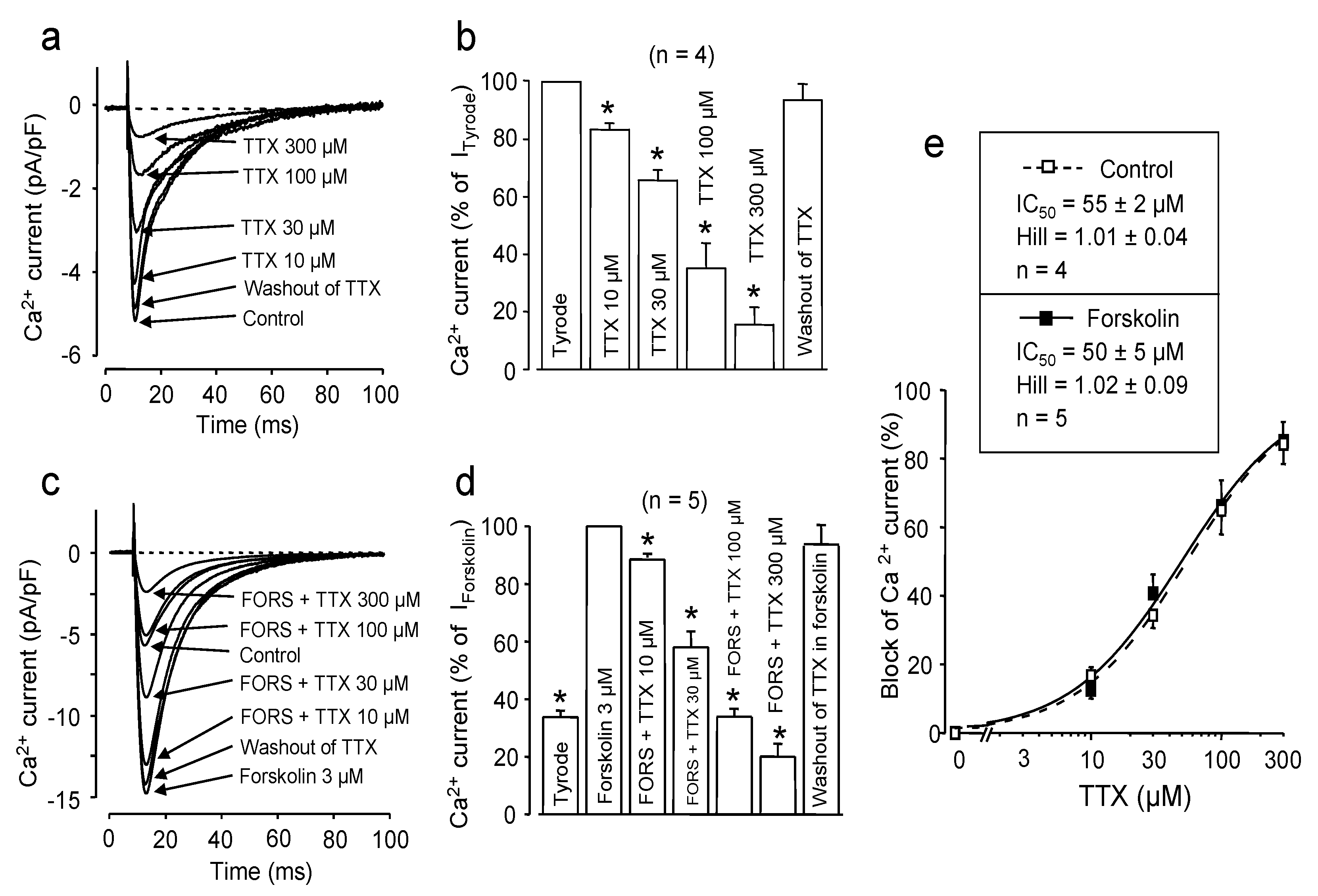
2.1. Effect of Channel Phosphorylation

Since phosphorylation of the channel protein is one of the most effective ways to control ICa in many excitable and non-excitable tissues, the blocking effect of TTX was compared on native and PKA-phosphorylated Ca channels. Forskolin is known to induce a stable activation of adenylate cyclase, resulting in high cAMP levels and, consequently, in full activation of protein kinase A, an enzyme responsible for phosphorylation of the pore forming α1 and the auxiliary β subunit of the Ca channel. This phosphorylation—through allosteric interactions—effectively increase the open probability of the pore-forming α1 channel subunit, while it has little effect on the selectivity filter [14]. As is demonstrated in Figure 1, 3 μM forskolin increased the peak amplitude of ICa (from 5.5 ± 0.4 pA/pF to 16.3 ± 0.8 pA/pF, p < 0.05, n = 5). TTX caused a concentration-dependent and reversible suppressive effect on ICa in both native and forskolin-treated cardiomyocytes. Furthermore, fitting the data to the Hill equation yielded very similar IC50 values: 55 ± 2 μM in control vs. 50 ± 5 μM in forskolin (n = 4 and n = 5, respectively, N.S.). The Hill coefficients in both cases were close to unity (1.01 ± 0.04 vs. 1.02 ± 0.09, N.S.). These results suggest that the phosphorylation-induced allosteric changes in the structure of the α1 subunit, although having serious consequences on channel gating, have little influence on TTX binding. The value of unity obtained for the Hill coefficients is congruent with the presence of one single TTX-binding site on each α1 subunit.
Figure 1. Effect of channel phosphorylation on the TTX-induced blockade. (a,c) Superimposed representative ICa records obtained in the presence of cumulatively increasing concentrations of TTX. Finally, TTX was washed out to demonstrate reversibility. Experiments were performed either in Tyrode solution (a) or in the presence of 3 μM forskolin (c). (b,d) Average results obtained with TTX in four untreated and five forskolin-treated myocytes (panels b and d, respectively). In both cases, peak ICa was normalized to the value measured prior to the first application of TTX. Columns and bars are the means ± SEM values; asterisks denote significant changes from the pre-TTX controls (Tyrode or forskolin, respectively). (e) Cumulative dose-response curves demonstrating the inhibitory effects of TTX in Tyrode solution (open symbols, dashed line) and in the presence of forskolin (filled symbols, solid line). IC50 values and Hill coefficients were determined by fitting data to the Hill equation.
Marinedrugs 11 02140 g001
The 55 μM value of IC50 obtained with TTX for ICa is relatively high, even if considering that the cardiac Na channel isoform (Nav1.5) shows much lower affinity to TTX than the neural (Nav1.1–1.3) or skeletal (Nav1.4) ones [15,16,17]. The aromatic side chain of the residue following the DEKA ring builder Asp residue was proposed to play a critical role in the specific TTX-ion channel interaction, and its replacement (e.g., to Cys residue in Nav1.5) may be the main reason for the reduced affinity [15,16]. In the sequence alignment proposed by Tikhonov and Zhorov [13], which was used when building the model in the present study, a Gly residue can be found at this key position. This fact can plausibly explain why Cav1.2 channels bind TTX with considerably lower affinity than neural or skeletal Na+ channels.

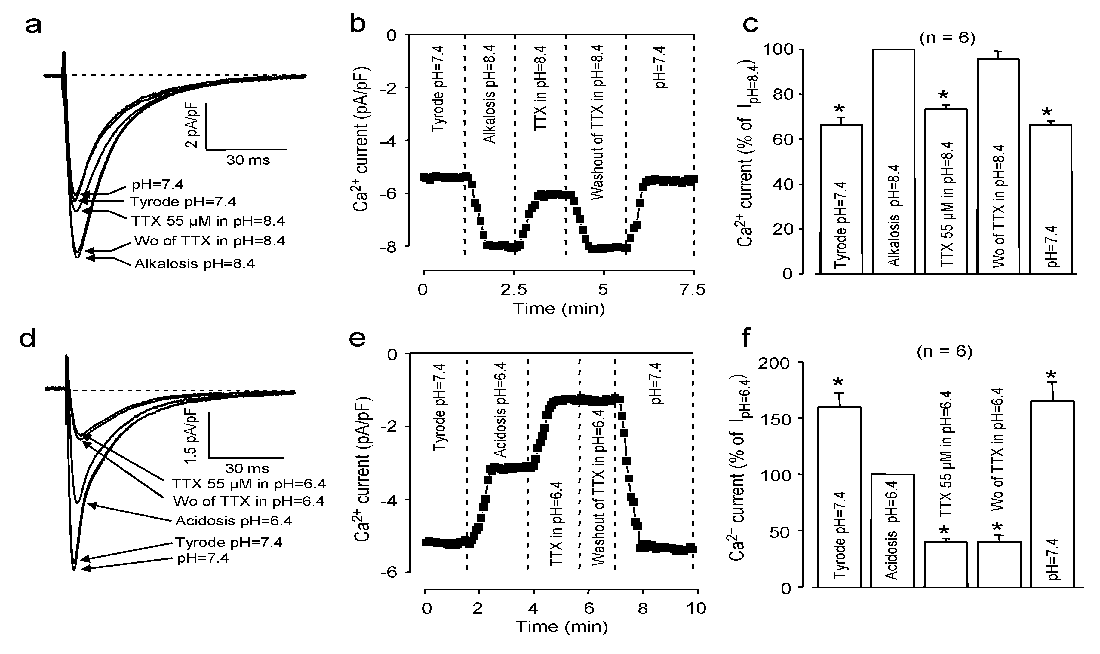
2.2. Effects of Extracellular pH

Our model, constructed to explain the TTX-sensitivity of ICa, predicts that TTX binds to the selectivity filter of the channel [6]. More specifically, this connection is based on the negatively charged (at pH values close to the physiological range) selectivity filter region of the channel and the positively charged guanidine group of the toxin. Consequently, changing the strengths of this electrostatic interaction by increasing or decreasing the protonation of either the selectivity filter region of the channel or of the TTX molecule itself is expected to modulate the magnitude of the TTX-induced blockade. As demonstrated in Figure 2, alkalization significantly decreased (to 26% ± 2% at pH = 8.4, n = 6), while acidification increased (to 60% ± 2% at pH = 6.4, n = 6) the inhibition of ICa caused by 55 μM TTX (which was estimated to be exactly 50% at the physiological pH value of 7.4).
Figure 2. Effect of extracellular alkalosis (ac) and acidosis (df) on the TTX-induced ICa blockade. Superimposed representative ICa records (a,d), time-dependent changes in peak ICa (b,e) and average values of peak ICa (c,f). Data were first obtained in Tyrode solution at pH = 7.4; then, the pH was shifted to either the alkaline or acidic direction (pH = 8.4 and pH = 6.4, respectively), and the cell was exposed to 55 μM TTX. Finally, TTX was removed, and in the next step, pH was turned back to its control value of 7.4. Columns and bars represent the means ± SEM; asterisks indicate significant changes from the pre-TTX controls (alkalosis or acidosis, respectively).
Marinedrugs 11 02140 g002
It is also worthy of noting that the effect of TTX could not be reverted at acidic pH, suggesting that the toxin was bound strongly under these conditions. Indeed, TTX could be removed from the channel, resulting in a full reversion of blockade, when pH was finally restored to 7.4 in TTX-free Tyrode solution (see Figure 2e,f).
Regarding the effect of pH on the amplitude of ICa, a very simplified interpretation can be given. Considering the approximately 6.5 pKa value of histidine [18], its imidazole side chain can be protonated, even in a mild acidic milieu. It should be noted, however, that the actual pKa depends strongly on the chemical environment. Accordingly, the histidine residues located in the conduction pathway of the channel (including the selectivity filter, as well) are at least partially protonated at the acidic pH of 6.4, which resulted in a less favorable environment for a positively charged ion. In addition, one (or even more than one) of the glutamate side chains at the highly negatively charged EEEE ring of the channel can be protonated, too, which can further reduce cationic current. Indeed, similarly to our results, ICa and INa have been reported to decrease in acidic milieu [15,16,19,20]. On the other hand, TTX exists almost exclusively in its positively charged form at pH 6.4, since the pKa value of the most acidic C10 hydroxyl group is close to 8.7 [21]. Therefore, the positively charged TTX molecule is expected to increase TTX binding (compared to the neutral zwitterionic form, which is dominant at the higher, alkaline pH range, but also existing partially at pH = 8.4 and in a small fraction even at pH = 7.4) to the negatively charged selectivity filter region resulting in a stronger inhibition of ICa. Furthermore, due to the reduced strength of electrostatic interactions (which is valid for both the double charged Ca2+ and—although to a lesser extent—for the protonated TTX molecule, because of its single positive charge), the relative contribution of the non-ionic hydrophilic and hydrophobic terms in the interaction between TTX and the selectivity filter of the channel is likely to increase. All these effects resulted in a reduction of ICa in acidic environment, which is still strongly inhibited by TTX. In contrast, at the alkaline pH of 8.4, most of the histidine side chains are neutral, and largely half of the free cysteine SH groups can be deprotonated, since the corresponding pKa is 8.5 [18]. This is certainly a more favorable environment (even compared to the neutral situation, i.e., to the case of pH = 7.4) for attracting and binding of the positively charged Ca2+ ions to the channel; consequently, the ion flux through the channel may increase. TTX may exist partially in its neutral form at mild alkaline pH values, due to deprotonation of the C-10 hydroxyl group, allowing for development of a weaker electrostatic attraction between TTX and the selectivity filter. The stronger Ca2+ channel interaction combined with a weaker interaction between TTX and the channel protein necessarily yields a smaller inhibition of ICa by TTX under alkaline conditions.
The increased amplitude of ICa and the decreased TTX blockade, observed at alkaline milieu, can easily be interpreted based on the simplified electrostatic model and the protonation equilibrium of the zwitterionic TTX outlined above in good agreement with the pH dependence of the TTX blockade observed on Na channels [22,23]. The increased inhibitory effect of TTX at acidic pH seems to contradict earlier results, suggesting a reduced TTX block at low pH values [22]. The reason for this discrepancy is not fully understood; however, one should consider that the structure of Ca and Na channels differs due to the limited sequence identity, and even in the case of Na channels, the corresponding effect is not especially large at a neutral or slightly acidic pH range. It is possible that at pH = 6.4, the conformation of the selectivity filter region of the Ca channel may change in a way, becoming more suitable for TTX-binding. Moreover, the 3%–5% non-effective (zwitterionic) fraction of TTX, estimated at pH = 7.4, is converted into cationic form at pH = 6.4, which may also contribute to the increase of the TTX-induced block.

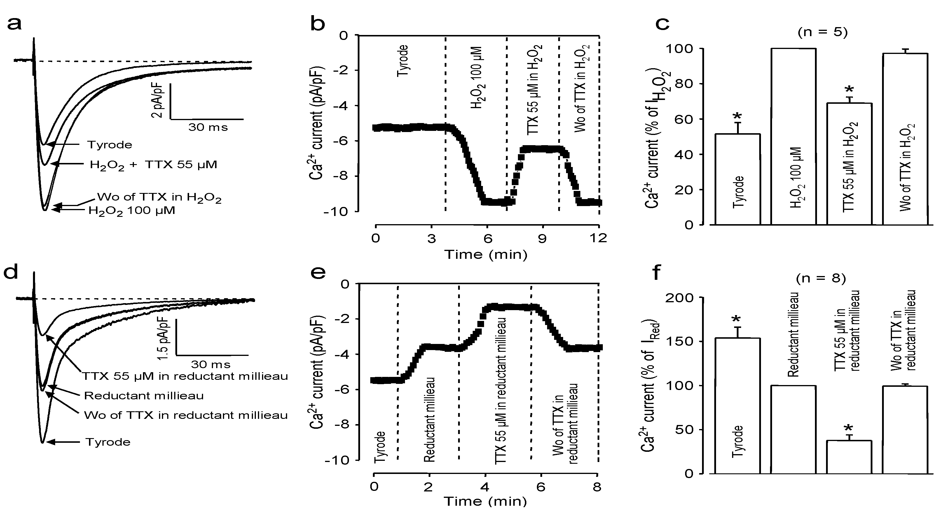
2.3. Effects of Redox Potential Changes

As demonstrated in Figure 3, the effects of redox potential changes were very similar to those initiated by changes in pH. Accordingly, in a strongly oxidant milieu, induced by application of 100 μM H2O2, 55 μM TTX suppressed only 31% ± 3% of ICa (Figure 3a–c, n = 5), in contrast to the 62% ± 6% ICa blockade observed in a reductant environment (Figure 3d–f, n = 8, 1 mM reduced glutathione + 1 mM ascorbic acid + 1 mM dithiothreitol). The role of the redox potential in modulating ICa and its suppressibility by TTX needs some more effort to be interpreted. In line with our observations, hydrogen peroxide has been shown to increase the amplitude of ICa in various mammalian cardiac tissues [24,25,26], while the current was decreased by a reductant environment [27]. Since H2O2 is a light oxidant, it oxidizes the free sulfhydryl groups to disulfide bonds. In contrast, a reducing environment may prevent the formation of disulfide bridges or break them once already performed. These disulfide bridges seem to stabilize a structure relatively resistant to TTX binding; thus, the inhibitory action of TTX on ICa increases in a reductant environment, while it decreases in the presence of H2O2. An additional explanation arises when considering that the density of ICa correlates well with oligomerization of the Cavβ subunits of Ca channels [28]. Assuming that H2O2 can stabilize these oligomeric states by the formation of disulfide bridges, both the higher ICa amplitudes, as well as the weaker inhibition by TTX can be explained.
Figure 3. Effect of oxidant (ac) and reductant (df) external milieu on the TTX-induced ICa blockade. Superimposed representative ICa records (a,d), time-dependent changes in peak ICa (b,e) and average values of peak ICa (c,f). Data were first obtained in Tyrode solution; then, the superfusate was made either oxidant (by 100 μM H2O2) or reductant (by application of glutathione + ascorbic acid + dithiothreitol, 1 mM each). The cells were exposed to 55 μM TTX in these solutions, followed by washout of TTX. Columns and bars represent the means ± SEM; asterisks indicate significant changes from the pre-TTX controls (H2O2 or reductant milieu, respectively).
Marinedrugs 11 02140 g003
Finally, it must be noted that the effects of a reductant milieu, both on the amplitude of ICa and on its suppressibility by TTX, were very similar to those of acidification. This strongly suggests that application of a reducing milieu may facilitate the protonation of some sensitive groups—in spite of the large buffering capacity of the system. This might explain the good coincidence observed in the TTX-induced inhibition of ICa, comparing it in an acidic vs. alkaline and in a reductant vs. oxidant milieu.

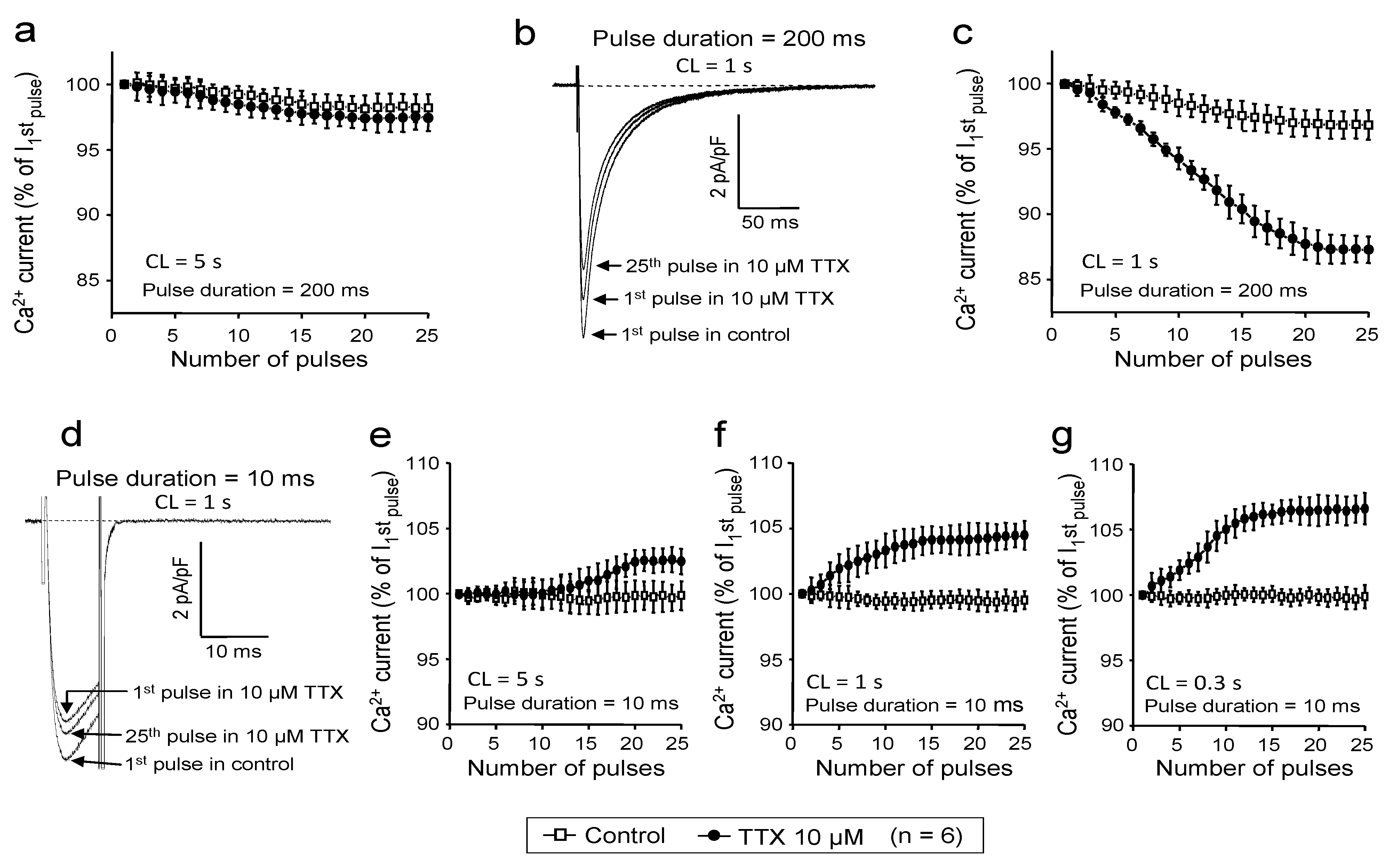
2.4. Use-Dependent Block
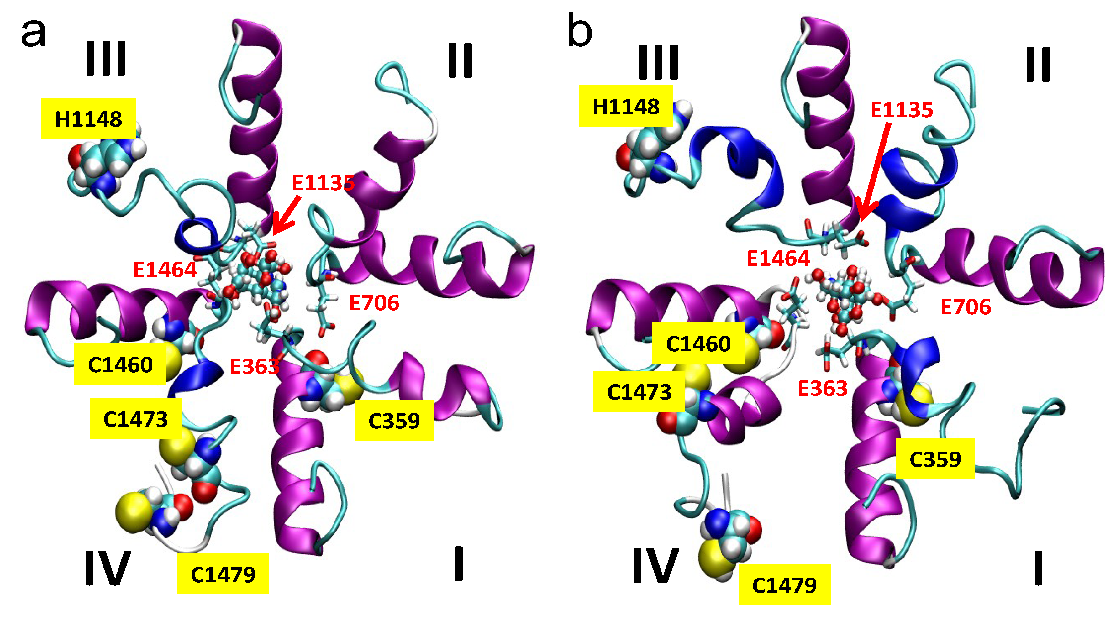
In the experiments above, ICa was activated by 400 ms-long depolarizations delivered at a frequency of 0.2 Hz. In other words, the repetition time of stimulation was 5 s. This was long enough to maintain a reasonably normal availability of ICa; therefore, the effect of TTX observed under these conditions was considered as the magnitude of tonic block. Indeed, as demonstrated in Figure 4a, this slow rate resulted in only a small reduction in ICa amplitude during the first 25 pulses applied following a 1 min period of rest both in the absence and presence of TTX (2.0 ± 1.0 and 2.8% ± 1.1% reduction, respectively, N.S., n = 6). However, a significant rate-dependent component of block was built up during the initial 25 pulses in the presence of 10 μM TTX, when the cycle length of stimulation was increased to 1 s. The magnitude of this rate-dependent block was 12.7% ± 1.0% vs. the 4.3% ± 1.1% decrease of ICa observed without TTX (Figure 4b,c, p < 0.05, n = 6). The 200 ms duration of these pulses did not allow the studying of the effect of TTX at higher frequencies; therefore, short depolarizations, having durations of only 10 ms, were applied in control and in the presence of TTX. Surprisingly, these short pulses caused a rate-dependent enhancement of ICa in the presence of 10 μM TTX, but not in control (Figure 4d–g). Increases in ICa amplitudes were larger at higher stimulation rates: increases of 2.1% ± 1.1%, 3.9% ± 1.2% and 6.3% ± 1.2% were obtained at cycle lengths of 5 s, 1 s and 0.3 s, respectively. These results indicate that in addition to the tonic block, an additional rate-dependent block is also evoked by TTX, however, the magnitude of this block was relatively moderate compared to the tonic component. More importantly, the TTX-induced inhibition of ICa seems to be restricted to resting and inactivated channel states, since frequent opening of the channel (by using short depolarizations at high frequencies) results in a relief from the block, suggesting that the affinity of TTX to the open channel state is markedly reduced.
Figure 4. Rate-dependent effects of TTX on ICa. Stimulating voltage pulses were applied at various cycle lengths, as indicated in each panel, following a 1 min period of rest (stimulation-free period). Peak values of ICa, obtained during the initial 25 pulses were plotted in the absence (open squares) and presence (filled circles) of 10 μM TTX. ICa was expressed as a percent of current amplitude observed during the first pulse. Pulse duration was set to either 200 ms (ac) or 10 ms (dg). Superimposed representative ICa recordings, obtained using 200 ms and 10 ms depolarizations at 1 Hz, are displayed in panels b and d, respectively.
Marinedrugs 11 02140 g004

2.5. Simulations

Since the pKa value (8.7) of the most acidic OH in the TTX molecule is relatively close to the pH range of 6.4–8.4 applied in our experiments, both the protonated form (i.e., positively charged due to the guanidinium group and the C10–OH) and the neutral form (the guanidinium group of TTX is still protonated, but the C10–OH is deprotonated to C10–O) of TTX were considered in our calculations (Figure 5).
Docking experiments revealed that TTX binds to the selectivity filter in both of its states; however, the binding is stronger (8.22 kcal/mol vs. 7.55 kcal/mol) when TTX is positively charged (i.e., when the proton is not dissociated from the C10–OH position). This can be the consequence of the dominantly negative formal charge of the selectivity filter. Molecular dynamics simulation started from the docked conformations supported the results obtained by docking, as the complexes remained stable during the 65 ns period of simulation (see the movies in the Supplementary Material).
Figure 5. Representative structure snapshots from dynamic trajectories of the selectivity filter region of the L-type Ca2+ channel with TTX being in a positively charged form (a), with protonated guanidine and the non-dissociated C10–OH group, and in neutral form (b), with protonated guanidine and the proton dissociated from the C10-OH group resulting in C10–O. The repeats are marked by Roman numerals. The EEEE ring of the selectivity filter and the TTX molecule are shown using “liquorice” and ball-and-stick representation, respectively. The position of the His and Cys residues marked by the van der Waals spheres of the atoms constitute these residues. The residue numbering is based on the human L-type Cav1.2 channel sequence obtained from the UNIPROT protein knowledgebase.
Marinedrugs 11 02140 g005

3. Experimental Section

3.1. Isolation of Single Canine Ventricular Myocytes

Adult beagle dogs of either sex were anaesthetized with intravenous injections of 10 mg/kg ketamine hydrochloride (Calypsol, Richter Gedeon, Budapest, Hungary) + 1 mg/kg xylazine hydrochloride (Sedaxylan, Eurovet Animal Health BV, Bladel, The Netherlands), according to a protocol approved by the local ethical committee and conforming to “principles of laboratory animal care” (NIH publication No. 85–23, revised 1985). The hearts were quickly removed and placed in Tyrode solution. Single myocytes were obtained by enzymatic dispersion using the segment perfusion technique [29,30]. Briefly, a wedge-shaped section of the ventricular wall supplied by the left anterior descending coronary artery was dissected, cannulated and perfused with oxygenized Tyrode solution containing (in mM): NaCl, 144; KCl, 5.6; CaCl2, 2.5; MgCl2, 1.2; HEPES, 5; dextrose, 11; at pH = 7.4. Perfusion was maintained until the removal of blood from the coronary system and, then, switched to a nominally Ca2+-free Joklik solution (Minimum Essential Medium Eagle, Joklik Modification, Sigma-Aldrich Co., St. Louis, MO, USA) for 5 min. This was followed by 30 min perfusion with Joklik solution supplemented with 1 mg/mL collagenase (Type II., Worthington Biochemical Co., Lakewood, NJ, USA) and 0.2% bovine serum albumin (Fraction V., Sigma-Aldrich Co., St. Louis, MO, USA) containing 50 μM Ca2+. Portions of the left ventricular wall were cut into small pieces, and the cell suspension, obtained at the end of the procedure predominantly from the mid-myocardial region of the left ventricle, was washed with Joklik solution. Finally, the Ca2+ concentration was gradually restored to 2.5 mM. The cells were stored in minimum essential medium until use.

3.2. Electrophysiology

All measurements were performed at 37 °C. The rod-shaped viable cells showing clear striation were sedimented in a Plexiglas chamber allowing continuous superfusion with oxygenized Tyrode solution. Suction pipettes, fabricated from borosilicate glass, had a tip resistance of 2 MΩ after filling with pipette solution containing in (mM): KCl, 110; KOH, 40; HEPES, 10; EGTA, 10; TEACl, 20; K-ATP, 3 mM, at pH = 7.2. ICa was recorded in Tyrode solution supplemented with 3 mM 4-aminopyridine with Axopatch-2B amplifier (Axon Instruments Inc., Foster City, CA, USA) using the whole cell configuration of the patch clamp technique [31]. The current was activated at +5 mV using 400 ms-long depolarizations arising from the holding potential of −40 mV. After establishing a high (1–10 GΩ) resistance seal by gentle suction, the cell membrane beneath the tip of the electrode was disrupted by further suction or by applying 1.5 V electrical pulses for 1 ms. The series resistance was typically 4–8 MΩ before compensation (usually 50%–80%). Experiments were discarded when the series resistance was high or substantially increasing during the measurement. Outputs from the clamp amplifier were digitized at 100 kHz under software control (pClamp 6.0, Axon Instruments Inc., Foster City, CA, USA). Ion currents were normalized to cell capacitance, which was determined in each cell using short hyperpolarizing pulses from −10 mV to −20 mV.

3.3. Simulation of TTX Binding to Cav1.2 Channels

The selectivity filter region of the human Cav1.2 channel was built on the basis of the X-ray structure of the selectivity filter region of the bacterial NavAb channel [12] and the existence of homology between the NavAb and Cav1.2 channels. The alignment between NavAb and Cav1.2 has been previously published [13], which was used without modification. Proper position of the EEEE ring, i.e., E363, E706, E1135 and E1464 residues in the human L-type Cav1.2 channel, confirmed this alignment [10]. The residue mutations corresponding to NavAb to Cav1.2 transformations were then carried out by means of the MODELER software [32] using the Chimera package as the graphical front-end [33]. The TTX molecule was then docked to the model using the Autodock4 software with the help of the graphical front-end “MGL tools” software suite [34]. In docking calculations, the flexible ligand (rotatable OH groups) rigid receptor model with genetic algorithm was applied [35]. Both the positively charged and the neutral (with deprotonated oxygen) form of TTX were docked to the selectivity filter of the Cav1.2 model. In order to assess the stability of the complexes, molecular dynamics simulations were carried out starting from the geometry obtained by docking. In the simulations for the selectivity filter and TTX molecule, AMBER99SB and GAFF force fields were applied, respectively [36,37]. Since only the selectivity filter region of Cav1.2 was included in the calculations, position restraints for the N- and C-terminal residues of each repeat-fragment and for the backbone atoms of the helical structures constituting the outer part of the filter region (N-terminal part of corresponding repeat-fragment) with no direct interaction with the TTX molecule were applied. All other atoms and residues were allowed to move freely during the simulation. The short constant particle number, constant pressure and constant temperature molecular dynamics (NPT) equilibration period was followed by the constant volume (NVT) simulation. In the calculations, explicit TIP3P water molecules and periodic boundary conditions were applied [38]. The long range electrostatic interactions were calculated using the particle mesh Ewald method [39]. Applying 2 fs time-steps, the total length of NPT and NVT simulations were 2 ns and 65 ns, respectively. The ionic strength was set to 150 mM by adding Na+ and Cl ions to the simulation box. The simulations were carried out using the Amber 12 software package [40], while for visualization and movie generation, the visual molecular dynamics (VMD) package was used [41].

3.4. Statistics

Results are expressed as the mean ± SEM values. Statistical significance of differences was evaluated using the Student’s t-test for paired or unpaired data, as pertinent. Differences were considered significant when p was less than 0.05. Drugs were obtained from Sigma-Aldrich Co. (St. Louis, MO, USA).

4. Conclusions

By applying acidic, alkaline, reductant and oxidant environments combined with in silico simulations, we have demonstrated in the present study that the guanidine-toxin TTX inhibits L-type Ca channels in the heart by binding to its selectivity filter—in a way similar to its action on fast Na channels [15,42].

Acknowledgments

Financial support was provided by grants from the Hungarian Scientific Research Fund (OTKA-K100151, OTKA-NK104331, OTKA101196, OTKA-PD101171). Further support was obtained from the Hungarian Government (TAMOP-4.2.2.A-11/1/KONV-2012-0045 research project).

References

  1. Baer, M.; Best, P.M.; Reuter, H. Voltage-dependent action of tetrodotoxin in mammalian cardiac muscle. Nature 1976, 263, 344–345. [Google Scholar] [CrossRef]
  2. Brown, A.M.; Lee, K.S.; Powell, T. Sodium current in single rat heart muscle cells. J. Physiol. 1981, 318, 479–500. [Google Scholar]
  3. Vornanen, M.; Hassinen, M.; Haverinen, J. Tetrodotoxin sensitivity of the vertebrate cardiac Na+ current. Mar. Drugs 2011, 9, 2409–2422. [Google Scholar] [CrossRef]
  4. Chorvatova, A.; Snowdon, R.; Hart, G.; Hussain, M. Effects of pressure overload-induced hypertrophy on TTX-sensitive inward currents in guinea pig left ventricle. Mol. Cell.Biochem. 2004, 261, 217–226. [Google Scholar] [CrossRef]
  5. Alvarez, J.L.; Salinas-Stefanon, E.; Orta, G.; Ferrer, T.; Talavera, K.; Galán, L.; Vassort, G. Occurrence of a tetrodotoxin-sensitive calcium current in rat ventricular myocytes after long-term myocardial infarction. Cardiovasc. Res. 2004, 63, 653–661. [Google Scholar] [CrossRef]
  6. Hegyi, B.; Horváth, B.; Bárándi, L.; Papp, F.; Magyar, J.; Bányász, T.; Krasznai, Z.; Szentandrássy, N.; Nánási, P.P. Tetrodotoxin blocks L-type Ca2+ channels in canine ventricular cardiomyocytes. Pflüg. Arch 2012, 464, 167–174. [Google Scholar] [CrossRef]
  7. Szabó, G.; Szentandrássy, N.; Bíró, T.; Tóth, I.B.; Czifra, G.; Magyar, J.; Bányász, T.; Varró, A.; Kovács, L.; Nánási, P.P. Asymmetrical distribution of ion channels in canine and human left ventricular wall: Epicardium versus midmyocardium. Pflüg. Arch. 2005, 450, 307–316. [Google Scholar] [CrossRef]
  8. Szentandrássy, N.; Bányász, T.; Bíró, T.; Szabó, G.; Tóth, I.B.; Magyar, J.; Lázár, J.; Varró, A.; Kovács, L.; Nánási, P.P. Apico-basal inhomogeneity in distribution of ion channels in canine and human ventricular myocardium. Cardiovasc. Res. 2005, 65, 851–860. [Google Scholar] [CrossRef]
  9. Tikhonov, D.B.; Zhorov, B.S. Modeling P-loops domain of sodium channel: Homology with potassium channels and interaction with ligands. Biophys. J. 2005, 88, 184–197. [Google Scholar] [CrossRef]
  10. Tikhonov, D.B.; Zhorov, B.S. Possible roles of exceptionally conserved residues around the selectivity filters of sodium and calcium channels. J. Biol. Chem. 2011, 286, 2998–3006. [Google Scholar] [CrossRef]
  11. Payandeh, J.; Scheuer, T.; Zheng, N.; Catterall, W.A. The crystal structure of a voltage-gated sodium channel. Nature 2011, 475, 353–358. [Google Scholar] [CrossRef]
  12. Payandeh, J.; Gamal El-Din, T.M.; Scheuer, T.; Zheng, N.; Catterall, W.A. Crystal structure of a voltage-gated sodium channel in two potentially inactivated states. Nature 2012, 486, 135–139. [Google Scholar]
  13. Tikhonov, D.B.; Zhorov, B.S. Architecture and pore block of eukaryotic voltage-gated sodium channels in view of NavAb bacterial sodium channel structure. Mol. Pharmacol. 2012, 82, 97–104. [Google Scholar] [CrossRef]
  14. Hove-Madsen, L.; Méry, P.F.; Jurevicius, J.; Skeberdis, A.V.; Fischmeister, R. Regulation of myocardial calcium channels by cyclic AMP metabolism. Basic Res. Cardiol. 1996, 91 (Suppl. 2), 1–8. [Google Scholar]
  15. Fozzard, H.A.; Lipkind, G.M. The tetrodotoxin binding site is within the outer vestibule of the sodium channel. Mar. Drugs 2010, 8, 219–234. [Google Scholar] [CrossRef]
  16. Satin, J.; Kyle, J.W.; Chen, M.; Bell, P.; Cribbs, L.L.; Fozzard, H.A.; Rogart, R.B. A mutant of TTX-resistant cardiac sodium channels with TTX-sensitive properties. Science 1992, 256, 1202–1205. [Google Scholar] [CrossRef]
  17. Favre, I.; Moczydlowski, E.; Schild, L. Specificity for block by saxitoxin and divalent cations at a residue which determines sensitivity of sodium channel subtypes to guanidinium toxins. J. Gen. Physiol. 1995, 106, 203–229. [Google Scholar] [CrossRef]
  18. Stryer, L. Biochemistry, 4th ed; V.H. Freeman and Company: New York, NY, USA, 1995; p. 23. [Google Scholar]
  19. Tombaugh, G.C.; Somjen, G.G. Effects of extracellular pH on voltage-gated Na+, K+ and Ca2+ currents in isolated rat CA1 neurons. J. Physiol. 1996, 493, 719–732. [Google Scholar]
  20. Saegusa, N.; Moorhouse, E.; Vaughan-Jones, R.D.; Spitzer, K.W. Influence of pH on Ca2+ current and its control of electrical and Ca2+ signaling in ventricular myocytes. J. Gen. Physiol. 2011, 138, 537–559. [Google Scholar] [CrossRef]
  21. Moczydlowski, E. The molecular mystique of tetrodotoxin. Toxicon 2013, 63, 711–722. [Google Scholar] [CrossRef]
  22. Ulbricht, W.; Wagner, H.H. The influence of pH on equilibrium effects of tetrodotoxin on myelinated nerve fibres of Rana esculenta. J. Physiol. 1975, 252, 159–184. [Google Scholar]
  23. Strickholm, A. Ionic permeability of K, Na, and Cl in potassium-depolarized nerve. Dependency on pH, cooperative effects, and action of tetrodotoxin. Biophys. J. 1981, 35, 677–697. [Google Scholar] [CrossRef]
  24. Guo, J.; Giles, W.R.; Ward, C.A. Effect of hydrogen peroxide on the membrane currents in sinoatrial node cells from rabbit heart. Am. J. Physiol. Heart Circ. Physiol. 2000, 279, H992–H999. [Google Scholar]
  25. Xie, L.H.; Chen, F.; Karagueuzian, H.S.; Weiss, J.N. Oxidative-stress-induced afterdepolarizations and calmodulin kinase II signaling. Circ. Res. 2009, 104, 79–86. [Google Scholar]
  26. Madhvani, R.V.; Xie, Y.; Pantazis, A.; Garfinkel, A.; Qu, Z.; Weiss, J.N.; Olcese, R.J. Shaping a new Ca2+ conductance to suppress early afterdepolarizations in cardiac myocytes. J.Physiol. 2011, 589, 6081–6092. [Google Scholar]
  27. Zhang, R.; Sun, Y.; Tsai, H.; Tang, C.; Jin, H.; Du, J. Hydrogen sulfide inhibits L-type calcium currents depending upon the protein sulfhydryl state in rat cardiomyocytes. PLoS One 2012, 7, e37073. [Google Scholar]
  28. Lao, Q.Z.; Kobrinsky, E.; Liu, Z.; Soldatov, N.M. oligomerization of Cavβ subunits is essential correlate of Ca2+ channel activity. FASEB J. 2010, 24, 5013–5023. [Google Scholar] [CrossRef]
  29. Magyar, J.; Bányász, T.; Szigligeti, P.; Körtvély, Á.; Jednákovits, A.; Nánási, P.P. Electrophysiological effects of bimoclomol in canine ventricular myocytes. Naunyn Schmiedebergs Arch. Pharmacol. 2000, 361, 303–310. [Google Scholar] [CrossRef]
  30. Bányász, T.; Magyar, J.; Körtvély, á.; Szigeti, G.y.; Szigligeti, P.; Papp, Z.; Mohácsi, A.; Kovács, L.; Nánási, P.P. Different effects of endothelin-1 on calcium and potassium currents in canine ventricular cells. Naunyn Schmiedebergs Arch. Pharmacol 2001, 363, 383–390. [Google Scholar] [CrossRef]
  31. Hamill, O.P.; Marty, A.; Neher, E.; Sakmann, B.; Sigworth, F.J. Improved patch-clamp techniques for high-resolution current recording from cells and cell-free membrane patches. Pflüg. Arch. 1981, 391, 85–100. [Google Scholar] [CrossRef]
  32. Sali, A.; Blundell, T.L. Comparative protein modelling by satisfaction of spatial restraints. J. Mol. Biol. 1993, 234, 779–815. [Google Scholar] [CrossRef]
  33. Pettersen, E.F.; Goddard, T.D.; Huang, C.C.; Couch, G.S.; Greenblatt, D.M.; Meng, E.C.; Ferrin, T.E. UCSF Chimera—A visualization system for exploratory research and analysis. J. Comput. Chem. 2004, 25, 1605–1612. [Google Scholar] [CrossRef]
  34. Morris, G.M.; Huey, R.; Lindstrom, W.; Sanner, M.F.; Belew, R.K.; Goodsell, D.S.; Olson, A.J. AutoDock4 and AutoDockTools4: Automated docking with selective receptor flexibility. J. Comput. Chem. 2009, 30, 2785–2791. [Google Scholar] [CrossRef]
  35. Morris, G.M.; Goodsell, D.S.; Halliday, R.S.; Huey, R.; Hart, W.E.; Belew, R.K.; Olson, A.J. Automated docking using a Lamarckian genetic algorithm and an empirical binding free energy function. J. Comput. Chem. 1998, 19, 1639–1662. [Google Scholar] [CrossRef]
  36. Hornak, V.; Abel, R.; Okur, A.; Strockbine, B.; Roitberg, A.; Simmerling, C. Comparison of multiple amber force fields and development of improved protein backbone parameters. Proteins 2006, 65, 712–725. [Google Scholar] [CrossRef]
  37. Wang, J.M.; Wolf, R.M.; Caldwell, J.W.; Kollman, P.A.; Case, D.A. Development and testing of a general amber force field. J. Comput. Chem. 2004, 25, 1157–1174. [Google Scholar] [CrossRef]
  38. Jorgensen, W.L.; Chandrasekhar, J.; Madura, J.D.; Impey, R.W.; Klein, M.L. Comparison of simple potential functions for simulating liquid water. J. Chem. Phys. 1983, 79, 926–935. [Google Scholar] [CrossRef]
  39. Darden, T.; York, D.; Pedersen, L. Particle mesh Ewald-an NlogN method for Ewald sums in large systems. J. Chem. Phys. 1993, 98, 10089–10092. [Google Scholar] [CrossRef]
  40. AMBER, version 12; University of California: San Francisco, CA, USA, 2012.
  41. Humphrey, W.; Dalke, A.; Schulten, K. VMD—Visual Molecular Dynamics. J. Mol. Graph. 1996, 14, 33–38. [Google Scholar] [CrossRef]
  42. Lipkind, G.M.; Fozzard, H.A. A structural model of the tetrodotoxin binding site of the Na+ channel. Biophys. J. 1994, 66, 1–13. [Google Scholar] [CrossRef]

Supplementary Files

  • Supplementary File 1:

    Supplementary (MPG, 8807 KB)

  • Share and Cite

    MDPI and ACS Style

    Hegyi, B.; Komáromi, I.; Kistamás, K.; Ruzsnavszky, F.; Váczi, K.; Horváth, B.; Magyar, J.; Bányász, T.; Nánási, P.P.; Szentandrássy, N. Tetrodotoxin Blockade on Canine Cardiac L-Type Ca2+ Channels Depends on pH and Redox Potential. Mar. Drugs 2013, 11, 2140-2153. https://doi.org/10.3390/md11062140

    AMA Style

    Hegyi B, Komáromi I, Kistamás K, Ruzsnavszky F, Váczi K, Horváth B, Magyar J, Bányász T, Nánási PP, Szentandrássy N. Tetrodotoxin Blockade on Canine Cardiac L-Type Ca2+ Channels Depends on pH and Redox Potential. Marine Drugs. 2013; 11(6):2140-2153. https://doi.org/10.3390/md11062140

    Chicago/Turabian Style

    Hegyi, Bence, István Komáromi, Kornél Kistamás, Ferenc Ruzsnavszky, Krisztina Váczi, Balázs Horváth, János Magyar, Tamás Bányász, Péter P. Nánási, and Norbert Szentandrássy. 2013. "Tetrodotoxin Blockade on Canine Cardiac L-Type Ca2+ Channels Depends on pH and Redox Potential" Marine Drugs 11, no. 6: 2140-2153. https://doi.org/10.3390/md11062140

    Article Metrics

    Back to TopTop