Article

Efficient Heterologous Transformation of Chlamydomonas reinhardtii npq2 Mutant with the Zeaxanthin Epoxidase Gene Isolated and Characterized from Chlorella zofingiensis

by
Inmaculada Couso
,
Baldo F. Cordero
,
María Ángeles Vargas
and
Herminia Rodríguez
*
Institute of Plant Biochemistry and Photosynthesis, CIC Cartuja, University of Seville and CSIC, Avda. Américo Vespucio no. 49, 41092-Seville, Spain
*
Author to whom correspondence should be addressed.
Mar. Drugs 2012, 10(9), 1955-1976; https://doi.org/10.3390/md10091955
Submission received: 4 July 2012 / Revised: 6 August 2012 / Accepted: 22 August 2012 / Published: 12 September 2012

Abstract

:
In the violaxanthin cycle, the violaxanthin de-epoxidase and zeaxanthin epoxidase catalyze the inter-conversion between violaxanthin and zeaxanthin in both plants and green algae. The zeaxanthin epoxidase gene from the green microalga Chlorella zofingiensis (Czzep) has been isolated. This gene encodes a polypeptide of 596 amino acids. A single copy of Czzep has been found in the C. zofingiensis genome by Southern blot analysis. qPCR analysis has shown that transcript levels of Czzep were increased after zeaxanthin formation under high light conditions. The functionality of Czzep gene by heterologous genetic complementation in the Chlamydomonas mutant npq2, which lacks zeaxanthin epoxidase (ZEP) activity and accumulates zeaxanthin in all conditions, was analyzed. The Czzep gene was adequately inserted in the pSI105 vector and expressed in npq2. The positive transformants were able to efficiently convert zeaxanthin into violaxanthin, as well as to restore their maximum quantum efficiency of the PSII (Fv/Fm). These results show that Chlamydomonas can be an efficient tool for heterologous expression and metabolic engineering for biotechnological applications.

Abbreviations

gDNA
Genomic DNA
UPGMA
Unweighted Pair Group Method with Arithmetic Mean
PSII
Photosystem II
qPCR
Quantitative Polymerase Chain Reaction

1. Introduction

In photosynthetic organisms, carotenoids participate in light harvesting within the reaction centers and the light harvesting complexes (LHCs), the protection of the photosynthetic apparatus via the direct quenching of chlorophyll reactive intermediates and/or singlet oxygen molecules, the dissipation of absorbed energy beyond that required for photosynthesis [1,2], and the structural maintenance of photosynthetic complexes [3]. Therefore, carotenoids participate in keeping a balance between capture, use of light and cell protection of excess excitation energy. In particular the xanthophylls zeaxanthin and lutein, whose formation is enhanced under high light conditions, help in the efficient transition of LHCs of PSII to a conformation that favors the thermal dissipation of parts of the excess of excitation energy. This energy dissipation, measured as non-photochemical quenching of the chlorophyll fluorescence (NPQ) prevents over-reduction of the reaction centers and must be strictly controlled to avoid photosynthetic efficiency loss [4,5].
The photo-protective functions of the de-epoxidazed carotenoid zeaxanthin have been intensively studied [6], however, it was not until 1989 when Demming-Adams and co-workers [7] first proposed a now widely accepted role linking zeaxanthin formation with photoprotection within the violaxanthin cycle. The dissipation of excess excitation energy mediated by zeaxanthin in this cycle is a key process in the photosynthetic systems of plants and algae [8,9]. A similar cycle, known as the diadinoxanthin cycle, has also been identified in certain algal groups with cycling occurring between the two xanthophylls diadinoxanthin and diatoxanthin, which only comprises one de-epoxidation step [10,11,12,13]. However, in these algae this cycle is the main one and the role of the violaxanthin cycle is mainly to produce intermediates for the biosynthesis of the diadinoxanthin cycle pigments.
The violaxanthin cycle is found in plants and the green (chlorophyta) and brown (phaeophyceae) algae. In this cycle, when the irradiance is higher than that required for photosynthesis in the chloroplasts of plants and algae, a reversible violaxanthin de-epoxidation reaction occurs to form the intermediate antheraxanthin, subsequently resulting in the accumulation of zeaxanthin in a second de-epoxidation reaction. The extent of the de-epoxidation depends on two factors at least, namely, the pigment pool size and the fraction of the violaxanthin pool that is available for de-epoxidation [14]. When the absorbed irradiance is lower than that required for the saturation of the photosynthetic apparatus, zeaxanthin is converted back into violaxanthin. This reaction occurs on the stromal side of the chloroplast thylakoid membrane, and it is relatively slower than the de-epoxidation reaction [15,16]. The de-epoxidation sequence of the violaxanthin cycle is catalyzed by the enzyme violaxanthin de-epoxidase (VDE), and the epoxidation from zeaxanthin into violaxanthin is carried out by the enzyme zeaxanthin epoxidase (ZEP).
ZEP has a specificity for carotenoids with a 3-hydroxy-β-hexenyl ring and requires the presence of NADPH and FAD as cofactors, ferredoxin and ferredoxin-like reducing agents are also required for its activity [17]. The first isolation of this gene was performed in a transposon-tagged Nicotiana plumbaginifolia ABA-deficient mutant called aba2 [18,19] and many genetic studies on this cycle have been performed in plants, such as Arabidopsis thaliana in which different aba mutants have been characterized, providing an opportunity to study the physiological roles of epoxy-carotenoids in photosynthesis [20], as well as the npq mutants in the green alga Chlamydomonas reinhardtii [21]. This unicellular green alga has been used as a good alternative molecular system to plants in order to study photosynthesis and other metabolic processes [22].
In 1997, Niyogi and co-workers [21] generated a series of violaxanthin cycle mutants in an Arg- auxotroph Chlamydomonas reinhardtii strain. These mutants were obtained by insertional mutagenesis using a linearized vector containing the wild type argininosuccinate lyase gene (arg7). These mutants exhibited an aberrant NPQ, being defective in two different NPQ phases, either being defective in the quick-turn-over first phase (npq4), or in the slower second phase (npq1) or in both of them (npq2 and npq11). Npq2 had lost the zeaxanthin epoxidase activity, since it exhibited a violaxanthin cycle pool of entirely zeaxanthin even after dark adaptation. After examining the co-segregation of the insertional mutagen (ARG7 DNA) and the npq aberrant phenotype in the npq2, it was concluded that this mutation was closely linked to a single insertion locus of the ARG7 vector.
The chlorophyta Chlorella zofingiensis is considered as a model organism to study the regulation of the carotenoid biosynthetic pathway since it produces both the primary carotenoid lutein as well as the secondary carotenoid astaxanthin [23]. Several carotenogenic genes have been isolated and characterized in this microalga, such as β-carotene oxygenase (bkt) [24], phytoene desaturase (pds) [25], carotene β-hydroxylase (chyB) [26], lycopene β-cyclase (lcyB) [27] and phytoene synthase (psy) [28]; however, genes involved in the violaxanthin cycle have not been isolated yet.
In this paper, we report the isolation and characterization of the zeaxanthin epoxidase gene involved in the violaxanthin cycle in Chlorella zofingiensis. The transcriptional regulation of this gene in response to light and nitrogen, as well as its functionality by heterologous transformation of Chlamydomonas reinhardtii npq2 mutant, which lacks ZEP activity, has also been performed.

2. Results

2.1. Isolation and Characterization of the ZEP Gene and Deduced Protein Sequence of C. zofingiensis

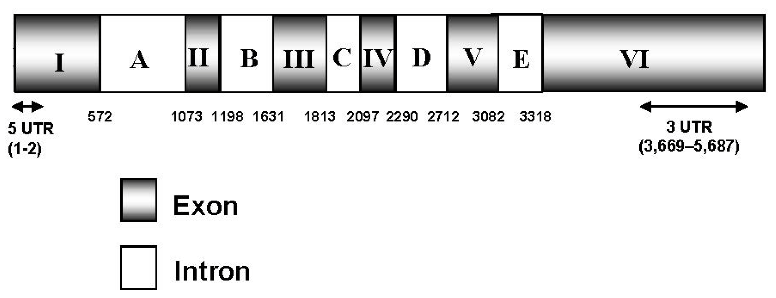
Different degenerate primers were designed on the basis of the conserved motifs present in ZEP amino acid sequences from microalgae and plants. A partial Czzep cDNA fragment of 1100 bp was isolated by PCR amplification using the degenerate primers ZEP-1F and ZEP-1R (Table 1). A complete basic local alignment search tool (BLAST) homology search in the Genbank database (NCBI) showed that this fragment had enough similarity with the ZEP genes from other species, and provided sequence information for designing specific primers for rapid amplification of 5′ and 3′ cDNA ends (RACE-PCR). This analysis generated a full-length cDNA of 3836 bp, which contained an Open Reading Frame (ORF) of 1791 bp, a very short two nucleotides of 5′ untranslated region (UTR), and a long 3′ UTR of 2043 nucleotides. The predicted protein has 596 amino acid residues, with an estimated molecular weight of 64.50 kDa, a theoretical isoelectric point of 8.31 and an instability index of 29.87 (data obtained with ProtParam program). The differences between the C. zofingiensis zep gene and the cDNA sequence were compared and revealed the presence of six exons and five introns (Figure 1).
Table 1. Nucleotide sequences of primer pairs used for PCR amplification.
PrimerSequence (5′→3′)
Partial ZEP fragment
ZEP-1Ftggtgggcgccgayggnathhg
ZEP-1Rcgcccacgtcggtgswnacraarta
5′ and 3′ RACE
NGSP-ZEP-5′Rgatcttggaccagataccatcagcacccaca
GSP-ZEP-5′Ragcatgtatagccgctgtagttggg
NGSP-ZEP-3′F cagatttcacgccagcagacattgacat
GSP-ZEP-3′Fgcggctatacatgctacacagggatct
Genomic DNA amplification
G-ZEP-1Fatggggacttcagtacctgca
G-ZEP-1Raacagccagcacaagacctcc
G-ZEP-2Fcaagaccagaaagcgtcacggc
G-ZEP-2Rtgctgcacacctgtgacagggt
G-ZEP-3Fgatcctgattgatgccgtggcg
G-ZEP-3Rtgctccatagcatctgccagat
G-ZEP-4Fatttgggtcagggtggctgcat
G-ZEP-4Rggtgcgaaaagggtatgaagaacg
G-ZEP-5Rcgctctacacttggctgtagtg
G-ZEP-6Fatcaatggcatggagtacagga
G-ZEP-6Rtagcacgatgctgcaagatcacgt
G-ZEP-7Fctggtgtgcagattggttttgccg
G-ZEP-7RAgtattcactcactatgaactgatgcc
G-ZEP-8Fccattgaagatgcttatcagttggcagc
G-ZEP-8Rggcatacatgctctgagcaggcataag
PCR for Chamydomonas reinhardtii transformation a
pStpZEPxhoFcgcctcgagcatggggacttcagtacctgcaaat
pStpZEPecoRccgaattcgtggctgttatatttgtgctgttg
CzZEP expression
RT-Czzep-Fcgaacgtgatcttacagccat
RT-Czzep-Rgatacgatctcctgtgatgcagcca
CBLP expression
RT-cblp-Fcgccacccagtcctccatcaaga
RT-cblp-RCtaggcgcggctgggcatttac
F, forward; R, reverse. a XhoI and EcoRI restriction sites (lowercase letters underlined) were added for cloning the gene into the corresponding cut sites of the pSI105Tp1 vector.
For the determination of the copy number of the zep gene in the genome of C. zofingiensis, genomic DNA was digested with four different restriction enzymes ScaI, NdeI, BamHI and NcoI and subjected to Southern blot analysis at different conditions of stringency. Using a 662 bp fragment of Czzep as a probe, strong hybridization signals were obtained with the different digestions. The digestion with NcoI and BamHI enzymes, which cut once inside the probe sequence, showed two bands, while digestion with NdeI or ScaI, with no restriction site in the probe, exhibited only one band (Figure 2). These results suggest the presence of a single copy of the zep gene in the genome of C. zofingiensis.
Figure 1. Czzep scheme. The diagram shows that the Czzep gene consists of six exons (I–VI) and five introns (A–E). The 5′ UTR and 3′ UTR sequences are indicated with arrows and correspond to the positions 1–2 bp and 3669–5687 bp, respectively. Numbers represent the cDNA coordinates (bp). UTR, untranslated region.
Marinedrugs 10 01955 g001
Figure 2. Southern blot analysis of genomic DNA from C. zofingiensis. gDNA was digested with ScaI (Lane 1), NdeI (Lane 2), BamHI (Lane 3) or NcoI (Lane 4), electrophoretically separated on a 0.8% agarose gel, blotted and hybridized at high stringency with a probe of 662 bp of the Czzep gene amplified by PCR.
Marinedrugs 10 01955 g002
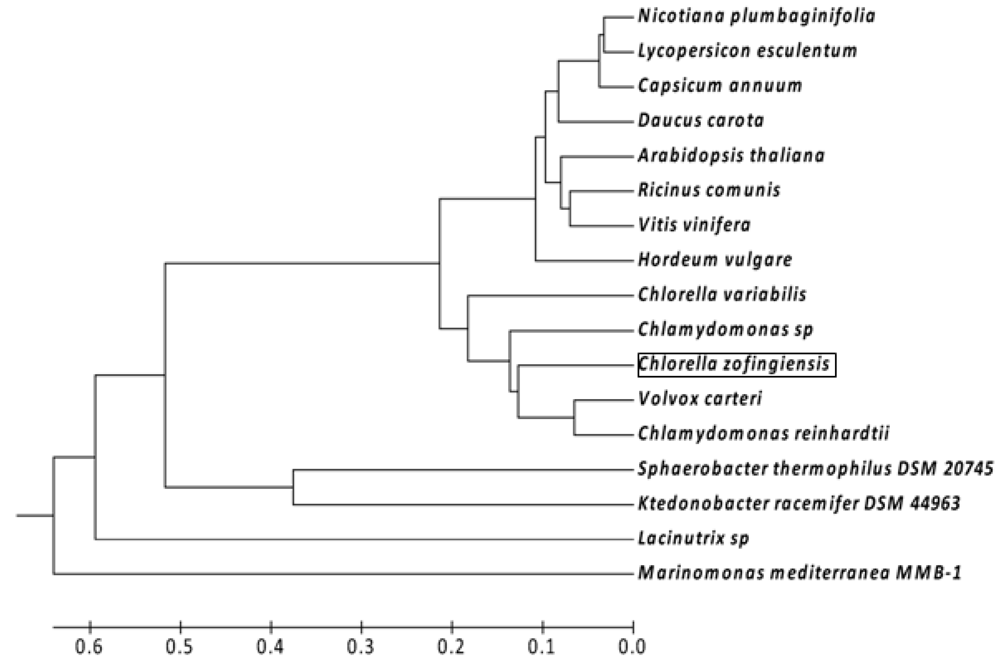
The BlastP search results demonstrated that the cloned CzZEP showed the highest overall homology sequence with other ZEP from green algae, such as Volvox carteri (identity, 66% and similarity, 77%), Chlorella variabilis (identity, 69% and similarity, 79%) and Chlamydomonas reinhardtii (identity, 69% and similarity, 80%). The GC content of the Czzep coding region was 52.4%, which was lower than that of V. carteri (60.5%), C. variabilis (68.1%) or of C. reinhardtii (68.8%). The phylogenetic analysis of ZEP from green algae, plants and bacteria is illustrated in Figure 3. This analysis was performed in MEGA5 software [29] using the unweighted pair group method with arithmetic mean (UPGMA) method. The predicted CzZEP forms a cluster with the ZEP of the green algae studied, which are phylogenetically close to ZEP of plants (between 60% and 63% of identity and between 73% and 76% similarity). CzZEP was distantly related to bacterial zeaxanthin epoxidases (between 30% and 34% of identity and 45%–50% of similarity).
Figure 3. Unweighted pair group method with arithmetic mean (UPGMA) tree analysis of the indicated plant, algal and bacterial ZEP amino acid sequences. The GenBank accession numbers for other species are as follows: Nicotiana plumbaginifolia (X95732.1); Lycopersicon esculentum (Z83835.1); Capsicum annuum (Q96375); Daucus carota (ABB52077.1); Arabidopsis thaliana (AAG17703); Ricinus comunis (XP_002523587.1); Vitis vinifera (AAR11195.1); Hordeum vulgare (BAK08085.1); Chlorella variabilis (EFN52633.1); Chamydomonas sp (AAO48941.1); Volvox carteri (XP_002953670.1); Chlamydomonas reinhardtii (XP_001701701.1); Sphaerobacter thermophilus DSM 20745 (ACZ40773.1); Ktedonobacter racemifer DSM 44963 (ZP_06974439.1); Lacinutrix sp. (AEH02389.1); Marinomonas mediterranea MMB-1 (ADZ93016.1). The tree is drawn to scale, with branch lengths in the same units as those of the evolutionary distances used to infer the phylogenetic tree. The evolutionary distances correspond to the number of amino acid substitutions per site and were computed using the Poisson correction method.
Marinedrugs 10 01955 g003
Since in microalgae and plants ZEP is located in the chloroplast membranes, we analyzed the CzZEP sequence with different programs to determine both the presence of a signal peptide and transmembrane domains. Target-P and Chloro-P programs identified an N-terminal chloroplast transit peptide of 60 aa length. Analysis with TMHMM and TopPred servers identified four deduced transmembrane domains of CzZEP located between amino acids 26–46, 144–164, 502–522 and 549–569. The domain structure analysis with Interpro Scan showed two monooxygenase FAD-binding domains located between amino acids 144–160 and 439–473, as well as four aromatic-ring hydroxylase-like domains located between amino acids 145–167, 308–323, 438–453, and 453–469 (data not shown).

2.2. Effect of Irradiance and Nitrogen on the Expression of Czzep Gene
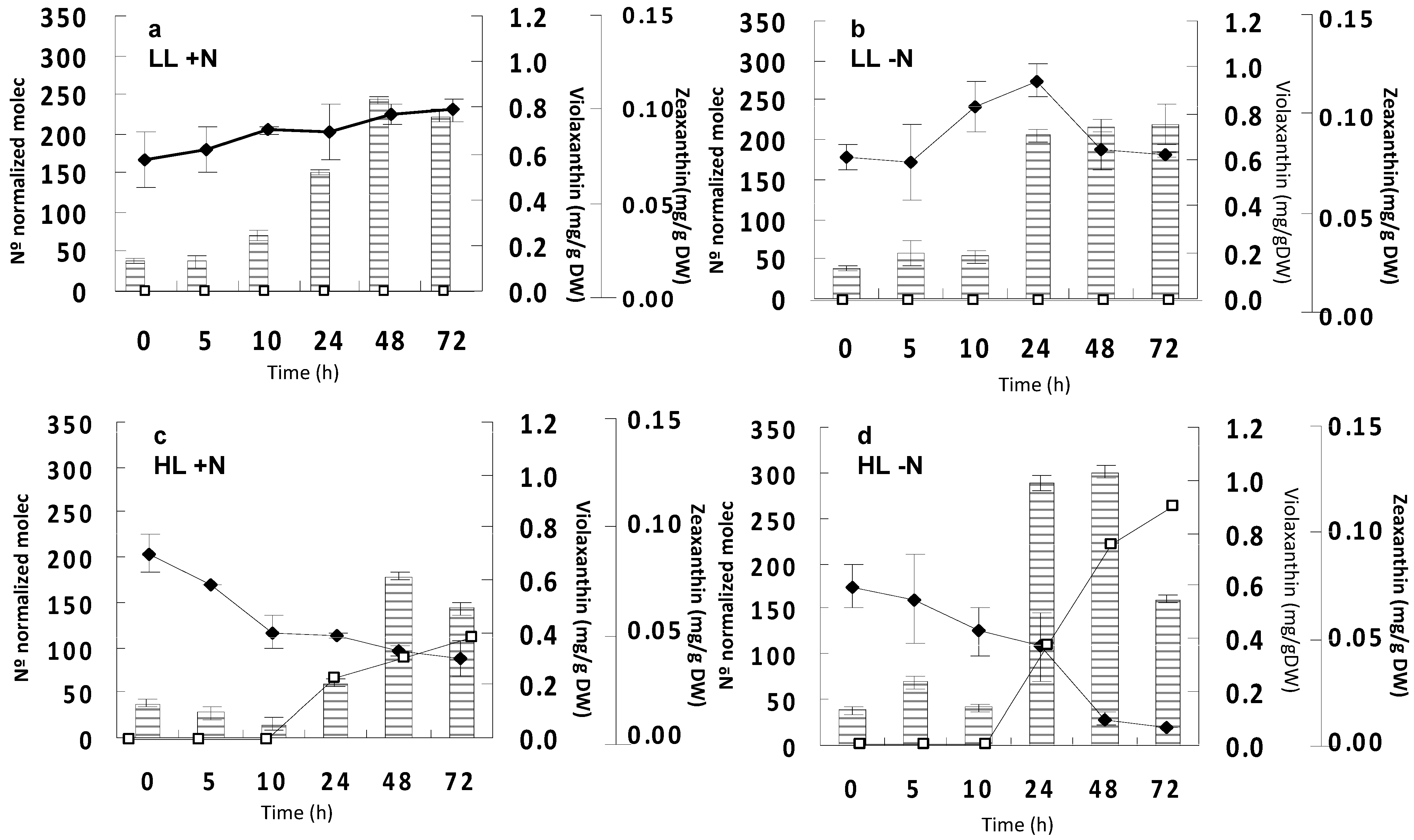
C. zofingiensis cells were grown photoautotrophically at low irradiance and constant nitrate, as indicated in the Experimental Section, until the middle of the exponential phase. Then, cells were kept in the dark for 18 h, in order to make the transcript levels come down to basal values. After this dark period, cells were subjected to either low or high irradiance, under either constant nitrogen or nitrogen-deprivation conditions. The evolution with time of mRNA levels of the Czzep gene as affected by irradiance and nitrogen availability was monitored by quantitative real time PCR (qRT-PCR). In addition, changes in zeaxanthin and violaxanthin cellular contents were also determined in order to correlate mRNA levels with the biosynthesis of these carotenoids. As shown in Figure 4a,b, at low irradiance the levels of transcripts did not start to increase until 24 h after the dark period, regardless of nitrate availability. Under high irradiance and constant nitrogen conditions, the transcript levels of Czzep decreased between 10% and 60% after 5 h and 10 h from the beginning of the experiment, respectively (Figure 4c). Although there was also a high increase of the transcripts levels after 24 h under these conditions, they did not reach the levels registered at low irradiance. However, an additive response was detected in the nitrogen-free culture under high light conditions (Figure 4d), transcript levels after 24 h of induction being higher than those exhibited under low irradiance. With regards to the cellular carotenoid content, zeaxanthin was only registered at high irradiance, the levels of this carotenoid increased with time after 10 h, with a much higher accumulation of this carotenoid taking place under nitrogen depletion conditions (Figure 4c,d). Antheraxanthin was only detected at high irradiance as well, its levels ranging from 0.12 mg∙g−1 DW at 10 h to 0.09 mg g−1 DW at 72 h of high light exposure. Both zeaxanthin and antheraxanthin contents seem to be correlated with the increased mRNA levels of Czzep. The concentration of violaxanthin decreased with time at high irradiance, especially in the case of nitrogen-free conditions, while in the case of low light conditions it remained almost constant and only a little increase was detected at the 10 h or 24 h under nitrogen deprivation. Under high irradiance, astaxanthin was also present both in nitrogen depleted and in nitrogen repleted conditions, reaching values of 0.21 mg g1 DW and 0.5 mg g1 DW after 48 h, respectively.
Figure 4. Effect of irradiance and nitrogen availability on the mRNA levels of the zeaxanthin epoxidase gene (zep) and cellular content of violaxanthin and zeaxanthin in Chlorella zofingiensis. (a) Low irradiance (20 µmol photons m2 s1) and constant nitrate concentration (LL +N); (b) low irradiance and nitrate deprivation (LL −N); (c) high irradiance (300 µmol photons m2 s1) and constant nitrate concentration (HL +N); and (d) high irradiance and nitrate deprivation (HL −N). Columns indicate mRNA levels of the zep gene; (♦), violaxanthin content (mg g1 DW); (□), zeaxanthin content (mg g1 DW). Error bars indicate the standard deviations of four independent measurements. DW, dry weight.
Marinedrugs 10 01955 g004

2.3. Functional Analysis of the ZEP from C. zofingiensis by Heterologous Genetic Complementation in C. reinhardtii npq2 Mutant

2.3.1. Transformation of C. reinhardtii npq2 Mutant with zep Gene from C. zofingiensis and Screening of the Obtained Transformants

Since the use of E. coli (DH5α) engineered to produce zeaxanthin did not allow the functional characterization of the Czzep gene, probably due to the lack of cofactors necessary for enzymatic activity, the functionality of this gene was assayed by heterologous genetic complementation in the C. reinhardtii npq2 mutant. For this purpose, the complete coding region of Czzep was amplified by PCR and cloned between the XhoI and EcoRI restriction sites of the Chlamydomonas expression vector pSI105-Tp1, resulting in the plasmid pSI105-Tp1-zep. C. reinhardtii npq2 mutant cells were transformed with this plasmid, and the transformants were a priori selected on the basis of their paromomycin resistance. The colonies obtained were screened for the insertion of Czzep cDNA in their genome by PCR. Figure 5 shows some of the positive transformants analyzed exhibiting a band of approximately 0.1 kb, which corresponds to the Czzep cDNA integrated in their genome. The primers used for PCR analysis to confirm this integration were G-ZEP-2F and G-ZEP-1R (Table 1) with a final amplification of 75 bp. More than 150 colonies resistant to paromomycin were analyzed and 113 of them (≈75%) were found to be positive.

2.3.2. Analysis of Carotenoid Content, Maximum Quantum Efficiency and mRNA Level of Czzep Gene in Both npq2 Mutant of C. reinhardtii and Czzep-Transformed Strains of npq2 Mutant

The mRNA relative abundance of Czzep gene of C. reinhardtii npq2 mutant, which lacks ZEP activity and accumulates zeaxanthin in all conditions, and Czzep-transformant strains of npq2 mutant was monitored by qRT-PCR, and changes in cellular carotenoids content were determined in order to correlate transcript levels with the biosynthesis of specific carotenoids. In addition, the photosynthetic capacity of npq2 mutant and the transformants was also determined by measuring their maximum quantum yield (Fv/Fm).
Lutein and β-carotene were the major carotenoids both in npq2 mutant and Czzep transformant cells, the levels of those carotenoids being higher in Czzep transformants (ranging from 1.67 to 2.29 mg g∙DW−1 in the case of lutein and 0.78 to 1.19 mg g∙DW−1 in the case of β-carotene) than in npq2 mutant (0.55 mg g∙DW−1 in the case of lutein and 0.41 mg g∙DW−1 in the case of β-carotene). Thus, the npq2-4 transformant showed a content of lutein and β-carotene six- and four-fold higher, respectively, than the mutant. Most of the transformants (approximately 80% of the selected positives) exhibited a lutein and β-carotene contents of 2.8- to 3.2- and 1.8- to 2.5-fold higher than the npq2 mutant, respectively. The violaxanthin and zeaxanthin contents and the relative mRNA levels of the Czzep gene in the npq2 mutant and nine selected Czzep npq2 transformants are illustrated in Figure 6. Most of the transformants analyzed showed a restored zeaxanthin epoxidase activity, some of them exhibiting a total conversion of zeaxanthin into violaxanthin, such as npq2-4, which seems also to exhibit the highest expression pattern of the Czzep gene. Some transformants, however, did not show a total conversion of zeaxanthin into violaxanthin or a restored zeaxanthin epoxidase activity, although in all of them the Czzep gene was adequately transcribed. Nevertheless, the expression levels of Czzep in transformants were rather heterogeneous coinciding with the differences found in the carotenoid levels. In fact, the highest Czzep mRNA level was found in the npq2-4 transformant, which reached about seven-fold the levels of the transformants npq2-7, npq2-5 and npq2-10. This correlated with the highest level of carotenoids shown by npq2-4 transformant. Npq2-9 and npq2-13 transformants also showed a high level of Czzep mRNA, reaching about five times the level of the lowest yielding transformants. Therefore, the functional characterization of the recently isolated Czzep gene could be achieved by expression of this gene in the transformants and the restoration of zeaxanthin epoxidase activity.
Figure 5. Verification of the plasmid pSI105-Tp1-Czzep insertion in the genome of C. reinhardtii by PCR. C. reinhardtii npq2 cells transformed with the plasmid pSI105-Tp1-Czzep were grown in TAP medium with paromomycin (30 µg mL−1), and paromomycin-resistant colonies were tested by PCR. Lane 10 is the 2log DNA ladder (0.1–10 kb, Biolabs). Lanes 1–9 correspond to some analyzed transformants that were found to be positive.
Marinedrugs 10 01955 g005
Figure 6. Zeaxanthin and violaxanthin content (a) and mRNA relative abundance of Czzep (b) in cells of C. reinhardtii npq2 mutant (npq2) and nine selected transformants (npq2-1, npq2-4, npq2-5, npq2-7, npq2-8, npq2-9, npq2-10, npq2-13 and npq2-14). a, Zeaxanthin (grey bar) and violaxanthin (black bar); b, Levels of Czzep transcripts were normalized in respect to the housekeeping control gene (cblp). Error bars indicate the standard deviations of four independent measurements.
Marinedrugs 10 01955 g006
The Fv/Fm parameter is widely considered to be a sensitive indicator of plant photosynthetic performance. In this sense, we wanted to determine why lacking a functional violaxanthin cycle npq2 mutant could determine a lower photosynthetic capacity of the npq2 mutant. At the same time, we also wanted to measure the differences, if they do exist, in the maximum quantum yields (Fv/Fm) among the different transformants and the parental strain. In Table 2, it is shown that the Fv/Fm value of the npq2 mutant is 25% lower than that of the Wild type. In addition, Fv/Fm values of the transformants were restored by the exogenous CzZEP activity from 84.6% for npq2-5 to 100% for npq2-4. The full recovery of the Fv/Fm value shown by the npq2-4 transformant was correlated with the fact that this transformant was able to completely transform the zeaxanthin pool into violaxanthin.
Table 2. Maximum quantum yield data and the percentages recovered in the transformants analyzed.
StrainsFv/Fm% Fv/Fm recovered
WT4A+0.65 ± 0.014---
npq20.485 ± 0.003---
npq2-10.585 ± 0.01789.2%
npq2-40.65 ± 0.014100%
npq2-50.555 ± 0.00384.6%
npq2-70.585 ± 0.00789.2%
npq2-80.6 ± 0.00792.3%
npq2-90.585 ± 089.2%
npq2-100.605 ± 0.00393.1%
npq2-130.58 ± 089.2%
npq2-140.61 ± 0.00793.8%

3. Discussion

Although previous work concerning zeaxanthin epoxidase has essentially focused on its role in ABA biosynthesis during drought stress and seed development in plants [19,20,30], its relationship with photosynthesis and the carotenoids synthesis pathway using mutants lacking the zeaxanthin epoxidase activity has also been reported both in plants and algae [21,31,32,33,34]. The zep gene cDNA has been isolated from higher plants such as Nicotiana plumbaginifolia (tobacco), Capsicum annuum (pepper), Lycopersicon esculentum (tomato) and Arabidopsis thaliana [15,18,34] and from some algae such as the well-known Chlamydomonas reinhardtii [35]. In this work, we describe the isolation and characterization of the zep gene from the green alga Chlorella zofingiensis. This new gene showed six exons and five introns, and the ORF of the cDNA encoded a hypothetical protein of 596 amino acids (Figure 1). The length of the putative protein was similar to those described for microalgae, being slightly shorter than plant enzymes, according to alignments performed with the Blocks program (data not shown). Southern blot analysis has demonstrated that in C. zofingiensis, as in other green algae such as C. reinhardtii [35] and Chlorella variabilis [36], as well as in angiosperms, only a single gene encoding ZEP is present. However, it has been recently described that other algae classes such as the diatoms P. tricornutum and T. pseudonana contain three and two copies, respectively, of the zep gene, which could be a reflection of the presence of both cycles, violaxanthin cycle and diadinoxanthin cycle in these organisms. The diadinoxanthin cycle is generally found in heterokons but it is absent in plants and chlorophytes [37]. On the other hand, the zep gene has not been found in the genomes of cyanobacteria or the red algae, such as C. merolae and G. sulphuraria [38].
The first zep gene isolated in Nicotiana plumbaginifolia [18], called aba2, encoded a chloroplast-imported flavin monooxygenase (FMO) containing a FAD-binding domain. The amino acid sequences of the pepper [15], tomato [34] and tobacco ZEP were similar to that of a flavoprotein monooxygenase (salicylate-1-monooxygenase) and those of a number of other bacterial hydroxylases [39]. Therefore, ZEP enzymes are considered FAD-dependent monooxygenases. The amino acid sequence analysis of the predicted protein encoded by the new gene isolated from C. zofingiensis revealed two monooxygenase FAD-binding domains [37,40]. In addition, the predicted CzZEP sequence showed four putative transmembrane domains that could play a role in the localization of this enzyme on the stromal side of the chloroplast thylakoid membrane, and a transit peptide which would allow CzZEP to travel from the cytoplasm, where the polypeptide is synthesized, to its final location in the chloroplast as in the case of other carotenogenic enzymes in this alga which are encoded by nuclear genes [23,27].
Different environmental conditions can show different regulations of the carotenoid synthesis pathway. As an example, either low light intensities or darkness stimulate the backward reaction of the violaxanthin cycle resulting in the conversion of zeaxanthin back to violaxanthin in Arabidopsis and C. reinhardtii [41,42]. In photoautotrophically grown C. zofingiensis cultures, it is known that the combination of both high irradiance and nitrogen starvation causes changes in the cellular content of several carotenoids [43,44,45]. Recently, it has been shown that high irradiance in this alga up-regulates the pds, chyB, and bkt genes, significantly enhancing astaxanthin synthesis [25,26]. In addition, the nitrogen deprivation per se up-regulated the lcyB gene, but did not trigger astaxanthin biosynthesis [27]. We have studied the regulation of the Czzep gene by nitrogen and irradiance, determining the mRNA levels as well as the cellular content of violaxanthin and zeaxanthin (Figure 4). The transcript levels of Czzep did not increase until 24 h under low irradiance conditions, coming from a dark period, regardless of the nitrogen availability, without any accumulation of zeaxanthin and with the violaxanthin content remaining virtually constant. Therefore, it is possible that nitrogen deprivation itself could not affect the level of zep transcripts. Under high irradiance and as previously described, the light activated the de-epoxidation of violaxanthin, registering zeaxanthin production after 10 h of exposure and the decrease of the violaxanthin content, the effect being more pronounced under nitrogen deprivation (about two-fold zeaxanthin level of the nitrogen complete culture), showing a possible additive effect of those two stress conditions. However, mRNA levels of Czzep surprisingly increased after 10 h, reaching the highest values under nitrogen starvation conditions. For both high light and high light combined with nitrogen starvation stresses, the increase in zeaxanthin content was lower than the decrease in violaxanthin content. This could be due to the presence of antheraxanthin as well as to the astaxanthin accumulation, since in C. zofingiensis it has been reported that astaxanthin is mainly synthesized from zeaxanthin instead of β-carotene as in H. pluvialis [46].
The high violaxanthin de-epoxidase activity under high irradiance has been described previously as being due to a result of the presence of a transmembrane proton gradient during periods of high irradiance [14,47,48]. Until now, the regulation of the ZEP is still not well understood. Some authors have proposed that the zeaxanthin epoxidase activity seems to be constitutive [49], as this is not regulated by the stromal pH. Since ZEP requires molecular oxygen as a second substrate and NADPH as a cofactor, both factors might principally control or limit ZEP activity [49]. The predicted protein encoded by the new gene isolated from C. zofingiensis showed a high homology degree with ZEP of plants and algae (Figure 3). This could be sufficient to consider this new gene as a zeaxanthin epoxidase, but the functional analysis has confirmed this hypothesis. By complementation in E. coli using the pAC25crtx plasmid, which [50] contains the carotenogenic genes responsible for the formation synthesis of zeaxanthin, together with the Czzep gene cloned into pQE80-L expression vector, no conversion of zeaxanthin into violaxanthin or the intermediate antheraxanthin was detected. Molecular oxygen (O2), NADPH and reduced ferredoxin are known to be co-substrates for the epoxidation reaction [15,17,51]. The prokaryotic host E. coli cannot provide reduced ferredoxin and this seems to be the reason why some zep genes do not work in this bacterium, as it was also the case of Nicotiana plumbaginifolia [18,52]. In Chlamydomonas reinhardtii, ZEP functionality has been studied by using forward genetics. The Chlamydomonas npq2 mutant was isolated because it exhibited an unusual violaxanthin cycle pool, consisting entirely of zeaxanthin in all conditions. In contrast to other npq mutations, npq2 phenotype co-segregated with arginine producer phenotype, demonstrating that the mutation affects a single gene encoding the zeaxanthin epoxidase enzyme [21]. As the CzZEP functionality could not be performed in E. coli, the Czzep gene was inserted into a Chlamydomonas expression vector to be heterologously expressed in vivo in the Chlamydomonas npq2 mutant. The transformants obtained were able to convert back zeaxanthin into violaxanthin, which confirmed the functionality of the Czzep gene isolated.
At the moment, Chlamydomonas ZEP is the only zeaxanthin epoxidase enzyme whose functionality has been reported in algae [21,53]. On the other hand, the complementation of the mutation that npq2 carried by Czzep, supports the idea that Chlamydomonas can be an efficient host for expressing exogenous genes.
Several studies about over-expression of the zeaxanthin epoxidase gene have been reported in plants [54,55]. Transgenic lines in tomatoes shown altered de-epoxidation kinetics of the violaxanthin cycle especially under stress conditions, but nothing was described regarding the concentration of the rest of carotenoids found in their profile. In the case of the CzZEP transformants of npq2 mutant obtained we found higher levels of β-carotene and lutein reaching between three to six times the levels of the npq2 control cells. Moreover, these increments in carotenoids also correspond to increased Czzep mRNA levels of the transformants analyzed. Thus, the npq2-4 transformant, which exhibited the highest expression pattern, also showed the highest carotenoids levels (Figure 6a) as well as a total conversion from zeaxanthin into violaxanthin (Figure 6b). Chlamydomonas transformants over-expressing the phytoene synthase gene of C. zofingiensis isolated also by us, exhibited a carotenoids content up to 2.2-fold the level of the control cultures [23]. Phytoene synthase catalyzes the first step of the carotenoid biosynthetic pathway and it is considered as a rate-limiting key enzyme in this pathway. Since the Czzep transformants isolated in this work showed higher carotenoids levels than parental ones, the violaxanthin cycle can be postulated as a key step to be considered for biotechnological applications and genetic manipulation.
It has been shown that the violaxanthin cycle pigments are bound to light-harvesting complexes of the two photosystems [56,57]. In fact, ZEP has been proposed to be located close to the light harvesting complex of PSII (LHCII) to enable the binding of violaxanthin and the intermediate antheraxanthin to the antenna complex [58]. The role of zeaxanthin in the chlorophyll fluorescence quenching has also been largely discussed, concluding that Lhcb proteins replace violaxanthin by zeaxanthin in the L2 site which induces the quenching of the chlorophyll fluorescence by increasing the relative amplitude of the short fluorescence lifetime component found in these Lhcb proteins. This correlates with energy dissipation and regulation of the energy transfer to the PSI reaction centers [59,60]. Kalituho and co-workers in 2007 [61] reported a reduced electron transport in Arabidopsis npq2 mutant due to sustained energy dissipation by the high levels of zeaxanthin, and how it might reflect the less efficient utilization of absorbed light energy measured as the maximum quantum yield (Fv/Fm). In the same way, Chlamydomoans npq2 mutant Fv/Fm values were 25% lower than those in the wild type strain. However, the obtained transformants showed restored Fv/Fm values, recovering the levels of the Chlamydomonas wild type cells almost completely (Table 2). These results were also correlated to those obtained from the transformants, some of which showed high expression levels and total conversion from zeaxanthin into violaxanthin. As an example, the npq2-4 transformant exhibited a 100% restored Fv/Fm, in addition to a higher concentration of carotenoids and mRNA levels of Czzep.

4. Experimental Section

4.1. Strains and Culture Conditions

Chlorella zofingiensis SAG 211-14 strain was obtained from the Culture Collection of Göttingen University (SAG, Germany). This green microalga was grown photoautotrophically in Arnon medium [62] modified to contain 4 mM K2HPO4 and 20 mM NaNO3, at 25 °C under continuous illumination (20 μmol photons m−2 s−1). The light intensity was measured at the surface of the flasks using a LI-COR quantum sensor (model L1-1905B, Li-Cor, Inc. Lincoln, NE, USA). The liquid cultures were continuously bubbled with air supplemented with 1% (v/v) CO2 as the only source of carbon. In order to analyze zep gene expression, cells were grown in Roux flasks of 1 L capacity laterally and continuously illuminated with mercury halide lamps at either 20 or 300 µmol photons m−2 s−1 either in the presence or in the absence of nitrate. The nitrate concentration was measured daily and the nitrate consumed by the cells was added to the cultures in order to keep the nitrate constant in the medium.
Chlamydomonas reinhardtii WT4a+ (CC-4051) and npq2 (CC-4101) strains were obtained from the Chlamydomonas center’s collection and cultured mixotrophically in either liquid or agar solidified Tris-acetate phosphate (TAP) medium [63] at 25 °C under a continuous irradiance of 20 μmol photons m−2 s−1. For the analysis of transformants, cells were grown in Erlenmeyer flasks of 100 mL capacity at 25 °C under continuous illumination (20 μmol photons m−2 s−1) in liquid TAP medium.
Escherichia coli DH5α strain was used as a host for DNA manipulation.

4.2. Genomic DNA and RNA Isolation and cDNA Preparation

DNA and total RNA were isolated using DNeasy Plant Mini Kit and RNeasy Plant Mini Kit (Qiagen, Düsseldorf, Germany), respectively. For PCR screening of npq2 transformants, a loopful of cells was scrapped from a plate and resuspended in 150 μL of cold distilled water and 350 μL of a buffered solution containing 50 mM Tris-HCl, pH 8, 0.3 M NaCl, 5 mM EDTA, and 2% SDS. The genomic DNA isolation was performed by phenol-chloroform-isoamyl alcohol (50:48:2) extraction and selective precipitation with ethanol, according to previously described protocols [64]. Regarding the quantitative real-time PCR analysis (qRT-PCR), first-strand cDNA synthesis was obtained from total RNA treated with DNase as recommended by the manufacturer, by using the SuperScript First-Strand Synthesis System (Invitrogen, Barcelona, Spain) primed with oligo(dT)18 according to the manufacturer’s instructions.

4.3. Cloning of C. zofingiensis zep cDNA and Genomic Gene

Amino acid sequences deduced from previously cloned zep genes from different kinds of algae and plants were aligned for the isolation of the cDNA clone coding the C. zofingiensis ZEP. Highly conserved regions were identified, and different pairs of degenerated primers were designed. The PCR products were cloned into pGEM-T vector (Promega, Madison, WI, USA) according to the manufacturer’s manual and then sequenced. The cDNA fragment obtained corresponding to partial zep clone analyzed provided sequence information for the designing of gene specific primers for the amplification of 5′ and 3′ cDNA ends by RACE-PCR. All reactions were performed with kits according to the manufacturer’s instructions (Smart RACE cDNA Amplification Kit, Clontech, Mountain View, CA, USA). 5′ and 3′ RACE products were cloned into pGEM-T vector and sequenced. Specific primers were synthesized for genomic DNA amplification based on cDNA sequence. The primer sets used in this study are listed in Table 1.

4.4. Nucleotide Sequence Accession Numbers

The Czzep cDNA and genomic DNA sequences have been registered in the EMBL database (EMBL, HE863825 and EMBL, HE863826, respectively).

4.5. Sequencing and Phylogenetic Analysis

The deduced amino acid sequence of the C. zofingiensis ZEP was compared with other ZEP sequences of algae, plants and bacteria. The sequence analysis and alignments were performed using CLUSTAL_W software. The construction of a phylogenetic tree was performed in MEGA5 [29] using the UPGMA method. The deduced amino acid sequence was subjected to the ProtParam application at the ExPASy server [65] for physical and chemical parameters, Target_P program [66] for the prediction of possible plastid localization, and ChloroP1.1 server [67] for the identification of a chloroplast transit peptide. For transmembrane domains analysis, TMHMM [68] and TopPred [69] servers were used. The domain structure of the predicted protein was analyzed by Interpro Scan [70].

4.6. Southern Blot Analysis

Genomic DNA was digested with ScaI, NdeI, BamHI and NcoI. The first two enzymes showed one recognition site inside the probed region of the zep gene, while the other two did not have any. The probe was prepared by amplifying the amplification of the genomic DNA with primers G-ZEP-4F and G-ZEP-4R, resulting in a 662 bp fragment of Czzep gene. The digested DNA was transferred to a Hybond-N membrane (GE Healthcare, Little Chanfont, UK) by capillary transfer and hybridized with the 32P labeled DNA probe at both low and high stringency overnight. After hybridization, the radioactivity of the membrane was monitored by the Cyclone Phosphor System (Perkin-Elmer, Waltham, MA, USA).

4.7. Chlamydomonas Nuclear Transformation

The complete coding region of Czzep was amplified by PCR with primers pStpZEPxhoF and pStpZEPecoR, which were designed to contain XhoI and EcoRI restriction sites respectively, and cloned into pGEM-T vector (Promega), which carries ampicillin resistance. The PCR product was digested with XhoI and EcoRI and sub-cloned into the pSI105-Tp1 vector, resulting in the plasmid pSI105-Tp1-zep that was used to transform Chlamydomonas cells. The plasmid pSI105-Tp1 is based on the plasmid pSI104-PLK [71], derived from the pSI103 [72], in which the aphVIII gene from Streptomyces rimosus, encoding an aminoglycoside 3′ phosphotransferase that confers resistance to the antibiotic paromomycin, is expressed under the control of the strong constitutive promoters rbcS2 and hsp70A and terminated by the 3′ untranslated region of rbcS2. The construction pSI105-Tp1 also carries a second expression cassette driven by the same two constitutive promoters and terminator region. This second cassette carries the transit peptide of RuBisCO small subunit (rbcS2) to target the final peptide into the chloroplast stroma and also carries a polilynker region in which the cDNA from C. zofingiensis zep was subcloned in frame with the transit peptide sequence.
Nuclear transformation was carried out using the glass beads method of Kindle (1990) with some modifications [71]. Chlamydomonas npq2 cells were grown to about 107 cells mL−1 then cells were harvested by centrifugation and resuspended in fresh TAP medium to obtain a 100-fold concentrated cell suspension. The concentrated cell suspension (0.6 mL) was added to a conical tube containing 0.3 g of sterile glass beads (Ø 0.4–0.6 mm), 0.2 mL of 20% polyethylene glycol (MW8000) and 1 μg of the desired plasmid. Cells were vortexed and resuspended in 50 mL of fresh sterile TAP medium where they were incubated in the dark overnight. After this incubation, cells were centrifuged and spread onto solid TAP medium with paromomycin (30 μg mL−1). Transformed colonies were visible after 4 to 5 days.

4.8. Quantitative RT-PCR

The mRNA relative abundance of C. zofingiensis zep was examined by qRT-PCR on an IQ5 Real-Time PCR Detection System (BioRad, Hercules, CA, USA), according to [27]. In each experiment, a series of standard dilutions containing a specific concentration of a PCR fragment was amplified in 20 μL of reaction solution containing 1 × SYBR Green PCR Master Mix (Quantimix Easy SYG kit, BioTools B & M Labs, Madrid, Spain) and corresponding primers for zep from C. zofingiensis (Table 1). After heating at 95 °C for 10 min, the cycling parameters were: fourty cycles of 95 °C for 30 s, 60 °C for 30 s, and 72 °C for 30 s. Finally, the specificity of the qRT-PCR products was confirmed by performing a melting temperature analysis at temperatures ranging from 55 °C to 95 °C at 0.5 °C/min and also by electrophoresis on a 2% agarose gel. Data were captured as amplification plots. Transcription levels of the target gene were calculated from the threshold cycle by interpolation from the standard curve. To standardize the results, the relative abundance of cblp gene, which encodes a G-protein β-subunit-like polypeptide [73], was also determined and used as the internal standard in the case of Chlamydomonas transformants. The actin gene was also used as the internal standard [74,75] for zep mRNA analysis in C. zofingiensis. The complete experiments (RNA isolation, cDNA synthesis followed with qRT-PCR) were repeated twice independently, and data are the average of at least three replicates.

4.9. Analytical Methods

4.9.1. Cell Concentration and Dry Weight Determinations

The cell number was determined with a Neubauer hemocytometer. For dry weight measurements, aliquots (5 mL) of the cell culture were filtered through Whatman GF/C paper (Whatman plc, Kent, UK), washed three times with ammonium formate 1 M solution, and dried at 80 °C for 24 h.

4.9.2. Carotenoid Extraction and HPLC Analysis

Total pigments were extracted with 80% of acetone (v/v) according to León et al. (2005) [76]. Then the samples were centrifuged and analyzed by HPLC using a Waters Spherisorb ODS2 column (4.6 × 250 mm, 5 μm particle size) (Waters, Mildford, MA, USA). The chromatographic method described by [27] was used. Pigments were eluted at a flow rate of 1.0 mL min−1 and were detected at 440 nm using a Waters 2996 photodiode-array detector. Identification of carotenoids was achieved by comparison of the individual characteristic absorption spectrum and the retention time with known standards. Quantification was performed using a calibration curve generated with commercially available carotenoids standards from Sigma-Aldrich (St. Louis, MO, USA) and DHI (Holsholm, Germany).

4.9.3. Photochemical Efficiency Analysis (Fv/Fm Measurements)

Fv/Fm is a parameter widely used to indicate the maximum quantum efficiency of Photosystem II. Fv/Fm is presented as a ratio of variable fluorescence (Fv) over the maximum fluorescence value (Fm). For these measurements, samples were taken from npq2 transformants obtained and from control cultures in Erlenmeyer flasks at 25 °C under continuous illumination (20 μmol photons m−2 s−1) and agitation. They were firstly adapted to the dark for 15 min and then these measurements were obtained using 2 mL of culture in an AquaPen-C-AP100 (Photon Systems Instruments, Drasov, Czech Republic). The experiments were performed in duplicate and the given data are the resulting averages of three replicates of each sample.

5. Conclusions

Until now, Chlamydomonas ZEP was the only zeaxanthin epoxidase enzyme which functionality was reported in algae. Czzep isolation and characterization was performed in this study. The recovered transformants able to convert back zeaxanthin into violaxanthin confirm the functionality of the Czzep gene isolated and support the idea that Chlamydomonas can be an efficient host for checking the functionality of exogenous genes. Our data on the characterization and regulation of this gene by light and nitrogen could contribute to the understanding of the regulatory mechanism of biosynthesis of carotenoids at the molecular level, which can be very useful for the optimization of the physiological conditions for higher carotenoid production by C. zofingiensis.

Acknowledgements

This work was supported by the Andalusian Government, Spain (group BIO299).

References

  1. Niyogi, K.K. Photoprotection revisited: Genetic and molecular approaches. Annu. Rev. Plant Physiol. Plant Mol. Biol. 1999, 50, 333–359. [Google Scholar] [CrossRef]
  2. Li, Z.; Wakao, S.; Fischer, B.B.; Niyogi, K. Sensing and responding to excess light. Annu. Rev. Plant Biol. 2009, 60, 239–260. [Google Scholar] [CrossRef]
  3. Freer, A.A.; Prince, S.M.; Sauer, K.; Papiz, M.Z.; Hawthornthwaite-Lawless, A.M.; McDermott, G.; Cogdell, R.J.; Isaacs, N.W. Pigment-protein interactions and energy transfer in the antennacomplex of the photosynthetic bacterium Rhodopseudomonas acidophila. Structure 1996, 4, 449–462. [Google Scholar] [CrossRef]
  4. Niyogi, K.K.; Shih, C.; Chow, W.S.; Pogson, B.J.; DellaPenna, D.; Björkman, O. Photoprotection in a zeaxanthin- and lutein-deficient double mutant of Arabidopsis. Photosynth. Res. 2001, 67, 139–145. [Google Scholar] [CrossRef]
  5. Pogson, B.J.; Niyogi, K.K.; Björkman, O.; DellaPenna, D. Altered xanthophyll compositions adversely affect chlorophyll accumulation and nonphotochemical quenching in Arabidopsis mutants. Proc. Natl. Acad. Sci. USA 1998, 95, 13324–13329. [Google Scholar]
  6. Young, A.J.; Phillip, D.; Savill, J. Carotenoids in higher plant photosynthesis. In Handbook of Photosynthesis; Pessaraki, M., Ed.; Marcel Dekker Inc.: New York, NY, USA, 1997; pp. 575–596. [Google Scholar]
  7. Demmig-Adams, B.; Adams, W.W.; Winter, K.; Meyer, A.; Schreiber, U.; Pereira, J.S.; Kriiger, A.; Czygan, F.C.; Lange, O.L. Photochemical efficiency of photosystem II, photon yield of O2 evolution, photosynthetic capacity, and carotenoid composition during the midday depression of net CO2 uptake in Arbutus unedo growing in Portugal. Planta 1989, 177, 377–387. [Google Scholar] [CrossRef]
  8. Goss, R.; Richter, M.; Wild, A. Role of DpH in the mechanism of zeaxanthin-dependent amplification of qE. J. Photochem. Photobiol. B Biol. 1995, 27, 147–152. [Google Scholar]
  9. Goss, R.; Jakob, T. Regulation and function of xanthophyll cycle-dependent photoprotection in algae. Photosynth. Res. 2010, 106, 103–122. [Google Scholar] [CrossRef]
  10. Stransky, H.; Hager, A. Das Carotinoidmuster und die Verbreitung des lichtinduzierten Xanthophyllcyclus in verschiedenen Algenklassen. Arch. Microbiol. 1970, 71, 164–190. [Google Scholar]
  11. Olaizola, M.; Roche, J.; Kolber, Z.; Falkowski, P.G. Nonphotochemical fluorescence quenching and the diadinoxanthin cycle in a marine diatom. Photosynth. Res. 1994, 41, 357–370. [Google Scholar] [CrossRef]
  12. Lohr, M.; Wilhelm, C. Algae displaying the diadinoxanthin cycle also possess the violaxanthin cycle. Proc. Natl. Acad. Sci. USA 1999, 96, 8784–8789. [Google Scholar] [CrossRef]
  13. Lohr, M.; Wilhelm, C. Xanthophyll synthesis in diatoms: quantification of putative intermediates and comparison of pigment conversion kinetics with rate constants derived from a model. Planta 2001, 212, 382–391. [Google Scholar] [CrossRef]
  14. Siefermann, D.; Yamamoto, H. NADPH and oxygen-dependent epoxidation of zeaxanthin in isolated chloroplasts. Biochem. Biophys. Res. Commun. 1975, 62, 456–461. [Google Scholar] [CrossRef]
  15. Bouvier, F.; d’Harlingue, A.; Hugueney, P.; Marin, E.; Marion-Poll, A.; Camara, B. Xanthophyll biosynthesis: Cloning, expression, functional reconstitution, and regulation of b-cyclohexenyl carotenoid epoxidase from pepper (Capsicum annuum). J. Biol. Chem. 1996, 271, 28861–28867. [Google Scholar]
  16. Holt, N.E.; Fleming, G.R.; Niyogi, K.K. Toward an understanding of the mechanism of nonphotochemical quenching in green plants. Biochemistry 2004, 43, 8281–8289. [Google Scholar]
  17. Büch, K.; Stransky, H.; Hager, A. FAD is a further essential cofactor of the NAD(P)H and O2-dependent zeaxanthin-epoxidase. FEBS Lett. 1995, 376, 45–48. [Google Scholar] [CrossRef]
  18. Marin, E.; Nussaume, L.; Quesada, A.; Gonneau, M.; Sotta, B.; Hugueney, P.; Frey, A.; Marion-Poll, A. Molecular identification of zeaxanthin epoxidase of Nicotiana plumbaginifolia, a gene involved in abscisic acid biosynthesis and corresponding to the ABA locus of Arabidopsis thaliana. EMBO J. 1996, 15, 2331–2342. [Google Scholar]
  19. Audran, C.; Borel, C.; Frey, A.; Sotta, B.; Meyer, C.; Simonneau, T.; Marion-Poll, A. Expression Studies of the Zeaxanthin Epoxidase Gene in Nicotiana plumbaginifolia. Plant Physiol. 1998, 118, 1021–1028. [Google Scholar] [CrossRef]
  20. Rock, C.D.; Zeevaart, J.A.D. The aba mutant of Arabidopsis thaliana is impaired in epoxy-carotenoid biosynthesis. Proc. Natl. Acad. Sci. USA 1991, 88, 7496–7499. [Google Scholar]
  21. Niyogi, K.K.; Bjirkman, O.; Grossman, A.R. Chlamydomonas xanthophyll cycle mutants identified by video imaging of chlorophyll fluorescence quenching. Plant Cell 1997, 9, 1369–1380. [Google Scholar]
  22. Eonseon, J.; Polle, J.W.; Lee, H.K.; Hyun, S.M.; Chang, M.J. Xanthophylls in microalgae: from biosynthesis to biotechnological mass production and application. Microbiol. Biotechnol. 2003, 13, 165–174. [Google Scholar]
  23. Cordero, B.F.; Couso, I.; León, R.; Rodríguez, H.; Vargas, M.A. Enhancement of carotenoids biosynthesis in Chlamydomonas reinhardtii by nuclear transformation using a phytoene synthase gene isolated from Chlorella zofingiensis. Appl. Microbiol. Biotechnol. 2011, 91, 341–351. [Google Scholar]
  24. Huang, J.C.; Chen, F.; Sandmann, G. Stress-related differential expression of multiple β-carotene ketolase genes in the unicellular green alga Haematococcus pluvialis. J. Biotechnol. 2006, 122, 176–185. [Google Scholar]
  25. Huang, J.C.; Liu, J.; Li, Y.; Chen, F. Isolation and characterization of the phytoene desaturase gene as a potential selective marker for genetic engineering of the astaxanthin-producing green alga Chlorella zofingiensis (Chlorophyta). J. Phycol. 2008, 44, 684–690. [Google Scholar] [CrossRef]
  26. Li, Y.; Huang, J.; Sandmann, G.; Chen, F. Glucose sensing and the mitochondrial alternative pathway are involved in the regulation of astaxanthin biosynthesis in the dark-grown Chlorella zofingiensis (Chlorophyceae). Planta 2008, 228, 735–743. [Google Scholar] [CrossRef]
  27. Cordero, B.F.; Obraztsova, I.; Martín, L.; Couso, I.; León, R.; Vargas, M.A.; Rodríguez, H. Isolation and characterization of a lycopene β-cyclase gene from the astaxanthin-producing green alga Chlorella zofingiensis (Chlorophyta). J. Phycol. 2010, 46, 1229–1238. [Google Scholar] [CrossRef]
  28. Cordero, B.F.; Obraztsova, I.; Couso, I.; León, R.; Vargas, M.A.; Rodriguez, H. Enhancement of lutein production in Chlorella sorokiniana (Chorophyta) by improvement of culture conditions and random mutagenesis. Mar. Drugs 2011, 9, 1607–1624. [Google Scholar] [CrossRef] [Green Version]
  29. Tamura, K.; Peterson, D.; Peterson, N.; Stecher, G.; Nei, M.; Kumar, S. MEGA5: Molecular evolutionary genetics analysis using maximum likelihood, evolutionary distance, and maximum parsimony methods. Mol. Biol. Evol. 2011, 28, 2731–2739. [Google Scholar] [CrossRef]
  30. Hurry, V.; Anderson, J.M.; Chow, W.S.; Osmond, C.B. Accumulation of zeaxanthin in abscisic acid-deficient mutants of Arabidopsis does not affect chlorophyll fluorescence quenching or sensitivity to photoinhibition in vivo. Plant Physiol. 1997, 113, 639–648. [Google Scholar]
  31. Tardy, F.; Havaux, M. Photosynthesis, chlorophyll fluorescence, light-harvesting system and photoinhibition resistance of a zeaxanthin-accumulating mutant of Arabidopsis thaliana. J. Photochem. Photobiol. 1996, 34, 87–94. [Google Scholar] [CrossRef]
  32. Baroli, I.; Do, A.D.; Yamane, T.; Niyogi, K.K. Zeaxanthin accumulation in the absence of a functional xanthophyll cycle protects Chlamydomonas reinhardtii from photooxidative stress. Plant Cell 2003, 15, 992–1008. [Google Scholar] [CrossRef]
  33. Jin, E.; Yokthongwattana, K.; Polle, J.E.W.; Melis, A. Role of the reversible xanthophyll cycle in the photosystem II damage and repair cycle in Dunaliella salina. Plant Physiol. 2003, 132, 352–364. [Google Scholar] [CrossRef]
  34. Burbidge, A.; Grieve, T.M.; Jackson, A.; Thompson, A.; Taylor, I.B. Structure and expression of a cDNA encoding a zeaxanthin epoxidase, isolated from a wilt-related tomato (Lycopersicon esculentum Mill.) library. J. Exp. Bot. 1997, 48, 1749–1750. [Google Scholar]
  35. Merchant, S.S.; Prochnik, S.E.; Vallon, O.; Harris, E.H.; Karpowicz, S.J.; Witman, G.B.; Terry, A.; Salamov, A.; Fritz-Laylin, L.K.; Maréchal-Drouard, L.; et al. The Chlamydomonas genome reveals the evolution of key animal and plant functions. Science 2007, 318, 245–250. [Google Scholar]
  36. Blanc, G.; Duncan, G.; Agarkova, I.; Borodovsky, M.; Gurnon, J.; Kuo, A.; Lindquist, E.; Lucas, S.; Pangilinan, J.; Polle, J.; et al. The Chlorella variabilis NC64A genome reveals adaptation to photosymbiosis, coevolution with viruses, and cryptic sex. Plant Cell 2010, 22, 2943–2955. [Google Scholar] [CrossRef]
  37. Coesel, S.; Oborník, M.; Varela, J.; Falciatore, A.; Bowler, C. Evolutionary origins and functions of the carotenoid biosynthetic pathway in marine diatoms. PLoS ONE 2008, 3, e2896. [Google Scholar] [CrossRef]
  38. Frommolt, R.; Werner, S.; Paulsen, H.; Goss, R.; Wilhelm, C.; Zauner, S.; Maier, U.G.; Grossman, A.R.; Bhattacharya, D.; Lohr, M. Ancient recruitment by chromists of green algal genes encoding enzymes for carotenoid biosynthesis. Mol. Biol. Evol. 2008, 25, 2653–2667. [Google Scholar] [CrossRef]
  39. Cunningham, F.X.; Gantt, E. Genes and enzymes of carotenoid biosynthesis in plants. Ann. Rev. Plant Physiol. Plant Mol. Biol. 1998, 49, 557–583. [Google Scholar] [CrossRef]
  40. Bugos, R.C.; Hieber, A.D.; Yamamoto, H.Y. Xanthophyll cycle enzymes are members of the lipocalin family, the first identified from plants. J. Biol. Chem. 1998, 273, 15321–15324. [Google Scholar] [CrossRef]
  41. Bohne, F.; Linden, H. Regulation of carotenoids biosynthesis in response to light in Chlamydomonas reinhardtii. Biochim. Biophys. Acta 2002, 1579, 26–34. [Google Scholar] [CrossRef]
  42. Botella-Pavía, P.; Besumbes, O.; Phillips, M.A.; Carretero-Paulet, L.; Boronat, A.; Rodríguez-Concepción, M. Regulation of carotenoid biosynthesis in plants: Evidence for a key role of hydroxymethylbutenyl diphosphate reductase in controlling the supply of plastidial isoprenoid precursors. Plant J. 2004, 40, 188–199. [Google Scholar] [CrossRef]
  43. Rise, M.; Cohen, E.; Vishkautsan, M.; Cojocaru, M.; Gottlieb, H.E.; Arad, S.M. Accumulation of secondary carotenoids in Chlorella zofingiensis. J. Plant Physiol. 1994, 144, 287–292. [Google Scholar] [CrossRef]
  44. Bar, E.; Rise, M.; Vishkantsan, M.; Arad, S. Pigment andstructural changes in Chlorella zofingiensis upon light and nitrogen stress. J. Plant Physiol. 1995, 146, 527–534. [Google Scholar] [CrossRef]
  45. Orosa, M.; Valero, J.F.; Herrero, C.; Abalde, J. Comparison of the accumulation of astaxanthin in Haematococcus pluvialis and other green microalgae under N-starvation and high light conditions. Biotechnol. Lett. 2001, 23, 1079–1085. [Google Scholar] [CrossRef]
  46. Huang, J.C.; Wang, Y.; Sandmann, G.; Chen, F. Isolation and characterization of a carotenoid oxygenase gene from Chlorella zofingiensis (Chlorophyta). Appl. Microbiol. Biotechnol. 2006, 71, 473–479. [Google Scholar] [CrossRef] [Green Version]
  47. Gilmore, N.; Mohanty, H.; Yamamoto, Y. Epoxidation of zeaxanthin and antheraxanthin reverses non-photochemical quenching of photosystem II chlorophyll a fluorescence in the presence of trans-thylakoid delta pH. FEBS Lett. 1994, 350, 271–274. [Google Scholar] [CrossRef]
  48. Goss, R.; Böhme, K.; Wilhelm, C. The xanthophyll cycle of Mantoniella squamata converts violaxanthin into antheraxanthin but not to zeaxanthin: consequences for the mechanism of enhanced non-photochemical energy dissipation. Planta 1998, 205, 613–621. [Google Scholar] [CrossRef]
  49. Jahns, P.; Latowski, D.; Strzalka, K. Mechanism and regulation of the violaxanthin cycle: The role of antenna proteins and membrane lipids. Biochim. Biophys. Acta 2009, 1787, 3–14. [Google Scholar] [CrossRef]
  50. Misawa, N.; Satomi, Y.; Kondo, K.; Yokoyama, A.; Kajiwara, S. Structure and functional analysis of a marine bacterial carotenoid biosynthesis gene cluster and astaxanthin biosynthetic pathway proposed at the gene level. J. Bacteriol. 1995, 177, 6575–6584. [Google Scholar]
  51. Eskling, M.; Arvidsson, P.O.; Åkerlund, H.E. The xanthophyll cycle, its regulation and components. Physiol. Plant 1997, 100, 806–816. [Google Scholar]
  52. Sandmann, G.; Albrecht, M.; Schnurr, G.; Knorzer, O.; Böger, P. The biotechnological potential and design of novel carotenoids by gene combination in Escherichia coli. Trends Biotechnol. 1999, 17, 233–237. [Google Scholar] [CrossRef]
  53. Takaichi, S. Carotenoids in algae: Distributions, biosyntheses and functions. Mar. Drugs 2011, 9, 1101–1118. [Google Scholar] [CrossRef]
  54. Frey, A.; Audran, C.; Marin, E.; Sotta, B.; Marion-Poll, A. Engineeringseed dormancy by the modification of zeaxanthin epoxidase gene expression. Plant Mol. Biol. 1999, 39, 1267–1274. [Google Scholar] [CrossRef]
  55. Wang, N.; Fang, W.; Han, H.; Sui, N.; Li, B.; Meng, Q.W. Overexpression of zeaxanthin epoxidase gene enhances thesensitivity of tomato PSII photoinhibition to high light andchilling stress. Physiol. Plant. 2008, 132, 384–396. [Google Scholar] [CrossRef]
  56. Ruban, A.V.; Young, A.J.; Pascal, A.A.; Horton, P. The effects of illumination on the xanthophyll composition of PSII light harvesting complexesof spinach thylakoid membranes. Plant Physiol. 1994, 104, 227–234. [Google Scholar]
  57. Lee, A.J.; Thornber, J.P. Analysis of the pigment-protein complexes from barley (Hordeum vulgare): The xanthophyll cycle intermediates occur mainly in the light-harvesting complexes of photosystem I and II. Plant Physiol. 1995, 107, 565–574. [Google Scholar]
  58. Goss, R.; Lepetit, B.; Wilhelm, C. Evidence for a rebinding of antheraxanthin to the light-harvesting complex during the epoxidation reaction of the violaxanthin cycle. J. Plant Physiol. 2006, 163, 585–590. [Google Scholar] [CrossRef]
  59. Kalituho, L.; Rech, J.; Jahns, P. The roles of specific xanthophylls in light utilization. Planta 2007, 225, 423–439. [Google Scholar]
  60. Dall’Osto, L.; Caffarri, S.; Bassi, R. A mechanism of non photochemical energy dissipation, independent from PsbS, revealed by a conformational change in the antenna protein CP26. Plant Cell 2005, 17, 1217–1232. [Google Scholar] [CrossRef]
  61. Kalituho, L.; Beran, K.C.; Jahns, P. The transiently generated nonphotochemical quenching of excitation energy in arabidopsis leaves is modulated by zeaxanthin. Plant Physiol. 2007, 143, 1861–1870. [Google Scholar] [CrossRef]
  62. Arnon, D.I.; McSwain, B.D.; Tsujimoto, H.Y.; Wada, K. Photochemical activity and components of membrane preparations from blue-green algae. I. Coexistence of two photosystems in relation to chlorophyll a and removal of phycocyanin. Biochim. Biophys. Acta 1974, 357, 231–245. [Google Scholar] [CrossRef]
  63. Gorman, D.S.; Levine, R.P. Cytochrome f and plastocyanin: Their sequence in the photosynthetic electron transport chain of Chlamydomonas reinhardtii. Proc. Natl. Acad. Sci. USA 1965, 54, 1665–1669. [Google Scholar] [CrossRef]
  64. Anwaruzzaman, N.; Chin, B.L.; Li, X.P.; Lohr, M.; Martínez, D.A.; Niyogi, K.K. Genomic analysis of mutants affecting xanthophyll biosynthesis and regulation of photosynthetic light harvesting in Chlamydomonas reinhardtii. Photosynth. Res. 2004, 82, 265–276. [Google Scholar] [CrossRef]
  65. Gasteiger, E.; Hoogland, C.; Gattiker, A.; Duvaud, S.; Wilkins, M.R.; Appel, R.D.; Bairoch, A. Protein Identification and Analysis Tools on the ExPASy Server. In The Proteomics Protocols Handbook; Walker, J., Walker, M., Eds.; Humana Press: Totowa, NJ, USA, 2005; pp. 571–607. [Google Scholar]
  66. Emanuelsson, O.; Nielsen, H.; Brunak, S.; von Heijne, G. Predicting subcellular localization of proteins based on their N-terminal amino acid sequence. J. Mol. Biol. 2000, 300, 1005–1016. [Google Scholar] [CrossRef]
  67. Emanuelsson, O.; Nielsen, H.; von Hejne, G. ChloroP, a neural network-based method for predicting chloroplast transit peptides and their cleavage sites. Protein Sci. 1999, 8, 978–984. [Google Scholar] [CrossRef]
  68. Krogh, A.; Larsson, B.; von Heijne, G.; Sonnhammer, E.L. Predicting transmembrane protein topology with a hidden Markov model: Application to complete genomes. J. Mol. Biol. 2001, 305, 567–580. [Google Scholar] [CrossRef]
  69. Von Heijne, G. Membrane protein structure prediction: hydrophobicity analysis and the “Positive inside”rule. J. Mol. Biol. 1992, 225, 487–494. [Google Scholar] [CrossRef]
  70. Hunter, S.; Apweiler, R.; Attwood, T.K.; Bairoch, A.; Bateman, A.; Binns, D.; Bork, P.; Das, U.; Daugherty, L.; Duquenne, L.; et al. InterPro: The integrative protein signature database. Nucleic Acids Res. 2009, 37, D211–D215. [Google Scholar] [CrossRef]
  71. León, R.; Couso, I.; Fernández, E. Metabolic engineering of ketocarotenoids biosynthesis in the unicelullar microalga Chlamydomonas reinhardtii. J. Biotechnol. 2007, 15, 143–152. [Google Scholar]
  72. Sizova, I.; Fuhrman, M.; Hegemann, P. A Streptomyces rimosus aphVIII gene coding for a new type phosphotransferase provides stable antibiotic resistance to Chlamydomonas reinhardtii. Gene 2001, 277, 221–229. [Google Scholar] [CrossRef]
  73. Von Kampen, J.; Nieländer, U.; von Wettern, M. Stress-dependent transcription of a gene encoding a Gb-like polypeptide from Chlamydomonas reinhardtii. J. Plant Physiol. 1994, 143, 756–758. [Google Scholar] [CrossRef]
  74. Sun, N.; Wang, Y.; Huang, J.C.; Chen, F. Sugar-based growth, astaxanthin accumulation and carotenogenic transcription of heterotrophic Chlorella zofingiensis (Chlorophyta). Process Biochem. 2008, 43, 1288–1292. [Google Scholar] [CrossRef]
  75. Vidhyavathi, R.; Venkatachalam, L.; Sarada, R.; Ravishankar, G.A. Regulation of carotenoid biosynthetic genes expression and carotenoid accumulation in the green alga Haematococcus pluvialis under nutrient stress conditions. J. Exp. Bot. 2008, 59, 1409–1418. [Google Scholar] [CrossRef]
  76. León, R.; Vila, M.; Hernánz, D.; Vilches, C. Production of phytoene by herbicide-treated microalgae Dunaliella bardawil in two-phase systems. Biotechnol. Bioeng. 2005, 92, 695–701. [Google Scholar] [CrossRef]
  • Samples Availability: Available from the authors.