Article

Metabolomic Analyses of Blood Plasma after Oral Administration of D-Glucosamine Hydrochloride to Dogs

1
Department of Veterinary Clinical Medicine, School of Veterinary Medicine, Tottori University, 4-101 Koyama-minami, Tottori 680-8553, Japan
2
Koyo Chemical Co. Ltd., 3-11-15 Iidabashi, Chiyodaku, Tokyo 102-0072, Japan
*
Author to whom correspondence should be addressed.
Mar. Drugs 2012, 10(8), 1873-1882; https://doi.org/10.3390/md10081873
Submission received: 12 June 2012 / Revised: 2 July 2012 / Accepted: 15 August 2012 / Published: 22 August 2012

Abstract

:
D-Glucosamine hydrochloride (GlcN∙HCl) is an endogenous amino monosaccharide synthesized from glucose that is useful in the treatment of joint diseases in both humans and animals. The aim of this study was to examine amino acid metabolism in dogs after oral administration of GlcN∙HCl. Accelerated fumarate respiration and elevated plasma levels of lactic acid and alanine were observed after administration. These results suggest that oral administration of GlcN∙HCl induces anaerobic respiration and starvation in cells, and we hypothesize that these conditions promote cartilage regeneration. Further studies are required to evaluate the expression of transforming growth factor-beta (TGF-β).

1. Introduction

Glucosamine (GlcN) is a derivative of cellular glucose metabolism and is also a component of glycosaminoglycans, hyaluronic acid, and proteoglycans in the cartilage matrix that form the ends of bones. Exogenous GlcN primarily exists in two forms, D-GlcN hydrochloride (HCl) and GlcN sulfate [1]. Both forms are immediately ionized into GlcN, and therefore D-GlcN∙HCl and GlcN sulfate are largely redundant [2]. In Japan, GlcN sulfate is a medical agent but not used as a nutritional supplement. In the Glucosamine/Chondroitin Arthritis Intervention Trial (GAIT), GlcN∙HCl supplements were used, and based on that, we chose the same form for our study [3].
GlcN is useful for the treatment of joint diseases in both humans and animals [4,5]. The bioavailability of GlcN has been reported to be 26% in humans [6], 19% in rats [7], 12% in dogs [8], and 2%–6.1% in horses [9,10,11]. The maximum drug concentration time (Tmax) after intravenous injection of GlcN in rats was 2 h [12], while Tmax after oral administration in dogs was 1 h [13]. These results suggest species-specific differences in GlcN absorption and metabolism.
Glycosaminoglycans (GAGs) and proteoglycans are found at high concentrations in cartilage. The results of our previous studies suggested that oral administration of GlcN∙HCl (1 g per head per day) led to regeneration of both GAGs and proteoglycans [14]. We also found that it protected cartilage in experimentally induced osteoarthritis by inhibition of type II collagen degradation and enhancement of type II collagen synthesis in articular cartilage [15].
In the GAIT study, GlcN was orally administered for 24 weeks [3]. However, there have been no studies investigating the relationship between long-term oral administration of amino monosaccharides and amino acid synthesis. The aim of this study was to examine the metabolism of amino acids in dogs after oral administration of GlcN∙HCl for five weeks.

2. Results

2.1. Principal Component Analysis of the Metabolic Profiles of GlcN∙HCl
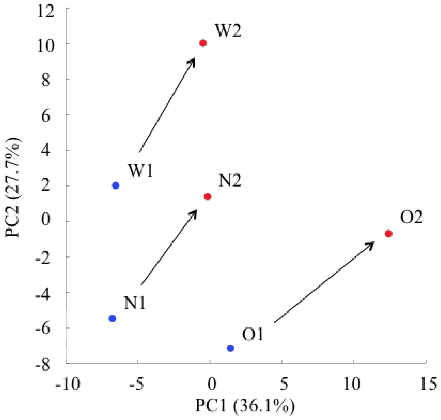
A capillary electrophoresis time-of-flight mass spectrometry (CE-TOFMS) system detected 49 peaks (33 cation and 16 anion). Principal component analysis (PCA) is a technique for obtaining high-dimensional data that uses dependencies between variables to present data in a more tractable, lower-dimensional form, without losing too much information [16]. The PCA score plots of each dog changed in a similar manner before (O1, N1, W1) and after (O2, N2, W2) oral administration of GlcN∙HCl (Figure 1). The results revealed that the individual variability in the change in amino acid metabolism was small.
Figure 1. Principal component analysis (PCA) score plots before (blue dots: O1, N1, W1) and after (red dots: O2, N2, W2) oral administration of D-Glucosamine hydrochloride (GlcN∙HCl). Arrows indicate changing power directions after GlcN∙HCl administration in each dog.
Marinedrugs 10 01873 g001

2.2. Changes in Plasma Metabolite Concentrations

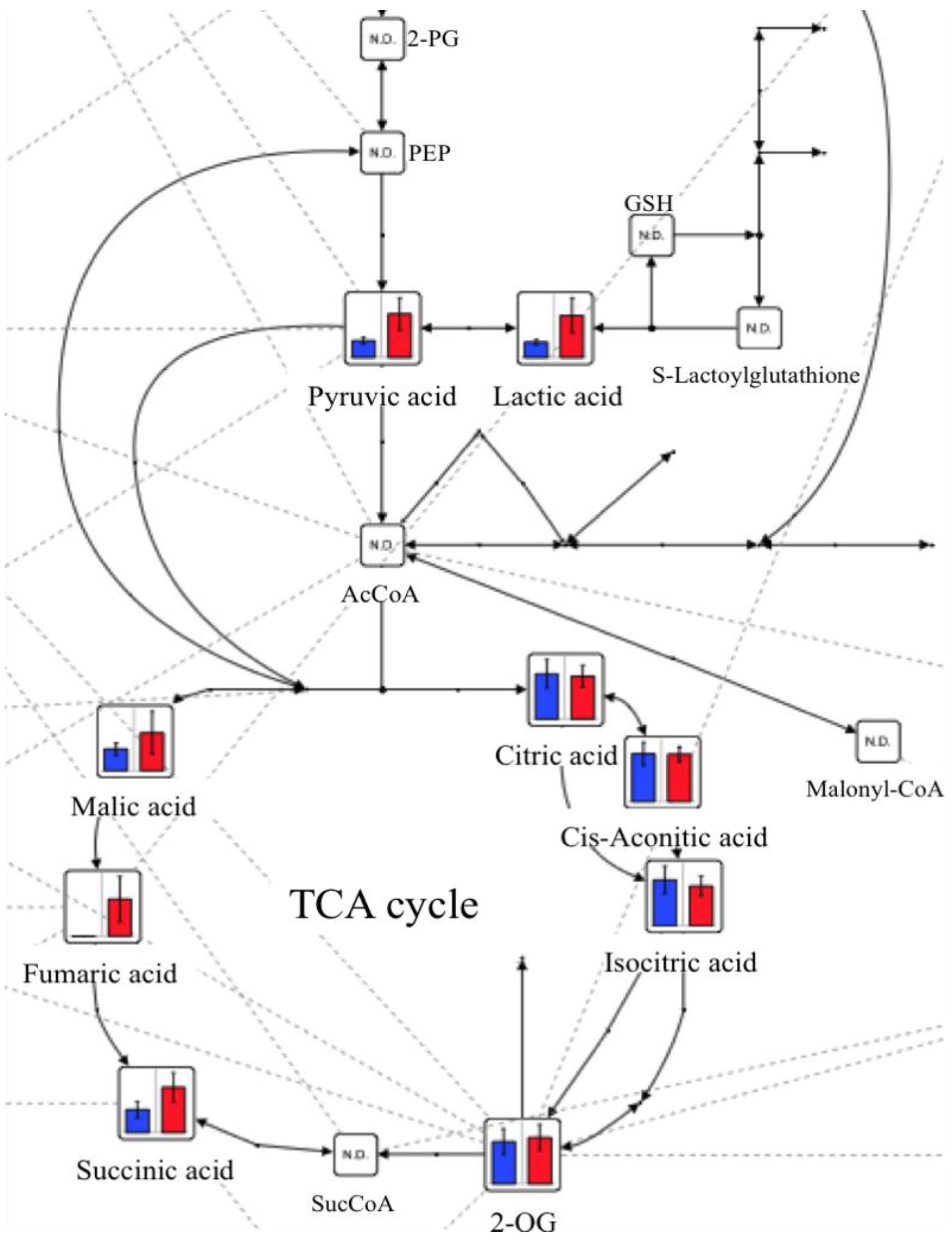
Table 1 shows the concentrations of major metabolites before and after GlcN∙HCl administration. Glycolysis and the tricarboxylic acid (TCA) cycle are illustrated in Figure 2. The levels of succinic acid, malic acid, lactic acid, and pyruvic acid all increased after administration of GlcN∙HCl. Fumaric acid was not detectable before administration but reached a discernable level afterwards. The levels of hydroxyproline and alanine were significantly higher after oral administration of GlcN∙HCl than before (p < 0.01 and p < 0.05, respectively).
Table 1. Concentrations (µM) of major metabolites before and after oral administration of D-Glucosamine hydrochloride (GlcN∙HCl).
MetabolitesBeforeAfter
MeanSDMeanSD
Glyoxylic acid3.20.61.7N.A.
Pyruvic acid942024089
Lactic acid137726337601582
3-Hydroxybutyric acid3915283.0
2-Hydroxybutyric acid3415207.3
Fumaric acidN.D.N.A.2.61.6
2-Oxoisovaleric acid105.8126.7
Succinic acid7.42.8154.8
Malic acid7.62.1137.6
2-Oxoglutaric acid195.7205.6
Glycerol 3-phosphate2.80.54.01.1
cis-Aconitic acid8.42.08.31.3
3-Phosphoglyceric acid1.00.31.60.3
Citric acid2818826668
Isocitric acid8.72.67.61.9
Gluconic acid5.31.96.11.1
Glycine1448.514825
Putrescine0.60.060.70.13
Sarcosine *3.80.65.90.3
Alanine *2914241739
β-Alanine0.60.20.70.2
N,N-Dimethylglycine4.90.96.11.6
Choline6.32.99.52.2
Serine1091413236
Creatinine484.7453.8
Proline871612726
Betaine233312895.3
Valine1054211535
Threonine1162815065
Hydroxyproline **3.70.115.70.3
Creatine173.1209.2
Isoleucine3613415.8
Leucine79269631
Asparagine374.4448.4
Ornithine124.9135.1
Aspartic acid **4.10.36.40.5
Glutamine755177907204
Lysine15310314329
Glutamic acid305.0367.7
Methionine294.0342.4
Histidine *793.5936.9
Phenylalanine538.7566.4
Arginine851410225
Citrulline53226419
Tyrosine375.8446.6
Tryptophan51175717
Carnosine253.7315.7
Cytidine0.70.081.20.2
Glutathione (GSSG) divalent0.60.090.80.3
GlucosamineN.D.N.A.N.D.N.A.
N-AcetylglucosamineN.A.N.A.N.A.N.A.
Asterisks indicate significant differences between the concentration of metabolites before and after oral administration of GlcN∙HCl. The p values were evaluated by the Welch’s t test. * p < 0.05; ** p < 0.01; SD: standard deviation; N.D.: not detected; N.A.: not available.
Figure 2. Levels of metabolites for glycolysis and the tricarboxylic acid (TCA) cycle before (blue bars) and after (red bars) oral administration of GlcN∙HCl.
Marinedrugs 10 01873 g002

3. Discussion

Glucosamine is a widely used dietary supplement for promoting joint health. There have been concerns that oral GlcN supplementation at usual doses may adversely affect glucose metabolism in subjects with impaired glucose tolerance. However, a recent report showed that GlcN had no effects on fasting blood glucose levels, glucose metabolism, or insulin sensitivity at any oral dose in healthy subjects, in those with diabetes, and in those with impaired glucose tolerance [17].
In this study, GlcN was not detected in the plasma the day after stopping oral administration of GlcN∙HCl (day 36), which is in agreement with the results of our previous study [13]. That report revealed that amino acid levels in the plasma changed 1 h after oral GlcN∙HCl administration and that the metabolomics profile could change within a day. The observed changes in metabolomic profiles in the current study were definitely related to GlcN∙HCl administration because all other life cycle variables, including diet, were constant. Because the plasma levels of GlcN increased slightly just after feeding, we assume that GlcN did not denature. However, in the future we need to investigate the stability of GlcN∙HCl in food.
In this study, levels of N-acetylglucosamine (GlcNAc) remained nearly unchanged before and after administration of GlcN∙HCl. Changes in GlcNAc levels were evaluated, but our measurement technique was not able to quantify its concentration in the plasma because of the lack of a standard substance. We assume that the administration of GlcN∙HCl did not affect the metabolism of GlcNAc. In future studies, the concentration of GlcNAc should be quantified using high performance liquid chromatography.
No prior reports have described the increases of succinic acid, malic acid, and fumaric acid resulting from oral administration of GlcN. It is known that some parasites and cancer cells depend on fumarate respiration using a reverse reaction involving succinate dehydrogenase, producing succinate as a byproduct [18,19]. Under conditions of glucose depletion and severe hypoxia, ATP is synthesized without oxygen by fumarate respiration. The active use of fumarate respiration after oral administration of GlcN∙HCl provides a possible explanation of the accumulation of fumaric acid observed in this study.
During glycolysis, the levels of lactic acid and pyruvic acid increased after administration of GlcN∙HCl. Lactic acid is a predominant source of carbon atoms for glucose synthesized by gluconeogenesis. Accumulated lactic acid is recycled by the liver via the Cori cycle to form glucose; however, this is an energy-consuming process. Yalamanchi et al. [20] showed that lactic acid significantly increased transforming growth factor-beta (TGF-β) peptide expression, receptor expression, and functional activity. TGF-β is a cytokine with numerous functions related to wound healing, including recruitment of fibroblasts and macrophages, stimulation of collagen production, and formation of new capillaries [21,22]. Cartilage regeneration might therefore result from its increase.
During fasting, muscles break down protein, and amino acids are released into circulation. In the present study, the levels of hydroxyproline and alanine after oral administration of GlcN∙HCl were significantly higher than levels prior to administration, with alanine higher to a greater extent. Alanine can be directly converted to pyruvic acid and used as a substrate for gluconeogenesis (via the alanine cycle) [23], which could possibly explain the increase in plasma concentrations of pyruvic acid we found.
Glucose depletion might result from hexosamine-induced insulin resistance [24]. GlcN taken up by cells is phosphorylated into GlcN-6-phosphate by hexokinase, which then bypasses GFAT to enter the hexosamine pathway [25]. It is then converted to uridine diphosphate-GlcNAc. Elevated levels of GlcNAc inhibit insulin-stimulated glucose uptake (via GLUT4 translocation) and glycogen synthesis [24]. GlcNAc also serves as a donor substrate for the synthesis of nucleotides, glycosyl phosphatidylinositol anchors, complex protein glycosylation, and O-linked-β-N-acetylglucosamine (O-GlcNAc) protein modification. O-GlcNAc modification of cytosolic and nuclear proteins occurs via the action of O-GlcNAc transferase [24]. This post-translational modification enhances the transcription of TGF-β and plasminogen activator inhibitor-1 [24,26]. Similarly, TGF-β induced by O-GlcNAc might stimulate collagen production.

4. Materials and Methods

4.1. Chemicals

GlcN∙HCl was supplied by Koyo Chemical Co., Ltd. (Tokyo, Japan).

4.2. Animals

Three healthy beagle dogs, mean age 6.3 years (range, 5–7 years) and mean body weight 10.4 kg (range, 9–11.1 kg), were used in this study. The use of these animals and all related procedures were approved by the Animal Research Committee of Tottori University.

4.3. Administration and Blood Sampling

GlcN∙HCl (25 mg/kg) was mixed with standard dog food (Dog Meal; Cainz Home Co., Ltd., Gunma, Japan) just before feeding. The dose was set in proportion to the recommended human dose (1.5 g/65 kg). The dogs were fed at the same time daily for 35 days. Blood samples from each dog were collected before beginning GlcN∙HCl administration and again 36 days after. The samples were drawn from the jugular vein using heparin as an anti-coagulant. They were centrifuged at 734× g for 10 min, and the plasma was then immediately separated and frozen at −80 °C.

4.4. Instrumentation

Capillary electrophoresis time-of-flight mass spectrometry (CE-TOF-MS) was carried out using an Agilent CE capillary electrophoresis system (Agilent Technologies, Waldbronn, Germany) equipped with an Agilent 6210 time-of-flight mass spectrometer, an Agilent 1100 isocratic HPLC pump, an Agilent G1603A CE-MS adapter kit, and an Agilent G1607A CE-ESI-MS sprayer kit. The overall system was controlled by Agilent G2201AA ChemStation software version B.03.01 for CE.

4.5. CE-TOFMS Conditions

Cationic metabolites were analyzed with a fused silica capillary column (50 µm i.d. × 80 cm total length) and commercial cation electrophoresis buffer (Solution ID: H3301-1001, Human Metabolome Technologies) as the electrolyte. Samples were injected at a pressure of 50 mbar for 10 s (approximately 10 nL), and the applied voltage was set at 27 kV. Electrospray ionization-mass spectrometry (ESI-MS) was conducted in the positive ion mode, and the capillary voltage was set at 4 kV. The spectrometer was scanned from m/z 50 to 1000. Other conditions were standard for cation analysis [27,28].
Anionic metabolites were similarly analyzed with a fused silica capillary column and commercial anion electrophoresis buffer (Solution ID: H3302-1021, Human Metabolome Technologies) as the electrolyte. Samples were injected at a pressure of 50 mbar for 25 s (approximately 25 nL), and the applied voltage was set at 30 kV. ESI-MS was conducted in the negative ion mode, and the capillary voltage was set at 3.5 kV. The spectrometer was scanned from m/z 50 to 1000. Other conditions were standard for anion analysis [28,29,30].

4.6. Data Analysis

Raw data obtained by CE-TOFMS were processed with MasterHands software [31]. Signal peaks corresponding to isotopomers, adduct ions, and other product ions of known metabolites were excluded from analysis. All signal peaks potentially corresponding to authentic compounds were extracted, and their migration times (MT) were normalized using internal standards (MetSul for cations and CSA for anions). The peaks were then aligned according to the m/z and normalized MT values. Finally, peak areas were normalized using the internal standards and by sample amount. Annotation tables were produced from CE-ESI-TOFMS measurements of standard compound and aligned with the datasets according to similar m/z and normalized MT values. The metabolic pathway map was provided using the public-domain software Visualization and Analysis of Networks containing Experimental Data (VANTED) [32,33].

4.7. Statistical Analysis

All values are presented as the mean ± standard deviation (SD). The Welch’s t-test was used to compare the amino acid concentrations in the plasma before and after oral administration of GlcN∙HCl. The results were considered significant at p < 0.05.

5. Conclusions

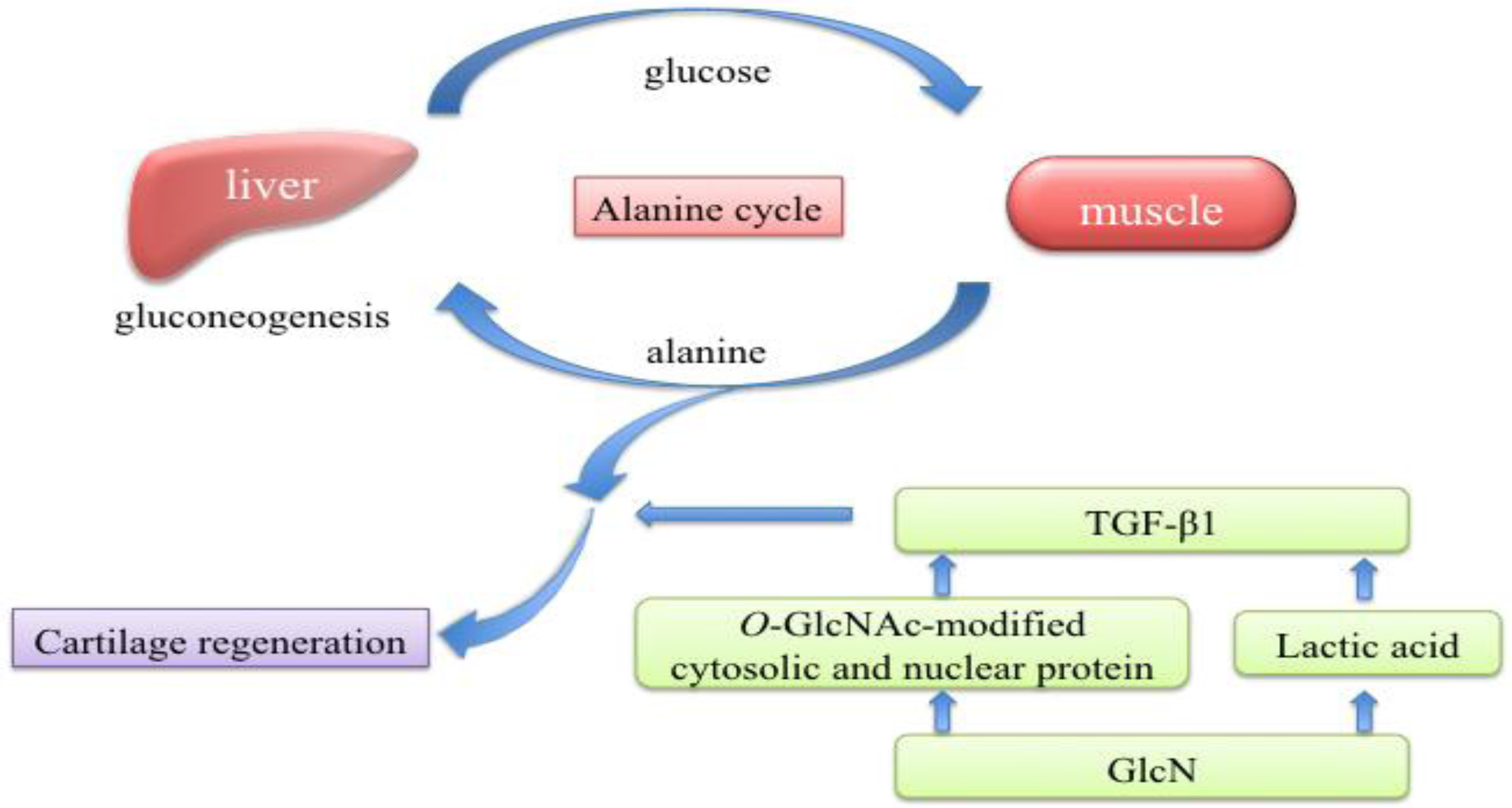
In this study, accelerated fumarate respiration and elevated plasma levels of lactic acid and alanine were observed after oral administration of GlcN∙HCl. These results indicate that oral administration of GlcN∙HCl induced anaerobic respiration and starvation in cells and further suggest that lactic acid and O-GlcNAc could potentially induce TGF-β production. We hypothesize that these conditions induced by oral administration of GlcN∙HCl promote cartilage regeneration as shown in the schematic diagram in Figure 3. However, further studies are required to evaluate the expression of TGF-β.
Figure 3. Schematic diagram of cartilage regeneration. Elevated plasma levels of lactic acid and alanine were observed after oral administration of GlcN∙HCl. We hypothesize that lactic acid and O-GlcNAc induce production of TGF-β, which would promote cartilage regeneration.
Marinedrugs 10 01873 g003

Acknowledgments

We thank Kazuki Watanabe for providing advice on data interpretation.

References

  1. Fox, B.A.; Stephens, M.M. Glucosamine hydrochloride for the treatment of osteoarthritis symptoms. Clin. Interv. Aging 2007, 2, 599–604. [Google Scholar]
  2. Aghazadeh-Habashi, A.; Jamali, F. The glucosamine controversy; a pharmacokinetic issue. J. Pharm. Pharm. Sci. 2011, 14, 264–273. [Google Scholar]
  3. Clegg, D.O.; Reda, D.J.; Harris, C.L.; Klein, M.A.; O’Dell, J.R.; Hooper, M.M.; Bradley, J.D.; Bingham, C.O., III.; Weisman, M.H.; Jackson, C.G.; et al. Glucosamine, chondroitin sulfate, and the two in combination for painful knee osteoarthritis. N. Engl. J. Med. 2006, 354, 795–808. [Google Scholar] [CrossRef]
  4. Goodrich, L.R.; Nixon, A.J. Medical treatment of osteoarthritis in the horse—a review. Vet. J. 2006, 171, 51–69. [Google Scholar] [CrossRef]
  5. Minami, S.; Hata, M.; Tamai, Y.; Hashida, M.; Takayama, T.; Yamamoto, S.; Okada, M.; Funatsu, T.; Tsuka, T.; Imagawa, T.; et al. Clinical application of D-glucosamine and scale collagen peptide on canine and feline orthopedic diseases and spondylitis deformans. Carbohydr. Polym. 2010, 84, 831–834. [Google Scholar]
  6. Barclay, T.S.; Tsourounis, C.; McCart, G.M. Glucosamine. Ann. Pharmacother. 1998, 32, 574–579. [Google Scholar] [CrossRef]
  7. Aghazadeh-Habashi, A.; Sattari, S.; Pasutto, F.; Jamali, F. Single dose pharmacokinetics and bioavailability of glucosamine in the rat. J. Pharm. Pharm. Sci. 2002, 5, 181–184. [Google Scholar]
  8. Adebowale, A.; Du, J.; Liang, Z.; Leslie, J.L.; Eddington, N.D. The bioavailability and pharmacokinetics of glucosamine hydrochloride and low molecular weight chondroitin sulfate after single and multiple doses to beagle dogs. Biopharm. Drug Dispos. 2002, 23, 217–225. [Google Scholar] [CrossRef]
  9. Du, J.; White, N.; Eddington, N.D. The bioavailability and pharmacokinetics of glucosamine hydrochloride and chondroitin sulfate after oral and intravenous single dose administration in the horse. Biopharm. Drug Dispos. 2004, 25, 109–116. [Google Scholar] [CrossRef]
  10. Laverty, S.; Sandy, J.D.; Celeste, C.; Vachon, P.; Marier, J.F.; Plaas, A.H. Synovial fluid levels and serum pharmacokinetics in a large animal model following treatment with oral glucosamine at clinically relevant doses. Arthritis Rheum. 2005, 52, 181–191. [Google Scholar] [CrossRef]
  11. Meulyzer, M.; Vachon, P.; Beaudry, F.; Vinardell, T.; Richard, H.; Beauchamp, G.; Laverty, S. Comparison of pharmacokinetics of glucosamine and synovial fluid levels following administration of glucosamine sulfate or glucosamine hydrochloride. Osteoarthr. Cartil. 2008, 16, 973–979. [Google Scholar]
  12. Setnikar, I.; Giachetti, C.; Zanolo, G. Absorption, distribution and excretion of radioactivity after a single intravenous or oral administration of [14C] glucosamine to the rat. Pharmatherapeutica 1984, 3, 538–550. [Google Scholar]
  13. Azuma, K.; Osaki, T.; Tsuka, T.; Imagawa, T.; Okamoto, Y.; Takamori, Y.; Minami, S. Effects of oral glucosamine hydrochloride administration on plasma free amino acid concentrations in dogs. Mar. Drugs 2011, 9, 712–718. [Google Scholar] [CrossRef]
  14. Tamai, Y.; Miyatake, K.; Okamoto, Y.; Takamori, Y.; Sakamoto, H.; Minami, S. Enhanced healing of cartilaginous injuries by glucosamine hydrochloride. Carbohydr. Polym. 2002, 48, 369–378. [Google Scholar] [CrossRef]
  15. Tamai, Y.; Miyatake, K.; Okamoto, Y.; Takamori, Y.; Sakamoto, K.; Minami, S. Enhanced healing of cartilaginous injuries by N-acetyl-D-glucosamine and glucuronic acid. Carbohydr. Polym. 54, 251–262.
  16. Ringnér, M. What is principal component analysis? Nat. Biotechnol. 2008, 26, 303–304. [Google Scholar] [CrossRef]
  17. Simon, R.R.; Marks, V.; Leeds, A.R.; Anderson, J.W. A comprehensive review of oral glucosamine use and effects on glucose metabolism in normal and diabetic individuals. Diabetes Metab. Res. Rev. 2011, 27, 14–27. [Google Scholar] [CrossRef]
  18. Kita, K.; Hirawake, H.; Miyadera, H.; Amino, H.; Takeo, S. Role of complex II in anaerobic respiration of the parasite mitochondria from Ascaris suum and Plasmodium falciparum. Biochim. Biophys. Acta 2002, 1553, 123–139. [Google Scholar]
  19. Hirayama, A.; Kami, K.; Sugimoto, M.; Sugawara, M.; Toki, N.; Onozuka, H.; Kinoshita, T.; Saito, N.; Ochiai, A.; Tomita, M.; et al. Quantitative metabolome profiling of colon and stomach cancer microenvironment by capillary electrophoresis time-of-flight mass spectrometry. Cancer Res. 2009, 69, 4918–4925. [Google Scholar]
  20. Yalamanchi, N.; Klein, M.B.; Pham, H.M.; Longaker, M.T.; Chang, J. Flexor tendon wound healing in vitro: Lactate up-regulation of TGF-beta expression and functional activity. Plast. Reconstr. Surg. 2004, 113, 625–632. [Google Scholar] [CrossRef]
  21. Folkman, J.; Klagsbrun, M. Angiogenic factors. Science 1987, 235, 442–447. [Google Scholar]
  22. Klein, M.B.; Yalamanchi, N.; Pham, H.; Longaker, M.T.; Chang, J. Flexor tendon healing in vitro: Effects of TGF-beta on tendon cell collagen production. J. Hand Surg. Am. 2002, 27, 615–620. [Google Scholar] [CrossRef]
  23. Felig, P. The glucose-alanine cycle. Metabolism 1973, 22, 179–207. [Google Scholar] [CrossRef]
  24. Cheatham, B. Enough is enough: Nutrient sensors and insulin resistance. Endocrinology 2004, 145, 2115–2117. [Google Scholar] [CrossRef]
  25. Uldry, M.; Ibberson, M.; Hosokawa, M.; Thorens, B. GLUT2 is a high affinity glucosamine transporter. FEBS Lett. 2002, 524, 199–203. [Google Scholar] [CrossRef]
  26. Luo, B.; Soesanto, Y.; McClain, D.A. Protein modification by O-linked GlcNAc reduces angiogenesis by inhibiting Akt activity in endothelial cells. Arterioscler. Thromb. Vasc. Biol. 2008, 28, 651–657. [Google Scholar] [CrossRef]
  27. Soga, T.; Heiger, D.N. Amino acid analysis by capillary electrophoresis electrospray ionization mass spectrometry. Anal. Chem. 2000, 72, 1236–1241. [Google Scholar] [CrossRef]
  28. Soga, T.; Ohashi, Y.; Ueno, Y.; Naraoka, H.; Tomita, M.; Nishioka, T. Quantitative metabolome analysis using capillary electrophoresis mass spectrometry. J. Proteome Res. 2003, 2, 488–494. [Google Scholar] [CrossRef]
  29. Soga, T.; Ueno, Y.; Naraoka, H.; Ohashi, Y.; Tomita, M.; Nishioka, T. Simultaneous determination of anionic intermediates for Bacillus subtilis metabolic pathways by capillary electrophoresis electrospray ionization mass spectrometry. Anal. Chem. 2002, 74, 2233–2239. [Google Scholar] [CrossRef]
  30. Soga, T.; Ishikawa, T.; Igarashi, S.; Sugawara, K.; Kakazu, Y.; Tomita, M. Analysis of nucleotides by pressure-assisted capillary electrophoresis-mass spectrometry using silanol mask technique. J. Chromatogr. 2007, 1159, 125–133. [Google Scholar]
  31. Sugimoto, M.; Hirayama, A.; Ihiskawa, T.; Baran, R.; Robert, M.; Uehara, K.; Soga, T.; Tomita, M. Differential metabolomics software for capillary electrophoresis-mass spectrometry data analysis. Metabolomics 2010, 6, 27–41. [Google Scholar] [CrossRef]
  32. Junker, B.H.; Klukas, C.; Schreiber, F. VANTED: A system for advanced data analysis and visualization in the context of biological networks. BMC Bioinforma. 2006, 7. [Google Scholar] [CrossRef]
  33. Klukas, C.; Schreiber, F. Integration of -omics data and networks for biomedical research with VANTED. J. Integr. Bioinform. 2010, 7, 112. [Google Scholar]