Article

Structure Elucidation and Anticancer Activity of 7-Oxostaurosporine Derivatives from the Brazilian Endemic Tunicate Eudistoma vannamei

by
Paula Christine Jimenez
1,2,
Diego Veras Wilke
2,
Elthon Gois Ferreira
1,
Renata Takeara
3,
Manoel Odorico De Moraes
2,
Edilberto Rocha Silveira
4,
Tito Monteiro Da Cruz Lotufo
1,
Norberto Peporine Lopes
3 and
Leticia Veras Costa-Lotufo
1,2,*
1
Institute of Marine Sciences, LABOMAR, Federal University of Ceará, Fortaleza, CE 60165-081, Brazil
2
Department of Physiology and Pharmacology, School of Medicine, Federal University of Ceará, Fortaleza, CE 60430-270, Brazil
3
Department of Chemistry and Physics, Faculty of Pharmaceutical Sciences of Ribeirão Preto, University of São Paulo, Ribeirão Preto, SP 14040-903, Brazil
4
Department of Organic and Inorganic Chemistry, Federal University of Ceará, Fortaleza, CE 60021-970, Brazil
*
Author to whom correspondence should be addressed.
Mar. Drugs 2012, 10(5), 1092-1102; https://doi.org/10.3390/md10051092
Submission received: 21 March 2012 / Revised: 11 May 2012 / Accepted: 11 May 2012 / Published: 21 May 2012

Abstract

:
The present study reports the identification of two new staurosporine derivatives, 2-hydroxy-7-oxostaurosporine (1) and 3-hydroxy-7-oxostaurosporine (2), obtained from mid-polar fractions of an aqueous methanol extract of the tunicate Eudistoma vannamei, endemic to the northeast coast of Brazil. The mixture of 1 and 2 displayed IC50 values in the nM range and was up to 14 times more cytotoxic than staurosporine across a panel of tumor cell lines, as evaluated using the MTT assay.

1. Introduction

Tunicates are a group of marine invertebrates particularly abundant along coastal regions, often found as the dominant organisms in sessile benthic communities [1]. In Brazil, as in many other regions, information regarding these animals is restricted to faunal inventories and, in many cases, is inaccurate or incomplete [2]. In regards to Brazilian tropical waters, the deficiency in species inventories is quite evident, as very few references are available [3,4,5].
The faunal list for the state of Ceará, on the northeastern coast of Brazil, indicated that 22 species of ascidians were registered [5,6], including novel species and many cases of regional endemism. This coastline has been scarcely explored as a source of natural products. In 2003, a pioneering study investigated the cytotoxicity of aqueous methanol extracts of the ten most abundant ascidians of Ceará. The results of these assays suggested that Eudistoma vannamei Millar, 1977 (Figure 1 a) might be of interest due to its marked bioactivity, especially the inhibition of growth in tumor cell lines [7]. Evaluation of the cytotoxicity data from these extracts suggested the presence of amino acid-derived compounds in fractions of intermediate polarity. Treatment of leukemia cells with the amino acid-derived compounds resulted in apoptosis [8].
Figure 1. (a) Eudistoma vannamei; (b) Structures of 2-hydroxy-7-oxostaurosporine (1), 3-hydroxy-7-oxostaurosporine (2) and staurosporine (3).
Marinedrugs 10 01092 g001
The genus Eudistoma is the most diverse among the Polycitoridae, with most of its species living in tropical regions [9]. On the Brazilian coast, only one species of Eudistoma was found in temperate waters [10], while in the warmer Northeast region, there are at least seven species of this genus. E. vannamei is endemic to the Northeast coast of Brazil where it forms large colonies on the underside of ledges of the beach rocks and is characterized by its conspicuous orange or yellow bulbus heads [3,5]. E. vannamei’s sessile habit and apparent lack of mechanical defenses would make it an easy target for predators. However, taking into account its dominance on the intertidal reefs, the species most probably relies on chemical deterrents for protection.
Several cytotoxic substances have been isolated from Eudistoma ascidians, including the alkaloids eudistomins A–T, eudistomidins A–F and eudistalbin [11,12,13,14,15,16,17,18,19] and the macrolides iejimalides A–D [20,21]. The present study reports the bioactivity-guided isolation of two new members of the staurosporine group of alkaloids from the extract of E. vannamei.

2. Results and Discussion

The aqueous methanol extract from E. vannamei was submitted to cytotoxicity-guided fractionation (flowchart provided in the supplementary material), resulting in the isolation of 5 mg of an active fraction, which appeared as a single spot by TLC and a single peak via LC-MS analysis.
This material was submitted to NMR and HRMS analysis. The ESI-HRMS spectrum showed one signal at m/z 497.1830 (error value: 2.21 ppm), suggesting the molecular formula C28H24N4O5. The initial analysis of the 13C NMR contained more carbons than expected, and appeared as two sets of peaks with similar chemical shifts. Detailed analysis of the MS and MS/MS data along with evaluation of the 13C NMR data suggested that the material was a mixture of two staurosporine derivatives, compounds 1 and 2.
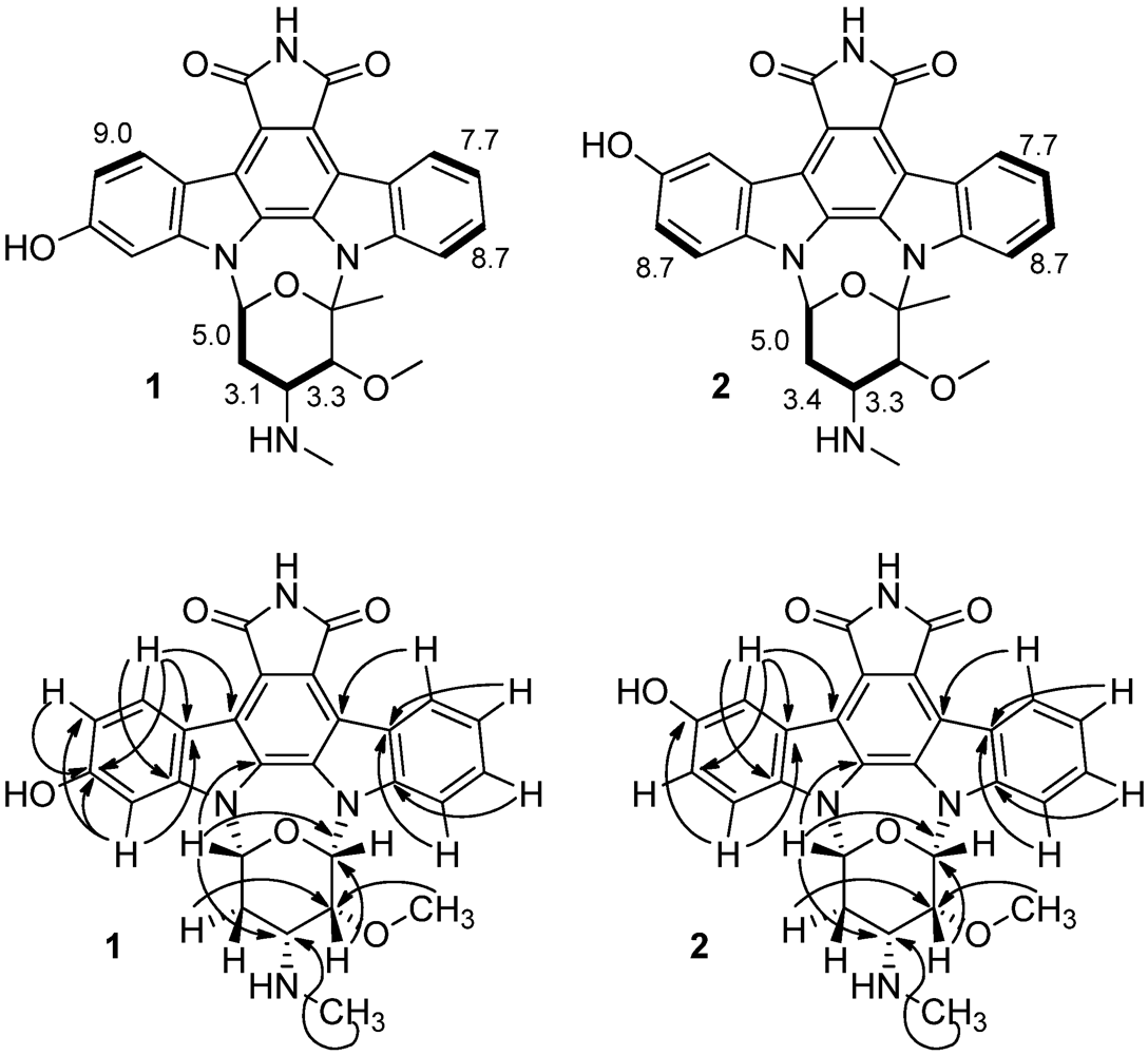
A detailed set of NMR spectral data was then collected including 1H, 13C, COSY, HSQC and HMBC spectra to identify the structures of 1 and 2. Each of the protons in 1 and 2 were assigned using a combination of chemical shift data and coupling constants from the 1H NMR spectrum (Table 1) and correlations from a COSY spectrum (Figure 2). Analysis of the chemical shift and coupling constant data indicated that the glycosidic unit matched that in staurosporine [22,23,24,25]. An HSQC experiment enabled the assignments of each methine, methylene and methyl carbon (Table 1). The COSY spectrum revealed the scalar coupling correlations for the most deshielded proton attached to sp3 carbons H-6′ (δH 6.62 for 1 and 6.69 for 2) with the heterotopic methylene protons 2H-5′ (δH 2.36/2.72 for 1 and δH 2.46/2.74 for 2) which, in turn, showed geminal coupling. The oxymethine protons at δH 3.95 (H-3′ for 1) and 3.97 (H-3′ for 2) showed correlations with the azomethine protons at δH 3.23 and 3.26 (H-4′) for 1 and 2, respectively. In the aromatic region, the most deshielded proton at δH 9.94 (H-8 for both 1 and 2) coupled to one at δH 7.46 (H-9 for both 1 and 2), which coupled with one at δH 7.61 (H-10 for both 1 and 2) and that, in turn, coupled with the ones at δH 8.15 (for 1) and 8.17 (for 2). On the hydroxyl substituted benzene ring, the most deshielded doublet (J = 8.4 Hz) at δH 9.63 (H-4 for 1) correlated with the one at δH 7.40 (H-3 of 1), while the correspondent H-4 of 2 at δH 9.58 (br, d) coupled weakly with the proton at δH 7.61 that, in turn, coupled with the one at δH 7.54 (d, J = 8.7 Hz, H-1 of 2).
Table 1. NMR Data (500 MHz, pyridine-d5) for 2-hydroxy-7-oxostaurosporine (1) and 3-hydroxy-7-oxostaurosporine (2).
2-Hydroxy-7-oxostaurosporine (1)3-Hydroxy-7-oxostaurosporine (2)
PositionδC, mult.δH ( J in Hz)HMBC dδC, mult.δH ( J in Hz)HMBC d
195.6, CH7.39 br. s2, 3, 4a109.7, CH 7.54 d (8.7)3
2159.5, C 117.4, CH7.61 c3
3111.3, CH7.40 d (9.6)4a153.8, C
4126.6, CH9.63 d (8.4)2, 4b, 13a111.7, CH9.58 br. d2, 13a
4a115.8, C 123.8, C
4b117.0, C 117.0, C
4c120.2 a, C 120.0 a, C
5172.7 b, C 172.6 b, C
7a123.4 a, C 122.1 a, C
7b116.8, C 116.8, C
7c124.7, C 124.7, C
8125.8, CH9.94 d (7.9)7b, 10, 11a125.9, CH9.94 d (7.9)7b, 10, 11a
9120.7, CH7.46 d (7.5)7c, 11120.7, CH7.46 t (7.5)7c, 11
10126.4, CH7.61 c8, 11a126.4, CH7.61c8, 11a
11116.1, CH8.15 d (8.7)7c, 9116.8, CH8.17 d (8.7)7c, 9
11a141.9, C 141.8, C
12a132.3, C 132.4, C
12b131.2, C 131.8, C
13a141.1, C 133.3, C
2′91.7, C 91.8, C
3′84.1, CH3.95 d (3.3)2′, CH3O, CH384.2, CH3.97 d (3.3)2′, CH3O, CH3
4′50.8, CH3.23 br. q (3.1)2′, 3′, 6′, CH3N50.8, CH3.26 br. q (3.4)2′, 3′, 6′, CH3N
5′29.9, CH22.72 m3′, 4′, 6′29.8, CH22.74 m3′, 4′, 6′
2.36 m 2.46 m
6′80.8, CH6.62 d (5.0)2′, 4′, 12b80.9, CH6.69 d (5.0)2′, 4′, 12b
CH3-NH30.5, CH31.48 s4′30.5, CH31.48 s4′
CH3O-C3′57.2, CH33.31 s3′57.2, CH33.32 s3′
CH3-C2′33.8, CH32.37 s2′, 3′33.8, CH32.38 s2′, 3′
a,b Values with the same superscript are interchangeable in the same column; c Due to partial overlapping the multiplicity and J values could not be determined precisely; d HMBC correlations, optimized for 7.25 Hz, are from proton(s) stated to the indicated carbon.
Figure 2. A summary of the NMR data including (top) coupling constants and COSY correlations (bottom) key long-range 1H,13C-correlations (depicted by arrows) observed in the HMBC experiment.
Marinedrugs 10 01092 g002
Subsequent HMBC data facilitated the assignment of the remaining carbons (Table 1) with the exception of C-4c, C-5, C-7 and C-7a, which were interpreted by comparison with the literature [22,23,24,25]. Further long range correlations observed in the HMBC spectrum (Figure 2) provided definitive proof for the connectivity and thereby confirmed the structural assignment of 1 and 2 as 2-hydroxy-7-oxostaurosporine and 3-hydroxy-7-oxostaurosporine, respectively.
Staurosporines are a group of highly cytotoxic indole-carbazole alkaloids of which the parental molecule was isolated in 1977 from the fermentation broth of the soil actinomycete Streptomyces staurosporeus in a screening protocol directed to identify PKC (protein kinase C) inhibitors [26]. Currently, the staurosporines are a group of over 50 structurally related substances of natural and synthetic origins and have been found frequently in extracts from Eudistoma tunicates. 11-Hydroxystaurosporine and 3,11-dihydroxystaurosporine were isolated from Eudistoma sp. collected on a Micronesian island [27], while the staurosporine aglicone (K252-c) was obtained from another Eudistoma species found on the west coast of Africa [28]. Between 1999 and 2002, Schupp and co-workers published three articles covering the isolation and nM-range cytotoxicity of five new (3-hydroxy-4′-N-methylstaurosporine, 3-hydroxy-4′-N-demethylstaurosporine, 3′-demethoxy-3′-hydroxy-4′-N-demethylestaurosporine, 3-hydroxy-3′-demethoxy-3′-hydroxystaurosporine and 11-hydroxy-4′-N-demethylstaurosporine) and seven previously known staurosporines found in the Micronesian ascidian Eudistoma toealensis and its predatory flatworm Pseudoceros sp. [24,29,30].
The mixture of compounds 12 presented strong cytotoxic activity against a panel of seven human tumor cell lines (HL-60, Molt-4, Jurkat, K562, HCT-8, SF-295 and MDA-MB-435) and normal proliferating lymphocytes (PBMC), with an IC50 ranging from 10.33 nM in Jurkat leukemia cells to 687.08 nM in normal PBMC cells (Table 2). Standard staurosporine (3), used as a positive control, was generally less potent than compounds 12 against tumor cells. Against normal proliferating lymphocytes, both 3 and 12 presented similar activity, suggesting a better selectivity index for 12 (Table 2). For example, 12 was 26.46 times more active against HL-60 cells than against PBMC, whereas STP was 2.00 times more active against HL-60 cells when compared with normal cells.
Table 2. Cytotoxicity of 2-hydroxy-7-oxostaurosporine/3-hydroxy-7-oxostaurosporine (1/2) and staurosporine (3), evaluated using the MTT assay after 72 h of incubation. The IC50 (nM) values and 95% CI were obtained via nonlinear regression (first values); the 95% CI are set in brackets.
Cell LineIC50 (nM) 1–2Selectivity Index a PBMC vs. Cancer CellsIC50 (nM) 3Selectivity Index a PBMC vs. Cancer Cells
HL-6025.97 [22.42–30.09]26.46391.83 [316.81–484.86]2.00
Molt-418.64 [15.97–21.74]36.86154.50 [128.12–186.33]5.08
Jurkat10.33 [7.12–15.00]70.0883.96 [51.38–137.25]9.34
K562144.47 [103.88–200.9]4.751960.86 N.C.0.40
HCT-858.24 [50.96–66.58]11.8083.83 [66.43–105.80]9.36
SF 29557.90 [47.10–71.16]11.87569.52 [444.13–730.28]1.38
MDA MB 43528.68 [25.64–32.06]23.96215.42 [153.64–301.80]3.64
PBMC687.08 [452.55–1043.48]-784.51 [566.95–1085.89]
N.C.: value was not converted; a ratio between the cytotoxicities expressed as IC50 (nM) against peripheral blood mononucleated cells (PBMCs) (normal cells) and cancer cell lines.
Structure-activity relationship studies involving staurosporine and structurally related compounds have shown that slight changes in structure lead to drastic changes in functionality or, essentially, selectivity towards a target [31,32,33,34]. A preliminary investigation on the mode of action of compounds 1 and 2 was conducted using HL-60 leukemia cells as a model. The mixture of 1 and 2 (80 nM) induced a notable and sustained cytostatic effect against HL-60 cells throughout the 72 h analysis. This observation was accompanied by an increase in the accumulation of cells in G2/M and a decrease of those in G0/G1 and S phases. The toxicity of STP (430 nM), however, was kinetically less consistent. Moreover, after 24 h, almost the entire STP-treated culture was arrested in G2/M, which was followed by an increase in polyploid cells at further time points, showing a less strict blockage. When the concentration of the mixture was increased to 160 nM, extensive DNA damage occurred, leading to cellular apoptosis (results not shown). However, in order to access the contribution of each isomer in the observed antiproliferative activity, it will be necessary to isolate the compounds and to access the mode of action of each one individually.

3. Experimental Section

3.1. Reagents

Cell culture media, fetal bovine serum and antibiotics were acquired from Gibco (Grand Island, NY, USA). Staurosporine, MTT and resazurin were obtained from Sigma-Aldrich (St. Louis, MO, USA). All other reagents used were of analytical grade.

3.2. Collection and Identification of Eudistoma vannamei

Samples of Eudistoma vannamei were collected in crevices or on the underside of beach rocks in the intertidal zone of Taíba Beach (03°34,931′S; 038°54,469′W), on the west coast of Ceará state, Brazil. The material was immediately immersed in methanol and stored at −4 °C. Part of the material was fixed in 70% ethanol and sent for identification. A voucher specimen #198 has been deposited at the ascidian collection from the Institute of Marine Sciences, Federal University of Ceará (Instituto de Ciências do Mar, Universidade Federal do Ceará).

3.3. Extraction and Bioguided Fractionation

The collected specimens (8.8 kg) were extracted with MeOH (1:5, m/v, wet weight). The suspension was filtered, concentrated under reduced pressure (TECNAL, model TE-120) and lyophilized (Thermo Electron Corporation, model: MicroModulyo Freeze Dryer 115) to obtain the dry raw methanol extract (351.80 g). The raw extract was then resuspended in MeOH to precipitate the salt, filtered and afterwards diluted with water to a proportion of 7:3 (MeOH:H2O). A successive partition of the aqueous phase followed, first with CH2Cl2 (2:1, v/v, 3-fold) and then with n-BuOH (2:1, v/v, 3-fold). The solvent of all the fractions was removed under reduced pressure. The CH2Cl2 partition was fractionated by flash chromatography on a glass column (55 × 25 cm) filled with 800 g of silica gel 60 GF254 70–230 mesh ASTM (Sigma), using an n-hexane/EtOAc gradient from 20–100% EtOAc. After analysis with thin layer chromatography (TLC), 20 fractions (1 to 20) were obtained and further studied. Fraction 14 (1.81 g) exhibited the most promising biological potential and therefore was subjected to flash chromatography on silica gel 60 GF254 (glass column 55 × 25 cm; 170 g silica) in an isocratic elution with CH2Cl2:MeOH (9:1, v/v), which provided six sub-fractions. Further purification, after the biological evaluation, led to the selection of sub-fraction 3 (i.e., SF3), which was purified by TLC. The major sub-sub-fraction 1 (i.e., SF3.1) after sequential TLC resulted in one active mixture of isomers (5 mg). The structure elucidation was based on infrared spectra (IR; Perkin-Elmer, model FT-IR 1000), nuclear magnetic resonance (NMR) and high-resolution mass spectrometry. All NMR experiments were recorded on a Bruker DRX-500 spectrometer operating at 499.80 and 125.69 MHz for 1H and 13C, respectively, using standard pulse sequences supplied by the manufacturer. The ESI-MS was performed with Bruker Daltonics™ equipment (UltrO-TOF, Billerica, MA, USA). The sample (0.5 µg/mL) was dissolved in methanol/water at a 1:1 ratio and was introduced into the electrospray source at 5 µL/min via an infusion pump (Cole-Parmer, Vernon Hills, IL, USA). Nitrogen was used as a nebulising gas, and the capillary voltage was set to 3500 V.

3.4. Evaluation of Cytotoxicity

3.4.1. Cell Lines and Cell Models

HL-60 (promyelocytic leukemia), Molt-4 (lymphocytic leukemia), Jurkat (T cell leukemia), K562 (chronic myeloid leukemia) HCT-8 (colon cancer), MDA MB-435 (melanoma), and SF-295 (glioblastoma) human tumor cell lines were obtained from the National Cancer Institute, Bethesda, MD, USA. Cells were grown in RPMI-1640 medium supplemented with 10% fetal bovine serum, 2 mM glutamine, 100 μg/mL streptomycin and 100 U/mL penicillin and incubated at 37 °C under a 5% CO2 atmosphere.
Peripheral blood mononucleated cells (PBMCs) were used as a model for the evaluation of cytotoxicity in normal cells. Peripheral blood samples were obtained from four healthy volunteers. Venous blood (8 mL) was collected via routine venipuncture into a sterile tube with EDTA and carefully layered over 2 mL of Ficoll-Histopaque (Sigma-Aldrich, St. Louis, MO, USA) and centrifuged at 1500 rpm for 20 min. The layer containing the lymphocytes was aspirated, washed twice with PBS and tested for viability using trypan blue. PBMCs were suspended to their final concentration in RPMI medium supplemented with 20% fetal bovine serum, 2 mm glutamine, 100 μg/mL streptomycin, 100 U/mL penicillin and 3% phytohemagglutinin to stimulate proliferation.

3.4.2. MTT Assay

Cells were plated into 96-well plates (3 × 10⁵ cells/mL for suspended leukemia cells and 1 × 10⁵ cells/mL for adherent solid tumor cells). Adherent cells were plated 24 h prior to addition of test substances, which were added using the HTS, and incubated for 72 h. Control groups received DMSO. Three hours before the end of the incubation, 150 µL of a stock solution (0.5 mg/mL) of MTT (3-(4,5-dimethyl-2-thiazolyl)-2,5-diphenyl-2H-tetrazolium bromide; Sigma-Aldrich Co., St. Louis, MO, USA) was added to each well. Absorbance was measured using a multiplate reader (DTX 880 Multimode Detector, Beckman Coulter, Inc. Fullerton, CA, USA). The effect was quantified as the percentage of the control absorbance at 570 nm [35].

3.4.3. AlamarBlue® Assay

Cells were plated into 96-well plates (3 × 10⁵ cells/mL). After 24 h, substances were added to each well using the HTS, and cells were incubated for 72 h. Control groups received DMSO. Twenty-four hours before the end of the incubation, 10 µL of a stock solution (0.436 mg/mL) of Alamar Blue (resazurin, Sigma-Aldrich Co., St. Louis, MO, USA) was added to each well. Absorbance was measured as above. The effect of each sample was quantified as the percentage of the control absorbance at 570 nm and 600 nm [36].

4. Conclusions

In the present study, two new 7-oxostaurosporine derivatives were obtained from the Brazilian endemic tunicate Eudistoma vannamei. The mixture of these compounds presented a strong antiproliferative effect against tumor cell lines, inducing a distinguished and persistent G2 arrest in sub-toxic concentrations, while at toxic concentrations, the treated cells underwent apoptosis.

Acknowledgments

The authors wish to thank the National Agencies CNPq, CAPES, InCB and FAPESP for financial support and Silvana França for technical assistance with the cell culture. The English was edited by American Journal Experts (key #BB18-0547-39E0-459E-6C6B).

Supplementary Files

  • Supplementary File 1::

    PDF-Document (PDF, 270 KB)

  • References

    1. Millar, R.H. The biology of ascidians. Adv. Mar. Biol. 1971, 9, 1–100. [Google Scholar] [CrossRef]
    2. Shenkar, N.; Swalla, B.J. Global diversity of ascidiacea. PLoS One 2011, 6, e20657. [Google Scholar] [CrossRef]
    3. Millar, R.H. Ascidians (Tunicata: Ascidiacea) from the northeastern Brazilian shelf. J. Nat. Hist. 1977, 11, 169–223. [Google Scholar] [CrossRef]
    4. Monniot, C. Ascidies Phlebobranches et Stolidobranches. XXXVI Campagne de la Calypso au large des cotes atlantiques del’Amérique du Sud (1961–1962). Première partie. Annales del’Institutte Océanographique 1969, 47, 35–59. [Google Scholar]
    5. Lotufo, T.M.C.; Silva, A.M.B. Ascidiacea do Litoral Cearense. In Biota Marinha da Costa Oeste do Ceará; Matthews-Cascon, H., Lotufo, T.M.C., Eds.; Ministério do Meio Ambiente: Brasília, Brasil, 2006; pp. 221–247. [Google Scholar]
    6. Lotufo, T.M.C.; Dias, G.M. Didemnum Galacteum, a New Species of White Didemnid (Chordata: Ascidiacea: Didemnidae) from Brazil. In Proceedings of the Biological Society of Washington; Biological Society of Washington: Washington, DC, USA, 2007; 120, pp. 137–142. [Google Scholar]
    7. Jimenez, P.C.; Fortier, S.C.; Lotufo, T.M.C.; Pessoa, C.; Moraes, M.E.A.; Moraes, M.O.; Costa-Lotufo, L.V. Biological activity in extracts of ascidians (Tunicata, Ascidiacea) from the northeastern Brazilian coast. J. Exp. Mar. Biol. Ecol. 2003, 1, 93–101. [Google Scholar]
    8. Jimenez, P.C.; Wilke, D.V.; Takeara, R.; Lotufo, T.M.C.; Pessoa, C.O.; Moraes, M.O.; Lopes, N.P.; Costa-Lotufo, L.V. Preliminary studies on the cytotoxic activity of a dichloromethane extract and fractions obtained from Eudistoma vannamei (Tunicata: Ascidiacea). Comp. Biochem. Physiol. C 2008, 151, 391–398. [Google Scholar]
    9. Kott, P. The Australian ascidiacea. Part 2, aplousobranchia (1). Mem. Qld. Mus. 1990, 29, 1–266. [Google Scholar]
    10. Rocha, R.M.; Moreno, T.R. Ascidians associated with Eudistoma carolinense Van Name, 1945. With description of a new species of Polycarpa. Ophelia 2000, 52, 9–16. [Google Scholar] [CrossRef]
    11. Kobayashi, J.; Harbour, G.C.; Gilmore, J.; Rinehart, K.L., Jr. Eudistomins A, D, G, H, I, J, M, N, O, P, and Q, bromo, hydroxy, pyrrolyl and iminoazepino beta-carbolines from the antiviral Caribbean tunicate Eudistoma olivaceum. J. Am. Chem. Soc. 1984, 106, 1526–1528. [Google Scholar]
    12. Kinzer, K.F.; Cardelina, J.H. Three beta-carbolines from the Bermudan tunicate Eudistoma olivaceum. Tetrahedron Lett. 1987, 28, 925–926. [Google Scholar]
    13. Rinehart, K.L., Jr.; Kobayashi, J.; Harbour, G.C.; Gilmore, J.; Mascal, M.; Holt, T.G.; Shield, L.S.; Lafargue, F. Eudistomins A–Q β-carbolines from the antiviral Caribbean tunicate Eudistoma olivaceum. J. Am. Chem. Soc. 1987, 109, 3378–3387. [Google Scholar]
    14. Murata, O.; Shigemori, H.; Ishibashi, M.; Sugama, K.; Hayashi, K.; Kobayashi, J. Eudistomidins E and F, new beta-carboline alkaloids from the Okinawan marine tunicate Eudistoma glaucus. Tetrahedron Lett. 1991, 32, 3539–3542. [Google Scholar]
    15. Adesanya, S.A.; Chbany, M.; Pais, M.; Debitus, C. Brominated beta-carbolines from the marine tunicate Eudistoma album. J. Nat. Prod. 1992, 55, 525–527. [Google Scholar] [CrossRef]
    16. Davis, R.A.; Christensen, L.V.; Richardson, A.D.; da Rocha, R.M.; Ireland, C.M. Rigidin E, a new pyrrolopyrimidine alkaloid from a Papua New Guinea tunicate Eudistoma species. Mar. Drugs 2003, 1, 27–33. [Google Scholar]
    17. Makarieva, T.N.; Dmitrenok, A.S.; Dmitrenok, P.S.; Grebnev, B.B.; Stonik, V.A. Pibocin B, the first N–O–methylindole marine alkaloid, a metabolite from the Far-Eastern ascidian Eudistoma species. J. Nat. Prod. 2001, 64, 1559–1561. [Google Scholar] [CrossRef]
    18. Rashid, M.A.; Gustafson, K.R.; Boyd, M.R. New cytotoxic N-methylated β-carboline alkaloids from the marine ascidian Eudistoma gilboverde. J. Nat. Prod. 2001, 64, 1454–1456. [Google Scholar] [CrossRef]
    19. Schupp, P.; Pochner, T.; Edrada, R.; Ebel, R.; Berg, A.; Wray, V.; Proksch, P. Eudistomins W and X, two new β-carbolines from the Micronesian tunicate Eudistoma sp. J. Nat. Prod. 2003, 66, 272–275. [Google Scholar] [CrossRef]
    20. Kobayashi, J.; Cheng, J.F.; Ohta, T.; Nakamura, H.; Nozoe, S.; Hirata, Y.; Ohizumi, Y.; Sasaki, T. Iejimalides A and B, novel 24-membered macrolides with a potent antileukemic activity from the Okinawan tunicate Eudistoma cf. rigida. J. Org. Chem. 1988, 53, 6147–6150. [Google Scholar]
    21. Kikuchi, Y.; Ishibashi, M.; Sasaki, T.; Kobayashi, J. Iejimalides C and D, new antineoplastic 24-membered macrolide sulfates from the Okinawan marine tunicate Eudistoma cf. rigida. Tetrahedron Lett. 1991, 32, 797–798. [Google Scholar]
    22. Koshino, H.; Osada, H.; Isono, K. A new inhibitor of protein kinase C, RK-1497 (7-Oxostaurosporine) II. Fermentation, isolation, physic-chemical properties and structure. J. Antibiot. 1992, 45, 195–198. [Google Scholar] [CrossRef]
    23. Anizon, F.; Moreau, P.; Sancelme, M.; Voldoire, A.; Prudhomme, M.; Ollier, M.; Sevère, D.; Riou, J.F.; Baily, C.; Fabbro, D.; et al. Syntheses, biochemical and biological evaluation of staurosporine analogues from the microbial metabolite rebeccamycin. Bioorg. Med. Chem. 1998, 6, 1597–1604. [Google Scholar] [CrossRef]
    24. Schupp, P.; Eder, C.; Proksch, P.; Wray, V.; Schneider, B.; Herderich, M.; Paul, V. Staurosporine derivatives from the ascidian Eudistoma toealensis and its predatory flatworm Pseudoceros sp. J. Nat. Prod. 1999, 62, 959–962. [Google Scholar] [CrossRef]
    25. Meksuriyen, D.; Cordell, G. Biosynthesis of staurosporine, 1. 1H and 13C NMR assignments. J. Nat. Prod. 1988, 51, 884–892. [Google Scholar] [CrossRef]
    26. Omura, S.; Iwai, Y.; Hirano, A.; Nakagawa, A.; Awaya, J.; Tsuchiya, H.; Takahashi, Y.; Masuma, R. A new alkaloid AM-2282 of Streptomyces origin: Taxonomy, isolation, fermentation and preliminary characterization. J. Antibiot. 1977, 30, 275–282. [Google Scholar] [CrossRef]
    27. Kinnel, R.B.; Scheuer, P.J. 11-hydroxystaurosporines a highly cytotoxic powerful protein kinase C inhibitor from a tunicate. J. Org. Chem. 1992, 57, 6327–6329. [Google Scholar] [CrossRef]
    28. Horton, P.A.; Longley, R.E.; Mcconnell, O.J.; Ballas, L.M. Staurosporine aglycone (K252-c) andarcyriaflavin A from the marine ascidian, Eudistoma sp. Experientia 1994, 50, 843–845. [Google Scholar] [CrossRef]
    29. Schupp, P.; Steube, K.; Meyer, C.; Proksch, P. Anti-proliferative effects of new staurosporine derivatives isolated from a marine ascidian and its predatory flatworm. Cancer Lett. 2001, 174, 165–172. [Google Scholar] [CrossRef]
    30. Schupp, P.; Proksch, P.; Wray, V. Further new staurosporine derivatives from the ascidian Eudistoma toealensis and its predatory flatworm Pseudoceros sp. J. Nat. Prod. 2002, 65, 295–298. [Google Scholar] [CrossRef]
    31. Takahashi, Y.; Kobayashi, E.; Asano, K.; Yoshida, M.; Nakano, H. UCN-01, a selective inhibitor of protein kinase C from Streptomyces. J. Antibiot. 1987, 40, 1782–1784. [Google Scholar] [CrossRef]
    32. Takahashi, Y.; Kobayashi, E.; Asano, K.; Kawamoto, I.; Tamaoki, T.; Nakano, H. UCN-01 and UCN-02, new selective inhibitors of protein kinase CI: Screening, producing organism and fermentation. J. Antibiot. 1989, 42, 564–570. [Google Scholar] [CrossRef]
    33. Davies, S.P.; Reddy, H.; Caviano, M.; Cohen, P. Specificity and mechanism of action of some commonly used protein kinase inhibitors. Biochem. J. 2000, 351, 95–105. [Google Scholar] [CrossRef]
    34. Jackson, J.R.; Gilmartin, A.; Imburgia, C.; Winkler, J.D.; Marshall, L.A.; Roshak, A. An indolocarbazole inhibitor oh human checkpoint kinase (Chk1) abrogates cell cycle arrest caused by DNA damage. Cancer Res. 2000, 60, 566–572. [Google Scholar]
    35. Mosmann, T. Rapid colorimetric assay for cellular growth and survival: Aplication to proliferation and cytotoxicity assays. J. Immunol. Methods 1983, 65, 55–63. [Google Scholar] [CrossRef]
    36. Ahmed, S.A.; Gogal, R.M., Jr.; Walsh, J.E. A new rapid and simple non-radioactive assay to monitor and determine the proliferation of lymphocytes: An alternative to [3H]thymidine incorporation assay. J. Immunol. Methods 1994, 170, 211–224. [Google Scholar] [CrossRef]
    • Samples Availability: Available from the authors.

    Share and Cite

    MDPI and ACS Style

    Jimenez, P.C.; Wilke, D.V.; Ferreira, E.G.; Takeara, R.; De Moraes, M.O.; Silveira, E.R.; Da Cruz Lotufo, T.M.; Lopes, N.P.; Costa-Lotufo, L.V. Structure Elucidation and Anticancer Activity of 7-Oxostaurosporine Derivatives from the Brazilian Endemic Tunicate Eudistoma vannamei. Mar. Drugs 2012, 10, 1092-1102. https://doi.org/10.3390/md10051092

    AMA Style

    Jimenez PC, Wilke DV, Ferreira EG, Takeara R, De Moraes MO, Silveira ER, Da Cruz Lotufo TM, Lopes NP, Costa-Lotufo LV. Structure Elucidation and Anticancer Activity of 7-Oxostaurosporine Derivatives from the Brazilian Endemic Tunicate Eudistoma vannamei. Marine Drugs. 2012; 10(5):1092-1102. https://doi.org/10.3390/md10051092

    Chicago/Turabian Style

    Jimenez, Paula Christine, Diego Veras Wilke, Elthon Gois Ferreira, Renata Takeara, Manoel Odorico De Moraes, Edilberto Rocha Silveira, Tito Monteiro Da Cruz Lotufo, Norberto Peporine Lopes, and Leticia Veras Costa-Lotufo. 2012. "Structure Elucidation and Anticancer Activity of 7-Oxostaurosporine Derivatives from the Brazilian Endemic Tunicate Eudistoma vannamei" Marine Drugs 10, no. 5: 1092-1102. https://doi.org/10.3390/md10051092

    APA Style

    Jimenez, P. C., Wilke, D. V., Ferreira, E. G., Takeara, R., De Moraes, M. O., Silveira, E. R., Da Cruz Lotufo, T. M., Lopes, N. P., & Costa-Lotufo, L. V. (2012). Structure Elucidation and Anticancer Activity of 7-Oxostaurosporine Derivatives from the Brazilian Endemic Tunicate Eudistoma vannamei. Marine Drugs, 10(5), 1092-1102. https://doi.org/10.3390/md10051092

    Article Metrics

    Back to TopTop