Article

Elevated Levels of sLAG-3 as a Possible Biomarker in Graves’ Disease With and Without Thyroid Eye Disease: A Prospective Observational Case–Control Study

1
Department of Tumor Markers, Holy Cross Cancer Center, 25-734 Kielce, Poland
2
Collegium Medicum, Jan Kochanowski University in Kielce, 25-317 Kielce, Poland
3
Department of Endocrinology, Holy Cross Cancer Center, 25-734 Kielce, Poland
*
Author to whom correspondence should be addressed.
Medicina 2025, 61(9), 1664; https://doi.org/10.3390/medicina61091664
Submission received: 19 August 2025 / Revised: 9 September 2025 / Accepted: 11 September 2025 / Published: 13 September 2025
(This article belongs to the Section Endocrinology)

Abstract

Background and Objectives: The pathogenesis of thyroid eye disease (TED) is driven by interactions between orbital fibroblasts and immune cells. Lymphocyte-activation gene 3 (LAG-3) is an immune checkpoint molecule with a similar structure to the T lymphocyte CD4 receptor but with higher affinity for MHC class II, and LAG-3–MHC class II interaction inhibits T lymphocyte activity. Lymphocytes shed LAG-3, generating soluble LAG-3 (sLAG-3), whose function is unclear. We investigated sLAG-3 involvement in Graves’ disease (GD) and GD-associated TED pathogenesis. Materials and Methods: Patients with GD-associated TED (n = 47) and GD without TED (n = 35) were enrolled alongside 37 healthy controls (HCs). Peripheral blood serum sLAG-3 levels were measured using enzyme-linked immunosorbent assays and compared across the three groups. The effect of intravenous glucocorticosteroid (IVGC) treatment (12 weeks) on sLAG-3 concentrations in patients with GD-associated TED was monitored, and associations of sLAG-3 levels with clinical characteristics were analyzed. Disease activity before and after IVGC treatment was assessed using Clinical Activity Score. Results: Relative to those in HCs, serum sLAG-3 levels were significantly higher in GD patients both with (p < 0.001) and without (p = 0.0129) TED. No significant difference in sLAG-3 levels was observed between the two patient groups (p = 1.000), and no significant change in sLAG-3 levels was detected in patients with TED after IVGC therapy (p = 0.0536). Conclusions: The higher sLAG-3 levels in patients compared to HCs suggest that sLAG-3 dysregulation may contribute to GD and GD with orbitopathy development and the pathomechanisms underlying these conditions. Metalloproteinase-mediated cleavage of LAG-3 from the lymphocyte surface enables T lymphocyte proliferation and activation, while released sLAG-3 may enhance the immune response. Further studies of sLAG-3’s mechanisms of action are needed to establish clear cut-off values and to define the diagnostic role of sLAG-3 in GD diagnosis.

1. Introduction

Graves’ disease (GD) is an autoimmune disorder characterized by loss of T lymphocyte tolerance to the self-antigen, thyrotropin hormone receptor (TSH-R). In GD, stimulatory autoantibodies activate TSH-R, leading to thyroid hypertrophy, as well as dysregulated thyroid hormone production and secretion [1,2,3,4,5].
Thyroid eye disease (TED), also known as Graves’ ophthalmopathy, is an extra-thyroidal manifestation that affects approximately 30% of patients with GD, although less than 10% have severe forms of TED requiring immunosuppressive treatment. Similarly to GD, TED is an autoimmune disease. The underlying pathophysiology of TED is complex, with key factors including activated CD4+ T cell invasion and subsequent production of cytokines that stimulate B lymphocytes to produce autoantibody binding and activation of TSH-R and insulin-like growth factor-1 receptor (IGF-1R) expressed on the cell membrane of orbital fibroblasts (OFs). This process manifests as orbital inflammation and leads to OF differentiation into myofibroblasts or adipocytes [6,7,8,9,10,11,12,13]. The interaction between OFs and immune cells is established as the primary mechanism promoting and sustaining orbital inflammation [14,15]. A positive correlation between the extent of T and B lymphocyte infiltration and the clinical activity score (CAS) of patients with TED has also been demonstrated [9]. Immune checkpoint molecules (ICs) regulate lymphocyte immune responses affecting anti-tumor immunity, defense against pathogens, and self-tolerance.
Lymphocyte-activation gene-3 (LAG-3, CD233) is referred to as a third IC, after the U.S. Food and Drug Administration approved the combination drug relatlimab/nivolumab (anti-LAG-3/anti-PD-1) for treatment of inoperable or metastatic melanoma in 2022 [16]. LAG-3 is expressed on exhausted CD4 and CD8 lymphocytes during prolonged immune responses; its protein structure resembles that of the CD4 T cell receptor, and it also binds to the canonical ligand of this receptor, major histocompatibility complex class II (MHC class II). Furthermore, MHC class II molecules have a higher affinity for LAG-3 than CD4, and the interaction between MHC class II and LAG-3 inhibits T cell activation and proliferation, contributing to maintenance of immune tolerance. Although alternative LAG-3 ligands, including galectin-3, liver and lymph node sinusoidal endothelial cell C-type lectin, fibrinogen-like protein 1, α-synuclein preformed fibrils, and (most recently) the T cell antigen receptor (TCR)-CD3 complex, have recently been reported, there are no data establishing biological roles of their interaction with LAG-3. LAG-3 expression has also been detected on regulatory lymphocytes (regulatory T (Treg) cells and Type 1 regulatory T (Tr1) cells), Natural Killer (NK) cells, Natural Killer T (NKT) cells, B cells, and plasmacytoid dendritic cells, but knowledge of its roles in these cells is limited [16,17,18,19,20].
Soluble LAG-3 (sLAG-3) is formed when lymphocytes shed the LAG-3 receptor from their surface through the activity of two TCR-induced transmembrane metalloproteases, ADAM10 and ADAM17, thereby terminating the inhibitory LAG-3 signal. A soluble isoform of the LAG-3 protein, known as LAG-3V3, which is formed by alternative RNA splicing, has also been described [17,18,19,20]. Li et al. showed that separation of LAG-3 from the cell surface increases T cell activation, proliferation, and cytokine production; although they presented preliminary data showing that sLAG-3 has no effect on antigen-driven T cell function [21], many investigators have since aimed to determine whether sLAG-3 has a specific function in immune regulation or is simply a byproduct of T cell activation. sLAG-3 has also been studied in the context of cancer, where its importance as a marker of disease stage and a predictor of treatment response has been confirmed [22,23,24,25,26,27,28].
Recently, soluble immune checkpoint molecules (sICs), including sLAG-3, have become the focus of increasing scientific interest regarding their impact on autoimmune disease pathomechanisms [29]. The aim of this study was to evaluate the role of sLAG-3 in the pathomechanism underlying TED, by comparing its levels in patients with GD, both with and without TED, as well as in healthy control subjects (HCs). We also analyzed whether sLAG-3 levels are associated with the response to intravenous glucocorticosteroid (IVGC) treatment or disease activity. Our results demonstrate that sLAG-3 levels are higher in patients with GD than those in HCs, suggesting that sLAG-3 dysregulation may contribute to GD and orbitopathy development in this disease.

2. Materials and Methods

2.1. Study Population

Patients with GD (n = 82), including 47 with TED, diagnosed based on European Graves’ Orbitopathy Group (EUGOGO) criteria, and 35 without TED, who were referred to the Department of Endocrinology at Holy Cross Cancer Centre between 1 January 2024 and 30 April 2025, were recruited to the study. Patients with a history of other autoimmune diseases or cancer, and those with active infection, were excluded from the study. Patients who had previously been treated with antithyroid drugs (ATD) were not excluded. All patients underwent a comprehensive clinical examination. TED severity was classified as mild, moderate-to-severe, or very severe based on the EUGOGO guidelines, and disease activity was assessed using CAS [7]. Data on medical history, demographic characteristics, and smoking status were collected from medical records (Table 1). According to the EUGOGO guidelines, patients with TED were qualified for treatment with IVGC and received 12 intravenous infusions of methylprednisolone at 7-day intervals (total dose, 4.5 g) [7]. To assess the effect of IVGC treatment on sLAG-3 levels, blood samples from GD patients with TED were collected before and after 12 weeks of steroid therapy. The control group included 37 healthy adults, recruited from employees of the Department of Endocrinology at the Holy Cross Cancer Center, matched for age and sex. Individuals with a history or current diagnosis of autoimmune diseases, cancer, or thyroid disorders were excluded. The study participant selection process is presented in Figure 1. All participants were informed about the purpose of the study and provided written informed consent. The study was approved by the Bioethics Committee of the Collegium Medicum of Jan Kochanowski University in Kielce (Resolution No. 18/2025) and was registered in the research registry of the Collegium Medicum of Jan Kochanowski University in Kielce.

2.2. Measurement and Analysis of Serum sLAG-3 Concentration

Serum sLAG-3 concentration was measured using a commercially available double-antibody sandwich enzyme-linked immunosorbent assay kit for human lymphocyte-activation gene (LAG-3) (Shanghai Sunred Biological Technology Co., Ltd., Shanghai, China) according to the manufacturer’s instructions. The material used for the analysis was serum that had been separated from whole blood by prior centrifugation and stored at −80 °C. Standards, comprising five-point serial dilutions, and samples were added to 96-well plates pre-coated with a monoclonal antibody specific for human LAG-3. Samples from HCs were analyzed simultaneously alongside patient samples. After incubation and washing, bound sLAG-3 was detected using a biotinylated anti-LAG-3 monoclonal antibody conjugated to Streptavidin-HRP, forming an immunological complex. Substrate was then added, and, after a suitable time, the reaction was stopped using stop solution. Absorbance of samples was measured at 450 nm using a microplate reader (Multiskan® EX, Thermo Fisher Scientific, Waltham, MA, USA). sLAG-3 concentration (ng/mL) was calculated using a standard dilution calibration curve; detection range was 0.1–30.0 ng/mL, with a minimum detectable dose of 0.085 ng/mL.

2.3. Additional Tests Including Routine Thyroid Assessment

Thyroid-stimulating hormone (TSH), free thyroxine (FT4), and free triiodothyronine (FT3) were measured using the chemiluminescence method with an ARCHITECT i2000 immunoassay analyzer (Abbott Laboratories, Chicago, IL, USA). Thyroid-stimulating immunoglobulin (TSI) and interleukin 6 (IL-6) were analyzed using an IMMULITE 2000 XPi analyzer (Siemens, Erlangen, Germany). The IMMULITE 2000 TSI test is a fully automated, two-step chemiluminescent immunoassay. The IMMULITE 2000 IL-6 assay is an immunoenzymatic, sequential sandwich solid-phase test. Antithyroid peroxidase (aTPO) and anti-thyroglobulin (aTG) antibodies were measured on the COBAS e411 Analyzer (Roche Diagnostics GmbH Mannheim, Germany using electrochemiluminescence technology. Anti-TSH-R was measured on a Maglumi 1000 Analyzer (SNIBE, Shenzhen, China) using two recombinant human TSH receptors (hTSHR) in a bridging immunoassay design; the capture receptor was fixed on a solid phase (polystyrene bead), while the signal receptor, a recombinant hTSHR labeled with alkaline phosphatase, was suspended in a buffer solution.

2.4. Statistical Analysis

Continuous variables are presented as means, standard deviations, medians, quartiles, and ranges (minimum and maximum), while categorical data are summarized as frequencies and percentages. The normality of distributions was verified using the Shapiro–Wilk test. The three experimental groups were compared using the chi-square test for categorical variables. Normally distributed continuous variables were analyzed using one-way ANOVA, followed by post hoc pairwise t-tests, with Welch’s correction applied in cases of unequal variances. The Kruskal–Wallis test was used for non-normally distributed continuous variables, with post hoc analysis performed using multiple comparisons of mean ranks for all groups. Differences in sLAG-3 concentrations before and after treatment were tested using the paired Wilcoxon signed-rank test. Due to non-normality, Spearman’s rank correlation coefficients were calculated to assess the strength of monotonic associations between sLAG-3 and CAS. Two-tailed p < 0.05 was considered statistically significant. A post hoc power analysis was performed for the comparison of sLAG-3 levels between GD patients and healthy controls, with effect size expressed as Cohen’s d. The diagnostic performance of sLAG-3 was evaluated by receiver operating characteristic (ROC) curve analysis with bootstrap estimation of the area under the curve (AUC) and 95% confidence intervals; the optimal cut-off point was determined by the Youden index, and sensitivity, specificity, false positives, and false negatives were reported. A sensitivity analysis including smoking status was conducted using linear regression models with sLAG-3 as the dependent variable. Finally, in the TED subgroup, a multivariable logistic regression model was fitted to assess associations between active disease (CAS ≥ 3) and sLAG-3, TSI, IL-6, age, smoking status, ATD treatment status, and GD duration. All statistical analyses were performed using Statistica software version 13.

3. Results

3.1. Patient Characteristics

Patient characteristics are summarized in Table 1. The GD with TED group comprised 30 (68.09%) women and 15 (31.92%) men, with a median (range) age of 54 (20–78) years. Mean duration of GD was 23.72 months (median (range), 12 (2–240) months), with disease duration > 12 months in 21 (44.68%) patients. TED symptoms persisted for a mean of 6.25 months (median (range), 6 (1–18) months), and the CAS was ≥3 in 41 (87.24%) patients. Of GD patients without TED, 26 (74.29%) were women and 9 (25.71%) were men, the median (range) age was 57 (20–80) years, and the mean duration of GD was 55.6 months (median 36, range (1–240) months).

3.2. Comparison of sLAG -3 Concentrations Among Study Groups

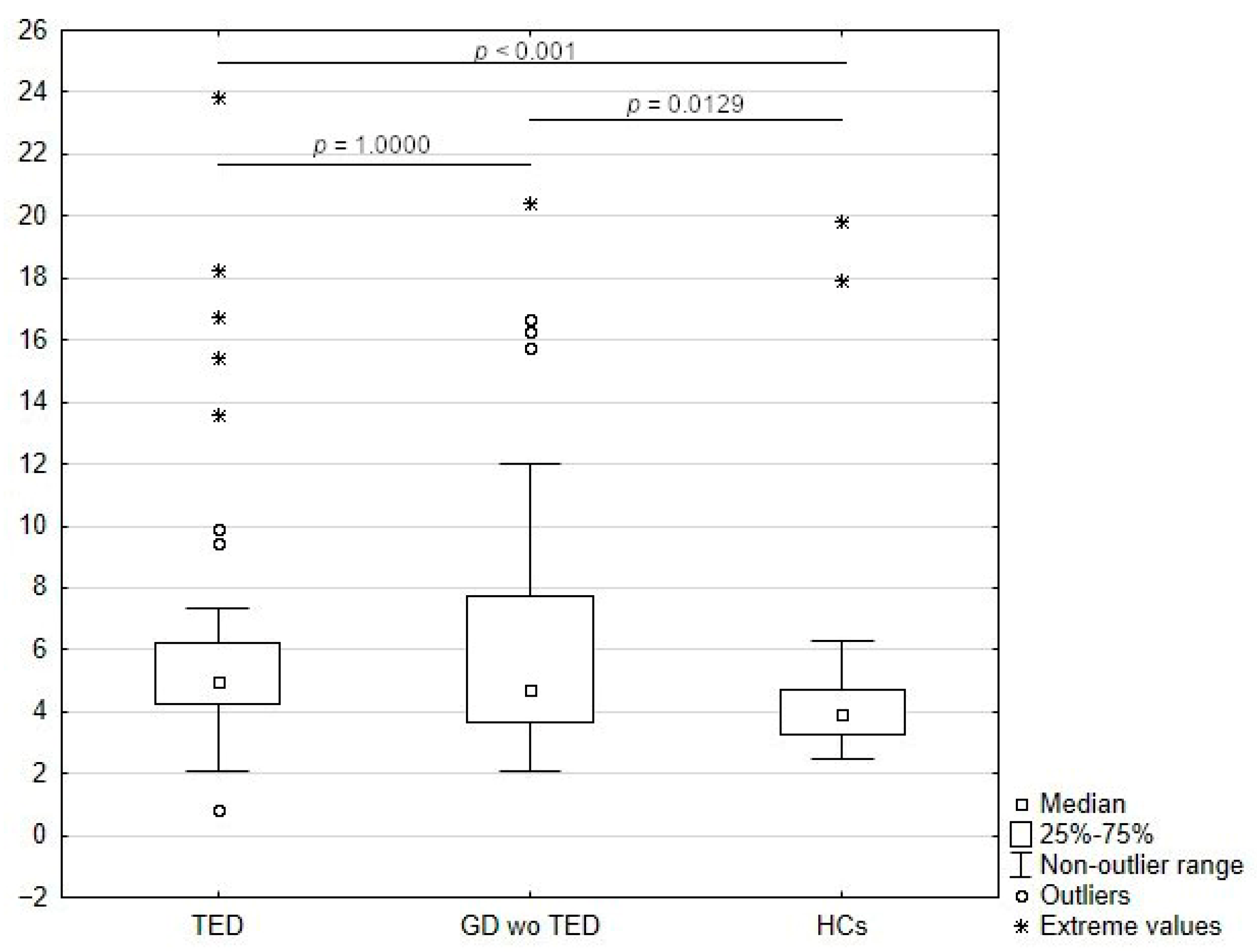
Levels of sLAG-3 were significantly elevated in serum from GD patients both without (p = 0.0129) and with (p < 0.001) TED relative to those of HC; however, no significant difference between levels of sLAG-3 was detected between the two groups of patients (i.e., GD with and without TED) (p = 1.0000) (Table 2; Figure 2).
The post hoc power analysis comparing sLAG-3 levels between GD patients (n = 35) and healthy controls (n = 37) indicated an effect size of medium magnitude (Cohen’s d = 0.49) and a statistical power of 0.53 at α = 0.05. Thus, while differences were detectable, the study was moderately underpowered to fully exclude type II error.
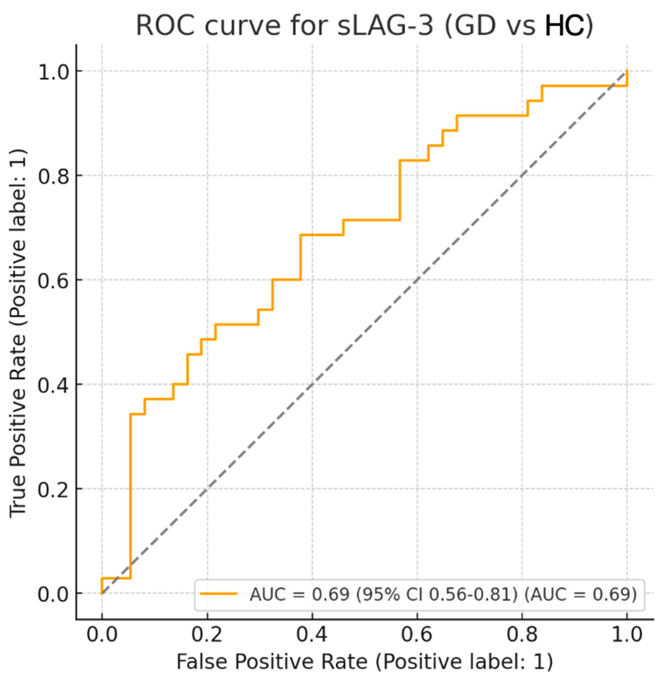
ROC analysis demonstrated a moderate ability of sLAG-3 to discriminate GD patients from healthy controls (AUC = 0.69). The optimal cut-off point, determined by the Youden index, was 4.18 ng/mL, corresponding to 68.6% sensitivity and 62.2% specificity (Table 3; Figure 3).

3.3. Effects of IVGC Treatment

In the TED group, IVGC treatment had no significant effect on sLAG-3 levels measured after 3 months of treatment relative to those at baseline (p = 0.0536) (Table 4).

3.4. Analysis of Correlation of sLAG-3 Levels with Clinical and Laboratory Parameters

Levels of sLAG-3 did not correlate with CAS (p = 0.9747, rho = −0.0048), indicating that the molecule is not a useful biomarker of disease activity. Also, there was no significant correlation of sLAG-3 levels with TSI, thyroid-stimulating hormone receptor antibodies (TRAb), aTPO, aTG, TSH, FT4, or FT3 levels. Furthermore, no effects of age, duration of GD or TED, or ATD treatment on sLAG-3 levels were detected (Table 3).
In the sensitivity analysis including smoking status, no significant association was observed between smoking and sLAG-3 levels. Compared with current smokers, neither former smokers (β = −1.11, 95% CI: −4.41 to 2.19, p = 0.50) nor non-smokers (β = 1.99, 95% CI: −0.65 to 4.62, p = 0.14) showed significant differences. Adjustment for smoking did not materially change the comparison between TED and healthy controls (β = 0.75, 95% CI: −1.55 to 3.05, p = 0.52).
In the multivariable logistic regression model restricted to TED patients, none of the analyzed variables (sLAG-3, TSI, IL-6, age, smoking status, ATD status, or disease duration) showed significant association with active disease defined as CAS ≥ 3 (all p > 0.3) (Table 5).

4. Discussion

There have been few studies focused on the interaction between soluble and membrane ICs expressed on cells involved in orbital inflammation [30]. Nevertheless, previous research has demonstrated the high potential of the sPD-L1 molecule as an immunological biomarker for assessing the activity of TED, as its levels are elevated in GD patients with TED compared with GD patients without TED [31]. sPD-L1 levels correlate with CAS and are higher in patients with active TED than those with non-active TED [32,33].
Recently, there has been increasing research interest in sLAG-3, which has been extensively studied in the context of cancer, but also in conditions with autoimmune etiology. Elevated plasma concentrations of sLAG-3 are observed in patients with active immune thrombocytopenia, while its levels normalize during remission [34]. Similarly, serum sLAG-3 levels are significantly higher in patients with rheumatoid arthritis (RA); however, no significant difference in levels was detected between patients with RA in the remission phase of the disease and those with moderate to high disease activity [35]. Furthermore, sLAG-3 levels are higher in patients with antineutrophil cytoplasmic antibody-associated vasculitis and primary biliary cholangitis relative to those in healthy subjects [36,37].
Yehudai-Ofir et al. demonstrated a potential role for sLAG-3 as a predictor of acute graft-versus-host disease development [38]. In late 2024, Qinglei Yin et al. published results showing that GD patients with TED had higher levels of sLAG-3 than both GD patients without orbitopathy and HCs. Furthermore, the same study showed that the group with active TED (CAS ≥ 3) had higher sLAG-3 levels than patients with inactive TED (CAS < 2) [33]. Consistent with these results, in our cohort, we found that sLAG-3 levels were significantly elevated in both GD patients with orbitopathy (p < 0.001) and those without TED (p = 0.0129) relative to HCs; however, unlike previously published findings [33], we did not detect a significant difference in sLAG-3 levels between the two groups of patients (i.e., those with GD with or without TED (p = 1.0000)). In our sample, the majority of patients (87.24%) in the GD with TED group were in an active disease phase (CAS ≥ 3, median: 4); however, unlike in the Chinese study, in which patients were newly diagnosed, our patients had longer disease duration (mean: 6.25 months) and had already been treated with ATD. We hypothesize that this difference influenced the results as ATD may suppress the immune response by reducing levels of pro-inflammatory cytokines; however, the underlying mechanism remains unclear [31].
This is the first published investigation of the effect of IVGC treatment on sLAG-3 levels in TED; we noted no significant difference before and after 3 months of steroid treatment (p = 0.0536). Furthermore, sLAG-3 levels did not correlate with CAS, indicating that the molecule is not a useful biomarker of disease activity. Also, there was no significant correlation of sLAG-3 levels with TSI, TRAK, aTPO, or aTG levels, nor with TSH, FT4, or FT3 hormone levels. We did not observe any influence of patient age or the durations of GD or TED on sLAG-3 levels. Furthermore, cigarette smoking influences the development of thyroid disease and TED [39] but had no effect on serum sLAG-3 levels (Table 3).
Elevated sLAG-3 levels are presumably associated with increased lymphocyte activation in the GD and TED autoimmune process as T cell activation and cytokine production require LAG-3 release from the cell membrane [21]. The function of released sLAG-3 remains unclear; it may block the interaction between CD4 and TCRs with MHC class II molecules, potentially attenuating the immune response to a given antigen. Furthermore, given the high affinity of sLAG-3 for MHC class II molecules, it may compete with cellular LAG-3 to reduce its function, resulting in an enhanced lymphocyte immune response [40].
Injection of recombinant sLAG-3 into mice with gastric cancer prolonged overall survival and increased CD8+ T cell responses; the authors hypothesized that this was due to the increased expansion of antigen-specific CD8+ T cells [41]. Administration of recombinant human LAG-3 (rhLAG-3) to cultures of peripheral blood mononucleate cells collected from patients with TED led to increased secretion of pro-inflammatory cytokines (RANTES, eotaxin, and interferon-gamma-inducible protein-10) and decreased secretion of anti-inflammatory IL-10, suggesting that sLAG-3 may enhance the inflammatory feedback loop by activating antigen-presenting cells (APCs) [33]. A form of LAG-3-related therapy involves the use of LAG-3:Fc dimer (a fusion of the extracellular Ig domains of LAG-3 with the Fc section of an antibody), which stimulates dendritic cells via MHC-II molecules, thereby enhancing antigen presentation to T lymphocytes and acting as an adjuvant to APCs, potentially improving vaccine response [42,43,44].
Studies of differences in the expression of ICs on cells in orbital tissues and levels of sICs in peripheral blood will be essential to improve the understanding of the interaction of immune cells and OFs in TED pathogenesis. Recent studies show that teprotumumab, an anti-IGF-1R inhibitor, attenuates constitutive expression of MHC-II and B7 family costimulatory molecules in OFs [45].
The strengths of our study include consistent comparisons between disease groups with and without orbitopathy and the inclusion of a control group. Our study also has limitations: first, the sample size was small due to the rarity of TED; second, some of our patients had previously been treated with ATD as the majority had GD prior to TED onset. Therefore, further studies on larger samples including patients in different phases of the disease are needed. Moreover, we did not investigate the mechanisms underlying the functional impact of sLAG-3 in GD and TED. Finally, we emphasize the need to assess the phenotype of immune cells in orbital tissues in the context of ICs, together with their interaction with other cells in the orbit and sICs, to gain a better understanding of TED pathophysiology.

5. Conclusions

In conclusion, although the mechanism underlying the function of sLAG-3 in GD and TED remains unclear, our results demonstrate a significant increase in this molecule in patients with GD relative to that in HCs, supporting a potential role for the dysregulation of sLAG-3 in the development of GD and its extra-thyroidal manifestations, such as TED.
sICs are established to influence lymphocyte function by interacting with their ligands on the cell surface, possibly enhancing the immune response or contributing to maintenance of a state of tolerance [46]. A minimally invasive assessment of the concentrations of soluble costimulatory molecules in patient blood could provide a novel approach to diagnose autoimmune diseases, including GD and TED. Therefore, further investigation of ICs and their soluble forms is warranted.

Author Contributions

Conceptualization, K.C.; methodology, K.C., E.N., A.S. and A.K.; software, E.N.; validation, K.C., A.K., E.N. and A.S.; formal analysis, K.C. and E.N.; investigation, K.C., E.N. and A.S.; resources, I.P. and E.J. data curation, K.C., E.N., E.J. and I.P.; writing—original draft preparation, K.C.; writing—review and editing, E.N., A.S. and A.K.; visualization, K.C. and E.N.; supervision, A.K.; project administration, A.K.; funding acquisition, A.K. All authors have read and agreed to the published version of the manuscript.

Funding

This research received no external funding.

Institutional Review Board Statement

This study was approved by the Bioethics Committee of the Collegium Medicum of Jan Kochanowski University in Kielce (Bioethics Committee Resolution No. 18/2025).

Informed Consent Statement

All subjects were informed about the purpose of the study and gave written informed consent to participate in the study.

Data Availability Statement

The raw data supporting the conclusions of this article will be made available by the authors upon request.

Conflicts of Interest

The authors declare no conflicts of interest.

Abbreviations

The following abbreviations are used in this manuscript:
APCAntigen-presenting cells
ATDAntithyroid drugs
CASClinical Activity Score
EUGOGOEuropean Graves’ Orbitopathy Group
GDGraves’ disease
HCsHealthy control subjects
hTSHRRecombinant human TSH receptors
ICsImmune checkpoint molecules
IVGCIntravenous glucocorticosteroid
MHC class IIMajor histocompatibility complex class II
NANot applicable
OFOrbital fibroblast
Q1First quartile
Q3Third quartile
RARheumatoid arthritis
rhLAG-3Recombinant human LAG-3
SDStandard deviation
sICPSoluble immune checkpoint
sICsSoluble immune checkpoint molecules
sLAG-3Soluble LAG-3
TCRT cell antigen receptor
TEDThyroid eye disease
TSHThyroid-stimulating hormone
TSH-RThyrotropin hormone receptor
TSIThyroid-stimulating immunoglobulin

References

  1. Kahaly, G.J.; Bartalena, L.; Hegedüs, L.; Leenhardt, L.; Poppe, K.; Pearce, S.H. 2018 European Thyroid Association Guideline for the Management of Graves’ Hyperthyroidism. Eur. Thyroid. J. 2018, 7, 167–186. [Google Scholar] [CrossRef] [PubMed] [PubMed Central]
  2. Kustrimovic, N.; Gallo, D.; Piantanida, E.; Bartalena, L.; Lai, A.; Zerbinati, N.; Tanda, M.L.; Mortara, L. Regulatory T Cells in the Pathogenesis of Graves’ Disease. Int. J. Mol. Sci. 2023, 24, 16432. [Google Scholar] [CrossRef] [PubMed] [PubMed Central]
  3. Gallo, D.; Piantanida, E.; Gallazzi, M.; Bartalena, L.; Tanda, M.L.; Bruno, A.; Mortara, L. Immunological Drivers in Graves’ Disease: NK Cells as a Master Switcher. Front. Endocrinol. 2020, 11, 406. [Google Scholar] [CrossRef] [PubMed] [PubMed Central]
  4. Fröhlich, E.; Wahl, R. Thyroid Autoimmunity: Role of Anti-thyroid Antibodies in Thyroid and Extra-Thyroidal Diseases. Front. Immunol. 2017, 8, 521. [Google Scholar] [CrossRef] [PubMed] [PubMed Central]
  5. Gallo, D.; Tanda, M.L.; Piantanida, E. Predicting the Risk of Graves Disease Relapse: Commentary on “Thyroid Peroxidase Antibody Positivity is Associated with Relapse-Free Survival Following Antithyroid Drug Treatment for Graves Disease”. Endocr. Pract. 2020, 26, 1039–1041. [Google Scholar] [CrossRef] [PubMed]
  6. Cieplińska, K.; Niedziela, E.; Kowalska, A. Immunological Processes in the Orbit and Indications for Current and Potential Drug Targets. J. Clin. Med. 2023, 13, 72. [Google Scholar] [CrossRef] [PubMed] [PubMed Central]
  7. Bartalena, L.; Kahaly, G.J.; Baldeschi, L.; Dayan, C.M.; Eckstein, A.; Marcocci, C.; Marino, M.; Vaidya, B.; Wiersinga, W.M. The 2021 European Group on Graves’ Orbitopathy (Eugogo) Clinical Practice Guidelines for the Medical Management of Graves’ Orbitopathy. Eur. J. Endocrinol. 2021, 185, G43–G67. [Google Scholar] [CrossRef] [PubMed]
  8. Bartalena, L.; Tanda, M.L. Current concepts regarding Graves’ orbitopathy. J. Intern. Med. 2022, 292, 692–716. [Google Scholar] [CrossRef] [PubMed] [PubMed Central]
  9. Rotondo Dottore, G.; Torregrossa, L.; Caturegli, P.; Ionni, I.; Sframeli, A.; Sabini, E.; Menconi, F.; Piaggi, P.; Sellari-Franceschini, S.; Nardi, M.; et al. Association of T and B Cells Infiltrating Orbital Tissues with Clinical Features of Graves Orbitopathy. JAMA Ophthalmol. 2018, 136, 613–619. [Google Scholar] [CrossRef] [PubMed] [PubMed Central]
  10. Bartalena, L.; Piantanida, E.; Gallo, D.; Lai, A.; Tanda, M.L. Epidemiology, Natural History, Risk Factors, and Prevention of Graves’ Orbitopathy. Front. Endocrinol. 2020, 11, 615993. [Google Scholar] [CrossRef] [PubMed] [PubMed Central]
  11. Marinò, M.; Rotondo Dottore, G.; Ionni, I.; Lanzolla, G.; Sabini, E.; Ricci, D.; Sframeli, A.; Mazzi, B.; Menconi, F.; Latrofa, F.; et al. Serum antibodies against the insulin-like growth factor-1 receptor (IGF-1R) in Graves’ disease and Graves’ orbitopathy. J. Endocrinol. Investig. 2019, 42, 471–480. [Google Scholar] [CrossRef] [PubMed]
  12. Tanda, M.L.; Gallo, D.; Ippolito, S.; Bartalena, L.; Piantanida, E. Treatment of moderate-to-severe and active Graves’ orbitopathy: A step forward from the OPTIC study. J. Endocrinol. Investig. 2020, 43, 1523–1525. [Google Scholar] [CrossRef] [PubMed]
  13. Rotondo Dottore, G.; Bucci, I.; Lanzolla, G.; Dallan, I.; Sframeli, A.; Torregrossa, L.; Casini, G.; Basolo, F.; Figus, M.; Nardi, M.; et al. Genetic Profiling of Orbital Fibroblasts from Patients with Graves’ Orbitopathy. J. Clin. Endocrinol. Metab. 2021, 106, e2176–e2190. [Google Scholar] [CrossRef] [PubMed]
  14. Lehmann, G.M.; Feldon, S.E.; Smith, T.J.; Phipps, R.P. Immune mechanisms in thyroid eye disease. Thyroid 2008, 18, 959–965. [Google Scholar] [CrossRef] [PubMed] [PubMed Central]
  15. Fang, S.; Lu, Y.; Huang, Y.; Zhou, H.; Fan, X. Mechanisms That Underly T Cell Immunity in Graves’ Orbitopathy. Front. Endocrinol. 2021, 12, 648732. [Google Scholar] [CrossRef] [PubMed] [PubMed Central]
  16. Aggarwal, V.; Workman, C.J.; Vignali, D.A.A. LAG-3 as the third checkpoint inhibitor. Nat. Immunol. 2023, 24, 1415–1422. [Google Scholar] [CrossRef] [PubMed] [PubMed Central]
  17. Burnell, S.E.A.; Capitani, L.; MacLachlan, B.J.; Mason, G.H.; Gallimore, A.M.; Godkin, A. Seven mysteries of LAG-3: A multi-faceted immune receptor of increasing complexity. Immunother. Adv. 2021, 2, ltab025. [Google Scholar] [CrossRef] [PubMed] [PubMed Central]
  18. Wang, J.; Klein, C.; Cochran, J.R.; Sockolosky, J.; Lippow, S.M. Exploring new frontiers in LAG-3 biology and therapeutics. Trends Pharmacol. Sci. 2025, 46, 638–652. [Google Scholar] [CrossRef] [PubMed]
  19. Ruffo, E.; Wu, R.C.; Bruno, T.C.; Workman, C.J.; Vignali, D.A.A. Lymphocyte-activation gene 3 (LAG3): The next immune checkpoint receptor. Semin. Immunol. 2019, 42, 101305. [Google Scholar] [CrossRef] [PubMed] [PubMed Central]
  20. Triebel, F. LAG-3: A regulator of T-cell and DC responses and its use in therapeutic vaccination. Trends Immunol. 2003, 24, 619–622. [Google Scholar] [CrossRef] [PubMed]
  21. Li, N.; Wang, Y.; Forbes, K.; Vignali, K.M.; Heale, B.S.; Saftig, P.; Hartmann, D.; Black, R.A.; Rossi, J.J.; Blobel, C.P.; et al. Metalloproteases regulate T-cell proliferation and effector function via LAG-3. EMBO J. 2007, 26, 494–504. [Google Scholar] [CrossRef] [PubMed] [PubMed Central]
  22. Malinga, N.Z.; Siwele, S.C.; Steel, H.C.; Kwofie, L.L.; Meyer, P.W.; Smit, T.; Anderson, R.; Rapoport, B.L.; Kgokolo, M.C. Systemic levels of the soluble co-inhibitory immune checkpoints, CTLA-4, LAG-3, PD-1/PD-L1 and TIM-3 are markedly increased in basal cell carcinoma. Transl. Oncol. 2022, 19, 101384. [Google Scholar] [CrossRef]
  23. Liu, X.; Xu, Y.; Wu, X.; Liu, Y.; Wu, Q.; Wu, J.; Zhang, H.; Zhou, M.; Qu, J. Soluble Immune-related proteins as new candidate serum biomarkers for the diagnosis and progression of Lymphangioleiomyomatosis. Front. Immunol. 2022, 13, 844914. [Google Scholar] [CrossRef] [PubMed]
  24. Li, Y.; Wang, W.; Tian, J.; Zhou, Y.; Shen, Y.; Wang, M.; Tang, L.; Liu, C.; Zhang, X.; Shen, F.; et al. Clinical significance of Soluble LAG-3 (sLAG-3) in patients with Cervical Cancer determined via enzyme-linked immunosorbent assay with monoclonal antibodies. Technol. Cancer Res. Treat. 2023, 22, 15330338231202650. [Google Scholar] [CrossRef]
  25. Wang, Q.; Zhang, J.; Tu, H.; Liang, D.; Chang, D.W.; Ye, Y.; Wu, X. Soluble immune checkpoint-related proteins as predictors of tumor recurrence, survival, and T cell phenotypes in clear cell renal cell carcinoma patients. J. Immunother. Cancer. 2019, 7, 334. [Google Scholar] [CrossRef] [PubMed]
  26. Gorgulho, J.; Roderburg, C.; Beier, F.; Bokemeyer, C.; Brümmendorf, T.H.; Loosen, S.H.; Luedde, T. Soluble lymphocyte activation gene-3 (sLAG3) and CD4/CD8 ratio dynamics as predictive biomarkers in patients undergoing immune checkpoint blockade for solid malignancies. Br. J. Cancer. 2024, 130, 1013–1022. [Google Scholar] [CrossRef] [PubMed] [PubMed Central]
  27. Botticelli, A.; Zizzari, I.G.; Scagnoli, S.; Pomati, G.; Strigari, L.; Cirillo, A.; Cerbelli, B.; Di Filippo, A.; Napoletano, C.; Scirocchi, F.; et al. The Role of Soluble LAG3 and Soluble Immune Checkpoints Profile in Advanced Head and Neck Cancer: A Pilot Study. J. Pers. Med. 2021, 11, 651. [Google Scholar] [CrossRef] [PubMed] [PubMed Central]
  28. Landeira-Viñuela, A.; Arias-Hidalgo, C.; Juanes-Velasco, P.; Alcoceba, M.; Navarro-Bailón, A.; Pedreira, C.E.; Lecrevisse, Q.; Díaz-Muñoz, L.; Sánchez-Santos, J.M.; Hernández, Á.P.; et al. Unravelling soluble immune checkpoints in chronic lymphocytic leukemia: Physiological immunomodulators or immune dysfunction. Front. Immunol. 2022, 13, 965905. [Google Scholar] [CrossRef] [PubMed] [PubMed Central]
  29. Yuan, L.; Wang, Y.; Shen, X.; Ma, F.; Wang, J.; Yan, F. Soluble form of immune checkpoints in autoimmune diseases. J. Autoimmun. 2024, 147, 103278. [Google Scholar] [CrossRef] [PubMed]
  30. Shu, X.; Shao, Y.; Chen, Y.; Zeng, C.; Huang, X.; Wei, R. Immune checkpoints: New insights into the pathogenesis of thyroid eye disease. Front. Immunol. 2024, 15, 1392956. [Google Scholar] [CrossRef] [PubMed] [PubMed Central]
  31. Khamisi, S.; Anders Karlsson, F.; Ljunggren, Ö.; Thulin, M.; Larsson, A. Increased plasma levels of soluble programmed death ligand 1 (sPD-L1) and fibroblast growth factor 23 (FGF-23) in patients with Graves’ ophthalmopathy in comparison to hyperthyroid patients without Graves’ ophthalmopathy. Cytokine 2023, 169, 156269. [Google Scholar] [CrossRef] [PubMed]
  32. Cieplińska, K.; Niedziela, E.; Rdzanek, A.K.; Słuszniak, A.; Chrapek, M.; Pałyga, I.; Kowalska, A. Association between clinical activity score and serum sPD-1 and sPD-L1 levels during systemic glucocorticoid treatment for active moderate-to-severe thyroid eye disease. Cytokine 2025, 187, 156862. [Google Scholar] [CrossRef] [PubMed]
  33. Yin, Q.; Zhu, T.; Song, D.; Fang, S.; Zhou, H.; Guan, H. Soluble immune checkpoints associated with disease activity and treatment response in GD and TED. J. Clin. Endocrinol. Metab. 2024, 110, 1537–1549. [Google Scholar] [CrossRef] [PubMed]
  34. Qiao, J.; Luo, Q.; Liu, N.; Wei, G.; Wu, X.; Lu, J.; Tang, K.; Wu, Y.; Zi, J.; Li, X.; et al. Increased ADAM10 expression in patients with immune thrombocytopenia. Int. Immunopharmacol. 2018, 55, 63–68. [Google Scholar] [CrossRef] [PubMed]
  35. Saadati, N.; Khodashahi, M.; Rezaieyazdi, Z.; Sahebari, M.; Saremi, Z.; Mohammadian Haftcheshmeh, S.; Rafatpanah, H.; Salehi, M. Serum Level of Soluble Lymphocyte-Activation Gene 3 Is Increased in Patients with Rheumatoid Arthritis. Iran. J. Immunol. 2020, 17, 324–332. [Google Scholar] [CrossRef] [PubMed]
  36. Pyo, J.Y.; Yoon, T.; Ahn, S.S.; Song, J.J.; Park, Y.B.; Lee, S.W. Soluble immune checkpoint molecules in patients with antineutrophil cytoplasmic antibody-associated vasculitis. Sci. Rep. 2022, 12, 21319. [Google Scholar] [CrossRef] [PubMed] [PubMed Central]
  37. Gao, X.; Wang, X.; Guan, Y.; Wang, L.; Gao, Y.; Niu, J. Soluble immune checkpoints are elevated in patients with primary biliary cholangitis. Eur. J. Med. Res. 2023, 28, 477. [Google Scholar] [CrossRef] [PubMed] [PubMed Central]
  38. Dana Yehudai-Ofir Nasren Eiza Israel, J. Henig, Tsila Zuckerman, Zahava Vadasz, Soluble LAG3: A Potential Marker for the Development and Activity of Acute Graft-Versus-Host Disease. Blood 2023, 142 (Suppl. S1), 4795. [Google Scholar] [CrossRef]
  39. Liu, J.; Zhu, T.; Yang, L.; Qian, W.; Fang, L.; Zhang, W.; Zhang, H.; Wang, Y.; Yu, B.; Sun, J.; et al. Cigarette Smoking Drives Thyroid Eye Disease Progression via RAGE Signaling Activation. Thyroid 2025, 35, 803–815. [Google Scholar] [CrossRef] [PubMed]
  40. Huard, B.; Mastrangeli, R.; Prigent, P.; Bruniquel, D.; Donini, S.; El-Tayar, N.; Maigret, B.; Dréano, M.; Triebel, F. Characterization of the major histocompatibility complex class II binding site on LAG-3 protein. Proc. Natl. Acad. Sci. USA 1997, 94, 5744–5749. [Google Scholar] [CrossRef]
  41. Li, N.; Jilisihan, B.; Wang, W.; Tang, Y.; Keyoumu, S. Soluble LAG3 acts as a potential prognostic marker of gastric cancer and its positive correlation with CD8+T cell frequency and secretion of IL-12 and INF-γ in peripheral blood. Cancer Biomark. 2018, 23, 341–351. [Google Scholar] [CrossRef] [PubMed]
  42. El Mir, S.; Triebel, F. A soluble lymphocyte activation gene-3 molecule used as a vaccine adjuvant elicits greater humoral and cellular immune responses to both particulate and soluble antigens. J. Immunol. 2000, 164, 5583–5589. [Google Scholar] [CrossRef] [PubMed]
  43. Andreae, S.; Piras, F.; Burdin, N.; Triebel, F. Maturation and activation of dendritic cells induced by lymphocyte activation gene-3 (CD223). J. Immunol. 2002, 168, 3874–3880. [Google Scholar] [CrossRef] [PubMed]
  44. Prigent, P.; El Mir, S.; Dréano, M.; Triebel, F. Lymphocyte activation gene-3 induces tumor regression and antitumor immune responses. Eur. J. Immunol. 1999, 29, 3867–3876. [Google Scholar] [CrossRef] [PubMed]
  45. Fernando, R.; Caldera, O.; Smith, T.J. Therapeutic IGF-I receptor inhibition alters fibrocyte immune phenotype in thyroid-associated ophthalmopathy. Proc. Natl. Acad. Sci. USA 2021, 118, e2114244118. [Google Scholar] [CrossRef] [PubMed] [PubMed Central]
  46. Riva, A. Editorial: Soluble immune checkpoints: Novel physiological immunomodulators. Front. Immunol. 2023, 14, 1178541. [Google Scholar] [CrossRef] [PubMed] [PubMed Central]
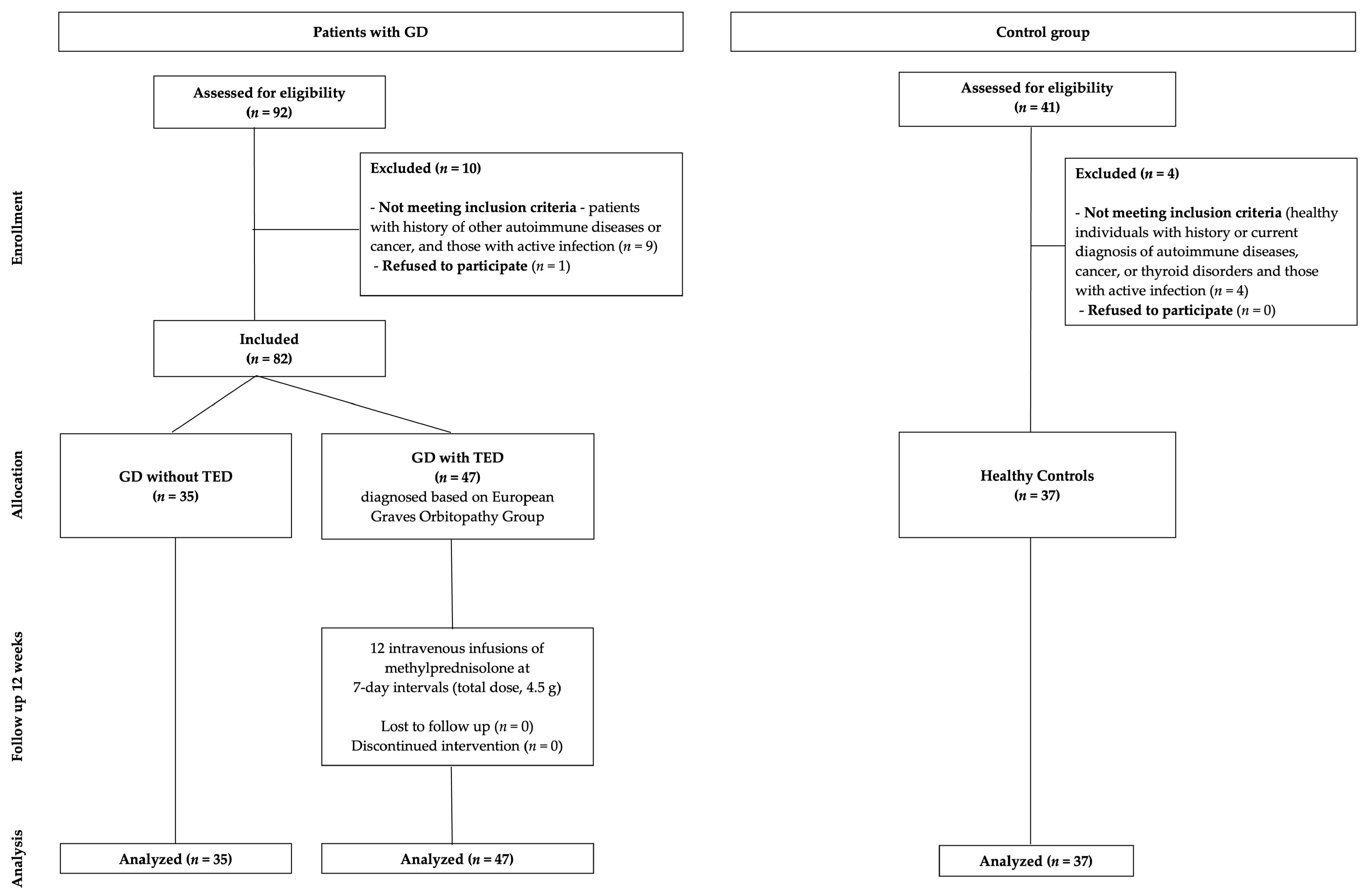
Figure 1. Flow diagram of participant selection. GD, Graves’ disease; TED, thyroid eye disease.
Medicina 61 01664 g001
Figure 2. sLAG-3 levels in patients with GD with and without TED and HCs. p-Values were calculated using the Kruskal–Wallis test, with post hoc analysis performed using multiple comparisons of mean ranks for all groups. TED, thyroid eye disease; GD wo TED, Graves’ disease without TED; HCs, healthy controls.
Medicina 61 01664 g002
Figure 3. ROC curve for sLAG-3 in discriminating Graves’ disease patients (GD, n = 35) from healthy controls (HC, n = 37). The area under the curve (AUC) was 0.69 (95% CI: 0.56–0.81, p = 0.004). The optimal cut-off value of 4.18 ng/mL provided 68.6% sensitivity and 62.2% specificity.
Medicina 61 01664 g003
Table 1. Patient characteristics.
CharacteristicPatients with GD Without TED (N = 35)Patients with GD and TED (N = 47)
Age (years)
 Mean (SD)53.09 (17.78)55.13 (12.02)
 Median (Q1, Q3)57 (39.00, 67.00)54 (46.00, 64.00)
 Range20–8020–78
Sex
 Female26 (74.29%)30 (68.09%)
 Male9 (25.71%)15 (31.92%)
Smoking history
 Non-smoker25 (75.76%)20 (42.55%)
 Former smoker3 (9.10%)11 (23.40%)
 Active smoker5 (15.15%)16 (34.04%)
Duration of GD (months)
 Mean (SD)55.60 (63.43)23.72 (37.40)
 Median (Q1, Q3)36 (4.00, 72.00)12 (7.00, 24.00)
 Range1–2402–240
Duration of GD (months)
 ≤12 12 (35.29%)26 (55.32%)
 >1222 (64.71%)21 (44.68%)
Duration of TED (months)
 Mean (SD)NA6.25 (3.98)
 Median (Q1, Q3)NA6 (3.00, 8.00)
 RangeNA1–18
Duration of TED (months)
 ≤12 NA45 (95.75%)
 >12NA2 (4.26%)
TSH (µIU/mL)
 Mean (SD)1.36 (2.62)4.49 (12.15)
 Median (Q1, Q3)0.39 (0.01, 1.41)0.92 (0.11–1.98)
 Range0–12.490–71.08
TSH (µIU/mL)
 <0.3515 (46.88%)16 (34.04%)
 0.35–4.9414 (43.75%)25 (53.19%)
 >4.943 (9.38%)6 (12.77%)
Maximum CAS at baseline
 1NA2 (4.25%)
 2NA4 (8.51%)
 3NA13 (27.66%)
 4NA14 (29.79%)
 5NA9 (19.15%)
 6NA4 (8.51%)
 7NA1 (2.13%)
Maximum CAS at baseline
 Mean (SD)NA3.85 (1.32)
 Median (Q1, Q3)NA4 (3, 5)
 RangeNA1–7
TSI at baseline (IU/L)
 Mean (SD)4.15 (4.12)6.40 (7.93)
 Median (Q1, Q3)2.17 (1.33, 5.98)2.90 (0.72, 10.3)
 Range0.31–15.200.1–37.4
TSI at baseline > 2 (IU/L)
 No17 (48.57%)19 (40.43%)
 Yes18 (51.43%)28 (59.57%)
sLAG-3 at baseline (ng/mL)
 Mean (SD)6.73 (4.52)6.86 (5.06)
 Median (Q1, Q3)4.73 (3.63, 7.75)4.94 (4.23, 6.23)
 Range2.10–20.430.848–23.84
IL-6 at baseline (pg/mL)
 Mean (SD)3.37 (2.40)3.30 (2.37)
 Median (Q1, Q3)2.40 (2.00, 3.36)2.2 (2, 3.51)
 Range2–11.6<2–12.5
IL-6 at baseline (pg/mL)
 <3.4 27 (77.14%)29 (61.70%)
 >3.48 (22.86%)10 (38.30%)
Antithyroid drugs
 Treatment-naïve2 (5.71%)6 (12.77%)
 Currently under treatment23 (65.71%)38 (80.85%)
 Previously treated, not currently10 (28.57%)3 (6.38%)
GD, Graves’ disease; TED, thyroid eye disease; SD, standard deviation; Q1, first quartile; Q3, third quartile; NA, not applicable; TSH, thyroid-stimulating hormone; CAS, Clinical Activity Score; TSI, thyroid-stimulating immunoglobulin; sLAG-3, soluble lymphocyte-activation gene 3; IL-6, interleukin 6.
Table 2. Comparison of sLAG-3 levels in patients with GD with and without TED and HCs.
HCs (N = 37)Patients with GD Without TED (N = 35)Patients with GD and TED (N = 47)p-Value (HC vs. GD Without TED)p-Value (HC vs. TED)p-Value (GD Without TED vs. TED)
Age (years)
 Mean (SD)55.11 (13.89)53.09 (17.78)55.13 (12.02)0.5913 *0.9945 *0.5597 **
 Median (Q1, Q3)55 (45, 67)57 (39, 67)54 (46, 64)
 Range23–7820–8020–78
Sex 0.2702 ***0.5708 ***0.5416 ***
 Female23 (62.16%)26 (74.29%)32 (68.09%)
 Male14 (37.84%)9 (25.71%)15 (31.92%)
sLAG-3 at baseline (ng/mL) 0.0129 ****<0.001 ****1.000 ****
 Mean (SD)4.76 (3.56)6.73 (4.52)6.68 (5.06)
 Median (Q1, Q3)3.94 (3.28, 4.71) 4.73 (3.63, 7.75)5.06 (4.23, 6.16)
 Range2.46–19.852.10–20.430.85–23.84
* t-test, ** t-test (Welch correction), *** Chi-squared test, **** post hoc multiple comparisons of mean ranks. HCs, healthy controls; GD, Graves’ disease; TED, thyroid eye disease; SD, standard deviation; Q1, first quartile; Q3, third quartile; NA, not applicable; TSH, thyroid-stimulating hormone; CAS, Clinical Activity Score; TSI, thyroid-stimulating immunoglobulin; sLAG-3, soluble lymphocyte-activation gene-3.
Table 3. ROC analysis of sLAG-3 for discriminating GD patients from healthy controls.
ParameterValue95% CIp-Value
AUC0.690.56–0.810.004
Optimal cut-off (ng/mL)4.18
Sensitivity (%)68.6
Specificity (%)62.2
False positives (n)14
False negatives (n)11
Youden index0.307
Table 4. sLAG-3 levels in patients with TED at baseline and after 12 weeks of IVGC treatment.
At Baseline
(N = 47)
sLAG-3 at Baseline (ng/mL)
 
Mean (SD)
Median (Q1, Q3)
Range
sLAG-3 at 12 Weeks (ng/mL)
 
Mean (SD)
Median (Q1, Q3)
Range
p-Value (for Difference in sLAG-3 Concentration)
All patients6.68 (5.06)6.91 (4.86)0.0536
4.94 (4.23, 6.23)5.13 (4.62, 6.54)
0.85–23.840.79–23.84
TSH 0.35–4. 94 (µIU/mL)
(N = 25)
7.12 (5.92)7.34 (5.61)0.0777
5.07 (4.19–6.44)5.19 (4.79–6.89)
0.85–23.840.79–23.84
TSH < 0.35 (µIU/mL)
(N = 16)
6.78 (4.47)7.05 (4.43)0.6051
5.19 (4.38–6.50)5.42 (4.68–7.30)
2.92–18.253.10–17.95
TSI > 2 (IU/L)
(N = 28)
7.36 (6.07)7.46 (5.81)0.1213
4.91 (4.29–5.98)5.04 (4.61–6.64)
2.92–23.843.10–23.84
TSI ≤ 2 (IU/L)
(N = 19)
5.67 (2.89)6.10 (2.96)0.2432
5.07 (4.17–6.90)5.35 (4.68–6.54)
0.85–13.590.79–13.32
IL-6 < 3.4 (pg/mL)
(N = 29)
6.62 (4.80)6.60 (4.52)0.1658
4.94 (4.23–6.09)5.03 (4.64–5.64)
2.09–23.842.99–23.84
IL-6 > 3.4 (pg/mL)
(N = 10)
7.53 (5.98)7.94 (5.81)0.4413
5.19 (4.69–7.33)6.07 (4.68–8.69)
4.10–23.844.60–23.84
Non-smokers
(N = 20)
8.12 (6.79)8.71 (6.64)0.1570
5.07 (4.29–8.61)5.42 (4.68–9.96)
2.09–23.842.99–23.84
Active Smokers
(N = 16)
6.01 (3.43)5.78 (2.36)0.3794
4.95 (4.29–5.72)5.01 (4.61–5.99)
3.15–15.413.60–13.32
Duration of TED ≤ 6 months
(N = 28)
7.61 (6.03)8.00 (5.95)0.0734
5.20 (4.21–8.38)5.35 (4.59–9.87)
2.09–23.842.99–23.84
Duration of TED > 6 months
(N = 19)
5.30 (2.74)5.31 (1.66)0.3144
4.84 (4.36–5.44)5.13 (4.64–5.92)
0.85–15.410.79–8.83
Duration of GD ≤ 12 months
(N = 26)
6.80 (4.69)7.29 (4.72)0.1094
5.14 (4.36–6.23)5.27 (4.68–8.69)
2.09–23.842.99–23.84
Duration of GD > 12 months
(N = 21)
6.53 (5.60)6.43 (5.11)0.2471
4.76 (4.10–5.44)5.13 (4.60–5.76)
0.848–23.840.787–23.84
sLAG-3, soluble lymphocyte-activation gene 3; SD, standard deviation; Q1, first quartile; Q3, third quartile; TSH, thyroid-stimulating hormone; TSI, thyroid-stimulating immunoglobulin; ATD, antithyroid drugs; IL-6, interleukin 6; TED, thyroid eye disease; GD, Graves’ disease; NA, not applicable; CAS, Clinical Activity Score.
Table 5. Multivariable logistic regression analysis of factors associated with active thyroid eye disease (CAS ≥ 3) in TED patients.
VariableOR95% CIp-Value
lag30.990.85–1.150.89
TSI1.040.93–1.170.46
IL61.000.72–1.391.00
Age1.020.96–1.100.51
Smoking (former vs. current)1.980.23–17.10.54
Smoking (never vs. current)0.440.07–2.760.38
ATD status (former vs. current)1.00 *
ATD status (never vs. current)0.460.01–16.20.67
GD duration (months)1.030.98–1.080.31
* Estimates for ATD status were unstable due to small subgroup sizes.
Disclaimer/Publisher’s Note: The statements, opinions and data contained in all publications are solely those of the individual author(s) and contributor(s) and not of MDPI and/or the editor(s). MDPI and/or the editor(s) disclaim responsibility for any injury to people or property resulting from any ideas, methods, instructions or products referred to in the content.