Article

Variable Phenotypic Expression of PAX2 Variants in Two Lithuanian Families with Kidney Disease

1
Department of Human and Medical Genetics, Institute of Biomedical Sciences, Faculty of Medicine, Vilnius University, 03101 Vilnius, Lithuania
2
Centre for Medical Genetics, Vilnius University Hospital Santaros Klinikos, 08661 Vilnius, Lithuania
3
Clinic of Gastroenterology, Nephro-Urology and Surgery, Faculty of Medicine, Vilnius University, 03101 Vilnius, Lithuania
*
Author to whom correspondence should be addressed.
Medicina 2025, 61(4), 597; https://doi.org/10.3390/medicina61040597
Submission received: 7 February 2025 / Revised: 17 March 2025 / Accepted: 23 March 2025 / Published: 26 March 2025
(This article belongs to the Section Genetics and Molecular Medicine)

Abstract

Background and Objectives: Pathogenic variants in the PAX2 gene have been associated with a spectrum of eye and kidney disorders, ranging from papillorenal syndrome (known as renal coloboma syndrome) to isolated nephrosis without kidney morphological anomalies (focal segmental glomerulosclerosis), inherited in an autosomal dominant manner. However, due to the growing number of reports of pathogenic variants in the PAX2 gene, it is observed that genotype–phenotype correlation is not always consistent. We present patients from two unrelated families with PAX2 pathogenic variants c.685C>T and c.250G>A, highlighting the diverse phenotypic expression of PAX2-related disorders. Materials and Methods: We analyzed clinical and genetic data from two families who were tested for genomic abnormalities using targeted next-generation sequencing and Sanger sequencing for segregation analysis. Results: In Family A, a 27-year-old male presented with chronic kidney disease stage 3, proteinuria, and multicystic kidney dysplasia diagnosed at 11 years old. An ophthalmologic examination revealed bilateral optic nerve dysplasia. In Family B, a 6-year-old female and her 4-year-old sister were clinically diagnosed with renal hypoplasia, while their 36-year-old father presented with chronic kidney disease stage 3, focal segmental glomerulosclerosis, and optic disc pits. Genetic analysis identified a heterozygous PAX2 pathogenic variant c.685C>T, p.(Arg229*), in Family A and a heterozygous PAX2 pathogenic variant c.250G>A, p.(Gly84Ser) in Family B. Conclusions: The literature and our data further support that the same PAX2 variants may cause diverse kidney and ocular phenotypes among unrelated families and within the same family. Due to variable expressivity, a wide range of clinical manifestations of rare hereditary kidney diseases are still underdiagnosed, and a multidisciplinary approach is required to detect extrarenal signs of PAX2-related disorder.

1. Introduction

The PAX2 (paired box gene 2) is a member of the PAX transcription factor gene family and plays a crucial role in the coordinated development of several organs during embryogenesis, including the kidneys and eyes [1]. Pathogenic variants in the PAX2 gene have been associated with a spectrum of eye and kidney disorders, ranging from papillorenal syndrome (known as renal coloboma syndrome; OMIM: 120330) to isolated nephrosis without kidney morphological anomalies (focal segmental glomerulosclerosis; OMIM: 616002), inherited in an autosomal dominant manner.
Heterozygous variants in the PAX2 gene were first described as the cause of the syndrome in 1995 in a family with renal hypoplasia and optic nerve colobomas [2]. To date, more than 160 unique reported DNA variants are documented in the updated database of PAX2 gene variants (accessed on 7 February 2025: https://www.LOVD.nl/PAX2). Various studies have identified PAX2 gene variants in both kidney and ophthalmologic disease cohorts, but the prevalence of PAX2-related disorder is still unknown. According to the literature, 92% of individuals with PAX2 gene variants have kidney disorders, while 77% present with ophthalmologic diseases [3]. Kidney disorders associated with PAX2 variants involve CAKUT, renal interstitial fibrosis, renal hypoplasia, cystic disorders, nephrotic syndrome, and urogenital cancers [4]. The most frequently described renal anomaly is hypodysplasia [5]. A review of reported cases of PAX2-related optic involvement summarized the most common ocular findings, which included optic nerve coloboma, optic disc excavation or pit, optic nerve hypoplasia, abnormal retinal vessels, strabismus, and retinal vessel abnormalities [6]. About one-half of patients presenting with optic nerve malformations and renal dysplasia harbor a pathogenic variant in the PAX2 gene [7].
The PAX2-related disorder is highly variable. However, due to the growing number of reports of pathogenic variants in the PAX2 gene, it is recognized that genotype–phenotype correlation is not always consistent. The inclusion of PAX2 gene in targeted next-generation sequencing (NGS) gene panels for kidney diseases allows for the identification of disease-causing variants, even in cases where clinical diagnosis may not be accurate [8,9].
We present three individuals from two unrelated families who carry different PAX2 gene variants to highlight the importance of PAX2-related disorders and the spectrum of phenotypic expression.

2. Materials and Methods

2.1. Ethical Compliance

This study was approved by the Vilnius Regional Biomedical Research Ethics Committee. Written informed consent was obtained from all patients.

2.2. Patients

In total, 3 patients with PAX2 variants were analyzed out of a cohort of 112 individuals who underwent a genetic investigation for hereditary kidney diseases.

2.3. Clinical Evaluation

A retrospective review of clinical findings, including the age and onset of the first reported symptoms, family history, laboratory test results, kidney findings from radiologic investigations, and/or renal biopsy results, and ophthalmologic examination results was conducted. The glomerular filtration rate was estimated by correcting 24 h creatinine clearance, and chronic kidney disease (CKD) staging was based on the KDIGO 2024 Clinical Practice Guideline for the Evaluation and Management of Chronic Kidney Disease [10].

2.4. Genetic Investigations

2.4.1. DNA Extraction

Patients’ genetic testing was performed using DNA extracted from peripheral blood samples via standard procedures using the phenol–chloroform–isoamyl alcohol extraction method.

2.4.2. Next-Generation Sequencing

For Patients 1 and 2, next-generation sequencing analysis of genomic DNA was performed. Sequencing and primary steps of pre-processing raw sequencing data were performed by the subcontracting NGS provider (CeGaT GmbH, Tübingen, Germany) using the high-throughput next-generation Illumina (Illumina, Inc., San Diego, CA, USA) platform. Obtained sequencing data (FASTQ, BAM, VCF files) analysis was further performed in our laboratory using a validated in-house bioinformatic pipeline.

2.4.3. Inherited Kidney Disease Gene Panel Analysis

Annotation and post-annotation of the sequencing data from the inherited kidney disease gene panel were further post-processed on-site using a validated in-house bioinformatic pipeline. Prioritization and interpretation of genome variants were performed using genomic tools and databases provided by the ANNOVAR 2020Jul08 program [11]. Variants were then classified by following the guidelines of the American College of Medical Genetics and Genomics.

2.4.4. Sanger Sequencing

For the following segregation analysis, Sanger sequencing was performed using DNA samples of Patient’s 2 father and sister. DNA sequence flanking the familial variant of the PAX2 gene was amplified using the PCR Master Mix (2X) according to the manufacturer’s protocol (Thermo Fisher Scientific, Waltham, MA, USA). Specific primers designed with Primer3web [12] for the PAX2 gene familial variant were available upon request. Sanger sequencing was performed using the BigDye Terminator v3.1 Cycle Sequencing Kit (Applied Biosystems, Foster City, CA, USA), and automatic genetic analyzer ABI PRISM 3130xl (Applied Biosystems, USA) according to the manufacturer’s protocol. The obtained sequences were aligned with the main reference sequence of PAX2 (NCBI: NM_000278.5).

3. Results

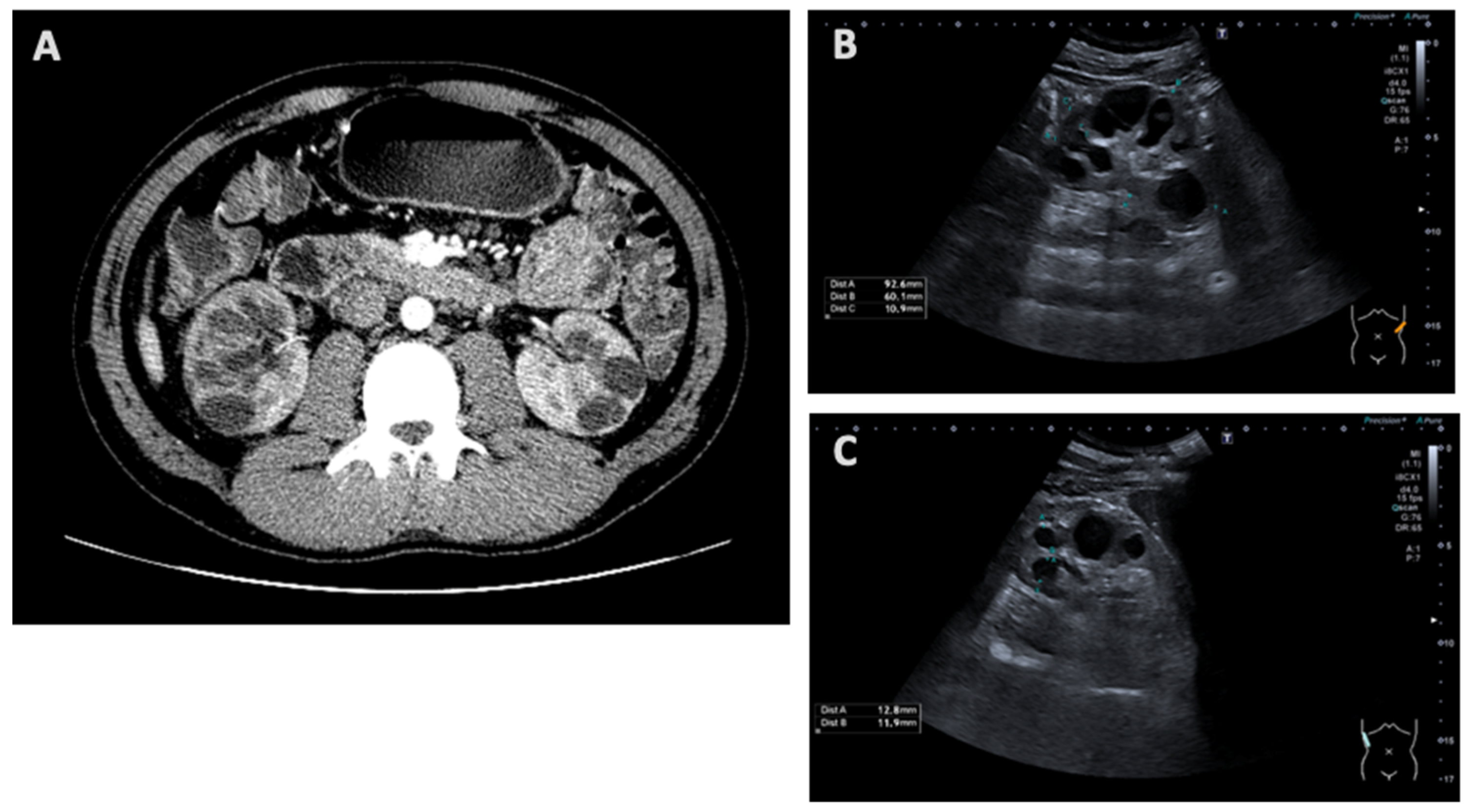
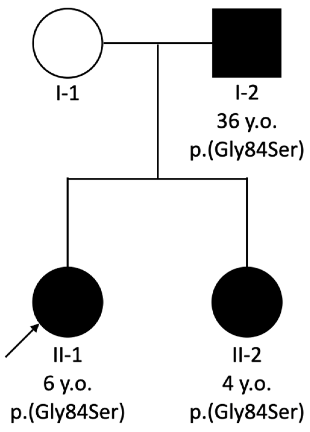
3.1. Clinical Characteristics

FAMILY A, PATIENT 1. A 27-year-old male was diagnosed with bilateral multicystic kidney dysplasia at 11 years old. Serum creatinine was 90 μmol/L at that time, but no information about urinalysis was available. The patient’s paternal grandmother had unspecified kidney disease and died at 28 years old.
Patient 1’s investigations were carried out to clarify the nature of his disease. The patient presented with proteinuria (1 g/L) and CKD stage G3aA3. His recent renal sonography and computed tomography showed cystic kidneys (shown in Figure 1), describing them as multicystic dysplastic or polycystic. Sonography showed that the right kidney was 96 × 48 mm and the left kidney was 100 × 49 mm in size; their parenchyma measured 14–15 mm and 15–16 mm, respectively. Both kidneys had regular contours and multiple cysts, up to 30 mm in size.
Laboratory tests revealed elevated levels of urea and creatinine and decreased estimated glomerular filtration rate (eGFR), while uric acid was within the normal range (all patients’ laboratory results are presented in Table 1). The ophthalmologic examination was performed and revealed bilateral optic nerve dysplasia without significant impact on vision. The condition remained stable over the year.
The renal function slightly decreased by the age of 30 years, and proteinuria reduced (0.3 g/L), but glucosuria was detected (up to 56 mmol/L). Kidney size remained similar to the primary sonography, and irregular contour was noted.
FAMILY B, PATIENT 2. A 6-year-old female was diagnosed with bilateral renal hypoplasia shortly after birth and had arterial hypertension from two months of age. The patient’s 4-year-old sister also presented with bilateral renal hypoplasia, and their father was diagnosed with focal segmental glomerulosclerosis (FSGS) (pedigree presented in Figure 2).
At 6 years old, the patient’s growth parameters were normal. Her recent renal ultrasound scan showed signs of chronic kidney disease, microcalcifications, and bilateral simple parenchymal cysts up to 2 mm in diameter. Laboratory tests showed elevated levels of urea and creatinine and decreased eGFR; the uric acid and urinalysis results were normal.
The renal function slightly decreased by 10 years old, and the patient also developed proteinuria (0.25 g/L).
FAMILY B, PATIENT 3. The 36-year-old father of Patient 2 was referred for segregation analysis. He presented with persistent proteinuria (1 g/L) from 20 years of age and CKD stage G3bA3. Two kidney biopsies were performed since his initial diagnosis: the first one showed signs of IgA nephropathy, which was ruled out; the second one showed focal segmental glomerulosclerosis. Laboratory analysis revealed elevated levels of urea, creatinine, uric acid, and decreased eGFR. His detailed ophthalmologic examination showed optic disc pits.
The renal function slightly decreased by 39 years old, proteinuria reduced (0.3 g/L), and glucosuria was detected (up to 56 mmol/L). The renal ultrasound scan showed normal kidney size and increased parenchymal echogenicity.

3.2. PAX2 Variants

Next-generation sequencing of Patient 1 showed a heterozygous PAX2 gene pathogenic variant NM_000278.5:c.685C>T, NP_000269.3:p.(Arg229*), rs76492282. Segregation analysis of this family was not possible as their parents were not available for testing.
Both Patient 2 and 3 were found to carry a heterozygous PAX2 gene pathogenic variant NM_000278.5:c.250G>A, NP_000269.3:p.(Gly84Ser), rs2133836340, confirming the genetic cause for their kidney disease. The familial variant was also detected in the sister of Patient 2.

4. Discussion

Papillorenal syndrome mainly manifests by ocular and renal abnormalities due to heterozygous pathogenic PAX2 gene variants. Both ocular and renal phenotypes present a spectrum of different findings with variable severity, even within members of the same family carrying PAX2 variants. PAX2 gene pathogenic variants have also been reported in individuals with isolated renal hypo/dysplasia and no ocular anomalies [13]. All this makes it difficult to determine the precise genotype–phenotype correlation of the PAX2-related disorder and establish diagnostic criteria.
Targeted next-generation sequencing analysis has proven to be a useful tool for both accurate etiology determination and differential diagnosis, especially in cases where patients are not necessarily extensively examined clinically before undergoing genetic testing. Domingo-Gallego et al. achieved a global diagnostic yield of 65% (300/460) in patients with early-onset CKD of suspected monogenic cause. Heterozygous variants in the PAX2 gene were found in the majority of clinical diagnostic categories: CAKUT (three cases with dysplastic and/or hypoplastic kidneys), glomerulopathies (one case with unspecified glomerulopathy and one case with suspected Alport syndrome, accompanied by a COL4A3 variant), and cystic kidney diseases (one case with suspected nephronophthisis-related ciliopathy and one case with unspecified polycystic kidney disease) [14]. Bullich et al. performed targeted next-generation sequencing of 140 genes associated with cystic or glomerular nephropathies in 421 patients. In this cohort, two patients carrying PAX2 gene variants were identified: one with a clinical suspicion of nephronophthisis-related ciliopathy and the other with an unspecified glomerular kidney disease [8]. In our study, early-onset kidney disease and positive family history led to thinking of a potential underlying genetic cause. NGS was used for genetic testing, and for Patient 1 in Family A, it showed a PAX2 gene heterozygous nonsense variant that changes the arginine at position 229 in exon 7 of the PAX2 protein homeodomain into a stop codon, leading to the production of an incomplete protein. This truncation likely disrupts the normal function of the protein and results in disease manifestation. Family B was identified with a heterozygous missense variant of PAX2 gene, that changes G to A at position 250 in exon 2, which substitutes serine with glycine at position 84 of the protein in the highly conserved paired domain. The variant segregated with disease in this family, and its etiology was explained.
Abnormal PAX2 gene expression disrupts kidney development, leading to various malformations. The overexpression of PAX2 gene can inhibit nephron progenitor cell differentiation, resulting in multicystic dysplastic kidneys, which are filled with cysts and lack normal structures [15]. Conversely, insufficient PAX2 gene expression can lead to renal hypodysplasia, characterized by underdeveloped kidneys with fewer nephrons [16]. Research and case reports on several FSGS patients revealed that PAX2 gene variants may contribute to the familial form of FSGS, both early and adult onset [15,17], as well as lead to glomerular basement membrane changes similar to Alport syndrome [18,19].
Based on the medical literature, various mutation types were detected in PAX2 gene, including missense, in-frame deletion, in-frame duplication, and nonsense variants [9]. A large study on 173 affected individuals seems to indicate that regardless of the type or location within the gene, heterozygous PAX2 pathogenic variants lead to a highly penetrant and highly variable phenotype involving abnormalities of both the kidneys and optic nerve [3]. In contrast, studies with PAX2 homozygous mutant mice demonstrated severely affected development of the optic nerve, metanephric kidney, and ventral regions of the inner ear [20], as well as early post-natal death with the absence of kidneys, ureters, and eyes [1]. Yang et al. suggested an approach predicting the pathogenic variants associated with the clinical phenotype that could be implemented in a diagnostic strategy for PAX2-related disorder. Their study demonstrated that renal coloboma syndrome (RCS) was highly correlated with likely/presumed gene disruptive variants (variants of deletion, frameshift, insertion, truncating, and splice site), while there were more cases with missense variants presenting with nephrosis compared to the cases with RCS, isolated congenital anomalies of the kidney and urinary tract (CAKUT), or unknown CKD [21].
To our knowledge, truncating variant p.(Arg229*) (also known as c.754C>T (p.(Arg252*)) in another transcript) detected in our study had been previously reported in a child with bilateral renal hypodysplasia, CKD stage 5, and intermittent strabismus [21]. In comparison, Patient 1 in our cohort presented with cystic kidneys diagnosed in childhood, CKD stage 3 in adulthood, and later diagnosed with bilateral optic nerve dysplasia. This contributes to the extended phenotypic spectrum associated with the same PAX2 gene variant; however, kidney manifestation in both cases fall into the CAKUT category. The PAX2 p.(Arg252*) genetic variant was reported in a series of severe prenatal CAKUT; a fetus with bilateral hyperechogenic kidneys, hypoplasia with cortical tubular microcysts, and focal retinal dysplasia [22]. This variant was also reported in a Chinese single-center study; however, it was not possible to match the exact clinical data of the affected individual [23].
The PAX2 p.(Gly84Ser) pathogenic variant detected in Family B in our cohort was once described by Bower et al. in a child, who presented with nystagmus and was later found to have bilateral optic nerve colobomas with pits, but no clinically apparent renal disease in the proband or his first-degree family members carrying the same variant [3]. The concordant feature in our Patient 3 was optic disc pits, but unlike the case reported by Bower et al., the dominant feature in our Family B was renal disease, in both the child and her father. Nevertheless, our case supports Yang’s et al. suggestion that missense variants are more likely to cause nephrosis than congenital kidney anomalies.
The published medical literature and our patients’ data further support that the same PAX2 gene variants may cause diverse kidney and ocular phenotypes among unrelated families and within the same family (as summarized in Table 2). In this study, only after establishing genetic diagnosis were patients referred to ophthalmologic assessment and were changes characteristic to PAX2-related disorders found. The underdiagnosis of eye lesions in patients might be attributed to the lack of awareness of PAX2-related disorders; the genetic diagnosis can assist in the timely detection of ocular abnormalities. On the other hand, a detailed funduscopic examination—including the optic disc—is useful for the diagnosis of kidney defects associated with PAX2 variants [24].
Unfortunately, in the era of advancing genomic medicine, therapeutic options for PAX2-related disorders have not been developed yet. Li et al. suggested that silencing PAX2 gene expression using siRNA could reduce renal tubular damage and delay interstitial fibrosis, potentially offering a new therapeutic approach for treating renal interstitial fibrosis [25]. Another study developed an induced pluripotent stem cell line from a 13-year-old boy with a heterozygous PAX2 variant (c.226G>A, p.Gly76Ser), which could serve as a model for exploring new treatments for FSGS7 [26]. However, specific therapies for PAX2-related disorders remain unavailable.

5. Conclusions

Hereditary kidney diseases are rare and still underdiagnosed due to variable expressivity and a wide range of clinical manifestations even within single families. While some individuals with PAX2 gene variants show mild symptoms such as isolated renal hypodysplasia, others may be affected by a more severe phenotype involving both kidney and eye abnormalities. In our patient cohort, we observed diverse phenotypes among family members and in other reported cases with the same PAX2 pathogenic variants. Some genotype–phenotype correlations, based on mutation type, were consistent with the findings in other studies. Our study contributes to expanding the knowledge of the PAX2-related disorder and may contribute to a better understanding of its prevalence. A multidisciplinary approach is crucial in detecting and treating the PAX2-related disorder. We emphasize that an ophthalmologic assessment should be offered for individuals with renal pathology as this may lead to a more precise clinical and genetic diagnosis. Although previous studies have indicated a low overall prevalence of PAX2 variants, the growing number of reported cases may improve our understanding of PAX2 and its role in kidney and urinary tract development and clinical presentations and provide insights for future treatment research.

Author Contributions

Conceptualization, D.B., M.M. and AU; methodology, D.B. and G.M.; formal analysis, D.B. and G.M.; investigation, D.B. and G.M.; data curation, D.B.; writing—original draft preparation, D.B.; writing—review and editing, G.M., M.M. and A.U.; supervision, M.M. and A.U. All authors have read and agreed to the published version of the manuscript.

Funding

This research received no external funding.

Institutional Review Board Statement

This study was approved by the Vilnius Regional Biomedical Research Ethics Committee of Lithuania (protocol code no. 2021/6-1356-831, date of approval: 29 June 2021).

Informed Consent Statement

Informed consent was obtained from all subjects involved in this study.

Data Availability Statement

The main data generated and analyzed during this study are included in this article. Any additional information is available from the authors upon request.

Acknowledgments

The authors are very thankful to the study individuals for their contribution.

Conflicts of Interest

The authors declare no conflicts of interest.

Abbreviations

The following abbreviations are used in this manuscript:
CAKUTCongenital anomalies of the kidney and urinary tract
CKDChronic kidney disease
eGFREstimated glomerular filtration rate
FSGSFocal segmental glomerulosclerosis
NGSNext-generation sequencing
OMIMOnline Mendelian Inheritance in Man
RCSRenal coloboma syndrome

References

  1. Torres, M.; Gómez-Pardo, E.; Dressler, G.R.; Gruss, P. Pax-2 controls multiple steps of urogenital development. Development 1995, 121, 4057–4065. [Google Scholar] [CrossRef]
  2. Sanyanusin, P.; Schimmenti, L.A.; McNoe, L.A.; Ward, T.A.; Pierpont, M.E.; Sullivan, M.J.; Dobyns, W.B.; Eccles, M.R. Mutation of the PAX2 gene in a family with optic nerve colobomas, renal anomalies and vesicoureteral reflux. Nat. Genet. 1995, 9, 358–364. [Google Scholar] [CrossRef] [PubMed]
  3. Bower, M.; Salomon, R.; Allanson, J.; Antignac, C.; Benedicenti, F.; Benetti, E.; Binenbaum, G.; Jensen, U.B.; Cochat, P.; DeCramer, S.; et al. Update of PAX2 mutations in renal coloboma syndrome and establishment of a locus-specific database. Hum. Mutat. 2012, 33, 457–466. [Google Scholar] [CrossRef] [PubMed]
  4. Muntean, C.; Chirtes, C.; Baczoni, B.; Banescu, C. PAX2 Gene Mutation in Pediatric Renal Disorders—A Narrative Review. Int. J. Mol. Sci. 2023, 24, 12737. [Google Scholar] [CrossRef]
  5. Chang, Y.M.; Chen, C.C.; Lee, N.C.; Sung, J.M.; Chou, Y.Y.; Chiou, Y.Y. PAX2 Mutation-Related Renal Hypodysplasia: Review of the Literature and Three Case Reports. Front. Pediatr. 2022, 9, 765929. [Google Scholar] [CrossRef] [PubMed]
  6. Liu, S.; Zhang, P.; Wu, J.; Chang, Q. A novel PAX2 heterozygous mutation in a family with Papillorenal syndrome: A case report and review of the literature. Am. J. Ophthalmol. Case Rep. 2021, 22, 101091. [Google Scholar] [CrossRef]
  7. Wall, P.B.; Traboulsi, E.I. Congenital abnormalities of the optic nerve: From gene mutation to clinical expression. Curr. Neurol. Neurosci. Rep. 2013, 13, 363. [Google Scholar] [CrossRef]
  8. Bullich, G.; Domingo-Gallego, A.; Vargas, I.; Ruiz, P.; Lorente-Grandoso, L.; Furlano, M.; Fraga, G.; Madrid, Á.; Ariceta, G.; Borregán, M.; et al. A kidney-disease gene panel allows a comprehensive genetic diagnosis of cystic and glomerular inherited kidney diseases. Kidney Int. 2018, 94, 363–371. [Google Scholar] [CrossRef]
  9. Rossanti, R.; Morisada, N.; Nozu, K.; Kamei, K.; Horinouchi, T.; Yamamura, T.; Minamikawa, S.; Fujimura, J.; Nagano, C.; Sakakibara, N.; et al. Clinical and genetic variability of PAX2-related disorder in the Japanese population. J. Hum. Genet. 2020, 65, 541–549. [Google Scholar] [CrossRef]
  10. Kidney Disease: Improving Global Outcomes (KDIGO) CKD Work Group. KDIGO 2024 Clinical Practice Guideline for the Evaluation and Management of Chronic Kidney Disease. Kidney Int. 2024, 105, S117–S314. [Google Scholar] [CrossRef]
  11. Wang, K.; Li, M.; Hakonarson, H. ANNOVAR: Functional annotation of genetic variants from high-throughput sequencing data. Nucleic Acids Res. 2010, 38, e164. [Google Scholar] [CrossRef] [PubMed]
  12. Koressaar, T.; Lepamets, M.; Kaplinski, L.; Raime, K.; Andreson, R.; Remm, M. Primer3_masker: Integrating masking of template sequence with primer design software. Bioinformatics 2018, 34, 1937–1938. [Google Scholar] [CrossRef]
  13. Negrisolo, S.; Benetti, E.; Centi, S.; Della Vella, M.; Ghirardo, G.; Zanon, G.F.; Murer, L.; Artifoni, L. PAX2 gene mutations in pediatric and young adult transplant recipients: Kidney and urinary tract malformations without ocular anomalies. Clin. Genet. 2011, 80, 581–585. [Google Scholar] [CrossRef]
  14. Domingo-Gallego, A.; Pybus, M.; Bullich, G.; Furlano, M.; Ejarque-Vila, L.; Lorente-Grandoso, L.; Ruiz, P.; Fraga, G.; González, M.L.; Piñero-Fernández, J.A.; et al. Clinical utility of genetic testing in early-onset kidney disease: Seven genes are the main players. Nephrol. Dial. Transplant. 2021, 37, 687–696. [Google Scholar] [CrossRef]
  15. Hu, X.; Lin, W.; Luo, Z.; Zhong, Y.; Xiao, X.; Tang, R. Frameshift Mutation in PAX2 Related to Focal Segmental Glomerular Sclerosis: A Case Report and Literature Review. Mol. Genet. Genomic Med. 2024, 12, e70006. [Google Scholar] [CrossRef] [PubMed]
  16. Zhang, L.; Zhai, S.B.; Zhao, L.Y.; Zhang, Y.; Sun, B.C.; Ma, Q.S. New PAX2 heterozygous mutation in a child with chronic kidney disease: A case report and review of the literature. BMC Nephrol. 2018, 19, 245. [Google Scholar] [CrossRef]
  17. Vivante, A.; Chacham, O.S.; Shril, S.; Schreiber, R.; Mane, S.M.; Pode-Shakked, B.; Soliman, N.A.; Koneth, I.; Schiffer, M.; Anikster, Y.; et al. Dominant PAX2 mutations may cause steroid-resistant nephrotic syndrome and FSGS in children. Pediatr. Nephrol. 2019, 34, 1607–1613. [Google Scholar] [CrossRef]
  18. Ohtsubo, H.; Morisada, N.; Kaito, H.; Nagatani, K.; Nakanishi, K.; Iijima, K. Alport-like glomerular basement membrane changes with renal-coloboma syndrome. Pediatr. Nephrol. 2012, 27, 1189–1192. [Google Scholar] [CrossRef]
  19. Yamada, Y.; Yokoyama, H.; Kinoshita, R.; Kitamoto, K.; Kawaba, Y.; Okada, S.; Horie, T.; Nagano, C.; Nozu, K.; Namba, N. Familial focal segmental glomerulosclerosis with Alport-like glomerular basement changes caused by paired box protein 2 gene variant. CEN Case Rep. 2024, 13, 204–208. [Google Scholar] [CrossRef]
  20. Favor, J.; Sandulache, R.; Neuhäuser-Klaus, A.; Pretsch, W.; Chatterjee, B.; Senft, E.; Wurst, W.; Blanquet, V.; Grimes, P.; Spörle, R.; et al. The mouse Pax21Neu mutation is identical to a human PAX2 mutation in a family with renal-coloboma syndrome and results in developmental defects of the brain, ear, eye, and kidney. Proc. Natl. Acad. Sci. USA 1996, 93, 13870–13875. [Google Scholar] [CrossRef]
  21. Yang, X.; Li, Y.; Fang, Y.; Shi, H.; Xiang, T.; Liu, J.; Liu, J.; Tang, X.; Fang, X.; Chen, J.; et al. Phenotypic spectrum and genetics of PAX2-related disorder in the Chinese cohort. BMC Med. Genom. 2021, 14, 250. [Google Scholar] [CrossRef] [PubMed]
  22. Madariaga, L.; Morinière, V.; Jeanpierre, C.; Bouvier, R.; Loget, P.; Martinovic, J.; Dechelotte, P.; Leporrier, N.; Thauvin-Robinet, C.; Jensen, U.B.; et al. Severe prenatal renal anomalies associated with mutations in HNF1B or PAX2 genes. Clin. J. Am. Soc. Nephrol. 2013, 8, 1179–1187. [Google Scholar] [CrossRef] [PubMed]
  23. Xiong, H.Y.; Shi, Y.Q.; Zhong, C.; Yang, Q.; Zhang, G.; Yang, H.; Wu, D.; Chen, Y.; Li, Q.; Wang, M. Detection of De Novo PAX2 Variants and Phenotypes in Chinese Population: A Single-Center Study. Front. Genet. 2022, 13, 799562. [Google Scholar] [CrossRef]
  24. Stevenson, M.; Pagnamenta, A.T.; Reichart, S.; Phlpott, C.; Lines, K.E.; OxClinWGS; Gorvin, C.M.; Lhotta, K.; Taylor, J.C.; Thakker, R.V. Whole genome sequence analysis identifies a PAX2 mutation to establish a correct diagnosis for a syndromic form of hyperuricemia. Am. J. Med. Genet. A 2020, 182, 2521–2528. [Google Scholar] [CrossRef] [PubMed]
  25. Li, L.; Wu, Y.; Wang, C.; Zhang, W. Inhibition of PAX2 Gene Expression by siRNA (Polyethylenimine) in Experimental Model of Obstructive Nephropathy. Ren. Fail. 2012, 34, 1288–1296. [Google Scholar] [CrossRef]
  26. Yang, X.; Zhang, H.; Gao, M.; Lv, Y.; Song, W.; Duan, C.; Liu, Y. An induced pluripotent stem cell line (SDQLCHi062-A) from a patient carrying a mutation in the PAX2 gene. Stem Cell Res. 2023, 73, 103260. [Google Scholar] [CrossRef]
Figure 1. Abdominal computed tomography scan (A) and renal ultrasound scan (B,C) showing cystic kidneys in Patient 1.
Medicina 61 00597 g001
Figure 2. Pedigree of Family B showing PAX2 variant segregation along with individuals’ age. The proband is marked with an arrow. Patient 2 and Patient 3 presented as II-1 and I-2, respectively.
Medicina 61 00597 g002
Table 1. Summary of laboratory test results.
Patient 1Patient 2Patient 3Reference Range
Age, Years27306103639
Urea, mmol/L13.517.212.3813.72020.42.5–7.5 (adults);
1.7–8.3 (children)
Creatinine, μmol/L1742287911023335464–104 (adults);
25–42 (children)
eGFR, mL/min/1.73 m2463255.7473018>90
Uric acid, μmol/L398338371476683379208–428 (adults);
205–420 (children)
Table 2. Summary of our three cases and previously reported patients carrying corresponding PAX2 gene variants.
Yang et al. [21]Madariaga et al. [22]Family A Patient 1Bower et al. [3]Family B Patient 2Family B Patient 3
GenderFMMNot specifiedFM
Age at first presentation2.8 yearsPrenatal11 years5.5 yearsAfter birth20 years
Phenotype categoryCAKUTCAKUTCAKUTN/ANephrosisNephrosis
Kidney imagingN/ABilateral hyperechogenic kidneysMulticystic dysplastic kidney/polycystic kidneyN/ASigns of chronic kidney disease, microcalcifications and simple cystsIncreased parenchymal echogenicity
Kidney histologyN/ABilateral hypoplasia with cortical tubular microcystsN/AN/AN/AFocal segmental glomerulosclerosis
Clinical diagnosis of kidney diseaseBilateral renal hypodysplasiaN/ABilateral multicystic kidney dysplasiaN/ABilateral renal hypoplasiaFocal segmental glomerulosclerosis
Renal functionCKD stage 5 at 6 years oldN/ACKD stage 3 at 27 years oldN/ACKD stage 3 at 6 years oldCKD stage 3 at 36 years old
Ocular findingsIntermittent strabismusFocal retinal dysplasiaBilateral optic nerve dysplasiaBilateral optic nerve coloboma;
enlarged optic nerve with pits;
photophobia
Normal examinationOptic disc pits
PAX2 gene variantc.685C>T, p.Arg229*c.754C>T, p.Arg252*c.685C>T, p.Arg229*c.250G>A, p.(Gly84Ser)c.250G>A, p.(Gly84Ser)c.250G>A, p.(Gly84Ser)
Type of mutationNonsenseNonsenseNonsenseMissenseMissenseMissense
ZygosityHetHetHetHetHetHet
SegregationN/AAffected fatherN/AAffected father and siblingsAffected father and siblingAffected daughters
CAKUT—congenital anomalies of the kidney and urinary tract; CKD—chronic kidney disease; Het—heterozygous.
Disclaimer/Publisher’s Note: The statements, opinions and data contained in all publications are solely those of the individual author(s) and contributor(s) and not of MDPI and/or the editor(s). MDPI and/or the editor(s) disclaim responsibility for any injury to people or property resulting from any ideas, methods, instructions or products referred to in the content.

