Article

Hybrid Minimally Invasive Esophagectomy vs. Open Esophagectomy: A Retrospective Propensity Score Matched Comparison

Department for General and Visceral Surgery, University Hospital Oldenburg, Klinikum Oldenburg AöR, Rahel-Straus-Straße 10, 26133 Oldenburg, Germany
*
Author to whom correspondence should be addressed.
Medicina 2023, 59(3), 434; https://doi.org/10.3390/medicina59030434
Submission received: 7 December 2022 / Revised: 31 January 2023 / Accepted: 20 February 2023 / Published: 22 February 2023

Abstract

:
Background and Objectives: Though widely used, only limited data is available that shows the superiority of hybrid minimally-invasive esophagectomy (HMIE) compared to open esophagectomy (OE). The present study aimed to analyze postoperative morbidity, mortality, and compare lengths of hospital stay. Materials and Methods: A total of 174 patients underwent Ivor Lewis esophagectomy in our surgical department, of which we retrospectively created a matched population of one hundred (HMIE n = 50, OE n = 50). Morbidity and mortality data was categorized, analyzed, and risk factor analyzed for complications. Results: The oncological results were found to be comparable in both groups. A median of 23.5 lymphnodes were harvested during OE, and 21.0 during HMIE. Negative tumor margins were achieved in 98% of OE and 100% of HMIE. In-hospital mortality rate showed no significant difference between techniques (OE 14.0%, HMIE 4.0%, p = 0.160). Hospital (OE Median 23.00 days, HMIE 16.50 days, p = 0.004) and ICU stay (OE 5.50 days, HMIE 3.00 days, p = 0.003) was significantly shorter after HMIE. The overall complication rate was 50%, but complications in general (OE 70.00%, HMIE 30%, p < 0.001) as well as severe complications (Clavien Dindo ≥ III: HMIE 16.0%, OE 48.0%, p < 0.001) were significantly more common after OE. In multivariate stepwise regressions the influence of OE proved to be independent for said outcomes. We observed more pulmonary complications in the OE group (46%) compared to HMIE patients (26%). This difference was statistically significant after adjustment for sex, age, BMI, ASA classification, histology, neoadjuvant treatment or not, smoking status, cardiac comorbidities, diabetes mellitus, and alcohol abuse (p = 0.019). Conclusions: HMIE is a feasible technique that significantly decreases morbidity, while ensuring equivalently good oncological resection compared to OE. HMIE should be performed whenever applicable for patients and surgeons.

1. Introduction

The German S3-Guideline for diagnosis and treatment of squamous cell carcinoma and adenocarcinoma of the esophagus recommends any kind of minimally-invasive esophagectomy, including the laparoscopic-thoracotomic hybrid approach (HMIE) as the surgical procedure of choice for surgical treatment of esophageal cancer instead of open esophagectomy (OE) [1]. A few studies showed less postoperative morbidity as well as shorter hospital and intensive care unit (ICU) stay for the total minimally invasive approach (MIC) compared to OE [2,3]. Nevertheless, for some patients or surgical centers MIC cannot be the method of choice. Instead, a 2020 meta-analysis with a total of 3732 patients showed some disadvantages of MIC compared to Ivor Lewis HMIE [4]. Not only was the operating time of MIC significantly higher, but also significantly more anastomotic leakages occurred. Another meta-analysis by Bras et al. showed a similar overall morbidity rate for HMIE and MIC (HMIE 40%, 95% CI: 25–59%; MIC 37%, 95% CI: 32–43%; OR 1.13) [5]. For the alternative HMIE approach, only a few current studies showed superiority regarding morbidity and non-inferiority regarding survival in comparison to the OE technique [6,7]. For example, the MIRO trial by Mariette et al. demonstrated a significantly lower major intraoperative and postoperative morbidity in the first 30 postoperative days after HMIE compared to OE [6]. The length of hospital stay was equal in both groups, and variables like postoperative anemia or need of red blood cells (RBCs) were not reported.
The objective of this retrospective study was to compare the HMIE and OE approaches regarding the aforementioned perioperative and oncological results in the setting of a single high-volume center with a large matched study population. An additional objective was to compare HMIE and OE regarding factors like hospital stay, as this kind of advantage has been shown for the equivalently recommended MIC technique [8].

2. Materials and Methods

2.1. Study Design and Patients

In our surgical department 174 patients received an Ivor Lewis esophagectomy for treatment of esophageal cancer from January 2010 until August 2022. HMIE was introduced as a new operating technique for underlying malignancy of the esophagus in 2019. Due to the new implementation, a hybrid approach was favored over MIC. In addition, HMIE is preferred over MIC at our institution, as it seems to be associated with a lower rate of anastomotic leakage [4,9]. This study retrospectively analyzed perioperative morbidity and mortality and the differences between two surgical techniques. HMIE was performed on 50 patients. We paired these with 50 patients treated with OE. All patients with an esophagectomy and reconstruction with gastric pull-up operated on in our surgical department in the aforementioned time span were considered for matching. This study was conducted in accordance with the ethics committee of the University of Oldenburg (AZ 2022-116). A flow chart of the study design is shown in Figure 1.

2.2. Preoperative Process

Patients were either referred by outpatient physicians or by the gastroenterology department of our hospital. Before receiving any treatment, patients were evaluated for their suitability for surgery by computed tomography (CT) of the neck, thorax, and abdomen and endoscopic ultrasound (EUS). Furthermore, all patients were discussed in a multidisciplinary tumor board to ensure a stage-related therapy. According to the S3-guideline for diagnosis and treatment of squamous cell carcinoma and adenocarcinoma of the esophagus, patients either received neoadjuvant treatment in the form of radiotherapy, chemotherapy, or radiochemotherapy [1,10,11]. Some patients received no neoadjuvant treatment and others were treated with endoscopic submucosal dissection prior to surgery. After completion of neoadjuvant therapy, restaging including CT and EUS was conducted.

2.3. Surgical Techniques

Directly before surgery an epidural catheter was placed in all patients to ensure adequate postoperative analgesia.
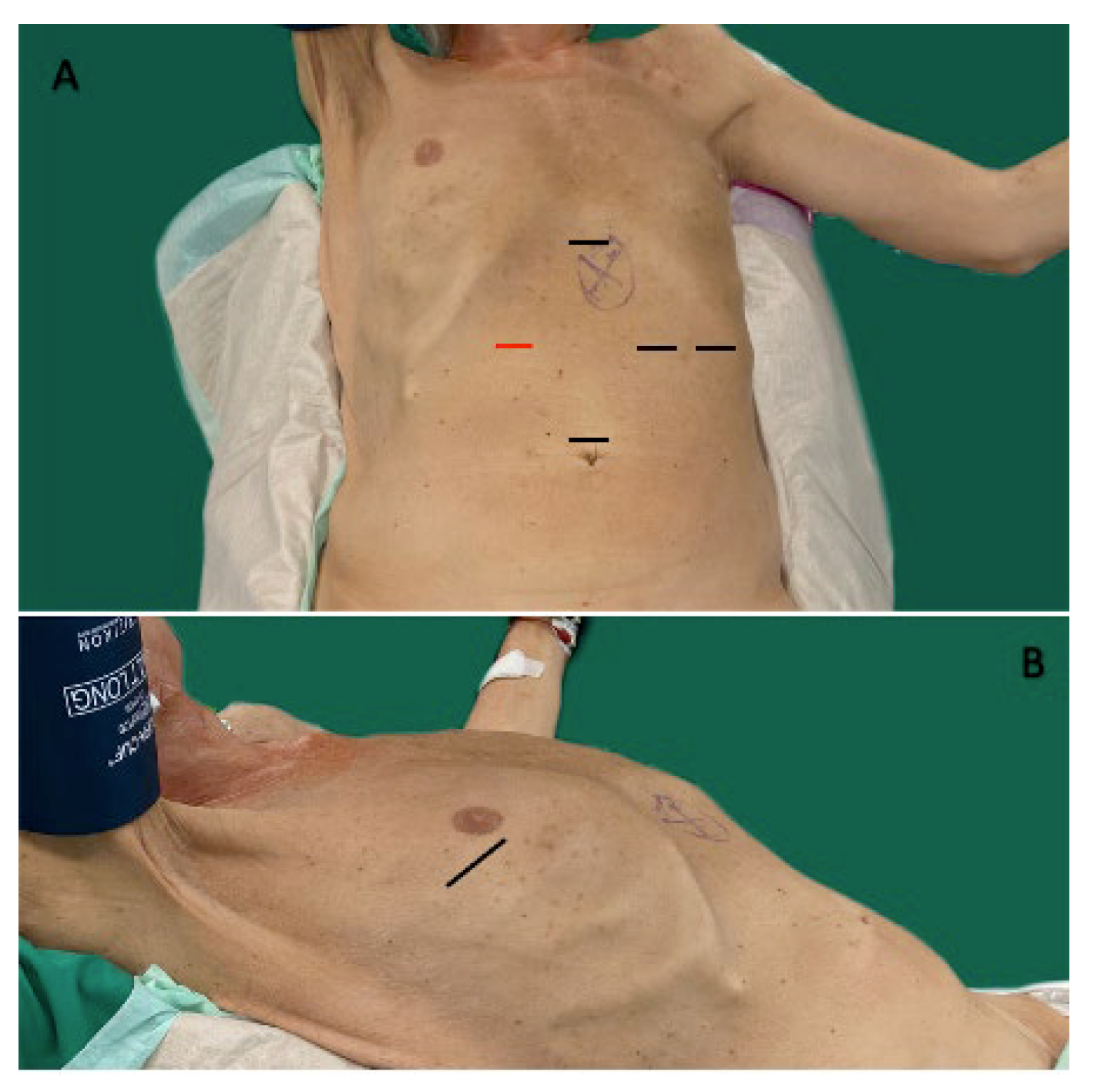
All operations were performed by two surgeons at all times. HMIE was done through a 2-stage approach consisting of a laparoscopic abdominal and a muscle-preserving open right thoracotomy phase. The patient was brought in a supine “left lateral” position in combination with a “beach-chair” position (Figure 2). This way, the 2-stage approach could be performed in one setting without the need for re-positioning.
During the abdominal phase, the first trocar was usually placed supra-umbilically. After induction of a pneumoperitoneum, four additional trocars were placed in a diamond-fashion where the one in the right upper quadrant must be at least a 12 mm port, which is needed for fashioning the gastric conduit (Figure 2). At first, the stomach was mobilized while preserving the gastric arcade and the right gastroepiploic artery and vein. A two-field lymphadenectomy was performed in concordance with the national treating guidelines for esophageal cancer [1]. Here, the left gastric artery and vein were identified, mobilized, and divided between two clips. Following complete mobilisation of the stomach, a 4–6 cm wide gastric conduit was fashioned by sequential firings of 45 mm Endo-GIA™ (Medtronic) cartridges parallel to the greater curvature, whereas the first firing was applied across the lesser curvature just above the pylorus and nearly perpendicular to the greater curvature. Following the complete mobilisation of the stomach and construction of the gastric conduit, an abdominal drain was inserted through the leftmost trocar and placed under the gastric conduit. This marked the end of the abdominal phase.
Following, a muscle-preserving right anterior thoracotomy was performed. The right lung was excluded using a double-lumen tube. The arch of the azygous vein was mobilized and divided using an Endo-GIA™ Curved Tip (Medtronic, Meerbusch, Germany). A routine infracarinal lymphadenectomy was performed. Before fashioning the esophagogastric anastomosis using a 25 mm EEA™ Auto Suture Circular Stapler (Medtronic, Meerbusch, Germany), an intraoperative pathology consultation took place to confirm free resection margins. Finally, two chest drains were placed before closing the thoracotomy—one perianastomotically and one at the base of the lung. All patients were extubated in the OR and transported to the ICU.

2.4. Postoperative Management

After surgery, all patients were primarily treated at the ICU and when stable enough, transferred to the intermediate care unit (IMC). Later, they were treated at the surgical ward. Enteral nutrition was standardized in all patients and started within 24 h postoperatively. For the first 4 postoperative days patients received nutrition via an intraoperatively inserted nasoduodenal feeding tube. From the fifth postoperative day, oral feeding was started with fluids and increased depending on clinical condition. Routine examination of the anastomosis was not performed. A combination of CT and upper endoscopy (UE) was only performed if an anastomotic leakage was suspected. In patients with severe complications (i.e., anastomotic leakage) a triple nasojejunal feeding tube was inserted after placing the endoluminal vacuum therapy.

2.5. Statistical Analysis

The data was obtained from the clinical database of our hospital. Statistical analysis was performed using IBM SPSS Statistics 27. Propensity score matching was used for matching OE patients to HMIE patients. The calculation of propensity scores was performed by logistic regression with the matching criteria as independent covariables. The propensity scores were then used to manually match pairs between OE and HMIE patients. The maximum caliper between propensity scores was 0.096 and the median caliper was 0.0056. Matching criteria were gender, age, preoperative BMI, American Society of Anesthesiologists (ASA) classification, location of the tumor in the esophagus, and the type of neoadjuvant treatment received. For statistical testing we used Mann-Whitney U tests for continuous and Fisher’s exact tests for categorical variables. To illustrate associations between possible risk factors and certain outcomes like complications we performed logistic regression. To obtain double robustness with identification of confounders we conducted a multivariate forward stepwise regression in cases where prior tests showed statistically significant differences.

3. Results

3.1. Patient Characteristics

Between January 2010 and August 2022, a total of 174 patients underwent Ivor Lewis esophagectomy at the University department for General and Visceral surgery, Klinikum Oldenburg. Among these, 50 patients received HMIE, and the rest were treated with OE (n = 124). After matching, the median age of the study population was 64 years in the OE group (Range: 32–81 years) and 64.5 years in the HMIE group (Range: 41–83 years). Median BMI was 25.50 in the OE group (Range: 18.00–39.00) and 26.0 in the HMIE group (Range: 19.00–37.00). Patients were ASA classified preoperatively. The distribution of ASA classification was comparable between groups, with the majority being classified as ASA II and III (OE 94.00%, HMIE 98.00%). About half of the patients received no neoadjuvant treatment (OE 48.00%, HMIE 50.00%), whereas the predominant rest was treated with either chemotherapy (OE 40.00%, n = 20; HMIE 36.00%, n = 18) or radiochemotherapy (OE 12.00%, n = 6; HMIE 12.00%, n = 6) prior to surgery. One patient of the OE group received endoscopic submucosal dissection. Most cancers were located in the lower third of the esophagus (OE and HMIE 84%, n = 42). The most frequent comorbidities were cardiac, e.g., hypertension or history of myocardial infarction (OE 68.00%, n = 34; HMIE 54.00%, n = 27), followed by pulmonary comorbidities such as allergic asthma or COPD (OE 16.00%, n = 8; HMIE 16.00%, n = 8). A third of the OE patients (32.00%, n = 16) and almost half of the HMIE patients (46.00%, n = 23) had a history of smoking, while excessive alcohol consumption was only reported by a few patients (OE 6.00%, n = 3; HMIE 10%, n = 5). Further patient and tumor characteristics are listed in Table 1.

3.2. Surgical Results

The exact surgical results are displayed in Table 2. The oncological results in the form of the number of harvested lymphnodes (OE Median 23.50; HMIE Median 21.00) and cases with negative tumor margin (R0-resection: OE 98.00%, n = 49; HMIE 100%, n = 50) were comparable across the groups. Only a few patients needed RBC transfusion perioperatively (OE 12%, n = 6; HMIE 2.00%, n = 1) and there was no significant difference between the groups (p = 0.112), but the number of RBCs that needed to be applied per patient was significantly higher in the OE group than in the HMIE group (OE Median 0.00, Maximum 6.00; HMIE Median 0.00, Maximum 1.00; p = 0.041). Hospital stay was significantly shorter for HMIE patients (OE Median 23.00 days; HMIE 16.50 days; p = 0.004). Furthermore, OE patients stayed almost twice as long on the ICU compared to HMIE patients (OE 5.50 days; HMIE 3.00 days; p = 0.003).

3.3. Perioperative Morbidity and Mortality

Half of the patients had at least one complication (n = 50). However, complications were significantly more common in the OE group (OE 70.00%, n = 35; HMIE 30%, n = 15; p < 0.001). Categorized for severity of complications according to the Clavien Dindo classification, severe complications (Clavien Dindo III–V) occurred in 48% of the OE patients (n = 24), whereas only in 16% of HMIE patients (n = 8). This difference was also statistically highly significant (p < 0.001). A chi-square test was performed to test for the influence of complication variables on the overall complication rate. All associations were statistically significant and the φ coefficient for effect size demonstrated the strongest influence for pulmonary complications (χ2(1) = 44.444, p ≤ 0.001, φ = 0667), followed by pneumonia alone (χ2(1) = 41.360, p ≤ 0.001, φ = 0.643) (see Appendix D, Table A4). These were also the most common complications, and though not statistically significant, were more common after OE than after HMIE (pulmonary complications OE 46%, HMIE 26%, p = 0.060; pneumonia OE 38%, HMIE 26%, p = 0.284). Nine patients died perioperatively after a median of 19 days, ranging from 7 to 82 postoperative days (OE 14.0%, n = 7; HMIE 4.0%, n = 2; p = 0.160). Anastomotic leakage occurred in a total of 16 patients of the matched study population. A statistically significant difference between the groups could not be proven (OE 22.0%, n = 11; HMIE 10.0%, n = 5; p = 0.171). The anastomotic leakages were categorized in accordance with the classification of the Esophagectomy Complications Consensus Group’s (ECCG) classification. Anastomotic leakages that required surgical therapy (ECCG III) occurred in five patients after OE, while not at all in the HMIE group, but the difference failed to be statistically significant (p = 0.056). Further morbidity data is listed in Table 3.

3.4. Risk Factor Analysis

Univariate testing with Fisher’s exact test revealed a significant difference between HMIE and OE patients regarding the occurrence of severe complications (Clavien Dindo III–V) (p = 0.001). The OR for HMIE compared to OE was 0.206 (95% CI 0.081–0.527). In order to test for confounding factors, a multivariate forward stepwise regression was conducted, where surgical technique was confirmed to be an independent risk factor for severe complications. Furthermore, Fisher’s test showed significant differences between ASA categories I/II and III/IV in relation to perioperative death (p = 0.034; OR 1.212; 95% CI 0.521–2.822). In the multivariate regression model, cardiac comorbidities were also significant risk factors for death (p = 0.037). Regarding the occurrence of any complications, OE (univariate p = 0.001, multivariate p < 0.001; OR 0.206; 95% CI 0.081–0.527) and cardiac comorbidities (univariate p = 0.040; multivariate p = 0.012; OR 2.571; 95% CI 1.122–5.895) could be identified as independent risk factors. No risk factors could be identified for pulmonary complications in univariate analysis. However, multivariate stepwise forward regression proved the differences between OE and HMIE groups (46%/26%; multivariate p = 0.019) and patients with or without preexisting pulmonary morbidities (18%/54%; multivariate p = 0.033) to be statistically significant after adjusting for sex, age, BMI, ASA classification, histology, neoadjuvant treatment or not, smoking status, cardiac comorbidities, diabetes mellitus, and alcohol abuse.
The detailed risk factor analysis for outcomes that were shown to have statistically significant risk factors are displayed in Table 4 and Table 5.
As mentioned above, Mann-Whitney U tests also showed statistically significant differences between surgical techniques regarding duration of hospital (median 23 vs. 16.5 days; p = 0.004) and ICU stay (median 5.50 vs. 3 days; p = 0.003), as well as regarding the number of applied RBCs (maximum 6.0 vs. 1.0; p = 0.041). A multivariate forward stepwise analysis confirmed this while showing no significant confounding by sex, BMI ≥ 25, ASA III/ IV, neoadjuvant treatment, smoking, pulmonary or cardiac comorbidities, diabetes mellitus, or alcohol abuse (see Appendixe A, Appendixe B and Appendixe C, Table A1, Table A2 and Table A3).

4. Discussion

In a meta-analysis with a total of 2397 patients from 2019, Yang et al. showed comparable results of HMIE and OE regarding the number of harvested lymphnodes [12]. This was also true for our study population, which not only showed similar numbers of harvested lymphnodes, but also almost the complete number of patients being R0-resected in both groups (OE 98%, HMIE 100%). These findings also go in line with other retrospective and prospective studies [6,13,14].
HMIE is undoubtedly on the rise in comparison to the open method. The aim of this study was to compare HMIE and OE regarding perioperative and oncological results and with that, to analyze whether HMIE could hold up to the expectations derived from other gastrointestinal procedures where advantages of laparoscopic compared to conventional open procedures have been shown numerous times [15,16]. Some studies have already shown similarly significant differences between HMIE and OE and the results are promising.
In our study we were able to confirm a lower overall postoperative complication rate for HMIE compared to OE, and demonstrated an 82% higher risk for the occurrence of complications for the OE group, while the overall complication rate was comparable to other studies with a similar number of patients [6,8]. Furthermore, the risk to develop severe complications, which were categorized by a Clavien Dindo classification of III–V, was 80% higher in the OE group than in the HMIE group. Several studies showed a high pulmonary complication rate for all esophagectomy approaches, and it was frequently shown that this rate was significantly lower for HMIE patients in comparison to OE [17,18]. We were not able to support this observation alone with statistically significant tests, but our data showed a clear tendency in the same direction, as pulmonary complications were observed in 46% of OE patients and only in 26% of HMIE patients (p = 0.06). Additionally, we could confirm the above mentioned assumption after adjustment for sex, age, BMI, ASA classification, histology, neoadjuvant treatment or not, smoking status, cardiac comorbidities, diabetes mellitus, and alcohol abuse (p = 0.019). We were also able to demonstrate a significant influence of pulmonary comorbidities on the development of pulmonary complications after any kind of esophagectomy in said adjusted setting (p = 0.033). The fact that pulmonary complications were significantly more common after OE than after HMIE raises the question of its cause, since all patients received the same kind of thoracotomy. However, the difference between HMIE and OE was the abdominal part, and considering that several studies have shown a higher rate of postoperative pneumonia after laparotomy compared to laparoscopy, the higher number of pulmonary complications in this study’s OE group could be partially caused by the abdominal approach [19,20]. Another possible explanation could be the difference in operating time, and with that the longer duration of mechanical ventilation, which has also been shown to be an independent risk factor for the development of postoperative pneumonia [21].
The rate of anastomotic leakages was higher after OE than after HMIE (OE 22%, HMIE 10%, p = 0.171). Although not statistically significant, the difference between rates of anastomotic leakages between the groups is remarkable. Known risk factors for said leakages, such as BMI > 30 kg/m2 or < 18.5 kg/m2, diabetes, or a smoking history are distributed equally between OE and HMIE groups, so the different frequencies cannot be explained by these [22]. However, a possible contribution could be a longer impaired circulation during the surgery since the groups are matched as homogeneously as possible, but we detected a difference in the duration of surgery and the needed number of RBCs [23,24]. In general, the results regarding anastomotic leakage rates are heterogenous throughout studies. While some report lower rates for OE compared to HMIE, other studies also show appreciably higher rates of anastomotic leakages after OE compared to HMIE [6,13]. Finally, a 2019 meta-analysis by Yang et al., including 17 studies and 2397 patients, also showed no difference between HMIE and OE (OR 0.95, 95% CI 0.67–1.35) [12]. Our results go in line with the literature here, since in our study the difference between anastomotic leakage rates is not statistically significant (p = 0.171).
Besides overall and pulmonary complications, we differentiated between severe complications and minor complications according to the Clavien Dindo classification. We considered complications classified Clavien Dindo III and above as severe, meaning at least the requirement of surgical, endoscopic, or radiological intervention [25]. In our study population these requirements were met by 48% of the OE group and 16% of HMIE patients with an Odds ratio of 0.206 (95% CI 0.081–0.527), which meant a large increase in risk of almost 80% for OE patients.
We were also able to show a significantly shorter hospital and ICU stay, as well as a significantly smaller number of applied RBCs for HMIE compared to OE patients. Since these are rather cost-relevant factors it would be interesting to perform a dedicated cost analysis of both procedures to see whether one is superior in this regard.

5. Limitations

This study had some limitations. The most important being the retrospective approach with all its weaknesses, including possible information bias, as it is not possible to fully ensure complete data in medical records. Another weakness is the single center setting that limited the size of our study population. Due to the matching procedure, additional patients had to be excluded from the analysis, which on one hand led to a robust handling of confounders, but on the other hand additionally reduced the number of patients.
Improvement of ICU management over time could have led to a lower overall complication rate in the HMIE group, of which the majority of patients received their surgery more recently than OE patients.
An analysis of risk factors was only conducted for the overall patient collective. To achieve statistically significant effects for analyses of subgroups a larger number of patients would be necessary. It would be interesting to know if the different surgical techniques come with different risk factors for certain complications.
One further limitation certainly is the restricted follow-up time. Our data collection was restricted to the hospital stay in which the operation took place. We were able to interpret the immediate oncological results and mortality, but long-term outcome, especially regarding recurrence rate or survival rates, could not be analyzed.

6. Conclusions

Our study clearly demonstrated reduced postoperative morbidity and in hospital-mortality for HMIE in comparison with OE. Not only were overall complications less likely to occur, but they were less severe than in OE cases. Furthermore, patients stayed significantly shorter in the ICU and were discharged faster when operated on with the HMIE approach. All this confirms the beneficial effects found in other studies and for other minimally-invasive surgical techniques of esophagectomy or other surgeries of the gastrointestinal tract.
Summed up, HMIE is a feasible technique that significantly decreases morbidity while ensuring equivalently good oncological resection compared to OE. HMIE should be performed whenever applicable for patients and surgeons.

Author Contributions

Conceptualization A.V. and N.E.-S. Methodology A.V. and N.E.-S.; Validation A.V. and N.E.-S.; Formal analysis A.V.; Investigation A.V.; Resources N.E.-S., M.B., S.M. and F.A.; Data curation A.V.; Interpretation A.V.; N.E.-S.; M.B., S.M. and F.A.; Writing—Original draft preparation A.V.; Writing—Review and editing N.E.-S. and M.B.; Visualization A.V. and N.E.-S.; Supervision N.E.-S. All authors have read and agreed to the published version of the manuscript.

Funding

This research received funding of the publication costs by the Carl-von-Ossietzky-University Oldenburg.

Institutional Review Board Statement

The study was conducted in accordance with the Declaration of Helsinki. Ethical review and approval were waived for this study, due to the unexceptional utilization of anonymous data that cannot be traced back to a person (Ethics committee of the Carl-von-Ossietzky-University Oldenburg, AZ 2022-116, 7 September 2022).

Informed Consent Statement

Patient consent was waived due to the sole utilization of anonymous data that cannot be traced back to a person—reviewed and approved by the Ethics committee (AZ 2022-116).

Data Availability Statement

Supporting results can be found in the Appendixe A, Appendixe B, Appendixe C and Appendixe D, Table A1, Table A2, Table A3 and Table A4 of this paper. The data presented in this study are available on request from the corresponding author. The data are not publicly available due to data protection restrictions of our institution.

Conflicts of Interest

The authors declare no conflict of interest.

Appendix A

Table A1. Step 1 of multivariate stepwise forward linear regression.
Regression Coefficient BStd ErrorWalddfpExp (B)
Step 1 aICU Stay (days)0.0390.0281.94710.1631.040
Hospital Stay (days)−0.0450.0214.60110.0320.956
Number of RBCs−1.4550.8193.15610.0760.233
Constant1.0300.4265.85610.0162.801
a In Step 1 inserted variables: ICU Stay (days), Hospital Stay (days), Number of RBCs.

Appendix B

Table A2. Step 2 of multivariate stepwise forward linear regression.
Regression Coefficient BStd ErrorWalddfpExp (B)
Step 2 aICU Stay (days)0.0360.0281.59710.2061.037
Hospital Stay (days)−0.0430.0214.39510.0360.958
Number of RBCs−1.6170.8403.70010.0540.199
Gender Female−0.0500.5480.00810.9270.951
BMI >/= 250.0000.4450.00010.9991.000
ASA 3/40.6070.4431.87610.1711.835
Constant0.7190.5651.62010.2032.052
a In Step 2 inserted variables: Gender Female, BMI >/= 25, ASA 3/4.

Appendix C

Table A3. Step 3 of multivariate stepwise forward linear regression.
Regression Coefficient BStd ErrorWalddfpExp (B)
Step 3 aICU Stay (days)0.0300.0310.94510.3311.031
Hospital Stay (days)−0.0430.0223.82110.0510.958
Number of RBCs−1.6560.9533.01910.0820.191
Gender Female−0.0680.5670.01410.9040.934
BMI >/= 250.1780.4650.14710.7011.195
ASA 3/40.8130.4992.65810.1032.256
Neoadjuvant Treatment0.2030.3290.38010.5371.225
Smoking0.4180.4750.77410.3791.518
Pulmonary Comorbidities0.4370.6880.40310.5261.547
Cardiac Comorbidities−0.8590.5032.92510.0870.423
Diabetes mellitus−0.1440.8370.03010.8630.866
Alcohol abuse0.2840.8400.11410.7351.328
Constant0.6870.7000.96410.3261.989
a In Step 3 inserted variables: Gender Female, BMI >/= 25, ASA 3/4, Neoadjuvant Treatment, Smoking, Pulmonary Comorbidities, Cardiac Comorbidities, Diabetes mellitus, Alcohol abuse.

Appendix D

Table A4. Chi-square test. Association between overall complications and individual complications.
Pearson-Chi-Square Valuedfpφ
Anastomotic leakage19.0481<0.0010.436
Pneumonia41.3601<0.0010.643
ARDS8.69610.0030.295
Pulmonary complication44.4441<0.0010.667
Pulmonary embolism4.16710.0410.204
Death5.98310.0140.245
ARDS = Acute respiratory distress syndrome.

References

  1. AWMF and Leitlinienprogramm Onkologie. S3-Leitlinie Diagnostik und Therapie der Plattenepithelkarzinome und Adenokarzinome des Ösophagus: Version 3.1., AWMF-Registernummer: 021/023OL; AWMF and Leitlinienprogramm Onkologie: Berlin, Germany, 2021. [Google Scholar]
  2. Moehler, M.; Al-Batran, S.E.; Andus, T.; Anthuber, M.; Arends, J.; Arnold, D.; Aust, D.; Baier, P.; Baretton, G.; Bernhardt, J.; et al. German S3-guideline “Diagnosis and treatment of esophagogastric cancer. Z. Fur Gastroenterol. 2011, 49, 461–531. (In German) [Google Scholar] [CrossRef] [PubMed] [Green Version]
  3. Schröder, W.; Gisbertz, S.S.; Voeten, D.M.; Gutschow, C.A.; Fuchs, H.F.; van Berge Henegouwen, M.I. Surgical Therapy of Esophageal Adenocarcinoma-Current Standards and Future Perspectives. Cancers 2021, 13, 5834. [Google Scholar] [CrossRef] [PubMed]
  4. van Workum, F.; Klarenbeek, B.R.; Baranov, N.; Rovers, M.M.; Rosman, C. Totally minimally invasive esophagectomy versus hybrid minimally invasive esophagectomy: Systematic review and meta-analysis. Dis. Esophagus 2020, 33, doaa021. [Google Scholar] [CrossRef] [PubMed]
  5. Harriott, C.B.; Angeramo, C.A.; Casas, M.A.; Schlottmann, F. Open versus hybrid versus totally minimally invasive Ivor Lewis esophagectomy: Systematic review and meta-analysis. J. Thorac. Cardiovasc. Surg. 2022, 164, e233–e254. [Google Scholar] [CrossRef]
  6. Mariette, C.; Markar, S.R.; Dabakuyo-Yonli, T.S.; Meunier, B.; Pezet, D.; Collet, D.; D’Journo, X.B.; Brigand, C.; Perniceni, T.; Carrère, N.; et al. Hybrid Minimally Invasive Esophagectomy for Esophageal Cancer. N. Engl. J. Med. 2019, 380, 152–162. [Google Scholar] [CrossRef]
  7. Nuytens, F.; Dabakuyo-Yonli, T.S.; Meunier, B.; Gagnière, J.; Collet, D.; D’Journo, X.B.; Brigand, C.; Perniceni, T.; Carrère, N.; Mabrut, J.Y.; et al. Five-Year Survival Outcomes of Hybrid Minimally Invasive Esophagectomy in Esophageal Cancer: Results of the MIRO Randomized Clinical Trial. JAMA Surg. 2021, 156, 323–332. [Google Scholar] [CrossRef]
  8. Biere, S.S.; van Berge Henegouwen, M.I.; Maas, K.W.; Bonavina, L.; Rosman, C.; Garcia, J.R.; Gisbertz, S.S.; Klinkenbijl, J.H.; Hollmann, M.W.; De Lange, E.S.; et al. Minimally invasive versus open oesophagectomy for patients with oesophageal cancer: A multicentre, open-label, randomised controlled trial. Lancet 2012, 379, 1887–1892. [Google Scholar] [CrossRef]
  9. Van der Wilk, B.J.; Hagens, E.R.; Eyck, B.M.; Gisbertz, S.S.; van Hillegersberg, R.; Nafteux, P.; Schröder, W.; Nilsson, M.; Wijnhoven, B.P.; Lagarde, S.M.; et al. Outcomes after totally minimally invasive versus hybrid and open Ivor Lewis oesophagectomy: Results from the International Esodata Study Group. Br. J. Surg. 2022, 109, 283–290. [Google Scholar] [CrossRef]
  10. AWMF and Leitlinienprogramm Onkologie. S3-Leitlinie Diagnostik und Therapie der Plattenepithelkarzinome und Adenokarzinome des Ösophagus.: Version 2.0. 2018. Available online: https://www.leitlinienprogramm-onkologie.de/leitlinien/oesophaguskarzinom/ (accessed on 27 September 2022).
  11. Porschen, R.; Buck, A.; Fischbach, W.; Gockel, I.; Görling, U.; Grenacher, L.; Hollerbach, S.; Hölscher, A.; Körber, J.; Messmann, H.; et al. S3-Leitlinie Diagnostik und Therapie der Plattenepithelkarzinome und Adenokarzinome des Ösophagus (Langversion 1.0—September 2015, AWMF-Registernummer: 021/023OL). Z. Fur Gastroenterol. 2015, 53, 1288–1347. (In German) [Google Scholar] [CrossRef] [Green Version]
  12. Yang, J.; Chen, L.; Ge, K.; Yang, J.L. Efficacy of hybrid minimally invasive esophagectomy vs. open esophagectomy for esophageal cancer: A meta-analysis. World J. Gastrointest. Oncol. 2019, 11, 1081–1091. [Google Scholar] [CrossRef]
  13. Glatz, T.; Marjanovic, G.; Kulemann, B.; Sick, O.; Hopt, U.T.; Hoeppner, J. Hybrid minimally invasive esophagectomy vs. open esophagectomy: A matched case analysis in 120 patients. Langenbeck’s Arch. Surg. 2017, 402, 323–331. [Google Scholar] [CrossRef] [PubMed]
  14. Bailey, L.; Khan, O.; Willows, E.; Somers, S.; Mercer, S.; Toh, S. Open and laparoscopically assisted oesophagectomy: A prospective comparative study. Eur. J. Cardio-Thorac. Surg. 2013, 43, 268–273. [Google Scholar] [CrossRef] [PubMed] [Green Version]
  15. Lacy, A.M.; García-Valdecasas, J.C.; Delgado, S.; Castells, A.; Taurá, P.; Piqué, J.M.; Visa, J. Laparoscopy-assisted colectomy versus open colectomy for treatment of non-metastatic colon cancer: A randomised trial. Lancet 2002, 359, 2224–2229. [Google Scholar] [CrossRef] [PubMed]
  16. Van der Pas, M.H.; Haglind, E.; Cuesta, M.A.; Fürst, A.; Lacy, A.M.; Hop, W.C.; Bonjer, H.J. Laparoscopic versus open surgery for rectal cancer (COLOR II): Short-term outcomes of a randomised, phase 3 trial. Lancet. Oncol. 2013, 14, 210–218. [Google Scholar] [CrossRef]
  17. Bjelovic, M.; Babic, T.; Spica, B.; Gunjic, D.; Veselinovic, M.; Trajkovic, G. Could hybrid minimally invasive esophagectomy improve the treatment results of esophageal cancer? Eur. J. Surg. Oncol. 2016, 42, 1196–1201. [Google Scholar] [CrossRef]
  18. Briez, N.; Piessen, G.; Claret, A.; Triboulet, J.; Mariette, C. Is minimally invasive œsophagectomy for cancer decreasing pulmonary complications? Results from a case-control study. J. Clin. Oncol. 2010, 28 (Suppl. 15), 4071. [Google Scholar] [CrossRef]
  19. Sparn, M.B.; Widmann, B.; Pietsch, U.; Weitzendorfer, M.; Warschkow, R.; Steffen, T. Risk factors and outcomes of postoperative aspiration pneumonia in abdominal surgery patients: An exact matching and weighting analysis. Surgery 2021, 170, 1432–1441. [Google Scholar] [CrossRef]
  20. Baba, H.; Tokai, R.; Hirano, K.; Watanabe, T.; Shibuya, K.; Hashimoto, I.; Hojo, S.; Yoshioka, I.; Okumura, T.; Nagata, T.; et al. Risk factors for postoperative pneumonia after general and digestive surgery: A retrospective single-center study. Surg. Today 2020, 50, 460–468. [Google Scholar] [CrossRef]
  21. Charles, M.P.; Kali, A.; Easow, J.M.; Joseph, N.M.; Ravishankar, M.; Srinivasan, S.; Kumar, S.; Umadevi, S. Ventilator-associated pneumonia. Australas. Med. J. 2014, 7, 334–344. [Google Scholar] [CrossRef]
  22. Fabbi, M.; Hagens, E.R.C.; van Berge Henegouwen, M.I.; Gisbertz, S.S. Anastomotic leakage after esophagectomy for esophageal cancer: Definitions, diagnostics, and treatment. Dis. Esophagus 2021, 34, doaa039. [Google Scholar] [CrossRef]
  23. Welte, M.; Saugel, B.; Reuter, D.A. Perioperative blood pressure management: What is the optimal pressure? Anaesthesist 2020, 69, 611–622. [Google Scholar] [CrossRef] [PubMed]
  24. Chughtai, M.; Gwam, C.U.; Mohamed, N.; Khlopas, A.; Newman, J.M.; Khan, R.; Nadhim, A.; Shaffiy, S.; Mont, M.A. The Epidemiology and Risk Factors for Postoperative Pneumonia. J. Clin. Med. Res. 2017, 9, 466–475. [Google Scholar] [CrossRef] [PubMed] [Green Version]
  25. Dindo, D. The Clavien–Dindo Classification of Surgical Complications. In Treatment of Postoperative Complications after Digestive Surgery; Cuesta, M., Bonjer, H.J., Eds.; Springer: London, UK, 2014; pp. 13–17. [Google Scholar]
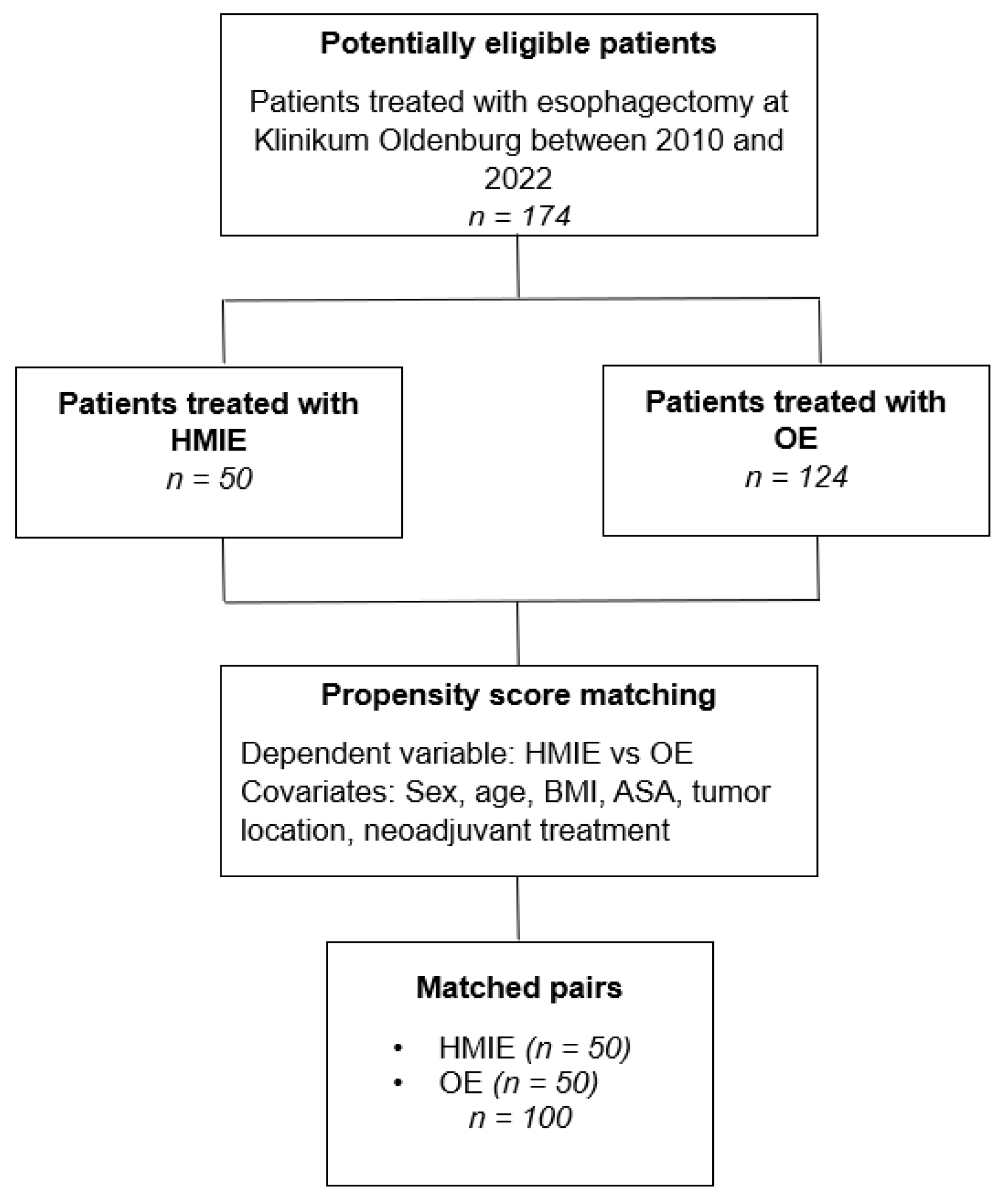
Figure 1. Study design.
Medicina 59 00434 g001
Figure 2. Positioning of the patient and placement of trocars. (A): Showing the placement of all five 5 mm–12 mm trocars. Red indicates placement of a 12 mm trocar. (B): Showing the positioning of the patient from the right side with the mark indicating the incision for open thoracotomy.
Medicina 59 00434 g002
Table 1. Clinicopathological features in association with surgical technique.
Open
(n = 50)
Hybrid
(n = 50)
n%Mediann%Medianp
SexMale4182.0% 4080.0% 1.000
Female918.0% 1020.0% 1.000
Age in years 64.00 64.500.373
Preoperative BMI 25.50 26.000.371
ASA-ClassificationASA I00.0% 00.0%
ASA II2652.0% 2142.0% 0.423
ASA III2142.0% 2856.0% 0.230
ASA IV36.0% 12.0% 0.617
ASA V00.0% 00.0%
Neoadjuvant TreatmentNone2448.0% 2550.0% 1.000
Chemotherapy2040.0% 1836.0% 0.837
Radio-Chemotherapy612.0% 612.0% 1.000
Endoscopic Submucosal Dissection00.0% 12.0% 1.000
Postoperative UICC-Classification07 8 1.000
IA14 12 0.820
IB2 9 0.051
IIA4 7 0.525
IIB9 0 0.003
III14 12 0.820
IV0 2 0.495
Histology of TumorNo malignancy12.0% 00.0% 1.000
Adenocarcinoma4081.6% 4386.0% 0.595
Squamous Cell Carcinoma816.3% 714.0% 1.000
Location of TumorLower third4284.0% 4284.0% 1.000
Middle third816.0% 714.0% 1.000
Upper third00.0% 12.0% 1.000
Smoking 1632.0% 2346.0% 0.218
Alcohol abuse 36.0% 510.0% 0.715
Diabetes mellitus 510.0% 48.0% 1.000
Cardiac Comorbidities 3468.0% 2754.0% 0.218
Pulmonary Comorbidities 816.0% 816.0% 1.000
Statistical testing was performed with Fisher’s exact test resp. Mann-Whitney U test. BMI = Body Mass Index, ASA = American Society of Anesthesiologists-Classification, UICC = Union Internationale Contre le Cancer-classification.
Table 2. Surgical outcome in association with surgical technique.
Open
(n = 50)
Hybrid
(n = 50)
MedianMaximumMinimumn (%)MedianMaximumMinimumn (%)p
Number of Harvested Lymphnodes23.5041.0012.00 21.0038.0013.00 0.666
Duration of Surgery288.00482.00127.00 225.50337.00122.00 0.243
Tumor MarginR0 49 (98.00) 50 (100.00)1.000
R1 0 0
R2 1 (2.00) 01.000
Required RBC Transfusion 6 (12.00) 1
(2.00)
0.112
Number of RBCs0.006.000.00 0.001.000.00 0.041
Hospital Stay (days)23.00123.007.00 16.50142.009.00 0.004
IMC Stay (days)4.0015.000.00 2.008.000.00 0.061
ICU Stay (days)5.5071.002.00 3.00117.001.00 0.003
Statistical testing was performed with Fisher’s exact test resp. Mann-Whitney U test. RBC = red blood cells units, IMC = intermediate care unit, ICU = intensive care unit.
Table 3. Postoperative Morbidity and Mortality.
Open
(n = 50)
Hybrid
(n = 50)
n%n%p
Any Complication3570.0%1530.0%<0.001
Clavien Dindo I-II2652.0%4284.0%0.001
Clavien Dindo III-V2448.0%816.0%0.001
Anastomotic Leakage1122.0%510.0%0.171
ECCG I00%00%
ECCG II612%510%1.000
ECCG III510%00%0.056
Pulmonary Embolism36.0%12.0%0.617
Pulmonary Complication2346.0%1326.0%0.060
Pneumonia1938.0%1326.0%0.284
ARDS612.0%24.0%0.269
Perioperative Death714.0%24.0%0.160
Statistical testing was performed with Fisher’s exact test. ECCG = Esophagectomy Complications Consensus Group classification, ARDS = Acute Respiratory Distress Syndrome.
Table 4. Risk factor analysis of overall complications and severe complications (Clavien Dindo III–V).
Any ComplicationClavien Dindo III–V
OE/
HMIE
(%)
paOR a
(95% CI)
pbOE/HMIE
(%)
paOR a
(95% CI)
pb
Surgical Technique (OE/HMIE)70/30<0.0010.184
(0.078–0.432)
< 0.00148/160.0010.206
(0.081–0.527)
<0.001
Sex (male/female)86/140.308 0.10352/121.000 0.483
Age (<65 years/≥65 years)54/460.689 0.27736/280.524 0.238
BMI (≥25/<25)60/401.000 0.42038/261.000 0.479
ASA (I–II/ III–IV)40/600.229 0.08228/360.674 0.329
Histology (adeno/SCC)40/160.595 0.21554/101.000 0.411
Neoadjuvant treatment (any/none)48/521.000 0.42236/280.524 0.306
Smoking (yes/no)34/660.412 0.15526/380.829 0.260
Pulmonary Comorbidities (yes/no)20/800.414 0.14012/520.771 0.057
Cardiac Comorbidities (yes/no)72/280.0402.571
(1.122–5.895)
0.01242/220.661 0.331
Diabetes mellitus (yes/no)14/860.160 0.04110/540.140 0.402
Alcohol abuse (yes/no)4/960.269 0.0724/601.000 0.238
a Univariate analysis including Fisher’s exact test. b Multivariate stepwise forward regression.
Table 5. Risk factor analysis of pulmonary complications and perioperative death.
Pulmonary ComplicationIn-Hospital Death
OE/HMIE
(%)
paOR a
(95% CI)
pbOE/HMIE
(%)
paOR a
(95% CI)
pb
Surgical Technique (OE/HMIE)46/260.060 0.01914/40.160 0.041
Sex (male/ female)60/120.793 0.33016/21.000 0.266
Age (<65 years/≥65 years)40/320.537 0.2504/140.089 0.036
BMI (≥25/<25)44/280.833 0.37512/60.733 0.314
ASA (I–II/III–IV)30/420.532 0.2142/160.0341.212
(0.521–2.822)
0.012
Histology (adeno/SCC)60/121.000 0.47418/00.351 0.079
Neoadjuvant treatment (any/none)36/361.000 0.4418/100.738 0.342
Smoking (yes/no)22/500.209 0.0998/100.733 0.364
Pulmonary Comorbidities (yes/no)18/540.089 0.0334/140.633 0.299
Cardiac Comorbidities (yes/no)50/220.209 0.09916/20.086 0.037
Diabetes mellitus (yes/no)6/661.000 0.4324/140.186 0.075
Alcohol abuse (yes/no)4/680.707 0.2520/181.00 0.179
a Univariate analysis including Fisher’s exact test. b Multivariate stepwise forward regression.
Disclaimer/Publisher’s Note: The statements, opinions and data contained in all publications are solely those of the individual author(s) and contributor(s) and not of MDPI and/or the editor(s). MDPI and/or the editor(s) disclaim responsibility for any injury to people or property resulting from any ideas, methods, instructions or products referred to in the content.