Article

Gramicidin, a Bactericidal Antibiotic, Is an Antiproliferative Agent for Ovarian Cancer Cells

1
Department of Biopharmaceutical Chemistry, Kookmin University, Seoul 02707, Republic of Korea
2
Biopharmaceutical Chemistry Major, School of Applied Chemistry, Kookmin University, Seoul 02707, Republic of Korea
3
Antibody Research Institute, Kookmin University, Seoul 02707, Republic of Korea
*
Authors to whom correspondence should be addressed.
These authors contributed equally to this work.
Medicina 2023, 59(12), 2059; https://doi.org/10.3390/medicina59122059
Submission received: 6 October 2023 / Revised: 25 October 2023 / Accepted: 21 November 2023 / Published: 22 November 2023
(This article belongs to the Section Oncology)

Abstract

:
Background and Objectives: Gramicidin, a bactericidal antibiotic used in dermatology and ophthalmology, has recently garnered attention for its inhibitory actions against cancer cell growth. However, the effects of gramicidin on ovarian cancer cells and the underlying mechanisms are still poorly understood. We aimed to elucidate the anticancer efficacy of gramicidin against ovarian cancer cells. Materials and Methods: The anticancer effect of gramicidin was investigated through an in vitro experiment. We analyzed cell proliferation, DNA fragmentation, cell cycle arrest and apoptosis in ovarian cancer cells using WST-1 assay, terminal deoxynucleotidyl transferase dUTP nick and labeling (TUNEL), DNA agarose gel electrophoresis, flow cytometry and western blot. Results: Gramicidin treatment induces dose- and time-dependent decreases in OVCAR8, SKOV3, and A2780 ovarian cancer cell proliferation. TUNEL assay and DNA agarose gel electrophoresis showed that gramicidin caused DNA fragmentation in ovarian cancer cells. Flow cytometry demonstrated that gramicidin induced cell cycle arrest. Furthermore, we confirmed via Western blot that gramicidin triggered apoptosis in ovarian cancer cells. Conclusions: Our results strongly suggest that gramicidin exerts its inhibitory effect on cancer cell growth by triggering apoptosis. Conclusively, this study provides new insights into the previously unexplored anticancer properties of gramicidin against ovarian cancer cells.

1. Introduction

Ovarian cancer (OC) is one of the most lethal forms of gynecological malignancies, ranking as the eighth leading cause of cancer-related deaths among women globally [1]. In 2023, 19,710 new cases of OC and 13,270 deaths from OC are expected in the United States alone [2]. Most OC cases (90%) are epithelial ovarian cancer, and serous carcinoma is the predominant histotype. The lack of discernible early symptoms, the limited early detection methods, the diagnosis at advanced stages (70% of cases), the dearth of efficient screening techniques, and the intricate tumor heterogeneity contribute to the high mortality of patients with OC [1,3,4,5]. Following debulking surgery, the current therapies for OC include platinum-based chemotherapy, such as carboplatin, angiogenesis inhibitors, such as bevacizumab, and poly-ADP-ribose polymerase inhibitors (PARPi), such as olaparib. Despite advances in advanced OC treatment, 70–80% of patients who undergo first-line chemotherapy eventually experience disease recurrence. The targeted application of PARPi is effective in treating patients with OC with breast cancer susceptibility gene (BRCA1/2) mutations and certain homologous recombination deficiency-positive tumors due to the selectivity of PARPi for cells with homologous recombination defects. However, the prevalence of BRCA1 or BRCA2 mutations in patients with OC is relatively low, comprising only approximately 15% of the entire patient cohort. Intriguingly, a substantial proportion, from 40% to 70% of patients with BRCA1/2-mutated OC, do not respond favorably to PARPi [4,6,7]. Given these challenges, innovative and novel therapeutic approaches targeting OC are urgently needed to address this persistent unmet medical requirement and improve the outcomes for patients with OC.
The development of novel and potent anticancer therapeutics typically requires 10–15 years and a substantial financial commitment ranging from USD 1 to 1.5 billion. In response to these formidable challenges, drug repositioning (DR) has emerged as a prominent strategy over the past decade. DR is a methodology for identifying fresh target molecules or applications for existing drugs via modifications in formulation, dosage, or route of administration, thereby obviating the need for structural alterations. Candidates for DR include drugs with established safety profiles from human clinical trials, but the drugs may have encountered setbacks during phase 2 or 3 trials or were withdrawn from the market due to adverse effects. By capitalizing on the pre-established safety, pharmacodynamics, and pharmacokinetics profiles verified via clinical trials, DR can abbreviate the development timeline (3–12 years) and substantially reduce costs (50%–60% of novel drug development). Furthermore, drugs developed via DR have a 150% higher probability of entering the market compared to entirely new drugs [8,9,10,11].
Gramicidin, a pentadecapeptide antibiotic derived from Bacillus brevis (ATCC 8185) [12,13], functions as an ionophore, exerting its antimicrobial effects by disrupting ion potential and interfering with monovalent cation transport. This multifaceted channel activity confers antimicrobial effects against a spectrum of microorganisms, including Gram-positive bacteria, fungi, and protozoa [13,14]. Gramicidin also exhibits potent anticancer properties in various cancer cell types, including renal cell carcinoma and gastric cancer cells. Gramicidin inhibits cell growth and angiogenesis in renal cell carcinoma and proliferation and the cell cycle in gastric cancer cells [15,16,17]. However, the anticancer properties of gramicidin in OC cells and the underlying mechanisms are relatively unexplored.
In this study, we employed terminal deoxynucleotidyl transferase dUTP nick and labeling (TUNEL) assays, DNA agarose gel electrophoresis, Western blot analysis, and flow cytometry to elucidate the inhibitory effects of gramicidin on OC cell proliferation via apoptosis. Collectively, our results indicate that gramicidin is a promising therapeutic agent for the treatment of OC.

2. Materials and Methods

2.1. Cell Culture

The human epithelial ovarian adenocarcinoma cell lines SKOV3 and A2780 were purchased from the Korea Biotechnology Commercialization Center (Incheon, Republic of Korea). The OVCAR8 cell line was purchased from the American Type Culture Collection (Rockville, MD, USA). The cell lines were maintained in Roswell Park Memorial Institute medium (Thermo Fisher Scientific; Waltham, MA, USA), supplemented with 10% heat-inactivated fetal bovine serum (Gibco, Carlsbad, CA, USA), 100 U/mL penicillin, and 100 μg/mL streptomycin. All cells were cultured at 37 °C in a humidified incubator with 5% CO2.

2.2. Cell Proliferation Assay

Gramicidin was acquired from TargetMol (Boston, MA, USA). Cell proliferation was assessed using a WST-1 Cell Proliferation Assay Kit (#MK400, Takara, Kyoto, Japan) according to the manufacturer’s instructions. Briefly, 5 × 10³ OVCAR8 and A2780 cells and 2 × 10³ SKOV3 cells in 200 μL culture medium were seeded into 96-well tissue culture plates. After 24 h, the cells were treated with 0, 0.33, 1, or 3 μM gramicidin for 24, 48, and 72 h. Then, 20 μL of WST-1 reagent was added to each well. After incubation for 1 h at 37 °C, the absorbance of the formazan product was measured at 450 nm using a microplate reader (Synergy H1, BioTek, Winooski, VT, USA). IC50 values were determined by fitting the dose–response curves to a four-parameter variable slope sigmoidal dose–response model in GraphPad Prism 5.0 software (GraphPad Software Inc., San Diego, CA, USA).

2.3. TUNEL Assay

The TUNEL assay was performed using an in situ cell death detection kit and fluorescein, according to the manufacturer’s instructions (#11684795910, Roche, Meylan, France) with slight modifications. In brief, 4 × 10³ OVCAR8 and SKOV3 cells were seeded into 8-well chamber slides (#177402, Thermo Fisher Scientific) coated with 1 μg/mL of poly-L-lysine (Sigma-Aldrich, St. Louis, MO, USA). After 24 h, the cells were treated with 0, 0.1, 0.3, or 1 μM gramicidin for 48 h. After the gramicidin treatment, the cells were fixed in phosphate-buffered saline (PBS) with 4% paraformaldehyde for 10 min and permeabilized using 0.1% Triton X-100 in PBS for 3 min at room temperature. Cells were then resuspended in 50 μL TUNEL-reaction mixture for 1 h at room temperature (RT) and stained with Hoechst 33342 (#H3570, Invitrogen, Carlsbad, CA, USA) for 3 min. After washing twice with PBS for 3 min each, the mounting medium (#S3023, DAKO, Santa Clara, CA, USA) was added, and the slides were examined using a confocal laser scanning microscope (Leica, Wetzlar, Germany). The TUNEL intensity of individual cells was quantified using Image J software (version 1.54d) (NIH, Bethesda, MD, USA). The TUNEL fluorescence intensity was determined by dividing the mean intensity of apoptotic cells by the mean intensity of control cells.

2.4. DNA Agarose Gel Electrophoresis

OVCAR8 (4 × 10⁵) and SKOV3 (2 × 10⁵) cells were seeded in 6-well culture plates. After 24 h, cells were treated with 0, 0.1, or 0.3 μM gramicidin for 48 h. The cells were then lysed with TE (Tris-EDTA) buffer (10 mM Tris, 1 mM EDTA, and 0.05% Tween 20, pH 9.0; Bio-sesang, Yongin, Republic of Korea) containing 0.2% Triton X-100 (Sigma-Aldrich). After centrifugation at 1500× g for 5 min, the supernatant was collected and treated with 2 μg/mL RNase A (Merck, Burlington, MA, USA) for 30 min at 37 °C followed by treatment with 2 μg/mL proteinase K (Sigma-Aldrich) for 30 min at 50 °C. The DNA was precipitated by adding ammonium acetate (Bio-sesang) and absolute cold ethanol (Merck), followed by a 1 h incubation at −20 h °C. After centrifugation at 14,000× g rpm for 10 min, the supernatant was discarded, and the pellet was washed with 70% ethanol and resuspended with TE buffer. Finally, 1 μg of DNA from each sample was subjected to electrophoresis on a 1.5% agarose gel and stained with ethidium bromide (Thermo Fisher Scientific). The DNA fragments were visualized under ultraviolet illumination.

2.5. Cell Cycle Analysis Using Propidium Iodine (PI) Staining

Cell cycle progression was analyzed via flow cytometry. OVCAR8 cells (2 × 10⁵) and SKOV3 cells (2 × 10⁵) were seeded in 6-well culture plates and treated with 0.1, 0.3, or 1 μM gramicidin for 48 h. Subsequently, the cells were fixed, permeabilized using ice-cold ethanol at −20 °C overnight and washed twice with PBS. The cells were then subjected to staining with a DNA staining buffer (dH2O supplemented with 50 μg/mL propidium iodide (BD, San Jose, CA, USA) and 2 μg/mL RNase A (Sigma-Aldrich)) at 37 °C for 10 min, followed by an additional 20 min incubation at room temperature. Cellular DNA content was measured using flow cytometry. A minimum of 10,000 events were recorded and analyzed using a flow cytometer (SONY, N First St, CA, USA).

2.6. Western Blot Analysis

OVCAR8 and SKOV3 cells (2 × 10⁵) in 6-well culture plates were lysed with RIPA buffer (25 mM Tris-HCl pH 7.6, 150 mM NaCl, 1% NP-40, 1% sodium deoxycholate, 0.1% SDS; Thermo Fisher Scientific) and protein concentrations were determined using a BCA Protein Assay Kit (Thermo Fisher Scientific). OVCAR8 and SKOV3 protein extracts (10 μg per sample) were separated by 12% SDS-PAGE and transferred to nitrocellulose membranes (Amersham, Little Chalfont, UK). Membranes were blocked with 5% bovine serum albumin in Tris-buffered saline plus 0.1% (v/v) Tween 20 (TBST) for 1 h at RT and then incubated overnight with primary antibodies against caspase-3 (1:1000; #14220), cleaved caspase-3 (1:200; #9664), PARP (1:1000; #9542), cleaved PARP (1:1000; #5625), and β-actin (1:1000; #sc-47778) (Santa Cruz, Heidelberg, Germany) at 4 °C. After washing six times with TBST, membranes were incubated with horseradish peroxidase-conjugated secondary antibody for 1 h at RT. Membranes were then washed six times with TBST, and chemiluminescence was detected using enhanced chemiluminescence (ECL; Thermo Fisher Scientific), followed by imaging using Amersham ImageQuant 800 (Cytiva, Logan, UT, USA).

2.7. Statistical Analyses

All statistical analyses were performed using the GraphPad Prism 5.0 software (GraphPad Software Inc., San Diego, CA, USA). Statistical differences between groups were assessed using one-way analysis of variance (ANOVA) with Dunnett’s multiple comparison test. p-values < 0.05 were considered statistically significant (* p < 0.05, ** p < 0.01, *** p < 0.001).

3. Results

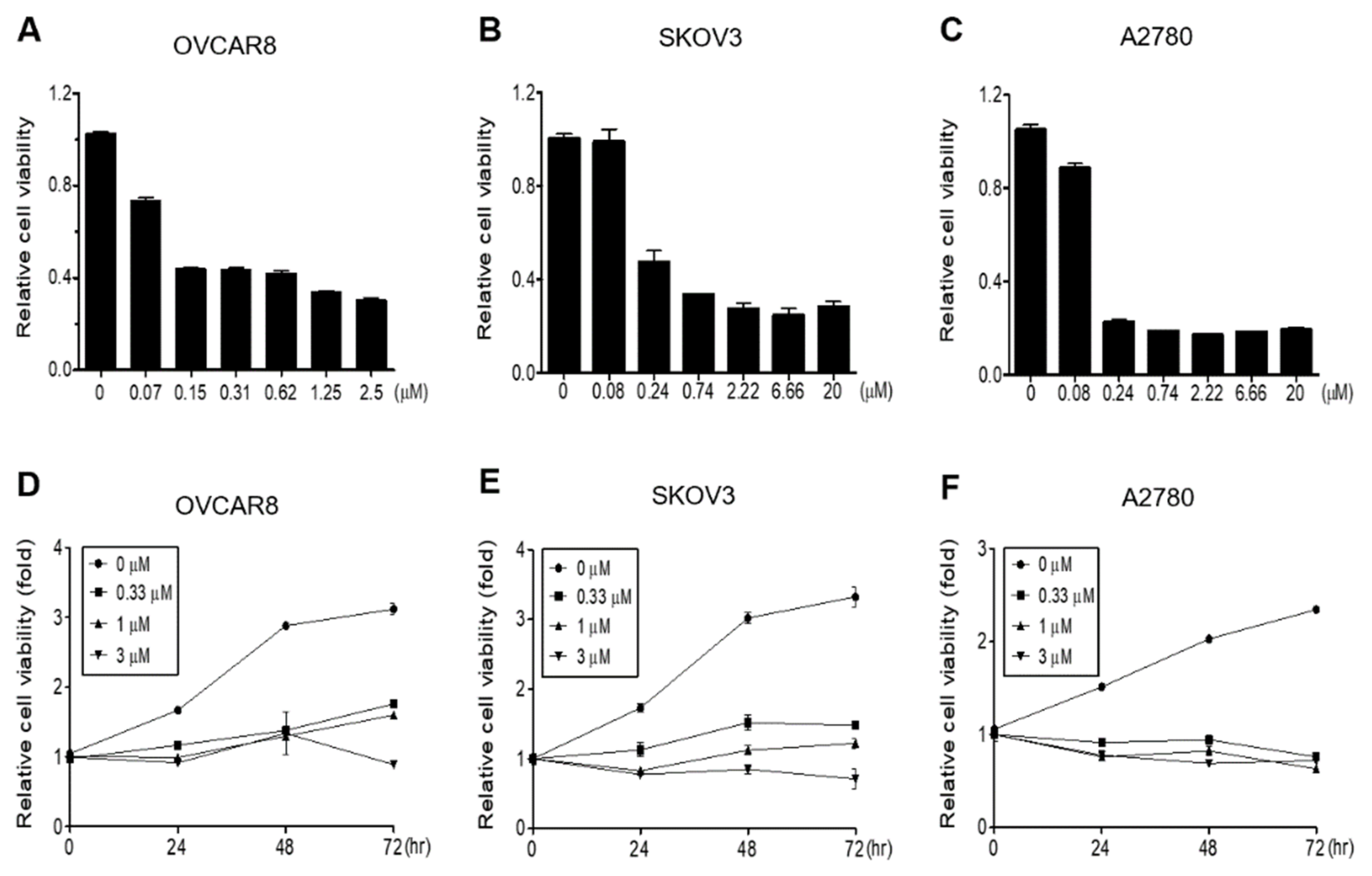
3.1. Gramicidin Inhibits the Proliferation of Ovarian Cancer Cells

To investigate the inhibitory effects of gramicidin on proliferation, OVCAR8, A2780, and SKOV3 OC cells were treated with gramicidin, and proliferation was measured using WST-1 assays. A dose-dependent reduction in cell growth was observed across all three OC cell lines with IC50 values of 0.0763, 0.1856, and 0.1148 μM for OVCAR8, SKOV3, and A2780 cells, respectively (Figure 1A–C). As shown in Figure 1D–F, the impact of gramicidin on OC cell growth was also evaluated over time (0, 24, 48, and 72 h). Gramicidin inhibited OC cell proliferation in a time-dependent manner. Collectively, these findings indicate that gramicidin inhibits the growth of OC cells in a dose- and time-dependent manner.

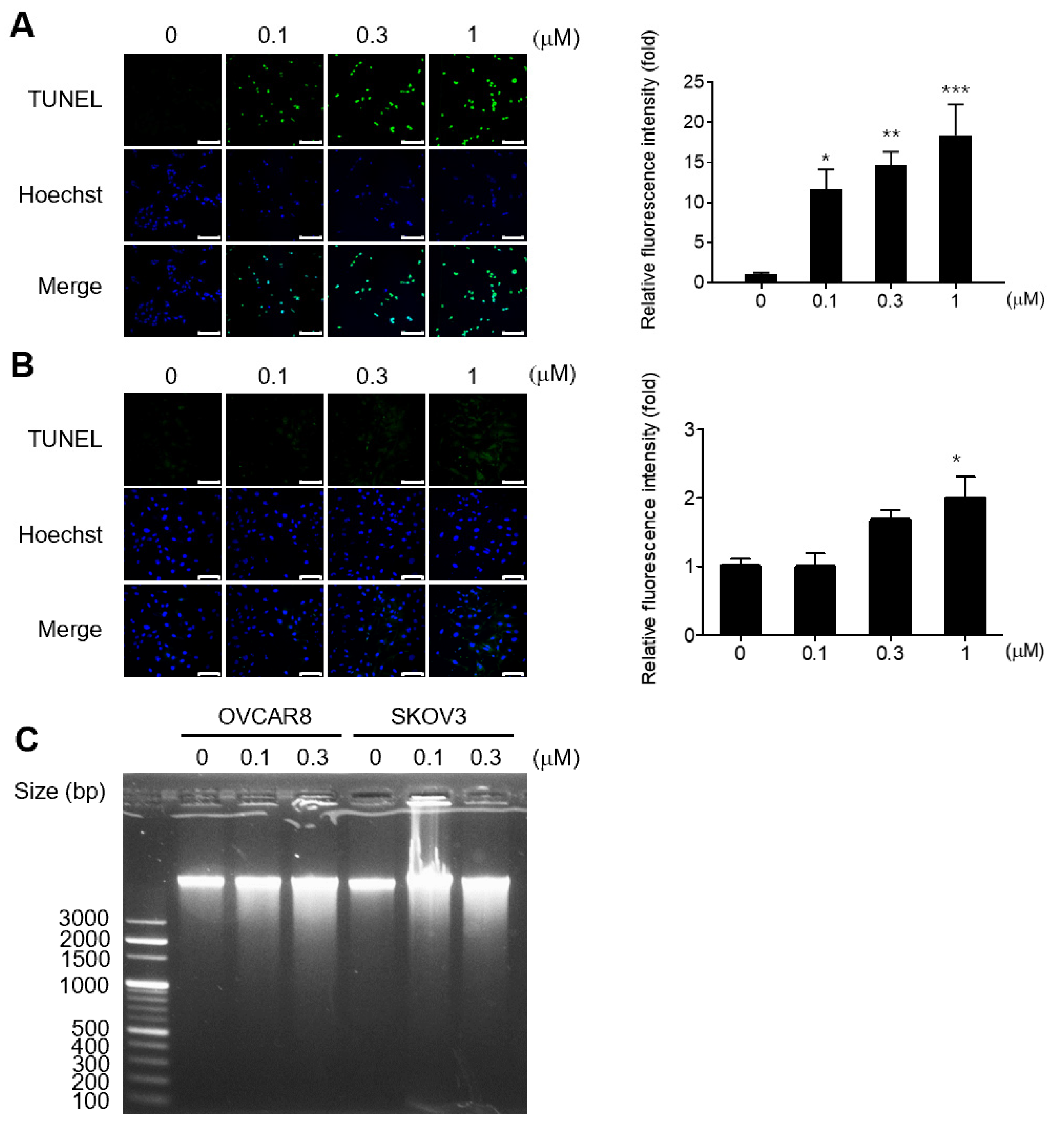
3.2. Gramicidin Induces Apoptosis by Mediating DNA Fragmentation in Ovarian Cancer Cells

TUNEL assay was used to assess the apoptotic effects of gramicidin in OC cells. Gramicidin induced DNA fragmentation, indicating apoptosis, in OVCAR8 and SKOV3 cells in a dose-dependent manner when compared to the negative control (Figure 2A,B). DNA fragmentation agarose gel electrophoresis was performed to confirm the DNA fragmentation. Consistent with the TUNEL assay results, DNA fragmentation tended to increase with increasing gramicidin concentrations (Figure 2C).
A previous study reported that gramicidin induced G2/M cycle arrest in SGC-7901 cells [15]. Thus, we investigated the effects of gramicidin on the cell cycle distribution of OC cells using flow cytometry. Gramicidin treatment induced a significant increase in the sub-G1 phase in both OVCAR8 and SKOV3 cells, as shown in Figure 3. These results strongly imply that gramicidin influences DNA fragmentation and the cell cycle via apoptosis.

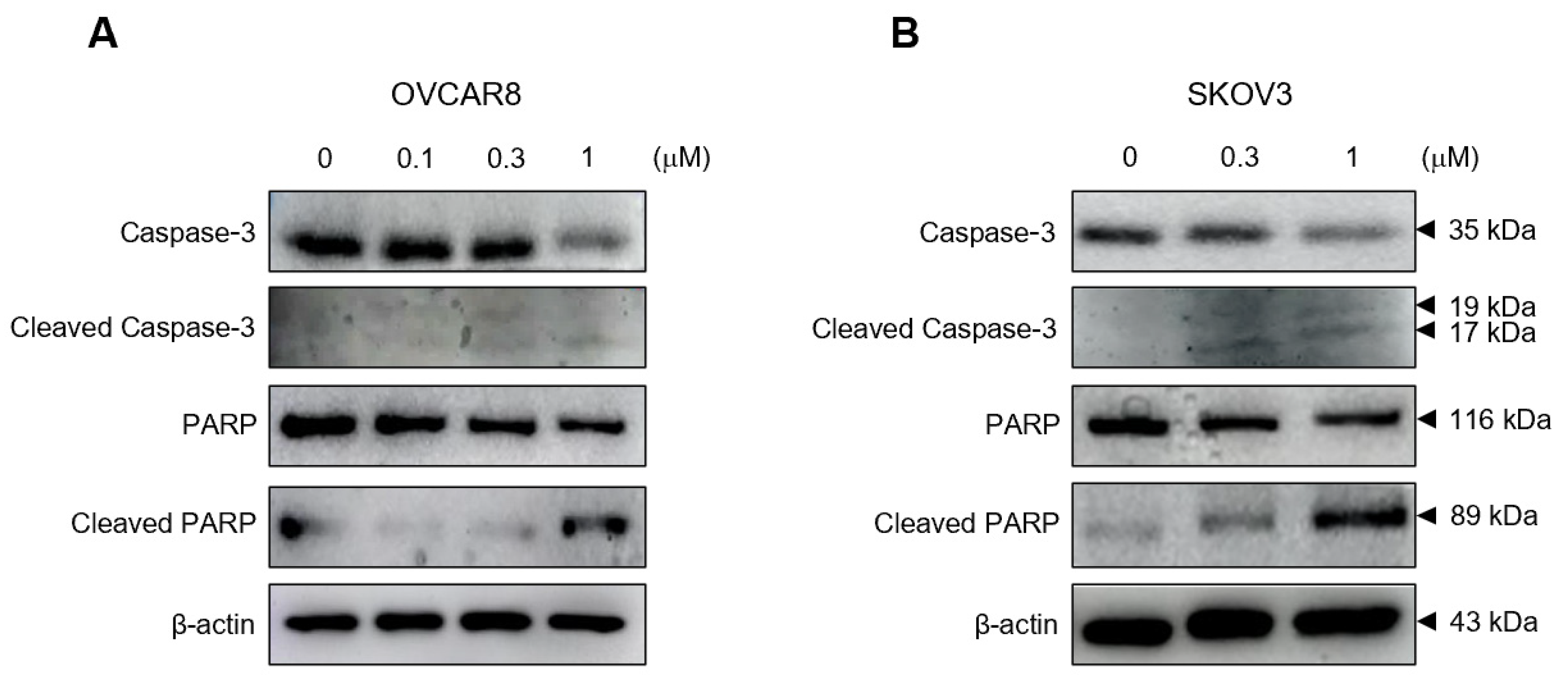
3.3. Gramicidin Mediates Apoptosis of Ovarian Cancer Cells via the Activation of Caspase-3

To determine the mechanisms underlying gramicidin-induced apoptosis in OC cells, the expression levels of the apoptosis-related proteins, caspase-3 and PARP, were determined with Western blot analysis. Gramicidin increased cleaved caspase-3 protein levels in both OVCAR8 and SKOV3 cells and substantially reduced the expression of caspase-3 protein. Cleaved PARP protein, a downstream target of caspase-3, was also increased, and the expression of PARP protein decreased (Figure 4). These data provide compelling evidence that gramicidin exerts its antitumor effect via the induction of apoptosis.

4. Discussion

In this study, we investigated the potential effectiveness of gramicidin for the treatment of OC via DR. Gramicidin inhibited the proliferation of OC cells, including the OVCAR8, SKOV3, and A2780 cell lines, in a dose- and time-dependent manner (Figure 1). TUNEL assay, cell cycle analysis, DNA agarose gel electrophoresis, and Western blot analysis using apoptosis-related antibodies in OVCAR8 and SKOV3 cell lines demonstrated that gramicidin induces apoptosis in OC cells. The apoptosis is characterized by DNA fragmentation and the cleavage of caspase-3 and PARP.
Gramicidin is a channel-forming polypeptide antibiotic composed of 15 L- and D-amino acids. The gramicidin monomer adopts a β-helix structure with an internal pore, and the monomers combine to form homodimers with β-helix pore structures that span the cell membrane. Dimerized gramicidin creates a pore through which water and monovalent cations can freely diffuse, resulting in Na+ influx and K+ efflux, membrane depolarization, osmotic swelling, and, ultimately, cell lysis [14,16,18]. Previous studies demonstrated the anticancer effects of gramicidin in various cancer cell types. For example, gramicidin inhibits the proliferation of cholangiocarcinoma cells (RBE and HuCCT1) [19]. Gramicidin also inhibited the proliferation of gastric cancer cells (SGC-7901), liver cancer cells (HepG2), and pancreatic cancer stem cells (BxPC3 and MIA PaCa-2) by inducing apoptosis [15,20,21]. Of note, the effects of gramicidin on OC cells have not been previously reported. This study represents the first investigation into the impact of gramicidin on OC cells, shedding light on its potential therapeutic significance in treating patients with OC.
Apoptosis is an energy-dependent programmed cell death process in multicellular organisms, leading to characteristic cellular changes via intrinsic and extrinsic pathways. Apoptotic cells exhibit morphological alterations, including cell shrinkage, blebbing, nuclear fragmentation, and chromatin condensation. Furthermore, apoptotic cells undergo biochemical modifications, including phosphatidylserine translocation from the inner to the outer membrane layer to facilitate phagocytosis by macrophages, protein degradation, DNA fragmentation, and the activation of caspases. Caspases, which can be classified into initiators and executioners, are cysteine proteases that cleave proteins after aspartic acid residues. The executioner caspases include caspase-3, which is activated via caspase cascades and plays a pivotal role in apoptosis. Caspase-3 cleaves various cellular proteins, cytoskeletal proteins, and DNA repair proteins [22,23]. In this study, cleaved caspase-3 and cleaved PARP increased in response to gramicidin treatment, suggesting that gramicidin induces the activation of caspase-3, which subsequently cleaves PARP—a known DNA repair protein—in OC cells (Figure 4). In apoptotic cells, chromosomal DNA is cleaved into oligonucleosomal-sized fragments by DNA fragmentation factors that are activated by cleaved caspase-3 in the late stages of apoptosis [24]. Our results demonstrate that gramicidin increases TUNEL fluorescent intensity (Figure 2), DNA fragmentation (Figure 2), and the percentage of cells in the sub-G1 phase (Figure 3) in OVCAR8 and SKOV3 cells. These results collectively indicate that DNA fragmentation is augmented in gramicidin-treated OC cells in a dose-dependent manner. Thus, our data strongly support the hypothesis that gramicidin induces apoptosis in OC cells.
The mechanism by which gramicidin induces apoptosis in cancer cells remains unknown. David et al. reported that gramicidin inhibited hypoxia-inducible factor 1α (HIF-1α) in renal cancer cells, consequently suppressing angiogenesis and tumor growth [25]. HIF-1α is an oxygen-sensitive subunit of HIF-1 and is expressed under hypoxia. Given that hypoxia is a common feature of many solid tumors, HIF-1α is frequently activated in cancer cells. Activated HIF-1α acts as a transcription factor, driving the expression of genes involved in various processes, such as survival, proliferation, metabolism, migration, and angiogenesis, in cancer cells [26,27]. Therefore, results indicate that gramicidin inhibits HIF-1α, in turn, inhibiting cell proliferation and inducing apoptosis in OC cells. However, further studies are required to investigate the anticancer effects of gramicidin via HIF-1α inhibition in OC cells.
In conclusion, our results support the DR of gramicidin as a potential therapeutic option for patients with OC. Gramicidin is a promising novel therapeutic agent for the treatment of OC.

Author Contributions

Conceptualization, M.S.C., C.Y.L., J.H.K., H.J.K. and K.H.; methodology, M.S.C. and K.H.; software, M.S.C. and C.Y.L.; validation, M.S.C. and C.Y.L.; formal analysis, M.S.C.; investigation, M.S.C., C.Y.L., J.H.K. and Y.M.L.; resources, S.L. and K.H.; data curation, M.S.C. and H.J.K.; writing—original draft preparation, M.S.C., C.Y.L., J.H.K. and K.H.; writing—review and editing, K.H.; visualization, K.H.; supervision, K.H. All authors have read and agreed to the published version of the manuscript.

Funding

This work was supported by a National Research Foundation of Korea (NRF) grant funded by the Korean government (MSIT) (No. 2023R1A2C1005434) and the Korea Health Technology R&D Project via the Korea Health Industry Development Institute (No. HI22C0360).

Institutional Review Board Statement

Not applicable.

Informed Consent Statement

Not applicable.

Data Availability Statement

The data presented in this study are available upon request from the corresponding authors.

Conflicts of Interest

The authors declare no conflict of interest. The funders have no role in the design of the study; the collection, analyses, or interpretation of data; the writing of the manuscript; or the decision to publish the results.

References

  1. Schoutrop, E.; Moyano-Galceran, L.; Lheureux, S.; Mattsson, J.; Lehti, K.; Dahlstrand, H.; Magalhaes, I. Molecular, cellular and systemic aspects of epithelial ovarian cancer and its tumor microenvironment. Semin. Cancer Biol. 2022, 86, 207–223. [Google Scholar] [CrossRef] [PubMed]
  2. Siegel, R.L.; Miller, K.D.; Wagle, N.S.; Jemal, A. Cancer statistics, 2023. CA Cancer J. Clin. 2023, 73, 17–48. [Google Scholar] [CrossRef] [PubMed]
  3. Torre, L.A.; Trabert, B.; DeSantis, C.E.; Miller, K.D.; Samimi, G.; Runowicz, C.D.; Gaudet, M.M.; Jemal, A.; Siegel, R.L. Ovarian cancer statistics, 2018. CA Cancer J. Clin. 2018, 68, 284–296. [Google Scholar] [CrossRef]
  4. Biegala, L.; Gajek, A.; Marczak, A.; Rogalska, A. PARP inhibitor resistance in ovarian cancer: Underlying mechanisms and therapeutic approaches targeting the ATR/CHK1 pathway. Biochim. Biophys. Acta Rev. Cancer 2021, 1876, 188633. [Google Scholar] [CrossRef] [PubMed]
  5. Liu, H.D.; Xia, B.R.; Jin, M.Z.; Lou, G. Organoid of ovarian cancer: Genomic analysis and drug screening. Clin. Transl. Oncol. 2020, 22, 1240–1251. [Google Scholar] [CrossRef] [PubMed]
  6. Bouberhan, S.; Bar-Peled, L.; Matoba, Y.; Mazina, V.; Philp, L.; Rueda, B.R. The evolving role of DNA damage response in overcoming therapeutic resistance in ovarian cancer. Cancer Drug Resist. 2023, 6, 345–357. [Google Scholar] [CrossRef]
  7. Jiang, X.; Li, X.; Li, W.; Bai, H.; Zhang, Z. PARP inhibitors in ovarian cancer: Sensitivity prediction and resistance mechanisms. J. Cell. Mol. Med. 2019, 23, 2303–2313. [Google Scholar] [CrossRef]
  8. Masuda, T.; Tsuruda, Y.; Matsumoto, Y.; Uchida, H.; Nakayama, K.I.; Mimori, K. Drug repositioning in cancer: The current situation in Japan. Cancer Sci. 2020, 111, 1039–1046. [Google Scholar] [CrossRef]
  9. Kobayashi, Y.; Banno, K.; Kunitomi, H.; Tominaga, E.; Aoki, D. Current state and outlook for drug repositioning anticipated in the field of ovarian cancer. J. Gynecol. Oncol. 2019, 30, e10. [Google Scholar] [CrossRef]
  10. Armando, R.G.; Gomez, D.L.M.; Gomez, D.E. New drugs are not enough-drug repositioning in oncology: An update. Int. J. Oncol. 2020, 56, 651–684. [Google Scholar] [CrossRef]
  11. Jourdan, J.P.; Bureau, R.; Rochais, C.; Dallemagne, P. Drug repositioning: A brief overview. J. Pharm. Pharmacol. 2020, 72, 1145–1151. [Google Scholar] [CrossRef] [PubMed]
  12. Sun, D.; He, S.; Bennett, W.F.D.; Bilodeau, C.L.; Andersen, O.S.; Lightstone, F.C.; Ingolfsson, H.I. Atomistic Characterization of Gramicidin Channel Formation. J. Chem. Theory Comput. 2021, 17, 7–12. [Google Scholar] [CrossRef] [PubMed]
  13. Kessler, N.; Schuhmann, H.; Morneweg, S.; Linne, U.; Marahiel, M.A. The linear pentadecapeptide gramicidin is assembled by four multimodular nonribosomal peptide synthetases that comprise 16 modules with 56 catalytic domains. J. Biol. Chem. 2004, 279, 7413–7419. [Google Scholar] [CrossRef] [PubMed]
  14. Wang, F.; Qin, L.H.; Pace, C.J.; Wong, P.; Malonis, R.; Gao, J.M. Solubilized Gramicidin A as Potential Systemic Antibiotics. Chembiochem 2012, 13, 51–55. [Google Scholar] [CrossRef] [PubMed]
  15. Chen, T.; Wang, Y.; Yang, Y.; Yu, K.; Cao, X.; Su, F.; Xu, H.; Peng, Y.; Hu, Y.; Qian, F.; et al. Gramicidin inhibits human gastric cancer cell proliferation, cell cycle and induced apoptosis. Biol. Res. 2019, 52, 57. [Google Scholar] [CrossRef] [PubMed]
  16. David, J.M.; Rajasekaran, A.K. Gramicidin A: A New Mission for an Old Antibiotic. J. Kidney Cancer VHL 2015, 2, 15–24. [Google Scholar] [CrossRef] [PubMed]
  17. David, J.M.; Owens, T.A.; Barwe, S.P.; Rajasekaran, A.K. Gramicidin A induces metabolic dysfunction and energy depletion leading to cell death in renal cell carcinoma cells. Mol. Cancer Ther. 2013, 12, 2296–2307. [Google Scholar] [CrossRef] [PubMed]
  18. Kelkar, D.A.; Chattopadhyay, A. The gramicidin ion channel: A model membrane protein. Biochim. Biophys. Acta 2007, 1768, 2011–2025. [Google Scholar] [CrossRef]
  19. Gong, X.; Zou, L.; Wang, M.; Zhang, Y.; Peng, S.; Zhong, M.; Zhou, J.; Li, X.; Ma, X. Gramicidin inhibits cholangiocarcinoma cell growth by suppressing EGR4. Artif. Cells Nanomed. Biotechnol. 2020, 48, 53–59. [Google Scholar] [CrossRef]
  20. Wang, R.Q.; Geng, J.; Sheng, W.J.; Liu, X.J.; Jiang, M.; Zhen, Y.S. The ionophore antibiotic gramicidin A inhibits pancreatic cancer stem cells associated with CD47 down-regulation. Cancer Cell Int. 2019, 19, 145. [Google Scholar] [CrossRef]
  21. Haoyang, W.W.; Xiao, Q.; Ye, Z.; Fu, Y.; Zhang, D.W.; Li, J.; Xiao, L.; Li, Z.T.; Hou, J.L. Gramicidin A-based unimolecular channel: Cancer cell-targeting behavior and ion transport-induced apoptosis. Chem. Commun. 2021, 57, 1097–1100. [Google Scholar] [CrossRef] [PubMed]
  22. Elmore, S. Apoptosis: A review of programmed cell death. Toxicol. Pathol. 2007, 35, 495–516. [Google Scholar] [CrossRef] [PubMed]
  23. Wong, R.S. Apoptosis in cancer: From pathogenesis to treatment. J. Exp. Clin. Cancer Res. 2011, 30, 87. [Google Scholar] [CrossRef] [PubMed]
  24. Zhang, J.H.; Xu, M. DNA fragmentation in apoptosis. Cell Res. 2000, 10, 205–211. [Google Scholar] [CrossRef]
  25. David, J.M.; Owens, T.A.; Inge, L.J.; Bremner, R.M.; Rajasekaran, A.K. Gramicidin A Blocks Tumor Growth and Angiogenesis through Inhibition of Hypoxia-Inducible Factor in Renal Cell Carcinoma. Mol. Cancer Ther. 2014, 13, 788–799. [Google Scholar] [CrossRef] [PubMed]
  26. Wang, X.; Du, Z.-W.; Xu, T.-M.; Wang, X.-J.; Li, W.; Gao, J.-L.; Li, J.; Zhu, H. HIF-1 alpha Is a Rational Target for Future Ovarian Cancer Therapies. Front. Oncol. 2021, 11, 785111. [Google Scholar] [CrossRef] [PubMed]
  27. Semenza, G.L. Defining the role of hypoxia-inducible factor 1 in cancer biology and therapeutics. Oncogene 2010, 29, 625–634. [Google Scholar] [CrossRef]
Figure 1. Gramicidin inhibited the proliferation of ovarian cancer (OC) cells. (A) OVCAR8, (B) SKOV3, and (C) A2780 cells were treated with the indicated concentrations of gramicidin for 72 h. (D) OVCAR8, (E) SKOV3, and (F) A2780 cells were treated with 0, 0.33, 1, or 3 μM gramicidin for 0, 24, 48, or 72 h. Relative cell proliferation rates were assessed using the WST-1 assay, which monitors mitochondrial succinate reductase activity at 450 nm. The data represent the mean ± SEM of duplicates from two independent experiments.
Medicina 59 02059 g001
Figure 2. Gramicidin-induced apoptosis, as indicated by DNA fragmentation in OC cells. Apoptotic cells were detected by DNA fragmentation using the TUNEL assay and DNA agarose gel electrophoresis. (A) OVCAR8 and (B) SKOV3 were treated with 0, 0.1, 0.3, or 1 μM gramicidin for 48 h and imaged using confocal microscopy. TUNEL-positive nuclei are shown in green, and total nuclei stained with Hoechst 33,342 are presented in blue (left panel). Relative TUNEL intensities, normalized to the negative control, were quantified in five randomly selected microscopic fields (right panel). (C) OVCAR8 and SKOV3 were treated with 0, 0.1, or 0.3 μM of gramicidin for 48 h, and genomic DNA was isolated. DNA fragmentation was assessed by agarose gel electrophoresis with ethidium bromide staining. The data are presented as mean ± SEM of a minimum of three independent experiments. Statistical significance was calculated using one-way ANOVA (* p < 0.05, ** p < 0.01, and *** p < 0.001).
Medicina 59 02059 g002
Figure 3. Gramicidin increased sub-G1 in OC cells. (A) OVCAR8 and (B) SKOV3 were treated with 0, 0.1, 0.3, or 1 μM of gramicidin for 48 h and subsequently stained with propidium iodide (PI) for cell cycle analysis using flow cytometry. The distribution of cells at different cell cycle phases was determined, and the percentages of cells in each phase relative to the total number of cells were calculated. The data are presented as mean ± SEM of three independent experiments.
Medicina 59 02059 g003
Figure 4. Gramicidin-induced cleavage of caspase-3 and PARP in OC cells. (A) OVCAR8 and (B) SKOV3 cells were treated with 0, 0.1, 0.3, or 1 μM of gramicidin for 24 h. Whole-cell lysates were analyzed via immunoblotting against caspase-3, cleaved caspase-3, PARP, cleaved PARP, or β-actin. The results represent a minimum of three independent experiments.
Medicina 59 02059 g004
Disclaimer/Publisher’s Note: The statements, opinions and data contained in all publications are solely those of the individual author(s) and contributor(s) and not of MDPI and/or the editor(s). MDPI and/or the editor(s) disclaim responsibility for any injury to people or property resulting from any ideas, methods, instructions or products referred to in the content.

