Article

Three Weeks of Pulmonary Rehabilitation Do Not Influence Oscillometry Parameters in Postoperative Lung Cancer Patients

by
Sabina Kostorz-Nosal
1,*,
Dariusz Jastrzębski
1,
Aleksandra Żebrowska
2,
Agnieszka Bartoszewicz
3 and
Dariusz Ziora
1
1
Department of Lung Diseases and Tuberculosis, Faculty of Medical Sciences in Zabrze, Medical University of Silesia in Katowice, 41-803 Zabrze, Poland
2
Department of Physiological and Medical Sciences, Institute of Sport Sciences, Academy of Physical Education, 40-065 Katowice, Poland
3
Independent Public Clinical Hospital No. 1, Medical University of Silesia, 41-800 Zabrze, Poland
*
Author to whom correspondence should be addressed.
Medicina 2022, 58(11), 1551; https://doi.org/10.3390/medicina58111551
Submission received: 10 September 2022 / Revised: 23 October 2022 / Accepted: 25 October 2022 / Published: 28 October 2022
(This article belongs to the Section Pulmonary)

Abstract

:

Simple Summary

Forced oscillation technique allows assessment of the respiratory system impedance, free from patient-dependent factors. Our study is the first to evaluate whether the implementation of physical activity has an influence on oscillometric results in patients after thoracic surgery due to lung cancer. The results indicate that despite the 6-min walking test and hand strength improvements, pulmonary rehabilitation has no impact on oscillometric values in this group of patients.

Abstract

Background: Thoracic surgery is a recommended treatment option for non-small cell lung cancer patients. An important part of a patient’s therapy, which helps to prevent postoperative complications and improve quality of life, is pulmonary rehabilitation (PR). The aim of this study was to assess whether the implementation of physical activity has an influence on forced oscillation technique (FOT) values in patients after thoracic surgery due to lung cancer. Methods: In this observational study, we enrolled 54 patients after thoracic surgery due to lung cancer, 49 patients with idiopathic interstitial fibrosis (IPF), and 54 patients with chronic obstructive pulmonary disease/asthma–COPD overlap (COPD/ACO). All patients were subjected to three weeks of in-hospital PR and assessed at the baseline as well as after completing PR by FOT, spirometry, grip strength measurement, and the 6-min walk test (6MWT). Results: We observed differences between FOT values under the influence of physical activity in studied groups, mostly between patients after thoracic surgery and COPD/ACO patients; however, no significant improvement after completing PR among FOT parameters was noticed in any group of patients. Improvements in the 6MWT distance, left hand strength, and right hand strength after PR were noticed (p < 0.001, 0.002, and 0.012, respectively). Conclusions: Three weeks of pulmonary rehabilitation had no impact on FOT values in patients after thoracic surgery due to lung cancer. Instead, we observed improvements in the 6MWT distance and the strength of both hands. Similarly, no FOT changes were observed in IPF and COPD/ACO patients after completing PR.

1. Introduction

Lung cancer has long been the most common malignant neoplasm in the world, with the highest mortality for both genders, accounting for approximately 18.4% of overall cancer mortality [1,2]. Nonetheless, at the time of diagnosis, only about 15% of cancers are operable [3]. Surgery may be the definitive available therapeutic strategy depending on the type of cancer, the stage of cancer, and other patient conditions (such as comorbidities and performance status). Complete tumor resection, if possible, is a recommended treatment option in non-small cell lung cancer patients [4,5]. However, thoracic surgery is fraught with the risk of postoperative pulmonary complications (PPCs), the most common of which are respiratory disorders (mainly respiratory failure) and cardiovascular events [5].
Among the methods of PPC prevention, chest physiotherapy plays a key role [5] because it increases mobility, breathing, and sputum clearance, as well as preserving lung function, especially when applied preoperatively [6,7,8]. Furthermore, the results of the latest studies clearly indicate the beneficial effects of pulmonary rehabilitation (PR) on the recuperation of functional efficiency, as well as improvements in the quality of life and life expectancy in this group of patients [9]. After thoracic surgery due to lung cancer, patients manifest obstructive disorders, so the rehabilitation model used in this group of patients corresponds to the rehabilitation program applied to patients with chronic obstructive pulmonary disease [10,11]. Some PPCs, such as atelectasis and fibrosis, may contribute to the prevalence of restrictive disorders that determine different lung function disorders. In these cases, it is possible that a completely different bedside manner, i.e., one with a rehabilitation scheme that brings greater benefits and advantages, should be applied.
Apart from quality-of-life questionnaires, the effectiveness of PR can also be objectively evaluated via lung function and exercise tests. Currently, spirometry is commonly performed for all patients [8]. However, this test requires forced expiration maneuvers that are highly dependent on the patient’s cooperation and effort. As a consequence, spirometry performance may prove to be problematic in children younger than 10 years and elderly populations older than 65 years [12,13]. Likewise, after thoracic surgery due to lung cancer, patients may be unable to correctly perform spirometry due to postoperative pain, lung stiffness, and fear associated with forced expiratory maneuvers, as well as the advanced age of diagnosis. These circumstances may have an effect on the obtained results.
In the case of the first two groups of patients (children and the elderly), the forced oscillation technique (FOT) has been successfully applied [14,15]. Thanks to the external excitation pressure generated by FOT devices and applied to airways during tidal breathing, oscillometry could be free from patient-dependent factors, such as coughing or pain, associated with recent thoracic surgery.
FOT allows one to assess respiratory, airway, lung parenchyma, or thoracic wall impedance. Therefore, an FOT device determines the resistance (R) and reactance (X) based on the response of the respiratory system to external stimuli [15,16]. R refers to the viscous resistance and is mostly dependent on airway diameter. The use of different frequencies allows one to separate airway resistance into total (R5, at 5 Hz), central (R19, at 19 Hz), and peripheral (R5–R19) resistance. The second impedance component is X, which is determined by elastic properties that dominate at low frequencies (X5) and inertia properties of the lung tissue that dominate at high frequencies (X19). The point when the value of X is equal to 0, at which elastic and inertia properties are equal, is called the resonant frequency (Fres).
Previous studies have indicated increased R5 and R5–20 and reduced X5 in obstructive lung diseases. Reduced X5 has mostly been observed in restrictive disorders [17,18,19,20,21]. The Fres parameter was found to be increased in both disorders. In contrast, only two studies so far have analyzed the impact of lobectomies on FOT values [22,23], as similar FOT abnormalities (however less expressed) were observed in chronic obstructive lung disease (COPD) patients. Additionally, there have been no reports assessing the impact of pulmonary rehabilitation on FOT parameters in postoperative lung cancer patients.
Therefore, the aim of this study was to assess whether the implementation of three weeks of in-hospital physical activity had any influence on FOT values in patients after thoracic surgery due to lung cancer. The secondary aim was to compare spirometry and effort test results before and after PR. These findings were further compared with a group of idiopathic interstitial fibrosis (IPF) and COPD or asthma–COPD overlap (ACO) patients undergoing the same 3-week PR program. The following article is presented in accordance with the STROBE reporting checklist presented in Table S1 (available as online Supplementary Materials).

2. Materials and Methods

2.1. Materials

Fifty-four patients after thoracic surgery due to lung cancer, forty-nine IPF patients, and fifty-four patients with preserved obstructive lung disease (COPD or ACO) were subjected to a 3-week inpatient rehabilitation program in the Department of Lung Diseases and Tuberculosis, Medical University of Silesia in Katowice between September 2018 and April 2022 (Figure 1). This was an observational study, so patients that matched inclusion criteria were consecutively enrolled in the project. The study was approved by the Bioethics Committee of Medical University of Silesia in Katowice (Act No. KNW/0022/KB1/85/I/17 from 19 December 2017) and was conducted in accordance with the Declaration of Helsinki. All participants provided written informed consent. The research was registered in the ISRCTN Trials Registry (ISRCTN31987937).
Inclusion criteria were defined as follows: the diagnosis of a disease based on actual guidelines for IPF [24,25], obstructive lung diseases [26,27], or non-small cell lung cancer (histologically or cytologically confirmed) after operative treatment within 6 months at stage I or II; a stable period of illness without infection/exacerbation during the previous 4 weeks; a distance in the 6-min walk test of no less than 250 m; and the ability and willingness to perform physical activity.
The exclusion criteria were as follows: patient’s refusal to engage in the rehabilitation program, infection/exacerbation during the previous 4 weeks, MRC Dyspnea Score > 3; unstable coronary artery disease, low performance level (Eastern Cooperative Oncology Group scale ≥ 3) [28], anemia (hemoglobin < 10 g/dL), and poor tolerability of pulmonary rehabilitation.

2.2. Physiological Measurements

All patients enrolled in the study performed lung function and exercise tests at the baseline and after 3 weeks of PR. A lung test apparatus (MES; Cracow, Poland) was used to perform spirometry in accordance with the manufacturer’s guidelines [29] and to assess forced expiratory volume during the first second (FEV1), forced vital capacity (FVC), and FEV1/FVC. The results are expressed as a percentage of predicted values. The grip strengths of the left and right hands were measured with a Meden-Inmed Baseline hydraulic hand dynamometer (Charder Medical, Taichung City, Taiwan) in compliance with the manufacturer’s guidelines [30]. Patients were asked to squeeze the dynamometer 3 times with each hand with all of their strength. Only the best score for each hand was included in the study. FOT measurements were evaluated with a Resmon Pro Full device (Restech Respiratory Technology SRL, Milano, Italy; marketed by MGC Diagnostics Cooperation, Saint Paul, MN, USA). These were performed over several seconds of tidal breathing through a mouthpiece in a sitting position. External small oscillatory pressures, applied to the airways, generated the respiratory system response and were used to record impedance (as appropriate): resistance (R, inspiratory and expiratory) at frequencies of 5 Hz, 11 Hz, and 19 Hz; reactance (X, inspiratory and expiratory) at frequencies of 5 Hz, 11 Hz, and 19 Hz; and resonant frequency (Fres). The impedance, resistance, and reactance results are expressed in cmH2O/L/s as percentages of the predicted values, and the Fres results are expressed in Hertz in accordance with the work of Oostveen et al. [15].

2.3. Pulmonary Rehabilitation

In-hospital PR was performed 5 days per week for a total of 3 weeks, under the supervision of a physical therapist and rehabilitation specialist. The program was carried out under the ATS/ERS recommendations [10]. Training intensity was based on the limit of the heart rate and the distance obtained during the 6MWT (6MWD), as previously described [31], and it included a combination of endurance and strength training [32].
The PR program consisted of 150 min per day: (1) aerobic exercises and flexibility training with elements of yoga and suspension UEU (Universal Exercise Unit allowing for relief and self-assisted exercises for 20 min) therapy; (2) breathing exercises (three times per day for 10 min); (3) lumbar and cervical stabilization exercises and equilibrium exercises (once per day for 20 min); (4) general rehabilitation gymnastics including stretching exercises, strengthening of the muscles of the upper and lower limbs and the back, as well as exercise for posture correction (once per day for 30 min); (5) continuous or interval endurance training (once per day for 30 min); (6) exercises on a stabilometric platform (once per day for 20 min); and (7) physiotherapy treatments depending on individual needs, e.g., ultrasound therapy or cryotherapy for back pain. Subsequent exercises of the daily PR program were performed within individual recovery periods.
This program was complemented by autogenic training, music therapy, and sessions with a psychologist (for 60 min per day).

2.4. Statistical Analysis

Statistica 13.3 (TIBCO Software Inc., Palo Alto, CA, USA, License SUM JPZ010A903827ARACD-F) was used for the statistical analysis. Descriptive statistics are presented as the means with standard deviation. The normality of distribution was checked with the Shapiro–Wilk test. The ANOVA Kruskal-Wallis test and Chi2 test were used to evaluate the differences in demographic data. Differences in lung function and exercise test results between groups, from baseline to the completion of pulmonary rehabilitation, and comparisons of parameter changes over time between groups were assessed with nonparametric analyses of longitudinal data, Analysis of Variance (ANOVA)-type statistics, and Nonparametric Analysis of Longitudinal Data (nparLD), using the nparLD library for R language in RStudio software [33]. Data are presented as medians with interquartile ranges. Significant results achieved in nparLD were evaluated in sequence via the pairwise multiple comparison Wilcoxon test. A p-value of 0.05 or less was considered statistically significant.

3. Results

All patients enrolled in the study (except for two due to infection) completed pulmonary rehabilitation and were re-evaluated with the same lung function and exercise tests used at the beginning (Figure 1). The PR program was carried out by the same staff, and there were no training modality changes during the study.
Demographic data are presented in Table 1. During the course of PR, no changes in the pharmacological treatment of underlying diseases occurred. None of the lung cancer patients enrolled in the study required analgesic treatment due to the recent thoracic surgery.
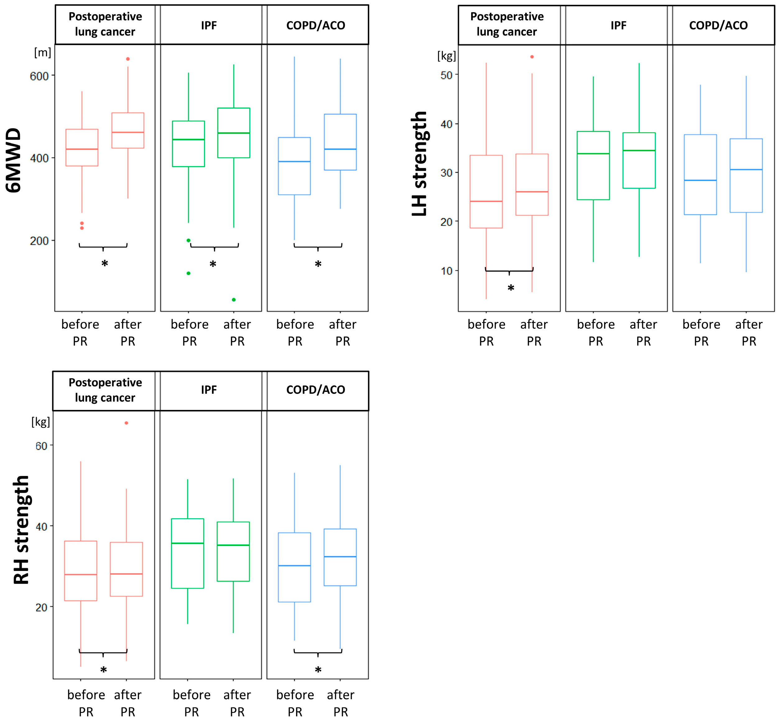
All lung function and exercise test results, including FOT parameters, significantly differed between the studied groups of patients at the baseline and/or at the end of the PR program (Table 2 and Table 3). After three weeks of PR, we observed improvements in 6MWD in all group of patients, as well as hand strength improvement in COPD/ACO patients regarding the left hand, and in thoracic surgery patients regarding both hands (Figure 2). Other parameters did not change after completion of the rehabilitation program.
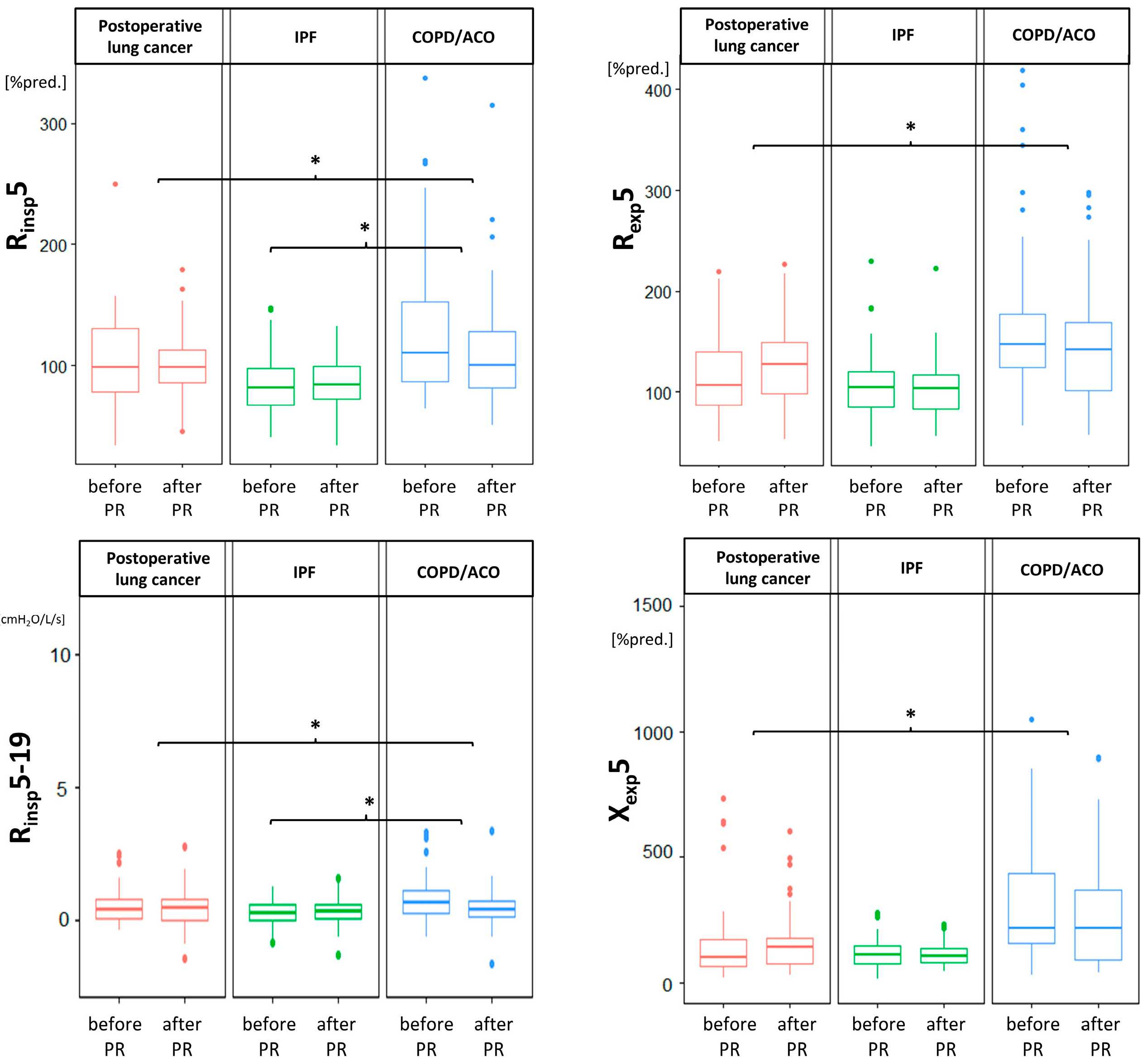
There were no differences in FOT values after PR (Table 3); however, we observed some significant differences between FOT changes in studied groups, mostly between patients after thoracic surgery and patients with obstructive lung disease (Figure 3).

4. Discussion

To the best of our knowledge, the impact of physical activity on oscillometry parameters in lung resection patients due to cancer has not yet been examined. According to our results, the FOT parameters in this group of patients remained stable after three weeks of pulmonary rehabilitation. As oscillometry is devoid of external factors that may have influenced the results (such as patient cooperation, muscle strength, and learning effect), we showed that three weeks of PR does not improve lung function, regardless of lung disease. Even if we observed significant disparities between changes in the FOT values due to PR in different groups of patients enrolled in the study (Table 3 and Figure 3), none of the studied groups demonstrated significant improvements or deteriorations with regard to oscillometric parameters.
However, these findings in no way imply that patients with lung diseases do not benefit from PR. We observed significant 6MWD improvements in all patients, as well as increases in right hand strength after training in COPD/ACO patients, and in both hands in patients after thoracic surgery due to lung cancer (p < 0.001, 0.016, 0.002, and 0.012, respectively; Table 2 and Figure 2).
These results may be considered to be rather disappointing when considering the well-documented positive impact of PR on quality of life, symptoms, muscle strength, and 6MWD [7,9,10,34,35]. However, these increases do not need to be consistent with lung function improvement, as studies have demonstrated PR-related improvements in skeletal muscle function and quality of life despite the absence of lung function improvements [36,37,38,39,40,41]. As COPD/ACO are systemic diseases, muscle dysfunction has a negative impact on these patients’ activities [42]. Lung cancer may also be considered to be a systemic disease with surgery-dependent consequences. Through peripheral muscle strength measurements, we showed that three weeks of PR may positively influence hand strength. These results are in concordance with previous meta-analyses showing improvements in muscle endurance and strength, as well as dyspnea reduction [43,44]. Although Brocki [45] reported improvements in respiratory muscle strength after two weeks of PR, regardless of the extent of the surgery or postoperative inspiratory muscle training, in this study, postoperative inspiration muscle training was connected to improvements in oxygenation.
Even though the authors of some studies have reported improvements in work breathing and lung volume influenced by PR in COPD patients, these have been argued to be caused by adjunctive therapies (such as medications, oxygen therapy, and exercise training) rather than breathing retraining alone [46]. As one PPC prevention method, perioperative PR has been recommended by the ERS [47,48]. A systematic review by Rodriguez-Larrad et al. [49] emphasized that pre-operative PR (compared to post-operative PR alone) is essential in functional capacity improvements and morbidity reductions. Contradictory data on pre-operative PR’s impact on lung function have been published. The authors of some studies observed no significant changes in spirometric results [40,41], while others reported improvements in FVC and FEV1 [50,51].
According to the ATS/ERS Guidelines, PR is one of the most effective non-pharmacological treatment options for COPD patients [34,52,53]. Dynamic lung hyperinflation, gas-exchange abnormalities, morphological alterations, and insufficient energy supplies are listed as pulmonary factors that may influence exercise limitations in COPD patients [54,55]. Until now, only two studies have presented analyses of the impact of PR on FOT measurements [31,56]. Zimmermann et al. [56] noticed that two 1 h sessions per week for a total of 16 sessions led to temporary improvements in Xinsp5 in COPD patients which were not observed at a 3-month follow-up; no other FOT changes were observed in that study. These improvements in lung volume were suggested to have resulted from reductions in ventilation heterogeneity. The differences between these results could derive from different PR settings, durations, and intensities. It is possible that the extension of PR duration would reveal improvements in FOT measurements. However, Zimmermann’s study was only conducted on 15 patients with no control group, so (as highlighted by the authors) the obtained results may not be representative and should be analyzed with caution [56].
In contrast, we previously reported that significant improvements in FVC were observed in patients with idiopathic interstitial pneumonia (IIP) subjected to three weeks of in-hospital PR, although, similarly, no changes in oscillometric results were noticed [31]. We reasoned that these results derived from better patient cooperation and compliance spirometry performance, improved diaphragm flexibility and strength, and the higher sensitivity of oscillometry in detecting SAD compared to spirometry [15,57].
It is commonly known that lung cancer surgery may be accompanied by many postoperative complications, with a frequency of between 2 and 40% [58,59,60,61], including pneumonia, atelectasis, acute respiratory failure, bronchospasm, pneumothorax, and prolonged air leaks. However, at the time of the admission to the Pulmonary Rehabilitation Department, we did not observe any serious pulmonary complications among postoperative patients. The authors of some studies have also addressed the subject of lung function deterioration after surgery and increased tissue resistance due to edematous changes and lung rigidity, which cause reduced lung compliance and decreases in ventilation due to intrathoracic pressure changes [22]. These factors contribute to the increased resistance of the thoracic wall, which, under normal conditions, represents one quarter of total airway resistance [62]. Local inflammation related to recent thoracic surgery may also lead to small airway disease [23].
Considering these results, lung function deterioration after thoracic surgery may be observed. Research on oscillometric changes in patients after lobectomy have consistently indicated increases in R5, R20, R5–R20, Fres, area of reactance (AX), and postoperative decreases in X5 and X11 [22,23]. Previous researchers have suggested that R depends on lung volume, as it was found to be correlated with decreases in VC and FEV1, as already described in this group of patients [63,64,65]. Additionally, a study performed by Drakou [66] indicated significant declines in FVC and FEV1 at one month after the thoracic operation was performed due to lung cancer diagnosis. Postoperative pulmonary function deterioration is mostly dependent on the extension of the surgery, so an R increase is coherent with VC and FEV1 decreases caused by lobectomy, according to volume reduction [65,67]. Additionally, there may be an impact of a postoperative anatomical repositioning or excursion of the remaining non-operated lobe which is even more pronounced in lobectomy compared to segmentectomy, due to its compensatory growth [68]. These results are in line with those of other studies, in which FVC was reported to improve over time as the stress applied by surgery declines [69,70]. However, the causes of postoperative lung function changes remain vague, as FOT changes were also described in surgery involving not the lungs, but the transurethral resection of a bladder tumor, under general anesthesia [71]. The authors of that study suggested that the observed lung function deterioration may have been caused by intraoperative lung injury triggered by intravascular fluid shifts, mechanical ventilation, and inhalational anesthetic drugs. Moreover, changes following lobectomy have been shown to be similar, regardless of concomitant lung diseases (COPD, IP, and CPFE) or their absence [22]. Similar findings were also obtained by Subotic et al. [72], who observed that there were no differences in lung function after lobectomy between patients with and without concomitant COPD.
As mentioned above, different lung diseases present different patterns of changes in FOT measurements [17,18,19,20,21]. Considering these conditions alone, more limitations in FOT results have been observed in patients after lobectomy compared to IPF patients, though they were less affected than COPD patients [23]. Interestingly, X11 decreases are more commonly seen in lobectomy patients than abnormalities in X5, though almost all previous studies considered only the X5 value. In a previous study, we observed only X11 decreases in IIP patients over three weeks, with or without PR [31]. However, in the current study, no changes in this parameter were observed, even in patients with IPF. Therefore, future studies are required to establish changes in oscillometric values as time passes, as well as to assess whether they are affected by physical activity.
Our study had several limitations. First of all, a small number of patients were enrolled in the study, which did not allow us to divide patients in terms of the extension of their lung surgery. Secondly, we did not include a control group consisting of postoperative lung cancer patients not subjected to PR. Thirdly, although lung cancer is more frequent in men, women presented greater willingness to undergo the pulmonary rehabilitation, and, therefore, applied more often for rehabilitation. Consequently, these circumstances had influenced the gender distribution and had contributed to the higher female participation. Finally, there was no possibility to extend the period of rehabilitation due to the National Health Fund limitations in Poland, which allow for a 3-week maximum in-hospital stay.
In conclusion, three weeks of pulmonary rehabilitation demonstrated no influence on FOT values in patients after thoracic surgery due to lung cancer. Instead, we observed improvements in 6MWD and the strength of both hands in this group of patients. Similarly, no FOT changes were observed in IPF and COPD/ACO patients. We believe that our data could facilitate the determination of the impact of a 3-week physical activity program on patient performance status and dispel doubts related to its influence on lung function.

Supplementary Materials

The following supporting information can be downloaded at: https://www.mdpi.com/article/10.3390/medicina58111551/s1, Table S1: STROBE Statement—Checklist of items that should be included in reports of cohort studies.

Author Contributions

Conceptualization, S.K.-N., D.J. and D.Z.; methodology, D.J.; software, S.K.-N. and A.B.; validation, A.Ż., D.J. and D.Z.; formal analysis, S.K.-N. and A.Ż.; investigation, S.K.-N.; resources, D.J.; data curation, A.B.; writing—original draft preparation, S.K.-N. and A.B.; writing—review and editing, D.J., D.Z. and A.Ż.; visualization, S.K.-N.; supervision, D.Z.; project administration, S.K.-N., A.B. and D.J.; funding acquisition, S.K.-N. All authors have read and agreed to the published version of the manuscript.

Funding

This study was supported by a grant from the National Science Centre in Poland (No. 2016/23/N/NZ7/02002) and the grants from Medical University of Silesia in Poland (No. PCN-1-090/N/2/K and No. PCN-1-194/N/0/K).

Institutional Review Board Statement

The study was conducted in accordance with the Declaration of Helsinki, and approved by the Bioethics Committee of the Medical University of Silesia in Katowice (Act No. KNW/0022/KB1/85/I/17 from 19 December 2017.

Informed Consent Statement

Informed consent was obtained from all subjects involved in the study.

Data Availability Statement

Datasets analyzed during the current study are available in the Figshare (https://doi.org/10.6084/m9.figshare.20373930.v1 accessed on 26 July 2022).

Conflicts of Interest

The authors declare no conflict of interest.

References

  1. Bray, F.; Ferlay, J.; Soerjomataram, I.; Siegel, R.L.; Torre, L.A.; Jemal, A. Global cancer statistics 2018: GLOBOCAN estimates of incidence and mortality worldwide for 36 cancers in 185 countries. CA Cancer J. Clin. 2018, 68, 394–424, Erratum in CA Cancer J. Clin. 2020, 70, 313. [Google Scholar] [CrossRef] [PubMed] [Green Version]
  2. Siegel, R.L.; Miller, K.D.; Fuchs, H.E.; Jemal, A. Cancer statistics, 2022. CA Cancer J. Clin. 2022, 72, 7–33. [Google Scholar] [CrossRef] [PubMed]
  3. American Cancer Society. Cancer Facts & Figures 2013; American Cancer Society: Atlanta, GA, USA, 2013; Available online: https://www.cancer.org/acs/groups/content/@epidemiologysurveilance/documents/document/acspc-036845.pdf (accessed on 11 November 2013).
  4. Howington, J.A.; Blum, M.G.; Chang, A.C.; Balekian, A.A.; Murthy, S.C. Treatment of Stage I and II Non-Small Cell Lung Cancer: Diagnosis and Management of Lung Cancer, 3rd ed.; American College of Chest Physicians Evidence-Based Clinical Practice Guidelines. Chest 2013, 143 (Suppl. 5), e278S–e313S. [Google Scholar] [CrossRef]
  5. Villeneuve, P.J. Interventions to avoid pulmonary complications after lung cancer resection. J. Thorac. Dis. 2018, 10 (Suppl. 32), S3781–S3788. [Google Scholar] [CrossRef] [PubMed]
  6. Novoa, N.; Ballesteros, E.; Aranda, J.L.; Jiménez, M.F.; Varela, G. Chest physiotherapy revisited: Evaluation of its influence on the pulmonary morbidity after pulmonary resection. Eur. J. Cardio-Thorac. Surg. 2011, 40, 130–134. [Google Scholar] [CrossRef] [Green Version]
  7. Mujovic, N.; Mujovic, N.; Subotic, D.; Ercegovac, M.; Milovanovic, A.; Nikcevic, L.; Zugic, V.; Nikolic, D. Influence of Pulmonary Rehabilitation on Lung Function Changes After the Lung Resection for Primary Lung Cancer in Patients with Chronic Obstructive Pulmonary Disease. Aging Dis. 2015, 6, 466–477. [Google Scholar] [CrossRef] [Green Version]
  8. Weiner, P.; Man, A.; Weiner, M.; Rabner, M.; Waizman, J.; Magadle, R.; Zamir, D.; Greiff, Y. The effect of incentive spirometry and inspiratory muscle training on pulmonary function after lung resection. J. Thorac. Cardiovasc. Surg. 1997, 113, 552–557. [Google Scholar] [CrossRef] [Green Version]
  9. Sano, Y. Perioperative Pulmonary Rehabilitation for Lung Cancer Surgeries in Patients with Poor Pulmonary Function. Kyobu Geka 2016, 69, 41–46. [Google Scholar]
  10. Spruit, M.A.; Singh, S.J.; Garvey, C.; ZuWallack, R.; Nici, L.; Rochester, C.; Hill, K.; Holland, A.E.; Lareau, S.C.; Man, W.D.-C.; et al. An Official American Thoracic Society/European Respiratory Society Statement: Key Concepts and Advances in Pulmonary Rehabilitation. Am. J. Respir. Crit. Care Med. 2013, 188, e13–e64. [Google Scholar] [CrossRef] [Green Version]
  11. Bolton, C.E.; Bevan-Smith, E.F.; Blakey, J.D.; Crowe, P.; Elkin, S.L.; Garrod, R.; Greening, N.J.; Heslop, K.; Hull, J.H.; Man, W.D.; et al. British Thoracic Society Pulmonary Rehabilitation Guideline Development Group; British Thoracic Society Standards of Care Committee. British Thoracic Society guideline on pulmonary rehabilitation in adults. Thorax 2013, 68 (Suppl. 2), ii1–ii30. [Google Scholar] [CrossRef] [PubMed] [Green Version]
  12. Tomalak, W.; Radliński, J.; Latawiec, W. Quality of spirometric measurements in children younger than 10 years of age in the light of the recommendations, Pneumonol. Alergol. Pol. 2008, 6, 421–425. [Google Scholar]
  13. Czajkowska-Malinowska, M.; Tomalak, W.; Radliński, J. Quality of spirometry in elderly. Pneumonol. Alergol. Pol. 2013, 81, 511–517. [Google Scholar] [CrossRef] [PubMed]
  14. Tomalak, W.; Radliński, J.; Czajkowska-Malinowska, M. Impulse oscillometry in assessing small airways properties. Electr. Rev. 2014, 90, 157–159. [Google Scholar]
  15. Oostveen, E.; MacLeod, D.; Lorino, H.; Farre, R.; Hantos, Z.; Desager, K.; Marchal, F. The forced oscillation technique in clinical practice: Methodology, recommendations and future developments. Eur. Respir. J. 2003, 22, 1026–1041. [Google Scholar] [CrossRef]
  16. Kaczka, D.W.; Dellacá, R.L. Oscillation mechanics of the respiratory system: Applications to lung disease. Crit. Rev. Biomed. Eng. 2011, 39, 337–359. [Google Scholar] [CrossRef] [Green Version]
  17. Mori, K.; Shirai, T.; Mikamo, M.; Shishido, Y.; Akita, T.; Morita, S.; Asada, K.; Fujii, M.; Suda, T.; Chida, K. Colored 3-dimenssional analyses of respiratory resistance and reactance in COPD and asthma. COPD 2011, 8, 456–463. [Google Scholar] [CrossRef]
  18. Kanda, S.; Fujimoto, K.; Komatsu, Y.; Yasuo, M.; Hanaoka, M.; Kubo, K. Evaluation of Respiratory Impedance in Asthma and COPD by an Impulse Oscillation System. Intern. Med. 2010, 49, 23–30. [Google Scholar] [CrossRef] [Green Version]
  19. Dandurand, R.; Dandurand, M.; Estepar, R.; Bourbeau, J.; Eidelman, D. Oscillometry in community practice ild is characterized by abnormal reactance but normal resistance. Quart. J. Med. 2016, 109, S50. [Google Scholar]
  20. Fujii, M.; Shirai, T.; Mori, K.; Mikamo, M.; Shishido, Y.; Akita, T.; Morita, S.; Asada, K.; Suda, T. Inspiratory resonant frequency of forced oscillation technique as a predictor of the composite physiologic index in interstitial lung disease. Respir. Physiol. Neurobiol. 2015, 207, 22–27. [Google Scholar] [CrossRef]
  21. Mori, K.; Shirai, T.; Mikamo, M.; Shishido, Y.; Akita, T.; Morita, S.; Asada, K.; Fujii, M.; Hozumi, H.; Suda, T.; et al. Respiratory mechanics measured by forced oscillation technique in combined pulmonary fibrosis and emphysema. Respir. Physiol. Neurobiol. 2013, 185, 235–240. [Google Scholar] [CrossRef]
  22. Kaku, R.; Yoden, M.; Shiratori, T.; Hayashi, K.; Okamoto, K.; Oshio, Y.; Nakano, Y.; Hanaoka, J. Perioperative changes in respiratory impedance in lobectomy and their clinical impact. J. Thorac. Dis. 2021, 13, 1347–1357. [Google Scholar] [CrossRef]
  23. Kostorz-Nosal, S.; Jastrzębski, D.; Ziora, D. Forced oscillation measurements in patients after lobectomy—A comparative analysis with IPF and COPD patients. Clin. Respir. J. 2021, 15, 310–319. [Google Scholar] [CrossRef] [PubMed]
  24. Raghu, G.; Remy-Jardin, M.; Myers, J.L.; Richeldi, L.; Ryerson, C.J.; Lederer, D.J.; Behr, J.; Cottin, V.; Danoff, S.K.; Morell, F.; et al. Diagnosis of Idiopathic Pulmonary Fibrosis. An Official ATS/ERS/JRS/ALAT Clinical Practice Guideline. Am. J. Respir. Crit. Care Med. 2018, 198, e44–e68. [Google Scholar] [CrossRef]
  25. Raghu, G.; Remy-Jardin, M.; Richeldi, L.; Thomson, C.C.; Inoue, Y.; Johkoh, T.; Kreuter, M.; Lynch, D.A.; Maher, T.M.; Martinez, F.J.; et al. Idiopathic Pulmonary Fibrosis (an Update) and Progressive Pulmonary Fibrosis in Adults: An Official ATS/ERS/JRS/ALAT Clinical Practice Guideline. Am. J. Respir. Crit. Care Med. 2022, 205, e18–e47. [Google Scholar] [CrossRef]
  26. Global Initiative for Chronic Obstructive Lung Disease. Global Strategy for the Diagnosis, Management, and Prevention of Chronic Obstructive Pulmonary Disease (2021 Report). Available online: https://goldcopd.org/ (accessed on 22 November 2020).
  27. Roman-Rodriguez, M.; Kaplan, A. GOLD 2021 Strategy Report: Implications for Asthma-COPD Overlap. Int. J. Chron. Obstruct. Pulmon. Dis. 2021, 16, 1709–1715. [Google Scholar] [CrossRef]
  28. Oken, M.M.; Creech, R.H.; Tormey, D.C.; Horton, J.; Davis, T.E.; McFadden, E.T.; Carbone, P.P. Toxicity and response criteria of the Eastern Cooperative Oncology Group. Am. J. Clin. Oncol. 1982, 5, 649–656. [Google Scholar] [CrossRef]
  29. Quanjer, P.H.; Stanojevic, S.; Cole, T.J.; Baur, X.; Hall, G.L.; Culver, B.H.; Enright, P.L.; Hankinson, J.L.; Ip, M.S.M.; Zheng, J.; et al. Multi-ethnic reference values for spirometry for the 3–95-yr age range: The global lung function 2012 equations. Eur. Respir. J. 2012, 40, 1324–1343. [Google Scholar] [CrossRef]
  30. Sallinen, J.; Stenholm, S.; Rantanen, T.; Heliövaara, M.; Sainio, P.; Koskinen, S. Hand-Grip Strength Cut Points to Screen Older Persons at Risk for Mobility Limitation. J. Am. Geriatr. Soc. 2010, 58, 1721–1726. [Google Scholar] [CrossRef] [Green Version]
  31. Kostorz-Nosal, S.; Jastrzębski, D.; Kubicki, P.; Galle, D.; Gałeczka-Turkiewicz, A.; Toczylowska, B.; Ziora, D. Forced Oscillation Measurements in Patients with Idiopathic Interstitial Pneumonia Subjected to Pulmonary Rehabilitation. J. Clin. Med. 2022, 11, 3657. [Google Scholar] [CrossRef]
  32. Gloeckl, R.; Marinov, B.; Pitta, F. Practical recommendations for exercise training in patients with COPD. Eur. Respir. Rev. 2013, 22, 178–186. [Google Scholar] [CrossRef]
  33. Kimihiro, N.; Gel, Y.R.; Brunner, E.; Konietschke, F. NparLD: An R Software Package for the Nonparametric Analysis of Longitudinal Data in Factorial Experiments. J. Stat. Softw. 2012, 50, 1–23. [Google Scholar]
  34. Maltais, F.; Decramer, M.; Casaburi, R.; Barreiro, E.; Burelle, Y.; Debigaré, R.; Dekhuijzen, P.N.; Franssen, F.; Gayan-Ramirez, G.; Gea, J.; et al. ATS/ERS Ad Hoc Committee on Limb Muscle Dysfunction in COPD. An official American Thoracic Society/European Respiratory Society statement: Update on limb muscle dysfunction in chronic obstructive pulmonary disease. Am. J. Respir. Crit. Care Med. 2014, 189, e15–e62. [Google Scholar] [CrossRef] [PubMed] [Green Version]
  35. Illini, O.; Valipour, A.; Gattinger, D.; Petrovic, M.; Fabikan, H.; Hochmair, M.J.; Zwick, R.H. Effectiveness of Outpatient Pulmonary Rehabilitation in Patients with Surgically Resected Lung Cancer: A Retrospective Real-World Analysis. Cancers 2022, 14, 3479. [Google Scholar] [CrossRef]
  36. Franssen, F.M.; Broekhuizen, R.; Janssen, P.P.; Wouters, E.F.; Schols, A.M. Effects of Whole-Body Exercise Training on Body Composition and Functional Capacity in Normal-Weight Patients with COPD. Chest 2004, 125, 2021–2028. [Google Scholar] [CrossRef] [Green Version]
  37. Spruit, M.; Gosselink, R.; Troosters, T.; De Paepe, K.; Decramer, M. Resistance versus endurance training in patients with COPD and peripheral muscle weakness. Eur. Respir. J. 2002, 19, 1072–1078. [Google Scholar] [CrossRef] [PubMed]
  38. King, T.E., Jr.; Bradford, W.Z.; Castro-Bernardini, S.; Fagan, E.A.; Glaspole, I.; Glassberg, M.K.; Gorina, E.; Hopkins, P.M.; Kardatzke, D.; Lancaster, L.; et al. A phase 3 trial of pirfenidone in patients with idiopathic pulmonary fibrosis. N. Engl. J. Med. 2014, 370, 2083–2092. [Google Scholar] [CrossRef] [Green Version]
  39. Danoff, S.K.; Schonhoft, E.H. Role of support measures and palliative care. Curr. Opin. Pulm. Med. 2013, 19, 480–484. [Google Scholar] [CrossRef]
  40. Bobbio, A.; Chetta, A.; Ampollini, L.; Primomo, G.L.; Internullo, E.; Carbognani, P.; Rusca, M.; Olivieri, D. Preoperative pulmonary rehabilitation in patients undergoing lung resection for non-small cell lung cancer. Eur. J. Cardio-Thorac. Surg. 2008, 33, 95–98. [Google Scholar] [CrossRef]
  41. Jones, L.W.; Peddle, C.J.; Eves, N.D.; Haykowsky, M.J.; Courneya, K.S.; Mackey, J.R.; Joy, A.A.; Kumar, V.; Winton, T.W.; Reiman, T. Effects of presurgical exercise training on cardiorespiratory fitness among patients undergoing thoracic surgery for malignant lung lesions. Cancer 2007, 110, 590–598. [Google Scholar] [CrossRef]
  42. Man, W.D.-C.; Kemp, P.; Moxham, J.; Polkey, M.I. Skeletal muscle dysfunction in COPD: Clinical and laboratory observations. Clin. Sci. 2009, 117, 251–264. [Google Scholar] [CrossRef] [Green Version]
  43. Geddes, E.L.; O’Brien, K.; Reid, W.D.; Brooks, D.; Crowe, J. Inspiratory muscle training in adults with chronic obstructive pulmonary disease: An update of a systematic review. Respir. Med. 2008, 102, 1715–1729. [Google Scholar] [CrossRef] [PubMed] [Green Version]
  44. Gosselink, R.; De Vos, J.; van den Heuvel, S.P.; Segers, J.; Decramer, M.; Kwakkel, G. Impact of inspiratory muscle training in patients with COPD: What is the evidence? Eur. Respir. J. 2011, 37, 416–425. [Google Scholar] [CrossRef] [PubMed] [Green Version]
  45. Brocki, B.C.; Andreasen, J.J.; Langer, D.; Souza, D.S.R.; Westerdahl, E. Postoperative inspiratory muscle training in addition to breathing exercises and early mobilization improves oxygenation in high-risk patients after lung cancer surgery: A randomized controlled trial. Eur. J. Cardio-Thorac. Surg. 2016, 49, 1483–1491. [Google Scholar] [CrossRef] [Green Version]
  46. Hillegass, E. Breathing retraining for individuals with chronic obstructive pulmonary disease—A role for clinicians. Chronic Respir. Dis. 2009, 6, 43–44. [Google Scholar] [CrossRef] [PubMed] [Green Version]
  47. Brunelli, A.; Charloux, A.; Bolliger, C.T.; Rocco, G.; Sculier, J.P.; Varela, G.; Ferguson, M.K.; Faivre-Finn, C.; Huber, R.M.; Clini, E.M.; et al. ERS/ESTS clinical guidelines on fitness for radical therapy in lung cancer patients (surgery and chemo-radiotherapy). Eur. Respir. J. 2009, 34, 17–41. [Google Scholar] [CrossRef]
  48. Makhabah, D.; Martino, F.; Ambrosino, N. Peri-operative physiotherapy. Multidisc. Respir. Med. 2013, 8, 4. [Google Scholar] [CrossRef] [Green Version]
  49. Rodriguez-Larrad, A.; Aguirrebeña, I.L.; Inchaurregui, L.C.A.; Seco, J. Perioperative physiotherapy in patients undergoing lung cancer resection. Interact. Cardiovasc. Thorac. Surg. 2014, 19, 269–281. [Google Scholar] [CrossRef] [Green Version]
  50. Liu, W.; Pan, Y.-L.; Gao, C.-X.; Shang, Z.; Ning, L.-J.; Liu, X. Breathing exercises improve post-operative pulmonary function and quality of life in patients with lung cancer: A meta-analysis. Exp. Ther. Med. 2013, 5, 1194–1200. [Google Scholar] [CrossRef] [Green Version]
  51. Divisi, D.; Di Francesco, C.; Di Leonardo, G.; Crisci, R. Preoperative pulmonary rehabilitation in patients with lung cancer and chronic obstructive pulmonary disease. Eur. J. Cardio-Thorac. Surg. 2013, 43, 293–296. [Google Scholar] [CrossRef] [PubMed] [Green Version]
  52. Lacasse, Y.; Martin, S.; Lasserson, T.J.; Goldstein, R.S. Meta-analysis of respiratory rehabilitation in chronic obstructive pulmonary disease. Cochrane Syst. Rev. Eur. Med. 2007, 43, 475–485. [Google Scholar] [CrossRef]
  53. Ries, A.L.; Bauldoff, G.S.; Carlin, B.W.; Casaburi, R.; Emery, C.F.; Mahler, D.A.; Make, B.; Rochester, C.L.; Zuwallack, R.; Herrerias, C. Pulmonary Rehabilitation: Joint ACCP/AACVPR Evidence-Based Clinical Practice Guidelines. Chest 2007, 131 (Suppl. 5), 4S–42S. [Google Scholar] [CrossRef]
  54. Eliason, G.; Abdel-Halim, S.; Arvidsson, B.; Kadi, F.; Piehl-Aulin, K. Physical performance and muscular characteristics in different stages of COPD. Scand. J. Med. Sci. Sports 2009, 19, 865–870. [Google Scholar] [CrossRef] [PubMed]
  55. Puente-Maestu, L.; Garcia de Pedro, J.; Martinez-Abad, Y.; Ruíz de Oña, J.M.; Llorente, D.; Cubillo, J.M. Dyspnea, ventilatory pattern, and changes in dynamic hyperinflation related to the intensity of constant work rate exercise in OPD. Chest 2005, 128, 651–656. [Google Scholar] [CrossRef] [PubMed]
  56. Zimmermann, S.C.; Thamrin, C.; Chan, A.S.; Bertolin, A.; Chapman, D.G.; King, G.G. Relationships Between Forced Oscillatory Impedance and 6-min Walk Distance After Pulmonary Rehabilitation in COPD. Int. J. Chron. Obstruct. Pulm. Dis. 2020, 15, 157–166. [Google Scholar] [CrossRef] [Green Version]
  57. Brashier, B.; Salvi, S. Measuring lung function using sound waves: Role of the forced oscillation technique and impulse oscillometry system. Breathe 2015, 11, 57–65. [Google Scholar] [CrossRef] [PubMed]
  58. Rock, P.; Rich, P.B. Postoperative pulmonary complications. Curr. Opin. Anaesthesiol. 2003, 16, 123–131. [Google Scholar] [CrossRef] [PubMed]
  59. Serejo, L.G.; da Silva-Junior, F.P.; Bastos, J.P.; de Bruin, G.S.; Mota, R.M.; de Bruin, P.F. Risk factors for pulmonary complications after emergency abdominal surgery. Respir. Med. 2007, 101, 808–813. [Google Scholar] [CrossRef] [PubMed] [Green Version]
  60. Lawrence, V.A.; Cornell, J.E.; Smetana, G.W. Strategies To Reduce Postoperative Pulmonary Complications after Noncardiothoracic Surgery: Systematic Review for the American College of Physicians. Ann. Intern. Med. 2006, 144, 596–608. [Google Scholar] [CrossRef] [PubMed] [Green Version]
  61. Qaseem, A.; Snow, V.; Fitterman, N.; Hornbake, R.; Lawrence, V.A.; Smetana, G.W.; Weiss, K.; Owens, D.K.; Aronson, M.; Barry, P. Risk assessment for and strategies to reduce perioperative pulmonary complications for patients undergoing noncardiothoracic surgery: A guideline from the American college of physicians. Ann. Intern. Med. 2006, 144, 575–580. [Google Scholar] [CrossRef] [PubMed]
  62. FOT GUIDE. Resmon Pro Full Device; Restech: Milan, Italy, 2018; p. 24. [Google Scholar]
  63. Matsumoto, R.; Takamori, S.; Yokoyama, S.; Hashiguchi, T.; Murakami, D.; Yoshiyama, K.; Nishi, T.; Kashihara, M.; Mitsuoka, M.; Hayashida, R.; et al. Lung function in the late postoperative phase and influencing factors in patients undergoing pulmonary lobectomy. J. Thorac. Dis. 2018, 10, 2916–2923. [Google Scholar] [CrossRef]
  64. Cukic, V. Reduction of Pulmonary Function After Surgical Lung Resections of Different Volume. Med. Arch. 2014, 68, 231–235. [Google Scholar] [CrossRef] [PubMed] [Green Version]
  65. Xu, Y.; Qin, Y.; Ma, D.; Liu, H. The impact of segmentectomy versus lobectomy on pulmonary function in patients with non-small-cell lung cancer: A meta-analysis. J. Cardiothorac. Surg. 2022, 17, 107. [Google Scholar] [CrossRef] [PubMed]
  66. Drakou, E.; Meletios, A.K.; Papadimitriou, L.; Lacovidou, N.; Vrachnis, N.; Nicolouzos, S.; Loukas, C.; Lioulias, A. Changes in Simple Spirometric Parameters After Lobectomy for Bronchial Carcinoma. J. Cardiovasc. Thorac. Res. 2015, 7, 68–71. [Google Scholar] [CrossRef] [PubMed]
  67. Nomori, H.; Shiraishi, A.; Cong, Y.; Sugimura, H.; Mishima, S. Differences in postoperative changes in pulmonary functions following segmentectomy compared with lobectomy. Eur. J. Cardio-Thorac. Surg. 2018, 53, 640–647. [Google Scholar] [CrossRef] [PubMed] [Green Version]
  68. Yoshimoto, K.; Nomori, H.; Mori, T.; Ohba, Y.; Shiraishi, K.; Tashiro, K.; Shiraishi, S. Postoperative change in pulmonary function of the ipsilateral preserved lung after segmentectomy versus lobectomy. Eur. J. Cardio-Thorac. Surg. 2010, 37, 36–39. [Google Scholar] [CrossRef]
  69. Santus, P.; Franceschi, E.; Radovanovic, D. Sublobar resection: Functional evaluation and pathophysiological considerations. J. Thorac. Dis. 2020, 12, 3363–3368. [Google Scholar] [CrossRef]
  70. Kobayashi, N.; Kikuchi, S.; Goto, Y.; Ichimura, H.; Endo, K.; Sato, Y. Long-term pulmonary function after surgery for lung cancer. Interact. Cardiovasc. Thorac. Surg. 2017, 24, 727–732. [Google Scholar] [CrossRef]
  71. Nakano, S.; Nakahira, J.; Sawai, T.; Kuzukawa, Y.; Ishio, J.; Minami, T. Perioperative evaluation of respiratory impedance using the forced oscillation technique: A prospective observational study. BMC Anesthesiol. 2016, 16, 32. [Google Scholar] [CrossRef] [Green Version]
  72. Subotic, D.; Mandaric, D.; Radosavljevic, G.; Stojsic, J.; Gajic, M. Lung function changes and complications after lobectomy for lung cancer in septuagenarians. Ann. Thorac. Med. 2009, 4, 54–59. [Google Scholar] [CrossRef]
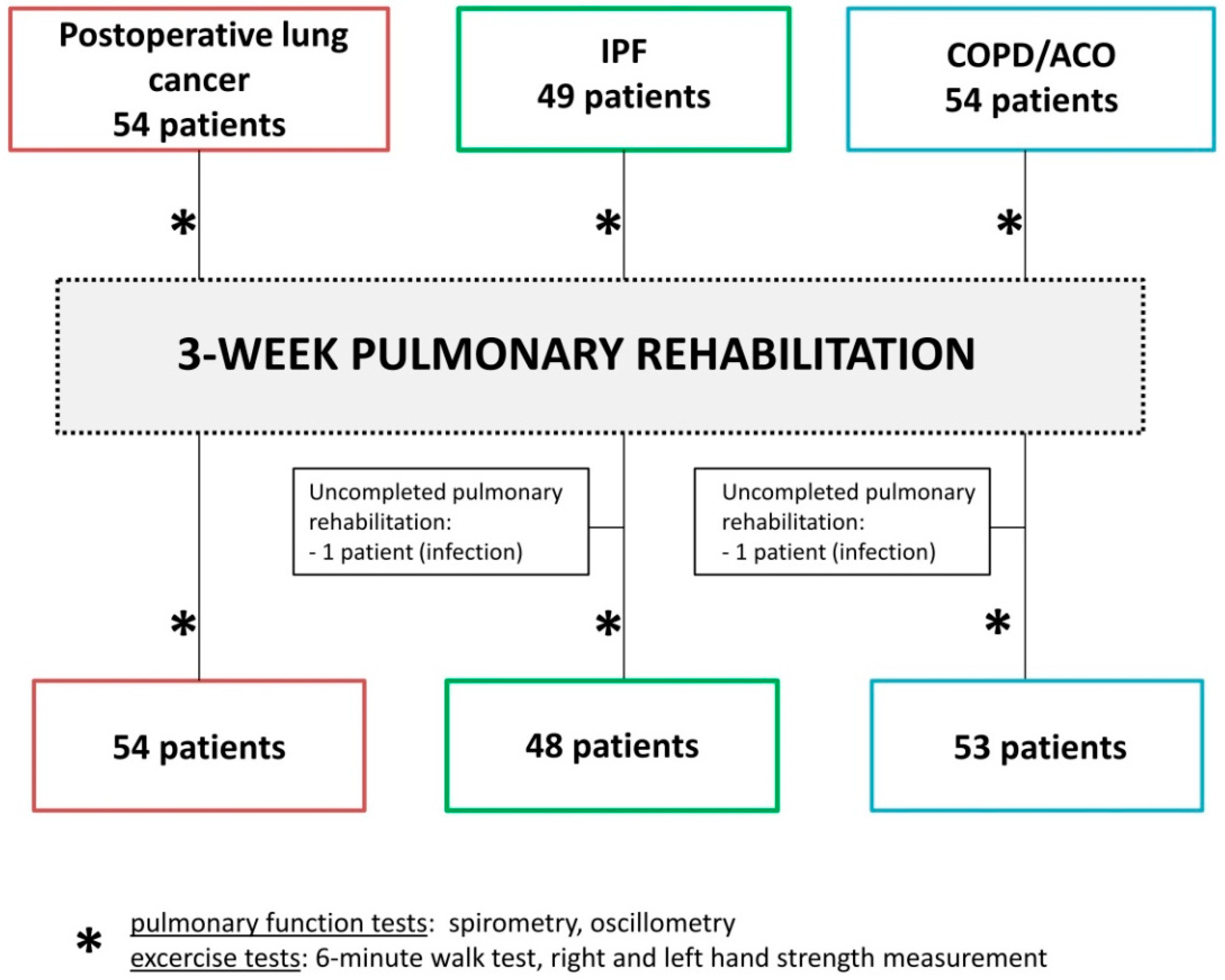
Figure 1. Study recruitment protocol.
Medicina 58 01551 g001
Figure 2. Significant improvements in the 6-min walk distance (6MWD), right hand (RH) strength, and left hand (LH) strength after completing 3 weeks of pulmonary rehabilitation (PR). The horizontal lines refer to medians, boxes to quartiles, and vertical lines to non-outlier range. IPF, idiopathic interstitial fibrosis; COPD, chronic obstructive pulmonary disease; ACO, asthma–COPD overlap; * p < 0.05.
Medicina 58 01551 g002
Figure 3. Differences between patient groups in terms of changes in resistance (R) and reactance (X) due to pulmonary rehabilitation (PR). The horizontal lines refer to medians, boxes to quartiles, and vertical lines to non-outlier range. IPF, idiopathic interstitial fibrosis; COPD, chronic obstructive pulmonary disease; ACO, asthma–COPD overlap; insp, inspiratory; exp, expiratory; * p < 0.05.
Medicina 58 01551 g003
Table 1. Demographic data.
Postoperative Lung Cancer
(54)
IPF
(48)
COPD/ACO
(53)
p
Age (years)67.4 ± 8.866.3 ± 6.165.9 ± 9.90.69
Sex (M/F)23/3134/1432/210.014
BMI (kg/m2)28.6 ± 4.527.2 ± 4.228.1 ± 6.00.308
Characteristics of
the patient group
Histopathological diagnosis
-
adenocarcinoma: 27 pts
-
squamous cell carcinoma: 19 pts
-
non-small cell lung cancer: 4 pts
-
carcinoid: 2 pts
-
large cell carcinoma: 1 pt
-
metastases from breast cancer: 1 pt
Type of operation (approximate
percentage of the lung tissue removal)
-
lobectomy: 44 pts (16–26%)
-
bilobectomy: 4 pts (25–30%)
-
pneumonectomy: 3 pts (47–53%)
-
segmentectomy: 2 pts (5–6%)
-
wedge resection: 1 pts (8%)
Neo-adjuvant therapy: 0 pts
Adjuvant therapy: 23 pts
Antifibrotic
treatment (77%):
Nintedanib: 9 pts
Pirfenidone: 28 pts
GAP Index:
Stage I: 31 pts (65%)
Stage II: 15 pts (31%)
Stage III: 2 pts (4%)
COPD: 41 pts (77%)
ACO: 12 pts (23%)
-
Comorbidities 0.908
Cardiovascular diseases42 (78%)38 (79%)39 (74%)
Diabetes mellitus13 (24%)8 (17%)14 (26%)
Musculoskeletal disorders22 (41%)19 (40%)31 (58%)
Gastrointestinal disorders13 (24%)13 (27%)11 (21%)
Urogenital disorders20 (37%)16 (33%)16 (30%)
Psychiatric disorders3 (6%)3 (6%)2 (4%)
Smoking history 0.119
Current4 (7%)-6 (11%)
Previous36 (67%)30 (63%)35 (66%)
Never14 (26%)18 (38%)12 (23%)
IPF, idiopathic interstitial fibrosis; COPD, chronic obstructive pulmonary disease; ACO, asthma–COPD overlap; M, male; F, female; BMI, body mass index; pt(s), patient(s); GAP, the gender-age-physiology index for IPF patients.
Table 2. Lung function and exercise test results at the baseline and after 3 weeks of pulmonary rehabilitation (PR).
ParameterPostoperative Lung
Cancer
(54)
IPF
(48)
COPD/ACO
(53)
npar LD Test
BaselineAfter PRBaselineAfter PRBaselineAfter PRGroupTimeGroup:
Time
FEV1 (%pred.)76.31 ± 21.579.03 ± 19.882.98 ± 16.984.06 ± 17.957.55 ± 23.557.5 ± 22.7<0.001 1,30.4090.313
FVC (%pred.)92.33 ± 21.094.2 ± 21.780.78 ± 18.181.69 ± 18.980.62 ± 21.780.59 ± 21.20.003 1,20.2180.392
FEV1/FVC (%)67.84 ± 8.867.81 ± 9.981.73 ± 6.981.92 ± 6.555.76 ± 14.456.12 ± 13.8<0.001 40.6430.781
SpO2 (%)95.88 ± 1.396.38 ± 1.794.77 ± 1.995.0 ± 2.494.68 ± 1.894.79 ± 1.5<0.001 1,20.1230.592
6MWD (m)415.7 ± 77.1469.3 ± 71.5426.6 ± 106.3456.6 ± 105.5382.3 ± 105.5431.8 ± 91.70.032 1,3<0.001 ***0.072
SpO2 after 6MWT (%)92.53 ± 3.192.26 ± 3.484.83 ± 8.986.82 ± 7.589.76 ± 6.290.39 ± 4.8<0.001 40.4910.263
RH strength (kg)25.76 ± 10.627.19 ± 10.332.27 ± 9.832.54 ± 8.928.63 ± 10.429.57 ± 10.00.039 20.002 *,**0.007 ****
LH strength (kg)28.37 ± 10.729.52 ± 11.234.19 ± 9.933.85 ± 9.330.33 ± 11.432.34 ± 10.50.007 20.012 *0.74
Results are expressed as means ± standard deviation. 1 Significant difference in lung cancer after thoracic surgery vs. COPD/ACO; 2 significant difference in lung cancer after thoracic surgery vs. IPF; 3 significant difference in IPF vs. COPD/ACO; 4 significant difference between all 3 groups; * significant improvement in lung cancer after thoracic surgery; ** significant improvement in COPD/ACO; *** significant improvement in all 3 groups; **** no significant difference based on the post hoc test. IPF, idiopathic interstitial fibrosis; COPD, chronic obstructive pulmonary disease; ACO, asthma–COPD overlap; FEV1, forced expiratory volume during the first second; FVC, forced vital capacity; SpO2, oxygen saturation; 6MWD, 6-min walk distance; 6MWT, 6-min walk test; RH, right hand; LH, left hand; group, differences between groups at the baseline and/or at the end of PR; time, changes in 3 weeks; group:time, and differences in changes after PR between studied groups.
Table 3. Forced oscillation results at the baseline and after 3 weeks of pulmonary rehabilitation (PR). IPF, idiopathic interstitial fibrosis; COPD, chronic obstructive pulmonary disease; ACO, asthma–COPD overlap; R, resistance; X, reactance; Fres, resonant frequency; insp, inspiratory; exp, expiratory; tot, total; group, differences between groups at the baseline and/or at the end of PR; time, changes in 3 weeks; group:time, differences of changes after PR between studied groups.
ParameterPostoperative Lung Cancer
(54)
IPF
(48)
COPD/ACO
(53)
npar LD Test
BaselineAfter PRBaselineAfter PRBaselineAfter PRGroupTimeGroup:Time
Rinsp5 (%pred.)104.97 ± 37.9101.3 ± 28.785.79 ± 26.585.66 ± 22.6129.08 ± 60.8109.36 ± 46.9<0.001 2,30.090.029 1,3
Rexp5 (%pred.)119.69 ± 44.5127.99 ± 39.9105.08 ± 34.8103.38 ± 31.4167.95 ± 80.1148.89 ± 59.6<0.001 40.850.005 1
Rinsp11 (%pred.)108.65 ± 34.1108.49 ± 24.892.91 ± 22.391.04 ± 17.6129.07 ± 54.9116.01 ± 43.0<0.001 2,30.4080.11
Rexp11 (%pred.)122.34 ± 36.3130.93 ± 34.7113.76 ± 31.6115.36 ± 28.2149.97 ± 61.5139.43 ± 47.3<0.001 40.2240.068
Rinsp19 (%pred.)95.99 ± 28.895.88 ± 21.081.94 ± 18.079.92 ± 14.6109.48 ± 44.899.83 ± 31.5<0.001 2,30.3930.206
Rexp19 (%pred.)100.98 ± 28.5108.68 ± 28.995.5 ± 24.596.43 ± 22.0118.46 ± 47.0111.52 ± 33.40.005 2,30.1560.133
Rinsp5-19 (cmH2O/L/s)0.51 ± 0.60.36 ± 0.70.25 ± 0.50.28 ± 0.60.77 ± 0.80.82 ± 3.20.021 1,30.1450.017 1,3
Rexp5-19 (cmH2O/L/s)0.89 ± 1.00.8 ± 0.90.47 ± 0.60.3 ± 0.51.69 ± 1.11.2 ± 1.0<0.001 40.0920.074
Xinsp5 (%pred.)82.89 ± 41.887.37 ± 40.4103.29 ± 54.2110.33 ± 45.9108.85 ± 58.5106.89 ± 87.10.039 1,20.4140.089
Xexp5 (%pred.)147.54 ± 154.4161.86 ± 122.8116.4 ± 55.6113.29 ± 49.0318.26 ± 318.8293.25 ± 294.8<0.001 1,30.6560.048 1
Xinsp11 (cmH2O/L/s)−0.37 ± 0.5−0.45 ± 0.6−0.37 ± 0.4−0.41 ± 0.5−0.68 ± 0.7−0.59 ± 0.60.029 1,30.4150.205
Xexp11 (cmH2O/L/s)−1.19 ± 1.5−1.4 ± 1.3−0.65 ± 0.5−0.76 ± 0.7−2.28 ± 1.5−2.13 ± 1.5<0.001 40.1170.025 *
Xinsp19 (cmH2O/L/s)0.3 ± 0.50.27 ± 0.6−0.26 ± 4.50.33 ± 0.50.02 ± 0.70.04 ± 0.60.002 1,30.6170.999
Xexp19 (cmH2O/L/s)−0.38 ± 1.0−0.55 ± 0.90.02 ± 0.5−0.03 ± 0.5−1.07 ± 0.9−1.01 ± 0.9<0.001 40.1510.057
Fres (Hz)15.84 ± 5.016.31 ± 5.615.2 ± 3.215.24 ± 3.619.08 ± 6.918.78 ± 6.30.003 1,30.530.838
Results are expressed as means ± standard deviation. 1 Significant difference in lung cancer after thoracic surgery vs. COPD/ACO; 2 significant difference in lung cancer after thoracic surgery vs. IPF; 3 significant difference in IPF vs. COPD/ACO; 4 significant difference between all 3 groups; * no significant difference based on the post hoc test. IPF, idiopathic interstitial fibrosis; COPD, chronic obstructive pulmonary disease; ACO, asthma–COPD overlap; R, resistance; X, reactance; Fres, resonant frequency; insp, inspiratory; exp, expiratory; tot, total; group, differences between groups at the baseline and/or at the end of PR; time, changes in 3 weeks; group:time, differences of changes after PR between studied groups.
Publisher’s Note: MDPI stays neutral with regard to jurisdictional claims in published maps and institutional affiliations.