Article

The Association between Admission Procalcitonin Level and The Severity of COVID-19 Pneumonia: A Retrospective Cohort Study

by
Mohamed Aon
1,*,
Abdullah Alsaeedi
2,
Azeez Alzafiri
2,
Mohamed M. Ibrahim
3,
Abdelrahman Al-Shammari
2,
Omar Al-Shammari
2,
Mahmoud Tawakul
2,
Sherif Taha
1,
Naser Alherz
2,
Jarrah Alshammari
2,
Ebraheem Albazee
2,
Teflah Alharbi
2,
Duaa Alshammari
2,
Zaid Alenezi
2,
Monerah Alenezi
2,
Salem Aldouseri
2,
Meshari Eyadah
2,
Mariam Aldhafeeri
2 and
Ahmed H. Aoun
4
1
Department of Internal Medicine, Faculty of Medicine, Cairo University, Giza 12613, Egypt
2
Department of Internal Medicine, Jahra Hospital, Jahra 2675, Kuwait
3
Jaber Al-Ahmad Military Hospital, Ministry of Defense, Kuwait City 46001, Kuwait
4
Department of Pediatrics, Faculty of Medicine, Cairo University, Giza 12613, Egypt
*
Author to whom correspondence should be addressed.
Medicina 2022, 58(10), 1389; https://doi.org/10.3390/medicina58101389
Submission received: 3 September 2022 / Revised: 28 September 2022 / Accepted: 29 September 2022 / Published: 3 October 2022
(This article belongs to the Section Infectious Disease)

Abstract

:
Background and Objectives: An elevated procalcitonin level has classically been linked to bacterial infections. Data on the association between elevated procalcitonin and the outcome of coronavirus disease 2019 (COVID-19) are conflicting. Some linked it to associated bacterial co-infections, while others correlated the elevation with disease severity without coexisting bacterial infections. We aimed to investigate the association between high procalcitonin and the severity of COVID-19. Materials and Methods: Hospitalized patients with confirmed COVID-19 pneumonia were divided into two groups: the normal-procalcitonin group and the high-procalcitonin group (>0.05 ng/mL). Patients with concomitant bacterial infections on admission were excluded. The primary outcomes were the need for intensive care unit (ICU) admission, progression to invasive mechanical ventilation (IMV), and in-hospital 28-day mortality. Results: We included 260 patients in the normal procalcitonin group and 397 patients in the high procalcitonin group. The mean age was 55 years and 49% were females. A higher number of patients in the elevated procalcitonin group required ICU admission (32.7% vs. 16.2%, p < 0.001) and IMV (27.2% vs. 13.5%, p < 0.001). In-hospital mortality was significantly higher in the elevated procalcitonin group (18.9% vs. 8.5%, p < 0.001). After adjusting for other covariates, procalcitonin > 0.05 ng/mL was an independent predictor of progression to IMV (OR, 1.71; 95% CI, 1.08–2.71; p = 0.022), ICU admission (OR, 1.73; 95% CI, 1.13–2.66; p = 0.011), and in-hospital mortality (OR, 1.99; 95% CI, 1.14–3.47; p = 0.015). An elevated procalcitonin level was the strongest predictor of in-hospital mortality. Conclusions: Measurement of procalcitonin can have a prognostic role among COVID-19 patients. The admission procalcitonin level can identify patients at risk of ICU admission, progression to IMV, and in-hospital mortality.

1. Introduction

The coronavirus disease 2019 (COVID-19), which started as a cluster of pneumonia cases in Wuhan, China, has affected nearly all countries throughout the world [1]. Although the majority of symptomatic COVID-19 infections are mild, a subset of patients requires hospitalization due to severe COVID-19 pneumonia, and about 5% of patients develop a critical disease characterized by respiratory failure and/or other organs’ failure [2]. The early recognition of severe COVID-19 features is crucial to identify patients at risk of poor outcome, create an opportunity for early interventions, personalize the treatment protocols, allow a better allocation of valuable resources, e.g., ventilators and intensive care unit (ICU) beds, and improve the outcomes [3]. The host factors are the main determinants of the disease severity, although viral factors have also been implicated [4,5]. Clinical predictors of disease severity include demographic features such as age and sex, pre-existing comorbidities, radiographic features, and laboratory parameters [6]. Plenty of biomarkers measured in COVID-19 patients were associated with disease severity and worse outcomes such as lymphopenia and elevated D-dimer, ferritin, lactate dehydrogenase (LDH), troponin, C-reactive protein (CRP), aspartate aminotransferase (AST), alanine aminotransferase (ALT), and creatinine [7,8]. The earliest reports from China showed that elevated procalcitonin (PCT) was not a prominent feature among COVID-19 patients [9]. Subsequently, elevated PCT levels were reported more frequently in severe cases [10,11].
PCT is a glycoprotein precursor of calcitonin that does not have a hormonal action of its own. PCT level is undetectable under normal circumstances but increases with bacterial infections to adjust the host′s immune response and vasoactivity in response to bacterial sepsis [12]. Bacterial infections induce PCT production via the stimulation of macrophages which produce inflammatory cytokines such as tumor necrosis factor (TNF)-α, interleukin (IL)-1β, and IL-6 that stimulate the synthesis of PCT by all cells within few hours. On the other hand, PCT is not induced by viral infections due to TNF- α inhibition by interferon (INF)-γ [13].
Despite being a viral infection, COVID-19 may be associated with elevated levels of PCT. The elevated PCT in this setting indicates a more severe illness and a higher risk of mortality, especially among elderly patients [14]. A meta-analysis revealed that high PCT levels were associated with higher disease severity and hypothesized that this relationship suggests bacterial co-infections that increase disease severity and add to the systemic sepsis [13]. Measurement of PCT levels may identify bacterial co-infections and guide antimicrobial therapy in COVID-19 patients [15]. However, there was heterogeneity in the thresholds used and the interpretation of PCT levels [16].
On the contrary, other studies failed to find an association between PCT and bacterial co-infections in COVID-19 patients and suggested that the high PCT levels are largely related to the disease severity and the associated inflammation rather than bacterial co-infection [17,18]. Current recommendations do not support the routine use of PCT to guide decisions about antibiotics in COVID-19 and draw attention to the uncertainty about the cut-off point to be used. Institutions using PCT are advised to participate in data collection and research [19]. Further research in a variety of settings is needed to evaluate the association of PCT with disease severity, mortality, and bacterial co-infections, and the impact of PCT-guided strategies on outcomes, antibiotic prescription patterns, and safety of patients.
In response to the gaps in the literature, we aimed to investigate the hypothesis that elevated PCT is associated with increasing disease severity, need for invasive mechanical ventilation (IMV), need for ICU admission, and in-hospital mortality.

2. Materials and Methods

2.1. Study Design and Settings

Our study was a single-center retrospective cohort study. The study included patients who were admitted to the medical ward at Jahra Hospital, Kuwait (the main district hospital) with COVID-19 pneumonia between May 2020 and May 2021. Patients were divided into 2 groups according to the admission PCT level: the normal PCT group with a PCT level ≤ 0.05 ng/mL (according to the manufacturer′s reference range) and the high PCT group with a PCT level > 0.05 ng/mL. PCT was measured using an automated Enzyme-Linked Fluorescent Assay (VIDAS® B.R.A.H.M.S. PCT™, BioMérieux Diagnostics, Inc., Lyon, France). All patients were treated according to the national treatment protocol that included pharmacologic thromboprophylaxis unless contraindicated and corticosteroids for hypoxemic patients (i.e., patients who need supplemental oxygen to keep SO2 ≥ 94%). Antibiotics were prescribed according to the treating physician′s discretion. Patient demographics, clinical data, and admission laboratory results were obtained from the hospital records.

2.2. Inclusion and Exclusion Criteria

Patients were included if they fulfilled all the following criteria: (1) admission to the medical ward with COVID-19 pneumonia (suggested by clinical and/or radiological features of lower respiratory infection); (2) COVID-19 diagnosis was confirmed with a positive reverse transcriptase-polymerase chain reaction (RT-PCR) assay for severe acute respiratory syndrome coronavirus-2 (SARS-CoV-2) on nasopharyngeal swab; (3) PCT concentration was measured on admission.
Patients who were transferred to another hospital before the outcome was known, had a bacterial co-infection, or did not have a PCT measurement on admission were excluded from the study. Bacterial co-infection was defined as positive bacterial culture from respiratory samples, blood, or urine, within 48 h from admission. Pregnant females were not included in our study.

2.3. Outcome Measures

The severity of COVID-19 respiratory illness on admission was assessed using a 7-category ordinal scale consisting of the following categories: (1) not hospitalized and ambulatory; (2) not hospitalized, but with limitation of activities; (3) hospitalized but did not require oxygen therapy; (4) hospitalized and required oxygen by nasal prongs or mask; (5) hospitalized and required oxygen therapy by high-flow nasal cannula or noninvasive mechanical ventilation; (6) hospitalized and required IMV; (7) hospitalized and required IMV plus additional organ support, e.g., extracorporeal membrane oxygenation, renal replacement therapy, or vasopressors [20]. The primary outcomes were the need for ICU admission, the requirement of IMV, and in-hospital 28-day mortality.

2.4. Statistical Analysis

Assuming an incidence of the main outcomes of 1% and 6.8% in the two groups, respectively [21], 174 patients in each group will be needed to achieve a study power of 80% and a confidence level of 95%. Validated data were tabulated, entered, and analyzed using Statistical Package for the Social Sciences (SPSS) version 22.0 (SPSS Inc., Chicago, IL, USA). Qualitative data were expressed as frequencies and percentages and comparisons between the groups were performed using the Chi-square (χ2) test. Quantitative data were expressed as mean and standard deviation (SD) if data were normally distributed. The student′s t-test was used for the comparison of normally distributed quantitative variables. If the quantitative data were not normally distributed, they were expressed as the median and interquartile range (IQR), and the differences between the study groups were compared by the Mann–Whitney test. Logistic regression was done to detect the independent predictors of the various primary outcomes. The association between determinants and outcome was presented as odds ratio (OR) and 95% confidence interval (CI) after controlling for other variables. A p-value < 0.05 was considered statistically significant.

3. Results

After assessment against inclusion and exclusion criteria, 657 patients were included in our study: 260 patients in the normal PCT group and 397 patients in the high PCT group, as shown in Figure 1.
The mean age of patients was 55 ± 15 years and 51% of them were males. Diabetes and hypertension were the most frequent comorbidities (60% and 42%, respectively). The median PCT in the normal and the high PCT groups was 0.05 and 0.21 ng/mL, respectively. Table 1 demonstrates the demographic features, comorbidities, and laboratory findings in both groups.
As demonstrated in Table 2, most patients in our cohort (89%) received empiric antibiotics therapy on admission to cover possible community-acquired pneumonia (CAP), and most of them (82%) presented with a severity scale of four. A higher number of patients deteriorated clinically in the elevated PCT group compared to the normal PCT group (p < 0.001), and the median length of stay was higher in the elevated PCT group compared to the normal PCT group (p < 0.001).
Figure 2 illustrates the primary outcomes in both groups. Compared to the normal PCT group, patients with elevated PCT required ICU admission and IMV more frequently (32.7% vs. 16.2% and 27.2% vs. 13.5%, respectively) and the difference between the groups was statistically significant (p < 0.001). The in-hospital mortality was significantly increased in the high PCT group compared to the normal PCT group (18.9% vs. 8.5%, p < 0.001).

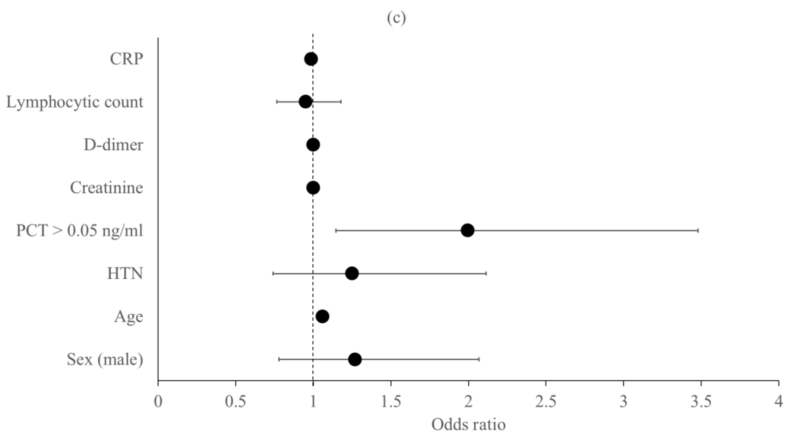
Predictors of the Primary Outcomes

Different models were designed, and regression analysis was performed to test the effect of the different variables on the primary outcomes (ICU admission, IMV, and in-hospital 28-day mortality). Our model identified male sex (OR, 1.82; 95% CI, 1.23–2.70; p = 0.003), older age (OR, 1.03; 95% CI, 1.01–1.05; p < 0.001), admission PCT > 0.05 ng/mL (OR, 1.73; 95% CI, 1.13–2.66; p = 0.011), and a higher CRP level (OR, 1.03; 95% CI, 1–1.05; p = 0.010) as risk factors for ICU admission after control of other variables, as demonstrated in Figure 3a. Progression to IMV predictors were male sex (OR, 1.69; 95% CI, 1.11–2.57; p = 0.013), older age (OR, 1.04; 95% CI, 1.02–1.05; p < 0.001), and admission PCT > 0.05 ng/mL (OR, 1.71; 95% CI, 1.08–2.71; p = 0.022) after adjusting for other covariables, as shown in Figure 3b. Older age (OR, 1.06; 95% CI, 1.04–1.08; p < 0.001) and admission PCT > 0.05 ng/mL (OR, 1.99; 95% CI, 1.14–3.47; p = 0.015) exhibited independent increasing risk of in-hospital mortality after adjusting for other covariates. As demonstrated in Figure 3c, PCT > 0.05 ng/mL was the strongest predictor of in-hospital mortality.

4. Discussion

In this study, we report our findings on the association between high PCT and COVID-19 severity. We found that older age and male sex were associated with disease severity; findings that have been demonstrated in previous studies [2,22]. The high PCT group patients had a higher neutrophil count and a lower lymphocytic count. These hematological parameters are known to be associated with severe COVID-19 infection [23]. Although neutrophilia is usually associated with bacterial infection, in COVID-19, it is usually linked to disease severity without clear evidence of associated bacterial co-infection. Furthermore, the use of corticosteroids contributes to the increase in the neutrophilic count. Thus, neutrophilia is not a reliable biomarker of bacterial co-infections among COVID-19 patients [18].
Other laboratory parameters, e.g., CRP, D-dimer, and creatinine were elevated in the high PCT group. These biomarkers are usually associated with more severe disease and a worse outcome [8]. Studies found that CRP levels correlate with PCT levels among COVID-19 patients [24]. Furthermore, some studies found that an elevated PCT level was associated with worsening renal functions and the development of disseminated intravascular coagulation in COVID-19 patients [25,26,27].
Consistent with previous studies, we found that increased PCT levels were associated with disease severity and the need for IMV. An early meta-analysis found that elevated PCT is associated with a 5-fold higher risk of severe COVID-19 infection [13]. Other studies found that elevated PCT levels were associated with a longer duration of IMV [18,28]. In a meta-analysis that included 3027 Chinese patients, PCT > 0.5 ng/mL was associated with progression to critical disease [29]. In another meta-analysis that included 3492 patients, Vazzana and his colleagues demonstrated that increased PCT was associated with severe COVID- 19 and a worse outcome. They suggested PCT as a specific, but not a sensitive, biomarker of COVID-19 severity [30].
SARS-CoV-2 binds to the host cells through angiotensin-converting enzyme 2 (ACE2) receptors leading to ACE2 dysregulation and renin–angiotensin–aldosterone system (RAAS) activation in several ACE2-expressing tissues, leading to a widespread inflammatory response and tissue injury [31]. The relationship between elevated PCT and COVID-19 severity can be explained by the associated systemic inflammation and the release of the proinflammatory cytokines such as IL-6, IL-1β, and TNF-α, with subsequent induction of PCT synthesis [25]. Gautam et al. demonstrated that PCT, in the setting of respiratory viral infections, may be a better indicator of disease severity than bacterial co-infections. The authors suggested that PCT induces an inflammatory response, via cytokines, which in turn increases the severity of illness and can lead to organ dysfunction and death. They also postulated that viral infections may induce PCT expression through cytokine-independent pathways [32]. In the study by Hu et al., patients with severe and critical COVID-19 had rates of bacterial co-infections that were lower than the rates of PCT elevation (20% vs. 50% and 50% vs. 80%, respectively). They suggested that, in severe and critical diseases, PCT can be a marker of disease severity independent from bacterial co-infections [33].
In our study, patients with elevated PCT had a significantly higher in-hospital 28-day mortality. In the study by Liu et al., PCT levels ≥0.05 ng/mL correlate with in-hospital mortality and worse survival [21]. Shen et al. performed a meta-analysis that included 7716 patients and showed that higher admission PCT levels were associated with disease severity and mortality [34].
A low burden of bacterial co-infections on admission was detected in our study (21 patients (3%)). A similar percentage of bacterial co-infections in the community-onset COVID-19 infections was detected in other studies (3.2% and 3.5%, respectively) [24,35]. A meta-analysis demonstrated that antibiotics were prescribed to 75% of COVID-19 patients, despite that the estimates of bacterial co-infections were much lesser. Of note, the Middle East region had a higher rate of antibiotic prescription (86%) compared to other regions [36]. Most of our patients received antibiotics during hospitalization. Previous studies showed that empiric antibiotics to treat CAP were administered to 91% and 77% of COVID-19 patients with high and normal PCT, respectively. Using PCT to guide antibiotic decisions in COVID-19 is biased by the possible cytokine production that may induce PCT levels [37]. The similarity of symptoms between COVID-19 and CAP has led to an upward trend of over-prescription of antibiotics [38].
The hypothesis of the association between elevated PCT and bacterial co-infections in severe COVID-19 was considered in many studies. Pink et al. demonstrated that the peak PCT levels coincide with the diagnosis of secondary bacterial infection, although it was not clear whether these infections were community-acquired or nosocomial. The authors did not establish a direct relation between high PCT levels and severity and stated that patients with secondary bacterial co-infections were expected to have worse clinical outcomes [15]. Many studies supporting PCT measurement in COVID-19 patients limit its use to antibiotics stewardship [24,39,40]. On the other hand, the guidelines endorse deficient evidence to support PCT as a marker of bacterial co-infections [19]. Fabre et al. showed that, among COVID-19 patients, admission PCT did not offer an added value to clinical evaluation in diagnosing bacterial co-infections. PCT is not uncommonly elevated in COVID-19 patients without evidence of bacterial pneumonia, thus limiting its use as a surrogate marker of bacterial infections in this setting [37]. Dolci et al. found that, among COVID-19 hospitalized patients, peak and initial PCT levels did not offer an additional value in diagnosing bacterial infections [41]. Another study from Italy demonstrated that PCT did not help distinguish COVID-19 pneumonia from non-COVID-19 pneumonia in patients presenting with respiratory symptoms and fever [42]. Vazzana et al. performed a meta-analysis that demonstrated the association between PCT and disease severity. They noticed that none of the included studies stratified PCT levels based on the presence or absence of bacterial co-infections. So, there is not enough evidence to conclude that elevated PCT in severe COVID-19 patients is related to bacterial co-infections [30]. Heer et al. found that PCT was associated with IMV requirements and inpatient death, independent of bacterial co-infections [18].
Our logistic regression model identified high PCT as a predictor of progression to IMV, ICU admission, and in-hospital mortality. In addition, PCT > 0.05 ng/mL was the strongest predictor of in-hospital mortality. Different studies have identified elevated PCT as a risk factor for worse outcomes and mortality. Xu et al. found that elevated baseline PCT is an independent predictor of mortality in hospitalized patients with COVID-19. They suggested using elevated PCT to identify patients with worse prognoses, especially among the elderly and the critically ill patients [14]. Another study found that elevated PCT, among other biomarkers, is a predictor of clinical deterioration, ICU admission, and death [8]. Sayah et al. suggested a PCT cut-off level of 0.138 and 0.16 ng/mL as predictors of severity and mortality, respectively. They found a positive correlation between the levels of IL-6 and PCT. IL-6 and PCT were the most accurate inflammatory biomarkers correlating with severity and mortality [43]. Another study found that severe and critical COVID-19 patients have higher levels of PCT and IL-6 suggesting a link between IL-6, PCT, and severity [44]. SARS-CoV-2 triggers a cytokine storm that includes a wide range of pro-inflammatory cytokines such as IL-6, IL-1β, and TNF-α. The main pathophysiological culprit of the cytokine storm in COVID-19 is IL-6. This hypercytokinemia is associated with the infiltration of lungs by inflammatory cells. Eventually, this can lead to a life-threatening multi-organ failure [45]. A study that analyzed the levels of 11 cytokines among COVID-19 patients found that the highest IL-6 levels coincide with disease severity, suggesting a strong link between this inflammatory cytokine and the pathogenesis of severe COVID-19 [4].
Although previous studies found an association between elevated PCT levels and COVID-19 severity, our study has specific features that should be emphasized. First, the timing of PCT measurement was comparable in all patients and all other laboratory parameters were measured simultaneously with PCT on admission. In addition, patients with positive bacterial cultures on admission were excluded. Finally, these findings share our real-life experience on PCT as a biomarker of COVID-19 severity. Nevertheless, our study has limitations. It was an observational retrospective study and included patients from a single hospital. Moreover, serial PCT was not measured; thus, we could not assess the dynamic PCT changes and their effect on the outcomes.

5. Conclusions

In conclusion, our findings demonstrate that early measurement of PCT can have a prognostic role among COVID-19 patients. Admission PCT measurement can identify patients at risk of requiring IMV and death. Our findings add to the growing evidence that elevated PCT level is a risk factor for worse COVID-19 outcomes, and we think that it can be incorporated in a practical strategy to early identify patients at risk of deterioration. Further research is required to determine the role of serial PCT measurement, the effect of drugs, and the mechanisms implicated in the association between elevated PCT and unfavorable outcomes in COVID-19. There is an urgent need to develop new and reliable strategies to diagnose bacterial co-infections among patients with COVID-19 infection.

Author Contributions

Conceptualization, M.A. (Mohamed Aon), M.M.I. and A.H.A.; data curation, M.A. (Mohamed Aon), M.M.I., N.A., J.A., E.A., T.A., D.A., Z.A., M.A. (Monerah Alenezi), S.A., M.E., M.A. (Mariam Aldhafeeri) and A.H.A.; formal analysis, M.A. (Mohamed Aon), E.A. and A.H.A.; investigation, M.A. (Mohamed Aon), A.A. (Abdullah Alsaeedi), A.A. (Azeez Alzafiri), M.M.I., A.A.-S., O.A.-S., M.T., S.T. and J.A.; methodology, M.A. (Mohamed Aon), M.M.I., A.A.-S., S.T., N.A., J.A., E.A., T.A., D.A., Z.A., M.A. (Monerah Alenezi), S.A., M.E., M.A. (Mariam Aldhafeeri) and A.H.A.; project administration, A.A. (Abdullah Alsaeedi), A.A. (Azeez Alzafiri), O.A.-S., M.T., N.A. and J.A.; resources, A.A. (Abdullah Alsaeedi), A.A. (Azeez Alzafiri), A.A.-S., O.A.-S., M.T., S.T. and N.A.; supervision, A.A. (Abdullah Alsaeedi), A.A. (Azeez Alzafiri), A.A.-S., O.A.-S., M.T. and N.A.; validation, M.A. (Mohamed Aon), J.A., T.A., D.A., Z.A., M.A. (Monerah Alenezi), S.A., M.E. and M.A. (Mariam Aldhafeeri); writing—original draft, M.A. (Mohamed Aon) and A.H.A.; writing—review and editing, M.A. (Mohamed Aon), M.M.I., S.T., E.A. and A.H.A. All authors have read and agreed to the published version of the manuscript.

Funding

This research received no external funding.

Institutional Review Board Statement

The study was conducted in accordance with the Declaration of Helsinki and approved by the institutional review board (IRB) of Jahra Hospital, Kuwait (J1-26102021).

Informed Consent Statement

Patient consent was waived by the IRB as the analysis included only retrospectively collected data.

Data Availability Statement

The data supporting the findings are available from the corresponding author upon reasonable request.

Conflicts of Interest

The authors declare no conflict of interest.

References

  1. WHO. Coronavirus (COVID-19) Dashboard. 2022. Available online: https://covid19.who.int (accessed on 1 February 2022).
  2. Wu, Z.; McGoogan, J.M. Characteristics of and Important Lessons from the Coronavirus Disease 2019 (COVID-19) Outbreak in China: Summary of a Report of 72 314 Cases from the Chinese Center for Disease Control and Prevention. JAMA 2020, 323, 1239. [Google Scholar] [CrossRef] [PubMed]
  3. Zhou, W.; Liu, Y.; Xu, B.; Wang, S.; Li, S.; Li, H.; Huang, Z.; Luo, Y.; Hu, M.; Wu, W.; et al. Early identification of patients with severe COVID-19 at increased risk of in-hospital death: A multicenter case-control study in Wuhan. J. Thorac. Dis. 2021, 13, 1380–1395. [Google Scholar] [CrossRef] [PubMed]
  4. Zhang, X.; Tan, Y.; Ling, Y.; Lu, G.; Liu, F.; Yi, Z.; Jia, X.; Shi, B.; Xu, S.; Chen, W.; et al. Viral and host factors related to the clinical outcome of COVID-19. Nature 2020, 583, 437–440. [Google Scholar] [CrossRef]
  5. Twohig, K.A.; Nyberg, T.; Zaidi, A.; Simon, T.; Sinnathamby, M.A.; Shirin, A.; Shaun, R.; Ross, H.J.; Russell, H.; Bernal, J.; et al. Hospital admission and emergency care attendance risk for SARS-CoV-2 delta (B.1.617.2) compared with alpha (B.1.1.7) variants of concern: A cohort study. Lancet Infect. Dis. 2021, 22, S1473309921004758. [Google Scholar] [CrossRef]
  6. Gallo Marin, B.; Aghagoli, G.; Lavine, K.; Yang, L.; Siff, E.J.; Chiang, S.S.; Salazar-Mather, T.P.; Dumenco, L.; Savaria, M.C.; Aung, S.N.; et al. Predictors of COVID-19 severity: A literature review. Rev. Med. Virol. 2021, 31, 1–10. [Google Scholar] [CrossRef] [PubMed]
  7. Liao, D.; Zhou, F.; Luo, L.; Xu, M.; Wang, H.; Xia, J.; Gao, Y.; Cai, L.; Wang, Z.; Yin, P.; et al. Haematological characteristics and risk factors in the classification and prognosis evaluation of COVID-19: A retrospective cohort study. Lancet Haematol. 2020, 7, e671–e678. [Google Scholar] [CrossRef]
  8. Cecconi, M.; Piovani, D.; Brunetta, E.; Aghemo, A.; Greco, M.; Ciccarelli, M.; Angelini, C.; Voza, A.; Omodei, P.; Vespa, E.; et al. Early Predictors of Clinical Deterioration in a Cohort of 239 Patients Hospitalized for Covid-19 Infection in Lombardy, Italy. J. Clin. Med. 2020, 9, 1548. [Google Scholar] [CrossRef]
  9. Chen, N.; Zhou, M.; Dong, X.; Qu, J.; Gong, F.; Han, Y.; Qiu, Y.; Wang, J.; Liu, Y.; Wei, Y.; et al. Epidemiological and clinical characteristics of 99 cases of 2019 novel coronavirus pneumonia in Wuhan, China: A descriptive study. Lancet 2020, 395, 507–513. [Google Scholar] [CrossRef] [Green Version]
  10. Chen, G.; Wu, D.; Guo, W.; Cao, Y.; Huang, D.; Wang, H.; Wang, T.; Zhang, X.; Chen, H.; Yu, H.; et al. Clinical and immunological features of severe and moderate coronavirus disease 2019. J. Clin. Investig. 2020, 130, 2620–2629. [Google Scholar] [CrossRef] [Green Version]
  11. Chen, T.; Wu, D.; Chen, H.; Yan, W.; Yang, D.; Chen, G.; Ma, K.; Xu, D.; Yu, H.; Wang, H.; et al. Clinical characteristics of 113 deceased patients with coronavirus disease 2019: Retrospective study. BMJ 2020, 368, m1091. [Google Scholar] [CrossRef]
  12. Meisner, M. Update on Procalcitonin Measurements. Ann. Lab. Med. 2014, 34, 263–273. [Google Scholar] [CrossRef] [PubMed] [Green Version]
  13. Lippi, G.; Plebani, M. Procalcitonin in patients with severe coronavirus disease 2019 (COVID-19): A meta-analysis. Clin. Chim. Acta 2020, 505, 190–191. [Google Scholar] [CrossRef] [PubMed]
  14. Xu, J.B.; Xu, C.; Zhang, R.; Bing, O.; Wu, M.; Pan, C.-K.; Li, X.-J.; Wang, Q.; Zeng, F.-F.; Zhu, S. Associations of procalcitonin, C-reaction protein and neutrophil-to-lymphocyte ratio with mortality in hospitalized COVID-19 patients in China. Sci. Rep. 2020, 10, 15058. [Google Scholar] [CrossRef] [PubMed]
  15. Pink, I.; Raupach, D.; Fuge, J.; Vonberg, R.-P.; Hoeper, M.M.; Welte, T.; Rademacher, J. C-reactive protein and procalcitonin for antimicrobial stewardship in COVID-19. Infection 2021, 49, 935–943. [Google Scholar] [CrossRef]
  16. Powell, N.; Howard, P.; Llewelyn, M.J.; Szakmany, T.; Albur, M.; Bond, S.E.; Euden, J.; Brookes-Howell, L.; Dark, P.; Hellyer, T.P.; et al. Use of Procalcitonin during the First Wave of COVID-19 in the Acute NHS Hospitals: A Retrospective Observational Study. Antibiotics 2021, 10, 516. [Google Scholar] [CrossRef]
  17. May, M.; Chang, M.; Dietz, D.; Shoucri, S.; Laracy, J.; Sobieszczyk, M.; Uhlemann, A.-C.; Zucker, J.; Kubin, C.J. Limited Utility of Procalcitonin in Identifying Community-Associated Bacterial Infections in Patients Presenting with Coronavirus Disease 2019. Antimicrob. Agents Chemother. 2021, 65, e02167–e02187. [Google Scholar] [CrossRef]
  18. Heer, R.S.; Mandal, A.K.; Kho, J.; Pinelli, V.; Novelli, F.; Carletti, A.M.; Chiesa, F.; Celi, A.; Sivori, M.; Giannoni, U. Elevated procalcitonin concentrations in severe Covid-19 may not reflect bacterial co-infection. Ann. Clin. Biochem. Int. J. Lab. Med. 2021, 58, 520–527. [Google Scholar] [CrossRef]
  19. NICE. Recommendations|COVID-19 Rapid Guideline: Managing COVID-19|Guidance|NICE. 2021. Available online: https://www.nice.org.uk/guidance/ng191/chapter/Recommendations (accessed on 10 November 2021).
  20. WHO. Novel Coronavirus COVID-19 Therapeutic Trial Synopsis. 2020. Available online: https://www.who.int/publications/i/item/covid-19-therapeutic-trial-synopsis (accessed on 2 January 2022).
  21. Liu, Z.M.; Li, J.P.; Wang, S.P.; Chen, D.-Y.; Zeng, W.; Chen, S.-C.; Huang, Y.-H.; Huang, J.-L.; Long, W.; Li, M.; et al. Association of procalcitonin levels with the progression and prognosis of hospitalized patients with COVID-19. Int. J. Med. Sci. 2020, 17, 2468–2476. [Google Scholar] [CrossRef]
  22. Abate, B.B.; Kassie, A.M.; Kassaw, M.W.; Aragie, T.G.; Masresha, S.A. Sex difference in coronavirus disease (COVID-19): A systematic review and meta-analysis. BMJ Open 2020, 10, e040129. [Google Scholar] [CrossRef]
  23. Saurabh, A.; Dey, B.; Raphael, V.; Prahbu, S.; Umakanth, S.; Bhat, V.; Sampathila, N. Evaluation of Hematological Parameters in Predicting Intensive Care Unit Admission in COVID-19 Patients. SN Compr. Clin. Med. 2022, 4, 39. [Google Scholar] [CrossRef]
  24. Hughes, S.; Mughal, N.; Moore, L.S.P. Procalcitonin to Guide Antibacterial Prescribing in Patients Hospitalised with COVID-19. Antibiotics 2021, 10, 1119. [Google Scholar] [CrossRef] [PubMed]
  25. Wang, R.R.; He, M.; Kang, Y. A risk score based on procalcitonin for predicting acute kidney injury in COVID-19 patients. J. Clin. Lab. Anal. 2021, 35, 23805. [Google Scholar] [CrossRef] [PubMed]
  26. Garrido, P.; Cueto, P.; Rovira, C.; Garcia, E.; Parra, A.; Enriquez, R.; Pinos, A.; Sosa, M.; Aguilera, A.; Vallversu, I. Clinical value of procalcitonin in critically ill patients infected by SARS-CoV-2. Am. J. Emerg. Med. 2021, 46, 525–531. [Google Scholar] [CrossRef] [PubMed]
  27. Asoğlu, R.; Tibilli, H.; Afşin, A.; Türkmen, S.; Barman, H.A.; Asoğlu, E. Procalcitonin is a predictor of disseminated intravascular coagulation in patients with fatal COVID-19. Eur. Rev. Med. Pharmacol. Sci. 2020, 24, 11953–11959. [Google Scholar] [CrossRef]
  28. Krause, M.; Douin, D.J.; Tran, T.T.; Fernandez-Bustamante, A.; Aftab, M.; Bartels, K. Association between procalcitonin levels and duration of mechanical ventilation in COVID-19 patients. PLoS ONE 2020, 15, e0239174. [Google Scholar] [CrossRef]
  29. Zheng, Z.; Peng, F.; Xu, B.; Zhao, J.; Liu, H.; Peng, J.; Li, Q.; Jiang, C.; Zhou, Y.; Liu, S.; et al. Risk factors of critical & mortal COVID-19 cases: A systematic literature review and meta-analysis. J. Infect. 2020, 81, e16–e25. [Google Scholar] [CrossRef]
  30. Vazzana, N.; Dipaola, F.; Ognibene, S. Procalcitonin and secondary bacterial infections in COVID-19: Association with disease severity and outcomes. Acta Clin. 2020, 77, 1–5. [Google Scholar] [CrossRef]
  31. Beyerstedt, S.; Casaro, E.B.; Rangel, É.B. COVID-19: Angiotensin-converting enzyme 2 (ACE2) expression and tissue susceptibility to SARS-CoV-2 infection. Eur. J. Clin. Microbiol. Infect. Dis. 2021, 40, 905–919. [Google Scholar] [CrossRef]
  32. Gautam, S.; Cohen, A.J.; Stahl, Y.; Toro, P.V.; Young, G.M.; Datta, R.; Yan, X.; Ristic, N.T.; Bermejo, S.D.; Sharma, L.; et al. Severe respiratory viral infection induces procalcitonin in the absence of bacterial pneumonia. Thorax 2020, 75, 974–981. [Google Scholar] [CrossRef]
  33. Hu, R.; Han, C.; Pei, S.; Yin, M.; Chen, X. Procalcitonin levels in COVID-19 patients. Int. J. Antimicrob. Agents 2020, 56, 106051. [Google Scholar] [CrossRef]
  34. Shen, Y.; Cheng, C.; Zheng, X.; Jin, Y.; Duan, G.; Chen, M.; Chen, S. Elevated Procalcitonin Is Positively Associated with the Severity of COVID-19: A Meta-Analysis Based on 10 Cohort Studies. Medicina 2021, 57, 594. [Google Scholar] [CrossRef] [PubMed]
  35. Langford, B.J.; So, M.; Raybardhan, S.; Leung, V.; Westwood, D.; MacFadden, D.R.; Soucy, J.-P.; Daneman, N. Bacterial co-infection and secondary infection in patients with COVID-19: A living rapid review and meta-analysis. Clin. Microbiol. Infect. 2020, 26, 1622–1629. [Google Scholar] [CrossRef] [PubMed]
  36. Langford, B.J.; So, M.; Raybardhan, S.; Leung, V.; Soucy, J.-P.; Westwood, D.; Daneman, N.; MacFadden, D.R. Antibiotic prescribing in patients with COVID-19: Rapid review and meta-analysis. Clin. Microbiol. Infect. 2021, 27, 520–531. [Google Scholar] [CrossRef] [PubMed]
  37. Fabre, V.; Karaba, S.; Amoah, J.; Robinson, M.; Jones, G.; Dzintars, K.; Katz, M.; Landrum, B.M.; Qasba, S.; Gupta, P.; et al. The role of procalcitonin results in antibiotic decision-making in coronavirus disease 2019 (COVID-19). Infect. Control Hosp. Epidemiol. 2021, 43, 1–6. [Google Scholar] [CrossRef] [PubMed]
  38. Hsu, J. How covid-19 is accelerating the threat of antimicrobial resistance. BMJ 2020, 369, m1983. [Google Scholar] [CrossRef]
  39. Calderon, M.; Li, A.; Bazo-Alvarez, J.C.; Dennis, J.; Baker, K.F.; van der Loeff, I.S.; Hanrath, A.T.; Capstick, R.; Payne, A.I.; Weiand, D. Evaluation of procalcitonin-guided antimicrobial stewardship in patients admitted to hospital with COVID-19 pneumonia. JAC-Antimicrob. Resist. 2021, 3, dlab133. [Google Scholar] [CrossRef]
  40. Peters, C.; Williams, K.; Un, E.A.; Little, L.; Saad, A.; Lendrum, K.; Thompson, N.; Weatherley, N.D.; Pegden, A. Use of procalcitonin for antibiotic stewardship in patients with COVID-19: A quality improvement project in a district general hospital. Clin. Med. 2021, 21, e71–e76. [Google Scholar] [CrossRef]
  41. Dolci, A.; Robbiano, C.; Aloisio, E.; Chibireva, M.; Serafini, L.; Falvella, F.S.; Pasqualetti, S.; Panteghini, M. Searching for a role of procalcitonin determination in COVID-19: A study on a selected cohort of hospitalized patients. Clin. Chem. Lab. Med. 2021, 59, 433–440. [Google Scholar] [CrossRef]
  42. Nazerian, P.; Gagliano, M.; Suardi, L.R.; Fanelli, A.; Rossolini, G.M.; Grifoni, S. Procalcitonin for the differential diagnosis of COVID-19 in the emergency department. Prospective monocentric study. Intern Emerg. Med. 2021, 16, 1733–1735. [Google Scholar] [CrossRef]
  43. Sayah, W.; Berkane, I.; Guermache, I.; Sabri, M.; Lakhal, F.Z.; Rahali, S.Y.; Djideli, A.; Mahammed, L.L.; Merah, F.; Belaid, B.; et al. Interleukin-6, procalcitonin and neutrophil-to-lymphocyte ratio: Potential immune-inflammatory parameters to identify severe and fatal forms of COVID-19. Cytokine 2021, 141, 155428. [Google Scholar] [CrossRef]
  44. Tang, J.; Lin, J.; Zhang, E.; Zhong, M.; Luo, Y.; Fu, Y.; Yang, Y. Serum IL-6 and procalcitonin are two promising novel biomarkers for evaluating the severity of COVID-19 patients. Medicine 2021, 100, e26131. [Google Scholar] [CrossRef] [PubMed]
  45. Girija, A.S.S.; Shankar, E.M.; Larsson, M. Could SARS-CoV-2-Induced Hyperinflammation Magnify the Severity of Coronavirus Disease (CoViD-19) Leading to Acute Respiratory Distress Syndrome? Front. Immunol. 2020, 11, 1206. [Google Scholar] [CrossRef] [PubMed]
Figure 1. Flowchart demonstrating the patient selection process. Abbreviations: COVID-19, coronavirus disease 2019; PCT, procalcitonin; SARS-CoV-2, severe acute respiratory syndrome coronavirus-2. a Transfer of patients to a tertiary dedicated COVID-19 center, to decrease the burden on the district hospitals, was regularly arranged by the national health authorities during the pandemic. b Patients who were discovered positive on screening and referred to the medical COVID-19 ward for isolation only without evidence of pneumonia.
Medicina 58 01389 g001
Figure 2. The primary outcomes in the normal and the high PCT groups. Abbreviations: ICU, intensive care unit; IMV, invasive mechanical ventilation; PCT, procalcitonin.
Medicina 58 01389 g002
Figure 3. Risk factors for the main primary outcomes: (a) ICU admission, (b) IMV, and (c) Hospital mortality. For each variable, the black dot represents the odds ratio, and the horizontal line represents the 95% confidence interval. Abbreviations: CRP, C-reactive protein; PCT, procalcitonin; HTN, hypertension; ICU, intensive care unit; IMV, invasive mechanical ventilation.
Medicina 58 01389 g003aMedicina 58 01389 g003b
Table 1. Demographics, comorbidities, and laboratory parameters for the normal and the high PCT groups.
Total
(n = 657)
Normal PCT Group
(n = 260)
High PCT Group
(n = 397)
p-ValueNR
Age (y), mean ± SD55.12 ± 15.152.55 ± 14.7856.8 ± 15.08<0.001
Sex
Male, n (%)
Female, n (%)
335 (51)
322 (49)
107 (41.2)
153 (58.8)
228 (57.4)
169 (42.6)
<0.001
Diabetes, n (%)
Hypertension, n (%)
Renal disease, n (%)
ASCVD a, n (%)
Lung disease, n (%)
393 (59.8)
278 (42.3)
78 (11.9)
210 (32)
58 (8.8)
145 (55.8)
92 (35.4)
18 (6.9)
73 (28.1)
24 (9.2)
248 (62.5)
186 (46.9)
60 (15.1)
137 (34.5)
34 (8.6)
0.088
0.004
0.001
0.088
0.78
White blood cells6.59 (4.73–9.66)5.6 (4.29–7.77)7.43 (5.39–10.3)<0.0014–10 × 109/L
Neutrophils4.89 (3.3–7.67)3.91 (2.7–6.14)5.9 (3.79–8.37)<0.0012–7 × 10⁹/L
Lymphocytes1.09 (0.7–1.54)1.18 (0.8–1.7)1 (0.65–1.48)0.0031–3 × 10⁹/L
Hemoglobin128 (114–141)128 (115–140)128 (114–141.5)0.807130–170 g/L
Platelets214 (164–265)209.5 (159.5–263)219 (169–265.5)0.227150–410 × 109/L
Creatinine83.91 (66–111.65)75.86 (60.05–92.81)93.24 (70.75–127)<0.00157–113 umol/L
ALT30 (20–46.5)24.5 (18–37.75)35 (22–54.35)<0.0018–41 IU/L
AST38 (28–58)32 (25–43)47 (30–69)<0.00110–40 IU/L
LDH313 (244–388)272 (217–320)322 (263.5–439)<0.00195–200 IU/L
D-dimer315 (205–620.8)251.5 (168.5–433.75)363 (240–859)<0.001<232 ng/ml
CRP9.5 (6.935–13.2)8.1 (4.93–9.65)10.2 (7.82–15.6)<0.0010–0.8 mg/dl
Admission PCT0.1 (0.05–0.265)0.05 (0.05–0.05)0.21 (0.11–0.56)<0.001≤0.05 ng/ml
a Includes coronary heart disease, peripheral arterial disease, and stroke. Abbreviations: ALT, alanine aminotransferase; ASCVD, atherosclerotic cardiovascular disease; AST, aspartate aminotransferase; CRP, C-reactive protein; IQR, interquartile range; LDH, lactate dehydrogenase; NR, normal range; PCT, procalcitonin; SD, standard deviation; y, years. Note: All variables were expressed as median and IQR unless stated otherwise.
Table 2. The hospital course of the normal and the high PCT groups.
Total
(n = 657)
Normal PCT Group (n = 260)High PCT Group (n = 397)p-Value
n%n%n%
Admission scale a
3
4
5
6
111
539
6
1
16.9
82
0.9
0.2
49
210
1
0
18.8
80.8
0.4
0
62
329
5
1
15.6
82.9
1.3
0.2
Patients who received empiric antibiotics on admission b58789.321984.236892.7
Clinical deterioration c14622.23613.811027.7<0.001
High flow O2 therapy d22634.45922.716742.1<0.001
Hospital LOS (days) e8 (5–13)7 (5–10)9 (6–15)<0.001
a Details of the ordinal scale are described in the Methods section. b Antibiotics were prescribed to cover the possibility of a community-acquired pneumonia. c Defined as worsening ≥ 2 steps on the ordinal scale. d Patients who required IMV, HFNC, or NIV. e Expressed as median and IQR. Abbreviations: ICU, intensive care unit; IQR, interquartile range; HFNC, high-flow nasal cannula; LOS, length of stay; NIV, noninvasive mechanical ventilation; O2, oxygen; PCT, procalcitonin.
Publisher’s Note: MDPI stays neutral with regard to jurisdictional claims in published maps and institutional affiliations.

