Radioprotective Effect of Hesperidin: A Systematic Review


Abstract
1. Introduction
2. Materials and Methods
2.1. Search Strategy
2.2. Inclusion Criteria
- Studies that were conducted to determine the radioprotective effect of hesperidin and were published in the English language;
- Studies in which ionizing radiation was used; and
- Experimental and clinical studies with full texts.
2.3. Exclusion Criteria
- Studies in which hesperidin was not used;
- Studies in which hesperidin was used in combination with other agents;
- Studies that made use of other forms of radiation such as ultraviolet (UV), fluorescence, cosmic, etc.;
- Studies that evaluated the effect of hesperidin with chemotherapy instead of radiation therapy; and
- Conference abstracts, simulation studies, review articles, case reports, letters, editorials, unpublished data, articles without full texts, and non-English articles.
2.4. Study Selection
2.5. Data Extraction
3. Results
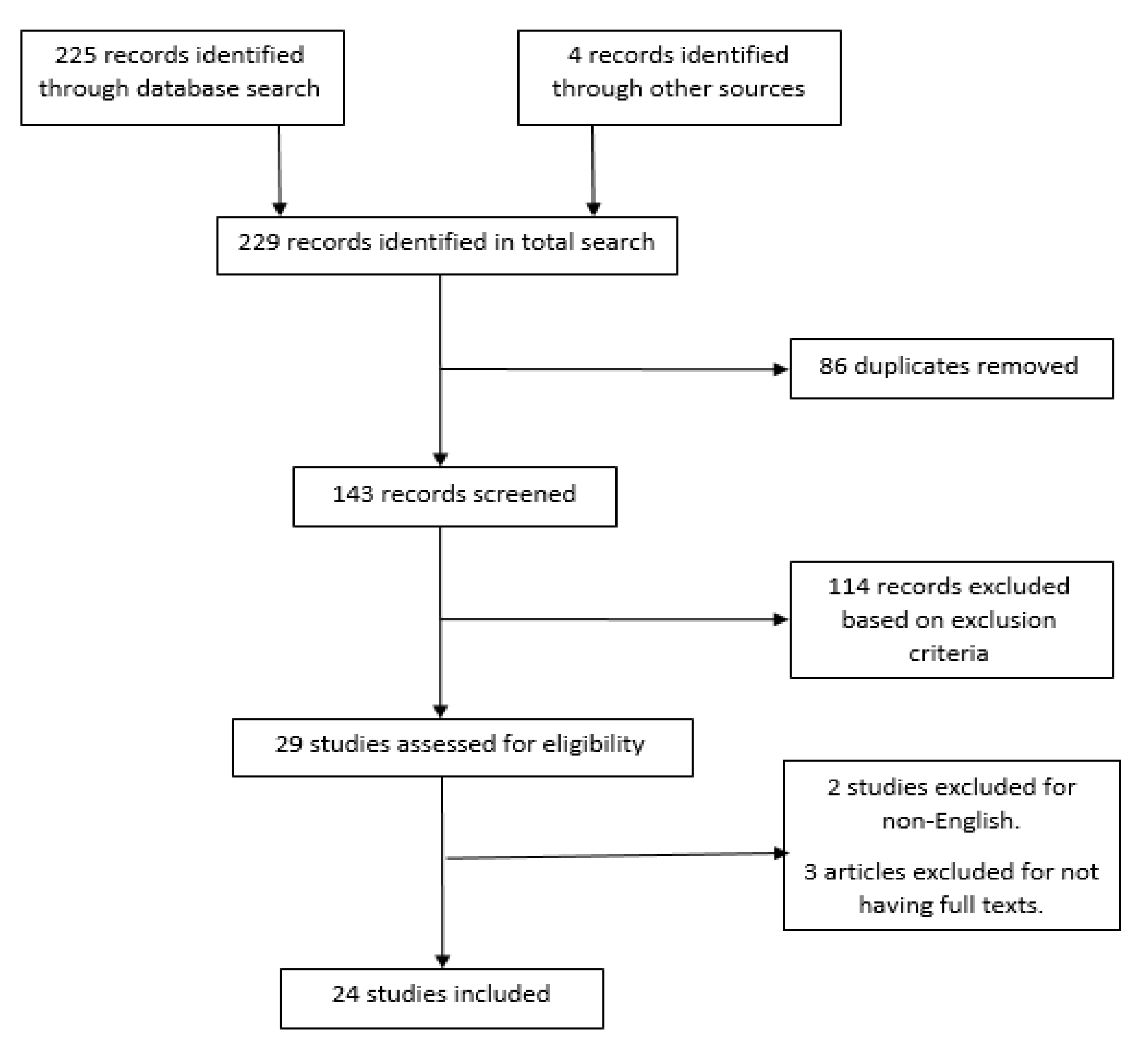
3.1. Literature Search
3.2. Study Characteristics
3.3. Hesperidin Dosage
3.4. Toxicity and Survival Analysis
4. Discussion
5. Conclusions
Author Contributions
Funding
Conflicts of Interest
References
- De Gonzalez, A.B.; Darby, S. Risk of cancer from diagnostic X-rays: Estimates for the UK and 14 other countries. Lancet 2004, 363, 345–351. [Google Scholar] [CrossRef]
- De González, A.B.; Mahesh, M.; Kim, K.-P.; Bhargavan, M.; Lewis, R.; Mettler, F.; Land, C. Projected cancer risks from computed tomographic scans performed in the United States in 2007. Arch. Intern. Med. 2009, 169, 2071–2077. [Google Scholar] [CrossRef] [PubMed]
- Ringborg, U.; Bergqvist, D.; Brorsson, B.; Cavallin-Ståhl, E.; Ceberg, J.; Einhorn, N.; Frödin, J.-E.; Järhult, J.; Lamnevik, G.; Lindholm, C. The Swedish Council on Technology Assessment in Health Care (SBU) systematic overview of radiotherapy for cancer including a prospective survey of radiotherapy practice in Sweden 2001—Summary and conclusions. Acta Oncol. 2003, 42, 357–365. [Google Scholar] [CrossRef] [PubMed]
- Wang, H.; Mu, X.; He, H.; Zhang, X.-D. Cancer radiosensitizers. Trends Pharmacol. Sci. 2018, 39, 24–48. [Google Scholar] [CrossRef] [PubMed]
- Shimizu, Y.; Kodama, K.; Nishi, N.; Kasagi, F.; Suyama, A.; Soda, M.; Grant, E.J.; Sugiyama, H.; Sakata, R.; Moriwaki, H. Radiation exposure and circulatory disease risk: Hiroshima and Nagasaki atomic bomb survivor data, 1950–2003. BMJ 2010, 340, b5349. [Google Scholar] [CrossRef] [PubMed]
- Berger, E.M. The Chernobyl disaster, concern about the environment, and life satisfaction. Kyklos 2010, 63, 1–8. [Google Scholar] [CrossRef]
- Devi, P.U.; Agrawala, P.K. Normal tissue protectors against radiation injury. Def. Sci. J. 2011, 61, 105–112. [Google Scholar] [CrossRef]
- Citrin, D.; Cotrim, A.P.; Hyodo, F.; Baum, B.J.; Krishna, M.C.; Mitchell, J.B. Radioprotectors and mitigators of radiation-induced normal tissue injury. Oncologist 2010, 15, 360–371. [Google Scholar] [CrossRef]
- Hosseinimehr, S.J. Trends in the development of radioprotective agents. Drug Discov. Today 2007, 12, 794–805. [Google Scholar] [CrossRef]
- Rosen, E.M.; Day, R.; Singh, V.K. New approaches to radiation protection. Front. Oncol. 2015, 4, 381. [Google Scholar] [CrossRef]
- Middleton, E. Effect of plant flavonoids on immune and inflammatory cell function. In Flavonoids in the Living System; Springer: Boston, MA, USA, 1998; pp. 175–182. [Google Scholar]
- Nones, J.; de Sampaio Spohr, T.C.L.; Gomes, F.C.A. Effects of the flavonoid hesperidin in cerebral cortical progenitors in vitro: Indirect action through astrocytes. Int. J. Dev. Neurosci. 2012, 30, 303–313. [Google Scholar] [CrossRef] [PubMed]
- Yahyapour, R.; Shabeeb, D.; Cheki, M.; Musa, A.E.; Farhood, B.; Rezaeyan, A.; Amini, P.; Fallah, H.; Najafi, M. Radiation Protection and Mitigation by Natural Antioxidants and Flavonoids: Implications to Radiotherapy and Radiation Disasters. Curr. Mol. Pharmacol. 2018, 11, 285–304. [Google Scholar] [CrossRef] [PubMed]
- Yamada, M.; Tanabe, F.; Arai, N.; Mitsuzumi, H.; Miwa, Y.; Kubota, M.; Chaen, H.; Kibata, M. Bioavailability of glucosyl hesperidin in rats. Biosci. Biotechnol. Biochem. 2006, 70, 1386–1394. [Google Scholar] [CrossRef] [PubMed]
- Cho, J. Antioxidant and neuroprotective effects of hesperidin and its aglycone hesperetin. Arch. Pharm. Res. 2006, 29, 699. [Google Scholar] [CrossRef] [PubMed]
- Roohbakhsh, A.; Parhiz, H.; Soltani, F.; Rezaee, R.; Iranshahi, M. Molecular mechanisms behind the biological effects of hesperidin and hesperetin for the prevention of cancer and cardiovascular diseases. Life Sci. 2015, 124, 64–74. [Google Scholar] [CrossRef] [PubMed]
- Garg, A.; Garg, S.; Zaneveld, L.; Singla, A. Chemistry and pharmacology of the citrus bioflavonoid hesperidin. Phytother. Res. 2001, 15, 655–669. [Google Scholar] [CrossRef] [PubMed]
- Moher, D.; Liberati, A.; Tetzlaff, J.; Altman, D.G. Preferred reporting items for systematic reviews and meta-analyses: The PRISMA statement. Ann. Intern. Med. 2009, 151, 264–269. [Google Scholar] [CrossRef]
- Katoch, O.; Kaushik, S.; Kumar, M.S.Y.; Agrawala, P.K.; Misra, K. Radioprotective property of an aqueous extract from Valeriana wallichii. J. Pharm. Bioallied Sci. 2012, 4, 327. [Google Scholar]
- Hosseinimehr, S.J.; Mahmoudzadeh, A.; Ahmadi, A.; Mohamadifar, S.; Akhlaghpoor, S. Radioprotective effects of hesperidin against genotoxicity induced by γ-irradiation in human lymphocytes. Mutagenesis 2009, 24, 233–235. [Google Scholar] [CrossRef]
- Kalpana, K.; Devipriya, N.; Srinivasan, M.; Menon, V.P. Investigation of the radioprotective efficacy of hesperidin against gamma-radiation induced cellular damage in cultured human peripheral blood lymphocytes. Mutat. Res. 2009, 676, 54–61. [Google Scholar] [CrossRef]
- Hosseinimehr, S.J.; Ahmadi, A.; Beiki, D.; Habibi, E.; Mahmoudzadeh, A. Protective effects of hesperidin against genotoxicity induced by 99mTc-MIBI in human cultured lymphocyte cells. Nucl. Med. Biol. 2009, 36, 863–867. [Google Scholar] [CrossRef]
- Kang, J.A.; Yoon, S.H.; Rho, J.K.; Jang, B.-s.; Choi, D.S.; Lee, D.-E.; Byun, E.-B.; Jeon, J.; Park, S.H. Radioprotective effect of hesperetin against γ-irradiation-induced DNA damage and immune dysfunction in murine splenocytes. Food Sci. Biotechnol. 2016, 25, 163–168. [Google Scholar] [CrossRef] [PubMed]
- Jagetia, G.C.; Rao, K. Acceleration in the Repair and Regenerative Responses by Different Doses of Hesperidin in the Deep Full Thickness Cutaneous Wound of Mice Whole Body Exposed To 6 Gy of γ -Radiation. Nurs. Health Care Int. J. 2018, 2, 1–12. [Google Scholar] [CrossRef]
- Jagetia, G.; Rao, K. Hesperidin treatment abates radiation-induced delay in healing of deep cutaneous excision wound of mice hemi-body exposed to different doses of γ-radiation. Clin. Dermatol. Dermatitis 2018, 1, 104. [Google Scholar]
- Jagetia, G.; Rao, K. Hesperidin, a citrus bioflavonoid potentiates repair and regeneration of deep dermal excision wounds of mice whole body exposed to different doses of 60 Co γ-radiation. Clin. Dermatol. Dermatitis 2018, 3, 000147. [Google Scholar]
- Jagetia, G.; Mallikarjuna Rao, K. Hesperidin, a citrus bioflavonoid reduces the oxidative stress in the skin of mouse exposed to partial body γ-radiation. Transcriptomics 2015, 3, 2. [Google Scholar]
- Kalpana, K.B.; Devipriya, N.; Srinivasan, M.; Vishwanathan, P.; Thayalan, K.; Menon, V.P. Evaluating the radioprotective effect of hesperidin in the liver of Swiss albino mice. Eur. J. Pharmacol. 2011, 658, 206–212. [Google Scholar] [CrossRef] [PubMed]
- Lee, Y.-R.; Jung, J.-H.; Kim, H.-S. Hesperidin partially restores impaired immune and nutritional function in irradiated mice. J. Med. Food 2011, 14, 475–482. [Google Scholar] [CrossRef] [PubMed]
- Hosseinimehr, S.; Nemati, A. Radioprotective effects of hesperidin against gamma irradiation in mouse bone marrow cells. Br. J. Radiol. 2006, 79, 415–418. [Google Scholar] [CrossRef]
- Haddadi, G.; Abbaszadeh, A.; Mosleh-Shirazi, M.A.; Okhovat, M.A.; Salajeghe, A.; Ghorbani, Z. Evaluation of the effect of hesperidin on vascular endothelial growth factor gene expression in rat skin animal models following cobalt-60 gamma irradiation. J. Cancer. Res. Ther. 2018, 14, S1098–S1104. [Google Scholar]
- Haddadi, G.H.; Rezaeyan, A.; Mosleh-Shirazi, M.A.; Hosseinzadeh, M.; Fardid, R.; Najafi, M.; Salajegheh, A. Hesperidin as Radioprotector against Radiation-induced Lung Damage in Rat: A Histopathological Study. J. Med. Phys. 2017, 42, 25–32. [Google Scholar] [PubMed]
- Shaban, N.Z.; Zahran, A.M.A.; El-Rashidy, F.H.; Kodous, A.S.A. Protective role of hesperidin against γ-radiation-induced oxidative stress and apoptosis in rat testis. J. Biol Res. (Thessalon). 2017, 24, 5. [Google Scholar] [CrossRef] [PubMed]
- Karimi, N.; Monfared, A.S.; Haddadi, G.H.; Soleymani, A.; Mohammadi, E.; Hajian-Tilaki, K.; Borzoueisileh, S. Radioprotective effect of hesperidin on reducing oxidative stress in the lens tissue of rats. Int. J. Pharm. Investig. 2017, 7, 149. [Google Scholar] [PubMed]
- El-Rahman, N.; El-Dein, E.; El-Hady, A.; Soliman, S.M. Effect of Hesperidin on γ-Radiation-and/or Paraquat Herbicide-Induced Biochemical, Hematological and Histopathological Changes in Rats. Pak. J. Zool. 2016, 48. [Google Scholar]
- Rezaeyan, A.; Fardid, R.; Haddadi, G.; Takhshid, M.; Hosseinzadeh, M.; Najafi, M.; Salajegheh, A. Evaluating radioprotective effect of hesperidin on acute radiation damage in the lung tissue of rats. J. Biomed. Phys. Eng. 2016, 6, 165. [Google Scholar]
- Fardid, R.; Ghorbani, Z.; Haddadi, G.; Behzad-Behbahani, A.; Arabsolghar, R.; Kazemi, E.; Okhovat, M.; Hosseinimehr, S. Effects of hesperidin as a radio-protector on apoptosis in rat peripheral blood lymphocytes after gamma radiation. J. Biomed. Phys. Eng. 2016, 6, 217. [Google Scholar] [PubMed]
- Rezaeyan, A.; Haddadi, G.H.; Hosseinzadeh, M.; Moradi, M.; Najafi, M. Radioprotective effects of hesperidin on oxidative damages and histopathological changes induced by X-irradiation in rats heart tissue. J. Med. Phys. 2016, 41, 182–191. [Google Scholar]
- Ahmed, H.M.; Hussein, M.A.; Alazonee, A.S. Radioprotective effect of hesperidin against gamma-irradiation-induced oxidative stress and biomechanical properties of bone in rats. Life Sci. J. 2013, 10, 2857–2865. [Google Scholar]
- Pradeep, K.; Ko, K.C.; Choi, M.H.; Kang, J.A.; Chung, Y.J.; Park, S.H. Protective effect of hesperidin, a citrus flavanoglycone, against γ-radiation-induced tissue damage in Sprague-Dawley rats. J. Med. Food 2012, 15, 419–427. [Google Scholar] [CrossRef]
- Said, U.Z.; Saada, H.N.; Abd-Alla, M.S.; Elsayed, M.E.; Amin, A.M. Hesperidin attenuates brain biochemical changes of irradiated rats. Int. J. Radiat. Biol. 2012, 88, 613–618. [Google Scholar] [CrossRef]
- Park, S.H.; Pradeep, K.; Ko, K.C. Protective effect of hesperidin against γ-radiation induced oxidative stress in Sprague-Dawley rats. Pharm. Biol. 2009, 47, 940–947. [Google Scholar] [CrossRef]
- Rodemann, H.P.; Blaese, M.A. Responses of normal cells to ionizing radiation. Semin. Radiat. Oncol. 2007, 17, 81–88. [Google Scholar] [CrossRef] [PubMed]
- Peña, L.A.; Fuks, Z.; Koksnick, R. Stress-induced apoptosis and the sphingomyelin pathway. Biochem Pharmacol. 1997, 53, 615–621. [Google Scholar] [CrossRef]
- Pena, L.A.; Fuks, Z.; Kolesnick, R.N. Radiation-induced apoptosis of endothelial cells in the murine central nervous system: Protection by fibroblast growth factor and sphingomyelinase deficiency. Cancer Res. 2000, 60, 321–327. [Google Scholar] [PubMed]
- Pearce, M.S.; Salotti, J.A.; Little, M.P.; McHugh, K.; Lee, C.; Kim, K.P.; Howe, N.L.; Ronckers, C.M.; Rajaraman, P.; Craft, A.W. Radiation exposure from CT scans in childhood and subsequent risk of leukaemia and brain tumours: A retrospective cohort study. Lancet 2012, 380, 499–505. [Google Scholar] [CrossRef]
- Prise, K.M.; Saran, A. Concise review: Stem cell effects in radiation risk. Stem Cells 2011, 29, 1315–1321. [Google Scholar] [PubMed]
- Ryan, J.L. Ionizing radiation: The good, the bad, and the ugly. J. Investig. Dermatol. 2012, 132, 985–993. [Google Scholar] [CrossRef]
- Porock, D.; Nikoletti, S.; Kristjanson, L. Management of radiation skin reactions: Literature review and clinical application. Plast. Surg. Nurs. 1999, 19, 185. [Google Scholar] [CrossRef]
- Yusuf, S.W.; Venkatesulu, B.P.; Mahadevan, L.S.; Krishnan, S. Radiation-induced cardiovascular disease: A clinical perspective. Front. Cardiovasc. Med. 2017, 4, 66. [Google Scholar] [CrossRef]
- Mathers, C.D.; Loncar, D. Projections of global mortality and burden of disease from 2002 to 2030. PLoS Med. 2006, 3, e442. [Google Scholar] [CrossRef]
- Darby, S.C.; Ewertz, M.; McGale, P.; Bennet, A.M.; Blom-Goldman, U.; Brønnum, D.; Correa, C.; Cutter, D.; Gagliardi, G.; Gigante, B. Risk of ischemic heart disease in women after radiotherapy for breast cancer. N. Engl. J. Med. 2013, 368, 987–998. [Google Scholar] [CrossRef] [PubMed]
- Ben-David, M.A.; Elkayam, R.; Gelernter, I.; Pfeffer, R.M. Melatonin for prevention of breast radiation dermatitis: A phase II, prospective, double-blind randomized trial. Isr. Med. Assoc. J. 2016, 18, 188–192. [Google Scholar] [PubMed]
- Lozano, A.; Marruecos, J.; Rubió-Casadevall, J.; Farre, N.; Lopez-Pousa, A.; Giralt, J.; Planas, I.; Cirauqui, B.; Lanzuela, M.; Morera, R. Phase II trial of high-dose melatonin oral gel for the prevention and treatment of oral mucositis in H&N cancer patients undergoing chemoradiation (MUCOMEL). J. Clin. Oncol. 2018. [Google Scholar] [CrossRef]


| First Author | Subject | Organ (or Tissue) of Interest | Radiation Type and Dose (Gy) | Hesperidin Dose/Concentration | Time for Outcome Assessment | Main Outcomes |
|---|---|---|---|---|---|---|
| Katoch et al. [19] | Cultured human fibroblast cells | Fibroblast cells | γ-ray, 5 | 6.18 ± 0.26 mg/g extract | 4 h | Countered radiation-induced free radicals post-irradiation, decreased prolonged oxidative stress, and protected against radiation-induced DNA damage. |
| Hosseinimehr et al. [20] | Cultured human blood lymphocytes | Lymphocytes | γ-ray, 1.5 | 250 mg/kg body weight | 0–3 h | Significant decrease in the incidence of micronuclei of blood lymphocytes collected 1 h after oral administration of hesperidin compared to those collected at 0 h. Maximum protection and decrease in frequency of micronuclei (33%) was observed at 1 h after ingestion of hesperidin. |
| Kalpana et al. [21] | Cultured human lymphocytes | Lymphocytes | γ-ray, 1–4 | 3.27–19.65 µM | 30 min | Here, 16.38 µM hesperidin pretreatment prior to irradiation had the maximum radioprotective effect, which included a significant decrease in the levels of MN and DC counts, as well as TBARS. Reduction in tail length, tail moment, olive tail moment, and % DNA in the tail. Increased levels of enzymatic (SOD, CAT, and GPx) and non-enzymatic (glutathione (GSH)) antioxidants and restored DNA damage to near-normal levels. |
| Hosseinimehr et al. [22] | Cultured human lymphocyte cells | Lymphocytes | γ-ray from 99mTc-MIBI radiopharmaceuticals, 200 μCi | 10–100 μM | 3 h | Significant reduction in micronuclei frequency in cultured lymphocytes, thereby leading to protection against genetic damage. Optimal effect of hesperidin was obtained at 100 μM concentration. |
| Kang et al. [23] | Cultured BALB/c mice splenocytes | Splenocytes | γ-ray, 2 and 4 | 20–500 µM | 24 h | Improved cell viability, prevented damage to DNA, and hindered proinflammatory cytokines, intracellular ROS, and NO. |
| Jagetia et al. [24] | Swiss albino mice | Skin wound | γ-ray, 6 | 0–500 mg/kg body weight | 3–15 days | Treatment with 100 mg/kg hesperidin before irradiation had the maximum radioprotective effect, leading to a steady increase in wound contraction and reduction in mean wound healing time by 2 days. |
| Jagetia et al. [25] | Swiss albino mice | Skin wound | γ-ray, 2–8 | 100 mg/kg body weight | 1–15 days | Enhancement of collagen, hexosamine, DNA, and nitric oxide synthesis in the granulation tissue, thereby improving wound healing compared to the irradiated group. |
| Jagetia et al. [26] | Swiss albino mice | Skin wound | γ-ray, 2–8 | 100 mg/kg body weight | 1–15 days | Significantly reduced both radiation-induced delay in wound contraction and mean wound healing time. |
| Jagetia et al. [27] | Swiss albino mice | Skin wound | γ-ray, 6 | 50 and 100 mg/kg body weight | 0–48 h | Reduced radiation-induced oxidative stress in the irradiated wounds of mice. |
| Kalpana et al. [28] | Swiss albino mice | Liver | X-ray, 4 | 12.5–100 mg/kg body weight | 30 days | Here, 25 mg/kg hesperidin pretreatment prior to irradiation had the maximum radioprotective effect, including restoring antioxidant status to near-normal as well as decreasing the levels of the lipid peroxidation index, DNA damage, and comet parameters. |
| Lee et al. [29] | ICR mice | Liver, intestine, splenocytes, and lymphocytes | X-ray, 15 | 50 and 200 mg/kg body weight | 10 and 30 days | Reduction of radiation-induced inflammation and partial restoration of immune and nutritional status. |
| Hosseinimehr et al. [30] | NMRI mice | Bone marrow cells | γ-ray, 2 | 10–160 mg/kg body weight | 24 h | Hesperidin dose of 80 mg/kg had the maximum reduction in the frequencies of MnPCEs. Significant increase in PCE/PCE + NCE ratio in mice bone marrow compared to nondrug-treated irradiated control. |
| Haddadi et al. [31] | Sprague-Dawley rats | Skin | γ-ray, 22 | 100 mg/kg body weight | 24 h | Initiated angiogenesis by inducing VEGF gene. Stimulated epithelialization and collagen deposition and enhanced cellular proliferation, thereby aiding wound healing and protecting skin from radiation damage. |
| Haddadi et al. [32] | Sprague-Dawley rats | Lung | γ-ray, 18 | 100 mg/kg body weight | 24 h and 8 weeks for acute and chronic histopathological evaluations, respectively. | Hesperidin administration led to significant decrease in radiation-induced inflammation and inflammatory cells at 24 h post-irradiation. Furthermore, there was a reduction in radiation pneumonitis and radiation fibrosis in the lung tissue at 8 weeks post-irradiation. |
| Shaban et al. [33] | Sprague-Dawley rats | Testes | γ-ray, 8 | 200 mg/kg body weight | 8 and 14 days | Reduction in OS, LPO, and apoptosis. Improvement in structure of testes and better protection of testes was observed when hesperidin was administered before irradiation compared to after irradiation. |
| Karimi et al. [34] | Rats | Lens | γ-ray, 15 | 100 mg/kg body weight | 2 days | Significant increase in the GSH level and decrease in MDA level, and hence, a reduction in oxidative stress. |
| Abd El-Rahman et al. [35] | Albino rats | Blood, lung, and dorsal aorta | γ-ray, 6 | 40 mg/kg body weight | 21 days | Significantly reduced lipid variation, decreased oxidative stress, improved blood cell counts, and attenuated lung and dorsal aorta tissue injury. |
| Rezaeyan et al. [36] | Rats | Lung | γ-ray, 18 | 100 mg/kg body weight | 24 h | Significant reduction in macrophages and neutrophils, as well as mild reduction in inflammation and lymphocytes. |
| Fardid et al. [37] | Rats | Peripheral blood lymphocytes | γ-ray, 2 and 8 | 50 and 100 mg/kg body weight | 24 h | Pretreatment with hesperidin significantly reduced apoptosis in irradiated rats. |
| Rezaeyan et al. [38] | Rats | Heart | X-ray, 18 | 100 mg/kg body weight | 24 h (for biochemical assay and acute histopathological evaluation) and 8 weeks (for chronic histopathological evaluation) | Decreased inflammation, fibrosis, mast cell, and macrophage numbers and myocyte necrosis. |
| Ahmed et al. [39] | Albino rats | Bone | γ-ray, 2 | 160 mg/kg body weight | 24 h | Improvement in antioxidant activities as well as biomechanical properties of bone and prevention of endothelial dysfunction. |
| Pradeep et al. [40] | Sprague-Dawley rats | Liver, heart, and kidney | γ-ray, 5 | 50 and 100 mg/kg body weight | 7 days | Reduction in necrotic and cellular damage, as well as oxidative stress. |
| Said et al. [41] | Albino rats | Brain | γ-ray, 5 | 50 mg/kg body weight | 14 days | Significant reduction in oxidative stress, monoamine alterations, and mitochondrial damage, and hence a reduction in the severity of radiation-induced biochemical brain disorders. |
| Park et al. [42] | Sprague-Dawley rats | Heart and kidney | γ-ray, 5 | 50 and 100 mg/kg body weight | 7 days | Treatment with hesperidin post-irradiation led to significant reduction in levels of lipid peroxidation, improvements in activities of endogenous antioxidants (SOD, CAT, GPx, and GSH), and minimal damage to the heart and kidney tissues. |
© 2019 by the authors. Licensee MDPI, Basel, Switzerland. This article is an open access article distributed under the terms and conditions of the Creative Commons Attribution (CC BY) license (http://creativecommons.org/licenses/by/4.0/).
Share and Cite
Musa, A.E.; Omyan, G.; Esmaely, F.; Shabeeb, D. Radioprotective Effect of Hesperidin: A Systematic Review. Medicina 2019, 55, 370. https://doi.org/10.3390/medicina55070370
Musa AE, Omyan G, Esmaely F, Shabeeb D. Radioprotective Effect of Hesperidin: A Systematic Review. Medicina. 2019; 55(7):370. https://doi.org/10.3390/medicina55070370
Chicago/Turabian StyleMusa, Ahmed Eleojo, Gilnaz Omyan, Farid Esmaely, and Dheyauldeen Shabeeb. 2019. "Radioprotective Effect of Hesperidin: A Systematic Review" Medicina 55, no. 7: 370. https://doi.org/10.3390/medicina55070370
APA StyleMusa, A. E., Omyan, G., Esmaely, F., & Shabeeb, D. (2019). Radioprotective Effect of Hesperidin: A Systematic Review. Medicina, 55(7), 370. https://doi.org/10.3390/medicina55070370