Next Article in Journal
Scaffold design for artificial tissue with bone marrow stem cells
Previous Article in Journal
Detection and evaluation of ventricular repolarization alternans: An approach to combined ECG, thoracic impedance, and beat-to-beat heart rate variability analysis
 
 
Medicina is published by MDPI from Volume 54 Issue 1 (2018). Previous articles were published by another publisher in Open Access under a CC-BY (or CC-BY-NC-ND) licence, and they are hosted by MDPI on mdpi.com as a courtesy and upon agreement with Lithuanian Medical Association, Lithuanian University of Health Sciences (LUHS), and Vilnius University.
Article

A role of alpha-tocopherol and phylloquinone in the modulation of uterine contractility and reproductive function in mouse models

by
Enitome E. Bafor
1,*,
Victory O. Ebidame
1,
Uloma B. Elvis-Offiah
2,
Osemelomen Omoruyi
1,
Gerald I. Eze
3,
Osamwonyi Igbinuwen
4 and
Kadiri P. Braimoh
5
1
Department of Pharmacology and Toxicology, Faculty of Pharmacy, University of Benin, Nigeria
2
Department of Science Laboratory Technology, Faculty of Life Sciences, University of Benin, Nigeria
3
Department of Anatomy, School of Basic Medical Sciences, University of Benin, Nigeria
4
Hematology Department, University of Benin Teaching Hospital, Benin City, Nigeria
5
Histopathology Department, University of Benin Teaching Hospital, Benin City, Nigeria
*
Author to whom correspondence should be addressed.
Medicina 2017, 53(3), 190-202; https://doi.org/10.1016/j.medici.2017.05.002
Submission received: 3 October 2016 / Revised: 5 April 2017 / Accepted: 8 May 2017 / Published: 30 May 2017

Abstract

:
Background and aim: Alpha-tocopherol has been implicated in reproduction processes, and deficiency of phylloquinone has been associated with serious complications in pregnancy. This study was therefore aimed at investigating the effects of phylloquinone and alpha-tocopherol on uterine contractility and female reproductive function using mouse models. Materials and methods: Both in vivo and ex vivo animal models were employed and designed to assess changes on uterine contractility and reproductive functions in the non-pregnant uterus. The effect of alpha-tocopherol and phylloquinone on spontaneous uterine contractions, oxytocin-induced uterine contractions (11.82 nM) and high KCl-induced tonic uterine contractions (80 mM) were assessed. The effect of subcutaneous administration of alpha-tocopherol (10 mg/kg) on reproductive hormone levels and reproductive tissues were also determined. Results: Alpha-tocopherol increased the force of contractions while phylloquinone decreased the force of uterine contractions. Plasma levels of luteinizing hormone (P < 0.01), estrogen (P < 0.01) and progesterone (P < 0.001) were elevated in the presence of alpha-tocopherol after 6 days subcutaneous administration. Conclusions: Alpha-tocopherol and phylloquinone have been shown to directly modulate uterine contractility and reproductive function and may contribute to the management and treatment of reproductive disorders.

1. Introduction

Phylloquinone and alpha-tocopherol are classified as fat-soluble vitamins [1,2]. The existence of fat-soluble vitamins was first reported in 1913 [2]. Alpha-tocopherol, which was first identified over 90 years ago as an essential dietary factor required to maintain normal reproduction in rats [3], has also been implicated in reproduction processes in general. Similarly, deficiency of phylloquinone had been associated with serious complications and risk in pregnancy which can include hemorrhage and eventually death [4,5]. Alpha-tocopherol is the major vitamin E compound found in animals and humans [6] and therefore understanding of their involvement in molecular signaling requires further study. Unlike the other three fat-soluble vitamins that have very specific molecular targets and actions, there are no reports on specific pathways and molecular targets for alpha-tocopherol that account for its requirement from the diet and in specific reproductive functions. Antenatal administration of phylloquinone supplementation for pregnant women may provide significant benefits for improving both maternal and neonatal outcomes. Maternal administration of phylloquinone has been suggested to improve prothrombin and partial thromboplastin activities and reduce the incidence and severity of intraventricular hemorrhage (IVH) in infants [7]. Taken together, a possible signal transduction role for alpha-tocopherol and phylloquinone vitamins appear to exist. Therefore research on identifying specific molecular targets and actions for alpha-tocopherol and other fat-soluble vitamins is encouraged [8]. Understanding and mapping out the involvement of these vitamins on regulation of uterine contractility, will therefore be useful in identifying signaling processes associated with the action of these vitamins on uterine signal transduction regulation. It will also increase the progress toward bridging the gaps that exist between therapeutic roles of nutritional drugs and female reproductive health. The uterus primarily functions to carry and sustain an embryo till parturition [9]. The myometrium therefore functions in order to prepare the uterus for such action in both keeping and expelling the fetus [9]. In the non-pregnant uterus, the myometrium undergoes significant changes in order to allow implantation of the fertilized embryo [9]. The myometrium has been described to be myogenic in nature being able to generate regular spontaneous contractions with no input from the nervous or hormonal systems [10]. They have also been described to exhibit phasic properties showing variations in frequency, amplitude and duration. These parameters equip the myometrium for its physiological function and are very often studied individually in order to provide a more concise picture into the workings of the myometrium. Uterine contractions are triggered by transient increases in intracellular calcium concentration ([Ca2+]i), which in turn are initiated and controlled by myometrial action potentials [11]. In the non-pregnant uterus, the myometrium is responsible for the contractions occurring during menstruation or estrous which may give rise to the cramping observed often referred to as dysmenorrhea [12]. The myometrium in this state is involved in a uterine peristaltic action that assists in the endometrial sloughing that occurs during menstruation [13]. Changes in female steroid hormones during this time greatly influence the pattern of myometrial activity [14]. It is important to note, particularly as regards this current study, that the pattern of contractile activity in the non-pregnant uterus is similar to uterine function in general, be it in the pregnant or non-pregnant states [15]. Contractions occurring in antegrade fashion and propagating from the fundus toward the cervical end of the uterus are necessary for emptying or discharge of uterine content i.e. menstrual blood [15]. On the other hand, cervico-fundal contractions assist in sperm transport or possibly electrolyte retention [16]. During pregnancy, retrograde contractions may contribute to the maintenance of early pregnancies within the uterine cavity [17].
Uterine contractility constitutes a part of female reproductive health [9,10,18]. Therefore understanding the roles of drugs on uterine contractility as a parameter of female reproductive health provides useful information toward either understanding or improvement of the female reproductive health in general. There has however been no study to investigate the direct effect of vitamins on uterine contractility. This study is aimed at investigating the roles of phylloquinone and alpha-tocopherol on uterine contractility and some female reproductive function parameters in vivo using mice models. This makes the study unique as it reports functional involvement of these dietary factors in female reproduction.

2. Materials and methods

2.1. Drugs and salts

Sodium chloride – NaCl (Guangdong GuanghuaSci-Tech Co. Ltd., China), potassium chloride – KCl (Guangdong GuanghuaSci-Tech Co. Ltd., China), sodium bicarbonate – NaHCO3 (Sigma–Aldrich, Inc.), d-glucose – C6H14O7 (Guangdong GuanghuaSci-Tech Co. Ltd., China) and calcium chloride – CaCl2 (XL®) were obtained. In addition, alpha-tocopherol (Embassy pharmaceutical and chemical Ltd., Nigeria), phylloquinone (Laborate Pharmaceuticals India), oxytocin (Roche pharmaceutical Ltd., UK), progesterone (Alpha pharmacy Ltd., Nigeria), and estradiol valerate (Bayer Health Care PLC, Berlin) were also acquired for the study.

2.2. Experimental animals

Female virgin Swiss albino laboratory mice weighing between 20.0–30.0 g purchased from the Animal House Department of Pharmacology & Toxicology, University of Benin, Nigeria were used. They were housed and cared for in well-ventilated plastic cages at environmentally controlled room temperature of approximately 29 ± 5 °C. All animals were handled as much as possible in accordance to standards of the Public Health Service policy on humane care and use of Laboratory Animals 2002. Ethical clearance for animal use was also obtained from the Ethical Committee of the Faculty of Pharmacy, University of Benin, Nigeria. Standard diet of animal pellets and clean tap water was provided. Adequate hygiene was maintained daily by regular cleaning.

2.3. Ex vivo experimental set-up

On the day of the experiment, the animal was weighed and only those in pro-estrous or estrous stages were used. This was confirmed by physical examination and observation of vaginal smears. The selected animal was humanely killed by cervical dislocation and the uterine horns were immediately but carefully excised, freed of accompanying mesenteries and fat and placed into previously warmed and aerated physiological salt solution (PSS) of the following composition in M: NaCl 154.00, NaHCO3 5.95, d-glucose 2.78, KCl 5.63, and CaCl2·2H2O 2.05. The uterine horns were transected medially and uterine segments (measuring between 0.5–0.7 cm in length) were mounted in 10 mL organ baths containing aerated, warmed (37 °C) PSS. One uteri per animal was used in each subgroup of experiments. For each subgroup, uteri from the fundus section were used in all cases in order to minimize variations across experiments. Tissues were placed under 4.90 mN tension and then equilibrated for no less than 30 min till stable regular contractions were obtained. The differential force (amplitude) and frequency of contractions generated from the longitudinal muscle layers of each uterine tissue segment were recorded using a 7003E-isometric force transducer (UgoBasile, Varise, Italy) connected to a 17400 data capsule digital recorder with an inbuilt bridge amplifier (UgoBasile, Varese, Italy). Concentrations of drugs used for each experimental protocol had been pre-determined as optimum working concentrations in preliminary experiments.

2.4. Studies on ex vivo spontaneous uterine contraction

The direct effect of cumulative concentrations of each vitamin drug on spontaneous uterine contractions was investigated as described by Bafor et al. [19]. Concentration–response relationships were obtained using alpha-tocopherol (ATE) at final concentrations of 0.069–30.62 μM. A contact time of 3 min was allowed following each concentration of ATE administered. At the end of the experiment, the drugs were washed off the tissues and the tissue was allowed to recover. Experiments were terminated for tissues that failed to recover. The same procedure was performed to obtain concentration–response relationships for phylloquinone (K1) using concentrations between 0.02–9.85 μM.

2.5. Studies on ex vivo oxytocin-induced uterine contraction

After tissue equilibration, the effects of ATE (0.05–54.12 μM) and K1 (0.15–17.26 μM) on oxytocin-induced uterine contraction were observed. The response of the tissue to ATE or K1 in the continued presence of 11.82 nM oxytocin (OT) was performed by the initial addition of OT to the bath for 5 min followed by cumulative additions of either ATE or K1 at 3 min per concentration. The last 3 min of the response of the tissue to OT prior to drug addition was taken as control (100%). At the end of each experiment, the tissue was completely washed of drugs and allowed to recover.

2.6. Studies on high KCl-induced uterine contraction in ex vivo models

In order to determine the effects of the drugs under study on the depolarized uterus, the effect of either ATE (0.05–54.12 μM) or K1 (0.15–17.26 μM) was investigated in the presence of high KCl (80 mM). KCl was applied to the bath containing the uterine tissues for 5 min and either ATE or K1 was added cumulatively in the continued presence of KCl. The last 3 min of the response of the tissue to KCl prior to drug addition was taken as control (100%).

2.7. Studies on some reproductive parameters in vivo

For this stage of experiments, ATE was selected for further study based on its use in the literatures for some reproductive conditions [20,21,22,23,24] and also due to its potency and consistent results in the current study. The stage of the estrus cycle for each mouse was determined by daily collection of vaginal smears and visual observations for the first 7 days according to methods previously described [25,26,27,28]. Observations were performed between the hours of 9 a.m. and noon daily. Briefly, vaginal smears examination was carried out to determine the stage of the estrus cycle of the mice by identification of cell types and the relative quantities present in the vagina swabs. Smear collection was obtained by gentle flushing and withdrawing of the vagina area with 0.1-mL distilled water using a soft pipette. The fluid obtained was placed on a clean glass slide and allowed to dry. To the dried smear was added a few drops of ethanol and gentian violet dye for fixing and for viewing respectively. The smears were subsequently observed under the microscope with the aid of a 10× objective lens. Specifically, the stage of the estrus was determined based on the presence or absence of leukocytes, cornified epithelial cells and nucleated epithelial cells [28]. In addition to the smear observation, visual examinations were performed by observation of the vagina appearance, the degree of vagina swelling, the color and presence of moisture, as well as the extent of vagina opening. The mammary glands were also observed to detect for changes in appearance.
At the end of the 7-day observation and staging period, the animals were grouped (4 animals per group) according to the stage of their estrus cycle: group 1 (negative control) received distilled water (0.1 mL p.o.); group 2 (test group) received ATE (10 mg/kg s.c.); group 3 (positive control) received estrogen (10 mg/kg p.o); and group 4 (positive control) received progesterone (0.01 mg/kg s.c.). The animals were administered the drugs every morning between the hours of 9 a.m. and 12 noon for the duration of their respective estrus cycles (determined to be 6 days). During the period of drug administration, regular smear collections and visual observations were continued. On the 7th day of drug administration, the animals were euthanized first via chloroform anesthesia and then careful cervical dislocation and cardiac puncture was performed to obtain blood samples. The blood samples obtained were placed in lithium-heparinized sample bottles and submitted for analyses of selected reproductive hormones. Measurement of reproductive hormones were performed as previously described [29] using the hormonal assay and involved the use of an automated qualitative test on serum or plasma (lithium heparin) using the Minividas Analyzer (VIDAS Kit France), and the Enzyme Linked Fluorescent Assay (ELFA) technique [30,31]. The uteri were isolated and weighed. The ovaries and cervix were then separated from the uterine horns and each organ placed in separate but properly labeled universal bottles containing 10% formalsaline (10 mL formaldehyde in 90 mL of 0.9% normal saline). The organs were then prepared for histological analysis and observed.

2.8. Data analysis

The mean frequency and amplitude were calculated from contractions occurring within the last 3 min of the phasic uterine contractions. Concentration–response plots were obtained using the GraphPad Prism version 7.0 (GraphPad software Inc., San Diego, CA, USA). Results were obtained as percentages of control applications (control = 100%) where applicable. All data shown were expressed as mean ± standard error of mean (SEM) and ‘‘n’’ was used to indicate the number of animals in each case. Significance was evaluated using appropriate t tests, and where necessary one-way analysis of variance with Dunnett’s post hoc was performed and P values ≤0.05 were taken to represent minimum significance. Mean log concentration–response curves were analyzed by non-linear regression. This was achieved by fitting data to a three-parameter logistic equation, using non-linear regression with GraphPad Prism 6.0 (GraphPad software, San Diego, CA, USA). Y = Bottom + (Top − Bottom)/(1 + 10(Log EC50X)*HillSlope), where Y = response which starts at the Bottom and goes to the Top in sigmoid shape, X = logarithm of concentration and EC50 is the concentration that produces half the maximal responses.

3. Results

3.1. Experiments on spontaneous uterine contractility

3.1.1. Effect of ATE and K1 on spontaneous uterine contractions

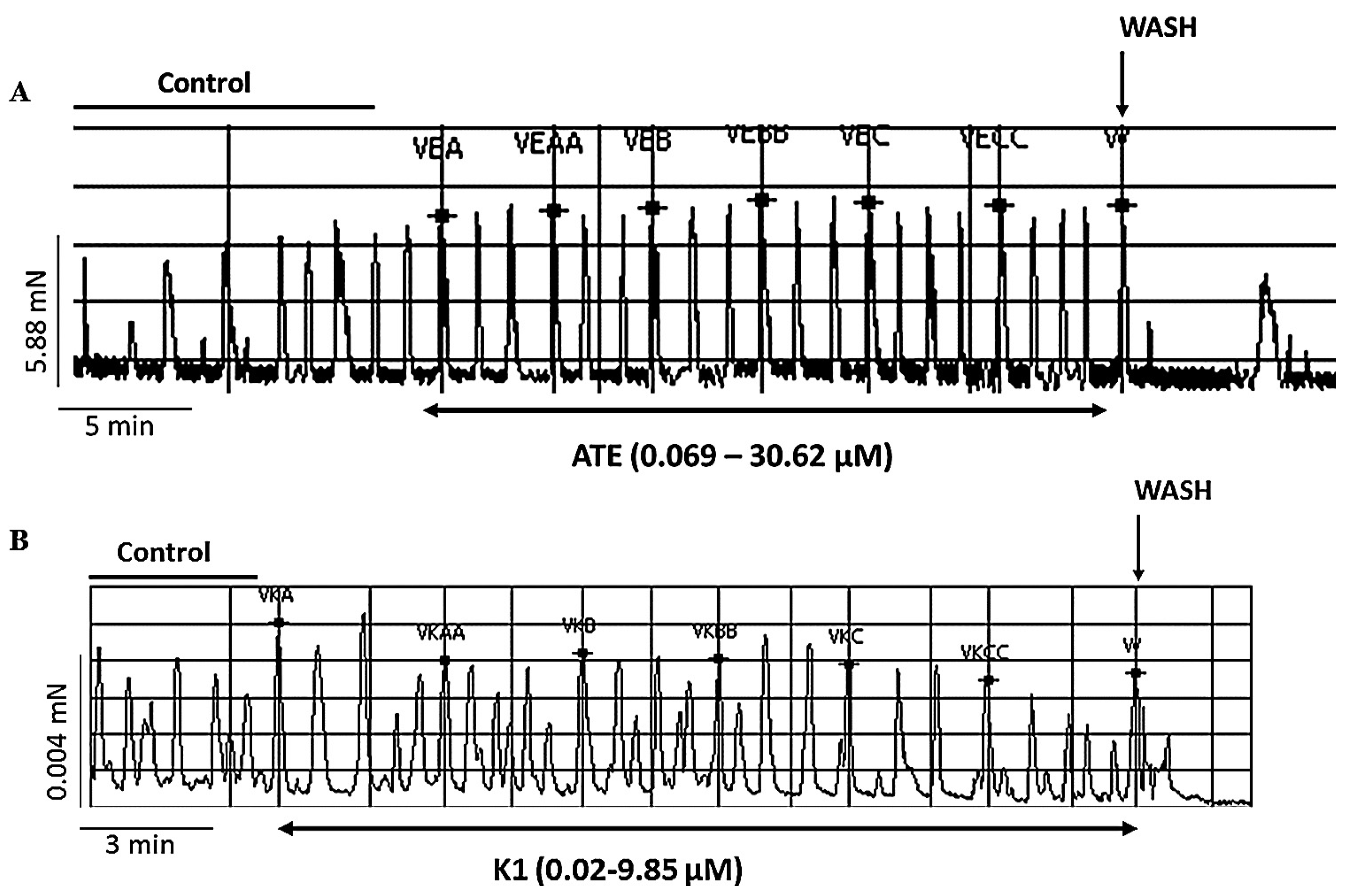
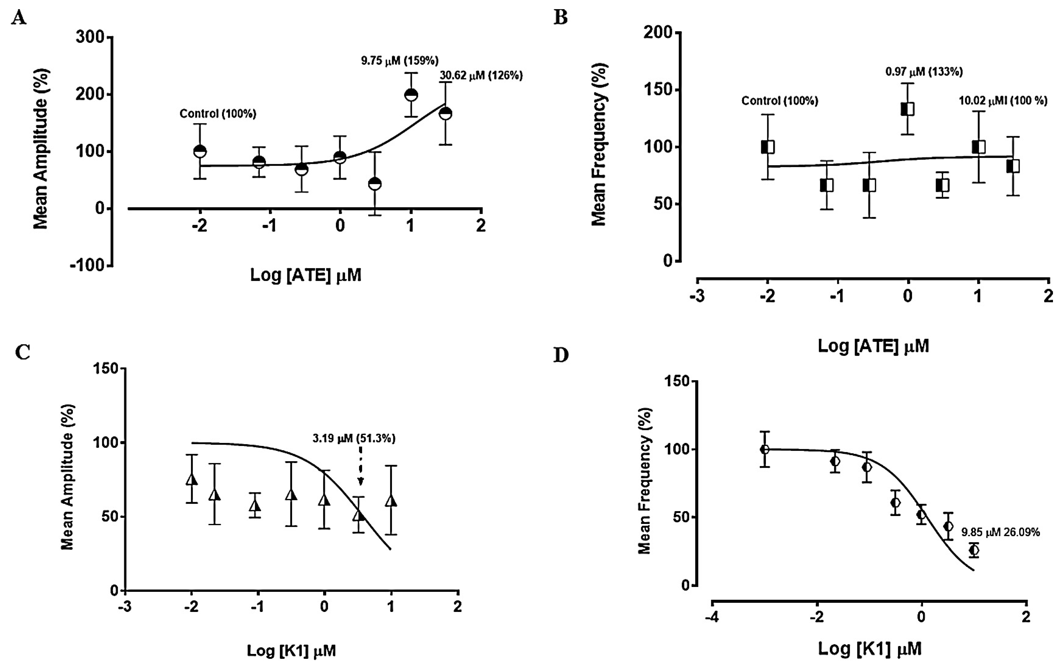
ATE produced a concentration-dependent stimulation of spontaneous uterine contractility while K1 produced a concentration-dependent inhibition (Fig. 1). This was also clearly depicted in a concentration–response plot that showed stimulation of the amplitude of spontaneous contraction by ATE up to 159% at a concentration of 9.75 μM and 126% at 30.62 μM and no significant changes were observed on the frequency of spontaneous contractions with ATE at lower concentrations though an increase to 133% from the control was observed at 9.75 μM (Fig. 2A and B respectively). The EC50 ATE for the force of contraction was found to be 5.33 ± 2.08 mg/mL while the EC50 ATE for the frequency was found to be 0.17 ± 1.52 mg/mL. A reduction of the amplitude of contraction from the control to 51.3% ≈ 51% at 3.19 μM K1 was observed and similarly a reduction to 26% of the frequency from the control at 9.85 μM K1 was also observed (Fig. 2C and D). The EC50 K1 was found to be 1.71 ± 0.26 mg/mL for the amplitude and 5.4 ± 0.21 mg/mL for the frequency.

3.2. Effect of ATE and K1 on oxytocin-induced uterine contractions

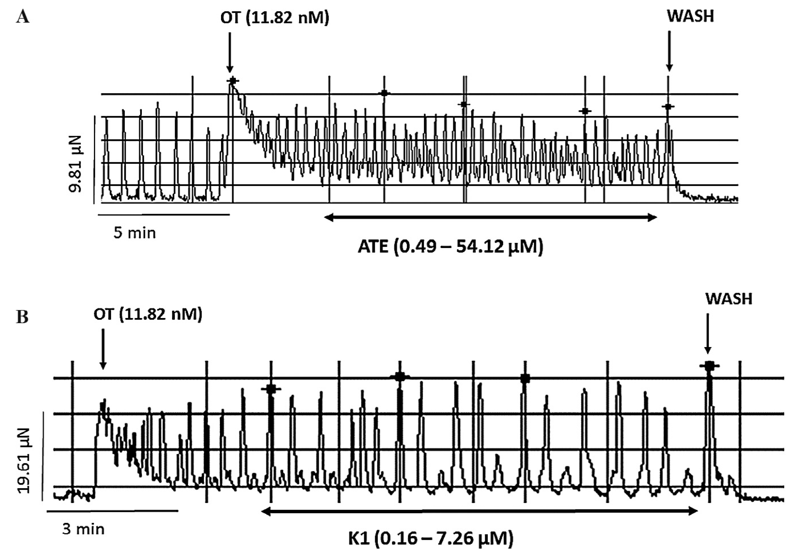
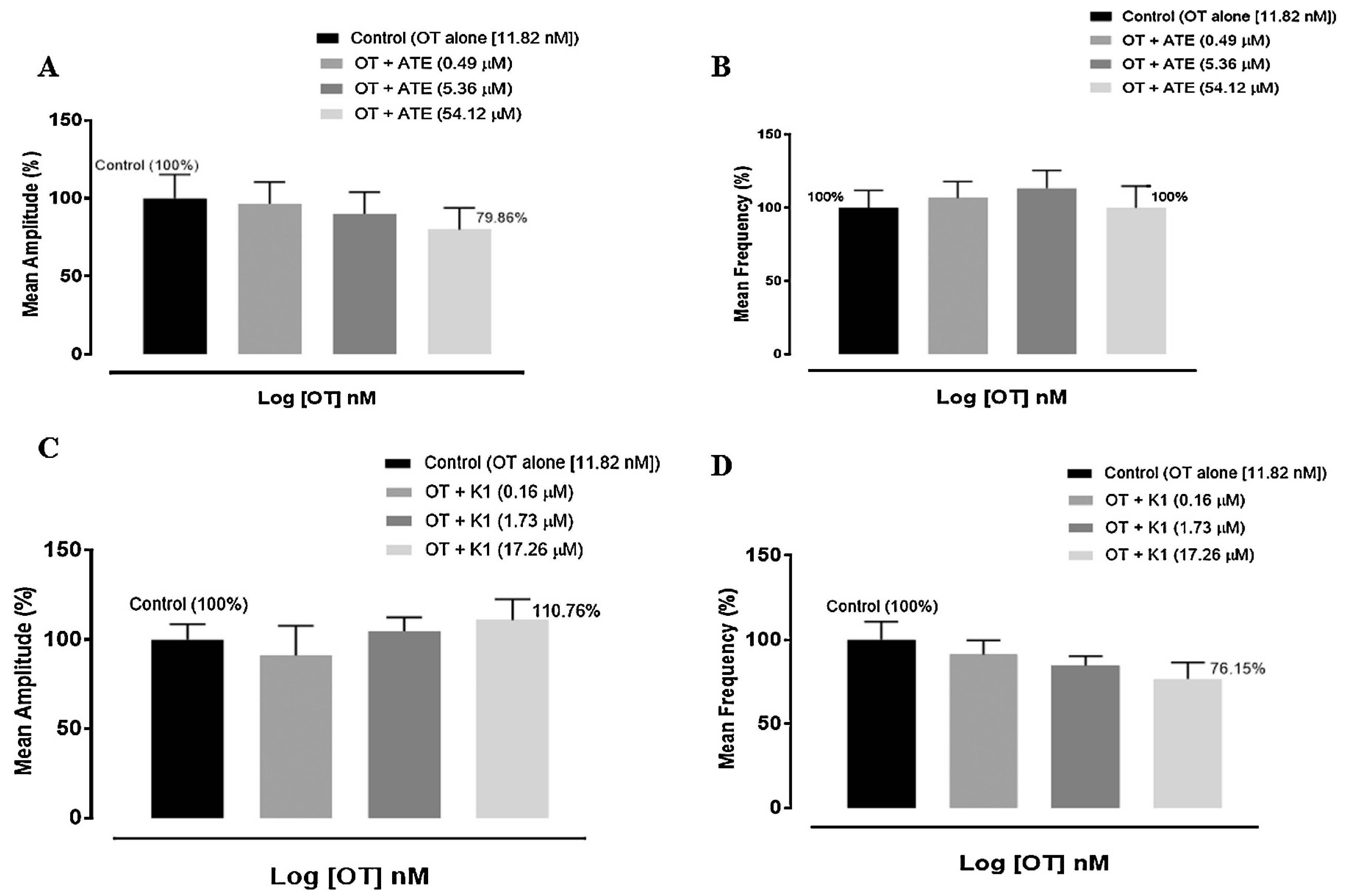
OT (11.82 nM) was used to determine the effect of the different drugs under study on agonist mediated uterine contractility as a means to assess effects on receptor-mediated activity and to also extrapolate possible mechanism(s) of activity. ATE was observed in this study to cause mild inhibitions on OT-induced uterine contractions (Fig. 3A) while K1 appeared to increase OT-induced contractility (Fig. 3B). Construction of the concentration–response plot showed ATE to inhibit the amplitude of OT-induced contraction to about 79% (Fig. 4A) with no distinct effect on the frequency (Fig. 4B). Concentration–response plots to K1 showed an initial decrease and then mild increase in amplitude (Fig. 4C) while a concentration-dependent decrease in frequency was observed to about 76% (Fig. 4D).

3.3. Effects of ATE and K1 on high KCl-induced uterine contraction

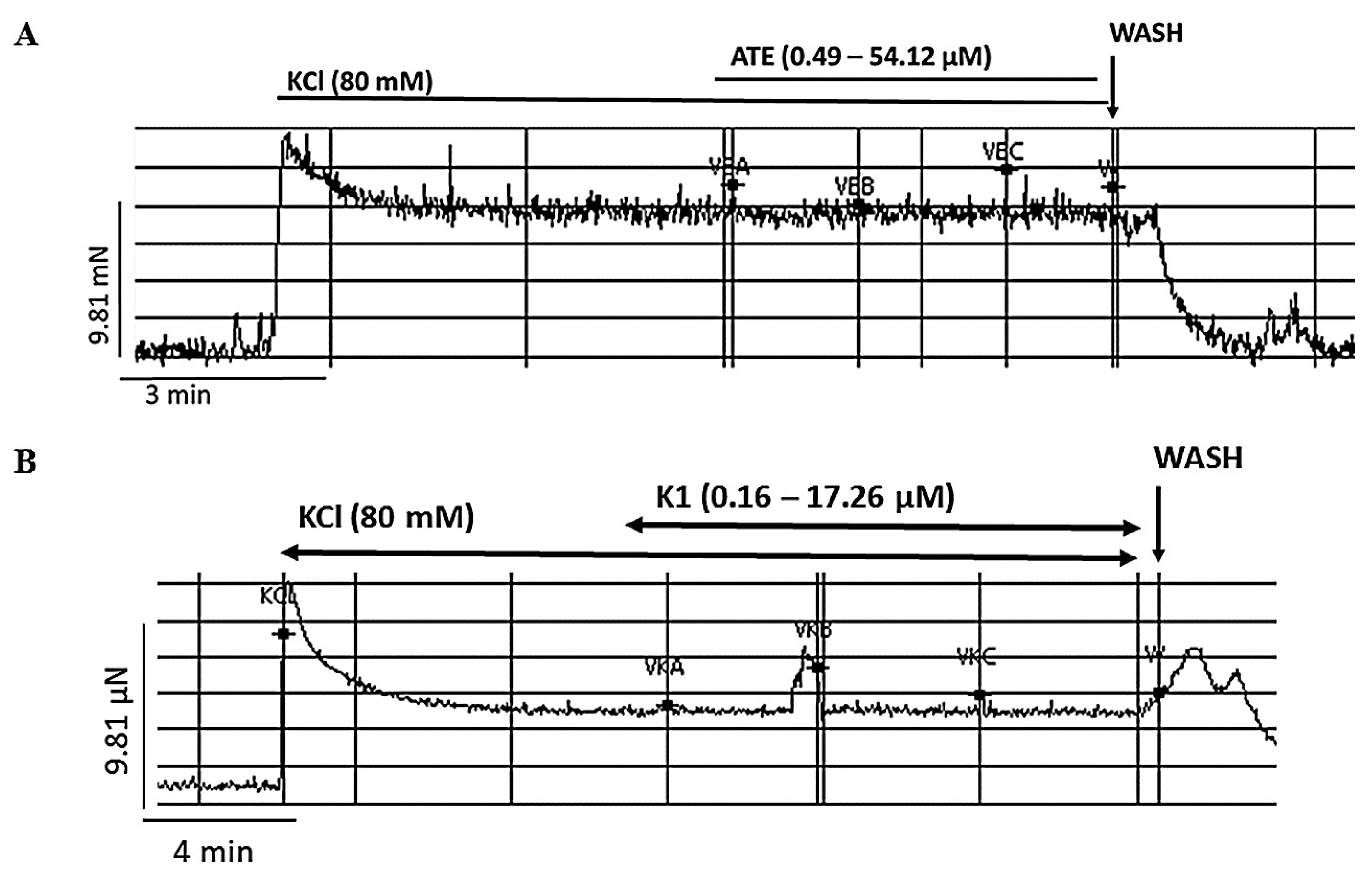
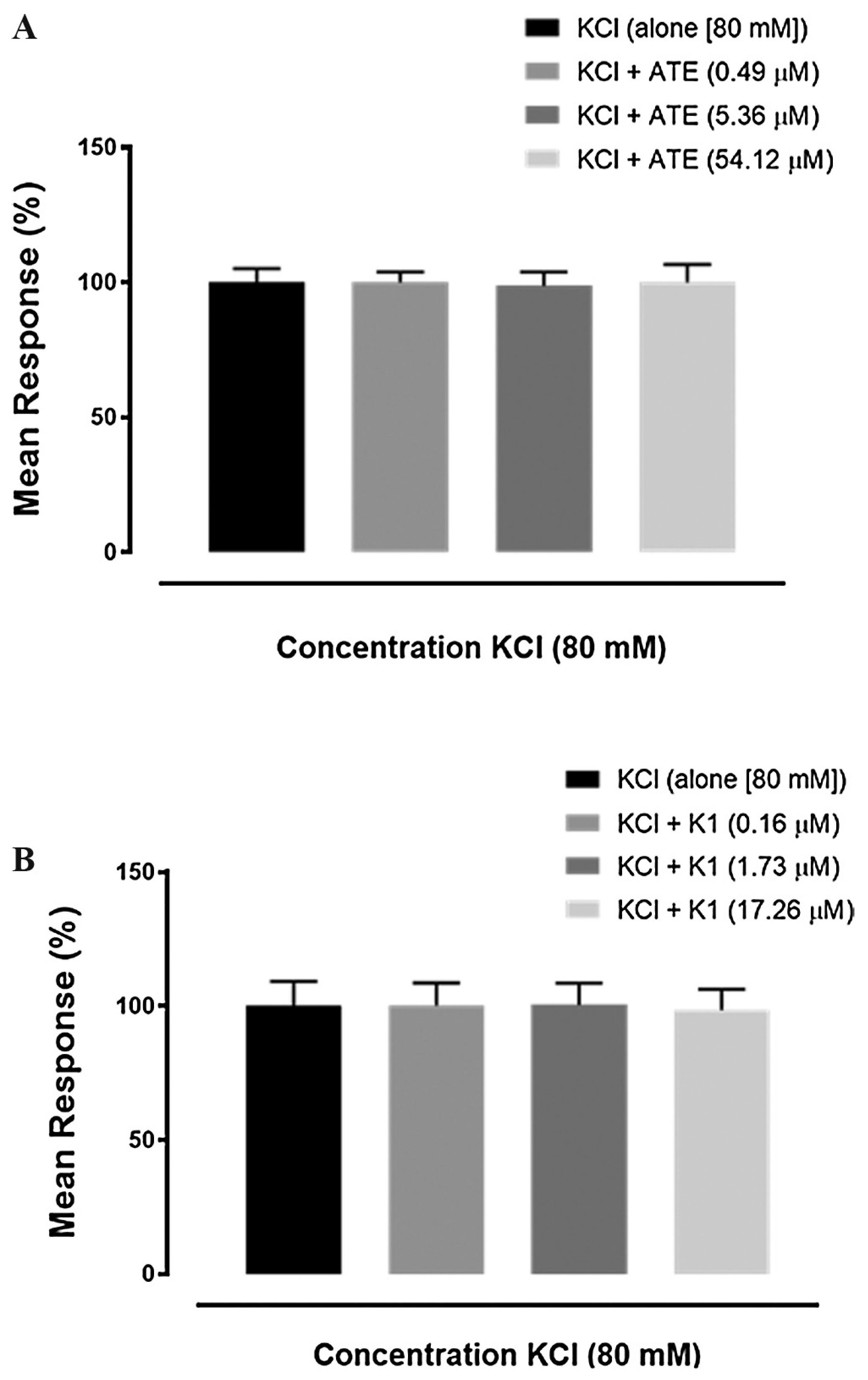
In order to investigate the effect of the drugs on the depolarized uterine tissue, the effect of a concentrated solution of KCl was examined. On addition of ATE (0.49–54.12 μM) or K1 (0.16–17.26 μM) on uterine contraction produced by high concentration of KCl (80 mM) no change was observed on the tonic contraction induced by KCl (Fig. 5A and B respectively). This is explicitly analyzed in bar plots which showed no significant change of KCl-induced response in the presence of either drugs (Fig. 6).

3.4. Effects of ATE on the concentrations of reproductive hormones, on mice body weight and weight of uterus

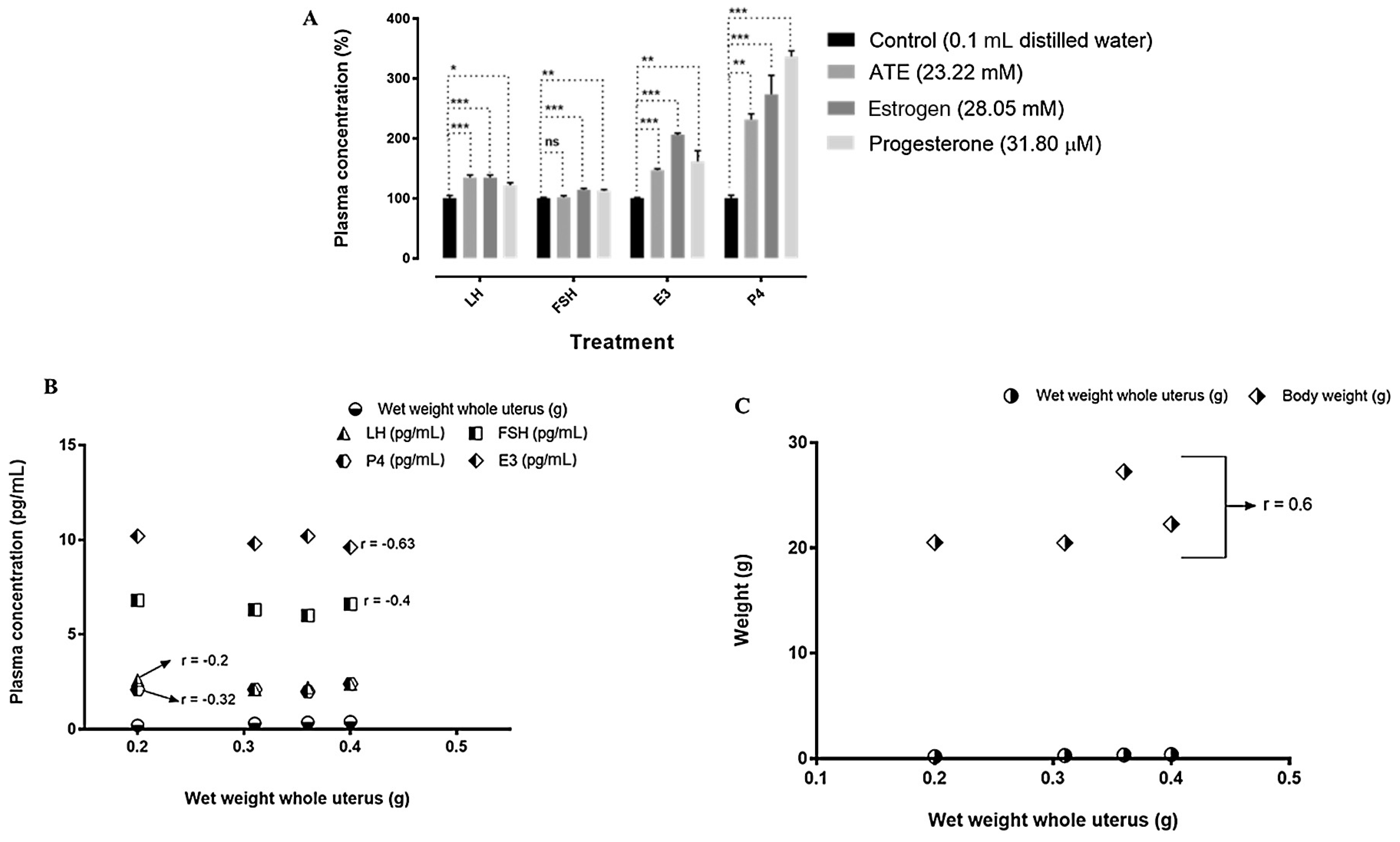
After 6 days daily treatment of ATE, significant increase in the plasma levels of luteinizing hormone (LH) (P < 0.001) estradiol (E3) (P < 0.001) and progesterone (P4) (P < 0.01) was observed compared to the respective controls (Fig. 7A). No significant effect was observed in the levels of follicle stimulating hormone on treatment with ATE (Fig. 7A). Estrogen and progesterone significantly increased (P < 0.001) levels of LH, FSH, E3 and P4 compared to control groups (Fig. 7A). On oral treatment with ATE (10 mg/kg) for 6 days (which spans the duration of the pre-determined estrus cycle), there was no significant change in animal body weights compared to the weights of the animals prior to drug administration in all treatment groups (Table 1). On day 7, after treatment had ended, the wet weights of the uterus were recorded (Table 2). However since it was impossible to determine the weights of the uterus prior to drug administration, a proper comparison could not be made. To make up for this, a correlation analysis was used to determine if changes observed were due to ATE treatment by checking for correlations between different parameters from animals that had undergone treatment under the same conditions. Correlation analyses showed an inverse relationship between the effect of ATE on uterine wet weights and estradiol plasma levels (r = −0.63), FSH levels (r = −0.4), and LH levels (r = −0.2) (Fig. 7B). However, a positive relationship was observed between the effect of ATE on uterine wet weights and plasma progesterone levels (Fig. 7B). Correlation analysis also showed a positive correlation between the weights of uterus and body weights of the animals on treatment with ATE (10 mg/kg) (Fig. 7C).

3.5. Effect of ATE on reproductive tissues

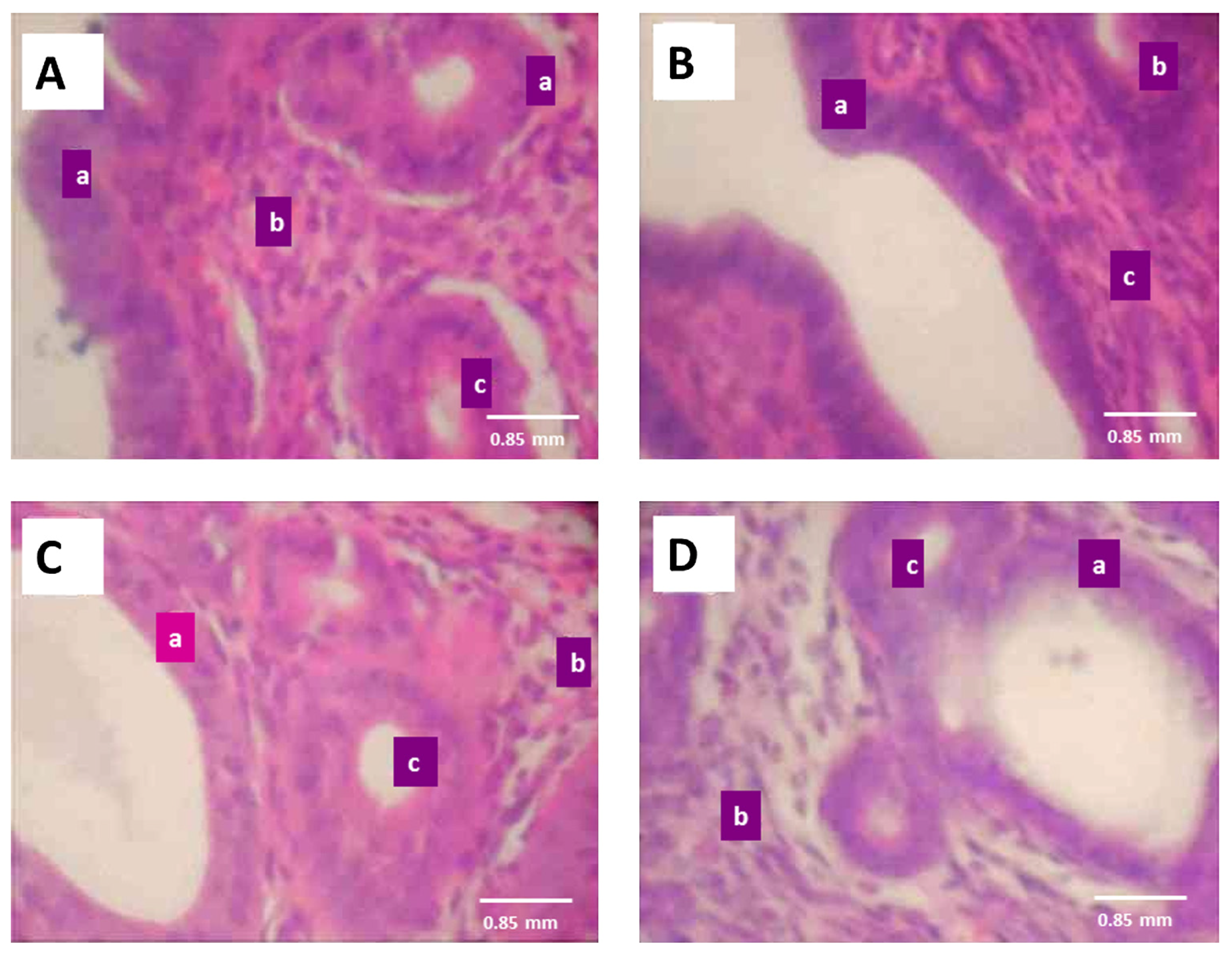
Morphological examinations revealed hypertrophy of the cervix on treatment with ATE compared with the control (Fig. 8). Hyperplasia of the uterine glands was also observed after treatment with ATE compared to the control but in the presence of progesterone and estrogen an increase in the number and size of uterine glands were observed when compared with the control under same conditions (Fig. 9).

4. Discussion

The uterus exhibits phasic contractions with varying and intermittent degrees of amplitude and frequency [32] which is primarily intertwined with calcium regulation [33]. Though the full complement of mechanisms involved in the generation of spontaneous uterine contractions besides calcium is largely unknown, a recent study suggested a strong involvement of the oxytocin receptor in the generation of spontaneous uterine contractions [34]. It would therefore seem that ATE may either have a direct effect on oxytocin receptors or may be involved in the direct release of calcium ions as these actions would increase spontaneous uterine contractility. Similarly, the inhibition observed in the presence of K1 suggests a reverse mechanism to that proposed for ATE, where K1 would either antagonize oxytocin receptors, or inhibit calcium release or directly block voltage–gated calcium channels which also plays a role in regulation of uterine contractility [35].
When ATE and OT were applied to the uterus, the force produced by OT was mildly reduced, which seemed in opposition to the effect observed on spontaneous uterine contraction. Similarly, K1 which had inhibited spontaneous contractions intriguingly appeared to augment the force of OT-induced contractions. This is indeed an intriguing outcome and one that lacks detailed and factual explanation at the moment but it would however seem that ATE and K1 have direct receptor interactions possibly with oxytocin, which further studies such as receptor ligand-binding and oxytocin antagonists-binding assays may elucidate. Nevertheless, it is also important to note that OT acts by the combination of a myriad of signaling events [36], it may well be therefore that ATE or K1 may interact with one or more of the processes involved in the action of OT which may have led to the interesting pattern noticed in this study. It has also been proposed that reduced uterine response to agonist treatment may result from a loss of phasic contractions due to a summation of the maximum contractile tension achievable, total occupancy of agonist receptor, or even a depletion in the number of agonist receptors [37,38,39]. These are a common phenomenon with the OT receptor [37]. In addition, varying patterns of action on either amplitude or frequency of OT-induced contractions were observed. The presence of an endogenous pacemaker in uterine tissues has been reported [37] and increased communication within uterine cells as a result of activation of the gap junction assembly has been reported to specifically increase the amplitude of uterine contractions [37]. Therefore activity on selected uterine cellular network and/or uterine pacemaker cells may affect cellular communication such that one parameter is affected separately from the other. It is also suggested that future studies.
Neither ATE nor K1 had any effect on tonic contractions induced by concentrated KCl solution. High KCl has been reported to depolarize uterine smooth muscle cells by opening of L-type Ca2+ channels which subsequently increases intracellular calcium ion concentration [32,40]. This infers that these compounds do not interact with L-type Ca2+ channels and therefore the receptor interaction stands as a greater possibility to explain the mechanisms of ATE and K1.
In mammals, estrogens produced from the developing follicles stimulate endometrial growth, and progesterone is responsible for converting the estrogen-primed endometrium into a receptive state [41]. The endometrium is the site of implantation in the non-pregnant uterus and fetal development in the pregnant uterus. Preparation for these important biological events relies primarily on progesterone, which takes the estrogen-primed endometrium toward a state of receptivity. It is the balance between estrogen and progesterone that maintains the endometrium in a state of health and provides the synchronous timing necessary for a successful implantation to occur [41]. Progesterone, the natural hormone produced by the corpus luteum and other steroid-secreting glands, is endowed with anti-osteogenic action and has a fundamental role in the initiation and maintenance of pregnancy and in the regulation of gonadotropin secretion [42]. That ATE was able to affect LH may suggest a central activity however the increase in estrogen (E3) and progesterone (P4) levels observed on treatment with ATE suggests a direct action on the ovaries as well [43] which may also have contributed to the levels of follicle stimulating hormone (FSH) being unaffected possibly due to a feedback mechanism [44]. A recent study showed ATE to be an estrogen receptor (ER) ligand and as such can be referred to as a phytoestrogen [45]. This may explain the effect observed on LH and on E3 levels but does not explain the rise in P4 levels. However a correlation between increased estrogen levels and increased progesterone receptors has been reported [46]. A previous study described the involvement of P4 in the reduction of uterine contractility [47]. This may account for the opposing effect observed with ATE in this study. Contractility studies such as these have also been shown to be useful in fertilization process and sperm transport [48] and therefore suggests a relevance for ATE and possibly K1 in the fertilization process. LH and FSH belong to the class of gonadotrophins and these are part of a neuronal network that regulates mammalian fertility [49]. A rise in plasma E3 results in LH surge from the anterior pituitary gland which induces ovulation [49]. In females, LH is involved in stimulating ovulation and the conversion of the ovulated ovarian follicle into a corpus luteum [50] while FSH stimulate the growth of ovarian follicles [50]. These gonadotrophins together with the sex hormones, E3 and P4 work in synchrony to modulate mammalian fertility and reproductive processes in general. The surge in LH, E3 and P4 in the presence of ATE therefore suggests a modulatory role for ATE or other fat soluble vitamins in mammalian fertility and possibly an indirect involvement in the control of the gonadotropins. Further research is suggested to clarify the exact roles played by ATE in modulation of these hormones.
ATE was also shown to have no effect on the weight of the animals. That a positive correlation was observed between the body weights and the uterine weights would therefore imply that the weights of the uterus were not significantly increased by ATE treatment and may also explain the inverse relationship observed between the weights of the uterus and the estradiol plasma levels. The estrogen type 17α-estradiol has been reported to show weak hypertrophic activity compared to other estrogen types which may again account for the effect observed in this study [51]. Mild cervical hypertrophy observed with ATE suggests mild osteogenic effect, similarly mild hyperplasia of the uterine glands in the presence of ATE also suggests a mild osteogenic effect and therefore a mild osteogenic nature for ATE. The uterus in both rodents and humans undergoes cyclical changes of growth and degeneration. Generally, the ovarian steroid hormones induce a number of physiological and biochemical changes on female reproductive organs that depend on these hormones [52,53]. ATE was observed to induce enlargement of the uterine endometrial glands compared to the control. A similar enlargement but with an accompanied increase in number of glands was also observed in the presence of progesterone and estrogen. During a normal menstrual cycle, the human endometrium undergoes series of proliferation, differentiation and degeneration cycles in response to steroid hormone concentrations variations. E3 has been known to induce an increase in the epithelial and stromal endometrial cells during the preovulatory proliferative phase and, post-ovulation, whereas progesterone appears to be more involved in glandular differentiation and glycogenesis [54]. An increase in the number of P4 receptors (PR) occurs in both the epithelial and stromal layers of the endometrium during the proliferative phase and it remains highly elevated during the early secretory phase. In the mid- to late secretory phase however, PR expression declines [55,56]. The persistence of PR during the secretory phase of the menstrual cycle suggests a constitutive expression of PR and emphasizes the continuous requirement for progesterone necessary for support of further tissue growth and development [55,56]. This observation also suggests a mitogenic and constitutive PR expression on basalis epithelial glands needed for tissue regeneration during menstrual reconstruction [57]. It would therefore appear that P4 has a dominant albeit constitutive role in uterine growth and regulation. This is supported by a recent study which reported that estrogen alone is not a mitogen for uterine growth [58]. It has also been shown that P4 activity via PR increases tissue volume through cell proliferation and extracellular matrix (ECM) accumulation [58]. This again supports that while E3 lacks mitogenic effect it is nonetheless essential for the growth and maintenance of uterine tissues as it also sensitizes uterine cells to P4 by inducing PR [58]. The requirement for P4 in uterine growth and glandular development may also support the role of P4 as a hormone of pregnancy required in all mammals for maternal support of conceptus (embryo/fetus and associated membranes) survival and development [59]. From the foregoing, administration of P4 or P4-like drugs can therefore be used to induce suitable environments for implantation and consequently improve fertility in the non-pregnant uterus but may promote maintenance of pregnancy in the pregnant uterus. ATE was observed in this study to act similar to progesterone and less so like E3 and may therefore be useful in promoting fertility in the non-pregnant uterus. Studies on the uterine glands of knockout ewes showed that the glands and their secretions which are supported by P4 are required for peri-implantation conceptus survival and growth. Stimulation of glandular epithelial proliferation and production of secretory proteins, during mid-pregnancy all act to induce growth hormone secretion that can also act on a progestinized uterus to stimulate glandular epithelial hypertrophy and increased glandular function and consequently an increased secretory function [59]. This further supports the role of P4 in the glandular and secretory activities of the myometrium and may explain the effect of ATE and progesterone on the endometrial glands observed in this study.
The cervix is the lower portion of the uterus and consists of the ectocervix and the endocervix layers. Prior to ovulation, estrogen levels rise resulting in an enlarged and softened cervix which promotes dilatation of the external os and assists in easier sperm passage to the uterus. However at the end of ovulation, the presence of progesterone will initiate hardening and closure of the cervix and also promote secretion of a thick milk-like mucus which plugs the cervix, and prevents passage of bacteria and/or sperm to the uterus [54,60]. Similar cervical changes also occur during parturition where changes in the connective tissue of the subepithelium and the muscular region are useful in cervical ripening [61]. A decrease in circulating progesterone (progesterone withdrawal) largely coordinates interaction between the cervix and the uterus at term [62]. Collagen remodeling also necessary for cervical ripening has been associated with a decrease in PR [63]. P4 also ensures that the cervix is firm and remains closed to hold the fetus till term [63]. The hypertrophy of the cervix observed in this study on administration of ATE appears similar to the effect of P4 on the cervix. This also supports a P4-like effect of ATE and possibly a role for ATE in controlling or improving fertility.

5. Conclusion

Conclusively, alpha-tocopherol and phylloquinone have been shown in this study to have direct effects on uterine contractility and to also exert modulatory effects on oxytocin. Alpha tocopherol exhibits inhibitory activities on OT-induced uterine contractility while phylloquinone exhibits more of stimulatory effects. In addition, alpha-tocopherol increased the levels of some of the reproductive hormones and may therefore have direct effects on the ovaries. Alpha-tocopherol has also been shown in this study to affect uterine and cervical tissue modeling, though exact mechanisms are unknown at this stage, the effects appear similar to those of progesterone which supports the use of ATE in promoting fertility. This study therefore reports for the first time a direct involvement of alpha-tocopherol and phylloquinone which are fat soluble vitamins with uterine contractility and may therefore be useful targets in the discovery of drugs for the management of female reproductive health pathologies.

Funding

This research did not receive any specific grant from funding agencies in the public, commercial, or not-for-profit sectors.

Conflict of Interest

The authors declare no conflict of interest.

Acknowledgments

The authors would like to acknowledge Mr. Osas Jim Eghianruwa for his assistance in blood collection during this research and Pharm. Osamuyi Uwumarongie for his assistance with the feeding tube.

R E F E R E N C E S

  1. Okuda, K. Fat soluble vitamins. Nippon Rinsho Jpn J Clin Med 1993, 51, 847–50. [Google Scholar] [CrossRef]
  2. McCollum, EV; Davis, M. The necessity of certain lipins in the diet during growth. J Biol Chem 1913, 15, 167–75. [Google Scholar] [CrossRef]
  3. Evans, HM; Bishop, KS. On the existence of a hitherto unrecognized dietary factor essential for reproduction. Science 1922, 56, 650–1. [Google Scholar] [CrossRef] [PubMed]
  4. Mulligan, ML; Felton, SK; Riek, AE; Bernal-Mizrachi, C. Implications of vitamin D deficiency in pregnancy and lactation. Am J Obstet Gynecol 2010, 202, 429.e1. [Google Scholar] [CrossRef] [PubMed]
  5. Beulens, JWJ; Booth, SL; van den Heuvel, EGHM; Stoecklin, E; Baka, A; Vermeer, C. The role of menaquinones (vitamin K2) in human health. Br J Nutr 2013, 110, 1357–68. [Google Scholar] [CrossRef] [PubMed]
  6. Kiyose, C; Muramatsu, R; Kameyama, Y; Ueda, T; Igarashi, O. Biodiscrimination of alpha-tocopherol stereoisomers in humans after oral administration. Am J Clin Nutr 1997, 65, 785–9. [Google Scholar] [CrossRef] [PubMed]
  7. El-Ganzoury, MM; El-Farrash, RA; Saad, AA; Ali, MS; El-Bhbiti, AR; Selem, AM. Antenatal administration of vitamin K1: relationship to vitamin K-dependent coagulation factors and incidence rate of periventricular–intraventricular hemorrhage in preterm infants; Egyptian randomized controlled trial. J Matern Fetal Neonatal Med 2014, 27, 816–20. [Google Scholar] [CrossRef] [PubMed]
  8. Traber, MG; Sies, H. Vitamin E in humans: demand and delivery. Annu Rev Nutr 1996, 16, 321–47. [Google Scholar] [CrossRef] [PubMed]
  9. Wray, S. Insights into the uterus. Exp Physiol 2007, 92, 621–31. [Google Scholar] [CrossRef] [PubMed]
  10. Wray, S. Uterine contraction and physiological mechanisms of modulation. Am J Physiol: Cell Physiol 1993, 264, C1–8. [Google Scholar] [CrossRef] [PubMed]
  11. Burdyga, T; Wray, S; Noble, K. In situ calcium signaling: no calcium sparks detected in rat myometrium. Ann N Y Acad Sci 2007, 1101, 85–96. [Google Scholar] [CrossRef] [PubMed]
  12. Togashi, K. Uterine contractility evaluated on cine magnetic resonance imaging. Ann N Y Acad Sci 2007, 1101, 62–71. [Google Scholar] [CrossRef] [PubMed]
  13. Bulletti, C; Ziegler, DD; Polli, V; Diotallevi, L; Ferro, ED; Flamigni, C. Uterine contractility during the menstrual cycle. Hum Reprod 2000, 15, 81–9. [Google Scholar] [CrossRef] [PubMed]
  14. Wray, S; Noble, K. Sex hormones and excitation–contraction coupling in the uterus: the effects of oestrous and hormones. J Neuroendocrinol 2008, 20, 451–61. [Google Scholar] [CrossRef] [PubMed]
  15. Lyons, EA; Taylor, PJ; Zheng, XH; Ballard, G; Levi, CS; Kredentser, JV. Characterization of subendometrial myometrial contractions throughout the menstrual cycle in normal fertile women. Fertil Steril 1991, 55, 771–4. [Google Scholar] [CrossRef]
  16. Kunz, G; Leyendecker, G. Uterine peristaltic activity during the menstrual cycle: characterization, regulation, function and dysfunction. Reprod Biomed Online 2002, 4 Suppl 3, 5–9. [Google Scholar] [CrossRef]
  17. De Vries, K; Lyons, EA; Ballard, G; Levi, CS; Lindsay, DJ. Contractions of the inner third of the myometrium. Am J Obstet Gynecol 1990, 162, 679–82. [Google Scholar] [CrossRef]
  18. Arrowsmith, S; Kendrick, A; Wray, S. Drugs acting on the pregnant uterus. Obstet Gynaecol Reprod Med 2010, 20, 241–7. [Google Scholar] [CrossRef] [PubMed]
  19. Bafor, EE; Lim, CV; Rowan, EG; Edrada-Ebel, R. The leaves of Ficus exasperata Vahl (Moraceae) generates uterine active chemical constituents. J Ethnopharmacol 2013, 145, 803–12. [Google Scholar] [CrossRef] [PubMed]
  20. Rumbold, A; Crowther, CA. Vitamin E supplementation in pregnancy. Cochrane Database Syst Rev 2005. [Google Scholar] [CrossRef]
  21. Boskovic, R; Gargaun, L; Oren, D; Djulus, J; Koren, G. Pregnancy outcome following high doses of Vitamin E supplementation. Reprod Toxicol 2005, 20, 85–8. [Google Scholar] [CrossRef] [PubMed]
  22. Cederberg, J; Simán, CM; Eriksson, UJ. Combined treatment with vitamin E and vitamin C decreases oxidative stress and improves fetal outcome in experimental diabetic pregnancy. Pediatr Res 2001, 49, 755–62. [Google Scholar] [CrossRef] [PubMed]
  23. Pruthi, S; Wahner-Roedler, DL; Torkelson, CJ; Cha, SS; Thicke, LS; Hazelton, JH; et al. Vitamin E and evening primrose oil for management of cyclical mastalgia: a randomized pilot study. Altern Med Rev 2010, 15, 59–67. [Google Scholar] [PubMed]
  24. Lanza, E; Forman, MR; Johnson, EJ; Muesing, RA; Graubard, BI; Beecher, GR. alpha-Tocopherol concentrations in plasma but not in lipoproteins fluctuate during the menstrual cycle in healthy premenopausal women. J Nutr 1998, 128, 1150–5. [Google Scholar] [CrossRef] [PubMed]
  25. Elvis-Offiah, U; Bafor, EE. Evaluation of the effects of oxytocin and diethylstilboestrol on mouse oestrous cycle using an index. J Med Biomed Res 2014, 13, 6–16. [Google Scholar]
  26. Green, EL. Biology of the laboratory mouse; The Blakiston Company: Philadelphia, 1966. [Google Scholar] [CrossRef]
  27. Pritchett, KR; Taft, RA. Reproductive Biology of the Laboratory Mouse The mouse in biomedical research: normative biology husbandry and models. 2007; vol. 3, pp. 91–121. [Google Scholar] [CrossRef]
  28. Champlin, AK; Dorr, DL; Gates, AH. Determining the stage of the estrous cycle in the mouse by the appearance of the vagina. Biol Reprod 1973, 8, 491–4. [Google Scholar] [CrossRef] [PubMed]
  29. Bafor, EE; Eyohan, SE; Omoruyi, O; Elvis-Offiah, UB; Ayinde, BA; Eze, GI; et al. Preliminary endocrinological, histological and haematological investigation of Alchornea laxiflora (Euphorbiaceae) leaf extract effects on the ovary, uterus and cervix of mouse models. J Sci Pract Pharm 2016, 2, 55–63. [Google Scholar]
  30. Miles, LE; Hales, C. Labelled antibodies and immunological assay systems. Nature 1968, 219, 186–9. [Google Scholar] [CrossRef] [PubMed]
  31. Lequin, RM. Enzyme immunoassay (EIA)/enzyme-linked immunosorbent assay (ELISA). Clin Chem 2005, 51, 2415–8. [Google Scholar] [CrossRef] [PubMed]
  32. Alotaibi, M. The physiological mechanism of uterine contraction with emphasis on calcium ion. Calcium Signal 2014, 1, 1168–76. [Google Scholar]
  33. Aguilar, HN; Mitchell, BF. Physiological pathways and molecular mechanisms regulating uterine contractility. Hum Reprod Update 2010, 16, 725–44. [Google Scholar] [CrossRef] [PubMed]
  34. Wilson, RJ; Allen, MJ; Nandi, M; Giles, H; Thornton, S. Spontaneous contractions of myometrium from humans, non-human primate and rodents are sensitive to selective oxytocin receptor antagonism in vitro. Br J Obstet Gynaecol 2001, 108, 960–6. [Google Scholar] [CrossRef]
  35. Wray, S; Kupittayanant, S; Shmygol, A; Smith, RD; Burdyga, T. The physiological basis of uterine contractility: a short review. Exp Physiol 2001, 86, 239–46. [Google Scholar] [CrossRef] [PubMed]
  36. Arrowsmith, S; Wray, S. Oxytocin: its mechanism of action and receptor signalling in the myometrium. J Neuroendocrinol 2014, 26, 356–69. [Google Scholar] [CrossRef] [PubMed]
  37. Mackler, AM; Ducsay, CA; Veldhuis, JD; Yellon, SM. Maturation of spontaneous and agonist-induced uterine contractions in the peripartum mouse uterus. Biol Reprod 1999, 61, 873–8. [Google Scholar] [CrossRef] [PubMed]
  38. Higuchi, T; Honda, K; Fukuoka, T; Negoro, H; Wakabayashi, K. Release of oxytocin during suckling and parturition in the rat. J Endocrinol 1985, 105, 339–46. [Google Scholar] [CrossRef] [PubMed]
  39. Chan, WY; Chen, DL. Myometrial oxytocin receptors and prostaglandin in the parturition process in the rat. Biol Reprod 1992, 46, 58–64. [Google Scholar] [CrossRef] [PubMed]
  40. Bolton, TB; Prestwich, SA; Zholos, AV; Gordienko, DV. Excitation–contraction coupling in gastrointestinal and other smooth muscles. Annu Rev Physiol 1999, 61, 85–115. [Google Scholar] [CrossRef] [PubMed]
  41. Croxatto, HB; Diaz, S. The place of progesterone in human contraception. J Steroid Biochem 1987, 27, 991–4. [Google Scholar] [CrossRef]
  42. Szekeres-Bartho, J; Halasz, M; Palkovics, T. Progesterone in pregnancy; receptor–ligand interaction and signaling pathways. J Reprod Immunol 2009, 83, 60–4. [Google Scholar] [CrossRef] [PubMed]
  43. Groom, GV; Griffiths, K. Effect of the anti-oestrogen tamoxifen on plasma levels of luteinizing hormone, follicle-stimulating hormone, prolactin, oestradiol and progesterone in normal pre-menopausal women. J Endocrinol 1976, 70, 421–8. [Google Scholar] [CrossRef] [PubMed]
  44. Rivier, C; Rivier, J; Vale, W. Inhibin-mediated feedback control of follicle-stimulating hormone secretion in the female rat. Science 1986, 234, 205–9. [Google Scholar] [CrossRef] [PubMed]
  45. Khallouki, F; de Medina, P; Caze-Subra, S; Bystricky, K; Balaguer, P; Poirot, M; et al. Molecular and biochemical analysis of the estrogenic and proliferative properties of vitamin E compounds. Front Oncol 2015, 5, 287. [Google Scholar] [CrossRef] [PubMed]
  46. Janne, O; Kontula, K; Luukkainen, T; Vihko, R. Oestrogen-induced progesterone receptor in human uterus. J Steroid Biochem 1975, 6, 501–9. [Google Scholar] [CrossRef]
  47. Künzel, J; Geisler, K; Maltaris, T; Müller, A; Hoffmann, I; Schneider, H; et al. Effects of interactions between progesterone and prostaglandin on uterine contractility in a perfused swine uterus model. In Vivo (Brooklyn) 2014, 28, 467–76. [Google Scholar]
  48. Dittrich, R; Henning, J; Maltaris, T; Hoffmann, I; Oppelt, PG; Cupisti, S; et al. Extracorporeal perfusion of the swine uterus: effect of human seminal plasma. Andrologia 2012, 44, 543–9. [Google Scholar] [CrossRef] [PubMed]
  49. Clarkson, J; Herbison, AE. Oestrogen, kisspeptin, GPR54 and the pre-ovulatory luteinising hormone surge. J Neuroendocrinol 2009, 21, 305–11. [Google Scholar] [CrossRef] [PubMed]
  50. Fox, SI. Human physiology, 12th ed.; McGraw Hill: New York, 2011. [Google Scholar]
  51. Perusquía, M; Navarrete, E. Evidence that 17alpha-estradiol is biologically active in the uterine tissue: antiuterotonic and antiuterotrophic action. Reprod Biol Endocrinol 2005, 3, 30. [Google Scholar] [CrossRef]
  52. Fischer, AH; Jacobson, KA; Rose, J; Zeller, R. Preparation of slides and coverslips for microscopy. Cold Spring Harbor Protoc 2008, 3. [Google Scholar] [CrossRef] [PubMed]
  53. Ruegg, MA; Meinen, S. Histopathology in hematoxylin & eosin stained muscle sections. Neuromuscul Netw 2012, 1–9. [Google Scholar]
  54. Graham, JD; Roman, SD; McGowan, E; Sutherland, RL; Clarke, CL. Preferential stimulation of human progesterone receptor B expression by estrogen in T-47D human breast cancer cells. J Biol Chem 1995, 270, 30693–700. [Google Scholar] [CrossRef] [PubMed]
  55. Fathalla, MF. Incessant ovulation – a factor in ovarian neoplasia? Lancet 1971, 2, 163. [Google Scholar] [CrossRef]
  56. Casagrande, JT; Pike, MC; Ross, RK; Louie, EW; Roy, S; Henderson, BE. “Incessant ovulation” and ovarian cancer. Lancet 1979, 314, 170–3. [Google Scholar] [CrossRef]
  57. Mote, PA; Balleine, RL; McGowan, EM; Clarke, CL. Heterogeneity of progesterone receptors A and B expression in human endometrial glands and stroma. Hum Reprod 2000, 15 Suppl 3, 48–56. [Google Scholar] [CrossRef] [PubMed]
  58. Ishikawa, H; Ishi, K; Ann Serna, V; Kakazu, R; Bulun, SE; Kurita, T. Progesterone is essential for maintenance and growth of uterine leiomyoma. Endocrinology 2010, 151, 2433–42. [Google Scholar] [CrossRef] [PubMed]
  59. Spencer, TE; Bazer, FW. Biology of progesterone action during pregnancy recognition and maintenance of pregnancy. Front Biosci 2002, 7, d1879–98. [Google Scholar] [CrossRef] [PubMed]
  60. Bergeron, C; Ferenczy, A; Toft, DO; Shyamala, G. Immunocytochemical study of progesterone receptors in hyperplastic and neoplastic endometrial tissues. Cancer Res 1988, 48, 6132–6. [Google Scholar] [PubMed]
  61. Huszar, GB; Michael, PW. Relationship between myometrial and cervical functions in pregnancy and labor. Semin Perinatol 1991, 15, 97. [Google Scholar] [PubMed]
  62. Challis, JRG; Lye, SJ. Parturition. In The physiology of reproduction; Knobil, E, Neill, JD, Eds.; Raven Press: New York, 1994; pp. 985–1031. [Google Scholar]
  63. Luque, EH; Ramos, JG; Rodriguez, Ha; Muñoz de Toro, MM. Dissociation in the control of cervical eosinophilic infiltration and collagenolysis at the end of pregnancy or after pseudopregnancy in ovariectomized steroid-treated rats. Biol Reprod 1996, 55, 1206–12. [Google Scholar] [CrossRef] [PubMed]
Fig. 1. Original recordings showing the effect of cumulative increases in concentration of ATE and K1 on spontaneous uterine contractions. Panel A shows recordings for ATE while panel B shows recordings for K1.
Medicina 53 00190 g001
Fig. 2. Concentration–response curves representative of the effect of ATE and K1 on the amplitude and frequency of spontaneous uterine contractions. A concentration-dependent increase in the amplitude was observed on ATE administration with only slight increases observed in the frequency (A and B). K1 on the hand concentration-dependently decreased the amplitude and frequency of spontaneous uterine contraction (C and D) (n = 4).
Medicina 53 00190 g002
Fig. 3. Original recordings showing the effect of cumulative increases in concentration of ATE and K1 on OT-induced uterine contractions. Panel A shows recordings for ATE while panel B shows recordings for K1.
Medicina 53 00190 g003
Fig. 4. Concentration–response plots showing the response of OT (11.82 nM) in the absence and presence of ATE (0.49–54.12 μM) and K1 (0.16–17.26 μM). ATE decreased the amplitude of OT-induced contraction with no distinct effect on the frequency. K1 exerted an initial decrease in amplitude of OT-induced contraction which appeared to have been attenuated as concentration increased. However, the frequency induced by OT was inhibited down to 76.15% by K1 (n = 5).
Medicina 53 00190 g004
Fig. 5. Original recordings showing the effect of cumulative increases in concentration of ATE and K1 on high KCl-induced (80 mM) uterine contractions. Panel A shows recordings for ATE while panel B shows recordings for K1.
Medicina 53 00190 g005
Fig. 6. Concentration–response plots showing the response of KCl (80 mM) in the absence and presence of ATE (0.49–54.12 μM) and K1 (0.16–17.26 μM). No significant change was observed in the presence of either ATE or K1 (n = 5).
Medicina 53 00190 g006
Fig. 7. Plots showing the effect of ATE on plasma levels of reproductive hormones and correlations with weights of animals and uterus. After 6 days administration to non-pregnant animals, ATE was observed to significantly increase plasma levels of luteinizing hormone (LH), estradiol (E3) and progesterone (P4) while producing no significant effect in the levels of follicle stimulating hormone (FSH) compared to the control (A). Correlation charts are shown in panels B and C. Inverse correlation relationships were observed between the weights of the uterus and plasma levels of LH, FSH and E3. However, a positive relationship was observed between plasma levels of P4 and the wet weights (B). A positive correlation was observed between the weights of the uterus and animal body weight (C). These changes were recorded based on treatment with ATE (10 mg/mL) (n = 4). Ns = not significant; *P < 0.05; **P < 0.001; ***P < 0.0001 compared to control.
Medicina 53 00190 g007
Fig. 8. Mouse uterine cervix (hematoxylin and eosin, 100×). Representative images of cervical tissue from mice after 6 days of ATE treatment: (A) control (0.9 mL of normal saline), a = ectocervix, b = stroma; (B) progesterone (0.01 mg/kg), a = normal ectocervix, b = stroma edema; (C) ATE (10 mg/kg), a = mild thickening of the ectocervix, b = mild stroma hypertrophy.
Medicina 53 00190 g008
Fig. 9. Mouse uterus (hematoxylin and eosin, 100×). Representative images of uterine tissue from mice after 6 days of ATE treatment: (A) control (0.9 mL of normal saline), a = endometrial lining, b = stroma, c = glands; (B) progesterone (0.01 mg/kg), a = normal endometrial lining, b = glands increased in number, c = increased blood vessel in the stroma; (C) estrogen (10 mg/kg), a = cystic glandular hyperplasia, b = plump stroma cell, c = glands which has increased in number; (D) ATE (10 mg/kg), a = cystic glandular hyperplasia, b = plump stroma cell, c = glands.
Medicina 53 00190 g009
Table 1. Weights of animals treated with ATE, estrogen and progesterone.
GroupsWeight, g
Day 0Day 7
Control (0.1 mL distilled water)21.00 ± 1.3021.00 ± 0.85
ATE (10 mg/kg)23.00 ± 1.70 23.00 ± 1.60
Estrogen (10 mg/kg)22.00 ± 0.9322.00 ± 1.30
Progesterone (0.01 mg/kg)24.00 ± 1.2024.00 ± 0.98
Values are presented in mean ± SEM; n = 4.
Table 2. Wet weights of whole uterus in control and treated groups.
GroupsWeight, g
Day 7
Control (0.1 mL distilled water)0.33 ± 0.07
ATE (10 mg/kg)0.32 ± 0.15
Estrogen (10 mg/kg)0.34 ± 0.02
Progesterone (0.01 mg/kg)0.26 ± 0.02
Values are presented in mean ± SEM; n = 4.