Article

MoWhi2 Mediates Mitophagy to Regulate Conidiation and Pathogenesis in Magnaporthe oryzae

1
State Key Lab of Rice Biology, China National Rice Research Institute, Hangzhou 311400, China
2
Hubei Key Lab of Plant Pathology, and College of Plant Science and Technology, Huazhong Agricultural University, Wuhan 430070, China
*
Authors to whom correspondence should be addressed.
Int. J. Mol. Sci. 2022, 23(10), 5311; https://doi.org/10.3390/ijms23105311
Submission received: 6 April 2022 / Revised: 5 May 2022 / Accepted: 5 May 2022 / Published: 10 May 2022

Abstract

:
Mitophagy refers to the specific process of degrading mitochondria, which is an important physiological process to maintain the balance of mitochondrial quantity and quality in cells. At present, the mechanisms of mitophagy in pathogenic fungi remain unclear. Magnaporthe oryzae (Syn. Pyricularia oryzae), the causal agent of rice blast disease, is responsible for the most serious disease of rice. In M. oryzae, mitophagy occurs in the foot cells and invasive hyphae to promote conidiation and infection. In this study, fluorescent observations and immunoblot analyses showed that general stress response protein MoWhi2 is required for mitophagy in M. oryzae. In addition, the activation of the autophagy, pexophagy and cytoplasm-to-vacuole targeting (CVT) pathway upon nitrogen starvation was determined using the GFP-MoATG8, GFP-SRL and MoAPE1-GFP strains and the ΔMowhi2 mutant in these backgrounds. The results indicated that MoWhi2 is specifically required for mitophagy in M. oryzae. Further studies showed that mitophagy in the foot cells and invasive hyphae of the ΔMowhi2 was interrupted, leading to reduced conidiation and virulence in the ΔMowhi2 mutant. Taken together, we found that MoWhi2 contributes to conidiation and invasive growth by regulating mitophagy in M. oryzae.

1. Introduction

In eukaryotes, mitochondria carry out oxidative metabolism and ultimately oxidize sugars, fats and amino acids to provide energy [1]. Mitochondria are also the main source of reactive oxygen species (ROS) in cells. However, multiple signals influence mitochondria and initiate mitochondrial dysfunction. Mitochondrial dysfunction is usually multifactorial and characterized by abnormal accumulation of ROS, leading to the peroxidation of mitochondrial DNA, protein and lipid [2]. Thus, dysfunctional mitochondria may disrupt intracellular cell homeostasis and be harmful to the cells [3]. Mitophagy is a process by which dysfunctional or excess mitochondria are selectively degraded by autophagy [4]. Upon nitrogen starvation or other stresses, mitophagy receptors recognize non-essential or dysfunctional mitochondria, then recruit core autophagic proteins and encapsulate mitochondria to degradation with autophagic vesicles [5,6]. To date, the mechanism of mitophagy has been deeply studied in mammals and yeast. However, research on plant pathogenic fungi is still in the exploratory stage and requires to further study.
In addition to mitophagy receptors and the core autophagic mechanism [7,8,9,10,11], it has been suggested that mitophagy requires the general stress response protein Whi2 (Whiskey 2) in yeast [12,13]. Whi2 was initially found to play a key role in inhibiting cell proliferation by sensing extracellular nutrient unavailability in yeast [14]. Subsequent studies showed that Whi2 interacts with phosphatase Psr1 to regulate the expression of a series of stress-responsive genes through dephosphorylation of translational factor Msn2 [15]. Recent studies have shown that Whi2 inactivates the cAMP/PKA signaling pathway by targeting Ras2 (GTPase) to vacuoles [12] and negatively regulates the activity of TOR signaling complex 1 (TORC1) under limited amino acid conditions [16]. It is worth noting that according to functional analysis of the Δfis1 mutant with a secondary mutation in WHI2, loss of Whi2 function has a stronger impact on mitophagy than autophagy (macroautophagy) in yeast [13,17]. In the plant pathogens Collectotrichum orbibulare and Magnaporthe oryzae, Whi2 plays an important role in pathogenesis by regulating TOR (target of rapamycin) signaling [18,19]. Whether Whi2 specifically links to mitophagy in plant pathogenic fungi is still unclear.
Rice blast is the most devastating plant disease caused by the filamentous pathogenic fungus M. oryzae, resulting in a loss of rice yield, accounting for 10–30% of total rice production. At present, cultivation of rice-blast-disease-resistant varieties in combination with chemical control is the most efficient way to control this disease. In M. oryzae, mitophagy was first observed in foot cells. Disruption of MoATG24, a sorting nexin related to yeast Snx4, led to disrupted mitophagy in foot cells and a decrease in conidiation. Further research found that mitophagy of invasive hyphae was blocked in the ΔMoatg24 mutant so that the invasive hyphae cannot spread in the host cells [20]. It is possible that MoAtg24-assisted mitophagy contributes to the transformation from biotrophy to necrotrophy in M. oryzae [20,21]. In addition, the mitochondrial fission machinery, including MoDNM1, MoFIS1 and MoMDV1, is also involved in mitophagy in M. oryzae. MoDnm1 is not only involved in mitophagy but also in pexophagy, which mediates invasive growth in the host. MoFis1 interacts with MoDnm1 through the connection of the scaffold protein MoMdv1 to participate in mitophagy in the form of a complex and regulates appressorium formation and virulence [22]. The mitochondrial fusion gene MoFZO1 is also required for mitophagy. Deletion of MoFZO1 resulted in a change in mitochondria morphology and reduced invasive hyphae expansion and virulence [21]. Moreover, transcription factor MoMsn2 participates in mitophagy by regulating the expression of MoAUH1 (3-methylglutaconyl-CoA hydratase), which affects growth, sporulation and pathogenicity [23]. The underlying mechanisms of mitophagy during development and virulence in M. oryzae still remain elusive and need to be further explored.
In a previous study, we found that MoWhi2 regulates appressorium formation via cAMP and MoTor signaling pathways in M. oryzae [19]. Here, we further demonstrate the function of MoWhi2 in mitophagy during conidiation and invasive hyphal growth. The results revealed that MoWhi2 is specifically involved in mitophagy (but not autophagy, pexophagy and the cytoplasm-to-vacuole targeting pathway) to regulate conidiation and invasive hyphae growth of M. oryzae.

2. Results

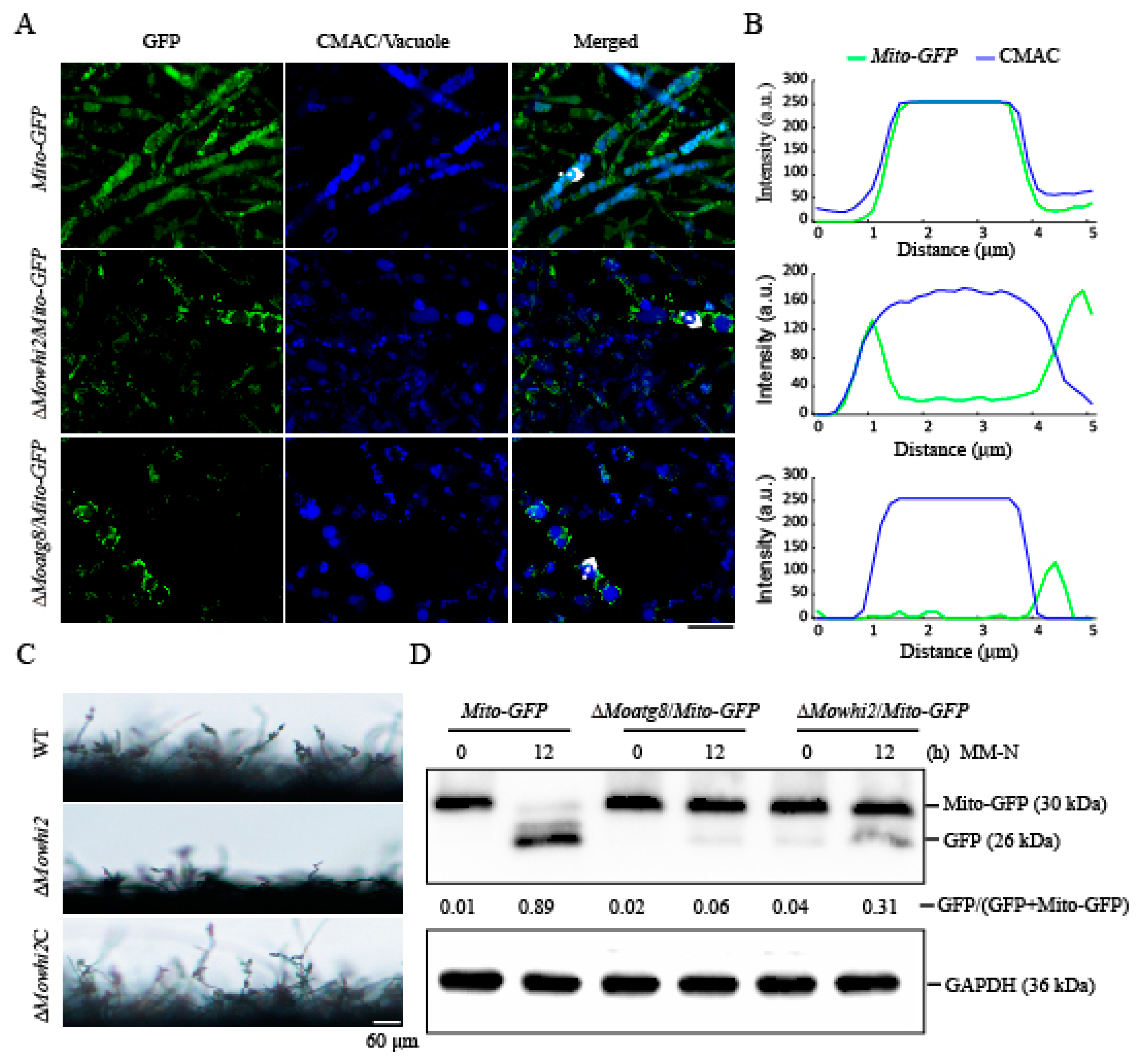
2.1. MoWHI2 Is Required for Mitophagy in M. oryzae

In M. oryzae, mitophagy occurs in the foot cells and invasive hyphae to promote conidiation and infection [20,21]. In a previous study, we found that the deletion of MoWHI2 not only led to abnormal appressorium formation but also highly reduced conidiation and invasive growth [19]. Thus, we speculated that MoWhi2 may participate in mitophagy as Whi2 in yeast [13]. To test this hypothesis, the MoWHI2 deletion mutant was generated in the Mito-GFP background, which is a well-used strain for monitoring mitophagy in M. oryzae [20,21,24]. The ΔMoatg8/Mito-GFP strain, which is defective for all types of autophagy, including mitophagy, served as a negative control. The resultant mutant ΔMowhi2/Mito-GFP, Mito-GFP and ΔMoatg8/Mito-GFP strains were grown in glycerol medium and transferred to nitrogen-starvation medium for 6 h to induce mitophagy. Then, the mycelia were stained with CMAC (7-amino-4-chloromethylcoumarin) dye to visualize the vacuoles and imaged with a confocal microscope. As shown in Figure 1A,B, the vacuolar localized GFP signal was evident in starved mycelia, indicating mitophagy was induced in the Mito-GFP strain. In contrast, similar to the negative control ΔMoatg8/Mito-GFP, the GFP signal could not be detected in the vacuoles in the ΔMowhi2/Mito-GFP strain, suggesting that MoWhi2 is involved in mitophagy. Similarly, microscopic observations showed that few conidiophores had differentiated in ΔMowhi2, revealing that the sporulation of the MoWHI2 mutant was significantly reduced (Figure 1C). To further confirm this result, a Western blot assay was performed with anti-GFP antibody to assess the degradation of fusion protein Mito-GFP. Consistent with the fluorescent observation in the ΔMoatg8/Mito-GFP strain, deletion of MoWHI2 resulted in a decreased degradation of Mito-GFP at indicated time points, suggesting that there are defects in mitophagy in the MoWHI2 deletion mutant (Figure 1D). Based on these results, we concluded that MoWHI2 is required for mitophagy in M. oryzae.

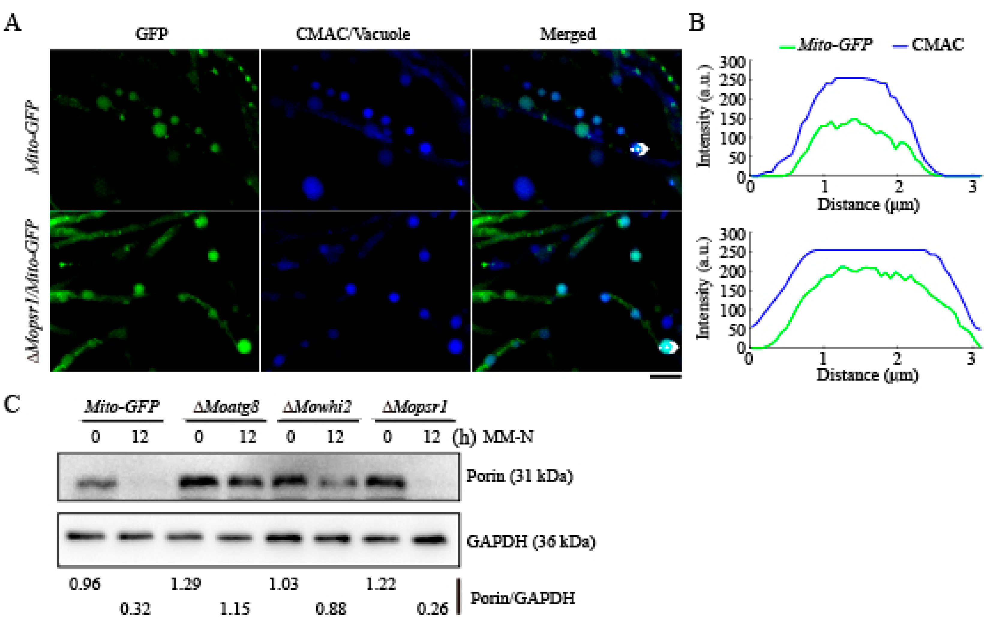
2.2. MoPsr1 Is Not Necessary for Mitophagy in M. oryzae

In our previous study, we found that MoWhi2 interacts with MoPsr1, and ΔMopsr1 showed similar defects in conidiation and invasive growth as the ΔMowhi2 mutant. It is possible that MoPsr1 participates in mitophagy as MoWhi2. To test this hypothesis, we constructed a ΔMopsr1 mutant in the Mito-GFP strain. The Mito-GFP, ΔMopsr1/Mito-GFP and ΔMoatg8/Mito-GFP strains were inoculated in CM medium for 2 days and then subjected to nitrogen starvation for 6 h. Confocal microscopic observation showed that the green fluorescence of Mito-GFP and ΔMopsr1/Mito-GFP strains overlapped with the CMAC-stained vacuole, indicating that parts of the mitochondria are delivered into vacuoles for degradation in the ΔMopsr1/Mito-GFP strain, as these in the Mito-GFP strain, upon nitrogen starvation (Figure 2A,B). These results suggest that MoPsr1 is not necessary for mitophagy. Furthermore, Western blot analyses were performed to reveal whether MoPsr1 is involved in mitophagy. The mitochondrial outer membrane protein Porin encoded by MGG_00968 was used as a marker to detect the occurrence of mitophagy [17]. With the same protein amount, the ratios of Porin to internal reference were calculated to indicate mitochondrial degradation with nitrogen starvation treatment in the ΔMopsr1/Mito-GFP, ΔMoatg8/Mito-GFP and ΔMowhi2/Mito-GFP strains. The results showed that upon nitrogen starvation, part of the mitochondria in the Mito-GFP and ΔMopsr1/Mito-GFP strains were degraded (Figure 2C). In contrast, the amount of Porin after induction was close to that before induction in the ΔMoatg8/Mito-GFP and ΔMowhi2/Mito-GFP strains (Figure 2B). Taken together, MoPsr1 is not necessary for mitophagy in M. oryzae.

2.3. Deletion of MoWHI2 Did Not Affect Autophagy (Aacroautophagy) upon Nitrogen Starvation

In yeast, disruption of WHI2 has no significant effects on autophagy [13,17]. To determine whether MoWhi2 is required for autophagy in M. oryzae, deletion mutants of MoWHI2 were generated in the GFP-MoATG8 strain, in which GFP-MoAtg8 is a commonly used marker of autophagy [25]. Under the autophagy-inductive condition, the colocalization of GFP-MoAtg8 with vacuoles was observed. As shown in the Figure 3A, the GFP fluorescence overlapped the CMAC-stained vacuoles in both GFP-MoATG8 and ΔMowhi2/GFP-MoATG8 strains (Figure 3A,B), suggesting that autophagy could be induced when MoWHI2 was disrupted. Furthermore, the number of autophagosomes increased in the GFP-MoATG8 and ΔMowhi2/GFP-MoATG8 strains, but there was no significant difference between GFP-MoATG8 and ΔMowhi2/GFP-MoATG8 in the SD-N (nitrogen starvation condition) medium (Figure 3C). In addition, the degradation of GFP-MoAtg8 fusion protein was detected by immunoblotting. The results demonstrated that the autophagy in mycelia of the ΔMowhi2/GFP-MoATG8 strain was comparable to that of the GFP-MoATG8 stain (Figure 3D). These results suggest that MoWhi2 is not required for autophagy in response to nitrogen starvation.

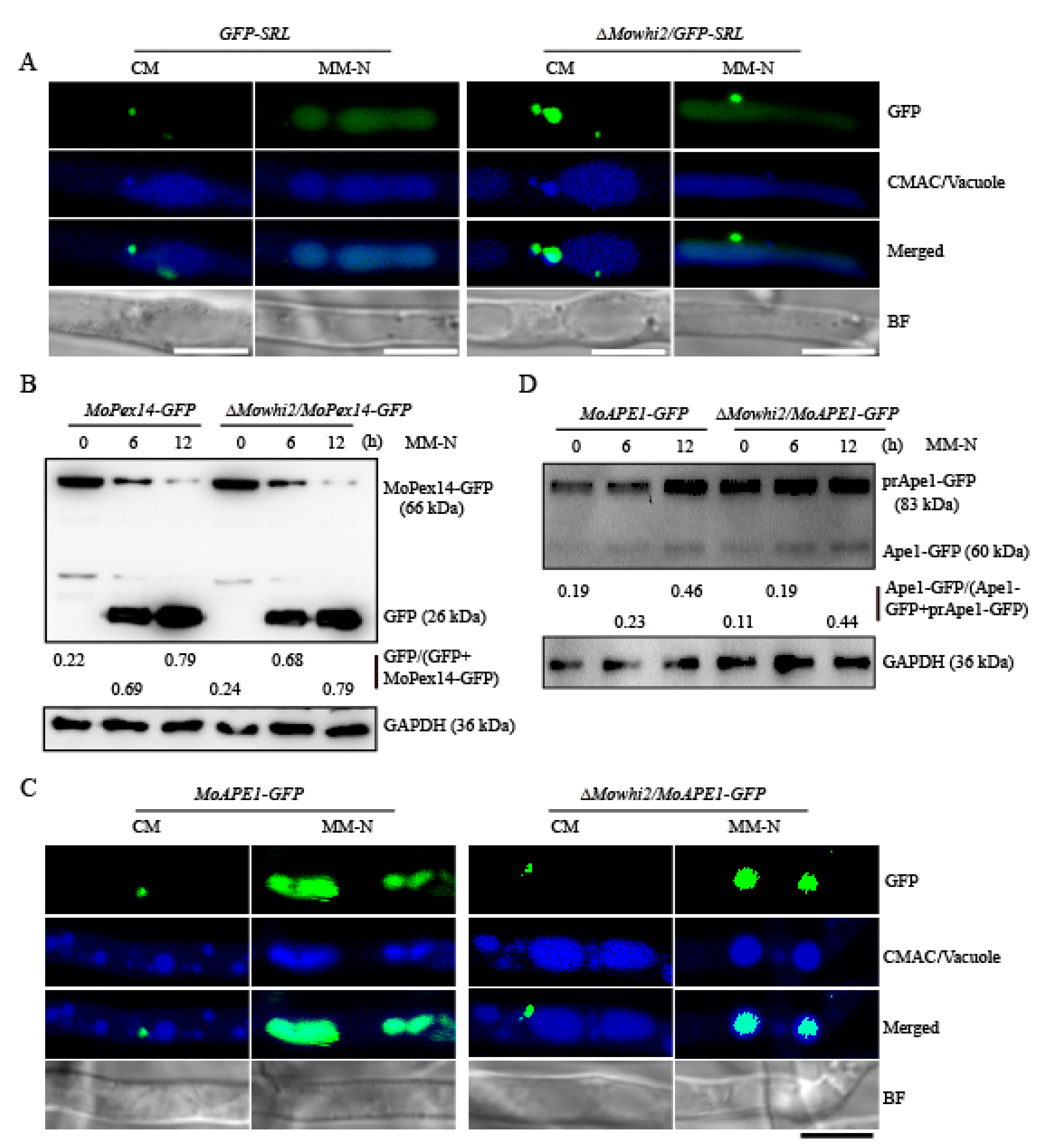
2.4. MoWHI2 Is Not Necessary for Pexophagy and the Cytoplasm-to-Vacuole Targeting (CVT) Pathway under Nitrogen Starvation

Autophagy is a highly conserved process that is responsible for the recycling of cytoplasmic components by the vacuole/lysosome, including selective and non-selective autophagic processes. Pexophagy and the cytoplasm-to-vacuole targeting (CVT) pathway are involved in the selective degradation of peroxisomes and the maturation of the precursor form of aminopeptidase I or α-mannosidase, respectively [5,26,27,28]. To detect whether MoWhi2 specifically participates in selective degradation of pexophagy, deletion mutants of MoWHI2 were generated in the GFP-SRL and MoAPE1-GFP strains. GFP-SRL is a pexosome-labelled GFP used to monitor pexosomes [29]. Moreover, the processing of the vacuolar aminopeptidase MoApe1 maturation served as an indicator of the activity of the cytoplasm-to-vacuole targeting (CVT) pathway [28,29,30]. The GFP-SRL, MoAPE1-GFP, ΔMowhi2/GFP-SRL and ΔMowhi2/MoAPE1-GFP strains were inoculated in CM medium for 2 days, then subjected to nitrogen starvation for 6 h. After CMAC staining, the mycelia were observed by confocal microscopy. The microscopic observation indicated that the pexophagy process occurs in both ΔMowhi2/GFP-SRL and GFP-SRL strains (Figure 4A). The activity of the CVT pathway in the ΔMowhi2/MoAPE1-GFP strain was similar to that in the MoAPE1-GFP strain (Figure 4C). Furthermore, the degradation rate of MoPex14-GFP, a pexophagy marker protein [31], and MoApe1-GFP was measured by immunoblot analyses. Consistent with the microscopic observation, the degradation rates of MoPex14-GFP were not significantly different in the MoPEX14-GFP and ΔMowhi2/MoPEX14-GFP strains (Figure 4B), and the maturation rate of prApe1-GFP in the ΔMowhi2/MoAPE1-GFP strain was consistent with that in the MoAPE1-GFP strain (Figure 4C,D). All these results suggest that MoWhi2 is not essential for pexophagy and the CVT pathway and may be specifically required for mitophagy.

2.5. MoWhi2 Is Necessary for Mitophagy in Foot Cells during Conidiation

Mitophagy is required for conidiation in M. oryzae [20,21]. Given that MoWhi2 plays an important role in conidial differentiation and morphology [19], mitophagy in foot cells was determined in the Mito-GFP and ΔMowhi2/Mito-GFP strains. As shown in Figure 5, the fluorescence signal of Mito-GFP was clearly presented in the vacuoles in the foot cells of the Mito-GFP strain, whereas the GFP signal could not be observed in the vacuoles of the ΔMowhi2/Mito-GFP strains, suggesting that disruption of MoWHI2 interrupts mitophagy in foot cells in M. oryzae. These results indicate that MoWhi2 is necessary for mitophagy in foot cells and likely contributes to conidiation.

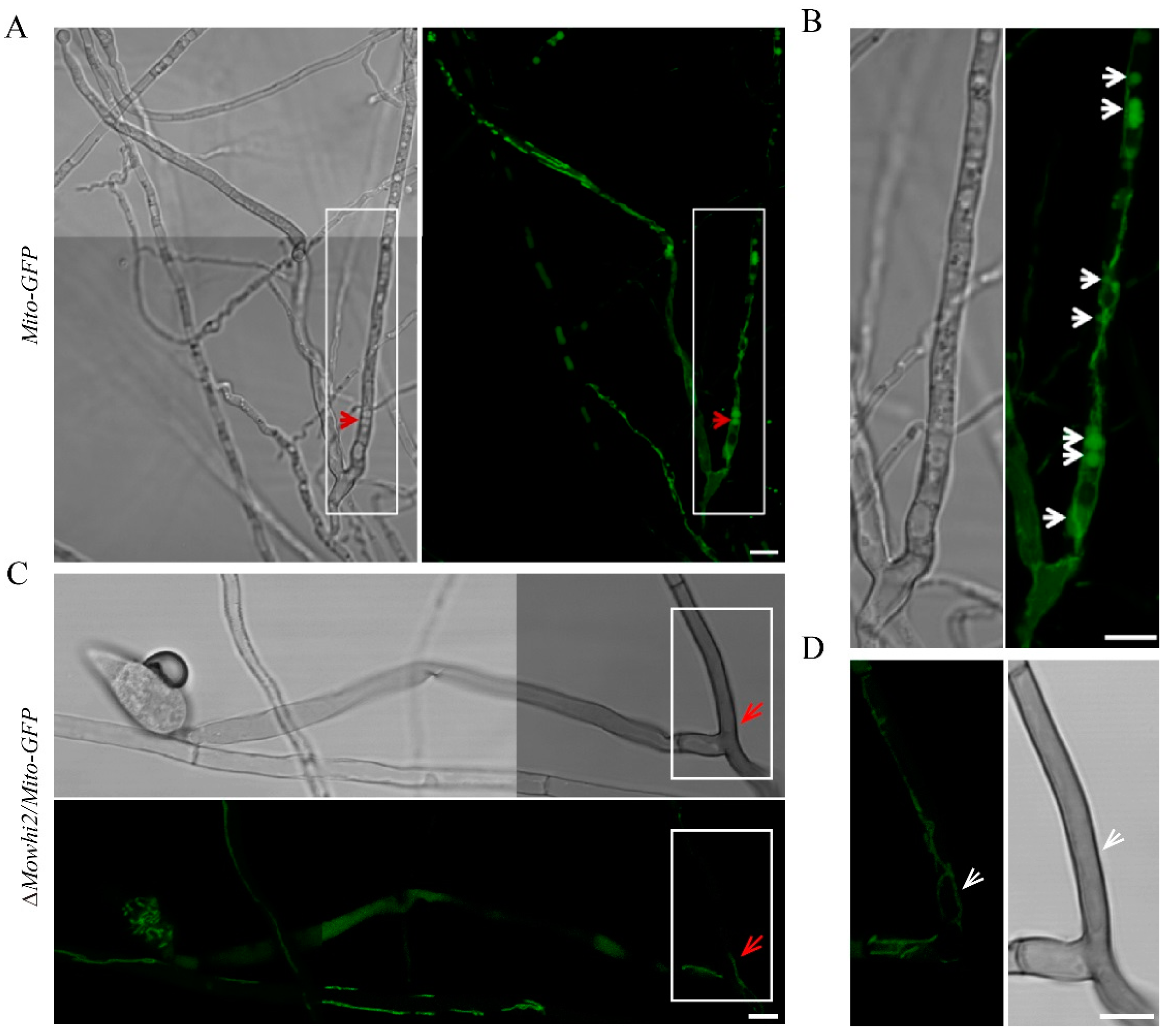
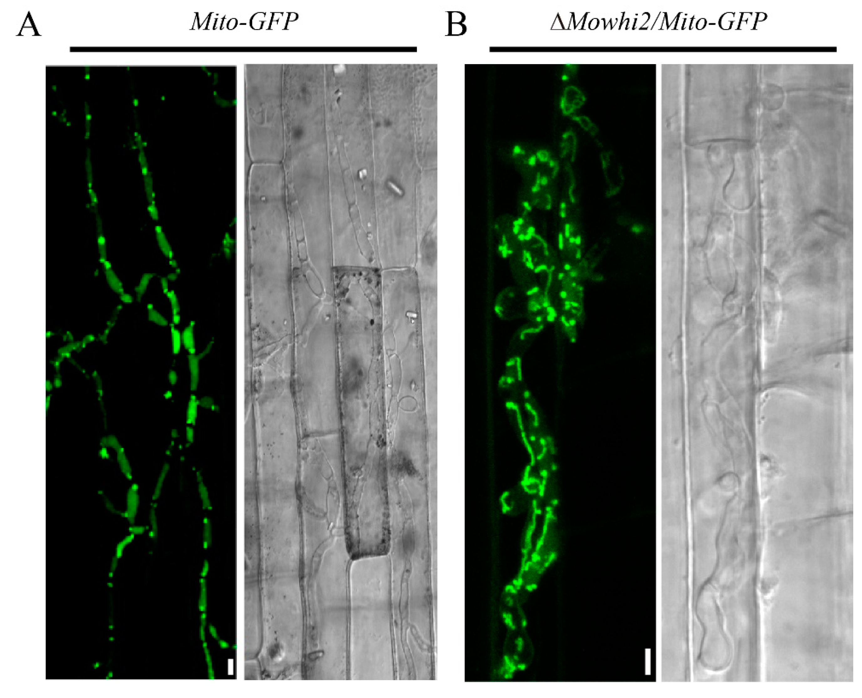
2.6. MoWhi2 Is Required for Mitophagy during Invasive Growth in M. oryzae

Mitophagy is not only necessary for conidiation but also for invasive hyphae growth in M. oryzae. To explore the involvement of MoWhi2 in mediating mitophagy during the invasive growth stage, mitophagy in the invasive hyphae was determined in the Mito-GFP and ΔMowhi2/Mito-GFP strains. In the Mito-GFP strain, the GFP entered into the vacuole, indicating that the mitochondria in the invasive hyphae are delivered into the vacuoles for degradation. In addition, the invasive hyphae had expanded into adjacent cells in the Mito-GFP strain (Figure 6A). However, the GFP signal of the ΔMowhi2/Mito-GFP strain was outside the vacuoles, suggesting that the mitochondria could not enter the vacuoles for degradation (Figure 6B). Meanwhile, the invasive hyphae of ΔMowhi2/Mito-GFP were mainly restricted to the first invaded cells of rice sheaths. In summary, mitophagy is impaired in the invasive hyphae of ΔMowhi2 in M. oryzae.

3. Discussion

In recent years, research on mitophagy has developed rapidly, especially concerning the identification of key receptors and regulatory factors [32,33], which provides a foundation for further understanding of the mechanism of this process. However, the molecular mechanism of mitophagy in plant pathogenic fungi has not been explored in detail. In this study, cell biology and biochemical analysis clearly demonstrated that MoWhi2 is specifically required for mitophagy in M. oryzae but not for autophagy, pexophagy and the CVT pathway. Furthermore, MoWhi2 plays important roles in mitophagy of foot cells and invasive hyphae, contributing to conidiation and virulence in M. oryzae.
Rice blast is a fungal disease caused by the filamentous ascomycete fungus M. oryzae. During the infection of M. oryzae, under suitable conditions, conidia germinate on the host surface to form germ tubes, which differentiate into appressoria. Turgor pressure accumulates in appressoria to penetrate the rigid rice cuticle; then, invasive hyphae grow in the host and spread to neighboring cells to form typical lesions. Conidiation is an essential step for infection, and spread of the invasive hyphae determines the formation of rice blast lesions. In M. oryzae, conidiation begins with the growth of a conidiophore stalk with apical extension from thick-walled vegetative cells called foot cells, which links aerial hyphae to the vegetative mycelia and ensures a supply of nutrients to the aerial hyphae [21]. Mitophagy occurs in the foot cells during conidiation in M. oryzae. When mitophagy in the foot cells is disrupted, conidiation is considerably reduced [20]. In addition, it was shown that mitophagy occurs in the invasive hyphae to promote invasive growth in M. oryzae. Knockout of the genes involved in mitophagy, such as MoATG24, MoDNM1, MoFIS1, MoMDV1, MoFZO1 and MoAUH1, resulted in reduced conidiation and spreading of invasive hyphae [20,22,23]. Similarly, the ΔMowhi2 strain showed defects in mitophagy in the foot cells, which likely led to decreased conidia. In addition, mitophagy in invasive hyphae was disrupted in the ΔMowhi2 strain, leading to defective invasive growth and restricted small, dark brown spot lesions. In summary, MoWhi2 plays vital roles in mitophagy and pathogenicity by regulating mitophagy in M. oryzae.
Whi2 was found to be required for the induction of mitophagy in yeast more than ten years ago [13]. Similar to yeast Whi2, we found that MoWhi2 is involved in mitophagy rather than autophagy, pexophagy and the CVT pathway in M. oryzae (Figure 3 and Figure 4). However, not much is known about the cellular functions of Whi2 in mitophagy. In yeast, Whi2 is better known to be involved in several fundamental cellular processes. Under conditions of low extracellular nutrients, including glucose and amino acids, Whi2 inhibits cell proliferation by regulating the expression of CLN1, CLN2 and G1 cyclin; suppresses the activity of TORC1; and inactivates the cAMP/PKA signaling pathway by targeting Ras2 to vacuoles [12,16]. In addition, Whi2 interacts with phosphatases Psr1 and Psr2, as well as the transcriptional factor Msn2, to activate the expression of general stress-response genes controlled by STREs (stress-responsive elements) [15]. In M. oryzae, transcription factor MoMsn2 participates in mitophagy by regulating the expression level of the 3-methylglutaconyl-CoA Hydratase encoding gene MoAUH1 [23]. In our previous study, we found that MoWhi2 also regulates appressorium formation and pathogenicity by the cAMP and MoTor signaling pathway in M. oryzae like ScWhi2 [19]. Based on all these results, we hypothesize that Whi2 might link cell cycle regulation, stress response, Ras-cAMP-PKA signaling pathways and TOR signaling to regulate the activity of Msn2, thus contributing to mitophagy [17].
In yeast, Whi2 and Psr1 form a complex that participates in the regulation of a series of stress responses to affect growth and development [15]. In Colletotrichum orbiculare, Whi2 interacts with Psr1 to promote biotrophic infection [18]. In Podospora anserina, Whi2 and Psr1 are necessary for sexual reproduction and nutrient-sensing signaling [34]. Moreover, the phosphatase MoPsr1, which interacts with MoWhi2, is involved in appressorium formation and pathogenicity in M. oryzae [19]. However, we found that MoPsr1 is not required for mitophagy. It is possible that MoWhi2 is involved in mitophagy through an MoPsr1-independent pathway. How Whi2 participates in mitophagy remains to be further studied.
In summary, we found that MoWhi2 is required for conidiation and invasive growth by regulating mitophagy in M. oryzae. The results deepen the understanding of the pathogenic mechanism of M. oryzae and provide a theoretical basis for the control of rice blast.

4. Materials and Methods

4.1. Fungal Strains and Culture Media

The Mito-GFP, ΔMoatg8/Mito-GFP and GFP-SRL strains were kindly provided by Prof. Naweed I. Naqvi of National University of Singapore (Singapore). All the M. oryzae strains were cultured on complete medium (CM: D-Glucose 10 g/L, Peptone 2 g/L, Casamino acid 1 g/L, Yeast extract 1 g/L, 20 × Nitrate salts 50 mL/L, 1000 × Vitamin solution 1 mL/L, 1000 × trace elements 1 mL/L, pH 6.5) under 16 h of light and 8 h of dark at 25 °C [21]. Transformants generated by Agrobacterium tumefaciens-mediated transformation (ATMT) were screened on basal medium (BM: yeast nitrogen base 1.6 g/L, asparagine 2.0 g/L, NH4NO3 1.0 g/L, glucose 10 g/L, agar 20g/L, pH 6.0) with chlorimuron-ethyl (50 μg/mL). Mycelia were collected from the liquid CM media for 2 d. The strains used in this study are summarized in Supplementary Table S1.

4.2. Plasmid Constructions

The MoWHI2 gene (MGG_11241) deletion mutant was generated using the standard one-step gene replacement strategy in the Mito-GFP background. Briefly, about 1 Kb of 5’ UTR and 3’ UTR regions were amplified by PCR and ligated sequentially to flanking restriction enzyme sites of the ILV2SUR sulfonylurea-resistance gene cassette in pFGL820 (Addgene, 58221). The primers used to amplify the 5’ and 3’ UTR of the MoWHI2 gene are listed in Supplementary Table S2. The resultant plasmid was confirmed by sequencing and subsequently transformed into the Mito-GFP strain by ATMT to delete the MoWHI2 gene [35]. The ΔMopsr1/Mito-GFP, ΔMopsr1/MoAPE1-GFP, ΔMowhi2/GFP-MoATG8, ΔMowhi2/MoAPE1-GFP and ΔMowhi2/GFP-SRL strains were constructed by a similar strategy.

4.3. Mitophagy, Autophagy, Pexophagy and CVT Pathway Analyses

The strains Mito-GFP, GFP-MoATG8, GFP-SRL and MoAPE1-GFP were used to monitor mitophagy, pexophagy and the CVT pathway in M. oryzae. For mitophagy, the Mito-GFP strain was first cultured in liquid CM media for 2 days, transferred to BM-G media (1.5% glycerol) for 30 h and finally transferred to MM-N media for 12 h at 28 °C [20]. To determine autophagy, pexophagy and the CVT pathway, the strains GFP-MoATG8, GFP-SRL and MoAPE1-GFP were cultured in liquid CM media for 2 days and then transferred to MM-N media for 12 h [28,29,36]. To stain vacuoles, the mycelia were incubated in 10 μM CellTracker™ Blue CMAC dye (7-amino-4-Chloromethylcoumarin, Molecular Probes, C2110, Carlsbad, CA, USA) for 30 min at room temperature and then washed with water before microscopic observation. Autophagosomes were counted in more than 25 hyphal cells. Linescan graph analysis was carried out by Image J software.
To observe mitophagy in foot cells, mycelial plugs (1 cm) were cut from marginal regions of a 7-day-old colony and incubated on glass slides covered with a thin layer of 1% agar medium. The slides were placed in the dark for 2 days and cultured in the light for another 12 h at 28 °C. Foot cells were imaged with a confocal microscope after removing the mycelial plugs. To observe mitophagy in the invasive hyphae, conidial suspensions (1 × 10⁵/mL) collected from 7-day-old culture plates were inoculated on the excised rice sheaths of susceptible cultivar CO39 (Oryza sativa). Fluorescence of Mito-GFP in the invasive hyphae was monitored at 60 hpi.

4.4. Fluorescence Microscopy

A fluorescent strain with GFP signal was observed and documented by confocal fluorescent LSM700 microscopy (Zeiss, Oberkochen, Germany). GFP and CMAC were imaged with 488 nm (Em. 505–530 nm) and 405 nm (Em. 430–470 nm) laser excitation, respectively. Images were processed with Image J and arranged using Photoshop CS6 software.

4.5. Western Blot Assay

The mycelia (0.1 g) cultured in liquid CM for 2 days were collected at 28 °C, and the total protein solutions were extracted with lysis buffer (50 mM Tris-HCl pH 7.4, 150 mM NaCl, 1 mM EDTA, 1% Triton 100, and 1 × protein inhibitor) at 12000 rpm for 10 min at 4 °C. For detection of Porin (mitochondria outer-membrane protein), total proteins were extracted with 10% SDS solution. The resultant protein solution was resolved by 8–15% SDS polyacrylamide gel electrophoresis (SDS-PAGE) and transferred to a PVDF membrane, followed by incubation with primary antibodies anti-GFP (Huabio, ET1607-31, Hangzhou, China) or anti-porin antibody (GenScript, A01419, Nanjing, China). The protein GAPDH served as a loading control. Western blots were detected using an ECL chemiluminescent kit (Biorad GS-710, Hercules, CA, USA), and the relative intensity of blots was quantified by Image J software (version 1.37).

Supplementary Materials

The following supporting information can be downloaded at: https://www.mdpi.com/article/10.3390/ijms23105311/s1.

Author Contributions

Methodology, Y.K. and H.S.; validation, S.M., J.S.J. and H.S.; formal analysis, S.M.; investigation, S.M. and J.S.J.; resources, C.L.; data curation, Y.K.; writing—original draft preparation, S.M.; writing—review and editing, S.M.; visualization, S.M. and H.S.; supervision, Y.K.; project administration, Y.K. and H.S.; funding acquisition, H.S. and Y.K. All authors have read and agreed to the published version of the manuscript.

Funding

This research was supported in part by the National Natural Science Foundation of China (31900127), the Zhejiang Provincial Natural Science Foundation of China (grant number “LY22C140006”) and the Key R&D project of China National Rice Research Institute (grant number “CNRRI-2020-04”). This project was also supported by the Chinese Academy of Agricultural Sciences under the Agricultural Sciences and Technologies Innovation Program.

Institutional Review Board Statement

Not applicable.

Informed Consent Statement

Not applicable.

Data Availability Statement

The data presented in this study are available on request from the corresponding authors.

Acknowledgments

This research was supported in part by the National Natural Science Foundation of China (31900127), the Zhejiang Provincial Natural Science Foundation of China (grant number “LY22C140006”) and the Key R&D Project of China National Rice Research Institute grant number “CNRRI-2020-04”). This project was also supported by the Chinese Academy of Agricultural Sciences under the Agricultural Sciences and Technologies Innovation Program. We thank the Rice–Pathogen Interaction group at China National Rice Research Institute for useful discussion and suggestions.

Conflicts of Interest

The authors declare that there are no conflicts of interest related to this article.

References

  1. Kanki, T.; Wang, K.; Baba, M.; Bartholomew, C.R.; Lynch-Day, M.A.; Du, Z.; Geng, J.; Mao, K.; Yang, Z.; Yen, W.L.; et al. A genomic screen for yeast mutants defective in selective mitochondria autophagy. Mol. Biol. Cell 2009, 20, 4730–4738. [Google Scholar] [CrossRef] [PubMed] [Green Version]
  2. Wallace, D.C. A mitochondrial paradigm of metabolic and degenerative diseases, aging, and cancer: A dawn for evolutionary medicine. Annu. Rev. Genet. 2005, 39, 359–407. [Google Scholar] [CrossRef] [PubMed] [Green Version]
  3. Zorov, D.B.; Juhaszova, M.; Sollott, S.J. Mitochondrial reactive oxygen species (ROS) and ROS-induced ROS release. Physiol. Rev. 2014, 94, 909–950. [Google Scholar] [CrossRef] [PubMed] [Green Version]
  4. Youle, R.J.; Narendra, D.P. Mechanisms of mitophagy. Nat. Rev. Mol. Cell Biol. 2011, 12, 9–14. [Google Scholar] [CrossRef] [PubMed]
  5. Zutphen, T.; Veenhuis, M.; van der Klei, I.J. Pex14 is the sole component of the peroxisomal translocon that is required for pexophagy. Autophagy 2008, 4, 63–66. [Google Scholar] [CrossRef] [PubMed] [Green Version]
  6. Jagernath, J.S.; Meng, S.; Qiu, J.; Shi, H.; Kou, Y. Selective degradation of mitochondria by mitophagy in pathogenic fungi. Am. J. Mol. Biol. 2021, 11, 15–27. [Google Scholar] [CrossRef]
  7. Kanki, T.; Wang, K.; Cao, Y.; Baba, M.; Klionsky, D.J. Atg32 is a mitochondrial protein that confers selectivity during mitophagy. Dev. Cell 2009, 17, 98–109. [Google Scholar] [CrossRef] [Green Version]
  8. Levchenko, M.; Lorenzi, I.; Dudek, J. The degradation pathway of the mitophagy receptor Atg32 is re-routed by a posttranslational modification. PLoS ONE 2016, 11, e0168518. [Google Scholar] [CrossRef]
  9. Liu, Y.; Okamoto, K. Regulatory mechanisms of mitophagy in yeast. Biochim. Biophys. Acta Gen. Subj. 2021, 1865, 129858. [Google Scholar] [CrossRef]
  10. Onishi, M.; Yamano, K.; Sato, M.; Matsuda, N.; Okamoto, K. Molecular mechanisms and physiological functions of mitophagy. EMBO J. 2021, 40, e104705. [Google Scholar] [CrossRef]
  11. Kumar, R.; Reichert, A.S. Common principles and specific mechanisms of mitophagy from yeast to humans. Int. J. Mol. Sci. 2021, 22, 4363. [Google Scholar] [CrossRef] [PubMed]
  12. Leadsham, J.E.; Miller, K.; Ayscough, K.R.; Colombo, S.; Martegani, E.; Sudbery, P.; Gourlay, C.W. Whi2p links nutritional sensing to actin-dependent Ras-cAMP-PKA regulation and apoptosis in yeast. J. Cell Sci. 2009, 122, 706–715. [Google Scholar] [CrossRef] [PubMed] [Green Version]
  13. Mendl, N.; Occhipinti, A.; Muller, M.; Wild, P.; Dikic, I.; Reichert, A.S. Mitophagy in yeast is independent of mitochondrial fission and requires the stress response gene WHI2. J. Cell Sci. 2011, 124, 1339–1350. [Google Scholar] [CrossRef] [PubMed] [Green Version]
  14. Saul, D.J.; Sudbery, P.E. Molecular cloning of WHI2, a gene involved in the regulation of cell proliferation in Saccharomyces cerevisiae. J. Gen. Microbiol. 1985, 131, 1797–1806. [Google Scholar] [CrossRef] [Green Version]
  15. Kaida, D.; Yashirodaa, H.; Toh-e, A.; Kikuchi, Y. Yeast Whi2 and Psr1-phosphatase form a complex and regulate STRE-mediated gene expression. Genes Cells 2002, 7, 543–552. [Google Scholar] [CrossRef]
  16. Chen, X.; Wang, G.; Zhang, Y.; Dayhoff-Brannigan, M.; Diny, N.L.; Zhao, M.; He, G.; Sing, C.N.; Metz, K.A.; Stolp, Z.D.; et al. Whi2 is a conserved negative regulator of TORC1 in response to low amino acids. PLoS Genet. 2018, 14, e1007592. [Google Scholar] [CrossRef]
  17. Muller, M.; Reichert, A.S. Mitophagy, mitochondrial dynamics and the general stress response in yeast. Biochem. Soc. Trans. 2011, 39, 1514–1519. [Google Scholar] [CrossRef] [Green Version]
  18. Harata, K.; Nishiuchi, T.; Kubo, Y. Colletotrichum orbiculare WHI2, a yeast stress-response regulator homolog, controls the biotrophic stage of hemibiotrophic infection through TOR signaling. Mol. Plant Microbe Interact. 2016, 29, 468–483. [Google Scholar] [CrossRef] [Green Version]
  19. Shi, H.; Meng, S.; Qiu, J.; Wang, C.; Shu, Y.; Luo, C.; Kou, Y. MoWhi2 regulates appressorium formation and pathogenicity via the MoTor signalling pathway in Magnaporthe oryzae. Mol. Plant Pathol. 2021, 22, 969–983. [Google Scholar] [CrossRef]
  20. He, Y.; Deng, Y.Z.; Naqvi, N.I. Atg24-assisted mitophagy in the foot cells is necessary for proper asexual differentiation in Magnaporthe oryzae. Autophagy 2013, 9, 1818–1827. [Google Scholar] [CrossRef] [Green Version]
  21. Kou, Y.; He, Y.; Qiu, J.; Shu, Y.; Yang, F.; Deng, Y.; Naqvi, N.I. Mitochondrial dynamics and mitophagy are necessary for proper invasive growth in rice blast. Mol. Plant Pathol. 2019, 20, 1147–1162. [Google Scholar] [CrossRef] [PubMed] [Green Version]
  22. Zhong, K.; Li, X.; Le, X.; Kong, X.; Zhang, H.; Zheng, X.; Wang, P.; Zhang, Z. MoDnm1 dynamin mediating peroxisomal and mitochondrial fission in complex with MoFis1 and MoMdv1 is important for development of functional appressorium in Magnaporthe oryzae. PLoS Pathog. 2016, 12, e1005823. [Google Scholar] [CrossRef] [PubMed]
  23. Xiao, Y.; Liu, L.; Zhang, T.; Zhou, R.; Ren, Y.; Li, X.; Shu, H.; Ye, W.; Zheng, X.; Zhang, Z.; et al. Transcription factor MoMsn2 targets the putative 3-methylglutaconyl-CoA hydratase-encoding gene MoAUH1 to govern infectious growth via mitochondrial fusion/fission balance in Magnaporthe oryzae. Environ. Microbiol. 2021, 23, 774–790. [Google Scholar] [CrossRef] [PubMed]
  24. Patkar, R.N.; Ramos-Pamplona, M.; Gupta, A.P.; Fan, Y.; Naqvi, N.I. Mitochondrial beta-oxidation regulates organellar integrity and is necessary for conidial germination and invasive growth in Magnaporthe oryzae. Mol. Microbiol. 2012, 86, 1345–1363. [Google Scholar] [CrossRef] [PubMed]
  25. Nakatogawa, H.; Ichimura, Y.; Ohsumi, Y. Atg8, a ubiquitin-like protein required for autophagosome formation, mediates membrane tethering and hemifusion. Cell 2007, 130, 165–178. [Google Scholar] [CrossRef] [Green Version]
  26. Dunn, W.A., Jr.; Cregg, J.M.; Kiel, J.A.; van der Klei, I.J.; Oku, M.; Sakai, Y.; Sibirny, A.A.; Stasyk, O.V.; Veenhuis, M. Pexophagy: The selective autophagy of peroxisomes. Autophagy 2005, 1, 75–83. [Google Scholar] [CrossRef] [Green Version]
  27. Li, L.; Wang, J.; Chen, H.; Chai, R.; Zhang, Z.; Mao, X.; Qiu, H.; Jiang, H.; Wang, Y.; Sun, G. Pex14/17, a filamentous fungus-specific peroxin, is required for the import of peroxisomal matrix proteins and full virulence of Magnaporthe oryzae. Mol. Plant. Pathol. 2017, 18, 1238–1252. [Google Scholar] [CrossRef] [Green Version]
  28. Lv, W.; Xu, Z.; Talbot, N.J.; Wang, Z. The sorting nexin FgAtg20 is involved in the Cvt pathway, non-selective macroautophagy, pexophagy and pathogenesis in Fusarium graminearum. Cell Microbiol. 2020, 22, e13208. [Google Scholar] [CrossRef]
  29. Deng, Y.; Qu, Z.; Naqvi, N.I. The role of snx41-based pexophagy in magnaporthe development. PLoS ONE 2013, 8, e79128. [Google Scholar] [CrossRef]
  30. Nair, U.; Cao, Y.; Xie, Z.; Klionsky, D.J. Roles of the lipid-binding motifs of Atg18 and Atg21 in the cytoplasm to vacuole targeting pathway and autophagy. J. Biol. Chem. 2010, 285, 11476–11488. [Google Scholar] [CrossRef] [Green Version]
  31. Mao, K.; Liu, X.; Feng, Y.; Klionsky, D.J. The progression of peroxisomal degradation through autophagy requires peroxisomal division. Autophagy 2014, 10, 652–661. [Google Scholar] [CrossRef] [PubMed] [Green Version]
  32. Liu, L.; Sakakibara, K.; Chen, Q.; Okamoto, K. Receptor-mediated mitophagy in yeast and mammalian systems. Cell Res. 2014, 24, 787–795. [Google Scholar] [CrossRef] [PubMed] [Green Version]
  33. Wei, Y.; Chiang, W.C.; Sumpter, R., Jr.; Mishra, P.; Levine, B. Prohibitin 2 is an inner mitochondrial membrane mitophagy receptor. Cell 2017, 168, 224–238. [Google Scholar] [CrossRef] [PubMed] [Green Version]
  34. Timpano, H.; Chan Ho Tong, L.; Gautier, V.; Lalucque, H.; Silar, P. The PaPsr1 and PaWhi2 genes are members of the regulatory network that connect stationary phase to mycelium differentiation and reproduction in Podospora anserina. Fungal Genet. Biol. 2016, 94, 1–10. [Google Scholar] [CrossRef] [PubMed]
  35. Yang, F.; Naqvi, N.I. Sulfonylurea resistance reconstitution as a novel strategy for ILV2-specific integration in Magnaporthe oryzae. Fungal Genet. Biol. 2014, 68, 71–76. [Google Scholar] [CrossRef] [PubMed]
  36. Veneault-Fourrey, C.; Barooah, M.; Egan, M.; Wakley, G.; Talbot, N.J. Autophagic fungal cell death is necessary for infection by the rice blast fungus. Science 2006, 312, 580–583. [Google Scholar] [CrossRef] [Green Version]
Figure 1. MoWhi2 is involved in mitophagy in M. oryzae. (A) The degradation of Mito-GFP was disrupted in the ΔMowhi2 strain. Mycelia cultured in complete media (CM) for 2 days were subjected to basal media with glycerol for 30 h and minimal media (MM) lacking a nitrogen source for 6 h. The mycelia were stained with CMAC (7-amino-4-Chloromethylcoumarin) to label vacuoles before confocal microscopic observation. Similarly to the ΔMoatg8/Mito-GFP strain, the mitochondria could not be delivered into vacuoles in the ΔMowhi2/Mito-GFP strain. In contrast, the GFP signals were overlapped with vacuoles in the Mito-GFP strain. Scale bar = 10 μm. (B) Linescan graph analysis of the region indicated by arrow in (A). (C) Conidiophore morphology of the wild type, ΔMowhi2 and complemented ΔMowhi2C strains. Scale bar = 60 μm. (D) Immunoblot analysis to detect the degradation of Mito-GFP. Equal amounts of mycelia of Mito-GFP, ΔMowhi2/Mito-GFP and ΔMoatg8/Mito-GFP strains were collected at 12 h post nitrogen starvation to perform immunoblot with anti-GFP antibody. The protein GAPDH served as a loading control. The values represent the percentage of free GFP in the indicated strain analyzed by Image J. Similar results were obtained from two biological repeats.
Ijms 23 05311 g001
Figure 2. Mitophagy occurs in the ΔMopsr1 mutant in M. oryzae. (A) The mitochondria of the ΔMopsr1 strain were delivered into vacuoles upon nitrogen starvation. Similar to the Mito-GFP strain, the Mito-GFP-marked mitochondria were delivered into vacuoles in the ΔMopsr1 mutant. Scale bar = 5 μm. (B) Linescan graph analysis of the region indicated by arrow in (A). (C) Immunoblotting detection of the mitochondria outer membrane protein Porin. The indicated strains were cultured in liquid CM for 2 days, then shifted to BM-G (1.5% glycerol) for 30 h, followed by starvation in MM-N for 12 h. Mycelia were harvested to perform immunoblot assays with anti-Porin antibody. The values represent the percentage of Porin/GAPDH in the indicated strain analyzed by Image J. Similar results were obtained from two biological repeats.
Ijms 23 05311 g002
Figure 3. MoWhi2 is not required for autophagy upon nitrogen starvation. (A) Autophagy occurs in the ΔMowhi2 strain. Mycelia of the GFP-MoATG8 and ΔMowhi2/GFP-MoATG8 strains cultured in CM medium were transferred to the SD-N medium for 12 h before imaging. Scale bar = 5 μm. (B) Linescan graph analysis of the region indicated by arrow in (A). (C) Statistics of autophagosomes in the indicated strains. Autophagosomes in more than 25 hyphal cells were counted. There were no significant differences in the numbers of autophagosomes in the hyphae of the GFP-MoATG8 and ΔMowhi2/GFP-MoATG8 strains. (D) Vacuolar degradation of the GFP-MoAtg8 fusion protein was detected by immunoblotting using anti-GFP antibody. The GFP-MoATG8 was degraded in the ΔMowhi2/GFP-MoATG8 strain as the GFP-MoATG8 strain upon nitrogen starvation. The numbers underneath the blots are the ratios of free GFP to total GFP, indicating the level of autophagy. Similar results were obtained from three replicates.
Ijms 23 05311 g003
Figure 4. MoWhi2 is not necessary for pexophagy and the activation of the cytoplasm-to-vacuole targeting pathway in M. oryzae. (A) MoWhi2 is not necessary for pexophagy. Upon nitrogen starvation, the green fluorescence of the fusion protein GFP-SRL was overlapped with the CMAC-stained vacuoles in the ΔMowhi2/GFP-SRL as the GFP-SRL strain. (B) Vacuolar degradation of peroxisomes was detected by immunoblotting assay. The wild-type and ΔMowhi2 strains expressing MoPEX14-GFP were cultured in CM for 2 days, then transferred to MM-N medium to induce pexophagy. The degradation dynamics of MoPex14-GFP were similar in the MoPEX14-GFP and ΔMowhi2/MoPEX14-GFP strains. (C,D) MoWhi2 is not necessary for activation of the cytoplasm-to-vacuole targeting pathway in M. oryzae. Fluorescent imaging (C) and immunoblot analysis (D) showed the maturation of MoApe1, a marker protein of the CVT pathway, in the MoAPE1-GFP and ΔMowhi2/MoAPE1-GFP strains. Similar results were obtained from three biological replicates. Scale bar = 5 μm.
Ijms 23 05311 g004
Figure 5. Mitophagy during conidiation in the Mito-GFP and ΔMowhi2/Mito-GFP strains. Mitophagy in foot cells was affected in the ΔMowhi2/Mito-GFP strain. Foot cells of the indicated strains were induced by culturing mycelial plugs on glass slides with a thin layer of agar medium. Pictures in (AD) were captured at 20 hpi (hours post inoculation). Among them, panels (B,D) display magnified areas labeled by white boxes in the left panels of (A,C), respectively. The red arrows represent vacuoles and the white arrows represent mitochondria. Scale bar = 10 μm.
Ijms 23 05311 g005
Figure 6. Fluorescent observation of GFP in the Mito-GFP (A) and ΔMowhi2/Mito-GFP strains (B) during the invasive growth stage. Conidial suspensions of the indicated strains were inoculated on rice sheaths from 3-week-old Co39 (Oryza sativa) seedlings. Photographs were taken by a confocal microscope at 60 hpi.
Ijms 23 05311 g006
Publisher’s Note: MDPI stays neutral with regard to jurisdictional claims in published maps and institutional affiliations.

