Exosome-Mediated Systemic Signaling: Mechanisms, Disease Integration, and Translational Potential

 , ,
, ,  , and
, and
Abstract
1. Introduction
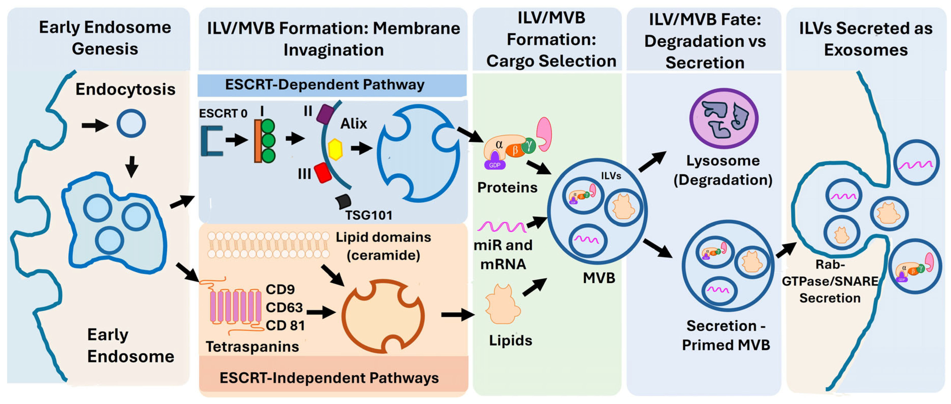
2. Exosome Biogenesis, Cargo Selection, and Functional Heterogeneity
2.1. Endosomal Biogenesis of Exosomes
2.2. ESCRT-Dependent Pathways
2.3. ESCRT-Independent Pathways
2.4. Selective Cargo Loading
2.5. Cellular Origin and Exosome Function
3. Exosome-Mediated Cellular Interactions and Signaling Pathways
4. Exosome Remodeling Under Pathological Conditions
4.1. Diabetes-Associated Remodeling
4.2. Chronic Kidney Disease
4.3. Cardiovascular Disease
4.4. Pathological Reprogramming
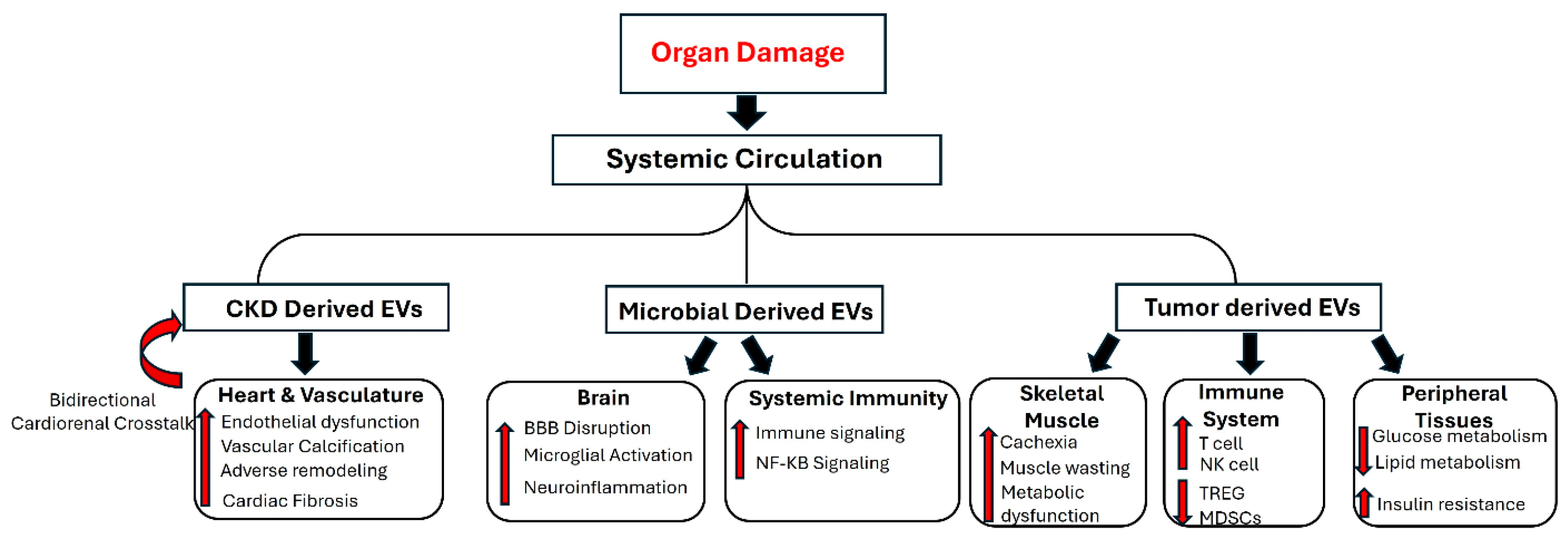
5. Inter-Organ Communication and Disease Integration
5.1. Kidney to Cardiovascular Axis
5.2. Gut–Brain Axis
5.3. Tumor to Systemic Metabolism
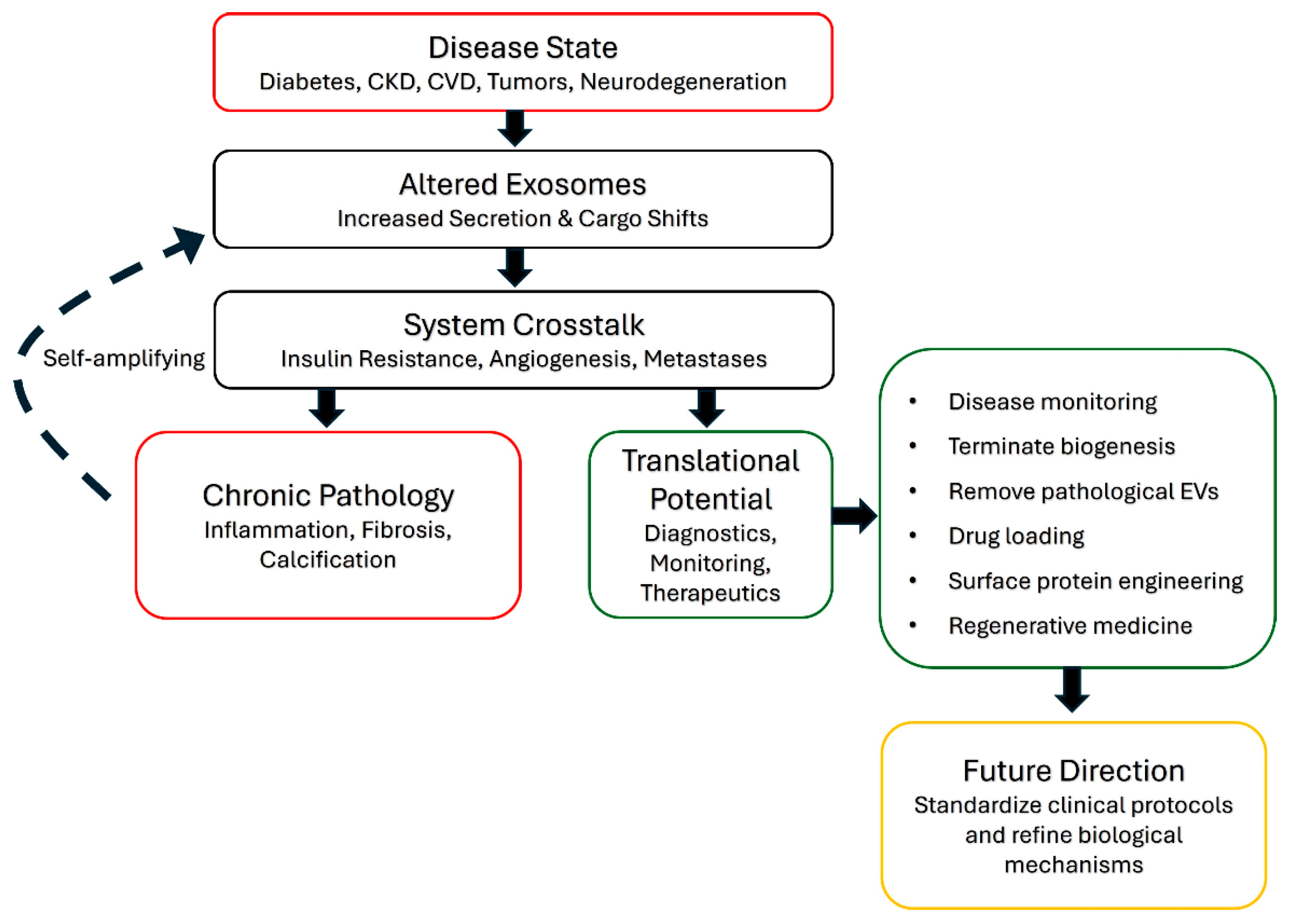
5.4. Disease Integration
6. Translational Applications: Biomarkers and Therapeutic Targeting
7. Conclusions
Author Contributions
Funding
Institutional Review Board Statement
Informed Consent Statement
Data Availability Statement
Conflicts of Interest
Abbreviations
| EVs | extracellular vesicles |
| miRs | microRNAs |
| EEs | early endosomes |
| ILVs | intraluminal vesicles |
| MVBs | multivesicular bodies |
| ESCRT | endosomal Sorting Complex Required for Transport |
| MHC | major histocompatibility complex |
| ALIX | aLG-2 interacting protein X |
| PA | phosphatidic acid |
| Cav-1 | Caveolin-1 |
| DAMPs | damage-associated molecular patterns |
| NF-κB | nuclear factor kappa-light-chain-enhancer of activated B cells |
| SNARE | soluble N-ethylmaleimide-sensitive agent-binding protein |
| SOX6 | SRY-box 6 |
| PI3K/AKT | phosphoinositide 3-kinase/protein kinase B |
| PTEN | phosphatase and tensin homolog deleted on chromosome 10 |
| STAT3 | transcription factor 3 |
| HMGB1 | high mobility group box 1 |
| TLR4 | toll-like receptor 4 |
| TGF-β | transforming growth factor-beta |
| NLRP3 | NLR family inflammasome containing the pyrin domain 3 |
| TNF-α | tumor necrosis factor alpha |
| IL | interleukin |
| Shh | sonic hedgehog |
| RBP4 | retinol-binding protein 4 |
| CKD | chronic kidney disease |
| SOCS1 | suppressor of cytokine signaling 1 |
| NOX | NADPH oxidase |
| MEK1/2 | mitogen-activated protein kinase 1 and 2 |
| ERK1/2 | extracellular signal-regulated kinases 1 and 2 |
| mTORC1-SCYL1 | mammalian target of rapamycin complex 1- SCY1-like pseudokinase 1 |
| AGE-RAGE | advanced glycation end-product–receptor for advanced glycation end-products |
| ROS | reactive oxygen species |
| FOXP1 | forkhead box P1 |
| IS | indoxyl sulfate |
| GAG | glycosaminoglycan |
| MMP1 | matrix metallopeptidase 1 |
| CTSL | cathepsin L |
| ICAM-1 | intercellular adhesion molecule 1 |
| VCAM-1 | vascular cell adhesion molecule-1 |
| OPN | β-catenin-osteopontin |
| TNFAIP8 | tumor necrosis factor alpha-induced protein 8 |
| CVD | cardiovascular disease |
| mTOR | mechanistic target of rapamycin |
| HIF-1α | hypoxia-inducible factor 1-alpha |
| Hsps | heat shock proteins |
| DUSP5 | dual-specificity protein phosphatase 5 |
| MAPK | mitogen-activated protein kinase |
| KLF4 | krüppel-like factor 4 |
| EGFR | epidermal growth factor receptor |
| IKK2 | IκB kinase β |
| PCSK8 | proprotein convertase subtilisin/kexin type 8 |
| VSMC | vascular smooth muscle cells |
| EndMT | endothelial–mesenchymal transition |
| LPS | lipopolysaccharide |
| TDEs | tumor-derived exosomes |
| GDF-15 | growth and differentiation factor 15 |
| NK | natural killer |
| Treg | regulatory T cell |
| MDSC | myeloid-derived suppressor cell |
| CSF | cerebrospinal fluid |
| pHERV-W ENV | human endogenous retrovirus type W exosomal envelope protein |
| LPPs | lipoproteins |
| AMPK/PGC-1α | AMP-activated protein kinase–peroxisome proliferator-activated receptor gamma coactivator 1-alpha |
References
- Tokizane, K.; Imai, S.I. Inter-organ communication is a critical machinery to regulate metabolism and aging. Trends Endocrinol. Metab. 2025, 36, 756–766. [Google Scholar] [CrossRef] [Scilit] [PubMed]
- Bhol, N.K.; Bhanjadeo, M.M.; Singh, A.K.; Dash, U.C.; Ojha, R.R.; Majhi, S.; Duttaroy, A.K.; Jena, A.B. The interplay between cytokines, inflammation, and antioxidants: Mechanistic insights and therapeutic potentials of various antioxidants and anti-cytokine compounds. Biomed. Pharmacother. 2024, 178, 117177. [Google Scholar] [CrossRef] [Scilit] [PubMed]
- Cao, C.K.; Shi, Z.Y.; Chen, C.B.; Li, X.K.; Su, Z.J. Fibroblast Growth Factor-Derived Peptides: Sources, Functions, and Applications. Bioengineering 2025, 12, 1019. [Google Scholar] [CrossRef] [Scilit] [PubMed]
- Castillo-Armengol, J.; Fajas, L.; Lopez-Mejia, I.C. Inter-organ communication: A gatekeeper for metabolic health. EMBO Rep. 2019, 20, e47903. [Google Scholar] [CrossRef] [Scilit] [PubMed]
- Donniacuo, A.; Mauro, A.; Cardamone, C.; Basile, A.; Manzo, P.; Dimitrov, J.; Cammarota, A.L.; Marzullo, L.; Triggiani, M.; Turco, M.C.; et al. Comprehensive Profiling of Cytokines and Growth Factors: Pathogenic Roles and Clinical Applications in Autoimmune Diseases. Int. J. Mol. Sci. 2025, 26, 8921. [Google Scholar] [CrossRef] [Scilit] [PubMed]
- Balachandran, S.; Arumugam, M.K.; Kalari Kandy, R.R.; Boopathy, L.K.; Gopal, T.; Rajamanickam, R.; Rasineni, K.; Kharbanda, K.K. Extracellular vesicles in molecular crosstalk and disease pathogenesis: A comprehensive review on mechanisms and therapeutic prospects. Biochem. Pharmacol. 2026, 251, 118186. [Google Scholar] [CrossRef] [Scilit] [PubMed]
- Li, X.; Raisinghani, N.; Gallinat, A.; Santos-Gallego, C.G.; Zhang, S.; La Salvia, S.; Yoon, S.; Yavuz, H.; Phan, A.; Shao, A.; et al. Circulating Extracellular Vesicles in the Pathogenesis of Heart Failure in Patients with Chronic Kidney Disease. Circulation 2026, 153, 94–114. [Google Scholar] [CrossRef] [Scilit] [PubMed]
- Chatterjee, E.; Rodosthenous, R.S.; Kujala, V.; Gokulnath, P.; Spanos, M.; Lehmann, H.I.; de Oliveira, G.P.; Shi, M.; Miller-Fleming, T.W.; Li, G.; et al. Circulating extracellular vesicles in human cardiorenal syndrome promote renal injury in a kidney-on-chip system. JCI Insight 2023, 8, e165172. [Google Scholar] [CrossRef] [Scilit] [PubMed]
- Miotto, P.M.; Yang, C.H.; Keenan, S.N.; De Nardo, W.; Beddows, C.A.; Fidelito, G.; Dodd, G.T.; Parker, B.L.; Hill, A.F.; Burton, P.R.; et al. Liver-derived extracellular vesicles improve whole-body glycaemic control via inter-organ communication. Nat. Metab. 2024, 6, 254–272. [Google Scholar] [CrossRef] [Scilit] [PubMed]
- Crescitelli, R.; Lasser, C.; Szabo, T.G.; Kittel, A.; Eldh, M.; Dianzani, I.; Buzas, E.I.; Lotvall, J. Distinct RNA profiles in subpopulations of extracellular vesicles: Apoptotic bodies, microvesicles and exosomes. J. Extracell. Vesicles 2013, 2, 20677. [Google Scholar] [CrossRef] [Scilit] [PubMed]
- Akers, J.C.; Gonda, D.; Kim, R.; Carter, B.S.; Chen, C.C. Biogenesis of extracellular vesicles (EV): Exosomes, microvesicles, retrovirus-like vesicles, and apoptotic bodies. J. Neuro-Oncol. 2013, 113, 1–11. [Google Scholar] [CrossRef] [Scilit] [PubMed]
- Gurung, S.; Perocheau, D.; Touramanidou, L.; Baruteau, J. The exosome journey: From biogenesis to uptake and intracellular signalling. Cell Commun. Signal. 2021, 19, 47. [Google Scholar] [CrossRef] [Scilit] [PubMed]
- Pescosolido, M.F.; Ouyang, Q.; Liu, J.S.; Morrow, E.M. Live-Imaging Detection of Multivesicular Body-Plasma Membrane Fusion and Exosome Release in Cultured Primary Neurons. Methods Mol. Biol. 2023, 2683, 213–220. [Google Scholar] [CrossRef] [Scilit] [PubMed]
- Krylova, S.V.; Feng, D. The Machinery of Exosomes: Biogenesis, Release, and Uptake. Int. J. Mol. Sci. 2023, 24, 1337. [Google Scholar] [CrossRef] [Scilit] [PubMed]
- Valadi, H.; Ekstrom, K.; Bossios, A.; Sjostrand, M.; Lee, J.J.; Lotvall, J.O. Exosome-mediated transfer of mRNAs and microRNAs is a novel mechanism of genetic exchange between cells. Nat. Cell Biol. 2007, 9, 654–659. [Google Scholar] [CrossRef] [Scilit] [PubMed]
- Lee, Y.J.; Shin, K.J.; Chae, Y.C. Regulation of cargo selection in exosome biogenesis and its biomedical applications in cancer. Exp. Mol. Med. 2024, 56, 877–889. [Google Scholar] [CrossRef] [Scilit] [PubMed]
- Zhao, H.; Yang, L.; Baddour, J.; Achreja, A.; Bernard, V.; Moss, T.; Marini, J.C.; Tudawe, T.; Seviour, E.G.; San Lucas, F.A.; et al. Tumor microenvironment derived exosomes pleiotropically modulate cancer cell metabolism. eLife 2016, 5, e10250. [Google Scholar] [CrossRef] [Scilit] [PubMed]
- Hoshino, A.; Costa-Silva, B.; Shen, T.L.; Rodrigues, G.; Hashimoto, A.; Tesic Mark, M.; Molina, H.; Kohsaka, S.; Di Giannatale, A.; Ceder, S.; et al. Tumour exosome integrins determine organotropic metastasis. Nature 2015, 527, 329–335. [Google Scholar] [CrossRef] [Scilit] [PubMed]
- Thery, C.; Boussac, M.; Veron, P.; Ricciardi-Castagnoli, P.; Raposo, G.; Garin, J.; Amigorena, S. Proteomic analysis of dendritic cell-derived exosomes: A secreted subcellular compartment distinct from apoptotic vesicles. J. Immunol. 2001, 166, 7309–7318. [Google Scholar] [CrossRef] [Scilit] [PubMed]
- Perez-Hernandez, D.; Gutierrez-Vazquez, C.; Jorge, I.; Lopez-Martin, S.; Ursa, A.; Sanchez-Madrid, F.; Vazquez, J.; Yanez-Mo, M. The intracellular interactome of tetraspanin-enriched microdomains reveals their function as sorting machineries toward exosomes. J. Biol. Chem. 2013, 288, 11649–11661. [Google Scholar] [CrossRef] [Scilit] [PubMed]
- Trajkovic, K.; Hsu, C.; Chiantia, S.; Rajendran, L.; Wenzel, D.; Wieland, F.; Schwille, P.; Brugger, B.; Simons, M. Ceramide triggers budding of exosome vesicles into multivesicular endosomes. Science 2008, 319, 1244–1247. [Google Scholar] [CrossRef] [Scilit] [PubMed]
- Ghadami, S.; Dellinger, K. The lipid composition of extracellular vesicles: Applications in diagnostics and therapeutic delivery. Front. Mol. Biosci. 2023, 10, 1198044. [Google Scholar] [CrossRef] [Scilit] [PubMed]
- Paba, C.; Dorigo, V.; Senigagliesi, B.; Tormena, N.; Parisse, P.; Voitchovsky, K.; Casalis, L. Lipid bilayer fluidity and degree of order regulates small EVs adsorption on model cell membrane. J. Colloid Interface Sci. 2023, 652, 1937–1943. [Google Scholar] [CrossRef] [Scilit] [PubMed]
- Peruzzi, J.A.; Gunnels, T.F.; Edelstein, H.I.; Lu, P.; Baker, D.; Leonard, J.N.; Kamat, N.P. Enhancing extracellular vesicle cargo loading and functional delivery by engineering protein-lipid interactions. Nat. Commun. 2024, 15, 5618. [Google Scholar] [CrossRef] [Scilit] [PubMed]
- Xie, S.; Zhang, Q.; Jiang, L. Current Knowledge on Exosome Biogenesis, Cargo-Sorting Mechanism and Therapeutic Implications. Membranes 2022, 12, 498. [Google Scholar] [CrossRef] [Scilit] [PubMed]
- Pedersen, R.T.A.; Hassinger, J.E.; Marchando, P.; Drubin, D.G. Spatial regulation of clathrin-mediated endocytosis through position-dependent site maturation. J. Cell Biol. 2020, 219, e202002160. [Google Scholar] [CrossRef] [Scilit] [PubMed]
- Toshima, J.Y.; Tsukahara, A.; Nagano, M.; Tojima, T.; Siekhaus, D.E.; Nakano, A.; Toshima, J. The yeast endocytic early/sorting compartment exists as an independent sub-compartment within the trans-Golgi network. eLife 2023, 12, e84850. [Google Scholar] [CrossRef] [Scilit] [PubMed]
- Nakashima, S.; Matsui, T.; Fukuda, M. Vps9d1 regulates tubular endosome formation through specific activation of Rab22A. J. Cell Sci. 2023, 136, jcs260522. [Google Scholar] [CrossRef] [Scilit] [PubMed]
- Cai, Z.; Li, Z.; Li, N.; He, X.; Jin, Y.; Lin, D.; Fu, B.; Ma, X.; Hong, D.; Lin, K.; et al. Coro1a promotes the multivesicular body and plasma membrane fusion by facilitating PKM2-mediated SNAP-23 phosphorylation. Commun. Biol. 2026. [Google Scholar] [CrossRef] [Scilit] [PubMed]
- Matsui, T.; Sakamaki, Y.; Hiragi, S.; Fukuda, M. VAMP5 and distinct sets of cognate Q-SNAREs mediate exosome release. Cell Struct. Funct. 2023, 48, 187–198. [Google Scholar] [CrossRef] [Scilit] [PubMed]
- Han, Q.F.; Li, W.J.; Hu, K.S.; Gao, J.; Zhai, W.L.; Yang, J.H.; Zhang, S.J. Exosome biogenesis: Machinery, regulation, and therapeutic implications in cancer. Mol. Cancer 2022, 21, 207. [Google Scholar] [CrossRef] [Scilit] [PubMed]
- Ostrowski, M.; Carmo, N.B.; Krumeich, S.; Fanget, I.; Raposo, G.; Savina, A.; Moita, C.F.; Schauer, K.; Hume, A.N.; Freitas, R.P.; et al. Rab27a and Rab27b control different steps of the exosome secretion pathway. Nat. Cell Biol. 2010, 12, 19–30. [Google Scholar] [CrossRef] [Scilit] [PubMed]
- Hsu, C.; Morohashi, Y.; Yoshimura, S.; Manrique-Hoyos, N.; Jung, S.; Lauterbach, M.A.; Bakhti, M.; Gronborg, M.; Mobius, W.; Rhee, J.; et al. Regulation of exosome secretion by Rab35 and its GTPase-activating proteins TBC1D10A-C. J. Cell Biol. 2010, 189, 223–232. [Google Scholar] [CrossRef] [Scilit] [PubMed]
- Verweij, F.J.; Bebelman, M.P.; George, A.E.; Couty, M.; Becot, A.; Palmulli, R.; Heiligenstein, X.; Sires-Campos, J.; Raposo, G.; Pegtel, D.M.; et al. ER membrane contact sites support endosomal small GTPase conversion for exosome secretion. J. Cell Biol. 2022, 221, e202112032. [Google Scholar] [CrossRef] [Scilit] [PubMed]
- Wu, J.; Zhao, Y.; Chen, Q.; Chen, Y.; Gu, J.; Mao, L. Enterovirus A71 Promotes Exosome Secretion by the Nonstructural Protein 3A Interacting with Rab27a. Microbiol. Spectr. 2023, 11, e0344622, Correction in Microbiol. Spectr. 2024, 12, e01766-24. [Google Scholar] [CrossRef] [Scilit] [PubMed]
- Yoshida, K.; Htike, K.; Eguchi, T.; Kawai, H.; Eain, H.S.; Tran, M.T.; Sogawa, C.; Umemori, K.; Ogawa, T.; Kanemoto, H.; et al. Rab11 suppresses head and neck carcinoma by regulating EGFR and EpCAM exosome secretion. J. Oral Biosci. 2024, 66, 205–216. [Google Scholar] [CrossRef] [Scilit] [PubMed]
- Savina, A.; Vidal, M.; Colombo, M.I. The exosome pathway in K562 cells is regulated by Rab11. J. Cell Sci. 2002, 115, 2505–2515. [Google Scholar] [CrossRef] [Scilit] [PubMed]
- Meneses, K.M.; Pandya, P.; Lindemann, J.A.; Al-Qasrawi, D.S.; Argo, R.A.; Weems, C.M.; Beetler, D.J.; Vijay, G.V.; Yan, I.K.; Wolfram, J.; et al. RAB27B Drives a Cancer Stem Cell Phenotype in NSCLC Cells Through Enhanced Extracellular Vesicle Secretion. Cancer Res. Commun. 2023, 3, 607–620. [Google Scholar] [CrossRef] [Scilit] [PubMed]
- Kugeratski, F.G.; Hodge, K.; Lilla, S.; McAndrews, K.M.; Zhou, X.; Hwang, R.F.; Zanivan, S.; Kalluri, R. Quantitative proteomics identifies the core proteome of exosomes with syntenin-1 as the highest abundant protein and a putative universal biomarker. Nat. Cell Biol. 2021, 23, 631–641. [Google Scholar] [CrossRef] [Scilit] [PubMed]
- Arya, S.B.; Collie, S.P.; Parent, C.A. The ins-and-outs of exosome biogenesis, secretion, and internalization. Trends Cell Biol. 2024, 34, 90–108. [Google Scholar] [CrossRef] [Scilit] [PubMed]
- Liu, M.; Wen, Z.; Zhang, T.; Zhang, L.; Liu, X.; Wang, M. The role of exosomal molecular cargo in exosome biogenesis and disease diagnosis. Front. Immunol. 2024, 15, 1417758. [Google Scholar] [CrossRef] [Scilit] [PubMed]
- Lee, Y.J.; Shin, K.J.; Jang, H.J.; Ryu, J.S.; Lee, C.Y.; Yoon, J.H.; Seo, J.K.; Park, S.; Lee, S.; Je, A.R.; et al. GPR143 controls ESCRT-dependent exosome biogenesis and promotes cancer metastasis. Dev. Cell 2023, 58, 320–334 e328. [Google Scholar] [CrossRef] [Scilit] [PubMed]
- Wu, C.Y.; Jhang, J.G.; Lin, W.S.; Chuang, P.H.; Lin, C.W.; Chu, L.A.; Chiang, A.S.; Ho, H.C.; Chan, C.C.; Huang, S.Y. Dihydroceramide desaturase promotes the formation of intraluminal vesicles and inhibits autophagy to increase exosome production. iScience 2021, 24, 103437. [Google Scholar] [CrossRef] [Scilit] [PubMed]
- Chigozie, V.U. Exosomal bioactive molecules: Molecular regulation, functional heterogeneity, and translational implications in disease diagnostics and therapeutics. Clin. Exp. Med. 2026, 26, 179. [Google Scholar] [CrossRef] [Scilit] [PubMed]
- Carvalho Ferraz, L.; Pereira, P.; Ferreira, J.V. Molecular Mechanisms of Extracellular Vesicle Biogenesis and Their Impact on the Design of Custom EVs. Adv. Healthc. Mater. 2025, 14, e2501349. [Google Scholar] [CrossRef] [Scilit] [PubMed]
- Lau, N.C.H.; Yam, J.W.P. From Exosome Biogenesis to Absorption: Key Takeaways for Cancer Research. Cancers 2023, 15, 1992. [Google Scholar] [CrossRef] [Scilit] [PubMed]
- Das Gupta, A.; Krawczynska, N.; Nelson, E.R. Extracellular Vesicles-The Next Frontier in Endocrinology. Endocrinology 2021, 162, bqab133. [Google Scholar] [CrossRef] [Scilit] [PubMed]
- Colombo, M.; Moita, C.; van Niel, G.; Kowal, J.; Vigneron, J.; Benaroch, P.; Manel, N.; Moita, L.F.; Thery, C.; Raposo, G. Analysis of ESCRT functions in exosome biogenesis, composition and secretion highlights the heterogeneity of extracellular vesicles. J. Cell Sci. 2013, 126, 5553–5565. [Google Scholar] [CrossRef] [Scilit] [PubMed]
- Adell, M.A.; Vogel, G.F.; Pakdel, M.; Muller, M.; Lindner, H.; Hess, M.W.; Teis, D. Coordinated binding of Vps4 to ESCRT-III drives membrane neck constriction during MVB vesicle formation. J. Cell Biol. 2014, 205, 33–49. [Google Scholar] [CrossRef] [Scilit] [PubMed]
- Adell, M.A.Y.; Migliano, S.M.; Upadhyayula, S.; Bykov, Y.S.; Sprenger, S.; Pakdel, M.; Vogel, G.F.; Jih, G.; Skillern, W.; Behrouzi, R.; et al. Recruitment dynamics of ESCRT-III and Vps4 to endosomes and implications for reverse membrane budding. eLife 2017, 6, e31652. [Google Scholar] [CrossRef] [Scilit] [PubMed]
- Liese, S.; Wenzel, E.M.; Kjos, I.; Rojas Molina, R.; Schultz, S.W.; Brech, A.; Stenmark, H.; Raiborg, C.; Carlson, A. Protein crowding mediates membrane remodeling in upstream ESCRT-induced formation of intraluminal vesicles. Proc. Natl. Acad. Sci. USA 2020, 117, 28614–28624. [Google Scholar] [CrossRef] [Scilit] [PubMed]
- Larios, J.; Mercier, V.; Roux, A.; Gruenberg, J. ALIX- and ESCRT-III-dependent sorting of tetraspanins to exosomes. J. Cell Biol. 2020, 219, e201904113. [Google Scholar] [CrossRef] [Scilit] [PubMed]
- Addi, C.; Presle, A.; Fremont, S.; Cuvelier, F.; Rocancourt, M.; Milin, F.; Schmutz, S.; Chamot-Rooke, J.; Douche, T.; Duchateau, M.; et al. The Flemmingsome reveals an ESCRT-to-membrane coupling via ALIX/syntenin/syndecan-4 required for completion of cytokinesis. Nat. Commun. 2020, 11, 1941. [Google Scholar] [CrossRef] [Scilit] [PubMed]
- Gabaran, S.G.; Nejati, V.; Dilsiz, N.; Rezaie, J. An updated review on the inhibition of exosome biogenesis, release, and uptake: A potential anticancer approach. Biochem. Pharmacol. 2025, 239, 117019. [Google Scholar] [CrossRef] [Scilit] [PubMed]
- Duke, L.C.; Cone, A.S.; Sun, L.; Dittmer, D.P.; Meckes, D.G., Jr.; Tomko, R.J., Jr. Tetraspanin CD9 alters cellular trafficking and endocytosis of tetraspanin CD63, affecting CD63 packaging into small extracellular vesicles. J. Biol. Chem. 2025, 301, 108255. [Google Scholar] [CrossRef] [Scilit] [PubMed]
- Kong, X.; Patel, N.A.; Chalfant, C.E.; Cooper, D.R. Ceramide synthesis regulates biogenesis and packaging of exosomal MALAT1 from adipose derived stem cells, increases dermal fibroblast migration and mitochondrial function. Cell Commun. Signal. 2023, 21, 221. [Google Scholar] [CrossRef] [Scilit] [PubMed]
- Cheng, Q.; Li, X.; Wang, Y.; Dong, M.; Zhan, F.H.; Liu, J. The ceramide pathway is involved in the survival, apoptosis and exosome functions of human multiple myeloma cells in vitro. Acta Pharmacol. Sin. 2018, 39, 561–568. [Google Scholar] [CrossRef] [Scilit] [PubMed]
- Hurwitz, S.N.; Cheerathodi, M.R.; Nkosi, D.; York, S.B.; Meckes, D.G., Jr. Tetraspanin CD63 Bridges Autophagic and Endosomal Processes To Regulate Exosomal Secretion and Intracellular Signaling of Epstein-Barr Virus LMP1. J. Virol. 2018, 92, e01969-17. [Google Scholar] [CrossRef] [Scilit] [PubMed]
- Albacete-Albacete, L.; Navarro-Lerida, I.; Lopez, J.A.; Martin-Padura, I.; Astudillo, A.M.; Ferrarini, A.; Van-Der-Heyden, M.; Balsinde, J.; Orend, G.; Vazquez, J.; et al. ECM deposition is driven by caveolin-1-dependent regulation of exosomal biogenesis and cargo sorting. J. Cell Biol. 2020, 219, e202006178. [Google Scholar] [CrossRef] [Scilit] [PubMed]
- Ghossoub, R.; Lembo, F.; Rubio, A.; Gaillard, C.B.; Bouchet, J.; Vitale, N.; Slavik, J.; Machala, M.; Zimmermann, P. Syntenin-ALIX exosome biogenesis and budding into multivesicular bodies are controlled by ARF6 and PLD2. Nat. Commun. 2014, 5, 3477. [Google Scholar] [CrossRef] [Scilit] [PubMed]
- Wu, B.X.; Clarke, C.J.; Matmati, N.; Montefusco, D.; Bartke, N.; Hannun, Y.A. Identification of novel anionic phospholipid binding domains in neutral sphingomyelinase 2 with selective binding preference. J. Biol. Chem. 2011, 286, 22362–22371. [Google Scholar] [CrossRef] [Scilit] [PubMed]
- Bills, B.L.; Knowles, M.K. Phosphatidic Acid Accumulates at Areas of Curvature in Tubulated Lipid Bilayers and Liposomes. Biomolecules 2022, 12, 1707. [Google Scholar] [CrossRef] [Scilit] [PubMed]
- Baietti, M.F.; Zhang, Z.; Mortier, E.; Melchior, A.; Degeest, G.; Geeraerts, A.; Ivarsson, Y.; Depoortere, F.; Coomans, C.; Vermeiren, E.; et al. Syndecan-syntenin-ALIX regulates the biogenesis of exosomes. Nat. Cell Biol. 2012, 14, 677–685. [Google Scholar] [CrossRef] [Scilit] [PubMed]
- Gauvreau, M.E.; Cote, M.H.; Bourgeois-Daigneault, M.C.; Rivard, L.D.; Xiu, F.; Brunet, A.; Shaw, A.; Steimle, V.; Thibodeau, J. Sorting of MHC class II molecules into exosomes through a ubiquitin-independent pathway. Traffic 2009, 10, 1518–1527. [Google Scholar] [CrossRef] [Scilit] [PubMed]
- Stuffers, S.; Sem Wegner, C.; Stenmark, H.; Brech, A. Multivesicular endosome biogenesis in the absence of ESCRTs. Traffic 2009, 10, 925–937. [Google Scholar] [CrossRef] [Scilit] [PubMed]
- Wu, Y.; Toldo, N.; Fabbri, M. The interplay between the extracellular matrix and extracellular vesicle-associated microRNAs. Cell Commun. Signal. 2026, 24, 89. [Google Scholar] [CrossRef] [Scilit] [PubMed]
- de Jong, O.G.; Verhaar, M.C.; Chen, Y.; Vader, P.; Gremmels, H.; Posthuma, G.; Schiffelers, R.M.; Gucek, M.; van Balkom, B.W. Cellular stress conditions are reflected in the protein and RNA content of endothelial cell-derived exosomes. J. Extracell. Vesicles 2012, 1, 18396. [Google Scholar] [CrossRef] [Scilit] [PubMed]
- Liu, X.M.; Ma, L.; Schekman, R. Selective sorting of microRNAs into exosomes by phase-separated YBX1 condensates. eLife 2021, 10, e71982. [Google Scholar] [CrossRef] [Scilit] [PubMed]
- Garcia-Martin, R.; Wang, G.; Brandao, B.B.; Zanotto, T.M.; Shah, S.; Kumar Patel, S.; Schilling, B.; Kahn, C.R. MicroRNA sequence codes for small extracellular vesicle release and cellular retention. Nature 2022, 601, 446–451. [Google Scholar] [CrossRef] [Scilit] [PubMed]
- Shurtleff, M.J.; Temoche-Diaz, M.M.; Karfilis, K.V.; Ri, S.; Schekman, R. Y-box protein 1 is required to sort microRNAs into exosomes in cells and in a cell-free reaction. eLife 2016, 5, e19276. [Google Scholar] [CrossRef] [Scilit] [PubMed]
- Santangelo, L.; Giurato, G.; Cicchini, C.; Montaldo, C.; Mancone, C.; Tarallo, R.; Battistelli, C.; Alonzi, T.; Weisz, A.; Tripodi, M. The RNA-Binding Protein SYNCRIP Is a Component of the Hepatocyte Exosomal Machinery Controlling MicroRNA Sorting. Cell Rep. 2016, 17, 799–808. [Google Scholar] [CrossRef] [Scilit] [PubMed]
- Gregorius, J.; Wang, C.; Stambouli, O.; Hussner, T.; Qi, Y.; Tertel, T.; Borger, V.; Mohamud Yusuf, A.; Hagemann, N.; Yin, D.; et al. Small extracellular vesicles obtained from hypoxic mesenchymal stromal cells have unique characteristics that promote cerebral angiogenesis, brain remodeling and neurological recovery after focal cerebral ischemia in mice. Basic Res. Cardiol. 2021, 116, 40. [Google Scholar] [CrossRef] [Scilit] [PubMed]
- Han, Y.; Ren, J.; Bai, Y.; Pei, X.; Han, Y. Exosomes from hypoxia-treated human adipose-derived mesenchymal stem cells enhance angiogenesis through VEGF/VEGF-R. Int. J. Biochem. Cell Biol. 2019, 109, 59–68, Erratum in Int. J. Biochem. Cell Biol. 2020, 126, 105805. [Google Scholar] [CrossRef] [Scilit] [PubMed]
- Gao, P.; Yi, J.; Chen, W.; Gu, J.; Miao, S.; Wang, X.; Huang, Y.; Jiang, T.; Li, Q.; Zhou, W.; et al. Pericyte-derived exosomal miR-210 improves mitochondrial function and inhibits lipid peroxidation in vascular endothelial cells after traumatic spinal cord injury by activating JAK1/STAT3 signaling pathway. J. Nanobiotechnol. 2023, 21, 452. [Google Scholar] [CrossRef] [Scilit] [PubMed]
- Wang, Z.; Gao, D.; Wang, S.; Lin, H.; Wang, Y.; Xu, W. Exosomal microRNA-1246 from human umbilical cord mesenchymal stem cells potentiates myocardial angiogenesis in chronic heart failure. Cell Biol. Int. 2021, 45, 2211–2225. [Google Scholar] [CrossRef] [Scilit] [PubMed]
- Jin, J.; Yu, G. Hypoxic lung cancer cell-derived exosomal miR-21 mediates macrophage M2 polarization and promotes cancer cell proliferation through targeting IRF1. World J. Surg. Oncol. 2022, 20, 241. [Google Scholar] [CrossRef] [Scilit] [PubMed]
- Chiaradia, E.; Tancini, B.; Emiliani, C.; Delo, F.; Pellegrino, R.M.; Tognoloni, A.; Urbanelli, L.; Buratta, S. Extracellular Vesicles under Oxidative Stress Conditions: Biological Properties and Physiological Roles. Cells 2021, 10, 1763. [Google Scholar] [CrossRef] [Scilit] [PubMed]
- Haque, S.; Kodidela, S.; Sinha, N.; Kumar, P.; Cory, T.J.; Kumar, S. Differential packaging of inflammatory cytokines/chemokines and oxidative stress modulators in U937 and U1 macrophages-derived extracellular vesicles upon exposure to tobacco constituents. PLoS ONE 2020, 15, e0233054. [Google Scholar] [CrossRef] [Scilit] [PubMed]
- Lu, W.; Yin, L.; Zou, S.; Wu, M.; Du, X.; Li, H.; Shi, B. A panel of serum-derived exosomal miRNAs as markers of cardiovascular risk assessed by carotid intima-media thickness in patients with type 2 diabetes. Am. J. Transl. Res. 2026, 18, 236–247. [Google Scholar] [CrossRef] [Scilit] [PubMed]
- Liang, W.; Sagar, S.; Ravindran, R.; Najor, R.H.; Quiles, J.M.; Chi, L.; Diao, R.Y.; Woodall, B.P.; Leon, L.J.; Zumaya, E.; et al. Mitochondria are secreted in extracellular vesicles when lysosomal function is impaired. Nat. Commun. 2023, 14, 5031. [Google Scholar] [CrossRef] [Scilit] [PubMed]
- Li, S.J.; Tetri, L.H.; Vijayan, V.; Elezaby, A.; Chiang, C.H.; Lopez, I.; Ostberg, N.P.; Cornell, T.T.; Chen, C.Y.; Haileselassie, B. Excessive mitochondrial fission and associated extracellular mitochondria mediate cardiac dysfunction in obesity cardiomyopathy. Life Sci. 2025, 373, 123658. [Google Scholar] [CrossRef] [Scilit] [PubMed]
- Yang, Y.; Ren, S.; Xue, J.; Dong, W.; He, W.; Luo, J.; Li, X.; Xu, H.; Zheng, Z.; Wang, X.; et al. DeSUMOylation of RBMX regulates exosomal sorting of cargo to promote renal tubulointerstitial fibrosis in diabetic kidney disease. J. Adv. Res. 2025, 74, 175–189. [Google Scholar] [CrossRef] [Scilit] [PubMed]
- Chen, Y.; Qi, W.; Wang, Z.; Niu, F. Exosome Source Matters: A Comprehensive Review from the Perspective of Diverse Cellular Origins. Pharmaceutics 2025, 17, 147. [Google Scholar] [CrossRef] [Scilit] [PubMed]
- Song, Y.; Hu, J.; Ma, C.; Liu, H.; Li, Z.; Yang, Y. Macrophage-Derived Exosomes as Advanced Therapeutics for Inflammation: Current Progress and Future Perspectives. Int. J. Nanomed. 2024, 19, 1597–1627. [Google Scholar] [CrossRef] [Scilit] [PubMed]
- Zhu, M.; Sun, X.; Qi, X.; Xia, L.; Wu, Y. Exosomes from high glucose-treated macrophages activate macrophages andinduce inflammatory responses via NF-kappaB signaling pathway in vitro and in vivo. Int. Immunopharmacol. 2020, 84, 106551. [Google Scholar] [CrossRef] [Scilit] [PubMed]
- Wong, P.F.; Tong, K.L.; Jamal, J.; Khor, E.S.; Lai, S.L.; Mustafa, M.R. Senescent HUVECs-secreted exosomes trigger endothelial barrier dysfunction in young endothelial cells. EXCLI J. 2019, 18, 764–776. [Google Scholar] [CrossRef] [Scilit] [PubMed]
- Bai, S.; Yin, Q.; Dong, T.; Dai, F.; Qin, Y.; Ye, L.; Du, J.; Zhang, Q.; Chen, H.; Shen, B. Endothelial progenitor cell-derived exosomes ameliorate endothelial dysfunction in a mouse model of diabetes. Biomed. Pharmacother. 2020, 131, 110756. [Google Scholar] [CrossRef] [Scilit] [PubMed]
- Tan, W.; Li, Y.; Ma, L.; Fu, X.; Long, Q.; Yan, F.; Li, W.; Liu, X.; Ding, H.; Wang, Y.; et al. Exosomes of endothelial progenitor cells repair injured vascular endothelial cells through the Bcl2/Bax/Caspase-3 pathway. Sci. Rep. 2024, 14, 4465. [Google Scholar] [CrossRef] [Scilit] [PubMed]
- Lee, S.H.; Shin, S.M.; Zhong, P.; Kim, H.T.; Kim, D.I.; Kim, J.M.; Heo, W.D.; Kim, D.W.; Yeo, C.Y.; Kim, C.H.; et al. Reciprocal control of excitatory synapse numbers by Wnt and Wnt inhibitor PRR7 secreted on exosomes. Nat. Commun. 2018, 9, 3434. [Google Scholar] [CrossRef] [Scilit] [PubMed]
- Men, Y.; Yelick, J.; Jin, S.; Tian, Y.; Chiang, M.S.R.; Higashimori, H.; Brown, E.; Jarvis, R.; Yang, Y. Exosome reporter mice reveal the involvement of exosomes in mediating neuron to astroglia communication in the CNS. Nat. Commun. 2019, 10, 4136. [Google Scholar] [CrossRef] [Scilit] [PubMed]
- Sardar Sinha, M.; Ansell-Schultz, A.; Civitelli, L.; Hildesjo, C.; Larsson, M.; Lannfelt, L.; Ingelsson, M.; Hallbeck, M. Alzheimer’s disease pathology propagation by exosomes containing toxic amyloid-beta oligomers. Acta Neuropathol. 2018, 136, 41–56. [Google Scholar] [CrossRef] [Scilit] [PubMed]
- Winston, C.N.; Aulston, B.; Rockenstein, E.M.; Adame, A.; Prikhodko, O.; Dave, K.N.; Mishra, P.; Rissman, R.A.; Yuan, S.H. Neuronal Exosome-Derived Human Tau is Toxic to Recipient Mouse Neurons in vivo. J. Alzheimers Dis. 2019, 67, 541–553. [Google Scholar] [CrossRef] [Scilit] [PubMed]
- Ma, Z.; Wei, K.; Yang, F.; Guo, Z.; Pan, C.; He, Y.; Wang, J.; Li, Z.; Chen, L.; Chen, Y.; et al. Tumor-derived exosomal miR-3157-3p promotes angiogenesis, vascular permeability and metastasis by targeting TIMP/KLF2 in non-small cell lung cancer. Cell Death Dis. 2021, 12, 840. [Google Scholar] [CrossRef] [Scilit] [PubMed]
- Zeng, Z.; Li, Y.; Pan, Y.; Lan, X.; Song, F.; Sun, J.; Zhou, K.; Liu, X.; Ren, X.; Wang, F.; et al. Cancer-derived exosomal miR-25-3p promotes pre-metastatic niche formation by inducing vascular permeability and angiogenesis. Nat. Commun. 2018, 9, 5395. [Google Scholar] [CrossRef] [Scilit] [PubMed]
- Chen, G.; Huang, A.C.; Zhang, W.; Zhang, G.; Wu, M.; Xu, W.; Yu, Z.; Yang, J.; Wang, B.; Sun, H.; et al. Exosomal PD-L1 contributes to immunosuppression and is associated with anti-PD-1 response. Nature 2018, 560, 382–386. [Google Scholar] [CrossRef] [Scilit] [PubMed]
- Ludwig, S.; Floros, T.; Theodoraki, M.N.; Hong, C.S.; Jackson, E.K.; Lang, S.; Whiteside, T.L. Suppression of Lymphocyte Functions by Plasma Exosomes Correlates with Disease Activity in Patients with Head and Neck Cancer. Clin. Cancer Res. 2017, 23, 4843–4854. [Google Scholar] [CrossRef] [Scilit] [PubMed]
- Chen, Y.X.; Wu, Y.S.; Yu, R.D.; Dong, Y.Y.; Zhao, J.X.; Zhang, Y.F. Exosome-mediated cell-cell communication: A new perspective on the mechanisms and therapeutic potential of diabetic microvascular complications. Front. Pharmacol. 2026, 17, 1839895. [Google Scholar] [CrossRef] [Scilit] [PubMed]
- Zhang, G.; Yang, P. A novel cell-cell communication mechanism in the nervous system: Exosomes. J. Neurosci. Res. 2018, 96, 45–52. [Google Scholar] [CrossRef] [Scilit] [PubMed]
- Zheng, W.; Ji, X.; Yin, Q.Q.; Wu, C.; Xu, C.; Pan, H.; Wu, C. Exosomes as Emerging Regulators of Immune Responses in Type 2 Diabetes Mellitus. J. Diabetes Res. 2024, 2024, 3759339. [Google Scholar] [CrossRef] [Scilit] [PubMed]
- Liu, C.; Liu, D.; Wang, S.; Gan, L.; Yang, X.; Ma, C. Identification of the SNARE complex that mediates the fusion of multivesicular bodies with the plasma membrane in exosome secretion. J. Extracell. Vesicles 2023, 12, e12356. [Google Scholar] [CrossRef] [Scilit] [PubMed]
- Takeda, A.; Tachibana, A.; Nagumo, H.; Sakai-Kato, K. An in vitro lipid-mixing assay to investigate the fusion between small extracellular vesicles and endosome. Anal. Biochem. 2023, 669, 115130. [Google Scholar] [CrossRef] [Scilit] [PubMed]
- Tian, T.; Zhu, Y.L.; Zhou, Y.Y.; Liang, G.F.; Wang, Y.Y.; Hu, F.H.; Xiao, Z.D. Exosome uptake through clathrin-mediated endocytosis and macropinocytosis and mediating miR-21 delivery. J. Biol. Chem. 2014, 289, 22258–22267. [Google Scholar] [CrossRef] [Scilit] [PubMed]
- Wisler, J.R.; Singh, K.; McCarty, A.; Harkless, R.; Karpurapu, M.; Hernandez, E.; Mukherjee, D.; Abouhashem, A.S.; Christman, J.W.; Sen, C.K. Exosomal Transfer of DNA Methyl-Transferase mRNA Induces an Immunosuppressive Phenotype in Human Monocytes. Shock 2022, 57, 218–227. [Google Scholar] [CrossRef] [Scilit] [PubMed]
- Long, R.; Gao, L.; Li, Y.; Li, G.; Qin, P.; Wei, Z.; Li, D.; Qian, C.; Li, J.; Yang, G. M2 macrophage-derived exosomes carry miR-1271-5p to alleviate cardiac injury in acute myocardial infarction through down-regulating SOX6. Mol. Immunol. 2021, 136, 26–35. [Google Scholar] [CrossRef] [Scilit] [PubMed]
- Chen, X.; Wang, X.; Cui, Z.; Luo, Q.; Jiang, Z.; Huang, Y.; Jiang, J.; Qiu, J.; Li, Y.; Yu, K.; et al. M1 Microglia-derived Exosomes Promote Activation of Resting Microglia and Amplifies Proangiogenic Effects through Irf1/miR-155-5p/Socs1 Axis in the Retina. Int. J. Biol. Sci. 2023, 19, 1791–1812. [Google Scholar] [CrossRef] [Scilit] [PubMed]
- Wang, X.; Xia, J.; Yang, L.; Dai, J.; He, L. Recent progress in exosome research: Isolation, characterization and clinical applications. Cancer Gene Ther. 2023, 30, 1051–1065. [Google Scholar] [CrossRef] [Scilit] [PubMed]
- Wang, Y.; Zhang, X.; Wang, J.; Zhang, Y.; Ye, Q.; Wang, Y.; Fei, D.; Wang, Q. Inflammatory Periodontal Ligament Stem Cells Drive M1 Macrophage Polarization via Exosomal miR-143-3p-Mediated Regulation of PI3K/AKT/NF-kappaB Signaling. Stem Cells 2023, 41, 184–199. [Google Scholar] [CrossRef] [Scilit] [PubMed]
- Sun, X.H.; Wang, X.; Zhang, Y.; Hui, J. Exosomes of bone-marrow stromal cells inhibit cardiomyocyte apoptosis under ischemic and hypoxic conditions via miR-486-5p targeting the PTEN/PI3K/AKT signaling pathway. Thromb. Res. 2019, 177, 23–32. [Google Scholar] [CrossRef] [Scilit] [PubMed]
- Huang, J.R.; Gu, W.C.; Yuan, Y.P.; Yang, J.X.; Chen, Y.; Guo, X.X.; Ding, W. Exosomal HMGB1 Orchestrates NSCLC Progression and Immunosuppressive Macrophage Polarisation Through the TLR4/NF-kappaB/IL-6/STAT3 Signalling Cascade. J. Cell. Mol. Med. 2026, 30, e71050. [Google Scholar] [CrossRef] [Scilit] [PubMed]
- Yang, Z.; Liang, Z.; Rao, J.; Xie, H.; Zhou, M.; Xu, X.; Lin, Y.; Lin, F.; Wang, C.; Chen, C. Hypoxic-preconditioned mesenchymal stem cell-derived small extracellular vesicles promote the recovery of spinal cord injury by affecting the phenotype of astrocytes through the miR-21/JAK2/STAT3 pathway. CNS Neurosci. Ther. 2024, 30, e14428. [Google Scholar] [CrossRef] [Scilit] [PubMed]
- Long, M.; Sun, J.; Zhou, D.; Chen, C.; Shu, J.; Mao, X.; Yu, K.; Liang, M.; Chen, L.; Wang, Y.; et al. Lung-Derived Small Extracellular Vesicles Containing miR-15a-5p/miR-96-5p Promote Myocardial Fibrosis in Hypertrophic Cardiomyopathy via Targeting SMAD7. Small 2025, 21, e01610. [Google Scholar] [CrossRef] [Scilit] [PubMed]
- Yang, J.; Bai, F.; Li, Y.; Gao, Q.; Li, L.; Zeng, Z.; Jia, J.; Jia, N.; Zhang, Y.; Gao, Y.; et al. Exosomal miR-122-5p from tubular cells ameliorates renal interstitial fibrosis by regulating fibroblasts via HIF-1alpha. Cell Death Discov. 2025, 11, 474. [Google Scholar] [CrossRef] [Scilit] [PubMed]
- Guinea-Viniegra, J.; Jimenez, M.; Schonthaler, H.B.; Navarro, R.; Delgado, Y.; Concha-Garzon, M.J.; Tschachler, E.; Obad, S.; Dauden, E.; Wagner, E.F. Targeting miR-21 to treat psoriasis. Sci. Transl. Med. 2014, 6, 225re221. [Google Scholar] [CrossRef] [Scilit] [PubMed]
- Zhang, X.; Ng, W.L.; Wang, P.; Tian, L.; Werner, E.; Wang, H.; Doetsch, P.; Wang, Y. MicroRNA-21 modulates the levels of reactive oxygen species by targeting SOD3 and TNFalpha. Cancer Res. 2012, 72, 4707–4713. [Google Scholar] [CrossRef] [Scilit] [PubMed]
- Chan, B.D.; Wong, W.Y.; Lee, M.M.; Cho, W.C.; Yee, B.K.; Kwan, Y.W.; Tai, W.C. Exosomes in Inflammation and Inflammatory Disease. Proteomics 2019, 19, e1800149. [Google Scholar] [CrossRef] [Scilit] [PubMed]
- Gao, K.; Jin, J.; Huang, C.; Li, J.; Luo, H.; Li, L.; Huang, Y.; Jiang, Y. Exosomes Derived From Septic Mouse Serum Modulate Immune Responses via Exosome-Associated Cytokines. Front. Immunol. 2019, 10, 1560. [Google Scholar] [CrossRef] [Scilit] [PubMed]
- Xiao, Y.; Liang, J.; Witwer, K.W.; Zhang, Y.; Wang, Q.; Yin, H. Extracellular vesicle-associated microRNA-30b-5p activates macrophages through the SIRT1/ NF-kappaB pathway in cell senescence. Front. Immunol. 2022, 13, 955175. [Google Scholar] [CrossRef] [Scilit] [PubMed]
- Salminen, A.; Kaarniranta, K.; Kauppinen, A. Exosomal vesicles enhance immunosuppression in chronic inflammation: Impact in cellular senescence and the aging process. Cell Signal. 2020, 75, 109771. [Google Scholar] [CrossRef] [Scilit] [PubMed]
- Song, M.; Han, L.; Chen, F.F.; Wang, D.; Wang, F.; Zhang, L.; Wang, Z.H.; Zhong, M.; Tang, M.X.; Zhang, W. Adipocyte-Derived Exosomes Carrying Sonic Hedgehog Mediate M1 Macrophage Polarization-Induced Insulin Resistance via Ptch and PI3K Pathways. Cell Physiol. Biochem. 2018, 48, 1416–1432. [Google Scholar] [CrossRef] [Scilit] [PubMed]
- Deng, Z.B.; Poliakov, A.; Hardy, R.W.; Clements, R.; Liu, C.; Liu, Y.; Wang, J.; Xiang, X.; Zhang, S.; Zhuang, X.; et al. Adipose tissue exosome-like vesicles mediate activation of macrophage-induced insulin resistance. Diabetes 2009, 58, 2498–2505. [Google Scholar] [CrossRef] [Scilit] [PubMed]
- Ying, W.; Riopel, M.; Bandyopadhyay, G.; Dong, Y.; Birmingham, A.; Seo, J.B.; Ofrecio, J.M.; Wollam, J.; Hernandez-Carretero, A.; Fu, W.; et al. Adipose Tissue Macrophage-Derived Exosomal miRNAs Can Modulate In Vivo and In Vitro Insulin Sensitivity. Cell 2017, 171, 372–384.e12. [Google Scholar] [CrossRef] [Scilit] [PubMed]
- Pan, Y.; Hui, X.; Hoo, R.L.C.; Ye, D.; Chan, C.Y.C.; Feng, T.; Wang, Y.; Lam, K.S.L.; Xu, A. Adipocyte-secreted exosomal microRNA-34a inhibits M2 macrophage polarization to promote obesity-induced adipose inflammation. J. Clin. Investig. 2019, 129, 834–849. [Google Scholar] [CrossRef] [Scilit] [PubMed]
- Castano, C.; Kalko, S.; Novials, A.; Parrizas, M. Obesity-associated exosomal miRNAs modulate glucose and lipid metabolism in mice. Proc. Natl. Acad. Sci. USA 2018, 115, 12158–12163. [Google Scholar] [CrossRef] [Scilit] [PubMed]
- Rolski, F.; Czepiel, M.; Tkacz, K.; Fryt, K.; Siedlar, M.; Kania, G.; Blyszczuk, P. T Lymphocyte-Derived Exosomes Transport MEK1/2 and ERK1/2 and Induce NOX4-Dependent Oxidative Stress in Cardiac Microvascular Endothelial Cells. Oxid. Med. Cell. Longev. 2022, 2022, 2457687. [Google Scholar] [CrossRef] [Scilit] [PubMed]
- Garcia-Martinez, I.; Alen, R.; Pereira, L.; Povo-Retana, A.; Astudillo, A.M.; Hitos, A.B.; Gomez-Hurtado, I.; Lopez-Collazo, E.; Bosca, L.; Frances, R.; et al. Saturated fatty acid-enriched small extracellular vesicles mediate a crosstalk inducing liver inflammation and hepatocyte insulin resistance. JHEP Rep. 2023, 5, 100756. [Google Scholar] [CrossRef] [Scilit] [PubMed]
- Kaeser-Pebernard, S.; Vionnet, C.; Mari, M.; Sankar, D.S.; Hu, Z.; Roubaty, C.; Martinez-Martinez, E.; Zhao, H.; Spuch-Calvar, M.; Petri-Fink, A.; et al. mTORC1 controls Golgi architecture and vesicle secretion by phosphorylation of SCYL1. Nat. Commun. 2022, 13, 4685. [Google Scholar] [CrossRef] [Scilit] [PubMed]
- Jiang, K.; Yang, J.; Guo, S.; Zhao, G.; Wu, H.; Deng, G. Peripheral Circulating Exosome-Mediated Delivery of miR-155 as a Novel Mechanism for Acute Lung Inflammation. Mol. Ther. 2019, 27, 1758–1771. [Google Scholar] [CrossRef] [Scilit] [PubMed]
- Wang, S.; Cai, X.; Weng, C.; Su, M.; Yang, Q.; Chen, B.; Zeng, J. GMSCs-Derived Exosome ZHX2 Improves Diabetes Nephropathy by Blocking AGEs/RAGE/NLRP3 Pathway to Inhibit Podocyte Pyroptosis and Inflammatory Response. FASEB J. 2026, 40, e71453. [Google Scholar] [CrossRef] [Scilit] [PubMed]
- Ding, M.; Shi, R.; Du, Y.; Chang, P.; Gao, T.; De, D.; Chen, Y.; Li, M.; Li, J.; Li, K.; et al. O-GlcNAcylation-mediated endothelial metabolic memory contributes to cardiac damage via small extracellular vesicles. Cell Metab. 2025, 37, 1344–1363 e1346. [Google Scholar] [CrossRef] [Scilit] [PubMed]
- Hempel, A.; Maasch, C.; Heintze, U.; Lindschau, C.; Dietz, R.; Luft, F.C.; Haller, H. High glucose concentrations increase endothelial cell permeability via activation of protein kinase C alpha. Circ. Res. 1997, 81, 363–371. [Google Scholar] [CrossRef] [Scilit] [PubMed]
- Li, M.; Zhang, T.; Wu, X.; Chen, Y.; Sun, L. High glucose provokes microvesicles generation from glomerular podocytes via NOX4/ROS pathway. Biosci. Rep. 2019, 39, BSR20192554. [Google Scholar] [CrossRef] [Scilit] [PubMed]
- Li, G.; Huang, D.; Li, N.; Ritter, J.K.; Li, P.L. Regulation of TRPML1 channel activity and inflammatory exosome release by endogenously produced reactive oxygen species in mouse podocytes. Redox Biol. 2021, 43, 102013. [Google Scholar] [CrossRef] [Scilit] [PubMed]
- Ji, Y.; Chen, H.; Pang, L.; Chen, C.; Wang, S.; Chen, J.; Fang, L.; Liu, B.; Cheng, Y.; Liu, S.; et al. AGE induced macrophage-derived exosomes induce endothelial dysfunction in diabetes via miR-22-5p/FOXP1. Cardiovasc. Diabetol. 2025, 24, 158. [Google Scholar] [CrossRef] [Scilit] [PubMed]
- Li, Y.Z.; Tian, Y.; Yang, C.; Liu, Y.F.; Qu, S.L.; Huang, L.; Zhang, C. Adipose tissue macrophages-derived exosomal MiR-500a-5p under high glucose promotes adipocytes inflammation by suppressing Nrf2 expression. Int. J. Biochem. Cell Biol. 2025, 178, 106713. [Google Scholar] [CrossRef] [Scilit] [PubMed]
- Liu, C.; Li, Q.; Ma, J.X.; Lu, B.; Criswell, T.; Zhang, Y. Exosome-mediated renal protection: Halting the progression of fibrosis. Genes Dis. 2024, 11, 101117. [Google Scholar] [CrossRef] [Scilit] [PubMed]
- Cao, H.; Li, Z.; Ye, J.; Lv, Y.; Zhang, C.; Liang, T.; Wang, Y. Emerging roles of exosomes in the diagnosis and treatment of kidney diseases. Front. Pharmacol. 2025, 16, 1525314. [Google Scholar] [CrossRef] [Scilit] [PubMed]
- Jing, H.; Tang, S.; Lin, S.; Liao, M.; Chen, H.; Zhou, J. The role of extracellular vesicles in renal fibrosis. Cell Death Dis. 2019, 10, 367. [Google Scholar] [CrossRef] [Scilit] [PubMed]
- Freise, C.; Zappe, A.; Lowa, N.; Schnorr, J.; Pagel, K.; Wiekhorst, F.; Taupitz, M. Uremic Toxin-Induced Exosome-like Extracellular Vesicles Contain Enhanced Levels of Sulfated Glycosaminoglycans which Facilitate the Interaction with Very Small Superparamagnetic Iron Oxide Particles. Int. J. Mol. Sci. 2023, 24, 14253. [Google Scholar] [CrossRef] [Scilit] [PubMed]
- Figuer, A.; Santos, F.M.; Ciordia, S.; Valera, G.; Martin-Jouve, B.; Hernandez-Fonseca, J.P.; Bodega, G.; Ceprian, N.; Ramirez, R.; Carracedo, J.; et al. Proteomic analysis of endothelial cells and extracellular vesicles in response to indoxyl sulfate: Mechanisms of endothelial dysfunction in chronic kidney disease. Life Sci. 2024, 351, 122810. [Google Scholar] [CrossRef] [Scilit] [PubMed]
- Zietzer, A.; Steffen, E.; Niepmann, S.; Dusing, P.; Hosen, M.R.; Liu, W.; Jamme, P.; Al-Kassou, B.; Goody, P.R.; Zimmer, S.; et al. MicroRNA-mediated vascular intercellular communication is altered in chronic kidney disease. Cardiovasc. Res. 2022, 118, 316–333. [Google Scholar] [CrossRef] [Scilit] [PubMed]
- Behrens, F.; Holle, J.; Chen, C.Y.; Ginsbach, L.F.; Krause, B.C.; Bruning, U.; Kriegel, F.L.; Kaiser, T.; Szijarto, I.A.; Anandakumar, H.; et al. Circulating Extracellular Vesicles as Putative Mediators of Cardiovascular Disease in Paediatric Chronic Kidney Disease. J. Extracell. Vesicles 2025, 14, e70062. [Google Scholar] [CrossRef] [Scilit] [PubMed]
- Wallquist, C.; Mansouri, L.; Norrback, M.; Hylander, B.; Jacobson, S.H.; Lundahl, J. Early Changes in Monocyte Adhesion Molecule Expression and Tumor Necrosis Factor-alpha Levels in Chronic Kidney Disease—A 5-Year Prospective Study. Am. J. Nephrol. 2016, 44, 268–275. [Google Scholar] [CrossRef] [Scilit] [PubMed]
- Jalal, D.; Sanford, B.; Renner, B.; Ten Eyck, P.; Laskowski, J.; Cooper, J.; Sun, M.; Zakharia, Y.; Spitz, D.; Dokun, A.; et al. Detection of pro angiogenic and inflammatory biomarkers in patients with CKD. Sci. Rep. 2021, 11, 8786. [Google Scholar] [CrossRef] [Scilit] [PubMed]
- Reck, M.; Baird, D.P.; Veizades, S.; Sutherland, C.; Bell, R.M.B.; Hur, H.; Cairns, C.; Janas, P.P.; Campbell, R.; Nam, A.; et al. Multiomic analysis of human kidney disease identifies a tractable inflammatory and pro-fibrotic tubular cell phenotype. Nat. Commun. 2025, 16, 4745. [Google Scholar] [CrossRef] [Scilit] [PubMed]
- Zhao, S.; Li, W.; Yu, W.; Rao, T.; Li, H.; Ruan, Y.; Yuan, R.; Li, C.; Ning, J.; Li, S.; et al. Exosomal miR-21 from tubular cells contributes to renal fibrosis by activating fibroblasts via targeting PTEN in obstructed kidneys. Theranostics 2021, 11, 8660–8673. [Google Scholar] [CrossRef] [Scilit] [PubMed]
- Glowacki, F.; Savary, G.; Gnemmi, V.; Buob, D.; Van der Hauwaert, C.; Lo-Guidice, J.M.; Bouye, S.; Hazzan, M.; Pottier, N.; Perrais, M.; et al. Increased circulating miR-21 levels are associated with kidney fibrosis. PLoS ONE 2013, 8, e58014. [Google Scholar] [CrossRef] [Scilit] [PubMed]
- Sun, Q.; Miao, J.; Luo, J.; Yuan, Q.; Cao, H.; Su, W.; Zhou, Y.; Jiang, L.; Fang, L.; Dai, C.; et al. The feedback loop between miR-21, PDCD4 and AP-1 functions as a driving force for renal fibrogenesis. J. Cell Sci. 2018, 131, jcs202317. [Google Scholar] [CrossRef] [Scilit] [PubMed]
- Liu, X.; Miao, J.; Wang, C.; Zhou, S.; Chen, S.; Ren, Q.; Hong, X.; Wang, Y.; Hou, F.F.; Zhou, L.; et al. Tubule-derived exosomes play a central role in fibroblast activation and kidney fibrosis. Kidney Int. 2020, 97, 1181–1195. [Google Scholar] [CrossRef] [Scilit] [PubMed]
- Chen, S.; Zhang, M.; Li, J.; Huang, J.; Zhou, S.; Hou, X.; Ye, H.; Liu, X.; Xiang, S.; Shen, W.; et al. beta-catenin-controlled tubular cell-derived exosomes play a key role in fibroblast activation via the OPN-CD44 axis. J. Extracell. Vesicles 2022, 11, e12203. [Google Scholar] [CrossRef] [Scilit] [PubMed]
- Viegas, C.S.B.; Santos, L.; Macedo, A.L.; Matos, A.A.; Silva, A.P.; Neves, P.L.; Staes, A.; Gevaert, K.; Morais, R.; Vermeer, C.; et al. Chronic Kidney Disease Circulating Calciprotein Particles and Extracellular Vesicles Promote Vascular Calcification: A Role for GRP (Gla-Rich Protein). Arter. Thromb. Vasc. Biol. 2018, 38, 575–587. [Google Scholar] [CrossRef] [Scilit] [PubMed]
- Kapustin, A.N.; Chatrou, M.L.; Drozdov, I.; Zheng, Y.; Davidson, S.M.; Soong, D.; Furmanik, M.; Sanchis, P.; De Rosales, R.T.; Alvarez-Hernandez, D.; et al. Vascular smooth muscle cell calcification is mediated by regulated exosome secretion. Circ. Res. 2015, 116, 1312–1323. [Google Scholar] [CrossRef] [Scilit] [PubMed]
- Koide, T.; Mandai, S.; Kitaoka, R.; Matsuki, H.; Chiga, M.; Yamamoto, K.; Yoshioka, K.; Yagi, Y.; Suzuki, S.; Fujiki, T.; et al. Circulating Extracellular Vesicle-Propagated microRNA Signature as a Vascular Calcification Factor in Chronic Kidney Disease. Circ. Res. 2023, 132, 415–431. [Google Scholar] [CrossRef] [Scilit] [PubMed]
- Reiss, A.B.; Ahmed, S.; Johnson, M.; Saeedullah, U.; De Leon, J. Exosomes in Cardiovascular Disease: From Mechanism to Therapeutic Target. Metabolites 2023, 13, 479. [Google Scholar] [CrossRef] [Scilit] [PubMed]
- Viola, M.; Bebelman, M.P.; Maas, R.G.C.; de Voogt, W.S.; Verweij, F.J.; Seinen, C.S.; de Jager, S.C.A.; Vader, P.; Pegtel, D.M.; Petrus Gerardus Sluijter, J. Hypoxia and TNF-alpha modulate extracellular vesicle release from human induced pluripotent stem cell-derived cardiomyocytes. J. Extracell. Vesicles 2024, 13, e70000. [Google Scholar] [CrossRef] [Scilit] [PubMed]
- Hafezi, L.A.; Duhaini, M.; Kondapalli, K.C. NHE9 regulates exosomal stress response to hypoxia in cardiomyocytes. Biochem. Biophys. Res. Commun. 2025, 776, 152191. [Google Scholar] [CrossRef] [Scilit] [PubMed]
- Yu, D.W.; Ge, P.P.; Liu, A.L.; Yu, X.Y.; Liu, T.T. HSP20-mediated cardiomyocyte exosomes improve cardiac function in mice with myocardial infarction by activating Akt signaling pathway. Eur. Rev. Med. Pharmacol. Sci. 2019, 23, 4873–4881. [Google Scholar] [CrossRef] [Scilit] [PubMed]
- Tian, J.; Guo, X.; Liu, X.M.; Liu, L.; Weng, Q.F.; Dong, S.J.; Knowlton, A.A.; Yuan, W.J.; Lin, L. Extracellular HSP60 induces inflammation through activating and up-regulating TLRs in cardiomyocytes. Cardiovasc. Res. 2013, 98, 391–401. [Google Scholar] [CrossRef] [Scilit] [PubMed]
- Datta, R.; Bansal, T.; Rana, S.; Datta, K.; Datta Chaudhuri, R.; Chawla-Sarkar, M.; Sarkar, S. Myocyte-Derived Hsp90 Modulates Collagen Upregulation via Biphasic Activation of STAT-3 in Fibroblasts during Cardiac Hypertrophy. Mol. Cell. Biol. 2017, 37, e00611-16. [Google Scholar] [CrossRef] [Scilit] [PubMed]
- Peng, M.; Sun, R.; Hong, Y.; Wang, J.; Xie, Y.; Zhang, X.; Li, J.; Guo, H.; Xu, P.; Li, Y.; et al. Extracellular vesicles carrying proinflammatory factors may spread atherosclerosis to remote locations. Cell. Mol. Life Sci. 2022, 79, 430. [Google Scholar] [CrossRef] [Scilit] [PubMed]
- Burger, D.; Turner, M.; Munkonda, M.N.; Touyz, R.M. Endothelial Microparticle-Derived Reactive Oxygen Species: Role in Endothelial Signaling and Vascular Function. Oxid. Med. Cell. Longev. 2016, 2016, 5047954. [Google Scholar] [CrossRef] [Scilit] [PubMed]
- DeSouza, C.S.; Stockelman, K.A.; Hijmans, J.G.; Bammert, T.D.; Lincenburg, G.M.; Greiner, J.J.; Garcia, V.P.; Stauffer, B.L.; DeSouza, C.A. Circulating endothelial extracellular vesicles progressively increase with age and are associated with endothelial vasodilator dysfunction. Am. J. Physiol. Regul. Integr. Comp. Physiol. 2025, 329, R509–R514. [Google Scholar] [CrossRef] [Scilit] [PubMed]
- Xu, X.H.; Shah, P.K.; Faure, E.; Equils, O.; Thomas, L.; Fishbein, M.C.; Luthringer, D.; Xu, X.P.; Rajavashisth, T.B.; Yano, J.; et al. Toll-like receptor-4 is expressed by macrophages in murine and human lipid-rich atherosclerotic plaques and upregulated by oxidized LDL. Circulation 2001, 104, 3103–3108. [Google Scholar] [CrossRef] [Scilit] [PubMed]
- Sun, L.; Ishida, T.; Yasuda, T.; Kojima, Y.; Honjo, T.; Yamamoto, Y.; Yamamoto, H.; Ishibashi, S.; Hirata, K.; Hayashi, Y. RAGE mediates oxidized LDL-induced pro-inflammatory effects and atherosclerosis in non-diabetic LDL receptor-deficient mice. Cardiovasc. Res. 2009, 82, 371–381. [Google Scholar] [CrossRef] [Scilit] [PubMed]
- Nie, X.; Fan, J.; Li, H.; Yin, Z.; Zhao, Y.; Dai, B.; Dong, N.; Chen, C.; Wang, D.W. miR-217 Promotes Cardiac Hypertrophy and Dysfunction by Targeting PTEN. Mol. Ther. Nucleic Acids 2018, 12, 254–266. [Google Scholar] [CrossRef] [Scilit] [PubMed]
- Wang, C.; Lai, Z.; Tan, H.; Zhang, H.; Tan, L.; Luo, Q.; Li, S.; Xiong, Z.; Yang, G.; Xiong, Z. Impaired cardiomyocytes accelerate cardiac hypertrophy and fibrosis by delivering exosomes containing Shh/N-Shh/Gli1 in angiotensin II infused mice. Heliyon 2024, 10, e39332. [Google Scholar] [CrossRef] [Scilit] [PubMed]
- Khalil, H.; Kanisicak, O.; Prasad, V.; Correll, R.N.; Fu, X.; Schips, T.; Vagnozzi, R.J.; Liu, R.; Huynh, T.; Lee, S.J.; et al. Fibroblast-specific TGF-beta-Smad2/3 signaling underlies cardiac fibrosis. J. Clin. Investig. 2017, 127, 3770–3783. [Google Scholar] [CrossRef] [Scilit] [PubMed]
- Eldh, M.; Ekstrom, K.; Valadi, H.; Sjostrand, M.; Olsson, B.; Jernas, M.; Lotvall, J. Exosomes communicate protective messages during oxidative stress; possible role of exosomal shuttle RNA. PLoS ONE 2010, 5, e15353. [Google Scholar] [CrossRef] [Scilit] [PubMed]
- Fang, T.; Lv, H.; Lv, G.; Li, T.; Wang, C.; Han, Q.; Yu, L.; Su, B.; Guo, L.; Huang, S.; et al. Tumor-derived exosomal miR-1247-3p induces cancer-associated fibroblast activation to foster lung metastasis of liver cancer. Nat. Commun. 2018, 9, 191. [Google Scholar] [CrossRef] [Scilit] [PubMed]
- Chen, L.; Yao, X.; Yao, H.; Ji, Q.; Ding, G.; Liu, X. Exosomal miR-103-3p from LPS-activated THP-1 macrophage contributes to the activation of hepatic stellate cells. FASEB J. 2020, 34, 5178–5192. [Google Scholar] [CrossRef] [Scilit] [PubMed]
- Che, H.; Gao, Y.; Xu, Y.; Xu, H.; Eils, R.; Tian, M. Organ cross-talk: Molecular mechanisms, biological functions, and therapeutic interventions for diseases. Signal Transduct. Target. Ther. 2026, 11, 8. [Google Scholar] [CrossRef] [Scilit] [PubMed]
- Isaac, R.; Reis, F.C.G.; Ying, W.; Olefsky, J.M. Exosomes as mediators of intercellular crosstalk in metabolism. Cell Metab. 2021, 33, 1744–1762. [Google Scholar] [CrossRef] [Scilit] [PubMed]
- Aljohani, M.A. Exosomes in Clinical Laboratory: From Biomarker Discovery to Diagnostic Implementation. Medicina 2025, 61, 1930. [Google Scholar] [CrossRef] [Scilit] [PubMed]
- Zhao, H.; Liu, H.; Liu, Y.; Jin, J.; He, Q.; Lin, B. The role of extracellular vesicles in vascular calcification in chronic kidney disease. Front. Med. 2022, 9, 997554. [Google Scholar] [CrossRef] [Scilit] [PubMed]
- Shroff, R.C.; McNair, R.; Skepper, J.N.; Figg, N.; Schurgers, L.J.; Deanfield, J.; Rees, L.; Shanahan, C.M. Chronic mineral dysregulation promotes vascular smooth muscle cell adaptation and extracellular matrix calcification. J. Am. Soc. Nephrol. 2010, 21, 103–112. [Google Scholar] [CrossRef] [Scilit] [PubMed]
- Kapustin, A.N.; Davies, J.D.; Reynolds, J.L.; McNair, R.; Jones, G.T.; Sidibe, A.; Schurgers, L.J.; Skepper, J.N.; Proudfoot, D.; Mayr, M.; et al. Calcium regulates key components of vascular smooth muscle cell-derived matrix vesicles to enhance mineralization. Circ. Res. 2011, 109, e1–e12. [Google Scholar] [CrossRef] [Scilit] [PubMed]
- Bakhshian Nik, A.; Ng, H.H.; Ashbrook, S.K.; Sun, P.; Iacoviello, F.; Shearing, P.R.; Bertazzo, S.; Mero, D.; Khomtchouk, B.B.; Hutcheson, J.D. Epidermal growth factor receptor inhibition prevents vascular calcifying extracellular vesicle biogenesis. Am. J. Physiol. Heart Circ. Physiol. 2023, 324, H553–H570. [Google Scholar] [CrossRef] [Scilit] [PubMed]
- Miyazaki-Anzai, S.; Masuda, M.; Keenan, A.L.; Shiozaki, Y.; Miranda, J.G.; Miyazaki, M. Activation of the IKK2/NF-kappaB pathway in VSMCs inhibits calcified vascular stiffness in CKD. JCI Insight 2024, 9, e174977. [Google Scholar] [CrossRef] [Scilit] [PubMed]
- Lupo, M.G.; Bressan, A.; Donato, M.; Canzano, P.; Camera, M.; Poggio, P.; Greco, M.F.; Garofalo, M.; De Martin, S.; Panighel, G.; et al. PCSK9 promotes arterial medial calcification. Atherosclerosis 2022, 346, 86–97. [Google Scholar] [CrossRef] [Scilit] [PubMed]
- Lin, X.; Shan, S.K.; Xu, F.; Zhong, J.Y.; Wu, F.; Duan, J.Y.; Guo, B.; Li, F.X.; Wang, Y.; Zheng, M.H.; et al. The crosstalk between endothelial cells and vascular smooth muscle cells aggravates high phosphorus-induced arterial calcification. Cell Death Dis. 2022, 13, 650. [Google Scholar] [CrossRef] [Scilit] [PubMed]
- Cohen-Hagai, K.; Kuchuk, E.; Matalon, S.T.; Benchetrit, S.; Zitman-Gal, T. Exosomes Derived from Dialysis Patients’ Serum Enhance Endothelial-Mesenchymal Transition and Calcification in Endothelial Cells. Kidney360 2025, 6, 351–360. [Google Scholar] [CrossRef] [Scilit] [PubMed]
- Scullion, K.M.; Vliegenthart, A.D.B.; Rivoli, L.; Oosthuyzen, W.; Farrah, T.E.; Czopek, A.; Webb, D.J.; Hunter, R.W.; Bailey, M.A.; Dhaun, N.; et al. Circulating argonaute-bound microRNA-126 reports vascular dysfunction and treatment response in acute and chronic kidney disease. iScience 2021, 24, 101937. [Google Scholar] [CrossRef] [Scilit] [PubMed]
- Inaba, S.; Hasegawa, T.; Nakano, Y.; Naito, S.; Suzukawa, R.; Koide, T.; Sekiya, H.; Matsuki, H.; Fujiki, T.; Kikuchi, H.; et al. Circulating Extracellular Vesicle MicroRNAs as Predictive Biomarkers for Kidney and Cardiovascular Events. J. Am. Heart Assoc. 2026, 15, e045148. [Google Scholar] [CrossRef] [Scilit] [PubMed]
- Wu, Q.; Kan, J.; Fu, C.; Liu, X.; Cui, Z.; Wang, S.; Le, Y.; Li, Z.; Liu, Q.; Zhang, Y.; et al. Insights into the unique roles of extracellular vesicles for gut health modulation: Mechanisms, challenges, and perspectives. Curr. Res. Microb. Sci. 2024, 7, 100301. [Google Scholar] [CrossRef] [Scilit] [PubMed]
- Sultan, S.; Mottawea, W.; Yeo, J.; Hammami, R. Gut Microbiota Extracellular Vesicles as Signaling Molecules Mediating Host-Microbiota Communications. Int. J. Mol. Sci. 2021, 22, 13166. [Google Scholar] [CrossRef] [Scilit] [PubMed]
- Mottawea, W.; Yousuf, B.; Sultan, S.; Ahmed, T.; Yeo, J.; Huttmann, N.; Li, Y.; Bouhlel, N.E.; Hassan, H.; Zhang, X.; et al. Multi-level analysis of gut microbiome extracellular vesicles-host interaction reveals a connection to gut-brain axis signaling. Microbiol. Spectr. 2025, 13, e0136824. [Google Scholar] [CrossRef] [Scilit] [PubMed]
- Tulkens, J.; Vergauwen, G.; Van Deun, J.; Geeurickx, E.; Dhondt, B.; Lippens, L.; De Scheerder, M.A.; Miinalainen, I.; Rappu, P.; De Geest, B.G.; et al. Increased levels of systemic LPS-positive bacterial extracellular vesicles in patients with intestinal barrier dysfunction. Gut 2020, 69, 191–193. [Google Scholar] [CrossRef] [Scilit] [PubMed]
- Song, M.; Zhou, W.; Fan, J.; Jia, C.; Xiong, W.; Wei, H.; Tao, S. Diarrheal microbiota-derived extracellular vesicles drive intestinal homeostasis dysfunction via miR-125b/NF-kappaB-mediated macrophage polarization. Gut Microbes 2025, 17, 2541036. [Google Scholar] [CrossRef] [Scilit] [PubMed]
- Wang, M.; Zhang, B. The Immunomodulation Potential of Exosomes in Tumor Microenvironment. J. Immunol. Res. 2021, 2021, 3710372. [Google Scholar] [CrossRef] [Scilit] [PubMed]
- Yang, E.; Wang, X.; Gong, Z.; Yu, M.; Wu, H.; Zhang, D. Exosome-mediated metabolic reprogramming: The emerging role in tumor microenvironment remodeling and its influence on cancer progression. Signal Transduct. Target. Ther. 2020, 5, 242. [Google Scholar] [CrossRef] [Scilit] [PubMed]
- Javdani-Mallak, A.; Mowla, S.J.; Alibolandi, M. Tumor-derived exosomes and their application in cancer treatment. J. Transl. Med. 2025, 23, 751. [Google Scholar] [CrossRef] [Scilit] [PubMed]
- Miao, C.; Zhang, W.; Feng, L.; Gu, X.; Shen, Q.; Lu, S.; Fan, M.; Li, Y.; Guo, X.; Ma, Y.; et al. Cancer-derived exosome miRNAs induce skeletal muscle wasting by Bcl-2-mediated apoptosis in colon cancer cachexia. Mol. Ther. Nucleic Acids 2021, 24, 923–938. [Google Scholar] [CrossRef] [Scilit] [PubMed]
- Zhang, W.; Sun, W.; Gu, X.; Miao, C.; Feng, L.; Shen, Q.; Liu, X.; Zhang, X. GDF-15 in tumor-derived exosomes promotes muscle atrophy via Bcl-2/caspase-3 pathway. Cell Death Discov. 2022, 8, 162. [Google Scholar] [CrossRef] [Scilit] [PubMed]
- Liu, Z.; Xiong, J.; Gao, S.; Zhu, M.X.; Sun, K.; Li, M.; Zhang, G.; Li, Y.P. Ameliorating cancer cachexia by inhibiting cancer cell release of Hsp70 and Hsp90 with omeprazole. J. Cachexia Sarcopenia Muscle 2022, 13, 636–647. [Google Scholar] [CrossRef] [Scilit] [PubMed]
- Cao, M.; Isaac, R.; Yan, W.; Ruan, X.; Jiang, L.; Wan, Y.; Wang, J.; Wang, E.; Caron, C.; Neben, S.; et al. Cancer-cell-secreted extracellular vesicles suppress insulin secretion through miR-122 to impair systemic glucose homeostasis and contribute to tumour growth. Nat. Cell Biol. 2022, 24, 954–967. [Google Scholar] [CrossRef] [Scilit] [PubMed]
- Fong, M.Y.; Zhou, W.; Liu, L.; Alontaga, A.Y.; Chandra, M.; Ashby, J.; Chow, A.; O’Connor, S.T.; Li, S.; Chin, A.R.; et al. Breast-cancer-secreted miR-122 reprograms glucose metabolism in premetastatic niche to promote metastasis. Nat. Cell Biol. 2015, 17, 183–194. [Google Scholar] [CrossRef] [Scilit] [PubMed]
- Tomida, R.; Ozaki, K.; Tanaka, Y.; Komatsu, R.; Hirata, S.; Uchida, T.; Furukawa, J.; Nikawa, T.; Fukawa, T. Regulatory mechanisms of mitochondrial function by cancer-derived exosomes in cachexia. Front. Oncol. 2026, 16, 1715589. [Google Scholar] [CrossRef] [Scilit] [PubMed]
- Zhou, L.; Zhang, T.; Shao, W.; Lu, R.; Wang, L.; Liu, H.; Jiang, B.; Li, S.; Zhuo, H.; Wang, S.; et al. Amiloride ameliorates muscle wasting in cancer cachexia through inhibiting tumor-derived exosome release. Skelet. Muscle 2021, 11, 17. [Google Scholar] [CrossRef] [Scilit] [PubMed]
- Gan, D.X.; Wang, Y.B.; He, M.Y.; Chen, Z.Y.; Qin, X.X.; Miao, Z.W.; Chen, Y.H.; Li, B. Lung Cancer Cells-Controlled Dkk-1 Production in Brain Metastatic Cascade Drive Microglia to Acquire a Pro-tumorigenic Phenotype. Front. Cell Dev. Biol. 2020, 8, 591405. [Google Scholar] [CrossRef] [Scilit] [PubMed]
- Kersten, K.; You, R.; Liang, S.; Tharp, K.M.; Pollack, J.; Weaver, V.M.; Krummel, M.F.; Headley, M.B. Uptake of tumor-derived microparticles induces metabolic reprogramming of macrophages in the early metastatic lung. Cell Rep. 2023, 42, 112582. [Google Scholar] [CrossRef] [Scilit] [PubMed]
- Wang, L.; Li, X.; Wu, J.; Tang, Q. Pancreatic Cancer-Derived Exosomal miR-Let-7b-5p Stimulates Insulin Resistance in Skeletal Muscle Cells Through RNF20/STAT3/FOXO1 Axis Regulation. Diabetes Metab. Syndr. Obes. 2023, 16, 3133–3145. [Google Scholar] [CrossRef] [Scilit] [PubMed]
- Wang, G.; Li, J.; Bojmar, L.; Chen, H.; Li, Z.; Tobias, G.C.; Hu, M.; Homan, E.A.; Lucotti, S.; Zhao, F.; et al. Tumour extracellular vesicles and particles induce liver metabolic dysfunction. Nature 2023, 618, 374–382. [Google Scholar] [CrossRef] [Scilit] [PubMed]
- Zhao, C.; Cheng, L.; Li, A.; Wang, H.; Li, X.; Xu, J. EGFR-Mutant Lung Adenocarcinoma Cell-Derived Exosomal miR-651-5p Induces CD8+ T Cell Apoptosis via Downregulating BCL2 Expression. Biomedicines 2025, 13, 482. [Google Scholar] [CrossRef] [Scilit] [PubMed]
- Huyan, T.; Gao, L.; Gao, N.; Wang, C.; Guo, W.; Zhou, X.; Li, Q. miR-221-5p and miR-186-5p Are the Critical Bladder Cancer Derived Exosomal miRNAs in Natural Killer Cell Dysfunction. Int. J. Mol. Sci. 2022, 23, 15177. [Google Scholar] [CrossRef] [Scilit] [PubMed]
- Lin, F.; Cai, D.; Jian, C.; Qi, Y.; Zheng, L.; Yang, Q.; Zhang, L.; Chen, D.; Li, L.; Cai, P.; et al. Tumor-Derived Exosomes Deliver Membrane-Bound Fgl2 to Activate FcgammaRIIB-Mediated Immunosuppression in Myeloid-Derived Suppressor Cells. Adv. Sci. 2026, 13, e21784. [Google Scholar] [CrossRef] [Scilit] [PubMed]
- Chen, J.; Zhang, K.; Zhi, Y.; Wu, Y.; Chen, B.; Bai, J.; Wang, X. Tumor-derived exosomal miR-19b-3p facilitates M2 macrophage polarization and exosomal LINC00273 secretion to promote lung adenocarcinoma metastasis via Hippo pathway. Clin. Transl. Med. 2021, 11, e478. [Google Scholar] [CrossRef] [Scilit] [PubMed]
- Ni, C.; Fang, Q.Q.; Chen, W.Z.; Jiang, J.X.; Jiang, Z.; Ye, J.; Zhang, T.; Yang, L.; Meng, F.B.; Xia, W.J.; et al. Breast cancer-derived exosomes transmit lncRNA SNHG16 to induce CD73+gammadelta1 Treg cells. Signal Transduct. Target. Ther. 2020, 5, 41. [Google Scholar] [CrossRef] [Scilit] [PubMed]
- Qin, Z.; Li, Y.; Li, J.; Jiang, L.; Zhang, Z.; Chang, K.; Yang, Q.; Chen, S.; Liao, R.; Su, B. Exosomal STAT1 derived from high phosphorus-stimulated vascular endothelial cells induces vascular smooth muscle cell calcification via the Wnt/beta-catenin signaling pathway. Int. J. Mol. Med. 2022, 50, 139. [Google Scholar] [CrossRef] [Scilit] [PubMed]
- Zhang, L.; Liu, H.; Jia, L.; Lyu, J.; Sun, Y.; Yu, H.; Li, H.; Liu, W.; Weng, Y.; Yu, W. Exosomes Mediate Hippocampal and Cortical Neuronal Injury Induced by Hepatic Ischemia-Reperfusion Injury through Activating Pyroptosis in Rats. Oxid. Med. Cell. Longev. 2019, 2019, 3753485. [Google Scholar] [CrossRef] [Scilit] [PubMed]
- Chavez, L.; Meguro, J.; Chen, S.; de Paiva, V.N.; Zambrano, R.; Eterno, J.M.; Kumar, R.; Duncan, M.R.; Benny, M.; Young, K.C.; et al. Circulating extracellular vesicles activate the pyroptosis pathway in the brain following ventilation-induced lung injury. J. Neuroinflamm. 2021, 18, 310. [Google Scholar] [CrossRef] [Scilit] [PubMed]
- Chen, E.; Zhu, X.; Chang, H.; Li, Q.; Zhao, Z.; Zhang, C.; Cao, Y.; Wang, Y.; Ma, X.; Zhu, T.; et al. Targeting cardiomyocyte-derived extracellular vesicle miR-574-5p protects against cognitive impairment induced by ischemia-reperfusion. BMC Med. 2026, 24, 298. [Google Scholar] [CrossRef] [Scilit] [PubMed]
- Huang, S.; Yan, F.; Qiu, Y.; Liu, T.; Zhang, W.; Yang, Y.; Zhong, R.; Yang, Y.; Peng, X. Exosomes in inflammation and cancer: From bench to bedside applications. Mol. Biomed. 2025, 6, 41. [Google Scholar] [CrossRef] [Scilit] [PubMed]
- Nikdoust, F.; Pazoki, M.; Mohammadtaghizadeh, M.; Aghaali, M.K.; Amrovani, M. Exosomes: Potential Player in Endothelial Dysfunction in Cardiovascular Disease. Cardiovasc. Toxicol. 2022, 22, 225–235. [Google Scholar] [CrossRef] [Scilit] [PubMed]
- Li, Y.; Waheed, Y.A.; Sun, D. Exosomes and Renal Fibrosis: Diagnostic Value, Therapeutic Potential and Challenges. Int. J. Nanomed. 2025, 20, 11267–11294. [Google Scholar] [CrossRef] [Scilit] [PubMed]
- Appiah, M.G.; Park, E.J.; Akama, Y.; Nakamori, Y.; Kawamoto, E.; Gaowa, A.; Shimaoka, M. Cellular and Exosomal Regulations of Sepsis-Induced Metabolic Alterations. Int. J. Mol. Sci. 2021, 22, 8295. [Google Scholar] [CrossRef] [Scilit] [PubMed]
- Muller, L.; Hong, C.S.; Stolz, D.B.; Watkins, S.C.; Whiteside, T.L. Isolation of biologically-active exosomes from human plasma. J. Immunol. Methods 2014, 411, 55–65. [Google Scholar] [CrossRef] [Scilit] [PubMed]
- Erozenci, L.A.; Bottger, F.; Bijnsdorp, I.V.; Jimenez, C.R. Urinary exosomal proteins as (pan-)cancer biomarkers: Insights from the proteome. FEBS Lett. 2019, 593, 1580–1597. [Google Scholar] [CrossRef] [Scilit] [PubMed]
- Mohammadinasr, M.; Montazersaheb, S.; Molavi, O.; Kahroba, H.; Talebi, M.; Ayromlou, H.; Hejazi, M.S. Multiplex Analysis of Cerebrospinal Fluid and Serum Exosomes MicroRNAs of Untreated Relapsing Remitting Multiple Sclerosis (RRMS) and Proposing Noninvasive Diagnostic Biomarkers. Neuromol. Med. 2023, 25, 402–414. [Google Scholar] [CrossRef] [Scilit] [PubMed]
- Boulestreau, J.; Molina, L.; Ouedraogo, A.; Laramy, L.; Grich, I.; Van, T.N.N.; Molina, F.; Kahli, M. Salivary extracellular vesicles isolation methods impact the robustness of downstream biomarkers detection. Sci. Rep. 2024, 14, 31233. [Google Scholar] [CrossRef] [Scilit] [PubMed]
- Xia, Y.; Zhang, J.; Liu, G.; Wolfram, J. Immunogenicity of Extracellular Vesicles. Adv. Mater. 2024, 36, e2403199. [Google Scholar] [CrossRef] [Scilit] [PubMed]
- Kalluri, R. The biology and function of extracellular vesicles in immune response and immunity. Immunity 2024, 57, 1752–1768. [Google Scholar] [CrossRef] [Scilit] [PubMed]
- Marino, J.; Babiker-Mohamed, M.H.; Crosby-Bertorini, P.; Paster, J.T.; LeGuern, C.; Germana, S.; Abdi, R.; Uehara, M.; Kim, J.I.; Markmann, J.F.; et al. Donor exosomes rather than passenger leukocytes initiate alloreactive T cell responses after transplantation. Sci. Immunol. 2016, 1, aaf8759. [Google Scholar] [CrossRef] [Scilit]
- Prunevieille, A.; Babiker-Mohamed, M.H.; Aslami, C.; Gonzalez-Nolasco, B.; Mooney, N.; Benichou, G. T cell antigenicity and immunogenicity of allogeneic exosomes. Am. J. Transpl. 2021, 21, 2583–2589. [Google Scholar] [CrossRef] [Scilit] [PubMed]
- Takakura, Y.; Hanayama, R.; Akiyoshi, K.; Futaki, S.; Hida, K.; Ichiki, T.; Ishii-Watabe, A.; Kuroda, M.; Maki, K.; Miura, Y.; et al. Quality and Safety Considerations for Therapeutic Products Based on Extracellular Vesicles. Pharm. Res. 2024, 41, 1573–1594. [Google Scholar] [CrossRef] [Scilit] [PubMed]
- Larssen, P.; Veerman, R.E.; Akpinar, G.G.; Hiltbrunner, S.; Karlsson, M.C.I.; Gabrielsson, S. Allogenicity Boosts Extracellular Vesicle-Induced Antigen-Specific Immunity and Mediates Tumor Protection and Long-Term Memory In Vivo. J. Immunol. 2019, 203, 825–834. [Google Scholar] [CrossRef] [Scilit] [PubMed]
- Van Delen, M.; Derdelinckx, J.; Wouters, K.; Nelissen, I.; Cools, N. A systematic review and meta-analysis of clinical trials assessing safety and efficacy of human extracellular vesicle-based therapy. J. Extracell. Vesicles 2024, 13, e12458. [Google Scholar] [CrossRef] [Scilit] [PubMed]
- Zhang, Y.; Wang, Y.; Shao, L.; Pan, X.; Liang, C.; Liu, B.; Zhang, Y.; Xie, W.; Yan, B.; Liu, F.; et al. Knockout of beta-2 microglobulin reduces stem cell-induced immune rejection and enhances ischaemic hindlimb repair via exosome/miR-24/Bim pathway. J. Cell. Mol. Med. 2020, 24, 695–710. [Google Scholar] [CrossRef] [Scilit] [PubMed]
- Cheng, L.; Hill, A.F. Therapeutically harnessing extracellular vesicles. Nat. Rev. Drug Discov. 2022, 21, 379–399. [Google Scholar] [CrossRef] [Scilit] [PubMed]
- Jia, L.; Zhu, M.; Yang, J.; Pang, Y.; Wang, Q.; Li, T.; Li, F.; Wang, Q.; Li, Y.; Wei, Y. Exosomal MicroRNA-Based Predictive Model for Preclinical Alzheimer’s Disease: A Multicenter Study. Biol. Psychiatry 2022, 92, 44–53. [Google Scholar] [CrossRef] [Scilit] [PubMed]
- Qadeer, A.; Ullah, A.; Khan, M.Z.; Alsharif, K.F.; Alzahrani, F.M.; Alzahrani, K.J.; Abuderman, A.A. Extracellular Vesicles Associated Metabolites as Intercellular Signalling Mediators in Disease and Therapy. Metabolites 2026, 16, 207. [Google Scholar] [CrossRef] [Scilit] [PubMed]
- Sui, S.; Xu, C.; Kanda, M.; Okugawa, Y.; Toiyama, Y.; Park, J.O.; Hur, H.; Kim, S.C.; Taketomi, A.; Kodera, Y.; et al. Exosomal Liquid Biopsy for the Early Detection of Gastric Cancer: The DESTINEX Multicenter Study. JAMA Surg. 2025, 160, 973–982. [Google Scholar] [CrossRef] [Scilit] [PubMed]
- Zhao, Y.; Shen, A.; Guo, F.; Song, Y.; Jing, N.; Ding, X.; Pan, M.; Zhang, H.; Wang, J.; Wu, L.; et al. Urinary Exosomal MiRNA-4534 as a Novel Diagnostic Biomarker for Diabetic Kidney Disease. Front. Endocrinol. 2020, 11, 590. [Google Scholar] [CrossRef] [Scilit] [PubMed]
- Kim, H.; Bae, Y.U.; Lee, H.; Kim, H.; Jeon, J.S.; Noh, H.; Han, D.C.; Byun, D.W.; Kim, S.H.; Park, H.K.; et al. Effect of diabetes on exosomal miRNA profile in patients with obesity. BMJ Open Diabetes Res. Care 2020, 8, e001403. [Google Scholar] [CrossRef] [Scilit] [PubMed]
- Perez-Hernandez, J.; Riffo-Campos, A.L.; Ortega, A.; Martinez-Arroyo, O.; Perez-Gil, D.; Olivares, D.; Solaz, E.; Martinez, F.; Martinez-Hervas, S.; Chaves, F.J.; et al. Urinary- and Plasma-Derived Exosomes Reveal a Distinct MicroRNA Signature Associated with Albuminuria in Hypertension. Hypertension 2021, 77, 960–971. [Google Scholar] [CrossRef] [Scilit] [PubMed]
- Zheng, S.; Li, J.; Ma, C.; Wang, B.; Lin, S.; Wang, Z.; Li, W.; Wang, P.; Chen, P.; Li, H.; et al. Expression of urinary exosomal miR-136-5p in diabetic kidney disease and evaluation of its clinical diagnostic value. Sci. Rep. 2025, 15, 23897. [Google Scholar] [CrossRef] [Scilit] [PubMed]
- Xu, C.; Mannucci, A.; Esposito, F.; Oliveres, H.; Alonso-Orduna, V.; Yubero, A.; Fernandez-Martos, C.; Salud, A.; Gallego, J.; Martin-Richard, M.; et al. An Exosome-Based Liquid Biopsy Predicts Depth of Response and Survival Outcomes to Cetuximab and Panitumumab in Metastatic Colorectal Cancer: The EXONERATE Study. Clin. Cancer Res. 2025, 31, 1002–1015. [Google Scholar] [CrossRef] [Scilit] [PubMed]
- Liu, Q.; Yu, Z.; Yuan, S.; Xie, W.; Li, C.; Hu, Z.; Xiang, Y.; Wu, N.; Wu, L.; Bai, L.; et al. Circulating exosomal microRNAs as prognostic biomarkers for non-small-cell lung cancer. Oncotarget 2017, 8, 13048–13058. [Google Scholar] [CrossRef] [Scilit] [PubMed]
- Gardiner, C.; Di Vizio, D.; Sahoo, S.; Thery, C.; Witwer, K.W.; Wauben, M.; Hill, A.F. Techniques used for the isolation and characterization of extracellular vesicles: Results of a worldwide survey. J. Extracell. Vesicles 2016, 5, 32945. [Google Scholar] [CrossRef] [Scilit] [PubMed]
- Theodoraki, M.N.; Laban, S.; Jackson, E.K.; Lotfi, R.; Schuler, P.J.; Brunner, C.; Hoffmann, T.K.; Whiteside, T.L.; Hofmann, L. Changes in circulating exosome molecular profiles following surgery/(chemo)radiotherapy: Early detection of response in head and neck cancer patients. BR J. Cancer 2021, 125, 1677–1686. [Google Scholar] [CrossRef] [Scilit] [PubMed]
- Saba, E.S.; Mrad, M.F.; Nakib, L.; Dermesrobian, V.; Abboud, J.; Khoury, S.J. Exosomal pHERV-W ENV as a dynamic biomarker for relapse prediction and prognosis in multiple sclerosis. J. Neuroinflamm. 2025, 22, 238. [Google Scholar] [CrossRef] [Scilit] [PubMed]
- Prendergast, E.N.; de Souza Fonseca, M.A.; Dezem, F.S.; Lester, J.; Karlan, B.Y.; Noushmehr, H.; Lin, X.; Lawrenson, K. Optimizing exosomal RNA isolation for RNA-Seq analyses of archival sera specimens. PLoS ONE 2018, 13, e0196913. [Google Scholar] [CrossRef] [Scilit] [PubMed]
- Meliciano, A.; Jacinto, J.; Freitas, C.; Costa, M.H.G.; de Almeida Fuzeta, M.; Serra, M. Clinical-scale bioreactor production of hiPSC-derived extracellular vesicles modulates miRNA and protein cargo to enhance angiogenic function. Trends Biotechnol. 2026, 44, 2102–2139. [Google Scholar] [CrossRef] [Scilit] [PubMed]
- Vicencio, J.M.; Yellon, D.M.; Sivaraman, V.; Das, D.; Boi-Doku, C.; Arjun, S.; Zheng, Y.; Riquelme, J.A.; Kearney, J.; Sharma, V.; et al. Plasma exosomes protect the myocardium from ischemia-reperfusion injury. J. Am. Coll. Cardiol. 2015, 65, 1525–1536. [Google Scholar] [CrossRef] [Scilit] [PubMed]
- Mathiyalagan, P.; Liang, Y.; Kim, D.; Misener, S.; Thorne, T.; Kamide, C.E.; Klyachko, E.; Losordo, D.W.; Hajjar, R.J.; Sahoo, S. Angiogenic Mechanisms of Human CD34(+) Stem Cell Exosomes in the Repair of Ischemic Hindlimb. Circ. Res. 2017, 120, 1466–1476. [Google Scholar] [CrossRef] [Scilit] [PubMed]
- Zhuang, Y.; Wang, Y.; Tang, X.; Zheng, N.; Lin, S.; Ke, J.; Chen, F. Exosomes generated from bone marrow mesenchymal stem cells limit the damage caused by myocardial ischemia-reperfusion via controlling the AMPK/PGC-1alpha signaling pathway. Biochim. Biophys. Acta Mol. Basis Dis. 2025, 1871, 167890. [Google Scholar] [CrossRef] [Scilit] [PubMed]
- Ioannou, S.; Kay, A.G.; Stone, A.P.; Rand, E.; Elberfeld, S.; Bolton, W.; Larson, T.; Crossland, R.E.; Kehoe, O.; Mentlak, D.A.; et al. Extracellular vesicle bioactivity and potential for clinical development are determined by mesenchymal stromal cell clonal subtype. Stem Cell Res. Ther. 2025, 16, 571. [Google Scholar] [CrossRef] [Scilit] [PubMed]
- Hung, M.E.; Leonard, J.N. Stabilization of exosome-targeting peptides via engineered glycosylation. J. Biol. Chem. 2015, 290, 8166–8172. [Google Scholar] [CrossRef] [Scilit] [PubMed]
- Kim, H.; Yun, N.; Mun, D.; Kang, J.Y.; Lee, S.H.; Park, H.; Park, H.; Joung, B. Cardiac-specific delivery by cardiac tissue-targeting peptide-expressing exosomes. Biochem. Biophys. Res. Commun. 2018, 499, 803–808. [Google Scholar] [CrossRef] [Scilit] [PubMed]
- Yerneni, S.S.; Lathwal, S.; Shrestha, P.; Shirwan, H.; Matyjaszewski, K.; Weiss, L.; Yolcu, E.S.; Campbell, P.G.; Das, S.R. Rapid On-Demand Extracellular Vesicle Augmentation with Versatile Oligonucleotide Tethers. ACS Nano 2019, 13, 10555–10565. [Google Scholar] [CrossRef] [Scilit] [PubMed]
- Sato, Y.T.; Umezaki, K.; Sawada, S.; Mukai, S.A.; Sasaki, Y.; Harada, N.; Shiku, H.; Akiyoshi, K. Engineering hybrid exosomes by membrane fusion with liposomes. Sci. Rep. 2016, 6, 21933. [Google Scholar] [CrossRef] [Scilit] [PubMed]
- Chen, C.; Li, Y.; Wang, Q.; Cai, N.; Wu, L.; Yan, X. Single-particle assessment of six different drug-loading strategies for incorporating doxorubicin into small extracellular vesicles. Anal. Bioanal. Chem. 2023, 415, 1287–1298. [Google Scholar] [CrossRef] [Scilit] [PubMed]
- Kumar, D.N.; Chaudhuri, A.; Kumar, D.; Singh, S.; Agrawal, A.K. Impact of the Drug Loading Method on the Drug Distribution and Biological Efficacy of Exosomes. AAPS PharmSciTech 2023, 24, 166. [Google Scholar] [CrossRef] [Scilit] [PubMed]
- Li, Z.; Zhou, X.; Wei, M.; Gao, X.; Zhao, L.; Shi, R.; Sun, W.; Duan, Y.; Yang, G.; Yuan, L. In Vitro and in Vivo RNA Inhibition by CD9-HuR Functionalized Exosomes Encapsulated with miRNA or CRISPR/dCas9. Nano Lett. 2019, 19, 19–28. [Google Scholar] [CrossRef] [Scilit] [PubMed]
- Alvarez-Erviti, L.; Seow, Y.; Yin, H.; Betts, C.; Lakhal, S.; Wood, M.J. Delivery of siRNA to the mouse brain by systemic injection of targeted exosomes. Nat. Biotechnol. 2011, 29, 341–345. [Google Scholar] [CrossRef] [Scilit] [PubMed]
- Tsai, S.J.; Atai, N.A.; Cacciottolo, M.; Nice, J.; Salehi, A.; Guo, C.; Sedgwick, A.; Kanagavelu, S.; Gould, S.J. Exosome-mediated mRNA delivery in vivo is safe and can be used to induce SARS-CoV-2 immunity. J. Biol. Chem. 2021, 297, 101266. [Google Scholar] [CrossRef] [Scilit] [PubMed]
- Shtam, T.; Evtushenko, V.; Samsonov, R.; Zabrodskaya, Y.; Kamyshinsky, R.; Zabegina, L.; Verlov, N.; Burdakov, V.; Garaeva, L.; Slyusarenko, M.; et al. Evaluation of immune and chemical precipitation methods for plasma exosome isolation. PLoS ONE 2020, 15, e0242732. [Google Scholar] [CrossRef] [Scilit] [PubMed]
- Lozano-Andres, E.; Enciso-Martinez, A.; Gijsbers, A.; Ridolfi, A.; Van Niel, G.; Libregts, S.; Pinheiro, C.; van Herwijnen, M.J.C.; Hendrix, A.; Brucale, M.; et al. Physical association of low density lipoprotein particles and extracellular vesicles unveiled by single particle analysis. J. Extracell. Vesicles 2023, 12, e12376. [Google Scholar] [CrossRef] [Scilit] [PubMed]
- Karimi, N.; Cvjetkovic, A.; Jang, S.C.; Crescitelli, R.; Hosseinpour Feizi, M.A.; Nieuwland, R.; Lotvall, J.; Lasser, C. Detailed analysis of the plasma extracellular vesicle proteome after separation from lipoproteins. Cell. Mol. Life Sci. 2018, 75, 2873–2886. [Google Scholar] [CrossRef] [Scilit] [PubMed]
- Musico, A.; Frigerio, R.; Normak, K.; Scolari, S.; Gori, A.; Arosio, P.; Radeghieri, A.; Paolini, L.; Romano, M.; Llarena, I.; et al. Orthogonal Investigation at Single-Particle and Ensemble Levels Uncovers Lipoprotein-Extracellular Vesicle Binding. Anal. Chem. 2026, 98, 1390–1405. [Google Scholar] [CrossRef] [Scilit] [PubMed]
- Baek, R.; Sondergaard, E.K.; Varming, K.; Jorgensen, M.M. The impact of various preanalytical treatments on the phenotype of small extracellular vesicles in blood analyzed by protein microarray. J. Immunol. Methods 2016, 438, 11–20. [Google Scholar] [CrossRef] [Scilit] [PubMed]
- Lotvall, J.; Hill, A.F.; Hochberg, F.; Buzas, E.I.; Di Vizio, D.; Gardiner, C.; Gho, Y.S.; Kurochkin, I.V.; Mathivanan, S.; Quesenberry, P.; et al. Minimal experimental requirements for definition of extracellular vesicles and their functions: A position statement from the International Society for Extracellular Vesicles. J. Extracell. Vesicles 2014, 3, 26913. [Google Scholar] [CrossRef] [Scilit] [PubMed]
- Pfaffl, M.W.; Kubista, M.; Vandesompele, J.; Bustin, S.A. MISEV and MIQE: Integrating domain-specific and general standards to strengthen extracellular vesicle biomarker research. Extracell. Vesicles Circ. Nucl. Acids 2025, 6, 669–676. [Google Scholar] [CrossRef] [Scilit] [PubMed]
- Ebrahimkhani, S.; Vafaee, F.; Young, P.E.; Hur, S.S.J.; Hawke, S.; Devenney, E.; Beadnall, H.; Barnett, M.H.; Suter, C.M.; Buckland, M.E. Exosomal microRNA signatures in multiple sclerosis reflect disease status. Sci. Rep. 2017, 7, 14293. [Google Scholar] [CrossRef] [Scilit] [PubMed]
- Avasthi, K.K.; Manley, B.J.; Pellini, B.; Zhang, J.; Bloomer, A.M.; Jean-Baptiste, E.; Marchion, D.C.; Fang, B.; Koomen, J.M.; Siegel, E.M.; et al. Different plasma exosome isolation methods generated distinct microRNA and protein profiles in healthy controls and patients with advanced prostate and lung cancer. BMC Cancer 2025, 26, 160. [Google Scholar] [CrossRef] [Scilit] [PubMed]

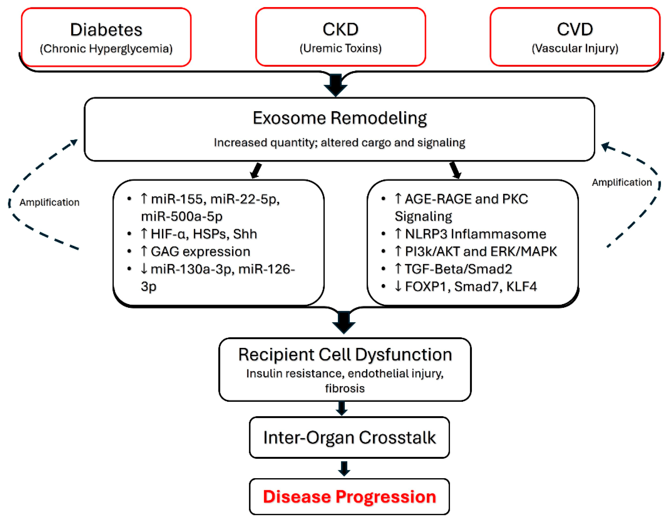



| Signaling Mechanism | Major Mediators | Representative Signaling Pathways | Major Biological Consequences | Reference |
|---|---|---|---|---|
| Recipient-cell interaction | PD-L1 | PD-1 | Immune evasion | [15,95,98,99,100,101,102,103,104] |
| Syntaxin-4; SNAP-23; VAMP-7 | Membrane fusion | Cargo delivery | ||
| DNMT mRNAs | Genome-wide DNA hypermethylation | Epigenetic regulation | ||
| miR-1271-5p | SOX6 repression | Cardiomyocyte survival | ||
| Signal transduction | Microglial exosomal miR-155-5p | NF-κB | Inflammation | [105,107,108,109,110,111,112,113] |
| miR-143-3p | PI3K/AKT | M1 macrophage polarization | ||
| miR-486-5p | PI3K/AKT | Cardiomyocyte survival | ||
| HMGB1 | TLR4/NF-κB/STAT3 | Tumor progression; immune suppression | ||
| miR-21 | STAT3 | Chronic inflammatory signaling | ||
| Inflammatory amplification | miR-30b-5p | NF-κB | Chronic inflammation | [7,115,117,118,119,120,127] |
| SHH | Hormone-sensitive lipase signaling | Insulin resistance | ||
| RBP4 | TNF-α; IL-6 | Macrophage activation | ||
| Metabolic regulation | Adipose tissue macrophage-derived exosomal miR-155 | SOCS1 | Insulin resistance | [121,122,123,124,125,126] |
| miR-34a | M2 macrophage polarization | Metabolic inflammation | ||
| NOX2; NOX4 | MEK1/2-ERK1/2 | Oxidative stress | ||
| Palmitic acid; stearic acid | TLR4 | Impaired insulin sensitivity | ||
| Nutrient availability | mTORC1-SCYL1 | Regulation of exosome secretion |
| Clinical Application | Biological Basis | Current Evidence | Major Limitation | Translational Readiness | References |
|---|---|---|---|---|---|
| Biomarkers & Diagnostics | Exosome- and EV-associated cargo reflects tissue pathophysiology | Human studies | Limited analytical standardization | Early clinical/clinical validation | [215,216,217,218,228,230] |
| Prognostic Value | Exosomal cargo correlates with disease outcome | Human studies | Limited prospective validation | Early clinical | [235,236] |
| Disease Monitoring | Dynamic changes in exosomal cargo reflect disease progression | Human studies | Limited analytical standardization | Early clinical | [238,239] |
| Plasma Exosomes & Systemic Disease | Exosome-mediated systemic signaling | Mechanistic and animal studies | Complex biodistribution and rapid organ uptake | Preclinical | [116,121,237] |
| Long-Term Stability | Protection of exosomal cargo by lipid bilayers | Human studies | Limited analytical standardization | Early clinical | [240] |
| Cargo Loading & Sorting | Selective cargo loading and sorting | Mechanistic studies | Limited mechanistic understanding and reproducibility | Preclinical | [71,241] |
| Therapeutic Use of Endogenous Exosomes | Activation of cytoprotective signaling pathways | Animal studies | Limited clinical validation and mechanistic understanding | Preclinical | [242] |
| MSC-Derived Regenerative Therapy | Transfer of regenerative bioactive molecules | Preclinical studies | Lack of standardized clinical protocols | Preclinical/early clinical | [243,244,245] |
| Engineered Drug Delivery | Surface engineering for targeted drug delivery | Proof-of-concept animal studies | Limited target efficiency, scalable manufacturing, and off-target biodistribution | Preclinical | [246,247,248] |
| Drug Loading | Loading of therapeutic cargo into exosomes and EVs | Preclinical models | Limited loading efficiency, cargo retention, and reproducibility | Preclinical | [249,250,251] |
| Gene Therapy | Delivery of therapeutic nucleic acids | Preclinical studies | Limited cargo loading efficiency and tissue specificity | Preclinical | [252,253,254] |
Disclaimer/Publisher’s Note: The statements, opinions and data contained in all publications are solely those of the individual author(s) and contributor(s) and not of MDPI and/or the editor(s). MDPI and/or the editor(s) disclaim responsibility for any injury to people or property resulting from any ideas, methods, instructions or products referred to in the content. |
© 2026 by the authors. Licensee MDPI, Basel, Switzerland. This article is an open access article distributed under the terms and conditions of the Creative Commons Attribution (CC BY) license.
Share and Cite
Madore, A.; Walsh, N.; Desai, K.; Udoh, G.; Gannavaram, N.; Kandula, A.; Yates, C.; Pillai, S.S.; Sodhi, K.; Goncalves, B.S. Exosome-Mediated Systemic Signaling: Mechanisms, Disease Integration, and Translational Potential. Curr. Issues Mol. Biol. 2026, 48, 845. https://doi.org/10.3390/cimb48080845
Madore A, Walsh N, Desai K, Udoh G, Gannavaram N, Kandula A, Yates C, Pillai SS, Sodhi K, Goncalves BS. Exosome-Mediated Systemic Signaling: Mechanisms, Disease Integration, and Translational Potential. Current Issues in Molecular Biology. 2026; 48(8):845. https://doi.org/10.3390/cimb48080845
Chicago/Turabian StyleMadore, Adam, Nigel Walsh, Kush Desai, Gideon Udoh, Naga Gannavaram, Aishniya Kandula, Cohen Yates, Sneha S. Pillai, Komal Sodhi, and Bruno S. Goncalves. 2026. "Exosome-Mediated Systemic Signaling: Mechanisms, Disease Integration, and Translational Potential" Current Issues in Molecular Biology 48, no. 8: 845. https://doi.org/10.3390/cimb48080845
APA StyleMadore, A., Walsh, N., Desai, K., Udoh, G., Gannavaram, N., Kandula, A., Yates, C., Pillai, S. S., Sodhi, K., & Goncalves, B. S. (2026). Exosome-Mediated Systemic Signaling: Mechanisms, Disease Integration, and Translational Potential. Current Issues in Molecular Biology, 48(8), 845. https://doi.org/10.3390/cimb48080845