Magnesium Sulfate in Neonatal Hypoxic–Ischemic Encephalopathy: Bridging the Gap Between Molecular Neuroprotection and Clinical Outcomes

 , ,
, ,  ,
,  and
and
Abstract
1. Introduction
1.1. Neonatal HIE as an Unmet Clinical Challenge
1.2. Limitations of Therapeutic Hypothermia
1.3. Why Magnesium Sulfate?
1.4. Aim of the Review
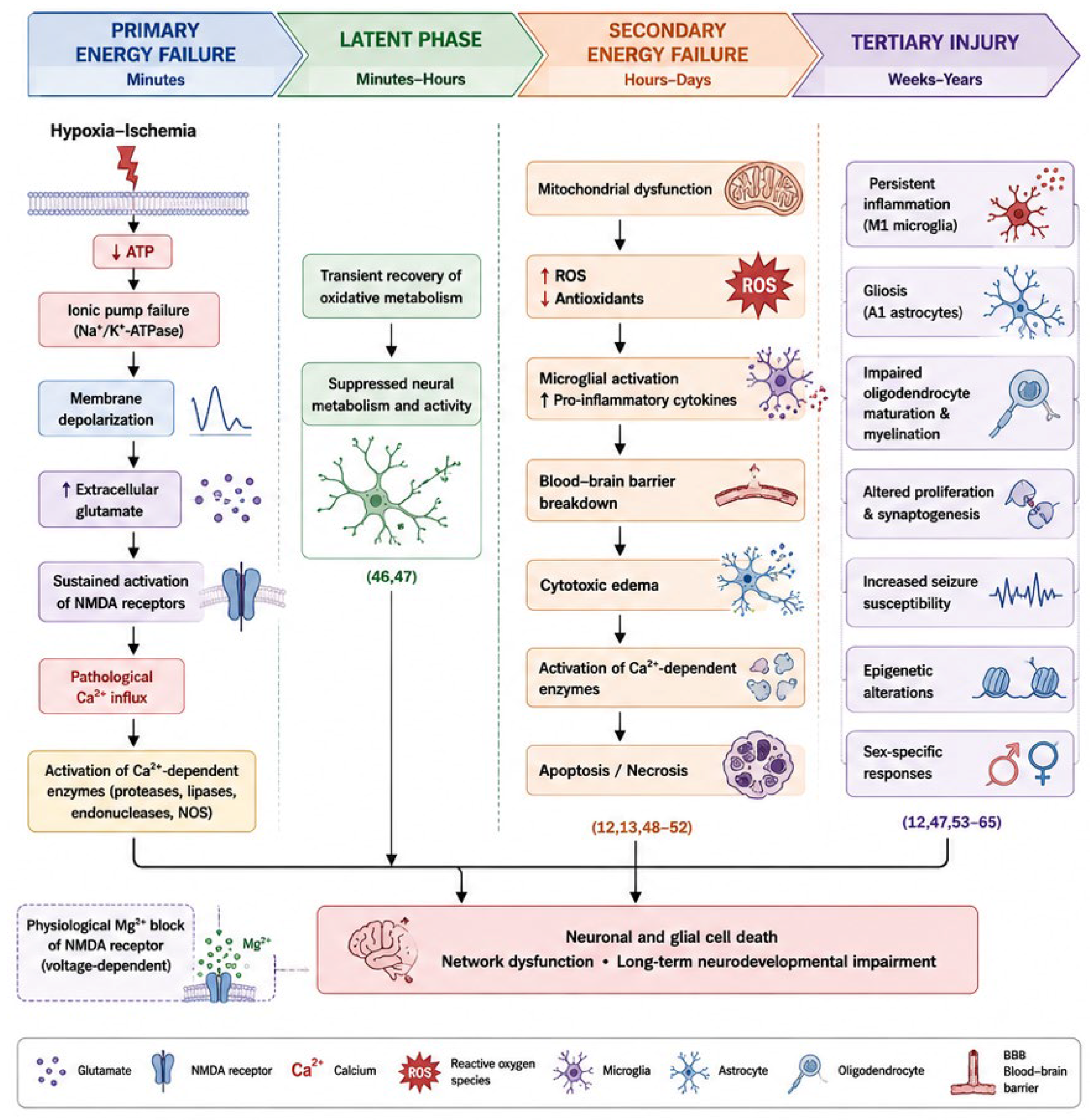
2. Pathophysiology of Hypoxic–Ischemic Brain Injury
2.1. Primary Energy Failure

2.2. Latent Phase
2.3. Secondary Energy Failure
2.4. Tertiary Injury
3. Molecular Basis of Magnesium Sulfate Neuroprotection
4. Clinical Evidence in Neonatal HIE
4.1. Early Pilot Studies
4.2. Recent Meta-Analyses and Systematic Reviews
5. The Translational Paradox
5.1. Why Does Molecular Efficacy Not Translate into Clinical Benefit?
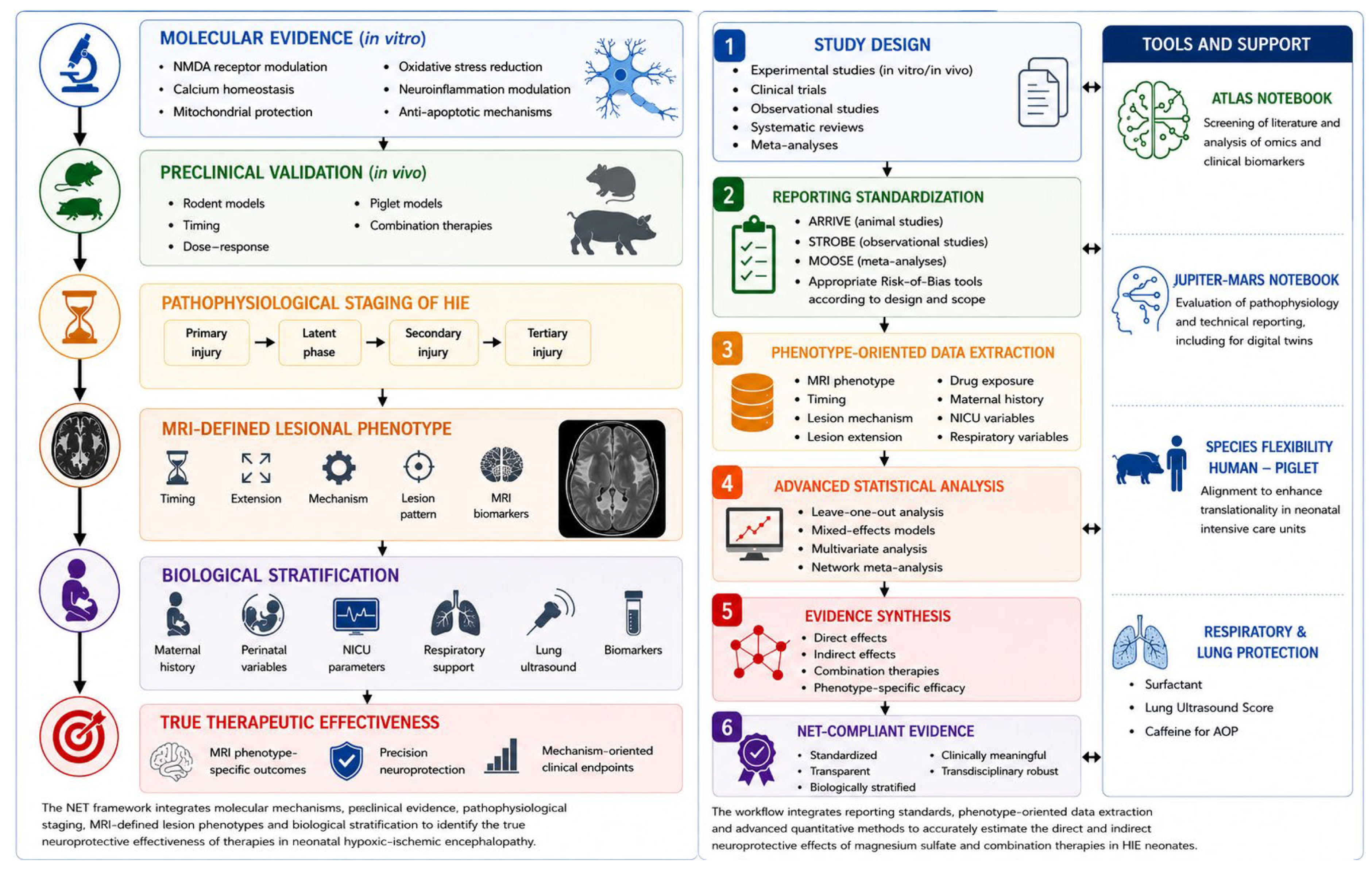
5.2. The Neuroprotection per Effective Therapy (NET) Framework: A Conceptual Proposal for Translational Neonatal Neuroprotection
5.3. Failure of “One Drug-One Mechanism” Approaches
5.4. Tools for Applying NET Principles
6. Towards MRI Biomarkers of Neuroprotective Drug Response
7. Towards Molecular Biomarkers of Neuroprotective Response: The Omics Contribution
8. Conclusions
Author Contributions
Funding
Institutional Review Board Statement
Informed Consent Statement
Data Availability Statement
Acknowledgments
Conflicts of Interest
References
- Global, Regional, and National Burden of Disorders Affecting the Nervous System, 1990-2021: A Systematic Analysis for the Global Burden of Disease Study 2021. Lancet Neurol. 2024, 23, 344–381. [CrossRef] [Scilit] [PubMed]
- Cimino, C.; Sortino, V.; Sapuppo, A.; Saporito, M.A.N.; Scarlata, F.; Falsaperla, R. Beyond Hypoxic–Ischemic Encephalopathy: Genetic Insights and Precision Diagnosis in Neonatal Encephalopathies. Front. Med. 2026, 13, 1868036. [Google Scholar] [CrossRef] [Scilit] [PubMed]
- Chakkarapani, E.; De Vries, L.S.; Ferriero, D.M.; Gunn, A.J. Neonatal Encephalopathy and Hypoxic-Ischemic Encephalopathy: The State of the Art. Pediatr. Res. 2025, 98, 2444–2458. [Google Scholar] [CrossRef] [Scilit] [PubMed]
- Christoffel, K.; Mulkey, S.B. Shifting Outlooks after Neonatal Encephalopathy in the Era of Therapeutic Hypothermia. Pediatr. Res. 2025, 98, 2518–2529. [Google Scholar] [CrossRef] [Scilit] [PubMed]
- Dani, C.; Poggi, C.; Fancelli, C.; Pratesi, S. Changes in Bilirubin in Infants with Hypoxic–Ischemic Encephalopathy. Eur. J. Pediatr. 2018, 177, 1795–1801. [Google Scholar] [CrossRef] [Scilit] [PubMed]
- Espinoza, M.L.; Brundler, M.-A.; Hasan, S.U.; Mohammad, K.; Momin, S.; Al Shaikh, B.; Yusuf, K. Placental Pathology as a Marker of Brain Injury in Infants with Hypoxic Ischemic Encephalopathy. Early Hum. Human. Dev. 2022, 174, 105683. [Google Scholar] [CrossRef] [Scilit] [PubMed]
- Kalogeropoulou, M.-S.; Thomson, L.; Beardsall, K. Continuous Glucose Monitoring during Therapeutic Hypothermia for Hypoxic Ischaemic Encephalopathy: A Feasibility Study. Arch. Dis. Child. Fetal Neonatal Ed. 2023, 108, 309–315. [Google Scholar] [CrossRef] [Scilit] [PubMed]
- Basu, S.K.; Salemi, J.L.; Gunn, A.J.; Kaiser, J.R. Hyperglycaemia in Infants with Hypoxic–Ischaemic Encephalopathy Is Associated with Improved Outcomes after Therapeutic Hypothermia: A Post Hoc Analysis of the CoolCap Study. Arch. Dis. Child. Fetal Neonatal Ed. 2017, 102, F299–F306. [Google Scholar] [CrossRef] [Scilit] [PubMed]
- Nadeem, M.; Murray, D.M.; Boylan, G.B.; Dempsey, E.M.; Ryan, C.A. Early Blood Glucose Profile and Neurodevelopmental Outcome at Two Years in Neonatal Hypoxic-Ischaemic Encephalopathy. BMC Pediatr. 2011, 11, 10. [Google Scholar] [CrossRef] [Scilit] [PubMed]
- Bersani, I.; Gasparroni, G.; Bashir, M.; Aboulgar, H.; Mufeed, H.; Iskander, I.; Kornacka, M.; Gruzfeld, D.; Dotta, A.; Campi, F.; et al. Early Predictors of Abnormal MRI Patterns in Asphyxiated Infants: S100B Protein Urine Levels. Clin. Chem. Lab. Med. (CCLM) 2022, 60, 1745–1752. [Google Scholar] [CrossRef] [Scilit] [PubMed]
- Chan, N.H.; Hawkins, C.C.; Rodrigues, B.V.; Cornet, M.; Gonzalez, F.F.; Wu, Y.W. Neuroprotection for Neonatal Hypoxic–Ischemic Encephalopathy: A Review of Novel Therapies Evaluated in Clinical Studies. Dev. Med. Child. Neuro 2025, 67, 591–599. [Google Scholar] [CrossRef] [Scilit]
- Zhou, K.Q.; Dhillon, S.K.; Bennet, L.; Davidson, J.O.; Gunn, A.J. How Do We Reach the Goal of Personalized Medicine for Neuroprotection in Neonatal Hypoxic-Ischemic Encephalopathy? Semin. Perinatol. 2024, 48, 151930. [Google Scholar] [CrossRef] [Scilit]
- Dixon, B.; Reis, C.; Ho, W.; Tang, J.; Zhang, J. Neuroprotective Strategies after Neonatal Hypoxic Ischemic Encephalopathy. Int. J. Mol. Sci. 2015, 16, 22368–22401. [Google Scholar] [CrossRef] [Scilit] [PubMed]
- Dhillon, S.K.; Lear, C.A.; Galinsky, R.; Wassink, G.; Davidson, J.O.; Juul, S.; Robertson, N.J.; Gunn, A.J.; Bennet, L. The Fetus at the Tipping Point: Modifying the Outcome of Fetal Asphyxia. J. Physiol. 2018, 596, 5571–5592. [Google Scholar] [CrossRef] [Scilit] [PubMed]
- Zhou, T.; Liang, P.; Dai, N.; Ma, M.; Lin, J.; Zhu, J.; Ren, G. Therapeutic Hypothermia for Neonates: A Bibliometric Analysis and Visualization Research. Front. Neurol. 2025, 16, 1565749. [Google Scholar] [CrossRef] [Scilit] [PubMed]
- Ghi, T.; Mercuri, E.; Ramenghi, L.A.; De Luca, D. Uncertainty in Brain Imaging after Therapeutic Hypothermia. Lancet Child. Adolesc. Health 2025, 9, 759–760. [Google Scholar] [CrossRef] [Scilit] [PubMed]
- Azzopardi, D.V.; Strohm, B.; Edwards, A.D.; Dyet, L.; Halliday, H.L.; Juszczak, E.; Kapellou, O.; Levene, M.; Marlow, N.; Porter, E.; et al. Moderate Hypothermia to Treat Perinatal Asphyxial Encephalopathy. N. Engl. J. Med. 2009, 361, 1349–1358. [Google Scholar] [CrossRef] [Scilit] [PubMed]
- Rutherford, M.; Ramenghi, L.A.; Edwards, A.D.; Brocklehurst, P.; Halliday, H.; Levene, M.; Strohm, B.; Thoresen, M.; Whitelaw, A.; Azzopardi, D. Assessment of Brain Tissue Injury after Moderate Hypothermia in Neonates with Hypoxic–Ischaemic Encephalopathy: A Nested Substudy of a Randomised Controlled Trial. Lancet Neurol. 2010, 9, 39–45. [Google Scholar] [CrossRef] [Scilit] [PubMed]
- Flyger, H.; Holdsworth, S.J.; Gunn, A.J.; Bennet, L.; Abbasi, H. Temporal Dynamics of Neonatal Hypoxic–Ischemic Encephalopathy Injuries on Magnetic Resonance Imaging. Neural Regen. Res. 2025, 20, 3144–3150. [Google Scholar] [CrossRef] [Scilit] [PubMed]
- Zhuang, X.; Jin, K.; Li, J.; Yin, Y.; Dong, X.; Lin, H. A Radiomics-Based Study of Deep Medullary Veins in Infants: Evaluation of Neonatal Brain Injury with Hypoxic-Ischemic Encephalopathy via Susceptibility-Weighted Imaging. Front. Neurosci. 2022, 16, 1093499. [Google Scholar] [CrossRef] [Scilit] [PubMed]
- Johnston, M.V.; Fatemi, A.; Wilson, M.A.; Northington, F. Treatment Advances in Neonatal Neuroprotection and Neurointensive Care. Lancet Neurol. 2011, 10, 372–382. [Google Scholar] [CrossRef] [Scilit] [PubMed]
- Robertson, N.J.; Tan, S.; Groenendaal, F.; Van Bel, F.; Juul, S.E.; Bennet, L.; Derrick, M.; Back, S.A.; Valdez, R.C.; Northington, F.; et al. Which Neuroprotective Agents Are Ready for Bench to Bedside Translation in the Newborn Infant? J. Pediatr. 2012, 160, 544–552.e4. [Google Scholar] [CrossRef] [Scilit] [PubMed]
- Lingam, I.; Robertson, N.J. Magnesium as a Neuroprotective Agent: A Review of Its Use in the Fetus, Term Infant with Neonatal Encephalopathy, and the Adult Stroke Patient. Dev. Neurosci. 2018, 40, 1–12. [Google Scholar] [CrossRef] [Scilit] [PubMed]
- Fawcett, W.J.; Haxby, E.J.; Male, D.A. Magnesium: Physiology and Pharmacology. Br. J. Anaesth. 1999, 83, 302–320. [Google Scholar] [CrossRef] [Scilit] [PubMed]
- Den Bakker, E.; Smith, D.E.C.; Finken, M.J.J.; Wamelink, M.M.C.; Salomons, G.S.; Van De Kamp, J.M.; Bökenkamp, A. Sulfate: A Neglected (but Potentially Highly Relevant) Anion. In Essays in Biochemistry; Portland Press Limited: London, UK, 2024; Volume 68, pp. 391–399. [Google Scholar] [CrossRef] [Scilit] [PubMed]
- Le Dieu-Lugon, B.; Dupré, N.; Derambure, C.; Janin, F.; Gonzalez, B.J.; Marret, S.; Arabo, A.; Leroux, P. Effect of Neuroprotective Magnesium Sulfate Treatment on Brain Transcription Response to Hypoxia Ischemia in Neonate Mice. Int. J. Mol. Sci. 2021, 22, 4253. [Google Scholar] [CrossRef] [Scilit] [PubMed]
- Galinsky, R.; Dean, J.M.; Lingam, I.; Robertson, N.J.; Mallard, C.; Bennet, L.; Gunn, A.J. A Systematic Review of Magnesium Sulfate for Perinatal Neuroprotection: What Have We Learnt from the Past Decade? Front. Neurol. 2020, 11, 449. [Google Scholar] [CrossRef] [Scilit] [PubMed]
- Robertson, C.S.; Foltz, R.; Grossman, R.G.; Goodman, J.C. Protection against Experimental Ischemic Spinal Cord Injury. J. Neurosurg. 1986, 64, 633–642. [Google Scholar] [CrossRef] [Scilit] [PubMed]
- Levene, M.; Blennow, M.; Whitelaw, A.; Hanko, E.; Fellman, V.; Hartley, R. Acute Effects of Two Different Doses of Magnesium Sulphate in Infants with Birth Asphyxia. Arch. Dis. Child.-Fetal Neonatal Ed. 1995, 73, F174–F177. [Google Scholar] [CrossRef] [Scilit] [PubMed]
- Levene, M.I.; Evans, D.J.; Mason, S.; Brown, J. An International Network for Evaluating Neuroprotective Therapy after Severe Birth Asphyxia. Semin. Perinatol. 1999, 23, 226–233. [Google Scholar] [CrossRef] [Scilit] [PubMed]
- Doyle, L.W.; Crowther, C.A.; Middleton, P.; Marret, S.; Rouse, D. Magnesium Sulphate for Women at Risk of Preterm Birth for Neuroprotection of the Fetus. In Cochrane Database of Systematic Reviews; John Wiley & Sons, Ltd.: Hoboken, NJ, USA, 2009. [Google Scholar] [CrossRef] [Scilit] [PubMed]
- Crowther, C.A. Effect of Magnesium Sulfate Given for Neuroprotection Before Preterm BirthA Randomized Controlled Trial. JAMA 2003, 290, 2669. [Google Scholar] [CrossRef] [Scilit] [PubMed]
- Wolf, H.; Huusom, L.; Henriksen, T.; Hegaard, H.; Brok, J.; Pinborg, A. Magnesium Sulphate for Fetal Neuroprotection at Imminent Risk for Preterm Delivery: A Systematic Review with Meta-analysis and Trial Sequential Analysis. BJOG Int. J. Obstet. Gynaecol. 2020, 127, 1180–1188. [Google Scholar] [CrossRef] [Scilit] [PubMed]
- Crowther, C.A.; Middleton, P.F.; Voysey, M.; Askie, L.; Duley, L.; Pryde, P.G.; Marret, S.; Doyle, L.W. for the AMICABLE Group Assessing the Neuroprotective Benefits for Babies of Antenatal Magnesium Sulphate: An Individual Participant Data Meta-Analysis. PLoS Med. 2017, 14, e1002398. [Google Scholar] [CrossRef] [Scilit] [PubMed]
- Maier, J.A.M.; Locatelli, L.; Fedele, G.; Cazzaniga, A.; Mazur, A. Magnesium and the Brain: A Focus on Neuroinflammation and Neurodegeneration. Int. J. Mol. Sci. 2022, 24, 223. [Google Scholar] [CrossRef] [Scilit] [PubMed]
- Koning, G.; Leverin, A.-L.; Nair, S.; Schwendimann, L.; Ek, J.; Carlsson, Y.; Gressens, P.; Thornton, C.; Wang, X.; Mallard, C.; et al. Magnesium Induces Preconditioning of the Neonatal Brain via Profound Mitochondrial Protection. J. Cereb. Blood Flow. Metab. 2019, 39, 1038–1055. [Google Scholar] [CrossRef] [Scilit] [PubMed]
- Chollat, C.; Marret, S. Magnesium Sulfate and Fetal Neuroprotection: Overview of Clinical Evidence. Neural Regen. Res. 2018, 13, 2044–2049. [Google Scholar] [CrossRef] [Scilit] [PubMed]
- Moradi, Y.; Khateri, R.; Haghighi, L.; Dehghani, S.; Hanis, S.M.; Valipour, M.; Najmi, Z.; Fathollahy, Z.; Allahmoradi, M.; Mansori, K. The Effect of Antenatal Magnesium Sulfate on Intraventricular Hemorrhage in Premature Infants: A Systematic Review and Meta-Analysis. Obs. Obstet. Gynecol. Sci. 2020, 63, 395–406. [Google Scholar] [CrossRef] [Scilit] [PubMed]
- Al Rifai, M.T.; Al Tawil, K.I. The Neurological Outcome of Isolated PVL and Severe IVH in Preterm Infants: Is It Fair to Compare? Pediatr. Neurol. 2015, 53, 427–433. [Google Scholar] [CrossRef] [Scilit] [PubMed]
- Jary, S.; De Carli, A.; Ramenghi, L.A.; Whitelaw, A. Impaired Brain Growth and Neurodevelopment in Preterm Infants with Posthaemorrhagic Ventricular Dilatation. Acta Paediatr. 2012, 101, 743–748. [Google Scholar] [CrossRef] [Scilit] [PubMed]
- El-Dib, M.; Abend, N.S.; Austin, T.; Boylan, G.; Chock, V.; Cilio, M.R.; Greisen, G.; Hellström-Westas, L.; Lemmers, P.; Pellicer, A.; et al. Neuromonitoring in Neonatal Critical Care Part I: Neonatal Encephalopathy and Neonates with Possible Seizures. Pediatr. Res. 2023, 94, 64–73. [Google Scholar] [CrossRef] [Scilit] [PubMed]
- Calandrino, A.; Minghetti, D.; Brigati, G.; Parodi, A.; Nozza, P.; Ramenghi, L.A. Disruption of Cerebellar Granular Layer as a Consequence of Germinal Matrix Intraventricular Hemorrhage in Extreme Prematurity: An Acute Direct Mechanism Too? Pediatr. Neurol. 2024, 152, 20–22. [Google Scholar] [CrossRef] [Scilit] [PubMed]
- Boswinkel, V.; Steggerda, S.J.; Fumagalli, M.; Parodi, A.; Ramenghi, L.A.; Groenendaal, F.; Dudink, J.; Benders, M.N.; Knol, R.; De Vries, L.S.; et al. The CHOPIn Study: A Multicenter Study on Cerebellar Hemorrhage and Outcome in Preterm Infants. Cerebellum 2019, 18, 989–998. [Google Scholar] [CrossRef] [Scilit] [PubMed]
- Benders, M.J.N.L.; Kersbergen, K.J.; De Vries, L.S. Neuroimaging of White Matter Injury, Intraventricular and Cerebellar Hemorrhage. Clin. Perinatol. 2014, 41, 69–82. [Google Scholar] [CrossRef] [Scilit] [PubMed]
- Poppe, T.; Thompson, B.; Boardman, J.P.; Bastin, M.E.; Alsweiler, J.; Deib, G.; Harding, J.E.; Crowther, C.A. Effect of Antenatal Magnesium Sulphate on MRI Biomarkers of White Matter Development at Term Equivalent Age: The MagNUM Study. eBioMedicine 2022, 78, 103923. [Google Scholar] [CrossRef] [Scilit] [PubMed]
- Ufkes, S.; Kennedy, E.; Poppe, T.; Miller, S.P.; Thompson, B.; Guo, J.; Harding, J.E.; Crowther, C.A. Prenatal Magnesium Sulfate and Functional Connectivity in Offspring at Term-Equivalent Age. JAMA Netw. Open 2024, 7, e2413508. [Google Scholar] [CrossRef] [Scilit] [PubMed]
- Lee, Y.A. White Matter Injury of Prematurity: Its Mechanisms and Clinical Features. J. Pathol. Transl. Med. 2017, 51, 449–455. [Google Scholar] [CrossRef] [Scilit] [PubMed]
- Ballabh, P.; de Vries, L.S. White Matter Injury in Infants with Intraventricular Haemorrhage: Mechanisms and Therapies. Nat. Rev. Neurol. 2021, 17, 199–214. [Google Scholar] [CrossRef] [Scilit] [PubMed]
- Galinsky, R.; Bennet, L.; Groenendaal, F.; Lear, C.A.; Tan, S.; Van Bel, F.; Juul, S.E.; Robertson, N.J.; Mallard, C.; Gunn, A.J. Magnesium Is Not Consistently Neuroprotective for Perinatal Hypoxia-Ischemia in Term-Equivalent Models in Preclinical Studies: A Systematic Review. Dev. Neurosci. 2014, 36, 73–82. [Google Scholar] [CrossRef] [Scilit] [PubMed]
- Fei, Q.; Wang, D.; Yuan, T. Comparison of Different Adjuvant Therapies for Hypothermia in Neonates with Hypoxic-Ischemic Encephalopathy: A Systematic Review and Network Meta-Analysis. Indian J. Pediatr. 2024, 91, 235–241. [Google Scholar] [CrossRef] [Scilit] [PubMed]
- Basak, S. Artificial Intelligence and Machine Learning-Guided Bio-Orthogonal Engineering of Smart Soft Polymeric Nanocarriers for Precision Drug Delivery and Translational Nanomedicine. Front. Soft Matter 2026, 6, 1883618. [Google Scholar] [CrossRef] [Scilit]
- Sukhera, J. Narrative Reviews: Flexible, Rigorous, and Practical. J. Grad. Med. Educ. 2022, 14, 414–417. [Google Scholar] [CrossRef] [Scilit] [PubMed]
- McLean, C.; Ferriero, D. Mechanisms of Hypoxic—Ischemic Injury in the Term Infant. Semin. Perinatol. 2004, 28, 425–432. [Google Scholar] [CrossRef] [Scilit] [PubMed]
- Zhang, X.; Peng, K.; Zhang, X. The Function of the NMDA Receptor in Hypoxic-Ischemic Encephalopathy. Front. Neurosci. 2020, 14, 567665. [Google Scholar] [CrossRef] [Scilit] [PubMed]
- Nair, J.; Kumar, V.H.S. Current and Emerging Therapies in the Management of Hypoxic Ischemic Encephalopathy in Neonates. Children 2018, 5, 99. [Google Scholar] [CrossRef] [Scilit] [PubMed]
- Chakkarapani, A.A.; Aly, H.; Benders, M.; Cotten, C.M.; El-Dib, M.; Gressens, P.; Hagberg, H.; Sabir, H.; Wintermark, P.; Robertson, N.J. Therapies for Neonatal Encephalopathy: Targeting the Latent, Secondary and Tertiary Phases of Evolving Brain Injury. Semin. Fetal Neonatal Med. 2021, 26, 101256. [Google Scholar] [CrossRef] [Scilit] [PubMed]
- Davidson, J.O.; Gonzalez, F.; Gressens, P.; Gunn, A.J. Update on Mechanisms of the Pathophysiology of Neonatal Encephalopathy. Semin. Fetal Neonatal Med. 2021, 26, 101267. [Google Scholar] [CrossRef] [Scilit] [PubMed]
- Li, B.; Concepcion, K.; Meng, X.; Zhang, L. Brain-Immune Interactions in Perinatal Hypoxic-Ischemic Brain Injury. Prog. Neurobiol. 2017, 159, 50–68. [Google Scholar] [CrossRef] [Scilit] [PubMed]
- Samaiya, P.K.; Krishnamurthy, S.; Kumar, A. Mitochondrial Dysfunction in Perinatal Asphyxia: Role in Pathogenesis and Potential Therapeutic Interventions. Mol. Cell Biochem. 2021, 476, 4421–4434. [Google Scholar] [CrossRef] [Scilit] [PubMed]
- Perrone, S.; Tataranno, L.M.; Stazzoni, G.; Ramenghi, L.; Buonocore, G. Brain Susceptibility to Oxidative Stress in the Perinatal Period. J. Matern.-Fetal Neonatal Med. 2015, 28, 2291–2295. [Google Scholar] [CrossRef] [Scilit] [PubMed]
- Panfoli, I.; Candiano, G.; Malova, M.; De Angelis, L.; Cardiello, V.; Buonocore, G.; Ramenghi, L.A. Oxidative Stress as a Primary Risk Factor for Brain Damage in Preterm Newborns. Front. Pediatr. 2018, 6, 369. [Google Scholar] [CrossRef] [Scilit] [PubMed]
- Polglase, G.R.; Miller, S.L.; Barton, S.K.; Kluckow, M.; Gill, A.W.; Hooper, S.B.; Tolcos, M. Respiratory Support for Premature Neonates in the Delivery Room: Effects on Cardiovascular Function and the Development of Brain Injury. Pediatr. Res. 2014, 75, 682–688. [Google Scholar] [CrossRef] [Scilit] [PubMed]
- Fleiss, B.; Gressens, P. Tertiary Mechanisms of Brain Damage: A New Hope for Treatment of Cerebral Palsy? Lancet Neurol. 2012, 11, 556–566. [Google Scholar] [CrossRef] [Scilit] [PubMed]
- Novak, I.; Morgan, C. High-Risk Follow-up: Early Intervention and Rehabilitation. Handb. Clin. Neurol. 2019, 162, 483–510. [Google Scholar] [CrossRef] [Scilit] [PubMed]
- Jaworska, J.; Ziemka-Nalecz, M.; Sypecka, J.; Zalewska, T. The Potential Neuroprotective Role of a Histone Deacetylase Inhibitor, Sodium Butyrate, after Neonatal Hypoxia-Ischemia. J. Neuroinflamm. 2017, 14, 34. [Google Scholar] [CrossRef] [Scilit] [PubMed]
- Zhou, D.; Ji, L.; Chen, Y. TSPO Modulates IL-4-Induced Microglia/Macrophage M2 Polarization via PPAR-γ Pathway. J. Mol. Neurosci. 2020, 70, 542–549. [Google Scholar] [CrossRef] [Scilit] [PubMed]
- Miyamoto, N.; Magami, S.; Inaba, T.; Ueno, Y.; Hira, K.; Kijima, C.; Nakajima, S.; Yamashiro, K.; Urabe, T.; Hattori, N. The Effects of A1/A2 Astrocytes on Oligodendrocyte Linage Cells against White Matter Injury under Prolonged Cerebral Hypoperfusion. Glia 2020, 68, 1910–1924. [Google Scholar] [CrossRef] [Scilit] [PubMed]
- Yun, S.P.; Kam, T.-I.; Panicker, N.; Kim, S.; Oh, Y.; Park, J.-S.; Kwon, S.-H.; Park, Y.J.; Karuppagounder, S.S.; Park, H.; et al. Block of A1 Astrocyte Conversion by Microglia Is Neuroprotective in Models of Parkinson’s Disease. Nat. Med. 2018, 24, 931–938. [Google Scholar] [CrossRef] [Scilit] [PubMed]
- Fleiss, B.; Van Steenwinckel, J.; Bokobza, C.; Shearer, I.K.; Ross-Munro, E.; Gressens, P. Microglia-Mediated Neurodegeneration in Perinatal Brain Injuries. Biomolecules 2021, 11, 99. [Google Scholar] [CrossRef] [Scilit] [PubMed]
- Karalis, F.; Soubasi, V.; Georgiou, T.; Nakas, C.T.; Simeonidou, C.; Guiba-Tziampiri, O.; Spandou, E. Resveratrol Ameliorates Hypoxia/Ischemia-Induced Behavioral Deficits and Brain Injury in the Neonatal Rat Brain. Brain Res. 2011, 1425, 98–110. [Google Scholar] [CrossRef] [Scilit] [PubMed]
- Mandrekar-Colucci, S.; Karlo, J.C.; Landreth, G.E. Mechanisms Underlying the Rapid Peroxisome Proliferator-Activated Receptor-γ-Mediated Amyloid Clearance and Reversal of Cognitive Deficits in a Murine Model of Alzheimer’s Disease. J. Neurosci. 2012, 32, 10117–10128. [Google Scholar] [CrossRef] [Scilit] [PubMed]
- Jin, Q.; Cheng, J.; Liu, Y.; Wu, J.; Wang, X.; Wei, S.; Zhou, X.; Qin, Z.; Jia, J.; Zhen, X. Improvement of Functional Recovery by Chronic Metformin Treatment Is Associated with Enhanced Alternative Activation of Microglia/Macrophages and Increased Angiogenesis and Neurogenesis Following Experimental Stroke. Brain Behav. Immun. 2014, 40, 131–142. [Google Scholar] [CrossRef] [Scilit] [PubMed]
- Zhao, Q.; Wang, Q.; Wang, J.; Tang, M.; Huang, S.; Peng, K.; Han, Y.; Zhang, J.; Liu, G.; Fang, Q.; et al. Maternal Immune Activation-Induced PPARγ-Dependent Dysfunction of Microglia Associated with Neurogenic Impairment and Aberrant Postnatal Behaviors in Offspring. Neurobiol. Dis. 2019, 125, 1–13. [Google Scholar] [CrossRef] [Scilit] [PubMed]
- Kalish, B.T.; Kim, E.; Finander, B.; Duffy, E.E.; Kim, H.; Gilman, C.K.; Yim, Y.S.; Tong, L.; Kaufman, R.J.; Griffith, E.C.; et al. Maternal Immune Activation in Mice Disrupts Proteostasis in the Fetal Brain. Nat. Neurosci. 2021, 24, 204–213. [Google Scholar] [CrossRef] [Scilit] [PubMed]
- Kim, S.; Kim, H.; Yim, Y.S.; Ha, S.; Atarashi, K.; Tan, T.G.; Longman, R.S.; Honda, K.; Littman, D.R.; Choi, G.B.; et al. Maternal Gut Bacteria Promote Neurodevelopmental Abnormalities in Mouse Offspring. Nature 2017, 549, 528–532. [Google Scholar] [CrossRef] [Scilit] [PubMed]
- Santoni, M.; Sagheddu, C.; Serra, V.; Mostallino, R.; Castelli, M.P.; Pisano, F.; Scherma, M.; Fadda, P.; Muntoni, A.L.; Zamberletti, E.; et al. Maternal Immune Activation Impairs Endocannabinoid Signaling in the Mesolimbic System of Adolescent Male Offspring. Brain Behav. Immun. 2023, 109, 271–284. [Google Scholar] [CrossRef] [Scilit] [PubMed]
- She, K.; Yuan, N.; Huang, M.; Zhu, W.; Tang, M.; Ma, Q.; Chen, J. Emerging Role of Microglia in the Developing Dopaminergic System: Perturbation by Early Life Stress. Neural Regen. Res. 2026, 21, 126–140. [Google Scholar] [CrossRef] [Scilit] [PubMed]
- McLeod, R.M.; Rosenkrantz, T.S.; Fitch, R.H. Antenatal Magnesium Sulfate Benefits Female Preterm Infants but Results in Poor Male Outcomes. Pharmaceuticals 2024, 17, 218. [Google Scholar] [CrossRef] [Scilit] [PubMed]
- Sugimoto, J.; Romani, A.M.; Valentin-Torres, A.M.; Luciano, A.A.; Ramirez Kitchen, C.M.; Funderburg, N.; Mesiano, S.; Bernstein, H.B. Magnesium Decreases Inflammatory Cytokine Production: A Novel Innate Immunomodulatory Mechanism. J. Immunol. 2012, 188, 6338–6346. [Google Scholar] [CrossRef] [Scilit] [PubMed]
- Peruche, B.; Krieglstein, J. Mechanisms of Drug Actions against Neuronal Damage Caused by Ischemia—An Overview. Prog. Neuro-Psychopharmacol. Biol. Psychiatry 1993, 17, 21–70. [Google Scholar] [CrossRef] [Scilit] [PubMed]
- Nowak, L.; Bregestovski, P.; Ascher, P.; Herbet, A.; Prochiantz, A. Magnesium Gates Glutamate-Activated Channels in Mouse Central Neurones. Nature 1984, 307, 462–465. [Google Scholar] [CrossRef] [Scilit] [PubMed]
- Okazaki, K.; Nakamura, S.; Koyano, K.; Konishi, Y.; Kondo, M.; Kusaka, T. Neonatal Asphyxia as an Inflammatory Disease: Reactive Oxygen Species and Cytokines. Front. Pediatr. 2023, 11, 1070743. [Google Scholar] [CrossRef] [Scilit] [PubMed]
- Kumar, C.; Adhisivam, B.; Bobby, Z.; Bhat, B.V. Magnesium Sulfate as an Adjunct to Therapeutic Hypothermia in the Management of Term Infants with Hypoxic–Ischemic Encephalopathy: A Randomized, Parallel-Group, Controlled Trial. Indian J. Pediatr. 2023, 90, 886–892. [Google Scholar] [CrossRef] [Scilit] [PubMed]
- Molloy, E.J.; El-Dib, M.; Juul, S.E.; Benders, M.; Gonzalez, F.; Bearer, C.; Wu, Y.W.; Robertson, N.J.; Hurley, T.; Branagan, A.; et al. Neuroprotective Therapies in the NICU in Term Infants: Present and Future. Pediatr. Res. 2023, 93, 1819–1827. [Google Scholar] [CrossRef] [Scilit] [PubMed]
- Molloy, E.J.; El-Dib, M.; Soul, J.; Juul, S.; Gunn, A.J.; Bender, M.; Gonzalez, F.; Bearer, C.; Wu, Y.; Robertson, N.J.; et al. Neuroprotective Therapies in the NICU in Preterm Infants: Present and Future (Neonatal Neurocritical Care Series). Pediatr. Res. 2024, 95, 1224–1236. [Google Scholar] [CrossRef] [Scilit] [PubMed]
- Fleiss, B.; Gressens, P. Neuroprotection of the Preterm Brain. Handb. Clin. Neurol. 2019, 162, 315–328. [Google Scholar] [CrossRef] [Scilit] [PubMed]
- Nonomura, M.; Harada, S.; Asada, Y.; Matsumura, H.; Iwami, H.; Tanaka, Y.; Ichiba, H. Combination Therapy with Erythropoietin, Magnesium Sulfate and Hypothermia for Hypoxic-Ischemic Encephalopathy: An Open-Label Pilot Study to Assess the Safety and Feasibility. BMC Pediatr. 2019, 19, 13. [Google Scholar] [CrossRef] [Scilit] [PubMed]
- Bhat, M.A.; Charoo, B.A.; Bhat, J.I.; Ahmad, S.M.; Ali, S.W.; Mufti, M.-H. Magnesium Sulfate in Severe Perinatal Asphyxia: A Randomized, Placebo-Controlled Trial. Pediatrics 2009, 123, e764–e769. [Google Scholar] [CrossRef] [Scilit] [PubMed]
- Ayeni, V.A.; Abdullahi, A.A.; Isah, A.; Ayamolowo, L.B.; Adeyemi, T.; Oladimeji, M.A.; Ahwinahwi, U.S.; Chukwuike, V.I.; Okeke, O.P.; Abodunrin, O.R.; et al. Pharmacologic Neuroprotective Agents for the Treatment of Perinatal Asphyxia in Low-Income and Lower-Middle-Income Countries: A Systematic Review and Meta-Analysis of Randomised Controlled Trials. PLoS ONE 2025, 20, e0337798. [Google Scholar] [CrossRef] [Scilit] [PubMed]
- Juul, S.E.; Comstock, B.A.; Wadhawan, R.; Mayock, D.E.; Courtney, S.E.; Robinson, T.; Ahmad, K.A.; Bendel-Stenzel, E.; Baserga, M.; LaGamma, E.F.; et al. A Randomized Trial of Erythropoietin for Neuroprotection in Preterm Infants. N. Engl. J. Med. 2020, 382, 233–243. [Google Scholar] [CrossRef] [Scilit] [PubMed]
- Bellini, V.; Ori, E.; Coccolini, F.; Bignami, E. Evidence Pyramid and Artificial Intelligence: A Metamorphosis of Clinical Research. Discov. Health Syst. 2023, 2, 40. [Google Scholar] [CrossRef] [Scilit]
- Gowda, B.B.; Rath, C.; Muthusamy, S.; Nagarajan, L.; Rao, S. Outcomes of Neonates with Hypoxic-Ischemic Encephalopathy Treated with Magnesium Sulfate: A Systematic Review with Meta-Analysis. J. Pediatr. 2023, 262, 113610. [Google Scholar] [CrossRef] [Scilit] [PubMed]
- Fumagalli, M.; Bassi, L.; Sirgiovanni, I.; Mosca, F.; Sannia, A.; Ramenghi, L.A. From Germinal Matrix to Cerebellar Haemorrhage. J. Matern.-Fetal Neonatal Med. 2015, 28, 2280–2285. [Google Scholar] [CrossRef] [Scilit] [PubMed]
- Sannia, A.; Natalizia, A.R.; Parodi, A.; Malova, M.; Fumagalli, M.; Rossi, A.; Ramenghi, L.A. Different Gestational Ages and Changing Vulnerability of the Premature Brain. J. Matern.-Fetal Neonatal Med. 2015, 28, 2268–2272. [Google Scholar] [CrossRef] [Scilit] [PubMed]
- Gressens, P.; Le Verche, V.; Fraser, M.; Rousset, C.I.; Schwendimann, L.; Bennet, L.; George, S.A.; Wang, X.; Mallard, C.; Tilley, B.C.; et al. Pitfalls in the Quest of Neuroprotectants for the Perinatal Brain. Dev. Neurosci. 2011, 33, 189–198. [Google Scholar] [CrossRef] [Scilit] [PubMed]
- Leonardo, C.C.; Pennypacker, K.R. Neuroinflammation and MMPs: Potential Therapeutic Targets in Neonatal Hypoxic-Ischemic Injury. J. Neuroinflamm. 2009, 6, 13. [Google Scholar] [CrossRef] [Scilit] [PubMed]
- Dammann, O.; Ferriero, D.; Gressens, P. Neonatal Encephalopathy or Hypoxic-Ischemic Encephalopathy? Appropriate Terminology Matters. Pediatr. Res. 2011, 70, 1–2. [Google Scholar] [CrossRef] [Scilit] [PubMed]
- Minor, K.C.; Liu, J.; Druzin, M.L.; El-Sayed, Y.Y.; Hintz, S.R.; Bonifacio, S.L.; Leonard, S.A.; Lee, H.C.; Profit, J.; Karakash, S.D. Magnesium Sulfate and Risk of Hypoxic-Ischemic Encephalopathy in a High-Risk Cohort. Am. J. Obs. Obstet. Gynecol. 2024, 231, 647.e1–647.e12. [Google Scholar] [CrossRef] [Scilit] [PubMed]
- Sera, F.; Armstrong, B.; Blangiardo, M.; Gasparrini, A. An Extended Mixed-effects Framework for Meta-analysis. Stat. Med. 2019, 38, 5429–5444. [Google Scholar] [CrossRef] [Scilit] [PubMed]
- Lunny, C.; Higgins, J.P.T.; White, I.R.; Dias, S.; Hutton, B.; Wright, J.M.; Veroniki, A.-A.; Whiting, P.F.; Tricco, A.C. Risk of Bias in Network Meta-Analysis (RoB NMA) Tool. BMJ 2025, 388, e079839. [Google Scholar] [CrossRef] [Scilit] [PubMed]
- Florez, I.D.; De La Cruz-Mena, J.E.; Veroniki, A.-A. Network Meta-Analysis: A Powerful Tool for Clinicians, Decision-Makers, and Methodologists. J. Clin. Epidemiol. 2024, 176, 111537. [Google Scholar] [CrossRef] [Scilit] [PubMed]
- Arun Babu, T.; Ballambattu, V.B. Charting the Course for Adjuvant Neuroprotective Therapies in Neonatal Hypoxic-Ischemic Encephalopathy: Insights from a Network Meta-Analysis. Indian J. Pediatr. 2024, 91, 215–216. [Google Scholar] [CrossRef] [Scilit] [PubMed]
- Dam, M.S.; Juhl, S.M.; Sangild, P.T.; Svendsen, M.N. Feeding Premature Neonates: Kinship and Species in Translational Neonatology. Soc. Social. Sci. Med. 2017, 179, 129–136. [Google Scholar] [CrossRef] [Scilit] [PubMed]
- Von Elm, E.; Altman, D.G.; Egger, M.; Pocock, S.J.; Gøtzsche, P.C.; Vandenbroucke, J.P. The Strengthening the Reporting of Observational Studies in Epidemiology (STROBE) Statement: Guidelines for Reporting Observational Studies. J. Clin. Epidemiol. 2008, 61, 344–349. [Google Scholar] [CrossRef] [Scilit] [PubMed]
- Brooke, B.S.; Schwartz, T.A.; Pawlik, T.M. MOOSE Reporting Guidelines for Meta-Analyses of Observational Studies. JAMA Surg. 2021, 156, 787. [Google Scholar] [CrossRef] [Scilit] [PubMed]
- Percie du Sert, N.; Hurst, V.; Ahluwalia, A.; Alam, S.; Avey, M.T.; Baker, M.; Browne, W.J.; Clark, A.; Cuthill, I.C.; Dirnagl, U.; et al. The ARRIVE Guidelines 2.0: Updated Guidelines for Reporting Animal Research. PLoS Biol. 2020, 18, e3000410. [Google Scholar] [CrossRef] [Scilit] [PubMed]
- Kelly, S.B.; Tran, N.T.; Polglase, G.R.; Hunt, R.W.; Nold, M.F.; Nold-Petry, C.A.; Olson, D.M.; Chemtob, S.; Lodygensky, G.A.; Robertson, S.A.; et al. A Systematic Review of Immune-Based Interventions for Perinatal Neuroprotection: Closing the Gap between Animal Studies and Human Trials. J. Neuroinflamm. 2023, 20, 241. [Google Scholar] [CrossRef] [Scilit] [PubMed]
- Hua, S.B.Z.; Heller, N.; He, P.; Towbin, A.J.; Chen, I.Y.; Lu, A.X.; Erdman, L. Underrepresentation of Children in Public Medical Imaging Datasets. Nat. Health 2026, 1, 888–897. [Google Scholar] [CrossRef] [Scilit]
- Tozzi, A.E.; Croci, I.; Voicu, P.; Dotta, F.; Colafati, G.S.; Carai, A.; Fabozzi, F.; Lacanna, G.; Premuselli, R.; Mastronuzzi, A. A Systematic Review of Data Sources for Artificial Intelligence Applications in Pediatric Brain Tumors in Europe: Implications for Bias and Generalizability. Front. Oncol. 2023, 13, 1285775. [Google Scholar] [CrossRef] [Scilit] [PubMed]
- Kocak, B.; Baessler, B.; Cuocolo, R.; Mercaldo, N.; Pinto Dos Santos, D. Trends and Statistics of Artificial Intelligence and Radiomics Research in Radiology, Nuclear Medicine, and Medical Imaging: Bibliometric Analysis. Eur. Radiol. 2023, 33, 7542–7555. [Google Scholar] [CrossRef] [Scilit] [PubMed]
- Schneck, A. Examining Publication Bias—a Simulation-Based Evaluation of Statistical Tests on Publication Bias. PeerJ 2017, 5, e4115. [Google Scholar] [CrossRef] [Scilit] [PubMed]
- Mathur, M.B. P.-hacking in Meta-analyses: A Formalization and New Meta-analytic Methods. Res. Synth. Methods 2024, 15, 483–499. [Google Scholar] [CrossRef] [Scilit] [PubMed]
- Ulrich, R.; Miller, J. Some Properties of P-Curves, with an Application to Gradual Publication Bias. Psychol. Methods 2018, 23, 546–560. [Google Scholar] [CrossRef] [Scilit] [PubMed]
- Yoneoka, D.; Rieck, B. A Note on Cherry-Picking in Meta-Analyses. Entropy 2023, 25, 691. [Google Scholar] [CrossRef] [Scilit] [PubMed]
- Sheth, S.A.; Saver, J.L.; Starkman, S.; Grunberg, I.D.; Guzy, J.; Ali, L.K.; Kim, D.; Gonzalez, N.R.; Jahan, R.; Tateshima, S.; et al. Enrollment Bias: Frequency and Impact on Patient Selection in Endovascular Stroke Trials. J. NeuroIntervent. Surg. 2016, 8, 353–359. [Google Scholar] [CrossRef] [Scilit] [PubMed]
- Saric, F.; Barcot, O.; Puljak, L. Risk of Bias Assessments for Selective Reporting Were Inadequate in the Majority of Cochrane Reviews. J. Clin. Epidemiol. 2019, 112, 53–58. [Google Scholar] [CrossRef] [Scilit] [PubMed]
- Zhang, S.; Liang, F.; Li, W. Comparison between Publicly Accessible Publications, Registries, and Protocols of Phase III Trials Indicated Persistence of Selective Outcome Reporting. J. Clin. Epidemiol. 2017, 91, 87–94. [Google Scholar] [CrossRef] [Scilit] [PubMed]
- Vassar, M.; Roberts, W.; Cooper, C.M.; Wayant, C.; Bibens, M. Evaluation of Selective Outcome Reporting and Trial Registration Practices among Addiction Clinical Trials. Addiction 2020, 115, 1172–1179. [Google Scholar] [CrossRef] [Scilit] [PubMed]
- Mayo-Wilson, E.; Li, T.; Fusco, N.; Bertizzolo, L.; Canner, J.K.; Cowley, T.; Doshi, P.; Ehmsen, J.; Gresham, G.; Guo, N.; et al. Cherry-Picking by Trialists and Meta-Analysts Can Drive Conclusions about Intervention Efficacy. J. Clin. Epidemiol. 2017, 91, 95–110. [Google Scholar] [CrossRef] [Scilit] [PubMed]
- Goyal, M.; Ganesh, A. Mitigating and Quantifying Cherry-Picking in Acute Stroke Trials. Stroke 2025, 56, 1343–1348. [Google Scholar] [CrossRef] [Scilit] [PubMed]
- Drummond, D.; Gonsard, A. Definitions and Characteristics of Patient Digital Twins Being Developed for Clinical Use: Scoping Review. J. Med. Internet Res. 2024, 26, e58504. [Google Scholar] [CrossRef] [Scilit] [PubMed]
- Canepa, M.E.; Striano, P.; Vinci, F.; Calandrino, A.; Cangelosi, D.; Ramenghi, L. JUPITER-MARS: A Reporting Framework and Preliminary Assessment Score for Pathophysiology-Driven Clinical Digital Twin Studies. JMIR Med. Inform. 2026. [Google Scholar] [CrossRef] [Scilit] [PubMed]
- Zhuang, X.; Jin, K.; Lin, H.; Li, J.; Yin, Y.; Dong, X. Can Radiomics Be Used to Detect Hypoxic–Ischemic Encephalopathy in Neonates without Magnetic Resonance Imaging Abnormalities? Pediatr. Radiol. 2023, 53, 1927–1940. [Google Scholar] [CrossRef] [Scilit] [PubMed]
- Zhuang, X.; Lin, H.; Li, J.; Yin, Y.; Dong, X.; Jin, K. Radiomics Based of Deep Medullary Veins on Susceptibility-Weighted Imaging in Infants: Predicting the Severity of Brain Injury of Neonates with Perinatal Asphyxia. Eur. J. Med. Res. 2023, 28, 9. [Google Scholar] [CrossRef] [Scilit] [PubMed]
- Pacchiano, F.; Tortora, M.; Bordin, V.; Gentile, F.; Cirillo, M.; Tortora, F.; Caranci, F.; Ugga, L. Radiomics in Fetal Brain MRI: A Narrative Review. Eur. Radiol. Exp. 2026, 10, 30. [Google Scholar] [CrossRef] [Scilit] [PubMed]
- Orlhac, F.; Lecler, A.; Savatovski, J.; Goya-Outi, J.; Nioche, C.; Charbonneau, F.; Ayache, N.; Frouin, F.; Duron, L.; Buvat, I. How Can We Combat Multicenter Variability in MR Radiomics? Validation of a Correction Procedure. Eur. Radiol. 2021, 31, 2272–2280. [Google Scholar] [CrossRef] [Scilit] [PubMed]
- Tassi, E.; Bianchi, A.M.; Calesella, F.; Vai, B.; Bellani, M.; Nenadić, I.; Piras, F.; Benedetti, F.; Brambilla, P.; Maggioni, E. Assessment of ComBat Harmonization Performance on Structural Magnetic Resonance Imaging Measurements. Hum. Brain Mapp. 2024, 45, e70085. [Google Scholar] [CrossRef] [Scilit] [PubMed]
- Bonthrone, A.F.; Blesa Cábez, M.; Edwards, A.D.; Hajnal, J.V.; Counsell, S.J.; Boardman, J.P. Harmonizing Multisite Neonatal Diffusion-Weighted Brain MRI Data for Developmental Neuroscience. Dev. Cogn. Neurosci. 2025, 71, 101488. [Google Scholar] [CrossRef] [Scilit] [PubMed]
- Pang, R.; Macneil, A.; Wadke, A.; Jaami, Y.; Marlow, N.; Standing, J.F.; Dehbi, H.-M.; Tranter, P.; Robertson, N.J. Acute High Dose Melatonin for Encephalopathy of the Newborn (ACUMEN) Study: A Protocol for a Multicentre Phase 1 Safety Trial of Melatonin to Augment Therapeutic Hypothermia for Moderate/Severe Hypoxic Ischaemic Encephalopathy. BMJ Open 2025, 15, e107083. [Google Scholar] [CrossRef] [Scilit] [PubMed]
- Lambin, P.; Leijenaar, R.T.H.; Deist, T.M.; Peerlings, J.; De Jong, E.E.C.; Van Timmeren, J.; Sanduleanu, S.; Larue, R.T.H.M.; Even, A.J.G.; Jochems, A.; et al. Radiomics: The Bridge between Medical Imaging and Personalized Medicine. Nat. Rev. Clin. Oncol. 2017, 14, 749–762. [Google Scholar] [CrossRef] [Scilit] [PubMed]
- Cascant-Vilaplana, M.M.; Piñeiro-Ramos, J.D.; Soláz-García, Á.; Lara-Cantón, I.; Izquierdo, I.; Llorens, R.; Marin, P.; Torres-Martínez, E.; Molitor, C.; Mohareb, F.; et al. Searching Molecular Biomarkers Correlating with BSID-III at 24 Months in Infants with Neonatal Hypoxic-Ischemic Encephalopathy. Eur. J. Pediatr. 2024, 183, 3933–3942. [Google Scholar] [CrossRef] [Scilit] [PubMed]
- Dupré, N.; Arabo, A.; Orset, C.; Maucotel, J.; Detroussel, Y.; Hauchecorne, M.; Gonzalez, B.J.; Marret, S.; Vivien, D.; Leroux, P. Neonatal Cerebral Hypoxia-Ischemia in Mice Triggers Age-Dependent Vascular Effects and Disabilities in Adults; Implication of Tissue Plasminogen Activator (tPA). Exp. Neurol. 2020, 323, 113087. [Google Scholar] [CrossRef] [Scilit] [PubMed]
- Rasineni, G.K.; Panigrahy, N.; Rath, S.N.; Chinnaboina, M.; Konanki, R.; Chirla, D.K.; Madduri, S. Diagnostic and Therapeutic Roles of the “Omics” in Hypoxic–Ischemic Encephalopathy in Neonates. Bioengineering 2022, 9, 498. [Google Scholar] [CrossRef] [Scilit] [PubMed]
- O’Sullivan, M.P.; Looney, A.M.; Moloney, G.M.; Finder, M.; Hallberg, B.; Clarke, G.; Boylan, G.B.; Murray, D.M. Validation of Altered Umbilical Cord Blood MicroRNA Expression in Neonatal Hypoxic-Ischemic Encephalopathy. JAMA Neurol. 2019, 76, 333. [Google Scholar] [CrossRef] [Scilit] [PubMed]
- Toorell, H.; Carlsson, Y.; Hallberg, B.; O’Riordian, M.N.; Walsh, B.H.; O’Sullivan, M.P.; Boylan, G.B.; Zetterberg, H.; Blennow, K.; Murray, D.; et al. Neuro-Specific and Immuno-Inflammatory Biomarkers in Umbilical Cord Blood in Neonatal Hypoxic-Ischemic Encephalopathy. Neonatology 2024, 121, 25–33. [Google Scholar] [CrossRef] [Scilit] [PubMed]
- Locci, E.; Bazzano, G.; Demontis, R.; Chighine, A.; Fanos, V.; d’Aloja, E. Exploring Perinatal Asphyxia by Metabolomics. Metabolites 2020, 10, 141. [Google Scholar] [CrossRef] [Scilit] [PubMed]
- O’Boyle, D.S.; Dunn, W.B.; O’Neill, D.; Kirwan, J.A.; Broadhurst, D.I.; Hallberg, B.; Boylan, G.B.; Murray, D.M. Improvement in the Prediction of Neonatal Hypoxic-Ischemic Encephalopathy with the Integration of Umbilical Cord Metabolites and Current Clinical Makers. J. Pediatr. 2021, 229, 175–181.e1. [Google Scholar] [CrossRef] [Scilit] [PubMed]
- Denihan, N.M.; Kirwan, J.A.; Walsh, B.H.; Dunn, W.B.; Broadhurst, D.I.; Boylan, G.B.; Murray, D.M. Untargeted Metabolomic Analysis and Pathway Discovery in Perinatal Asphyxia and Hypoxic-Ischaemic Encephalopathy. J. Cereb. Blood Flow. Metab. 2019, 39, 147–162. [Google Scholar] [CrossRef] [Scilit] [PubMed]
- Piñeiro-Ramos, J.D.; Núñez-Ramiro, A.; Llorens-Salvador, R.; Parra-Llorca, A.; Sánchez-Illana, Á.; Quintás, G.; Boronat-González, N.; Martínez-Rodilla, J.; Kuligowski, J.; Vento, M.; et al. Metabolic Phenotypes of Hypoxic-Ischemic Encephalopathy with Normal vs. Pathologic Magnetic Resonance Imaging Outcomes. Metabolites 2020, 10, 109. [Google Scholar] [CrossRef] [Scilit] [PubMed]
- Solevåg, A.L.; Zykova, S.N.; Thorsby, P.M.; Schmölzer, G.M. Metabolomics to Diagnose Oxidative Stress in Perinatal Asphyxia: Towards a Non-Invasive Approach. Antioxidants 2021, 10, 1753. [Google Scholar] [CrossRef] [Scilit] [PubMed]
- Piñeiro-Ramos, J.D.; Cascant, M.M.; Núñez-Ramiro, A.; López-Gonzálvez, Á.; Solaz-García, Á.; Albiach-Delgado, A.; Martínez-Rodilla, J.; Llorens-Salvador, R.; Sanjuan-Herraez, D.; Quintás, G.; et al. Noninvasive Monitoring of Evolving Urinary Metabolic Patterns in Neonatal Encephalopathy. Pediatr. Res. 2022, 91, 598–605. [Google Scholar] [CrossRef] [Scilit] [PubMed]
- Nixon, R.; Ip, T.H.R.; Jenkins, B.; Yip, P.K.; Clarke, P.; Ponnusamy, V.; Michael-Titus, A.T.; Koulman, A.; Shah, D.K. Lipid Profiles from Dried Blood Spots Reveal Lipidomic Signatures of Newborns Undergoing Mild Therapeutic Hypothermia after Hypoxic-Ischemic Encephalopathy. Nutrients 2021, 13, 4301. [Google Scholar] [CrossRef] [Scilit] [PubMed]

Disclaimer/Publisher’s Note: The statements, opinions and data contained in all publications are solely those of the individual author(s) and contributor(s) and not of MDPI and/or the editor(s). MDPI and/or the editor(s) disclaim responsibility for any injury to people or property resulting from any ideas, methods, instructions or products referred to in the content. |
© 2026 by the authors. Licensee MDPI, Basel, Switzerland. This article is an open access article distributed under the terms and conditions of the Creative Commons Attribution (CC BY) license.
Share and Cite
Canepa, M.E.; Prefumo, F.; Striano, P.; Calandrino, A.; Ramenghi, L.A. Magnesium Sulfate in Neonatal Hypoxic–Ischemic Encephalopathy: Bridging the Gap Between Molecular Neuroprotection and Clinical Outcomes. Curr. Issues Mol. Biol. 2026, 48, 844. https://doi.org/10.3390/cimb48080844
Canepa ME, Prefumo F, Striano P, Calandrino A, Ramenghi LA. Magnesium Sulfate in Neonatal Hypoxic–Ischemic Encephalopathy: Bridging the Gap Between Molecular Neuroprotection and Clinical Outcomes. Current Issues in Molecular Biology. 2026; 48(8):844. https://doi.org/10.3390/cimb48080844
Chicago/Turabian StyleCanepa, Maria Ester, Federico Prefumo, Pasquale Striano, Andrea Calandrino, and Luca Antonio Ramenghi. 2026. "Magnesium Sulfate in Neonatal Hypoxic–Ischemic Encephalopathy: Bridging the Gap Between Molecular Neuroprotection and Clinical Outcomes" Current Issues in Molecular Biology 48, no. 8: 844. https://doi.org/10.3390/cimb48080844
APA StyleCanepa, M. E., Prefumo, F., Striano, P., Calandrino, A., & Ramenghi, L. A. (2026). Magnesium Sulfate in Neonatal Hypoxic–Ischemic Encephalopathy: Bridging the Gap Between Molecular Neuroprotection and Clinical Outcomes. Current Issues in Molecular Biology, 48(8), 844. https://doi.org/10.3390/cimb48080844