Bioactive Assessment of MMA-Based Dental Materials: Molecular Docking and Network Topology Analysis of Stress-Regulated Survival, Apoptosis, and Mechanotransduction Pathways


Abstract
1. Introduction
2. Material and Methods
2.1. Preliminary Network Analysis of MMA Toxicity
2.2. Construction of the PPI Network
2.3. Molecular Docking Protocol
2.4. Independent Molecular Docking Using AutoDock Vina
2.5. Post-Docking Interaction Analysis
2.6. Swiss ADME and Swiss Target Prediction Analysis
3. Results
3.1. Selection and Structural Curation of Target Proteins
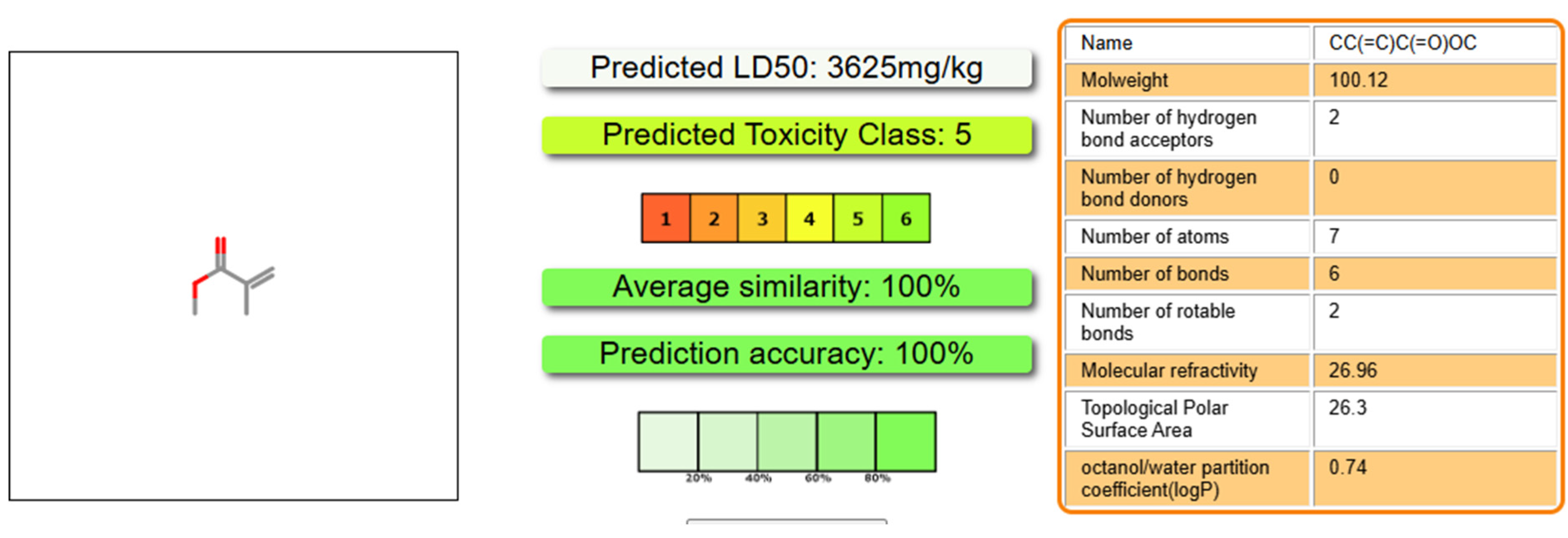
3.2. In Silico Toxicity Profiling of MMA
3.3. PPI Network Topology and Hub Protein Identification
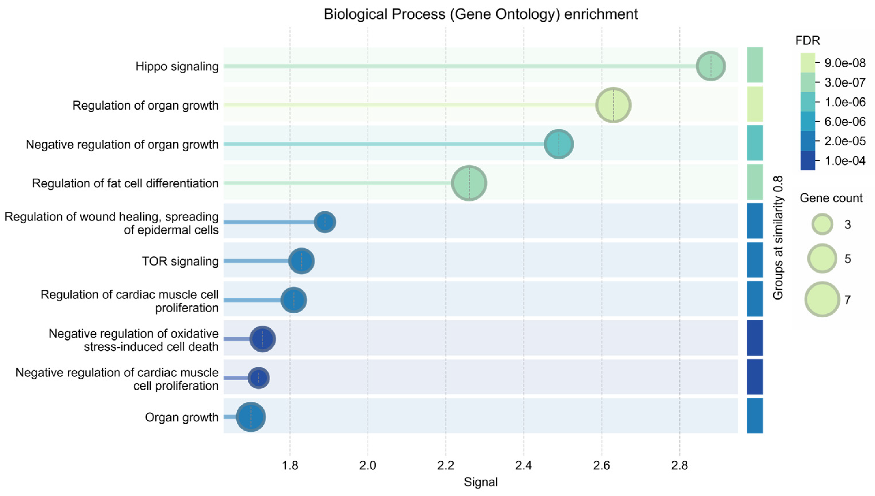
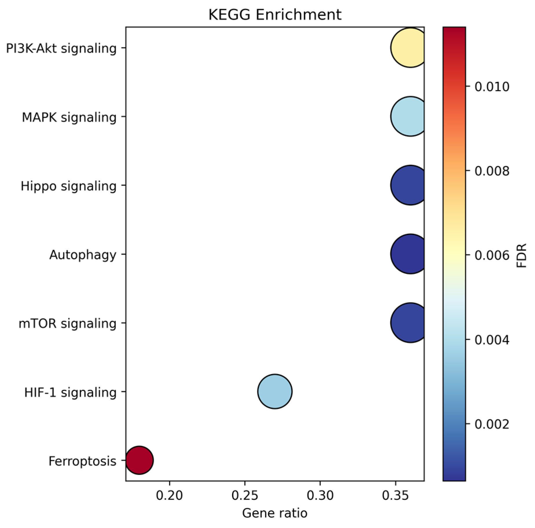
3.4. Molecular Docking and Pathway Interconnectivity
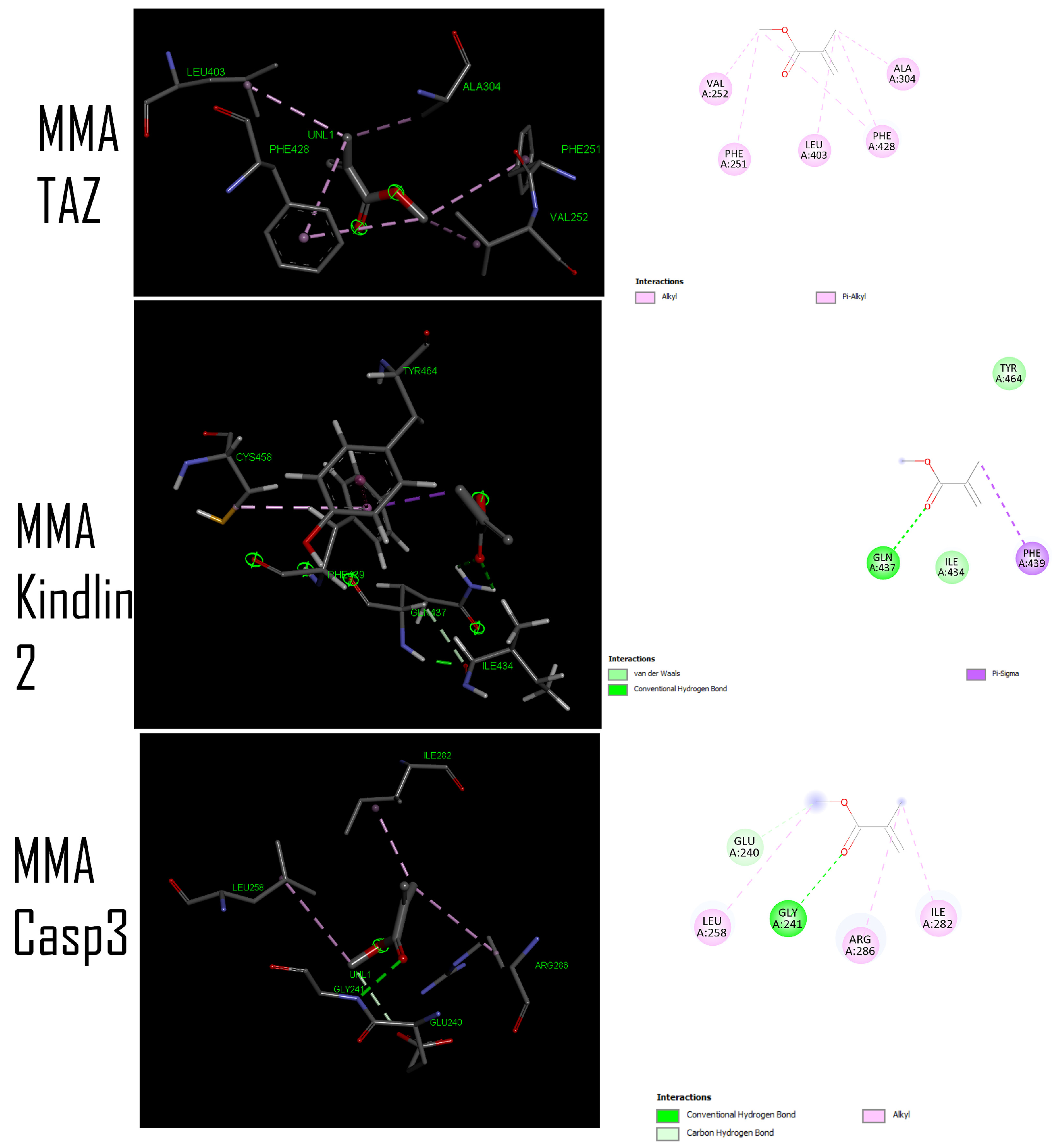
3.5. Pharmacophore Modeling and Residue-Specific Interaction Analysis
3.6. Putative Target Identification via Swiss ADME Prediction
4. Discussion
Limitations and Clinical Implications
5. Conclusions
Supplementary Materials
Author Contributions
Funding
Institutional Review Board Statement
Informed Consent Statement
Data Availability Statement
Conflicts of Interest
Abbreviations
References
- Gratton, D.G.; Aquilino, S.A. Interim restorations. Dent. Clin. N. Am. 2004, 48, 487–497. [Google Scholar] [CrossRef] [PubMed]
- Saini, R.S.; Binduhayyim, R.I.H.; Gurumurthy, V.; Alshadidi, A.A.F.; Bavabeedu, S.S.; Vyas, R.; Dermawan, D.; Naseef, P.P.; Mosaddad, S.A.; Heboyan, A. In silico assessment of biocompatibility and toxicity: Molecular docking and dynamics simulation of PMMA-based dental materials for interim prosthetic restorations. J. Mater. Sci. Mater. Med. 2024, 35, 28. [Google Scholar] [CrossRef] [PubMed]
- Alshadidi, A.A.F.; Alshahrani, A.A.; Aldosari, L.I.N.; Chaturvedi, S.; Saini, R.S.; Hassan, S.A.B.; Cicciù, M.; Minervini, G. Investigation on the application of artificial intelligence in prosthodontics. Appl. Sci. 2023, 13, 5004. [Google Scholar] [CrossRef]
- Alterman, M.; Jensen, O.T.; Glick, P.L.; Mazor, D.; Casap, N. Dental implant risk factors for peri-implant disease: A narrative review. Front. Oral Maxillofac. Med. 2023, 5, 26. [Google Scholar] [CrossRef]
- Jokstad, A.; Alkumru, H. Immediate function on the day of surgery compared with a delayed implant loading process in the mandible: A randomized clinical trial over 5 years. Clin. Oral Implant. Res. 2014, 25, 1325–1335. [Google Scholar] [CrossRef][Green Version]
- Mobarak, M.H.; Islam, M.A.; Hossain, N.; Al Mahmud, M.Z.; Rayhan, M.T.; Nishi, N.J.; Chowdhury, M.A. Recent advances of additive manufacturing in implant fabrication—A review. Apply Surf. Sci. Adv. 2023, 18, 100462. [Google Scholar] [CrossRef]
- Ainoosah, S.; Farghal, A.E.; Alzemei, M.S.; Saini, R.S.; Gurumurthy, V.; Quadri, S.A.; Okshah, A.; Mosaddad, S.A.; Heboyan, A. Comparative analysis of different types of occlusal splints for the management of sleep bruxism: A systematic review. BMC Oral Health 2024, 24, 29. [Google Scholar] [CrossRef]
- Diez-Pascual, A.M. PMMA-Based Nanocomposites for Odontology Applications: A State-of-the-Art. Int. J. Mol. Sci. 2022, 23, 10288. [Google Scholar] [CrossRef]
- Zafar, M.S. Prosthodontic Applications of Polymethyl Methacrylate (PMMA): An Update. Polymers 2020, 12, 2299. [Google Scholar] [CrossRef]
- Alqutaibi, A.Y.; Baik, A.; Almuzaini, S.A.; Farghal, A.E.; Alnazzawi, A.A.; Borzangy, S.; Aboalrejal, A.N.; AbdElaziz, M.H.; Mahmoud, I.I.; Zafar, M.S. Polymeric Denture Base Materials: A Review. Polymers 2023, 15, 3258. [Google Scholar] [CrossRef]
- Mohd Farid, D.A.; Zahari, N.A.H.; Said, Z.; Ghazali, M.I.M.; Hao-Ern, L.; Mohamad Zol, S.; Aldhuwayhi, S.; Alauddin, M.S. Modification of Polymer Based Dentures on Biological Properties: Current Update, Status, and Findings. Int. J. Mol. Sci. 2022, 23, 10426. [Google Scholar] [CrossRef]
- Kollmuss, M.; Edelhoff, D.; Schwendicke, F.; Wuersching, S.N. In Vitro Cytotoxic and Inflammatory Response of Gingival Fibroblasts and Oral Mucosal Keratinocytes to 3D Printed Oral Devices. Polymers 2024, 16, 1336. [Google Scholar] [CrossRef] [PubMed]
- Lim, J.H.; Lee, S.Y.; Gu, H.; Jin, G.; Kim, J.E. Evaluating oxygen shielding effect using glycerin or vacuum with varying temperature on 3D printed photopolymer in post-polymerization. J. Mech. Behav. Biomed. Mater. 2022, 130, 105170. [Google Scholar] [CrossRef]
- Berghaus, E.; Klocke, T.; Maletz, R.; Petersen, S. Degree of conversion and residual monomer elution of 3D-printed, milled and self-cured resin-based composite materials for temporary dental crowns and bridges. J. Mater. Sci. Mater. Med. 2023, 34, 23. [Google Scholar] [CrossRef] [PubMed]
- Imre, M.; Saramet, V.; Ciocan, L.T.; Vasilescu, V.G.; Biru, E.I.; Ghitman, J.; Pantea, M.; Ripszky, A.; Celebidache, A.L.; Iovu, H. Influence of the Processing Method on the Nano-Mechanical Properties and Porosity of Dental Acrylic Resins Fabricated by Heat-Curing, 3D Printing and Milling Techniques. Dent. J. 2025, 13, 311. [Google Scholar] [CrossRef] [PubMed]
- Sahin, Z.; Ozkan Vardar, D.; Erdogmus, E.; Calamak, S.; Kocer Gumusel, B. Monomer release, cytotoxicity, and surface roughness of temporary fixed prosthetic materials produced by digital and conventional methods. Odontology 2025, 113, 1643–1658. [Google Scholar] [CrossRef]
- Tamboura, B.; Yudaev, P.; Butorova, I.; Klyukin, B.; Chuev, V.; Chistyakov, E. Hexaallylaminocyclotriphosphazene-Modified Dental Compositions for 3D Printing of Dental Crowns. Polymers 2025, 18, 53. [Google Scholar] [CrossRef]
- Gong, B.; Zhang, H.; Li, S.; Lin, Y.; Xu, S.; Jia, B.; Lu, Y.; Liu, Z. Strategies for enhancing biocompatibility, bioactivity, and resistance to biodegradation in dental resin composites: A narrative review. Biomater. Adv. 2026, 179, 214496. [Google Scholar] [CrossRef]
- Mousavinasab, S.M. Biocompatibility of composite resins. Dent. Res. J. 2011, 8, S21–S29. [Google Scholar]
- Schmalz, G.; Galler, K.M. Biocompatibility of biomaterials—Lessons learned and considerations for the design of novel materials. Dent. Mater. 2017, 33, 382–393. [Google Scholar] [CrossRef]
- Gallorini, M.; Petzel, C.; Bolay, C.; Hiller, K.A.; Cataldi, A.; Buchalla, W.; Krifka, S.; Schweikl, H. Activation of the Nrf2-regulated antioxidant cell response inhibits HEMA-induced oxidative stress and supports cell viability. Biomaterials 2015, 56, 114–128. [Google Scholar] [CrossRef] [PubMed]
- Yamamoto, M.; Kensler, T.W.; Motohashi, H. The KEAP1-NRF2 System: A Thiol-Based Sensor-Effector Apparatus for Maintaining Redox Homeostasis. Physiol. Rev. 2018, 98, 1169–1203. [Google Scholar] [CrossRef] [PubMed]
- Manning, B.D.; Toker, A. AKT/PKB Signaling: Navigating the Network. Cell 2017, 169, 381–405. [Google Scholar] [CrossRef] [PubMed]
- Okkenhaug, K.; Graupera, M.; Vanhaesebroeck, B. Targeting PI3K in cancer: Impact on tumor cells, their protective stroma, angiogenesis, and immunotherapy. Cancer Discov. 2016, 6, 1090–1105. [Google Scholar] [CrossRef]
- Saxton, R.A.; Sabatini, D.M. mTOR Signaling in Growth, Metabolism, and Disease. Cell 2017, 168, 960–976. [Google Scholar] [CrossRef]
- Semenza, G.L. Hypoxia-inducible factors in physiology and medicine. Cell 2012, 148, 399–408. [Google Scholar] [CrossRef]
- Zhang, G.; Song, S.; Chen, Z.; Liu, X.; Zheng, J.; Wang, Y.; Chen, X.; Song, Y. Inhibition of PTEN promotes osteointegration of titanium implants in type 2 diabetes by enhancing anti-inflammation and osteogenic capacity of adipose-derived stem cells. Front. Bioeng. Biotechnol. 2024, 12, 1358802. [Google Scholar] [CrossRef]
- Julien, O.; Wells, J.A. Caspases and their substrates. Cell Death Differ. 2017, 24, 1380–1389. [Google Scholar] [CrossRef]
- Kale, J.; Osterlund, E.J.; Andrews, D.W. BCL-2 family proteins: Changing partners in the dance towards death. Cell Death Differ. 2018, 25, 65–80. [Google Scholar] [CrossRef]
- Singh, R.; Letai, A.; Sarosiek, K. Regulation of apoptosis in health and disease: The balancing act of BCL-2 family proteins. Nat. Rev. Mol. Cell Biol. 2019, 20, 175–193. [Google Scholar] [CrossRef]
- Wei, Y.; Hui, V.L.Z.; Chen, Y.; Han, R.; Han, X.; Guo, Y. YAP/TAZ: Molecular pathway and disease therapy. MedComm 2023, 4, e340. [Google Scholar] [CrossRef] [PubMed]
- Berman, H.M.; Westbrook, J.; Feng, Z.; Gilliland, G.; Bhat, T.N.; Weissig, H.; Shindyalov, I.N.; Bourne, P.E. The Protein Data Bank. Nucleic Acids Res. 2000, 28, 235–242. [Google Scholar] [CrossRef]
- Tian, W.; Chen, C.; Lei, X.; Zhao, J.; Liang, J. CASTp 3.0: Computed atlas of surface topography of proteins. Nucleic Acids Res. 2018, 46, W363–W367. [Google Scholar] [CrossRef]
- Jurowski, K.; Krosniak, A. Prediction of key toxicity endpoints of AP-238 a new psychoactive substance for clinical toxicology and forensic purposes using in silico methods. Sci. Rep. 2024, 14, 28977. [Google Scholar] [CrossRef] [PubMed]
- Banerjee, P.; Ulker, O.; Ozkan, I.; Ulker, O.C. The investigation of the toxicity of organophosphorus flame retardants (OPFRs) by using in silico toxicity prediction platform ProTox- 3.0. Toxicol. Mech. Methods 2025, 35, 32–42. [Google Scholar] [CrossRef] [PubMed]
- Daghighi, A.; Casanola-Martin, G.M.; Iduoku, K.; Kusic, H.; Gonzalez-Diaz, H.; Rasulev, B. Multi-Endpoint Acute Toxicity Assessment of Organic Compounds Using Large-Scale Machine Learning Modeling. Environ. Sci. Technol. 2024, 58, 10116–10127. [Google Scholar] [CrossRef]
- Tafaki, E.; Chalaris, M. Investigating QSAR models for chemical warfare agents: Biological, biochemical, and environmental perspectives. WSEAS Trans. Biol. Biomed. 2024, 21, 281–301. [Google Scholar] [CrossRef]
- Krifka, S.; Spagnuolo, G.; Schmalz, G.; Schweikl, H. A review of adaptive mechanisms in cell responses towards oxidative stress caused by dental resin monomers. Biomaterials 2013, 34, 4555–4563. [Google Scholar] [CrossRef]
- Samuelsen, J.T.; Dahl, J.E. Biological aspects of modern dental composites. Biomater. Investig. Dent. 2023, 10, 2223223. [Google Scholar] [CrossRef]
- de Baat, A.; Meier, D.T.; Rachid, L.; Fontana, A.; Boni-Schnetzler, M.; Donath, M.Y. Cystine/glutamate antiporter System x(c)(-) deficiency impairs insulin secretion in mice. Diabetologia 2023, 66, 2062–2074. [Google Scholar] [CrossRef]
- Liu, C.; Wu, X.; Bing, X.; Qi, W.; Zhu, F.; Guo, N.; Li, C.; Gao, X.; Cao, X.; Zhao, M.; et al. H1N1 influenza virus infection through NRF2-KEAP1-GCLC pathway induces ferroptosis in nasal mucosal epithelial cells. Free Radic. Biol. Med. 2023, 204, 226–242. [Google Scholar] [CrossRef]
- Xie, Y.; Kang, R.; Klionsky, D.J.; Tang, D. GPX4 in cell death, autophagy, and disease. Autophagy 2023, 19, 2621–2638. [Google Scholar] [CrossRef] [PubMed]
- Yang, D.; Gong, G.; Song, J.; Chen, J.; Wang, S.; Li, J.; Wang, G. Ferroptosis-mediated osteoclast-osteoblast crosstalk: Signaling pathways governing bone remodeling in osteoporosis. J. Orthop. Surg. Res. 2025, 20, 888. [Google Scholar] [CrossRef] [PubMed]
- Zhang, X.; Hou, L.; Guo, Z.; Wang, G.; Xu, J.; Zheng, Z.; Sun, K.; Guo, F. Lipid peroxidation in osteoarthritis: Focusing on 4-hydroxynonenal, malondialdehyde, and ferroptosis. Cell Death Discov. 2023, 9, 320. [Google Scholar] [CrossRef] [PubMed]
- Fujisawa, S.; Atsumi, T. Cytotoxicities of a 4-META/MMA-TBBO resin against human pulp fibroblasts. Dent. Mater. J. 2004, 23, 106–108. [Google Scholar] [CrossRef]
- Zhang, Y.; Xiao, J.F.; Yang, H.F.; Jiao, Y.; Cao, W.W.; Shi, H.M.; Cun, J.F.; Tay, F.R.; Ping, J.; Xiao, Y.H. N-Acetyl Cysteine as a Novel Polymethyl Methacrylate Resin Component: Protection against Cell Apoptosis and Genotoxicity. Oxid. Med. Cell Longev. 2019, 2019, 1301736, Erratum in Oxid. Med. Cell Longev. 2020, 2020, 2768238. [Google Scholar] [CrossRef]
- Pituru, S.M.; Greabu, M.; Totan, A.; Imre, M.; Pantea, M.; Spinu, T.; Tancu, A.M.C.; Popoviciu, N.O.; Stanescu, I.-I.; Ionescu, E. A Review on the Biocompatibility of PMMA-Based Dental Materials for Interim Prosthetic Restorations with a Glimpse into their Modern Manufacturing Techniques. Materials 2020, 13, 2894. [Google Scholar] [CrossRef]
- Schweikl, H.; Spagnuolo, G.; Schmalz, G. Genetic and cellular toxicology of dental resin monomers. J. Dent. Res. 2006, 85, 870–877. [Google Scholar] [CrossRef]
- Elbediwy, A.; Thompson, B.J. Evolution of mechanotransduction via YAP/TAZ in animal epithelia. Curr. Opin. Cell Biol. 2018, 51, 117–123. [Google Scholar] [CrossRef]
- Park, C.; Alahari, S.; Ausman, J.; Liu, R.; Nguyen, F.; Sallais, J.; Post, M.; Caniggia, I. Placental Hypoxia-Induced Ferroptosis Drives Vascular Damage in Preeclampsia. Circ. Res. 2025, 136, 361–378. [Google Scholar] [CrossRef]
- Wang, C.; Gu, W.; Sun, B.; Zhang, Y.; Ji, Y.; Xu, X.; Wen, Y. CTHRC1 promotes osteogenic differentiation of periodontal ligament stem cells by regulating TAZ. J. Mol. Histol. 2017, 48, 311–319. [Google Scholar] [CrossRef]
- Nardone, G.; Oliver-De La Cruz, J.; Vrbsky, J.; Martini, C.; Pribyl, J.; Skladal, P.; Pesl, M.; Caluori, G.; Pagliari, S.; Martino, F.; et al. YAP regulates cell mechanics by controlling focal adhesion assembly. Nat. Commun. 2017, 8, 15321. [Google Scholar] [CrossRef]
- Bellacosa, A.; Chan, T.O.; Ahmed, N.N.; Datta, K.; Malstrom, S.; Stokoe, D.; McCormick, F.; Feng, J.; Tsichlis, P. Akt activation by growth factors is a multiple-step process: The role of the PH domain. Oncogene 1998, 17, 313–325. [Google Scholar] [CrossRef]
- Sancak, Y.; Thoreen, C.C.; Peterson, T.R.; Lindquist, R.A.; Kang, S.A.; Spooner, E.; Carr, S.A.; Sabatini, D.M. PRAS40 is an insulin-regulated inhibitor of the mTORC1 protein kinase. Mol. Cell 2007, 25, 903–915. [Google Scholar] [CrossRef] [PubMed]
- Shaw, R.J.; Cantley, L.C. Ras, PI (3) K and mTOR signalling controls tumour cell growth. Nature 2006, 441, 424–430. [Google Scholar] [CrossRef] [PubMed]
- Vivanco, I.; Sawyers, C.L. The phosphatidylinositol 3-kinase–AKT pathway in human cancer. Nat. Rev. Cancer 2002, 2, 489–501. [Google Scholar] [CrossRef] [PubMed]
- Borak, J.; Fields, C.; Andrews, L.S.; Pemberton, M.A. Methyl methacrylate and respiratory sensitization: A critical review. Crit. Rev. Toxicol. 2011, 41, 230–268. [Google Scholar] [CrossRef]
- Leggat, P.A.; Kedjarune, U. Toxicity of methyl methacrylate in dentistry. Int. Dent. J. 2003, 53, 126–131. [Google Scholar] [CrossRef]
- Lanevskij, K.; Dapkunas, J.; Juska, L.; Japertas, P.; Didziapetris, R. QSAR analysis of blood-brain distribution: The influence of plasma and brain tissue binding. J. Pharm. Sci. 2011, 100, 2147–2160. [Google Scholar] [CrossRef]
- Bandarra, S.; Mascarenhas, P.; Luis, A.R.; Catrau, M.; Bekman, E.; Ribeiro, A.C.; Felix, S.; Caldeira, J.; Barahona, I. In vitro and in silico evaluations of resin-based dental restorative material toxicity. Clin. Oral Investig. 2020, 24, 2691–2700. [Google Scholar] [CrossRef]
- Bereznowski, Z. In vivo assessment of methyl methacrylate metabolism and toxicity. Int. J. Biochem. Cell Biol. 1995, 27, 1311–1316. [Google Scholar] [CrossRef]
- Warren, G.L.; Andrews, C.W.; Capelli, A.M.; Clarke, B.; LaLonde, J.; Lambert, M.H.; Lindvall, M.; Nevins, N.; Semus, S.F.; Senger, S.; et al. A critical assessment of docking programs and scoring functions. J. Med. Chem. 2006, 49, 5912–5931. [Google Scholar] [CrossRef]
- Moitessier, N.; Englebienne, P.; Lee, D.; Lawandi, J.; Corbeil, C.R. Towards the development of universal, fast and highly accurate docking/scoring methods: A long way to go. Br. J. Pharmacol. 2008, 153, S7–S26. [Google Scholar] [CrossRef]
- Ramírez, D.; Caballero, J. Is It Reliable to Take the Molecular Docking Top Scoring Position as the Best Solution without Considering Available Structural Data? Molecules 2018, 23, 1038. [Google Scholar] [CrossRef]
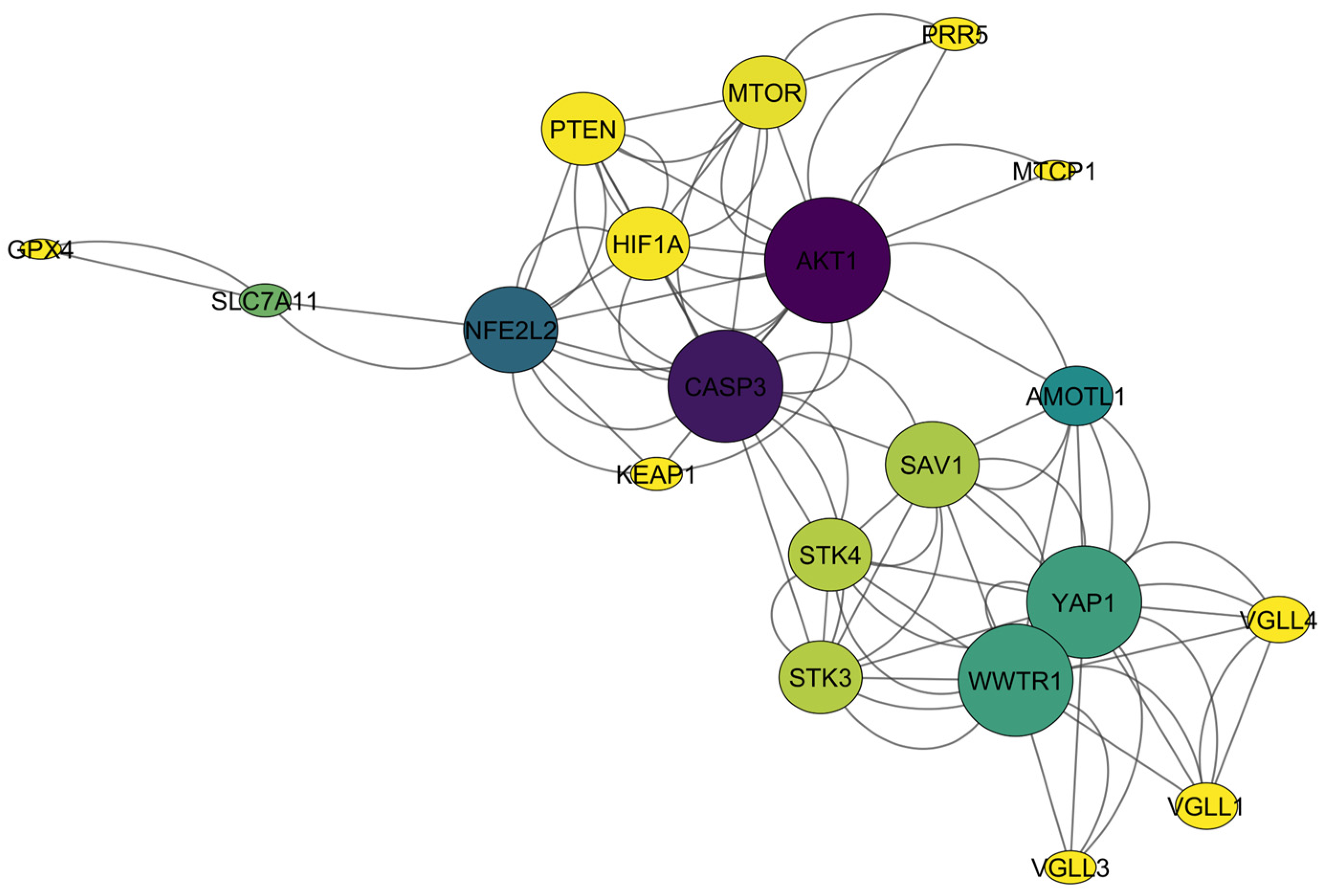


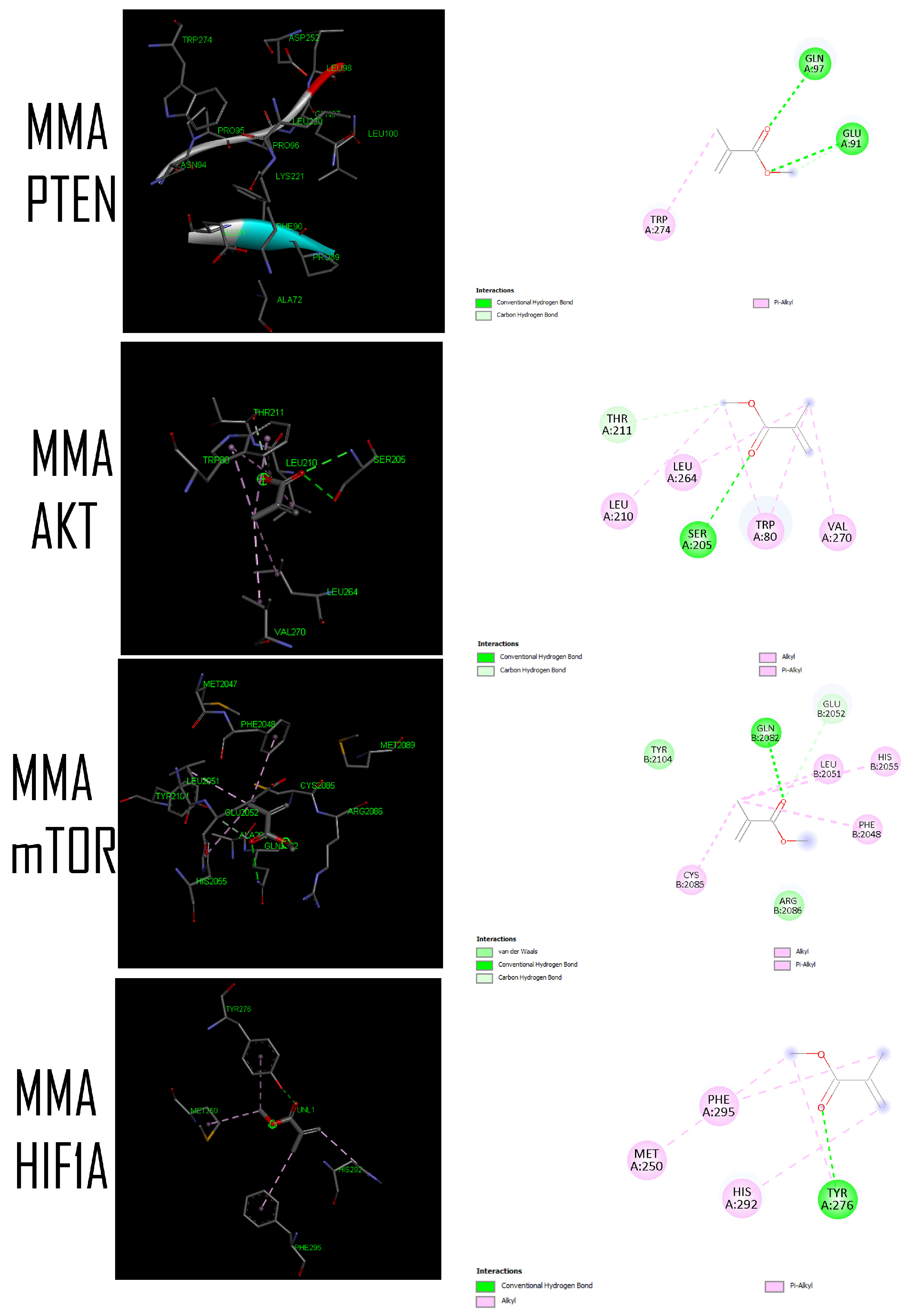

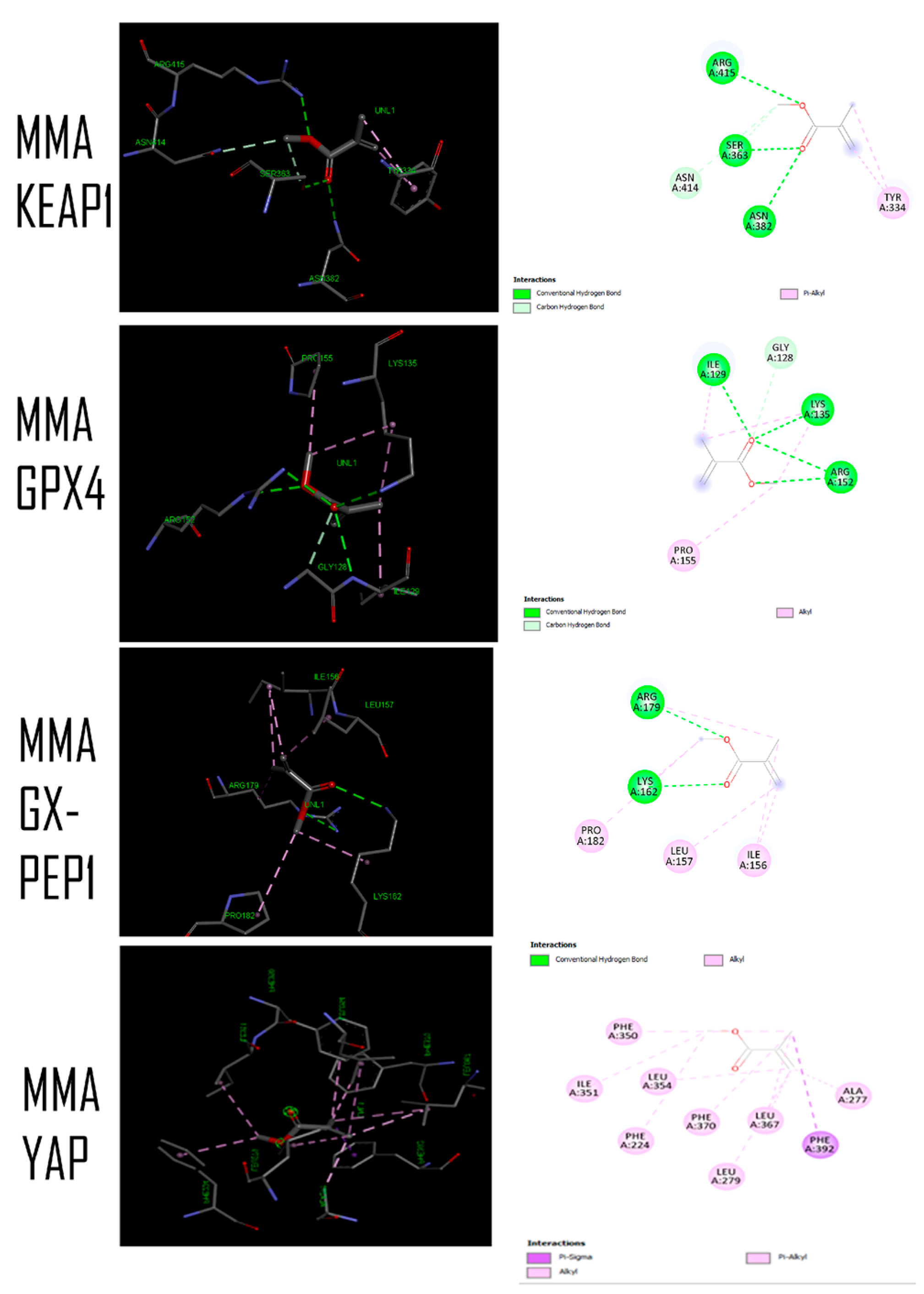

| Name | PDB/UniProt ID | Resolution (Å) | Chain | Weight (kDa) | Sequence Length | Active Site (Residue Number) | Biological Relevance of Each Target |
|---|---|---|---|---|---|---|---|
| PTEN | 1D5R | 2.10 | A | 38.65 | 324 | 16, 24, 92, 93, 124, 125, 126, 127, 128, 129, 130, 159 160, 162, 164, 167, 168, 171 | Central negative potential regulator of AKT signaling; recently implicated in osteoimmune modulation and peri-implant bone formation. |
| AKT1 | 4EJN | 2.19 | A | 52.45 | 446 | 13, 14, 15, 16, 17, 18, 20, 21, 22, 25, 27, 28, 30, 32, 34, 36, 37, 38, 50, 51, 52, 53, 54, 55, 56, 58, 59, 60, 68, 69, 72, 77, 79, 80, 81, 82, 83, 84, 86, 87, 88, 89, 156, 157, 158, 159, 161, 164, 181, 203, 204, 205, 207, 210, 211, 212, 229, 234, 261, 264, 268, 270, 271, 272, 273, 274, 275, 276, 277, 278, 279, 281, 290, 291 ,292, 293, 294, 295, 296, 297, 298, 299, 308, 309, 310, 311, 312, 31, 316, 317, 320, 323, 324, 325, 326, 327, 330, 331, 333, 334 | Key survival kinase linking metabolic adaptation, osteoblast viability, and angiogenic signaling. |
| mTOR | 4DRI | 1.45 | B | 28.36 | 98 | 52, 57, 67, 68, 6970, 72, 73, 75, 77, 78, 79, 80, 84, 85, 86, 87, 88, 90, 91 112, 113, 116, 117, 118, 119, 120, 121, 122, 130 | Co-crystal structure of the PPIase domain of FKBP51, rapamycin and the FRB fragment of mTOR. Master regulator of cellular growth and osteogenic metabolism. |
| HIF1A | 4H6J | 1.52 | A | 26.69 | 113 | 324, 325, 326, 327, 328, 333, 334 | Integrates hypoxia-driven angiogenesis with osteogenic adaptation. |
| KEAP1 | 4L7B | 2.41 | A | 66.78 | 300 | 334, 336, 337, 363, 364, 365, 366, 367, 368, 369, 380, 382, 383, 384, 385, 386, 387, 389, 414, 415, 416, 417, 418, 419, 420, 431, 433, 434, 435, 436, 450, 460, 461, 462, 463, 464, 465, 466, 467, 478, 479, 480 | Redox sensor protein regulating oxidative stress-dependent cell survival. |
| GPX4 | 2OBI | 1.55 | A | 20.96 | 183 | 127, 128, 129, 135, 139, 152, 153, 154, 155, 157 | Key inhibitor of ferroptosis; recently linked to implant-induced oxidative stress responses. |
| GX-PEP1 (GXpep-1-bound structure) | 5H5Q | 1.10 | A | 21.34 | 169 | 152, 154, 155, 156, 157, 158, 162, 166, 179, 180, 181, 182, 183, 184 | Included as an alternative ligand-bound structural state of GPX4 to examine whether MMA docking behavior is consistent across different conformational templates. |
| TEAD1–YAP complex | 4RE1 | 2.20 | A | 83.01 | 220 | 194, 195, 197, 198, 199, 200, 202, 237, 238, 239, 266, 267, 268, 269, 270, 271, 383, 384, 385, 407, 408 | Key downstream effector of Hippo signaling and mechanotransduction-related transcriptional regulation. |
| TEAD2-palmitate TAZ | 5HGU | 2.05 | A | 54.77 | 237 | 281, 283, 288, 291, 292, 326, 327, 340, 342, 344, 346, 353 356, 357, 358, 359, 360, 361, 362, 374, 375, 376, 377, 378, 379, 380, 381, 384, 411, 413, 415, 418, 420 | TAZ (WWTR1): Key mediator of Hippo signaling and mechanotransduction, with relevance to osteogenic differentiation and biomaterial-responsive cellular signaling. |
| Kindlin-2 (FERMT2) | 2LKO | 2.10 | A | 55.47 | 474 | 102, 104, 106, 141, 143, 144, 236, 238, 240, 242, 245, 246, 248, 249, 252, 271, 273, 274, 275, 277, 297, 300, 301, 304, 305, 306, 307, 308, 562, 567, 568, 569, 570 571, 572, 600, 601, 649 | Essential for integrin-mediated osteoblast adhesion to implant surfaces. |
| Caspase-3 | 1PAU | 2.50 | A | 29.04 | 147 | 294, 295 | Final executioner of apoptosis; key indicator of PMMA/MMA-induced cytotoxic signaling. |
| Classification | Target | Shorthand | Prediction | Probability |
|---|---|---|---|---|
| MMA | MMA | |||
| Metabolism | Cytochrome CYP2C9 | CYP2C9 | Active | 0.64 |
| Organ toxicity | Respiratory toxicity | respi | Active | 0.70 |
| Toxicity endpoints | BBB-barrier | bbb | Active | 0.90 |
| Gene | Degree | Closeness Centrality | Betweenness Centrality | Neighborhood Connectivity |
|---|---|---|---|---|
| AKT1 | 18 | 0.575 | 0.327 | 4.22 |
| CASP3 | 16 | 0.558 | 0.301 | 5.75 |
| WWTR1 | 16 | 0.452 | 0.141 | 4.5 |
| YAP1 | 16 | 0.452 | 0.141 | 4.5 |
| SAV1 | 12 | 0.5 | 0.060 | 6.33 |
| NFE2L2 | 12 | 0.475 | 0.215 | 5.16 |
| STK3 | 10 | 0.487 | 0.053 | 7.0 |
| STK4 | 10 | 0.487 | 0.053 | 7.0 |
| mTOR | 10 | 0.431 | 0.016 | 5.8 |
| PTEN | 10 | 0.452 | 0.004 | 6.6 |
| HIF1A | 10 | 0.452 | 0.004 | 6.6 |
| AMOTL1 | 8 | 0.513 | 0.170 | 7.75 |
| CASP3 | 8 | 0.76 | 0.13 | 6.50 |
| AKT1 | 7 | 0.68 | 0.00 | 6.25 |
| SAV1 | 6 | 1.00 | 0.11 | 6.00 |
| VGLL1 | 6 | 0.327 | 0.0 | 6.33 |
| VGLL4 | 6 | 0.327 | 0.0 | 6.33 |
| PTEN | 5 | 1.00 | 0.00 | 5.80 |
| HIF1A | 5 | 0.80 | 0.00 | 5.80 |
| WWTR1 | 5 | 1.00 | 0.00 | 5.75 |
| YAP1 | 5 | 0.80 | 0.00 | 5.37 |
| STK3 | 5 | 1.00 | 0.03 | 5.33 |
| STK4 | 5 | 1.00 | 0.03 | 5.00 |
| SLC7A11 | 4 | 0.339 | 0.105 | 3.5 |
| mTOR | 4 | 1.00 | 0.00 | 5.00 |
| KEAP1 | 4 | 0.395 | 0.0 | 7.5 |
| PRR5 | 4 | 0.379 | 0.0 | 7.0 |
| VGLL3 | 4 | 0.322 | 0.0 | 8.0 |
| GX-PEP1 (GXpep-1-bound structure) | 2 | 0.40 | 0.00 | 3.50 |
| GPX4 | 1 | 1.00 | 0.00 | 2.00 |
| Target (Gene/Protein) | PDB ID | Chain | CB-Dock Cavity Rank | Best Pose (Vina Mode) | Binding Affinity (kcal/mol) | RMSD l.b. (Å) | RMSD u.b. (Å) | Best Cavity ID | Cavity Size (x, y, z) | Cavity Center (x, y, z) |
|---|---|---|---|---|---|---|---|---|---|---|
| PTEN | 1d5r | A | 1 | 1 | −4.00 | 0.00 | 0.00 | 1 | 16, 16, 16 | 44.771 78.910 29.029 |
| AKT1 | 4ejn | A | 1 | 1 | −4.10 | 0.00 | 0.00 | 1 | 15, 15, 15 | 29.817 43.243 15.125 |
| mTOR | 4dri | B | 1 | 1 | −4.20 | 0.00 | 0.00 | 2 | 16, 16, 16 | 35 43 36 |
| HIF1A | 4h6j | A | 1 | 1 | −3.00 | 0.00 | 0.00 | 1 | 16, 16, 16 | 19.087 −17.259 −34.909 |
| KEAP1 | 4l7b | A | 1 | 1 | −3.90 | 0.00 | 0.00 | 1 | 22, 23, 32 | 7.472 −14.479 −14.411 |
| GX-PEP1 (GXpep-1-bound structure) | 5h5q | A | 1 | 1 | −4.00 | 0.00 | 0.00 | 3 | 16, 16, 16 | 7.203 11.455 −6.512 |
| GPX4 | 2obi | A | 1 | 1 | −3.90 | 0.00 | 0.00 | 1 | 16, 16, 16 | 32.320 −27.856 −8.825 |
| YAP | 4re1 | A | 1 | 1 | −4.20 | 0.00 | 0.00 | 1 | 16, 23, 16 | 17.210 −6.130 −20.962 |
| TAZ | 5hgu | A | 1 | 1 | −4.20 | 0.00 | 0.00 | 1 | 16, 16, 16 | −9.622 15.434 92.162 |
| Kindlin-2 | 2lko | A | 1 | 1 | −3.8 | 0.00 | 0.00 | 7 | 16, 16, 16 | −15 −7 −4 |
| CASP3 | 1pau | A | 1 | 1 | −3.50 | 0.00 | 0.00 | 1 | 16, 16, 16 | 32.458 96.173 8.113 |
| Parameter | MMA |
|---|---|
| Formula | C5H8O2 |
| Molecular weight (g/mol) | 100.12 |
| cLogP (XLOGP3) | 1.38 |
| cLogS (SILICOS-IT) | −0.71 |
| TPSA (Å2) | 26.30 |
| H-bond acceptors | 2 |
| H-bond donors | 0 |
| Rotatable bonds | 2 |
| Heavy atoms | 7 |
| Fraction Csp3 | 0.40 |
| Molar refractivity | 26.96 |
| GI absorption | High |
| BBB permeant | Yes |
| Bioavailability score | 0.55 |
| Lipinski | Yes; 0 violation |
| Ghose | No; 3 violations: MW < 160, MR < 40, #atoms < 20 |
| Veber | Yes |
| Egan | Yes |
| Muegge | No; 1 violation: MW < 200 |
| PAINS | 0 alert |
| Brenk | 1 alert: michael_acceptor_1 |
| Leadlikeness | No; 1 violation: MW < 250 |
| Synthetic accessibility | 1.12 |
Disclaimer/Publisher’s Note: The statements, opinions and data contained in all publications are solely those of the individual author(s) and contributor(s) and not of MDPI and/or the editor(s). MDPI and/or the editor(s) disclaim responsibility for any injury to people or property resulting from any ideas, methods, instructions or products referred to in the content. |
© 2026 by the authors. Licensee MDPI, Basel, Switzerland. This article is an open access article distributed under the terms and conditions of the Creative Commons Attribution (CC BY) license.
Share and Cite
Dilber, Y.; Dilber, E.; Yıldız Domaniç, K. Bioactive Assessment of MMA-Based Dental Materials: Molecular Docking and Network Topology Analysis of Stress-Regulated Survival, Apoptosis, and Mechanotransduction Pathways. Curr. Issues Mol. Biol. 2026, 48, 630. https://doi.org/10.3390/cimb48060630
Dilber Y, Dilber E, Yıldız Domaniç K. Bioactive Assessment of MMA-Based Dental Materials: Molecular Docking and Network Topology Analysis of Stress-Regulated Survival, Apoptosis, and Mechanotransduction Pathways. Current Issues in Molecular Biology. 2026; 48(6):630. https://doi.org/10.3390/cimb48060630
Chicago/Turabian StyleDilber, Yağmur, Erhan Dilber, and Kübra Yıldız Domaniç. 2026. "Bioactive Assessment of MMA-Based Dental Materials: Molecular Docking and Network Topology Analysis of Stress-Regulated Survival, Apoptosis, and Mechanotransduction Pathways" Current Issues in Molecular Biology 48, no. 6: 630. https://doi.org/10.3390/cimb48060630
APA StyleDilber, Y., Dilber, E., & Yıldız Domaniç, K. (2026). Bioactive Assessment of MMA-Based Dental Materials: Molecular Docking and Network Topology Analysis of Stress-Regulated Survival, Apoptosis, and Mechanotransduction Pathways. Current Issues in Molecular Biology, 48(6), 630. https://doi.org/10.3390/cimb48060630