1. Introduction
Pulmonary arterial hypertension (PAH) is a lethal, progressive disorder and a subtype of the broader condition of pulmonary hypertension. It is defined as a mean pulmonary artery pressure of >20 mmHg at rest, pulmonary arterial wedge pressure ≤ 15 mmHg, and pulmonary vascular resistance > 2 Wood units [
1]. PAH is an incurable disorder estimated to affect 146,000 people across the EU, USA, and Japan [
2]. Due to limited surveillance and healthcare access, the true prevalence is likely higher in developing countries. The condition is characterized by intima and media hyperplasia, formation of plexiform lesions in the pulmonary vasculature leading to elevated pulmonary arterial pressure, vascular resistance, and consequential right-heart failure. Symptoms of pulmonary arterial hypertension include difficulty in breathing, fatigue, weakness, syncope, angina, and edema [
3].
Clinical classification of PAH
Idiopathic PAH
Heritable PAH
Drug- and toxin-induced PAH
PAH associated with:
Connective tissue disease
HIV infection
Portal hypertension
Congenital heart disease
Schistosomiasis
PAH in response to long-term calcium channel blocker use
PAH with overt features of venous/capillary involvement
Persistent PH of the newborn syndrome [
4]
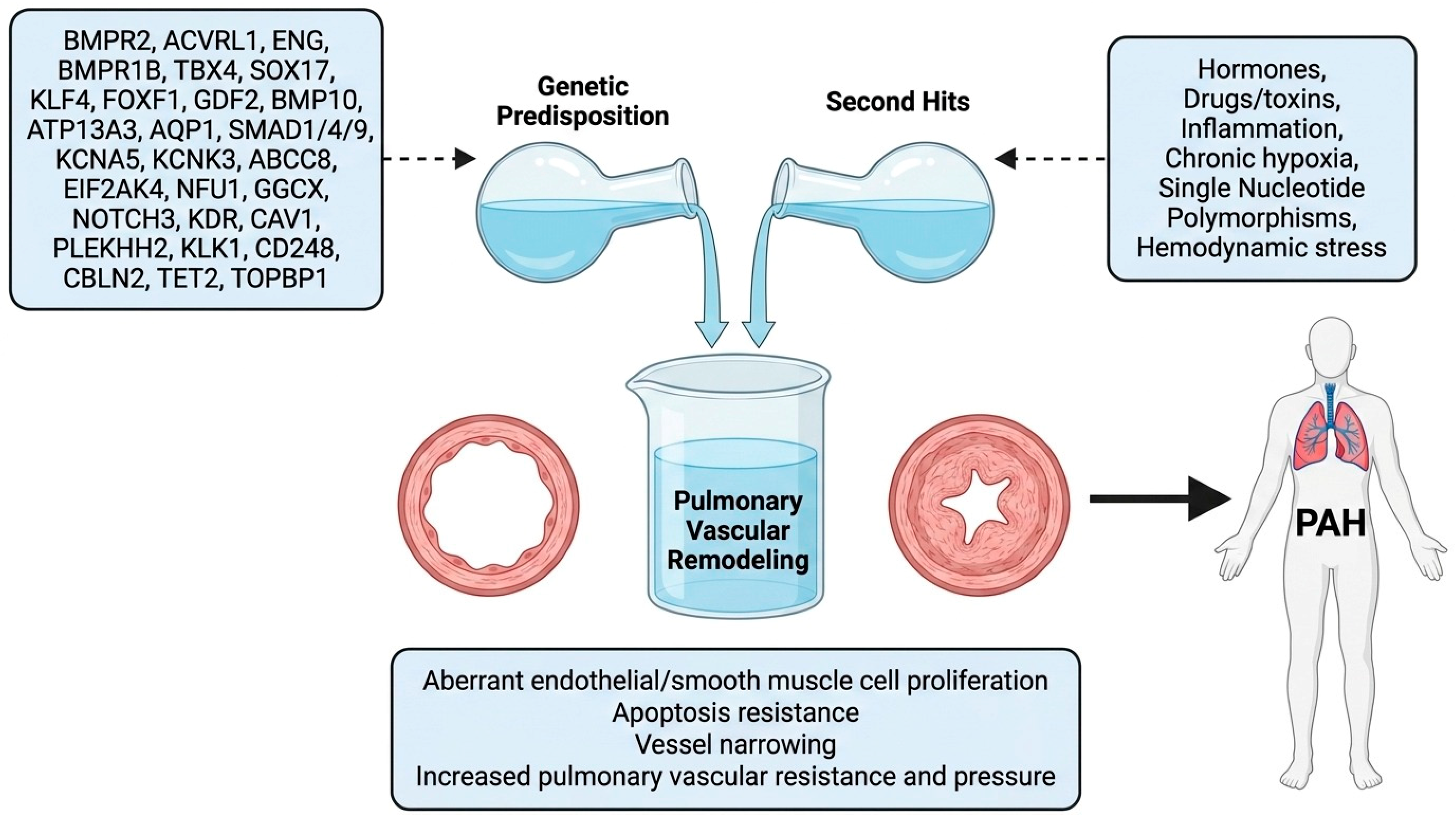
All heritable PAH and some cases of idiopathic PAH are caused by dysfunctions in certain genes (
Figure 1). According to [
5], at least 5% of idiopathic PAH patients have a family history of the disease. The presence of mutations in these genes can trigger the condition, especially in the presence of a “second hit” such as chronic hypoxia, inflammation, hemodynamic stress, hormones, drugs, and single-nucleotide polymorphisms (SNPs) (
Figure 1). A second hit refers to a secondary exposure that acts on a genetically predisposed background, initiating or accelerating the pathological processes (pulmonary vascular remodeling) that lead to the development of overt PAH. PAH is inherited in an autosomal dominant manner with an incomplete penetrance of around 20% [
5]. This low penetrance, even in the presence of a mutation, corroborates the requirement for a “second hit” to manifest PAH in some cases. Sex (estrogen) plays a role in penetrance, evidenced by females having a 2.3 times higher preponderance of PAH than males [
6]. Chronic hypoxia, hemodynamic stress, and inflammation cause pulmonary vascular injury, which leads to vascular remodeling, thereby precipitating PAH. Drugs such as methamphetamines, anorexigens, and amphetamines increase serotonin activity; serotonin is both a potent vasoconstrictor and a smooth muscle cell mitogen, promoting vascular remodeling. SNPs alter transcription efficiency of the
TGF-β1 gene, hence distorting the BMP/TGF-β balance by upregulating TGF-β signaling [
7] (
Table 1). Aberrant TGF-β signaling disrupts pulmonary vascular cell proliferation, survival, and migration, thereby causing vascular remodeling—a hallmark feature of PAH. Cigarette smoking represents a further environmental second hit of clinical relevance. Tobacco smoke constituents cause direct oxidative endothelial injury, promote pulmonary vascular inflammation, and impair nitric oxide-mediated vasodilation, thereby lowering the threshold for disease manifestation in genetically predisposed individuals. This is particularly pertinent in PAH cases associated with chronic obstructive pulmonary disease (COPD) or combined pulmonary fibrosis and emphysema, where smoking-induced vascular injury acts synergistically with genetic susceptibility to accelerate pulmonary vascular remodeling [
8].
This review discusses all genes currently implicated in PAH pathogenesis (
Figure 1). These genes have been grouped according to function: receptors of the TGF-β family (
BMPR2,
ACVRL1,
ENG, and
BMPR1B), ligands of the BMP signaling pathway (
GDF2 and
BMP10), transcription factors (
TBX4,
SOX17,
KLF4,
FOXF1, and
SMAD1/
4/8), membrane transporters (
ATP13A3 and
AQP1), potassium ion transporters (
KCNA5,
KCNK3, and
ABCC8), metabolic genes (
EIF2AK4,
NFU-1, and
GGCX), other signaling receptors (
NOTCH3 and
KDR), membrane/structural proteins (
CAV-1 and
PLEKHH2), vasoactive regulation (
KLK1), extracellular/matrix-related (
CD248 and
CBLN2), and epigenetic/nuclear regulation (
TET2 and
TOPBP1).
It should be noted that the depth of coverage across gene sections in this review reflects the available published literature. Genes such as BMPR2, ACVRL1, GDF2, SOX17, and EIF2AK4 are discussed in greater detail because they are supported by extensive genetic, functional, and clinical data accumulated over two decades. By contrast, genes such as PLEKHH2, KLK1, CBLN2, CD248, TOPBP1, NFU1, and GGCX are covered more concisely because the published evidence base remains limited, consisting largely of small cohort reports, single functional studies, or preliminary genetic associations. Where evidence is sparse, this review explicitly acknowledges that limitation rather than over-interpreting available data.
3. Genes Implicated in PAH Pathogenesis
3.1. Receptors of the TGF-β Family
The TGF-β family comprises a diverse group of cytokines such as bone morphogenetic proteins (BMPs), activins, growth differentiation factors (GDFs), nodal, and inhibins [
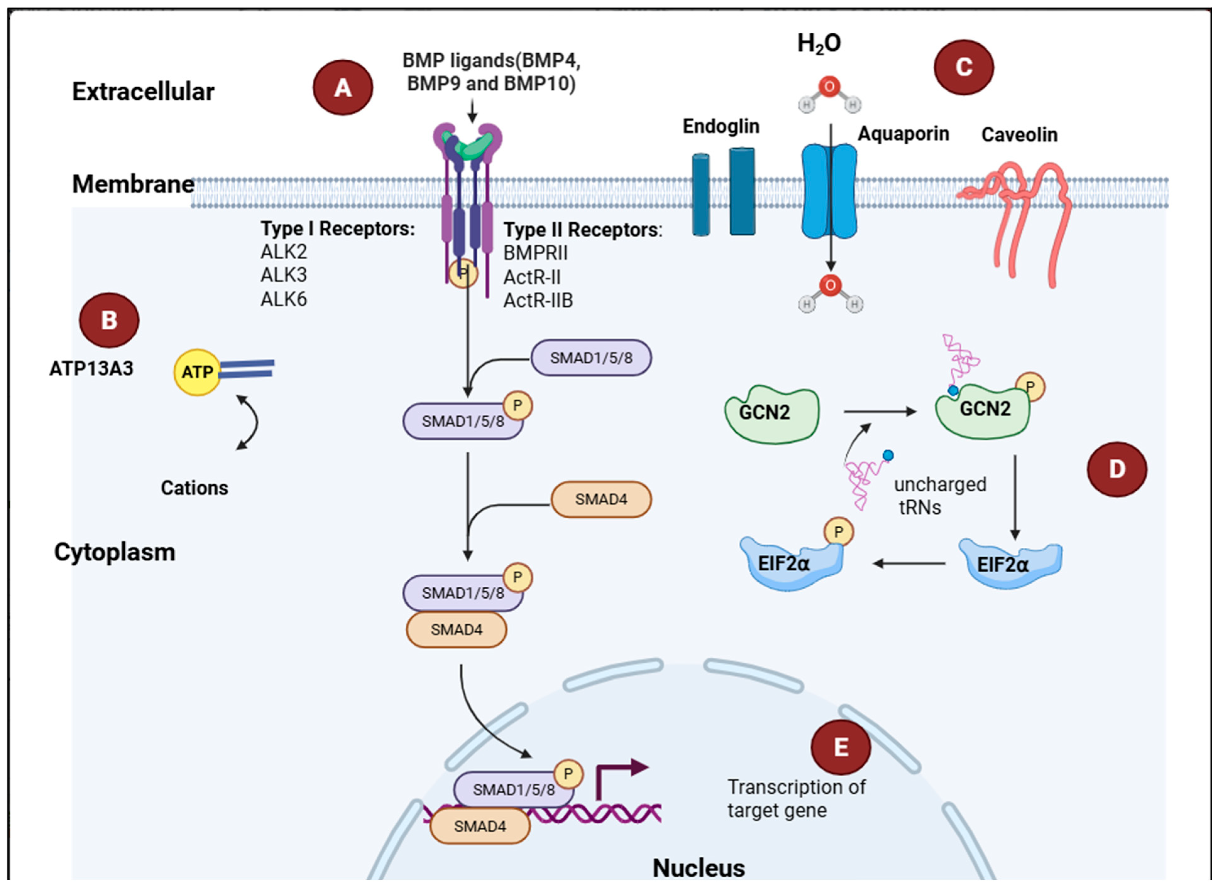
9] that modulate key cellular processes such as proliferation, differentiation, apoptosis, and extracellular matrix formation. TGF-β ligands signal through a receptor system composed of type I, type II, and type III co-receptors. In the BMP signaling pathway, the type II (BMPRII, ActRIIA, and ActRIIB)-type I receptor (
ALK1,
ALK2,
ALK3,
ALK6) complex activates SMAD1/5/8 (
Figure 2). In the TGF-β signaling pathway, the type II (TGFβRII, ActRIIA, ActRIIB)-type I receptor (
ALK4,
ALK5,
ALK7) complex activates SMAD2/3, which leads to downstream transcription of genes such as
PAI-1 that are responsible for the cellular changes (aberrant proliferation and apoptosis resistance) seen in PAH [
9]. Type III receptors (endoglin and betaglycan) function as auxiliary receptors, facilitating ligand presentation to type II–type I receptor complexes.
3.1.1. BMPR2
The bone morphogenetic protein receptor type 2 (
BMPR2), a member of the transforming growth factor-β (TGF-β) superfamily, plays a central role in regulating vascular cell proliferation, differentiation, and apoptosis. The
BMPR2 gene, located on chromosome 2q33, encodes a receptor comprising an extracellular ligand-binding domain (exons 1–3), a transmembrane domain (exon 4), a serine/threonine kinase domain (exons 5–11), and a cytoplasmic C-terminal domain (exons 12–13) [
10,
11].
BMPR2 was first identified as a causal gene for heritable pulmonary arterial hypertension (PAH) in 2000 through independent studies demonstrating heterozygous germline mutations in affected families [
12,
13,
14]. According to the ClinGen Pulmonary Hypertension Gene Curation Expert Panel,
BMPR2 is classified as a definitive gene for PAH, representing the most robust and reproducible genetic association in the disease [
15].
Pathogenic
BMPR2 variants are highly heterogeneous, including nonsense, frameshift, splice-site mutations, and large genomic rearrangements, most of which result in haploinsufficiency through nonsense-mediated decay or truncated protein products [
16,
17,
18]. These variants account for approximately 53–86% of heritable PAH cases and 14–35% of idiopathic PAH [
19]. However, despite this strong association, BMPR2 mutations exhibit incomplete penetrance of approximately 20–30% [
20], indicating that mutation carriage alone is insufficient to drive disease development.
This incomplete penetrance represents a key area of uncertainty and suggests that additional genetic modifiers, epigenetic mechanisms, and environmental exposures contribute to disease expression. Variability in penetrance and disease severity among carriers highlights substantial heterogeneity that is not fully explained by current genetic models, underscoring limitations in our understanding of disease initiation.
At the molecular level,
BMPR2 mediates signaling through both canonical and non-canonical pathways. Canonical signaling involves ligand binding (particularly
BMP9 and
BMP10), receptor complex formation, and phosphorylation of
SMAD1/
5/8 (
Figure 2), which regulate transcription of genes essential for maintaining vascular quiescence [
21,
22]. Disruption of this pathway leads to impaired anti-proliferative signaling in endothelial cells.
In parallel,
BMPR2 dysfunction is associated with activation of non-canonical signaling pathways, including ERK, p38 MAPK, JNK, and PI3K/AKT cascades, which contribute to endothelial dysfunction, apoptosis resistance, and vascular remodeling [
22]. However, the relative contribution of these pathways to disease initiation versus progression remains incompletely defined, reflecting a broader limitation in mechanistic resolution across PAH studies.
A central pathogenic feature of PAH is the imbalance between BMP and TGF-β signaling. Reduced
BMPR2-mediated signaling, coupled with relatively enhanced TGF-β pathway activity, promotes a pro-proliferative and anti-apoptotic vascular phenotype. Similar pathway dysregulation is observed in other PAH-associated genes within the BMP signaling axis, including
ACVRL1,
ENG,
SMAD9, and
GDF2. Compared with these genes,
BMPR2 has the most extensive genetic and functional evidence supporting causality, whereas several others demonstrate more variable or context-dependent associations [
15]. This distinction underscores the importance of differentiating between definitive causal genes and those with emerging or more limited evidence.
Clinically,
BMPR2 mutation carriers tend to present at a younger age, exhibit more severe hemodynamic impairment, and have poorer survival compared with non-carriers [
8]. However, reduced
BMPR2 protein expression has also been observed in PAH patients without identifiable
BMPR2 mutations, suggesting that
BMPR2 downregulation may reflect both primary genetic effects and secondary disease-associated processes [
23]. This observation complicates direct attribution of causality and highlights the interplay between genetic predisposition and disease progression.
Experimental animal models have confirmed the importance of
BMPR2 in vascular development and signaling [
24]; however, these models incompletely recapitulate the chronic, heterogeneous, and incompletely penetrant nature of human PAH. While these systems provide important mechanistic insights, their translational relevance remains limited, particularly in modeling late-onset or environmentally influenced disease.
Taken together, BMPR2 represents the most well-established genetic contributor to PAH, but its incomplete penetrance, variable expressivity, and interaction with broader signaling networks emphasize that it functions within a complex, multifactorial disease framework rather than as a singular deterministic cause.
3.1.2. ACVRL1
The
ACVRL1 gene encodes activin receptor-like kinase 1 (
ALK1), a type I serine/threonine kinase receptor within the transforming growth factor-β (TGF-β) superfamily that is predominantly expressed in endothelial cells. Located on chromosome 12q13,
ALK1 forms part of a heteromeric receptor complex that mediates signaling in response to circulating ligands such as
BMP9 and
BMP10 (
Figure 2), thereby promoting endothelial quiescence and vascular stability [
25,
26].
Pathogenic variants in
ACVRL1 were first identified in patients with hereditary hemorrhagic telangiectasia (HHT), establishing its role in vascular dysplasia [
27,
28]. Subsequent studies demonstrated that heterozygous
ACVRL1 mutations are also associated with pulmonary arterial hypertension (PAH), particularly in the context of HHT-associated PAH [
29,
30]. According to the ClinGen Pulmonary Hypertension Gene Curation Expert Panel,
ACVRL1 is classified as a definitive PAH gene, although it contributes less frequently to disease compared with
BMPR2 [
15].
The mutation spectrum of
ACVRL1 includes missense, nonsense, frameshift, and splice-site variants [
31], many of which affect functionally critical regions such as the kinase domain. These mutations account for approximately 3–5% of heritable PAH cases and a smaller proportion of idiopathic PAH, underscoring their relatively modest contribution compared with
BMPR2-associated disease [
30]. Clinically,
ACVRL1 mutation carriers tend to present at a younger age and may exhibit more severe disease, particularly in individuals with overlapping HHT features, suggesting a phenotype shaped by combined vascular dysplasia and pulmonary vascular remodeling [
30].
At the signaling level,
ALK1 is a key mediator of endothelial BMP signaling. Ligand binding promotes phosphorylation of
SMAD1/
5/8 and downstream transcriptional programs that maintain vascular homeostasis. Loss of
ACVRL1 function attenuates this pathway, reducing endothelial quiescence and increasing susceptibility to pathological remodeling [
25,
26]. In parallel, diminished
ALK1 activity has been associated with relative enhancement of TGF-β signaling, shifting endothelial cells toward pro-proliferative and anti-apoptotic states. However, much of this mechanistic framework is derived from in vitro systems and animal models, and direct confirmation in human pulmonary vascular tissue remains limited, constraining definitive causal interpretation.
Comparison with BMPR2-related PAH highlights both shared and distinct features within the BMP/TGF-β signaling axis. While both genes converge on impaired SMAD-mediated signaling, BMPR2 mutations are more prevalent and better characterized in isolated PAH, whereas ACVRL1-associated disease is frequently embedded within a broader syndromic vascular context. This distinction suggests that perturbations at different nodes of the same pathway may confer varying degrees of penetrance and tissue specificity, rather than equivalent pathogenic effects.
Several unresolved questions remain. The relative contribution of endothelial versus smooth muscle dysfunction, the extent to which ALK1 signaling defects initiate disease versus amplify existing vascular injury, and the marked variability in clinical expression among mutation carriers all point to incomplete mechanistic resolution. These uncertainties support a model in which ACVRL1 mutations create a sensitized endothelial state that requires additional genetic or environmental factors to produce overt PAH.
This endothelial signaling vulnerability is further exemplified by co-receptors such as ENG, which function alongside ALK1 within the same receptor complex and provide complementary insight into the role of disrupted BMP signaling in PAH pathogenesis.
3.1.3. ENDOGLIN
Endoglin (
ENG) is a transmembrane glycoprotein predominantly expressed in endothelial cells, where it functions as a co-receptor within the transforming growth factor-β (TGF-β)/bone morphogenetic protein (BMP) signaling pathway. Located on chromosome 9q34,
ENG encodes a protein belonging to the zona pellucida family, characterized by a conserved extracellular domain that facilitates ligand binding and stabilization of receptor complexes [
32,
33]. Through its interaction with type I receptors such as
ALK1 and type II receptors, endoglin modulates endothelial responses to circulating ligands, particularly
BMP9 and
BMP10 (
Figure 2) [
26].
Pathogenic variants in
ENG were identified in patients with hereditary hemorrhagic telangiectasia (HHT), establishing its role in vascular dysplasia [
27,
32]. Subsequent studies have demonstrated that
ENG mutations are also associated with pulmonary arterial hypertension (PAH), particularly in the context of HHT-associated disease [
34]. According to the ClinGen Pulmonary Hypertension Gene Curation Expert Panel,
ENG is classified as a definitive PAH gene, although its contribution is less frequent than
BMPR2 and is often observed within a syndromic vascular context [
15].
Endoglin enhances BMP signaling by facilitating ligand–receptor complex formation and promoting activation of the
SMAD1/
5/8 pathway, thereby supporting endothelial quiescence and vascular integrity. Loss of
ENG disrupts this signaling axis and alters endothelial proliferation, migration, and apoptosis. Experimental models demonstrate reduced BMP pathway activity, evidenced by decreased
SMAD1/
5/8 phosphorylation in
ENG-deficient systems [
35]. This signaling impairment closely parallels that observed in
BMPR2- and
ACVRL1-deficient models, indicating convergence on a shared downstream pathway rather than gene-specific effects.
Despite this mechanistic alignment, several limitations constrain interpretation. Much of the available evidence derives from murine models and in vitro systems, which do not fully capture the structural and hemodynamic complexity of the human pulmonary vasculature. Consequently, it remains unclear whether ENG deficiency directly initiates pulmonary vascular disease or instead amplifies remodeling processes in an already compromised vascular environment. This distinction is particularly relevant given that ENG-associated PAH frequently occurs in the setting of systemic vascular abnormalities such as HHT.
Clinically,
ENG mutation carriers, similar to those with
ACVRL1 mutations, tend to present with earlier disease onset and may experience more severe outcomes, including reduced survival [
34]. However, available cohort sizes are limited, and marked phenotypic variability suggests the influence of additional genetic or environmental modifiers.
In comparison with BMPR2, ENG contributes less frequently to PAH and is more commonly associated with syndromic vascular disease, underscoring differences in penetrance and disease context despite shared pathway involvement. This contrast supports a model in which disruption of endothelial BMP signaling represents a central pathogenic axis, while the specific clinical phenotype depends on the level and context of pathway perturbation.
Extending this pathway-level framework, circulating ligands such as GDF2 (BMP9) provide further insight into how upstream modulation of BMP signaling influences disease susceptibility.
3.1.4. BMPR1B
Bone morphogenetic protein receptor type 1B (
BMPR1B), also known as
ALK6, is a type I serine/threonine kinase receptor within the transforming growth factor-β (TGF-β) superfamily that participates in canonical BMP signaling. Structurally,
BMPR1B comprises an extracellular ligand-binding domain, a glycine–serine (GS) domain required for receptor activation, a transmembrane domain, and an intracellular kinase domain (
Table 2) [
36]. While these structural features are conserved across BMP type I receptors,
BMPR1B appears to have a more restricted role in pulmonary vascular biology, with comparatively lower endothelial expression than receptors such as
BMPR2 and
ACVRL1.
Evidence linking
BMPR1B to pulmonary arterial hypertension (PAH) remains limited. Initial sequencing studies identified two missense variants (S160N and F392L) in pediatric idiopathic PAH patients [
37]. Functional assessment demonstrated that the F392L variant, located within the kinase domain, more substantially altered receptor signaling than the S160N variant, consistent with observations in
BMPR2 where kinase-domain mutations exert greater functional impact. However, unlike canonical loss-of-function mutations in
BMPR2 or
ACVRL1, both
BMPR1B variants retained the capacity to induce
SMAD1/5/8 signaling in vitro, raising uncertainty regarding whether these alterations represent partial loss, gain, or context-dependent modulation of receptor activity.
This ambiguity highlights an important distinction between BMPR1B and established PAH genes within the BMP pathway. In contrast to BMPR2, ACVRL1, and ENG, where convergent evidence supports reduced BMP signaling as a central pathogenic mechanism, BMPR1B-associated variants do not consistently demonstrate impaired downstream signaling. This lack of directional consistency weakens causal inference and suggests that BMPR1B may not operate as a primary driver of pathway insufficiency, but rather as a context-dependent modulator of signaling output.
Consistent with this interpretation,
BMPR1B is not currently classified as a PAH-associated gene by the Clinical Genome Resource Pulmonary Hypertension Gene Curation Expert Panel, which designates the gene as having limited evidence [
15]. This classification reflects the small number of reported variants, the absence of replication in independent cohorts, and limited in vivo functional validation.
From a mechanistic perspective, BMPR1B variants may influence receptor complex composition or ligand responsiveness, thereby subtly altering SMAD signaling dynamics rather than producing the marked signaling deficits observed in BMPR2-related disease. This contrasts with ligand-level defects (e.g., BMP9/BMP10) or receptor haploinsufficiency, where reduced signaling amplitude is more clearly established. In this context, BMPR1B is better positioned alongside emerging or modifier genes, where effects on pathway tuning rather than pathway disruption may contribute to disease susceptibility.
A key unresolved issue is whether BMPR1B variants exert measurable effects under physiological conditions, where receptor stoichiometry, ligand gradients, and hemodynamic forces differ substantially from in vitro systems. The restriction of reported variants to small pediatric cohorts further limits generalizability and prevents robust genotype–phenotype correlation.
Current evidence does not support a definitive pathogenic role for BMPR1B in PAH. Instead, the gene represents a biologically plausible but genetically under-validated component of the BMP signaling network, illustrating the importance of distinguishing between core pathway drivers and peripheral modulators when interpreting PAH genetics.
3.2. Ligands of the BMP Signaling Pathway
BMPs are a subclass of the TGF-β superfamily that specifically signal through type I receptors
ACVRL1 (
ALK1), ACVR1 (
ALK2), BMPR1A (
ALK3), and
BMPR1B (
ALK6), consequently leading to the phosphorylation of
SMAD1/
5/8 (
Figure 2) [
38]. BMPs such as GDF5, GDF6, GDF7,
BMP2,
BMP4, BMP5, BMP6, BMP7, BMP8A, BMP8B,
BMP9,
BMP10, and BMP15 modulate several cellular processes, including proliferation, growth, differentiation, and apoptosis. Mutations in genes encoding these ligands disrupt the BMP/TGF-β signaling balance, triggering the vascular remodeling changes characteristic of PAH.
3.2.1. GDF2
The
GDF2 gene encodes bone morphogenetic protein 9 (
BMP9), a circulating ligand within the transforming growth factor-β (TGF-β) superfamily that plays a central role in maintaining vascular homeostasis (
Table 2).
BMP9 is synthesized predominantly in the liver and circulates in an active form, signaling through receptor complexes composed of
BMPR2 and type I receptors such as
ACVRL1 on endothelial cells [
25]. Through activation of canonical
SMAD1/
5/8 signaling,
BMP9 promotes endothelial quiescence and suppresses inappropriate vascular proliferation.
In contrast to receptor-level mutations (e.g.,
BMPR2,
ACVRL1,
ENG),
GDF2 variants disrupt BMP signaling at the level of ligand availability, demonstrating that pulmonary arterial hypertension (PAH) can arise from impaired signal input as well as defective signal transduction. Sequencing studies first identified pathogenic
GDF2 variants in PAH cohorts, with functional analyses showing reduced circulating
BMP9 levels and diminished downstream SMAD activation [
39,
40]. Consistent with this, the Clinical Genome Resource Pulmonary Hypertension Gene Curation Expert Panel classifies
GDF2 as a definitive PAH gene [
15].
Most GDF2 mutations result in reduced secretion or stability of BMP9, leading to decreased activation of BMPR2/ALK1 receptor complexes. The downstream consequence is attenuation of SMAD1/5/8 signaling and a relative shift toward TGF-β–dominant pathways, promoting endothelial dysfunction, apoptosis resistance, and vascular remodeling. This signaling imbalance closely mirrors that observed in receptor-level defects, indicating that diverse genetic lesions converge on a shared pathway-level deficiency rather than producing gene-specific phenotypes.
A key mechanistic insight from
GDF2-associated PAH is the apparent importance of ligand dosage. Biallelic loss-of-function mutations are associated with early-onset and severe disease, whereas heterozygous carriers demonstrate variable penetrance and later onset [
39,
41]. This dosage effect parallels observations in
BMPR2 haploinsufficiency and supports a threshold model in which partial reduction in BMP signaling lowers endothelial resilience without being independently sufficient to cause disease.
However, this framework is complicated by the presence of functional redundancy within the BMP ligand axis. BMP9 shares substantial overlap with BMP10, which can also activate ALK1-mediated signaling. This raises an unresolved question: whether GDF2 mutations cause disease primarily through absolute reduction in signaling, or through disruption of the relative balance between circulating BMP ligands. The partial preservation of signaling in some mutation carriers suggests that compensatory mechanisms may exist, but these are not sufficient to fully prevent disease under stress conditions.
Despite strong genetic and biochemical evidence, several limitations remain. Much of the current understanding is derived from circulating BMP9 measurements and in vitro endothelial assays, which may not fully reflect ligand gradients, receptor distribution, or hemodynamic influences present in the pulmonary circulation. Furthermore, incomplete penetrance in heterozygous carriers indicates that GDF2 deficiency alone is often insufficient to initiate PAH, supporting a multi-hit model involving additional genetic, inflammatory, or environmental modifiers.
Compared with receptor-associated genes, GDF2 highlights the importance of upstream signal availability in maintaining endothelial homeostasis. Together with defects in receptors (BMPR2, ACVRL1, ENG) and downstream mediators (SMADs), these findings support a unified model in which PAH arises from quantitative and context-dependent disruption of BMP signaling across multiple levels of the pathway, rather than from isolated gene-specific mechanisms.
3.2.2. BMP10
The
BMP10 gene encodes bone morphogenetic protein 10, a circulating ligand within the transforming growth factor-β (TGF-β) superfamily (
Figure 2) that plays a critical role in cardiovascular development, particularly in cardiac morphogenesis and ventricular trabeculation during embryogenesis. In adults,
BMP10 is predominantly produced in the right atrium and circulates as an endocrine-like vascular ligand [
42,
43].
BMP10 signals through type II BMP receptors in complex with type I receptors such as
ACVRL1 (
ALK1), activating canonical
SMAD1/
5/8 signaling and downstream transcriptional programs that promote endothelial quiescence and vascular stability [
25].
BMP10 exhibits substantial functional overlap with BMP9 (encoded by GDF2), with both ligands acting as high-affinity activators of ALK1-mediated signaling in endothelial cells. However, an important distinction lies in their physiological roles: BMP9 is the dominant circulating ligand under basal conditions, whereas BMP10 appears to contribute more selectively, with expression enriched in cardiac tissue. This asymmetry suggests that BMP10 may function as a context-dependent or compensatory ligand rather than a primary regulator of endothelial homeostasis.
From a pathobiological perspective, disruption of
BMP10 signaling would be expected to impair SMAD-dependent pathways and promote endothelial dysfunction, similar to defects observed in
GDF2 or receptor-level mutations. However, direct genetic evidence linking
BMP10 variants to pulmonary arterial hypertension (PAH) remains limited. Unlike
GDF2, which is classified as a definitive PAH gene,
BMP10 has not yet achieved equivalent evidentiary support within frameworks such as the Clinical Genome Resource Pulmonary Hypertension Gene Curation Expert Panel [
15]. This distinction is critical, as it places
BMP10 within the category of biologically plausible but genetically under-validated contributors.
A central unresolved issue is the extent to which BMP10 contributes independently to disease versus acting within a redundant ligand network. Functional overlap with BMP9 raises the possibility that isolated BMP10 deficiency may be buffered by preserved BMP9 signaling, thereby limiting phenotypic expression. Conversely, combined or cumulative reductions in BMP ligand availability could reduce overall signaling below a critical threshold, predisposing to disease. This “ligand dosage” model aligns with observations in GDF2 and BMPR2-associated PAH, where partial signaling deficits confer susceptibility rather than deterministic causality.
Emerging studies also suggest that
BMP10 may exert effects beyond canonical SMAD signaling. Experimental data indicate that altered
BMP10 activity can modulate inflammatory pathways, including upregulation of chemokines such as
CCL2, potentially promoting monocyte recruitment and vascular inflammation [
44]. While this introduces an additional mechanistic dimension linking BMP signaling to immune-mediated remodeling, these findings remain preliminary and are largely derived from in vitro systems.
Several limitations constrain current interpretation. The majority of mechanistic data originate from developmental biology and experimental models, which may not accurately reflect adult pulmonary vascular disease. The absence of well-characterized human mutation cohorts limits genotype–phenotype correlation and prevents clear attribution of causality. These gaps contrast with the robust genetic and functional evidence supporting BMP9 (GDF2), BMPR2, and ACVRL1, highlighting a gradient of evidence within the BMP signaling axis.
Therefore, BMP10 is best conceptualized as a complementary ligand within the BMP signaling network that contributes to endothelial homeostasis but has not yet been definitively implicated in PAH. Its role likely reflects modulation of overall ligand availability rather than a primary pathogenic driver, reinforcing the broader concept that quantitative and context-dependent disruption of BMP signaling, rather than single-gene defects, underlies pulmonary vascular disease.
3.3. Transcription Factors
Transcription factors play a crucial role in the pathogenesis of PAH by regulating the expression of genes that drive endothelial dysfunction, PASMC proliferation, metabolic reprogramming, inflammation, and extracellular matrix remodeling. Abnormal activation, suppression, or mutation of transcription factors contributes to the pro-proliferative, anti-apoptotic, and vasoconstrictive phenotype characteristic of PAH.
3.3.1. TBX4
Mutations in the
TBX4 gene have emerged as an important genetic contributor to childhood-onset pulmonary arterial hypertension (PAH). Early cohort studies demonstrated that a substantial proportion of pediatric PAH cases harbor either microdeletions at chromosome 17q23.2 encompassing
TBX4 or pathogenic sequence variants within the gene [
45]. In contrast,
TBX4 variants are identified in only a small fraction of adult PAH cases (approximately 1–2%), indicating a marked age-dependent penetrance and supporting a distinct developmental contribution to disease susceptibility.
TBX4 encodes a T-box transcription factor that plays a central role in embryonic limb and pulmonary development. A key downstream target is fibroblast growth factor 10 (
FGF10), which is essential for lung branching morphogenesis and pulmonary vascular formation [
46]. Disruption of the
TBX4–
FGF10 axis therefore provides a biologically plausible mechanism for early-life vulnerability, whereby abnormal lung and vascular development results in a structurally compromised pulmonary vascular bed that is less capable of adapting to postnatal hemodynamic demands.
This developmental framework distinguishes TBX4 from canonical PAH genes such as BMPR2, ACVRL1, and GDF2, which primarily perturb endothelial signaling pathways in established vasculature. Instead, TBX4-associated PAH is best conceptualized as a developmentally primed condition, in which impaired formation of the pulmonary vascular architecture lowers the threshold for later disease expression rather than directly initiating pathogenic signaling in adulthood.
However, this model remains partially inferential. While developmental defects are well supported by experimental and genetic data, the direct molecular link between TBX4 disruption and the characteristic vascular remodeling seen in PAH—namely smooth muscle proliferation, endothelial dysfunction, and occlusive lesion formation—has not been fully delineated in human systems. Most mechanistic insights derive from developmental biology and animal models, which may not fully recapitulate the postnatal pulmonary vascular environment.
Genotype–phenotype relationships further complicate interpretation. TBX4 variants are associated with a broad phenotypic spectrum, ranging from isolated PAH to small patella syndrome and other skeletal abnormalities, indicating pleiotropic effects of gene disruption. This variability, together with incomplete penetrance, suggests that TBX4 mutations alone are often insufficient to cause disease and that additional genetic modifiers or environmental stressors are required for clinical manifestation. Such observations are consistent with a multi-hit model of PAH pathogenesis.
From an evidence standpoint,
TBX4 is classified as a definitive PAH gene by the Clinical Genome Resource Pulmonary Hypertension Gene Curation Expert Panel [
15], supported by reproducible genetic associations and consistent clinical phenotypes, particularly in pediatric populations. This classification reflects strong genetic evidence rather than complete mechanistic resolution, highlighting a gap between genotype identification and pathway-level understanding.
In comparison with BMP pathway genes, TBX4 occupies a distinct position within the PAH genetic landscape. Whereas BMP-related genes converge on dysregulated endothelial signaling, TBX4 reflects disruption of early developmental programming. This distinction underscores the biological heterogeneity of PAH and supports the concept that disease can arise from both structural predisposition and signaling imbalance, which may ultimately converge on shared downstream pathways of vascular remodeling.
TBX4 represents a key determinant of pediatric PAH characterized by developmental dysregulation, variable penetrance, and mechanistic divergence from classical signaling-driven forms of disease. Studies integrating human pulmonary tissue analysis, developmental modeling, and longitudinal clinical data are needed to clarify how early developmental defects translate into progressive vascular pathology.
3.3.2. SOX17
The
SOX17 gene, located on chromosome 8q11.23, encodes the SRY-box transcription factor 17, a key regulator of endothelial development, arterial specification, and vascular homeostasis.
SOX17 functions as a lineage-defining transcription factor that maintains endothelial identity, particularly in arterial endothelial cells, by coordinating developmental and postnatal gene expression programs involved in vascular stability [
47]. The gene consists of a single coding exon (exon 2), encoding the conserved high-mobility group (HMG) DNA-binding domain and a C-terminal region involved in protein–protein interactions, including β-catenin binding.
Pathogenic
SOX17 variants identified in pulmonary arterial hypertension (PAH) cohorts include missense mutations affecting the HMG-box domain, as well as truncating variants such as nonsense and frameshift mutations [
48]. These variants are predicted to impair DNA binding and disrupt transcriptional regulation of endothelial gene networks. According to the Clinical Genome Resource Pulmonary Hypertension Gene Curation Expert Panel,
SOX17 is classified as a definitive PAH gene [
15], reflecting replicated genetic association and consistent functional evidence across independent cohorts.
SOX17 regulates endothelial signaling networks that intersect with both Wnt/β-catenin and Notch pathways. Loss-of-function studies demonstrate derepression of β-catenin-dependent transcriptional programs and impaired maintenance of endothelial quiescence, contributing to proliferative and migratory phenotypes in vascular endothelium [
47]. Additional experimental evidence suggests modulation of hepatocyte growth factor (HGF)/c-Met signaling, although this remains primarily based on cellular models and has not been fully validated in human pulmonary vascular tissue [
49]. These data support a model in which
SOX17 does not initiate proliferative signaling directly but rather destabilizes transcriptional control of multiple downstream pathways that govern endothelial behavior.
A key mechanistic feature of
SOX17-associated PAH is its strong context dependence. Endothelial-specific
SOX17 loss is insufficient to induce pulmonary arterial hypertension under baseline conditions, whereas disease phenotypes emerge in the presence of additional stressors such as chronic hypoxia or vascular injury [
49]. This provides experimental support for a multi-hit model in which
SOX17 deficiency lowers endothelial resilience but requires secondary environmental or physiological triggers for full disease expression. This behavior closely parallels that observed in
BMPR2 and
GDF2-associated PAH, despite differences in molecular level of action.
From a comparative standpoint, SOX17 occupies a distinct mechanistic position relative to BMP pathway genes such as BMPR2, ACVRL1, ENG, and GDF2. Whereas these genes regulate extracellular ligand–receptor signaling within the TGF-β superfamily, SOX17 functions downstream as an intracellular transcriptional integrator of endothelial identity. Despite this hierarchical difference, both classes converge on a shared endpoint of impaired endothelial quiescence and maladaptive vascular remodeling, suggesting pathway convergence rather than pathway exclusivity in PAH pathogenesis.
Clinically,
SOX17 mutations account for approximately 3% of PAH cases, with variable penetrance and expressivity across carriers [
50]. This variability further supports a non-deterministic model of disease in which
SOX17 variants act as susceptibility alleles rather than sole causal drivers, with phenotypic expression shaped by genetic modifiers and environmental exposures.
SOX17 represents a definitive PAH gene that contributes to disease through disruption of endothelial transcriptional stability rather than extracellular signaling. However, key mechanistic gaps remain, particularly in defining how SOX17-dependent transcriptional programs interface with BMP signaling and inflammatory or hypoxic stress pathways in human pulmonary vascular tissue.
3.3.3. KLF4
Kruppel-like factor 4 (
KLF4), located on chromosome 9q31, encodes a zinc finger transcription factor of the Kruppel-like family, characterized by three C-terminal C2H2 zinc finger domains that mediate DNA binding. In vascular biology,
KLF4 is highly expressed in endothelial cells, where it contributes to the maintenance of endothelial identity and adaptive responses to biomechanical and inflammatory stress [
51] (
Table 2). Unlike monogenic pulmonary arterial hypertension (PAH) genes,
KLF4 is not currently classified as a disease-causing locus but is increasingly recognized as a context-dependent regulator of endothelial quiescence.
At the transcriptional level,
KLF4 regulates multiple endothelial effector pathways, including nitric oxide signaling via endothelial nitric oxide synthase (eNOS), prostacyclin synthesis, and endothelin signaling. In PAH lung tissue, reduced
KLF4 expression has been associated with downregulation of eNOS, endothelin receptor subtype B (ETB), and prostacyclin synthase, alongside increased endothelin-1 expression [
52]. This coordinated shift reflects a reprogramming of endothelial signaling toward vasoconstriction, inflammation, and smooth muscle activation, which are central features of pulmonary vascular remodeling.
KLF4 functions less as a pathway-specific signaling component and more as a transcriptional integration node that stabilizes endothelial quiescence under shear stress and inflammatory stimulation. Its downregulation therefore does not initiate a discrete signaling defect but instead lowers the transcriptional threshold for activation of vasoconstrictive and proliferative programs. However, the current evidence base is largely derived from human tissue expression profiling and in vitro endothelial models, limiting causal inference regarding whether KLF4 loss is primary or secondary to established vascular injury.
From a genetic perspective, KLF4 differs fundamentally from established PAH genes such as SOX17, BMPR2, or TBX4, which harbor recurrent germline pathogenic variants with demonstrable disease segregation. In contrast, no reproducible monogenic KLF4 variant spectrum has been established in PAH, and its contribution is not currently supported at the level of a definitive or moderate ClinGen gene–disease relationship. This positions KLF4 outside the core heritable PAH gene set and instead within a broader category of transcriptional susceptibility factors.
KLF4 aligns more closely with endothelial state regulators such as CAV1 or FOXF1, which modulate vascular phenotype stability without acting as primary signaling initiators. In this framework, KLF4 loss is best interpreted as a permissive alteration that amplifies downstream consequences of established pathogenic pathways, including BMP/TGF-β dysregulation, inflammatory activation, and hypoxia-driven remodeling.
Several limitations constrain current interpretation. First, most available data is correlative, and reduced KLF4 expression may represent a downstream consequence of endothelial injury rather than a primary event. Second, in vivo models of endothelial-specific KLF4 loss in pulmonary hypertension remain limited and context-dependent, preventing definitive assignment of causality. Third, the absence of validated pathogenic germline variants further limits its classification as a monogenic PAH gene.
KLF4 is best conceptualized as a transcriptional gatekeeper of endothelial stability whose downregulation facilitates, rather than initiates, the pathological vascular phenotype in PAH. Its role is therefore most consistent with a disease modifier that integrates biomechanical and inflammatory stress into transcriptional programs that converge on vascular remodeling.
3.3.4. FOXF1
FOXF1 (Forkhead box F1), located on chromosome 16p24.1, encodes a forkhead family transcription factor characterized by a conserved winged-helix DNA-binding domain. During embryogenesis,
FOXF1 is a critical regulator of mesenchymal–endothelial crosstalk and is required for proper pulmonary vascular and alveolar capillary network development [
53] (
Table 2). In contrast, its role in adult pulmonary vascular homeostasis is less well defined, although emerging evidence supports a continued contribution to endothelial maintenance and injury responses.
Functional studies in pulmonary endothelial systems suggest that
FOXF1 contributes to endothelial stability by regulating angiogenic competence and DNA damage response pathways. In pulmonary artery endothelial cells derived from pulmonary arterial hypertension (PAH) patients,
FOXF1 overexpression has been associated with increased angiogenic signaling, including upregulation of
VEGFR2 and
CLDN5, alongside enhanced expression of DNA repair-related mediators such as TP53 and ATM [
54]. Conversely,
FOXF1 suppression in endothelial models impairs migration, reduces angiogenic capacity, and disrupts DNA damage response signaling, indicating a role in maintaining endothelial adaptability under stress rather than directly controlling proliferative signaling.
This evidence positions FOXF1 as a regulator of endothelial repair capacity and genomic stability rather than a primary determinant of vascular tone or canonical proliferative signaling pathways. Its function appears to center on preserving endothelial resilience following injury, particularly under conditions of oxidative stress or impaired vascular regeneration. However, the current evidence base is largely derived from in vitro systems, and the extent to which these mechanisms operate in vivo within the human pulmonary circulation remains uncertain.
From a genetic standpoint, FOXF1 differs from definitive PAH genes such as BMPR2, SOX17, and ACVRL1, which exhibit recurrent pathogenic variants with established disease segregation. In contrast, FOXF1 has not been consistently implicated in monogenic PAH, and its contribution is not currently established as a definitive gene–disease relationship. This supports its interpretation as a susceptibility or modifier locus rather than a primary causal driver in most PAH contexts.
FOXF1 occupies a distinct mechanistic position within PAH biology. Unlike SOX17, which directly regulates endothelial transcriptional programs linked to vascular identity, or KLF4, which integrates inflammatory and biomechanical cues, FOXF1 primarily influences endothelial repair and genomic maintenance pathways. This positions it within a complementary axis of vascular resilience, functioning alongside, but mechanistically distinct from, core BMP/TGF-β signaling components and ion-channel or metabolic regulators.
Key limitations persist. Most mechanistic evidence derives from endothelial cell culture models, which do not fully replicate the multicellular and hemodynamic complexity of pulmonary vascular remodeling. Second, definitive human genetic evidence linking FOXF1 variation to heritable PAH is limited, and its role may be context-dependent rather than deterministic. Third, the upstream and downstream integration of FOXF1 with established PAH pathways, including BMP signaling and inflammatory cascades, remains incompletely defined.
Therefore, FOXF1 is best conceptualized as a transcriptional regulator of endothelial repair and genomic integrity that modulates susceptibility to pulmonary vascular injury. Rather than initiating PAH, FOXF1 dysfunction likely lowers endothelial resilience, thereby amplifying disease progression in the presence of primary pathogenic insults.
3.3.5. SMADs 1, 4, and 8
SMAD1 is a receptor-regulated SMAD (R-SMAD) that functions as a key intracellular effector of bone morphogenetic protein (BMP) signaling. Following ligand binding to BMP receptor complexes, type II receptors phosphorylate type I receptors, which in turn phosphorylate
SMAD1. Activated
SMAD1 then forms a complex with the common mediator
SMAD4, translocates to the nucleus, and regulates transcription of downstream target genes involved in endothelial homeostasis, proliferation, and vascular stability (
Figure 2). In this context,
SMAD1 serves as a central intracellular signal transducer that converts extracellular BMP signaling intensity into transcriptional responses within pulmonary vascular cells.
Genetic studies have identified rare variants in SMAD pathway genes in pulmonary arterial hypertension (PAH) cohorts, including
SMAD1 missense variants (e.g., c.8T>C; p.V3A) and splice-site alterations, as well as
SMAD4 variants, which are associated with impaired BMP signaling activity [
55]. These findings provide important genetic evidence that disruption at the level of intracellular signal transduction can phenocopy upstream defects in BMP receptors and ligands. However, these variants are extremely rare, and robust genotype–phenotype correlations remain limited, preventing definitive conclusions regarding their overall contribution to PAH susceptibility.
SMAD8 (also known as
SMAD9) is another receptor-regulated SMAD that participates in BMP signaling but exhibits distinct functional properties compared with
SMAD1/
5. Although
SMAD8 can bind DNA and participate in transcriptional complexes, it demonstrates weaker transcriptional activation capacity and context-dependent regulatory effects, partly due to structural differences in its linker region [
56]. These differences allow
SMAD8 to modulate BMP-driven transcriptional output in a manner that can attenuate or fine-tune signaling responses depending on cellular context.
Alterations in SMAD8 have been proposed to disrupt endothelial BMP signaling by interfering with the balance of SMAD1/5-mediated transcriptional programs. One potential mechanism is competition for receptor-mediated phosphorylation or SMAD4 binding, which may reduce the effective transcriptional output of canonical BMP signaling. Alternatively, altered SMAD8 structure may shift transcriptional complexes toward a less active or dysregulated state, thereby contributing to endothelial dysfunction and vascular remodeling. However, these mechanistic models remain largely based on in vitro experimental systems and have not been fully validated in human pulmonary vascular tissue.
Why do alterations in a relatively weak transcriptional effector such as SMAD8 produce meaningful biological effects in the BMP pathway? One possibility is that even modest perturbations in SMAD complex composition can significantly alter transcriptional programs in endothelial cells, given the sensitivity of BMP signaling to dosage and stoichiometric balance. Another possibility is that SMAD8 variants exert dominant-negative effects under specific cellular conditions, thereby amplifying downstream signaling disruption.
Despite this, the overall evidence base remains limited, and in vivo models are needed to clarify whether SMAD1/SMAD4 dysfunction and SMAD8 dysregulation converge on shared pathogenic pathways or represent distinct mechanisms of endothelial injury. In particular, future studies should address whether SMAD-level perturbations act primarily as upstream amplifiers of BMP signaling defects or as independent modifiers of transcriptional endothelial states within the pulmonary circulation.
3.4. Membrane Transporters
Membrane transporters are integral proteins that regulate the movement of ions, water, metabolites, lipids, and signaling molecules across the cell membrane, thereby controlling essential processes such as osmosis, nutrient uptake, intercellular communication, and mitochondrial function. These proteins are either channels or carriers/pumps. Channels allow molecules to move across the cell membrane by passive diffusion driven by electrochemical gradients, whereas carriers/pumps rely on either primary (ATP hydrolysis) or secondary active transporters.
3.4.1. ATP13A3
ATP13A3 encodes a transmembrane P5-type ATPase localized predominantly to recycling endosomes, where it plays a key role in polyamine transport and intracellular polyamine homeostasis. Polyamines are essential regulators of cellular proliferation, survival, and cytoskeletal organization, and are particularly important for maintaining endothelial integrity and vascular barrier function (
Figure 2) (
Table 2). In this context,
ATP13A3 is increasingly recognized as a metabolic regulator of endothelial fitness rather than a classical signaling molecule within the transforming growth factor-β (TGF-β)/bone morphogenetic protein (BMP) pathway [
57].
Pathogenic variants in
ATP13A3, including biallelic missense and loss-of-function mutations, have been identified in pediatric and early-onset pulmonary arterial hypertension (PAH), suggesting that reduced gene dosage is associated with more severe disease phenotypes [
57,
58]. Functional studies indicate that
ATP13A3 deficiency impairs polyamine uptake, leading to reduced endothelial cell proliferation, increased apoptosis, and disruption of monolayer integrity in pulmonary artery endothelial cells. These alterations are consistent with a primary defect in endothelial survival rather than direct induction of a proliferative phenotype.
ATP13A3 dysfunction has been shown to compromise endothelial junctional stability, resulting in increased vascular permeability, including enhanced susceptibility to thrombin-induced barrier disruption. This loss of barrier integrity provides a plausible link between metabolic dysregulation and early endothelial injury in PAH. These effects converge with pathways implicated in other endothelial stability genes such as AQP1, CAV1, and SOX17, suggesting that impaired barrier maintenance represents a shared pathogenic axis across genetically diverse forms of PAH.
A paradox arises: that ATP13A3 deficiency reduces endothelial proliferation in vitro, whereas PAH is characterized by pathological vascular remodeling and proliferative vascular lesions. This discrepancy suggests that ATP13A3 is unlikely to function as a direct pro-proliferative driver. Instead, current evidence supports a model in which ATP13A3 loss primarily induces endothelial injury and metabolic stress, thereby creating a permissive environment for secondary proliferative responses in adjacent smooth muscle cells and fibroblasts. In this framework, vascular remodeling is interpreted as a compensatory and maladaptive response to primary endothelial dysfunction rather than a direct consequence of ATP13A3-driven proliferation.
According to the ClinGen Pulmonary Hypertension Gene Curation Expert Panel,
ATP13A3 is classified as a definitive PAH gene, supporting a reproducible association between loss-of-function variants and disease across multiple cohorts [
15]. However, despite this genetic strength, key mechanistic gaps remain. In particular, the absence of endothelial-specific
ATP13A3 knockout models and limited in vivo human functional data restricts understanding of how polyamine dysregulation translates into pulmonary vascular remodeling over time.
ATP13A3 represents a distinct class of PAH-associated genes that act through metabolic and membrane transport dysfunction rather than canonical signaling pathways. Its role in disease is best understood as a contributor to endothelial vulnerability and barrier failure, which synergizes with other genetic and environmental insults to initiate and amplify pulmonary vascular injury.
3.4.2. AQP1
Aquaporin 1 (
AQP1), encoded by the
AQP1 gene, is a transmembrane water channel widely expressed in endothelial cells, where it facilitates rapid, bidirectional water transport across the plasma membrane. Structural and functional studies established that
AQP1 forms tetrameric channels that selectively permit water flux in response to osmotic gradients, thereby regulating [
59,
60] (
Figure 2) (
Table 2). Beyond passive transport,
AQP1 has been implicated in endothelial cell migration through localized water influx at the leading edge, which supports lamellipodia formation and cytoskeletal remodeling [
61].
In pulmonary arterial hypertension (PAH), altered
AQP1 expression has been reported in pulmonary vascular cells. Experimental studies demonstrated that
AQP1 overexpression enhances endothelial and pulmonary artery smooth muscle cell proliferation and migration, whereas
AQP1 inhibition attenuates these responses [
62]. In parallel, functional assays have shown that
AQP1 increases endothelial monolayer permeability and reduces barrier resistance, consistent with disruption of cell–cell junction integrity. These findings suggest that
AQP1 influences vascular remodeling not through classical receptor-mediated signaling, but by modulating biophysical properties of the endothelial barrier.
Despite these observations, the direction of causality remains unresolved. Increased AQP1 expression may represent an adaptive response to hypoxia, shear stress, or endothelial injury rather than a primary pathogenic trigger. This interpretation is supported by the limited availability of longitudinal human data and the predominance of in vitro experimental systems, which do not fully recapitulate pulmonary vascular hemodynamics.
Genetic analyses have identified rare variants in
AQP1 among PAH patients, including the recurrent missense variant c.376C>T (p.Arg126Cys) [
63]. However, direct functional validation linking these variants to altered channel activity or endothelial dysfunction in vivo remains limited. Consistent with current evidence-based classifications,
AQP1 is not considered a definitive PAH gene by the Clinical Genome Resource and is best categorized as an emerging or candidate gene with incomplete mechanistic validation [
15].
AQP1-associated phenotypes appear to reflect dysregulated water flux and altered cell volume dynamics, which can influence endothelial migration, barrier permeability, and responsiveness to mechanical stress. However, these effects are likely context-dependent and may require coexisting environmental or molecular “second hits” to contribute meaningfully to disease progression.
Comparison with ATP13A3 highlights a notable convergence in pathogenic outcome despite divergent upstream mechanisms. Whereas ATP13A3 deficiency impairs polyamine transport, leading to reduced endothelial proliferation and increased apoptosis, AQP1 upregulation has been associated with enhanced proliferative and migratory behavior. Despite these opposing cellular phenotypes, both pathways converge on endothelial barrier dysfunction, supporting the concept that barrier failure represents a common downstream event in PAH pathogenesis.
In summary, AQP1 is best conceptualized as a modulator of endothelial barrier dynamics whose dysregulation may contribute to pulmonary vascular remodeling indirectly, rather than functioning as a primary driver of disease. Further in vivo studies, particularly those integrating hemodynamic stress models and human endothelial systems, will be required to define its precise role within the broader PAH genetic and mechanistic landscape.
3.5. Potassium Ion Transporters
Potassium ion transporters, including voltage-gated, ATP-sensitive, and two-pore potassium channels, are membrane proteins that modulate the movement of potassium ions across the cell membrane, thereby playing a pivotal role in regulating membrane potential, vascular tone, and smooth muscle cell proliferation.
3.5.1. KCNA5
The
KCNA5 gene, located on chromosome 12p13, encodes the Kv1.5 channel alpha subunit, a voltage-gated potassium (K
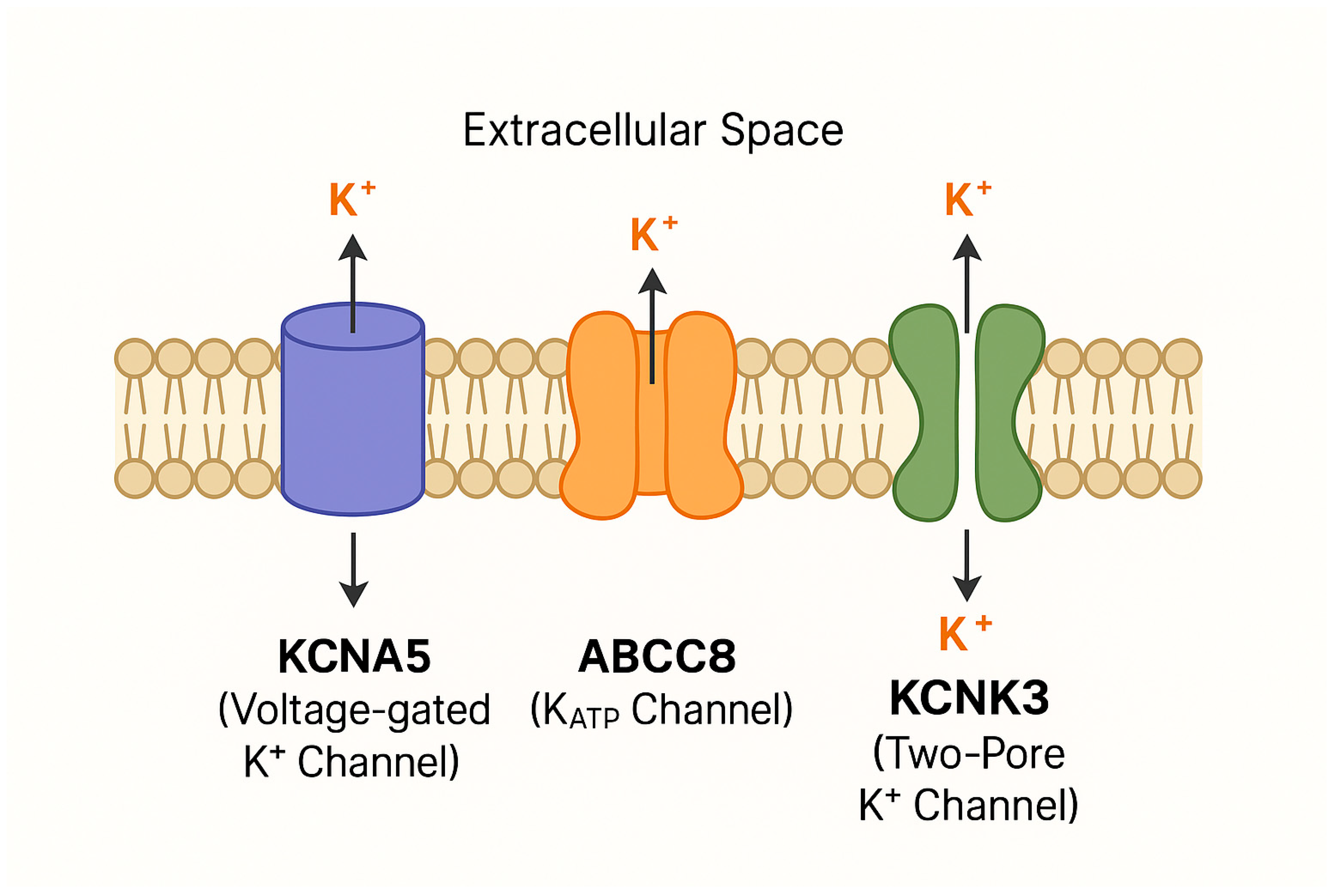
+) channel that is a key determinant of resting membrane potential in pulmonary artery smooth muscle cells (
Figure 3). Unlike multi-exon ion channel genes,
KCNA5 contains a single exon, which limits alternative splicing and makes functional variants more likely to directly disrupt channel structure. Kv1.5 plays an important role in maintaining pulmonary vascular tone through modulation of calcium ions (Ca
2+) influx across voltage-dependent Ca
2+ channels (VDCC) (
Figure 3). This places
KCNA5 within a distinct smooth muscle-centric pathogenic axis, in contrast to endothelial-focused genes such as
SOX17,
ATP13A3, and
AQP1.
Under physiological conditions, Kv1.5 activity maintains a hyperpolarized membrane state, limiting calcium entry and thereby preventing excessive vasoconstriction. Loss or inhibition of Kv1.5 function leads to membrane depolarization, increased calcium influx, and enhanced PASMC contraction, resulting in sustained vasoconstriction. In addition, Kv1.5 dysfunction has been linked to impaired apoptosis through reduced apoptotic volume decrease and altered caspase activation, promoting PASMC survival and contributing to vascular remodeling [
64,
65]. Together, these effects, vasoconstriction and apoptosis resistance, recapitulate core pathological features of pulmonary arterial hypertension (PAH) and align mechanistically with other ion channel-associated PAH genes such as
KCNK3 and
ABCC8.
KCNA5 encodes a channel protein with a relatively simple gene structure compared with multi-exon regulatory genes, meaning that missense variants are more likely to have direct functional consequences on channel gating, conductance, or membrane expression. Rare
KCNA5 variants, including Arg184Pro and Gly384Arg, have been identified in PAH cohorts and shown to impair channel function, reduce Kv1.5 current density, and promote a pro-proliferative, anti-apoptotic PASMC phenotype [
65]. Although these variants account for a small proportion of PAH cases, their consistent electrophysiological effects provide strong mechanistic support for the role of membrane depolarization and ion handling abnormalities in disease pathogenesis.
However, several important limitations must be acknowledged. Much of the mechanistic evidence is derived from in vitro electrophysiological studies and experimental modulation of channel expression, which may not fully replicate the physiological effects of endogenous pathogenic variants in human pulmonary vasculature. In addition, the rarity of KCNA5 variants and limited replication across independent cohorts constrain robust genotype–phenotype correlations, making it difficult to determine their true population-level contribution to PAH.
From a pathogenic standpoint, KCNA5 is best understood as a moderate-effect susceptibility gene rather than a high-penetrance monogenic driver. Its variants likely reduce electrical and apoptotic reserve in PASMCs, thereby increasing vulnerability to secondary insults such as hypoxia, inflammation, or upstream genetic defects (e.g., BMPR2 dysfunction). This positions KCNA5 within a broader framework of ion channel dysregulation in PAH, where impaired membrane excitability contributes to sustained vasoconstriction and vascular remodeling but typically requires additional “second hits” for full disease expression.
In conclusion, KCNA5 dysfunction highlights the importance of bioelectric signaling in pulmonary vascular homeostasis and reinforces the concept that PAH pathogenesis arises from the convergence of endothelial, smooth muscle, and metabolic abnormalities across multiple biological layers.
3.5.2. KCNK3
Whole-exome sequencing studies have identified pathogenic variants in
KCNK3, which encodes the pH-sensitive two-pore domain potassium channel TASK-1. These variants account for approximately 1–3% of pulmonary arterial hypertension (PAH) cases, establishing
KCNK3 as one of the earliest identified ion channel–associated PAH genes [
66] (
Figure 3).
TASK-1 is widely expressed across multiple tissues, including pulmonary artery smooth muscle cells (PASMCs), cardiac atria, right ventricle, adrenal gland, endothelium, and pancreas, where it contributes to the regulation of resting membrane potential. Unlike voltage-gated potassium channels such as Kv1.5 (KCNA5), TASK-1 functions as a constitutively active “leak” channel that stabilizes membrane potential independent of voltage-dependent activation. This continuous background activity makes TASK-1 particularly important for maintaining basal electrical stability in pulmonary vascular cells and preventing inappropriate depolarization.
Loss-of-function mutations in
KCNK3 reduce potassium efflux, leading to membrane depolarization and secondary activation of voltage-dependent calcium channels. The resulting increase in intracellular calcium promotes sustained pulmonary artery smooth muscle cell contraction and vasoconstriction, thereby contributing to elevated pulmonary vascular resistance and increased mean pulmonary arterial pressure [
66]. Because TASK-1 operates continuously under physiological conditions, its dysfunction produces persistent rather than episodic electrical instability, resulting in chronic vasoconstrictive tone.
In addition to effects on vascular tone, KCNK3 deficiency promotes vascular remodeling through enhanced PASMC proliferation and resistance to apoptosis. This dual phenotype, vasoconstriction combined with proliferative remodeling, closely mirrors that observed in other ion channel-associated PAH genes such as KCNA5 and ABCC8, reinforcing the concept that disrupted potassium homeostasis represents a shared mechanistic class in PAH pathogenesis.
Despite strong mechanistic plausibility supported by electrophysiological and experimental studies, several limitations remain. Notably, penetrance among KCNK3 mutation carriers is variable, indicating that channel dysfunction alone is insufficient to cause disease in all individuals. This variability supports a multi-hit model in which KCNK3 loss reduces electrophysiological reserve and increases susceptibility to secondary stressors such as hypoxia, inflammation, metabolic dysfunction, or additional genetic variants.
Based on the ClinGen Pulmonary Hypertension Gene Curation Expert Panel,
KCNK3 is regarded as a definitive genetic contributor to PAH [
15]. Its pathogenic role is most appropriately understood within a broader framework of ion channel dysregulation in PAH, where impaired potassium conductance contributes to sustained depolarization, calcium overload, and maladaptive vascular remodeling in conjunction with other genetic and environmental factors.
3.5.3. ABCC8
The ATP-binding cassette sub-family C member 8 (
ABCC8) gene encodes the sulfonylurea receptor 1 (SUR1), a regulatory subunit of ATP-sensitive potassium (KATP) channels. SUR1 is a key metabolic sensor that links intracellular energy status—particularly ATP/ADP ratios—to potassium channel activity, thereby coupling cellular metabolism to membrane excitability. In the pulmonary vasculature, KATP channels are important determinants of resting membrane potential and vascular tone, placing
ABCC8 at the intersection of metabolic regulation and electrophysiological control of vascular function (
Figure 3) (
Table 2).
Loss-of-function variants in
ABCC8 impair SUR1 function, resulting in reduced KATP channel activity and diminished potassium efflux [
67]. Consistent with other potassium channelopathies implicated in pulmonary arterial hypertension (PAH), reduced KATP activity leads to membrane depolarization, increased opening of voltage-dependent calcium channels, and subsequent intracellular calcium overload. This promotes sustained pulmonary artery smooth muscle cell contraction, contributing to increased pulmonary vascular resistance and vasoconstriction. In parallel,
ABCC8 dysfunction has also been associated with increased proliferation and reduced apoptosis in both endothelial cells and smooth muscle cells, thereby contributing to vascular remodeling [
68].
Unlike KCNA5 (voltage-gated Kv1.5) and KCNK3 (TASK-1 background leak channel), which regulate membrane potential primarily through electrical conductance properties, ABCC8 uniquely integrates metabolic status with electrophysiological signaling. By gating channel activity in response to ATP availability, SUR1 links cellular energetic state to vascular tone regulation. This metabolic–electrical coupling is particularly relevant in PAH, a disease increasingly recognized as involving mitochondrial dysfunction, altered glycolytic flux, and metabolic reprogramming of vascular cells. In this framework, ABCC8 represents a mechanistic bridge between metabolic stress and ion channel dysfunction, contributing to sustained depolarization under conditions of cellular energy imbalance.
Despite strong mechanistic plausibility supported by experimental models, several limitations remain. Most evidence for ABCC8-associated vascular dysfunction is derived from in vitro systems or heterologous expression models, which may not fully recapitulate the complex metabolic and hemodynamic environment of the human pulmonary circulation. The frequency, penetrance, and spectrum of ABCC8 variants in large, well-characterized PAH cohorts remain incompletely defined, limiting precise genotype–phenotype correlations.
ABCC8 is best conceptualized as a metabolic regulator of ion channel function whose dysfunction contributes to PAH through impaired ATP-sensitive potassium channel activity, leading to membrane depolarization, calcium overload, and downstream vascular remodeling. Rather than acting as an isolated driver, ABCC8 fits within a broader class of ion channel–metabolic convergence genes that amplify susceptibility to pulmonary vascular disease in the presence of additional genetic or environmental stressors.
3.6. Metabolic Genes
Although PAH has long been associated with dysfunctional vascular signaling pathways (most notably the BMP/TGF-β pathway), recent genetic studies have identified a group of genes, such as EIF2AK4, NFU1, and GGCX, that link PAH to disturbances in cellular metabolism, mitochondrial function, and protein modification. Mutations of these genes lead to metabolic stress responses that trigger pulmonary vascular remodeling.
3.6.1. EIF2AK4
The Eukaryotic Translation Initiation Factor 2 Alpha Kinase 4 (
EIF2AK4) gene encodes General Control Nonderepressible 2 (GCN2), a serine/threonine kinase that functions as a central regulator of the integrated stress response (ISR). GCN2 is activated by accumulation of uncharged transfer RNAs during amino acid deprivation and other cellular stress conditions. Once activated, it phosphorylates the α-subunit of eukaryotic initiation factor 2 (eIF2α), leading to global attenuation of cap-dependent protein translation while selectively permitting translation of stress-adaptive transcripts [
69]. This adaptive translational reprogramming is essential for maintaining endothelial cell survival under hypoxic, inflammatory, and metabolic stress conditions characteristic of the pulmonary vascular microenvironment in pulmonary hypertension (
Figure 2) (
Table 2).
According to ClinGen curation,
EIF2AK4 is classified as a definitive gene for pulmonary veno-occlusive disease and pulmonary capillary hemangiomatosis, conditions within the pulmonary hypertension spectrum that can clinically overlap with PAH [
15]. Biallelic loss-of-function mutations in
EIF2AK4 account for a significant proportion of heritable PVOD/PCH cases and have also been reported in a subset of PAH cohorts [
70]. Although early studies established a predominantly autosomal recessive inheritance pattern [
70,
71], subsequent reports suggest that
EIF2AK4 variants may also modify disease severity in the presence of additional genetic lesions, particularly
BMPR2 mutations [
72]. In such contexts, splice-site or truncating variants affecting functional domains of GCN2 can impair ribosomal association and stress response activation, thereby lowering the threshold for disease manifestation and accelerating disease progression.
EIF2AK4 dysfunction disrupts the integrated stress response, resulting in impaired translational control during periods of cellular stress. Loss of appropriate eIF2α phosphorylation compromises the ability of endothelial and smooth muscle cells to adapt to hypoxia, oxidative stress, and inflammatory signaling. This leads to cellular vulnerability, maladaptive protein synthesis, and accumulation of proteotoxic stress, which can indirectly amplify dysregulation of key vascular signaling pathways, including BMP and TGF-β cascades.
EIF2AK4 occupies a distinct mechanistic position compared with metabolic regulators such as ABCC8 and ATP13A3. Whereas ABCC8 primarily couples ATP availability to membrane excitability and ATP13A3 regulates polyamine-dependent cellular metabolism, EIF2AK4 governs cellular stress tolerance through translational control. This places EIF2AK4 as a key determinant of endothelial resilience rather than a direct effector of proliferative or electrophysiological signaling.
Despite strong genetic evidence linking EIF2AK4 loss-of-function to PVOD/PCH, several important limitations remain. Functional studies in human pulmonary vascular tissue are limited, making it difficult to determine whether EIF2AK4 mutations act as primary initiators of vascular remodeling or as modifiers that exacerbate pre-existing injury. Furthermore, the molecular basis underlying the phenotypic divergence between PVOD/PCH and PAH in EIF2AK4-deficient states remains incompletely understood, despite shared vascular stress signatures. This suggests that EIF2AK4 mutations define a stress-sensitized vascular phenotype, whose clinical expression is shaped by genetic background, environmental exposure, and co-existing molecular abnormalities within the pulmonary circulation.
3.6.2. NFU1
NFU1 encodes a mitochondrial iron–sulfur (Fe–S) cluster assembly factor required for the maturation and delivery of Fe–S cofactors to multiple mitochondrial enzymes, including components of the electron transport chain (notably complex II) and lipoic acid-dependent enzymes (
Table 2). Fe–S clusters are essential prosthetic groups involved in electron transfer within respiratory complexes I–III, thereby supporting oxidative phosphorylation, mitochondrial ATP production, and redox homeostasis [
73].
NFU1 functions as a core component of mitochondrial biosynthetic machinery required for cellular energetic stability.
Pathogenic biallelic
NFU1 variants are classically associated with severe early-onset mitochondrial disease, and pulmonary hypertension has been reported as part of this systemic phenotype in a subset of cases. Functional studies of specific variants (e.g., G208C) suggest impaired Fe–S cluster transfer with downstream disruption of respiratory chain activity and a compensatory shift toward glycolytic metabolism [
74]. While this metabolic reprogramming resembles the glycolytic phenotype observed in pulmonary arterial hypertension (PAH), current evidence does not establish
NFU1 as a recurrent monogenic cause of isolated PAH in large cohorts. Instead, its contribution appears to be strongest in multisystem mitochondrial disease contexts where pulmonary vascular disease may emerge as part of broader metabolic failure.
In addition to impaired oxidative phosphorylation, NFU1 dysfunction is associated with increased mitochondrial reactive oxygen species (ROS) production and reduced antioxidant buffering capacity. These changes may promote endothelial injury and impair adaptive signaling.
When compared with EIF2AK4, which regulates adaptive translational control during cellular stress, NFU1 operates further upstream by affecting mitochondrial bioenergetic capacity itself. EIF2AK4-related disease represents impaired stress adaptation, whereas NFU1-related pathology reflects impaired energy generation capacity. In both cases, pulmonary vascular involvement likely arises secondarily from reduced cellular resilience rather than pathway-specific vascular signaling defects typical of established PAH genes (e.g., BMPR2, SOX17, or ATP13A3).
Current evidence supporting a direct role for NFU1 in PAH is limited and largely derived from case reports and small series. No large-scale genotype–phenotype studies have established its prevalence in idiopathic or heritable PAH cohorts, and disease specificity remains uncertain given its established role in systemic mitochondrial disorders. Accordingly, NFU1 is best interpreted as a candidate metabolic gene associated with pulmonary vascular disease in the context of mitochondrial syndromes, rather than a definitively established PAH gene. Its precise role in isolated PAH remains to be defined, and further systematic genetic and functional studies are required to clarify its position within the pulmonary hypertension gene landscape.
3.6.3. GGCX
The
GGCX gene encodes γ-glutamyl carboxylase, an integral endoplasmic reticulum membrane enzyme responsible for vitamin K-dependent post-translational γ-carboxylation of glutamate residues to γ-carboxyglutamate (Gla) (
Table 2). This modification is essential for the functional activity of multiple extracellular proteins involved in coagulation and vascular biology, including matrix Gla protein (
MGP), which has been implicated in vascular calcification and growth factor regulation [
75,
76].
GGCX represents a post-translational regulatory node linking vitamin K metabolism to extracellular modulation of vascular signaling pathways.
The relevance of
GGCX to pulmonary vascular disease is primarily mediated through its role in activating
MGP. Fully γ-carboxylated
MGP acts as an extracellular inhibitor of bone morphogenetic protein (BMP) signaling by binding BMP ligands (notably
BMP2 and
BMP4) and limiting their interaction with BMP receptors, thereby modulating local signaling intensity within the vascular extracellular matrix [
77]. In contrast, undercarboxylated (inactive)
MGP loses this inhibitory capacity, which may result in altered extracellular BMP ligand bioavailability and dysregulated spatial control of signaling. However, the net functional consequence in pulmonary vascular tissue is not fully resolved and may differ depending on local ligand abundance, vascular bed, and disease stage.
Rare variants in
GGCX have been reported in small pulmonary arterial hypertension (PAH) cohorts, with an estimated frequency of ~1% in selected genetic screens [
75]. However, in a ClinGen-consistent framework, the current evidence base remains limited, as replication across large independent PAH cohorts and robust segregation or functional validation data are still lacking. Accordingly,
GGCX should not yet be considered a definitive PAH disease gene.
From a mechanistic standpoint, GGCX-related perturbation introduces a distinct regulatory layer in the BMP pathway that differs fundamentally from canonical PAH genes such as BMPR2, ACVRL1, or SMADs, which directly alter intracellular signaling. Instead, GGCX acts indirectly by modulating extracellular BMP ligand regulation through MGP activity. This places it within an emerging category of extracellular matrix-mediated signaling modulators, rather than classical receptor–ligand–transducer defects.
A key conceptual complexity arises from the fact that impaired
MGP carboxylation may increase extracellular BMP ligand availability, potentially enhancing BMP signaling. This contrasts with the reduced BMP signaling typically observed in
BMPR2- or
GDF2-associated PAH. Experimental studies in vascular calcification biology suggest that
MGP function is context-dependent and may differentially regulate BMP signaling intensity and localization rather than acting as a simple on/off inhibitor [
77]. Consequently, whether altered
GGCX activity results in net BMP pathway activation or dysregulated signaling remains unresolved, and likely depends on spatial compartmentalization and receptor context within the pulmonary vasculature.
While BMP signaling is often considered vasculoprotective, increasing evidence suggests that both insufficient and mislocalized BMP activity can contribute to vascular pathology depending on cellular context and downstream transcriptional integration. This supports a model in which PAH pathogenesis may arise not only from reduced BMP signaling but also from loss of spatial and regulatory control over BMP signaling dynamics.
Altogether, current evidence supports a limited but biologically plausible association between GGCX variants and PAH. However, the absence of large-scale replication studies, limited in vivo functional validation, and incomplete understanding of extracellular BMP regulation constrain causal interpretation. Within a ClinGen-aligned framework, GGCX is best classified as a candidate modifier gene influencing extracellular regulation of BMP signaling, whose role in pulmonary vascular disease remains to be definitively established.
3.7. Other Signaling Receptors
Signaling receptors are proteins located on the surface of pulmonary endothelial or smooth muscle cells that respond to external stimuli and regulate cell proliferation, growth, and differentiation. Mutations of the genes encoding for these receptors can lead to their overactivation or inhibition, shifting the balance from a stable, quiescent vessel wall to one that is progressively constricted and remodeled as in PAH.
3.7.1. NOTCH3
NOTCH3 is a member of the Notch receptor family that encodes a single-pass transmembrane receptor predominantly expressed in vascular smooth muscle cells, where it plays a central role in vascular development, arterial identity maintenance, and regulation of smooth muscle cell survival (
Table 2). In the pulmonary circulation,
NOTCH3 is particularly relevant to pulmonary artery smooth muscle cell (PASMC) phenotype stability and proliferative capacity [
78].
Following ligand binding by Jagged or Delta-like family ligands on adjacent cells, NOTCH3 undergoes sequential proteolytic cleavage mediated by ADAM-family proteases and γ-secretase, resulting in release of the Notch3 intracellular domain (N3ICD). The N3ICD translocates to the nucleus, where it forms a transcriptional activation complex that regulates downstream targets, including the Hes and Hey (HRT) gene families. These transcriptional programs promote PASMC survival, maintain a synthetic/proliferative phenotype, and inhibit apoptosis, thereby influencing vascular remodeling capacity.
Experimental and observational studies suggest that
NOTCH3 signaling is upregulated in pulmonary arterial hypertension (PAH). Increased
NOTCH3 expression has been reported in remodeled pulmonary arteries from PAH patients, with levels correlating with disease severity, supporting an association between enhanced
NOTCH3 activity and adverse vascular remodeling [
79]. Hypoxic exposure, an established trigger of pulmonary hypertension, has been shown to increase
NOTCH3 expression in experimental models, linking environmental stress to activation of this signaling axis [
80]. Collectively, these findings position
NOTCH3 as a context-sensitive signaling pathway that may integrate hypoxia-driven stress responses with PASMC proliferative signaling.
Rare
NOTCH3 variants, including missense substitutions such as c.2519G>A (p.G840E) and c.2698A>C (p.T900P), have been identified in PAH cohorts [
81]. Functional analyses suggest that these variants may alter
NOTCH3 signaling fidelity, including downstream transcriptional output through HES/HEY pathways, potentially contributing to dysregulated PASMC proliferation and survival. However, the available genetic evidence remains limited to small cohorts, and replication across independent PAH populations is lacking. Robust standardized functional validation of individual
NOTCH3 variants in pulmonary vascular models remains incomplete.
From a mechanistic perspective, NOTCH3 differs from canonical PAH genes such as BMPR2 or SOX17 in that it primarily regulates cell fate and phenotype stability in smooth muscle cells, rather than endothelial signaling or ligand–receptor BMP pathway integrity. It therefore occupies a distinct but potentially convergent position within the broader PAH network, acting at the level of PASMC proliferation, survival, and vascular remodeling responsiveness.
A key unresolved question is whether NOTCH3 activation represents a primary pathogenic driver, a downstream response to vascular injury (e.g., hypoxia or metabolic stress), or a secondary amplification pathway that reinforces established remodeling signals. Current evidence supports all three possibilities in different experimental contexts but does not yet allow clear causal stratification in human disease. This uncertainty is reinforced by the variability in expression patterns and the absence of large-scale genotype–phenotype correlation studies.
Within a ClinGen-consistent interpretative framework, NOTCH3 is regarded as a candidate or emerging PAH-associated gene with supportive functional evidence but limited definitive genetic validation. Its role in pulmonary vascular disease likely reflects a modulatory pathway that amplifies PASMC proliferation and survival in response to upstream pathogenic stimuli rather than a singular initiating event. Further large-cohort genetic studies and in vivo functional modeling will be required to clarify whether NOTCH3 contributes primarily as a disease driver, modifier, or convergence node within the broader PAH pathogenic network.
3.7.2. KDR
KDR encodes vascular endothelial growth factor receptor 2 (
VEGFR2), a receptor tyrosine kinase that mediates VEGF-driven endothelial signaling and is central to angiogenesis and vascular maintenance (
Table 2). Through activation by VEGF-A,
VEGFR2 regulates endothelial proliferation, migration, survival, and permeability, placing it at the core of endothelial adaptive responses to hemodynamic stress [
82].
Rare
KDR variants have been identified in pulmonary arterial hypertension (PAH), with reported frequencies of approximately 1–2% depending on cohort composition and sequencing strategy [
83]. Both missense and truncating variants have been described. Clinically, truncating variants have been associated with reduced diffusing capacity for carbon monoxide (DLCO) and later disease onset, suggesting a phenotype that reflects impaired endothelial maintenance rather than developmental vascular malformation.
Functional studies indicate that reduced
KDR activity compromises VEGF-dependent endothelial survival signaling, leading to endothelial dysfunction and impaired repair responses under stress conditions [
84]. Rather than driving proliferation directly, this loss appears to weaken endothelial resilience, making the pulmonary microvasculature less able to recover from hypoxic or inflammatory injury.
This places VEGFR2 in a distinct but conceptually overlapping category with genes such as SOX17 and FOXF1, where the primary defect is not constitutive pathway activation but reduced endothelial stability. A recurring theme across these genes is that endothelial vulnerability, rather than intrinsic hyperproliferation, lowers the threshold for vascular remodeling once secondary insults are present.
At the same time, VEGF signaling introduces a mechanistic paradox in pulmonary vascular disease. While excessive VEGF activity can promote aberrant angiogenesis in some vascular beds, insufficient VEGFR2 signaling in the lung appears to impair endothelial survival and regenerative capacity. The consequence is not simply reduced angiogenesis but a failure to maintain a stable endothelial layer under chronic stress, which may secondarily permit remodeling processes driven by other pathways such as BMP/TGF-β dysregulation.
Genetically, KDR-associated PAH remains incompletely defined. Penetrance is variable, reported cohorts are small, and functional validation of specific variants is still limited. These uncertainties make it difficult to determine whether KDR haploinsufficiency is sufficient to initiate disease or whether it acts primarily as a modifying factor within a broader multi-hit framework involving hypoxia, inflammation, or metabolic stress.
KDR is regarded as a limited-to-moderate confidence PAH-associated gene [
15], with supportive but not definitive genetic and functional data. Its main contribution to the disease landscape appears to lie in weakening endothelial adaptive capacity rather than acting as a primary driver of proliferative remodeling.
3.8. Membrane/Structural Proteins
Beyond dysfunctional growth factor signaling and metabolic reprogramming, PAH also involves irregularities in structural proteins that maintain the physical architecture of vascular cells. These proteins regulate cell-to-cell interaction, barrier integrity, mechanotransduction, and vesicular trafficking, processes that are essential in maintaining vascular homeostasis. The two structural proteins implicated in PAH are caveolin-1 and pleckstrin homology domain-containing protein family H member 2.
3.8.1. CAV1
CAV1 encodes caveolin-1, the principal structural protein of caveolae—plasma membrane invaginations involved in endocytosis, mechanosensing, and spatial organization of signaling complexes in endothelial and smooth muscle cells (
Figure 2) [
85,
86]. By organizing receptor localization and trafficking, caveolin-1 regulates multiple signaling pathways that control endothelial survival, proliferation, and vascular tone, placing it at a central regulatory node in pulmonary vascular homeostasis.
Pathogenic variants in
CAV1 were first implicated in pulmonary arterial hypertension (PAH) through the identification of loss-of-function mutations in familial and idiopathic cases [
87]. These findings established
CAV1 as a rare but biologically plausible PAH-associated gene. Subsequent studies in patient-derived samples and experimental systems confirmed reduced caveolin-1 expression in pulmonary vascular tissue, supporting a disease-associated deficiency state rather than simple receptor-level dysregulation [
87].
Caveolin-1 deficiency disrupts membrane microdomain organization and alters receptor compartmentalization, leading to dysregulated signaling through multiple pathways, including BMP and TGF-β signaling cascades. In pulmonary vascular cells, this imbalance favors enhanced TGF-β-driven proliferative signaling and reduced BMP-mediated quiescence, a pattern consistent with vascular remodeling phenotypes observed in established PAH genes such as
BMPR2 and
ACVRL1 [
87]. However, unlike receptor or ligand mutations,
CAV1 does not directly impair ligand binding or SMAD phosphorylation; instead, it modifies the spatial and temporal dynamics of signaling, amplifying downstream pathway dysregulation in a context-dependent manner.
In endothelial cells, CAV1 loss impairs caveolae-dependent mechanotransduction and nitric oxide signaling, contributing to endothelial dysfunction and barrier instability. In smooth muscle cells, altered signaling compartmentalization promotes a pro-proliferative, apoptosis-resistant phenotype. These dual effects provide a mechanistic bridge between endothelial injury and smooth muscle remodeling, although the relative contribution of each compartment remains incompletely defined in human PAH tissue.
Key uncertainties nonetheless remain. Although
CAV1 is widely expressed across multiple tissues, pathogenic variants predominantly manifest as pulmonary vascular disease, suggesting tissue-specific vulnerability. One proposed explanation is the unique dependence of the pulmonary circulation on finely tuned mechanosensing and growth factor signaling under conditions of chronic shear stress. However, direct human in vivo evidence supporting pulmonary selectivity is still limited, and most mechanistic data derive from murine models and cultured endothelial systems [
85,
87].
Within current evidence frameworks,
CAV1 is classified as a definitive PAH gene by the Clinical Genome Resource Pulmonary Hypertension Gene Curation Expert Panel [
15]. Unlike canonical BMP/TGF-β ligands or receptors, caveolin-1 does not directly regulate signal initiation but instead controls the spatial organization and trafficking of multiple signaling complexes at the plasma membrane. This positions
CAV1 as a regulator of signaling architecture, where loss of membrane compartmentalization amplifies dysregulated pathways rather than initiating them de novo. In contrast to genes such as
KDR or
AQP1, which remain at moderate or emerging levels of evidence,
CAV1 illustrates how disruption of membrane-level signal integration can represent a fully established pathogenic mechanism in pulmonary arterial hypertension.
3.8.2. PLEKHH2
PLEKHH2 is located on chromosome 2p21 and encodes a cytoskeletal adaptor protein involved in cell–matrix adhesion and structural stabilization of specialized barrier-forming cells (
Table 2) [
88]. In renal tissue, it has been characterized in podocytes, where it contributes to maintenance of foot process architecture and anchoring to the glomerular basement membrane, a structure essential for filtration barrier integrity. This functional role in highly specialized barrier cells suggests that
PLEKHH2 participates in maintaining mechanical stability in vascular and epithelial interfaces.
Beyond the kidney,
PLEKHH2 is expressed in pulmonary vascular endothelial cells, where emerging evidence suggests a role in maintaining endothelial structural integrity and homeostatic signaling [
89]. This shared expression across podocytes and pulmonary endothelium supports the broader concept that barrier-forming cells rely on conserved cytoskeletal–matrix coupling systems to withstand mechanical stress and maintain selective permeability.
Rare missense variants in
PLEKHH2 have recently been identified in pulmonary arterial hypertension (PAH) cohorts and have been associated with increased endothelial proliferation and reduced apoptosis in experimental endothelial models [
89]. These observations suggest that
PLEKHH2 disruption may destabilize endothelial anchorage to the extracellular matrix, thereby altering mechanotransduction pathways that normally regulate survival and controlled cell cycling under shear stress conditions.
Unlike canonical PAH genes such as BMPR2 or ACVRL1, PLEKHH2 does not directly participate in ligand–receptor signaling or intracellular SMAD-mediated transcriptional regulation. Instead, its putative role is structural, influencing how endothelial cells sense and respond to mechanical cues from the extracellular matrix. In this sense, it aligns with a growing subset of PAH-associated genes, including ATP13A3, AQP1, CAV1, and SOX17, where the primary defect is not constitutive pathway activation but reduced endothelial resilience to environmental and mechanical stressors.
This structural vulnerability model helps reconcile the observed phenotype: impaired cytoskeletal anchoring may not directly induce proliferation but may lower the threshold for maladaptive responses when endothelial cells are exposed to secondary insults such as hypoxia, inflammation, or dysregulated BMP/TGF-β signaling. However, current evidence does not establish PLEKHH2 as an initiating driver of disease; instead, it is more consistent with a modifier role that amplifies susceptibility to vascular remodeling.
Evidence supporting PLEKHH2 involvement in PAH remains preliminary. The number of reported pathogenic variants is still small, functional studies are largely limited to in vitro endothelial systems, and in vivo validation in pulmonary vascular models is currently lacking. As a result, genotype–phenotype correlations remain uncertain, and disease penetrance cannot yet be reliably estimated.
3.9. Vasoactive Regulation
PAH involves a dysregulated balance between vasodilators (downregulated) and vasoconstrictors (upregulated), contributing to the elevated pulmonary artery pressure and resistance. The KLK1 gene, which plays a crucial role in regulating vascular tone through the kallikrein–kinin system, has been implicated in the distortion of this vasodilator-vasoconstrictor balance.
KLK1
KLK1 encodes tissue kallikrein, a secreted serine protease of the kallikrein family that is widely expressed in multiple organs, including the lung, kidney, heart, and pancreas [
90]. Its primary physiological role is the generation of kinins, particularly bradykinin, through cleavage of kininogen substrates. Bradykinin signaling promotes vasodilation via nitric oxide and prostacyclin release, and contributes to endothelial repair, angiogenesis, and modulation of vascular tone, positioning
KLK1 within a protective endothelial signaling axis in normal pulmonary vascular physiology.
Rare
KLK1 variants have been reported in pulmonary arterial hypertension (PAH) cohorts, with an estimated frequency of approximately 0.5% in selected genetic studies [
75]. However, the available evidence remains limited in scale and replication. Clinically, carriers have been described as presenting at older ages with comparatively milder phenotypes than
BMPR2-associated disease, suggesting a weaker or modulatory contribution to disease susceptibility rather than a high-penetrance pathogenic effect.
KLK1 deficiency is thought to impair the local kinin–bradykinin axis, reducing nitric oxide-mediated vasodilation and endothelial repair signaling. Rather than directly promoting proliferative remodeling, this mechanism may weaken adaptive responses to vascular injury, particularly in distal pulmonary arterioles, where regenerative capacity is critical. In this sense, KLK1 dysfunction is more consistent with impaired vascular maintenance and repair than with primary activation of proliferative signaling pathways.
This distinguishes KLK1 from canonical PAH genes involved in BMP/TGF-β signaling, ion channel regulation, or metabolic stress responses. Instead, its functional profile aligns more closely with endothelial competence and angiogenic support genes such as KDR and FOXF1, where disease risk arises from reduced ability to maintain or restore vascular integrity under stress conditions. This supports a broader view of PAH pathobiology in which impaired vascular regeneration contributes alongside excessive vasoconstrictive or proliferative signaling.
An additional modifying layer may involve hormonal regulation. Experimental studies have shown that estrogen can upregulate
KLK1 expression [
91], raising the possibility that sex-dependent modulation of the kallikrein–kinin system contributes to variability in disease expression. This interaction has been proposed as one potential contributor to sex bias in PAH cohorts, although direct mechanistic evidence in
KLK1-associated disease remains limited and should be interpreted cautiously.
Despite these associations, current evidence is constrained by small patient numbers, limited replication across independent cohorts, and a lack of robust functional validation in human pulmonary vascular systems. It therefore remains unresolved whether KLK1 variants can independently initiate disease or whether they primarily act as low-penetrance modifiers that reduce endothelial repair capacity in the presence of additional genetic or environmental stressors.
Within a ClinGen-consistent framework,
KLK1 is best regarded as a limited-evidence, candidate modifier gene [
15], with a mechanistic role centered on endothelial repair signaling rather than primary pathway disruption.
3.10. Extracellular/Matrix Related
Recent genetic studies have identified CBLN2 and CD248 as active drivers of PAH and not just passive consequences of vascular injury. Extracellular matrix remodeling contributes to PAH by influencing stiffness, signaling, mechanotransduction, and recruitment of inflammatory cells in the pulmonary vasculature. These findings underscore the role of adventitial fibroblasts, in addition to endothelial and pulmonary artery smooth muscle cells, in PAH pathogenesis.
3.10.1. CBLN2
CBLN2 (Cerebellin 2) is a member of the cerebellin glycoprotein family, which is predominantly expressed in the central nervous system but has also been detected in pulmonary tissue, including lungs from patients with pulmonary arterial hypertension (PAH) (
Table 2). This atypical expression pattern in a non-neuronal vascular context suggests a stress-associated reactivation or dysregulation of developmental signaling programs in diseased pulmonary endothelium.
A common regulatory variant (rs2217560 G allele) within the
CBLN2 locus (18q22.3) has been associated with PAH, with a reported odds ratio of approximately 2.16 in case–control analyses [
92]. In addition to genetic association, increased
CBLN2 mRNA expression has been observed in PAH lung tissue, with preferential enrichment in endothelial cells compared with pulmonary artery smooth muscle cells. This cell-type specificity suggests that
CBLN2 may act primarily within the endothelial compartment during disease development.
In pulmonary vascular cells,
CBLN2 has been implicated in the activation of inflammatory and hypoxia-responsive signaling pathways, including NF-κB,
HIF-1α, and TWIST1, which converge on endothelial-to-mesenchymal transition (EndMT) programs [
93]. EndMT is increasingly recognized as a key contributor to pulmonary vascular remodeling, linking inflammatory stress and hypoxic signaling to endothelial plasticity, loss of barrier integrity, and acquisition of mesenchymal-like features. Through this mechanism,
CBLN2 is positioned within a broader network of PAH-associated genes that influence endothelial phenotype stability, including
SOX17,
CAV1,
KDR, and
NOTCH3, although it operates through distinct upstream regulatory inputs.
Unlike canonical PAH genes that directly regulate BMP/TGF-β signaling, ion channel activity, or mitochondrial metabolism, CBLN2 appears to function as an inflammation- and hypoxia-responsive modulator of endothelial fate. Its activity is therefore more consistent with a disease-amplifying role, in which pre-existing vascular stressors promote maladaptive transcriptional reprogramming rather than CBLN2 acting as a primary initiating lesion.
However, the current evidence base remains preliminary. The initial genetic association study was limited by moderate cohort size, and subsequent mechanistic insights are largely derived from in vitro signaling and endothelial transition models. Robust in vivo functional validation in pulmonary vascular systems, as well as replication in independent large-scale genetic cohorts, is still lacking. Consequently, it remains uncertain whether CBLN2 represents a true disease gene, a context-dependent modifier of endothelial injury responses, or a downstream marker of inflammatory endothelial activation.
3.10.2. CD248
CD248 (also known as endosialin or tumor endothelial marker 1) encodes a type I transmembrane glycoprotein belonging to the C-type lectin-like receptor family, with established roles in mesenchymal–vascular interactions during development (
Table 2). During embryogenesis,
CD248 is widely expressed in angiogenic vasculature, but its expression becomes largely silenced in most adult tissues, persisting mainly in specialized stromal and perivascular compartments such as fibroblasts and pericyte-rich vascular niches [
94,
95] dely expressed in angiogenic vasculature, but its expression becomes largely silenced in most adult tissues, persisting mainly in specialized stromal and perivascular compartments such as fibroblasts and pericyte-rich vascular niches.
Emerging genetic and experimental evidence implicates
CD248 in pulmonary arterial hypertension (PAH), although the data remain limited. Rare variants and altered expression patterns have been reported in PAH cohorts, alongside observations of reduced circulating
CD248 levels and decreased tissue expression in pulmonary vascular lesions [
96]. These findings suggest that
CD248 deficiency may be associated with impaired vascular integrity, although causality has not been firmly established.
CD248 is thought to regulate pericyte behavior, including proliferation, migration, and interaction with endothelial cells. Disruption of this signaling axis may impair pericyte–endothelial communication, weakening microvascular stability and contributing to aberrant vascular remodeling [
96]. In the pulmonary circulation, where pericyte–endothelial coupling is essential for maintaining vessel caliber and resistance, such disruption could lower the threshold for endothelial dysfunction and progressive occlusive remodeling.
Unlike canonical PAH genes involved in BMP/TGF-β signaling, ion channel regulation, or metabolic stress responses, CD248 does not directly participate in intracellular signaling cascades. Instead, it operates at the level of vascular structural organization and cell–cell communication, particularly within the perivascular compartment. This places CD248 in a broader category of vascular support genes that influence disease susceptibility indirectly by modulating the stability of the endothelial–pericyte unit, conceptually aligning with genes involved in endothelial resilience and vascular niche maintenance.
However, the current evidence base remains preliminary. The number of reported pathogenic variants is small, cohort sizes are limited, and functional studies are largely observational or in vitro in nature. In addition, the developmental regulation of CD248 raises unresolved questions regarding its contribution to adult-onset PAH, particularly whether disease-associated alterations represent pathological reactivation of developmental programs or primary adult-onset dysregulation in pulmonary vascular cells. This distinction remains to be established through systematic genetic and functional studies in adult pulmonary vascular models.
3.11. Epigenetic/Nuclear Regulation
Dysregulated epigenetic and nuclear mechanisms suppress vasoprotective gene expression, triggering pulmonary vascular remodeling, a hallmark feature of PAH. TET2 (epigenetic dysregulation) and TOPBP1 (DNA damage) mutations have been identified to contribute to PAH.
3.11.1. TET2
The
TET2 gene, located on chromosome 4q24, encodes a Fe(II)/α-ketoglutarate-dependent dioxygenase that catalyzes the conversion of 5-methylcytosine to 5-hydroxymethylcytosine, thereby enabling active DNA demethylation and dynamic epigenetic regulation (
Table 2). Through this function,
TET2 contributes to maintaining transcriptional programs that preserve endothelial quiescence and immune balance. Epigenetic homeostasis is governed by the opposing activities of DNA methyltransferases and TET enzymes; disruption of this balance may shift vascular cells toward persistent transcriptional repression of protective pathways [
97].
Rare
TET2 variants, predominantly germline in reported PAH cohorts, account for approximately 0.39% of cases [
98]. Most reported pathogenic variants cluster within the
TET2 expression has been demonstrated in peripheral blood mononuclear cells from affected individuals, alongside increased expression of pro-inflammatory mediators, particularly interleukin-1β (IL-1β).
Mechanistic evidence from experimental models suggests that
TET2 deficiency may contribute to pulmonary vascular disease through an inflammation-linked epigenetic mechanism. Loss of
TET2 activity is associated with increased DNA methylation and sustained activation of inflammatory signaling, while pharmacological inhibition of IL-1β has been shown to attenuate pulmonary hypertension phenotypes in animal models. IL-1β promotes pulmonary artery smooth muscle cell proliferation and migration [
99], providing a biologically plausible link between epigenetic dysregulation and vascular remodeling. These findings place
TET2 within an inflammatory–epigenetic axis that overlaps conceptually with genes such as
EIF2AK4 and
CBLN2, which also connect stress or inflammatory signaling to vascular dysfunction.
In contrast to canonical PAH genes involved in BMP/TGF-β signaling, ion channel function, or mitochondrial metabolism, TET2 acts as a broad epigenetic regulator capable of influencing multiple downstream pathways simultaneously. This positions TET2 more as a modifier of vascular transcriptional responsiveness rather than a pathway-specific signaling defect. Such a role is consistent with its association with later-onset disease and systemic inflammatory features, suggesting that epigenetic drift may lower the threshold for vascular remodeling rather than directly initiating it.
However, the current evidence base remains limited by small patient numbers and a reliance on peripheral blood and animal model data, with few studies directly examining pulmonary vascular tissue. It therefore remains unclear whether TET2 dysfunction primarily originates within the pulmonary endothelium or reflects systemic immune-driven epigenetic reprogramming that secondarily impacts the lung circulation. Future integrative studies combining pulmonary epigenomics, single-cell profiling, and longitudinal clinical data will be essential to define whether TET2 acts as a primary disease contributor or an amplifier within established inflammatory and remodeling pathways in PAH.
3.11.2. TOPBP1
Topoisomerase IIβ-binding protein 1 (
TOPBP1), located on chromosome 3, encodes a multifunctional scaffold protein that regulates DNA replication initiation, ATR-dependent checkpoint activation, and DNA damage repair (
Table 2). Through its BRCA1 C-terminal (BRCT) domains,
TOPBP1 mediates phospho-protein interactions required for assembly of DNA damage response complexes, thereby preserving genomic stability during replicative stress. Biochemical studies demonstrated that
TOPBP1 directly activates ATR kinase via its ATR-activation domain, establishing its central role in DNA damage checkpoint signaling [
100], while structural and functional analyses have shown that BRCT domains enable selective binding to phosphorylated substrates at sites of DNA damage [
101]. This function is particularly relevant in vascular endothelial cells, which are continuously exposed to shear stress and oxidative injury that can induce DNA damage.
Whole-exome sequencing identified several single-nucleotide variants in
TOPBP1 (rs55633281, rs17301766, rs10935070) in idiopathic PAH patients lacking
BMPR2 mutations [
102]. However, these observations were derived from a relatively small cohort and have not been independently replicated. Consistent with this limited evidence base,
TOPBP1 is not currently classified as a PAH-associated gene by the Clinical Genome Resource [
15] and is therefore best considered a candidate susceptibility gene rather than a confirmed causal factor.
At the cellular level, reduced TOPBP1 expression has been reported in pulmonary microvascular endothelial cells from PAH patients, accompanied by increased DNA damage and attenuated apoptotic responses. Functional knockdown experiments further demonstrated accumulation of DNA damage alongside persistence of apoptosis-resistant cells, suggesting impaired genomic quality control. Rather than directly promoting proliferation, these findings support a model in which defective DNA damage surveillance permits survival of dysfunctional endothelial cells, thereby compromising vascular integrity and predisposing to maladaptive remodeling.
In contrast to established PAH genes such as BMPR2, SOX17, and KDR, which have well-defined roles in endothelial signaling, angiogenesis, and vascular homeostasis, the evidence supporting TOPBP1 remains limited and largely hypothesis-generating. Its proposed role instead aligns more closely with genes such as FOXF1 and TET2, which influence endothelial resilience through regulation of genomic stability and epigenetic integrity rather than direct modulation of proliferative signaling pathways.
A key conceptual challenge is reconciling TOPBP1-associated genomic instability with the hyperproliferative vascular lesions characteristic of PAH. One plausible explanation is that accumulation of DNA-damaged but apoptosis-resistant endothelial cells promotes maladaptive repair responses, inflammation, or clonal expansion. Alternatively, localized endothelial injury may trigger compensatory proliferation in neighboring vascular cells. These mechanisms remain speculative and have not been validated in pulmonary vascular models.
In summary, the current evidence base is constrained by small sample sizes, lack of independent replication, and reliance on in vitro endothelial systems. In vivo studies examining endothelial-specific TOPBP1 dysfunction in pulmonary vascular contexts are lacking, and genotype–phenotype relationships remain undefined. Accordingly, TOPBP1 is best interpreted as a biologically plausible but genetically unvalidated contributor, highlighting a potential genomic instability axis in PAH that requires further investigation rather than representing an established pathogenic driver.
3.12. Single-Nucleotide Polymorphisms (SNPs)
SNPs, being more common than rare variants, often act as modifiers of PAH susceptibility, severity, or response to treatment. SNPs in the
TGF-β1 gene (509 and codon 10) of familial PAH patients with known
BMPR2 mutations are associated with earlier onsets and increased penetrance of the disease [
7] (
Table 1). This is attributed to the resultant imbalance of the TGF-β and BMP signaling pathways that trigger aberrant vascular cell proliferation and apoptosis resistance.
Table 1.
Single-Nucleotide Polymorphisms associated with PAH.
Table 1.
Single-Nucleotide Polymorphisms associated with PAH.
| Gene | SNP Location | Proposed Mechanism Leading to PAH | References |
|---|
| TGF-β1 | Chromosome 19 (509 C>T, Codon 10 T>C) | Imbalanced TGF-β/BMP signaling with enhanced TGF-β-mediated SMAD2/3 activation, promoting endothelial dysfunction, PASMC proliferation, extracellular matrix remodeling, and vascular fibrosis. | [7] |
| SOX17 | Chromosome 8 (rs13266183, rs10103692) | Impaired endothelial SOX17 signaling leading to dysregulated Wnt/β-catenin signaling, endothelial dysfunction, defective angiogenesis, and pulmonary vascular remodeling. | [103] |
| GNG2 | Chromosome 14 (rs11157866) | Aberrant G-protein signaling with enhanced endothelin-1-mediated vasoconstrictive and proliferative pathways contributing to pulmonary vascular remodeling. | [104] |
| KCNA5 | Chromosome 12 (rs10744676) | Loss of Kv1.5 channel function causing membrane depolarization, reduced K+ efflux, increased intracellular Ca2+, PASMC proliferation, and apoptosis resistance. | [105] |
| HIF1A | Chromosome 14 (rs12434438) | Enhanced HIF-1α activity induces pyruvate dehydrogenase kinase-1 (PDK1), suppressing mitochondrial oxidative phosphorylation and promoting aerobic glycolysis, PASMC hyperproliferation, and apoptosis resistance. | [106,107] |
| SIRT3 | Chromosome 11 | Reduced mitochondrial deacetylase activity causing hyperacetylation of mitochondrial enzymes, impaired oxidative phosphorylation, increased oxidative stress, and metabolic reprogramming toward aerobic glycolysis in PASMCs. | [108] |
| UCP2 | Chromosome 11 | Mitochondrial dysfunction and excess reactive oxygen species (ROS) generation stabilize HIF-1α signaling, promoting PASMC proliferation, apoptosis resistance, and pulmonary vascular remodeling. | [108] |
3.13. Non-Coding RNAs
Long non-coding RNAs (lncRNAs), which are RNA molecules >200 nucleotides in length and lack protein-coding ability, have also been implicated in PAH pathogenesis. lncRNAs such as tyrosine kinase inducing receptor lncRNA (TYKRIL), H19, lncRNA cancer susceptibility candidate 2 (CASC2), lncTNA taurine-upregulated gene 1 (TUG1), and paxip1 antisense RNA1 (PAXIP1-AS1) contribute to PAH by inducing uncontrolled proliferation, survival, and migration of pulmonary artery endothelial and smooth muscle cells [
109]. LncRNAs are also associated with oxidative stress and metabolism dysregulation, established drivers in PAH pathogenesis.
The genes involved in the pathogenesis of PAH are summarized in
Table 2.
Table 2.
Genes involved in the pathogenesis of PAH.
Table 2.
Genes involved in the pathogenesis of PAH.
| Grouping According to Function | Gene Symbol | Location | Product Name | Function | References |
|---|
| Receptors of the TGF-β family | BMPR2 | 2q33 (13 exons) | BMPRII | The BMPR2 receptor, phosphorylates type I receptors upon ligand binding, which in turn activate the SMAD1/5/8 pathway (BMP signaling). BMP signaling regulates smooth muscle and endothelial cell proliferation and migration. It also modulates inflammation through decreased expression of interleukin 6, tumor necrosis factor alpha, and monocyte chemoattractant protein 1. | [10,110] |
| ACVRL1 | 12q13.13 (11 exons) | ALK1 | ALK1, encoded by ACVRL1, functions as a modulator of BMP pathway signaling. | [111] |
| ENG | 9q34.11 (15 exons) | Endoglin | Encoded by ENG, the integral membrane protein Endoglin plays an important role in angiogenesis. | [33] |
| BMPR1B | 4q22.3 (21 exons) | BMPR1B Recombinant | Essential in BMP signaling, this receptor activates R-SMADs (1/5/8). | [36] |
| Ligands of the BMP signaling pathway | GDF2 | 10q11.22 (2 exons) | BMP-9 | BMP-9, encoded by GDF2, acts as a ligand for the BMPRII receptor, with receptor binding stimulating BMP signaling. | [41] |
| BMP10 | 2p13.3 (2 exons) | BMP-10 | BMP10, primarily produced in the right atrium, is essential for cardiac development and angiogenesis. | [43] |
| Transcription factors | TBX4 | 17q23.2 (11 exons) | TBX4 | TBX4, a T-box transcription factor encoded by TBX4, directly regulates the promoter of fibroblast growth factor 10 (FGF10), which is pivotal in the development of the lungs and pulmonary vascular beds. | [45,46] |
| SOX17 | 8q11.23 (2 exons) | SOX17 | SOX17 plays an important role in embryonic development, particularly in cardiogenesis and postnatal vascular remodeling. | [112] |
| KLF4 | 9q31 (5 exons) | KLF Transcription Factor 4 | KLF4, a modulator of angiogenesis, preserves adherens junction integrity to prevent vascular leakage and also inhibits epithelial-to-mesenchymal transition. | [51] |
| FOXF1 | 16q24.1 (2 exons) | Foxhead box protein F1 | FOXF1 encodes forkhead box protein F1, which is involved in embryonic angiogenesis and DNA repair within the pulmonary vasculature. | [53,54] |
| SMAD1 | 4q31.21 (12 exons) | SMAD1 | SMAD1, upon phosphorylation by type I receptors, complexes with SMAD4 and translocates to the nucleus. | [44] |
| SMAD4 | 18q21.2 (12 exons) | SMAD4 | SMAD4 functions as the common co-SMAD that binds phosphorylated SMAD1/5/8 and SMAD2/3 complexes before their translocation to the nucleus to regulate target gene transcription. | [44] |
| SMAD8 | 13q13.3 (10 exons) | SMAD8 | SMAD8 acts as a downstream modulator of BMP signaling; following activation by BMP type I receptors, it complexes with SMAD4 and translocates to the nucleus to regulate gene expression. | [56] |
| Membrane Transporters | ATP13A3 | 3q29 (36 exons) | ATPase13A3 | ATP13A3 is predominantly expressed in recycling endosomes and plays an essential role in polyamine metabolism and uptake. | [58] |
| AQP1 | 7p14.3 (4 exons) | Aquaporin-1 | AQP1 encodes aquaporin-1, a membrane protein that regulates water transport across the cell membrane. | [62] |
| Potassium ion transporters | KCNA5 | 12p13 (1 exon) | Potassium voltage-gated channel subfamily A member 5 | KCNA5 encodes the Kv1.5 channel α-subunit, which plays an important role in maintaining pulmonary vascular tone. | [64] |
| KCNK3 | 2p23.3 (3 exons) | TASK-1 | KCNK3 encodes the TASK1 potassium channel protein, which regulates the resting membrane potential in various tissues. | [66] |
| ABCC8 | 11p15.1 (38 exons) | SUR1 | ABCC8 encodes SUR1, a regulatory subunit of the ATP-sensitive potassium channel. | [67] |
| Metabolic genes | EIF2AK4 | 15q15.1 (39 exons) | GCN2 | EIF2AK4 encodes GCN2, a kinase that phosphorylates EIF2α to regulate protein synthesis and function as a sensor of cellular stress. | [70] |
| NFU1 | 2q36.1 (8 exons) | NFU1 iron–sulfur cluster scaffold | NFU1 encodes a protein essential for mitochondrial Fe–S cluster assembly, with these clusters serving as critical cofactors in the electron transport chain. | [73] |
| GGCX | 2p11.2 (15 exons) | GGCX | GGCX encodes gamma-glutamyl carboxylase, an enzyme crucial for regulating coagulation, cell proliferation, and inflammation. | [75] |
| Other Signaling receptors | NOTCH3 | 19p13.12 (33 exons) | Notch 3 protein | NOTCH3 encodes a transmembrane protein that is essential for blood vessel maintenance by regulating the survival and function of vascular smooth muscle cells. | [78] |
| KDR | 4q12 (30 exons) | VEGFR2 | VEGFR2 is encoded by KDR and is essential for angiogenesis, endothelial cell proliferation, survival, and migration. | [84] |
| Membrane/structural proteins | CAV1 | 7q31.2 (4 exons) | Caveolin-1 | Caveolin-1, encoded by CAV1, is a structural component of caveolae that are crucial for endocytosis, mechanosensing, and the spatial organization of signaling complexes. | [87] |
| PLEKHH2 | 2p21 (30 exons) | PLEKHH2 | PLEKHH2 encodes a protein that links podocyte foot processes to the glomerular basement membrane, supporting renal filtration, and is also implicated in the regulation of endothelial cell proliferation and apoptosis. | [88,89] |
| Vasoactive regulation | KLK1 | 19q13.33 (6 exons) | Kallikrein 1 | KLK1 encodes tissue kallikrein, which regulates blood pressure, inflammation, cell proliferation, and vascular tone. | [90] |
| Extracellular/matrix-related | CBLN2 | 18q22.3 (3 exons) | Cerebellin 2 Precursor | CBLN2 encodes cerebellin-2, a protein responsible for maintaining synaptic integrity and function. | [92] |
| CD248 | 11q13.2 (1 exon) | Endosialin/Tumor Endothelial Marker 1 | CD248 encodes the CD248 protein essential for embryonic angiogenesis and for maintaining vascular tone and pulmonary homeostasis. | [94,96] |
| Epigenetic/nuclear regulation | TET2 | 4q24 (11 exons) | TET2 | TET2 encodes the TET2 protein, an enzyme that regulates DNA methylation and plays a key role in epigenetic programming. | [97,98] |
| TOPBP1 | 3q22.1 (28 exons) | TOPBP1 | TOPBP1 encodes a DNA topoisomerase II-binding protein that plays a crucial role in angiogenesis, DNA replication, and DNA repair. | [102] |
4. Contribution of New Technologies and Multi-Omics to Expanding the Genetic Architecture of PAH
The genetic architecture of PAH has historically been defined through candidate-gene studies and linkage analyses in familial cohorts. Technological advances in next-generation sequencing, multi-omics platforms, and patient-derived cellular models have fundamentally expanded the known PAH gene catalog, illuminated molecular mechanisms extending well beyond the canonical BMP/TGF-β axis, and begun to explain the incomplete penetrance and phenotypic heterogeneity that characterize this disease (
Figure 4). Critically, the contribution of these technologies is considered here at the methodological and systems level; the specific gene-level discoveries enabled by each platform are discussed in detail within the individual gene sections above.
4.1. Whole-Exome and Whole-Genome Sequencing
Whole-exome sequencing (WES) transformed PAH gene discovery by enabling unbiased interrogation of the protein-coding genome across large patient cohorts. Applied to 2572 PAH patients by [
75] through the PAH Biobank, WES extended the genetic diagnostic yield from approximately 20% (attributable to BMPR2 alone) to nearly 30%, identifying nine novel risk genes simultaneously. This single study illustrated the superiority of WES over sequential candidate-gene testing for rare disease gene discovery by allowing simultaneous interrogation of thousands of coding regions without reliance on prior mechanistic assumptions. Importantly, WES revealed that the PAH gene landscape encompasses not only BMP/TGF-β signaling receptors but also polyamine transporters, water channels, vascular growth factor receptors, epigenetic regulators, and coagulation enzymes, classes of genes that would not have been included in hypothesis-driven panels [
75]. However, despite its transformative contribution to rare variant discovery, WES remains intrinsically limited by incomplete exon capture, poor coverage of GC-rich regions, and inability to reliably detect structural variants, repeat expansions, or pathogenic regulatory mutations outside protein-coding regions. Consequently, a substantial proportion of heritable PAH remains genetically unresolved even in large exome cohorts, highlighting the limitations of coding-centric approaches for fully defining disease architecture.
Whole-genome sequencing (WGS) extends this reach by capturing non-coding variants, deep intronic changes creating cryptic splice sites, and structural rearrangements invisible to exome-centric approaches. WGS analyses performed within the NIHR BioResource–Rare Diseases program identified pathogenic deep-intronic
BMPR2 variants in exome-negative familial PAH cases, resolving the genetic cause in a subset of families previously classified as idiopathic [
4]. Compared with WES, WGS provides more uniform genomic coverage and improved detection of structural and regulatory variation, making it particularly valuable for unresolved familial disease. Nevertheless, interpretation of non-coding variation remains a major analytical challenge because the functional consequences of most intronic and intergenic variants remain poorly annotated in pulmonary vascular tissues. In addition, the substantially higher financial, computational, and bioinformatic demands of WGS currently limit its widespread clinical implementation relative to WES. As WGS datasets mature and are integrated with epigenomic and chromatin accessibility maps, interpretation of non-coding variants at disease-relevant regulatory elements will become an increasingly important frontier for expanding the PAH genetic architecture. Together, WES and WGS are increasingly viewed as complementary rather than competing technologies, with WES providing cost-effective large-scale coding variant discovery and WGS enabling comprehensive interrogation of unresolved non-coding and structural genetic variation.
4.2. Genome-Wide Association Studies and Polygenic Architecture
Genome-wide association studies (GWAS) have revealed that common polymorphisms contribute to PAH susceptibility at the population level, complementing rare variant discovery by identifying disease-relevant loci that would not segregate in family-based studies. The international GWAS meta-analysis by [
92,
103], encompassing 2085 cases and over 9000 controls, identified genome-wide significant loci at SOX17 (chromosome 8q11.23) and HLA-DPA1/DPB1 (chromosome 6p21), implicating endothelial transcriptional dysregulation and immune-mediated mechanisms as common pathogenic themes. An earlier GWAS by identified a susceptibility locus at
CBLN2 (18q22.3). Unlike WES and WGS, which are optimized for detection of rare, high-penetrance variants in familial disease, GWAS is specifically designed to detect common low-effect polymorphisms contributing to population-level disease susceptibility. This complementary methodological framework has broadened understanding of PAH beyond monogenic inheritance by implicating regulatory and immune-related pathways that may modify disease risk across genetically diverse populations.
Crucially, the convergence of GWAS loci with rare variant genes, most notably
SOX17, which harbors both common risk variants and rare loss-of-function mutations, demonstrates that PAH genetic risk is distributed across the allele frequency spectrum. This convergence supports a polygenic component in which common variants at disease-relevant loci modify penetrance and expressivity of rare high-impact mutations, providing a framework for understanding why carriers of identical
BMPR2 mutations can manifest strikingly different disease severity [
92,
103]. However, despite their statistical power for detecting susceptibility loci, GWAS findings typically confer modest individual effect sizes and frequently localize to non-coding regions, making causal gene assignment and mechanistic interpretation challenging. In contrast to rare variant sequencing, which can directly identify pathogenic mutations, GWAS often requires integration with epigenomic mapping, chromatin accessibility profiling, and transcriptomic datasets to establish biological relevance. Furthermore, because PAH is a rare disease, GWAS studies remain constrained by relatively limited cohort sizes compared with more common complex diseases, reducing power to detect additional low-frequency susceptibility loci. Consequently, the greatest insight into PAH genetic architecture has emerged not from GWAS or sequencing independently, but from their convergence, where common variant associations, rare pathogenic mutations, and downstream molecular profiling collectively reveal interconnected pathways governing endothelial dysfunction, immune activation, and vascular remodeling.
4.3. Single-Cell RNA Sequencing and Spatial Transcriptomics
Single-cell RNA sequencing (scRNA-seq) has addressed a fundamental limitation of bulk transcriptomics—the averaging of signals across heterogeneous cell populations—by enabling transcriptomic profiling at individual cell resolution. In PAH, scRNA-seq applied to patient lung tissue has resolved disease-associated endothelial subpopulations characterized by transcriptional signatures of endothelial-to-mesenchymal transition (EndMT), impaired quiescence, and inflammatory activation. These cell states provide a cellular framework for contextualizing where and in which cell types PAH risk genes, including
SOX17,
FOXF1,
KLF4, and
TET2, exert their primary pathogenic effects [
113]. scRNA-seq has also resolved the immune cell landscape of PAH lungs, identifying macrophage and T-cell subpopulations that drive IL-1β-mediated vascular inflammation, providing cellular context for the
TET2-driven inflammatory axis described by [
97,
98]. Compared with bulk RNA sequencing, which provides broad transcriptomic coverage across tissue samples, scRNA-seq enables identification of rare pathogenic cell populations and cell-state transitions that would otherwise remain obscured within averaged gene-expression profiles. However, preparation of tissues for scRNA-seq requires enzymatic and mechanical dissociation of lung samples into single-cell suspensions, a process that may alter stress-sensitive transcriptional programs, damage fragile cell populations, preferentially underrepresent structurally embedded vascular cells, and disrupt native cell–cell interactions that are central to pulmonary vascular remodeling. In addition, sparse transcript capture, batch effects, and substantial computational demands complicate quantitative comparisons across studies, while limited availability of high-quality PAH lung tissue remains a major practical constraint. Consequently, although scRNA-seq provides unprecedented cellular resolution, it incompletely captures the spatial and multicellular architecture of the diseased pulmonary vasculature when used in isolation.
Spatial transcriptomics extends scRNA-seq by preserving the anatomical context of gene expression within tissue sections. Ref. [
84] applied spatial transcriptomics in familial PAH harboring a novel
KDR mutation, revealing spatially restricted transcriptional programs of angiogenic dysregulation localized to plexiform lesions and remodeled small pulmonary arteries. This approach demonstrated that
KDR mutation disrupts endothelial quiescence specifically within remodeled vascular compartments, providing mechanistic resolution that bulk or single-cell dissociation approaches cannot offer. By maintaining tissue architecture and spatial relationships between endothelial, immune, and mesenchymal cell populations, spatial transcriptomics enables interrogation of how pathogenic signaling networks are organized within remodeled vascular lesions. Nevertheless, current spatial transcriptomic platforms remain constrained by lower transcript depth, reduced single-cell resolution in some implementations, high analytical complexity, and substantial financial cost relative to conventional RNA sequencing approaches. As a result, spatial methods are currently less scalable for large cohort studies than bulk or single-cell transcriptomics. Genotype-stratified spatial transcriptomic studies will be essential for establishing whether distinct PAH gene mutations, such as
BMPR2 versus
SOX17 versus
TBX4, produce convergent or divergent spatial vascular remodeling programs [
84]. Collectively, bulk RNA sequencing, scRNA-seq, and spatial transcriptomics are increasingly viewed as complementary rather than competing technologies, with bulk approaches providing broad transcriptional coverage, scRNA-seq resolving cellular heterogeneity, and spatial transcriptomics restoring the anatomical context required to understand how disease-associated cell states interact within the pulmonary vascular microenvironment.
4.4. Epigenomics and DNA Methylation Profiling
Genome-wide DNA methylation profiling has established heritable and environmentally acquired epigenetic dysregulation as a central feature of PAH pathogenesis. Using whole-genome bisulfite sequencing and methylation arrays, ref. [
98] demonstrated a global DNA hypermethylation signature in peripheral blood mononuclear cells from
TET2-mutant PAH patients, with differentially methylated regions enriched at promoters of inflammatory response and vascular homeostasis genes. Ref. [
97] extended these findings by correlating the
TET2 hypermethylation signature with specific T-cell phenotypes, further supporting the contribution of epigenetically mediated immune dysregulation to pulmonary vascular disease. Unlike genomic sequencing approaches, which identify relatively stable inherited variants, epigenomic profiling captures dynamic regulatory alterations shaped by both genetic susceptibility and environmental exposures, providing a potential mechanistic link between inherited predisposition, inflammation, hypoxia, and vascular remodeling.
Chromatin accessibility profiling using ATAC-seq in PAH patient-derived pulmonary artery endothelial cells has revealed disease-specific open chromatin at regulatory elements governing
KLF4-dependent vasoprotective gene programs [
52], and
FOXF1-bound regulatory elements linked to DNA repair capacity [
54]. The integration of epigenomic maps with GWAS data can prioritize functional variants disrupting transcription factor binding at disease-relevant regulatory regions, thereby bridging statistical associations to molecular mechanisms. This integrative approach is particularly important for the interpretation of non-coding GWAS loci, where epigenomic annotation can identify disease-relevant enhancers and regulatory elements that would otherwise remain biologically unresolved.
However, interpretation of epigenomic data in PAH remains challenging because many observed methylation and chromatin accessibility changes may represent secondary consequences of inflammation, hypoxia, vascular remodeling, aging, or pharmacological treatment rather than primary pathogenic drivers. Furthermore, epigenetic signatures are highly cell-type specific, and analyses performed using bulk peripheral blood or mixed lung tissue samples may obscure critical regulatory programs operating within rare endothelial or immune cell populations. Technical variability between methylation platforms and the temporal instability of epigenetic modifications also complicate reproducibility across cohorts. Consequently, integration with single-cell transcriptomics, chromatin accessibility profiling, and functional genomic approaches will be essential for distinguishing causal epigenetic mechanisms from downstream disease-associated regulatory alterations. Collectively, epigenomic technologies provide a critical intermediary layer linking inherited genetic variation to altered transcriptional states, but their greatest value emerges when interpreted alongside genomic, transcriptomic, and functional datasets within a broader multi-omics framework.
4.5. Proteomics and Metabolomics
High-throughput plasma proteomics using aptamer-based affinity platforms has identified circulating protein signatures that discriminate PAH from other forms of pulmonary hypertension. Of particular note, plasma BMP9 (encoded by
GDF2) and BMP10 serve as quantitative biomarkers of BMP pathway activity: ref. [
40] demonstrated that PAH patients with
GDF2 mutations have significantly reduced plasma BMP9, confirming that ligand insufficiency is directly measurable in the circulation. Compared with genomic approaches, which primarily identify inherited susceptibility, proteomics provides a dynamic functional readout of downstream pathway activity and disease state, thereby offering potential utility for disease monitoring, prognostication, and therapeutic stratification. Integration of proteomics with genetic variant data via protein quantitative trait loci (pQTL) analyses further enables causal inference, distinguishing genetically regulated proteins mechanistically linked to PAH risk from those merely co-expressed with disease. This represents an important advance over conventional biomarker studies, which often struggle to differentiate causal pathogenic mediators from secondary inflammatory or hemodynamic consequences of advanced disease.
Untargeted metabolomics of PAH patient plasma and lung tissue has consistently identified perturbations in glycolysis, the TCA cycle, fatty acid oxidation, and polyamine metabolism, reflecting the Warburg-like metabolic shift characteristic of hyperproliferative pulmonary vascular cells. Ref. [
74] demonstrated that a single pathogenic
NFU1 mutation is sufficient to reprogram smooth muscle cellular metabolism from oxidative phosphorylation to aerobic glycolysis, establishing a direct genotype-to-metabolome link. Similarly, ref. [
58] showed that
ATP13A3 variants disrupt polyamine transport and intracellular polyamine homeostasis in pulmonary artery endothelial cells, producing a distinct metabolic vulnerability detectable by metabolomics. These genotype-specific metabolic signatures represent both mechanistic evidence and potential biomarkers for disease monitoring and therapeutic stratification, illustrating how metabolomics can bridge inherited genetic defects to downstream cellular phenotypes.
However, despite their translational promise, both proteomic and metabolomic profiles are highly sensitive to comorbidities, medications, diet, tissue handling, circadian variation, and analytical platform differences, which can complicate reproducibility across independent cohorts. Furthermore, many observed metabolic and proteomic alterations may reflect downstream adaptive responses to chronic vascular remodeling, hypoxia, or right heart dysfunction rather than primary pathogenic drivers. In contrast to genomic variants, which are relatively stable throughout life, proteomic and metabolomic states are dynamic and context dependent, making longitudinal interpretation more complex. Consequently, the biological relevance of candidate biomarkers often requires validation through integration with genomic, transcriptomic, and functional datasets. Collectively, proteomics and metabolomics provide an essential functional dimension to PAH systems biology by capturing the downstream biochemical consequences of genetic and epigenetic dysregulation, but their greatest mechanistic value emerges when interpreted alongside complementary multi-omics and experimental approaches.
4.6. iPSC-Derived Models and CRISPR Functional Genomics
Patient-derived induced pluripotent stem cells (iPSCs) reprogrammed into pulmonary artery endothelial or smooth muscle cells provide a human genetic platform for dissecting the cellular consequences of PAH-associated variants in native cellular backgrounds. Unlike genomic and transcriptomic approaches, which primarily identify disease-associated variants or molecular signatures, iPSC-derived models enable direct experimental interrogation of variant function within disease-relevant human vascular cell types. iPSC-derived pulmonary artery endothelial cells (PAECs) from patients with BMPR2 mutations recapitulate reduced BMP signaling, increased apoptosis susceptibility, and metabolic reprogramming toward glycolysis [
114], while iPSC models of
SOX17 deficiency demonstrate β-catenin-driven hyperproliferation and impaired endothelial identity maintenance [
49]. These systems therefore provide an important mechanistic bridge between genetic association and cellular phenotype, allowing functional validation of candidate disease genes identified through sequencing studies.
CRISPR-Cas9 genome editing applied to iPSC-derived endothelial cells further allows isogenic comparison of wild-type and mutant genotypes, isolating the contribution of individual variants from confounding genetic background effects. Ref. [
54] used patient-derived PAECs overexpressing
FOXF1 to demonstrate restoration of angiogenic capacity,
KDR expression, and DNA repair gene activity, establishing a functional rescue paradigm applicable to other PAH transcription factor genes. The combination of iPSC modeling and CRISPR-mediated genome editing has therefore emerged as a powerful complementary framework for validating pathogenicity, interrogating gene regulatory networks, and identifying potential therapeutic targets.
Nevertheless, important limitations remain. iPSC-derived vascular cells frequently retain developmentally immature phenotypes and incompletely reproduce the complex multicellular and hemodynamic environment of the pulmonary circulation. Critical features of PAH pathobiology, including endothelial–smooth muscle interactions, inflammatory cell recruitment, extracellular matrix remodeling, and chronic shear stress exposure, are difficult to fully model in conventional two-dimensional culture systems. In addition, prolonged culture, clonal variability, and epigenetic drift may introduce experimental heterogeneity, while CRISPR-based approaches remain susceptible to off-target editing effects and may oversimplify the polygenic interactions underlying disease penetrance and expressivity. Consequently, iPSC and genome-editing technologies are most informative when integrated with animal models, patient tissue analyses, and multi-omics profiling. Collectively, these approaches provide a crucial experimental platform for translating genetic discoveries into mechanistic insight, bridging the gap between variant identification and functional understanding of pulmonary vascular disease.
4.7. Integrative Multi-Omics and Artificial Intelligence
The integration of genomic, transcriptomic, epigenomic, proteomic, and metabolomic datasets using frameworks such as multi-omics factor analysis (MOFA) and weighted gene co-expression network analysis (WGCNA) enables the identification of molecular programs coordinately dysregulated across omics layers. Unlike single-platform approaches, which capture only isolated dimensions of disease biology, integrative multi-omics frameworks provide systems-level insight into how inherited genetic variants propagate through regulatory, transcriptional, and metabolic networks to produce pulmonary vascular remodeling. These integrative analyses have demonstrated that diverse genetic lesions, spanning
BMPR2 receptor haploinsufficiency,
SOX17 transcriptional dysregulation,
TET2 epigenetic reprogramming, and
ATP13A3 metabolic vulnerability, converge on shared downstream molecular programs of endothelial inflammatory activation, BMP pathway suppression, and metabolic reprogramming [
4]. Importantly, this convergence suggests that apparently distinct genetic subtypes of PAH may share common downstream pathogenic pathways, thereby providing a rationale for pathway-directed rather than purely gene-specific therapeutic strategies.
Artificial intelligence and machine learning approaches are increasingly being deployed to leverage multi-omics PAH datasets for risk prediction, disease progression modeling, and treatment response stratification at the individual patient level. Graph neural networks applied to multi-omics interaction networks hold the potential to identify non-obvious genetic modifiers and gene–gene interactions contributing to PAH penetrance and expressivity. These computational approaches are particularly valuable for analyzing the high dimensionality and network complexity generated by integrated omics datasets, which often exceed the interpretive capacity of conventional statistical frameworks.
However, despite their conceptual power, multi-omics and AI-driven approaches remain constrained by several important limitations. Most PAH cohorts remain relatively small because of the rarity of the disease, increasing susceptibility to overfitting, and limiting statistical generalizability across populations. Heterogeneity in tissue sources, sequencing platforms, clinical phenotyping, and bioinformatic pipelines further complicate cross-study integration and reproducibility. In addition, many machine learning models function as “black box” systems with limited interpretability, raising concerns regarding biological plausibility, transparency, and clinical applicability. The integration of multiple omics layers also substantially increases computational demands and analytical complexity, while distinguishing causal molecular drivers from secondary downstream alterations remains a major unresolved challenge. Consequently, robust multicenter datasets, standardized analytical frameworks, and experimental validation using functional genomic models will be essential before precision multi-omics approaches can be reliably translated into routine clinical practice.
Taken together, the convergence of next-generation sequencing, multi-omics integration, patient-derived cellular models, and AI-powered analytics is transitioning PAH research from single-gene discovery toward a comprehensive systems understanding of a genetically complex disease. Importantly, no single technology independently captures the full biological complexity of PAH; rather, the major advances in defining PAH genetic architecture have emerged through the convergence of complementary platforms spanning variant discovery, regulatory mapping, cellular resolution, functional validation, and systems-level computational integration.
5. Clinical Translation and Implications for Practice
The molecular-genetic discoveries reviewed in this article have direct and expanding implications for clinical practice across four principal domains: genetic diagnosis and cascade screening, risk stratification and prognostication, therapeutic decision-making, and genotype-guided intervention strategies.
5.1. Genetic Diagnosis and Cascade Screening
Genetic testing is now recommended for all patients with heritable PAH and idiopathic PAH by international guidelines, including the European Society of Cardiology/European Respiratory Society (ESC/ERS) and the American College of Chest Physicians [
4,
19]. Panel-based next-generation sequencing encompassing the 30 genes reviewed here, particularly the eight ClinGen-classified Definitive genes (
BMPR2,
ACVRL1,
ENG,
GDF2,
TBX4,
SOX17,
ATP13A3,
EIF2AK4), achieves a diagnostic yield of approximately 20–30% in idiopathic PAH and greater than 75% in familial PAH when combined with whole-genome sequencing approaches [
4,
75]. Identification of a pathogenic variant enables cascade genetic screening of first-degree relatives, facilitating pre-symptomatic diagnosis in mutation carriers before hemodynamic compromise develops. Early identification of at-risk individuals enables enhanced surveillance, lifestyle modification, and earlier initiation of therapy. However, the incomplete penetrance of most PAH-associated variants, particularly
BMPR2 (~20–30%),
ENG, and
ACVRL1, requires that genetic testing results be interpreted within a multidisciplinary framework that integrates clinical, physiological, and imaging data, rather than used in isolation to assign disease risk [
19,
20].
Critically, despite the increasing accessibility of next-generation sequencing, important limitations remain in the clinical implementation of genetic screening for PAH. A substantial proportion of patients with strong clinical phenotypes still lack identifiable pathogenic variants, highlighting the likelihood that additional undiscovered genes, non-coding regulatory variants, epigenetic mechanisms, and gene–environment interactions contribute to disease pathogenesis. Furthermore, many detected variants are classified as variants of uncertain significance (VUS), creating challenges in clinical interpretation and genetic counseling. This limitation is particularly relevant in underrepresented populations, where limited reference genomic databases may increase the risk of variant misclassification and reduce diagnostic accuracy. Consequently, the expanding use of broad sequencing panels may improve sensitivity but can simultaneously increase interpretative complexity without necessarily improving clinical prognostication.
Another important consideration is that the presence of a pathogenic mutation does not reliably predict disease severity, age of onset, or therapeutic response. Even among carriers of the same BMPR2 mutation, marked phenotypic heterogeneity is observed, suggesting that secondary genetic modifiers, sex hormones, inflammation, metabolic dysregulation, and environmental exposures substantially influence disease penetrance and progression. This variability complicates risk stratification and raises important ethical and psychological concerns during cascade screening, particularly in asymptomatic relatives who may never develop clinically significant PAH. Therefore, while genetic diagnosis represents a major advance toward precision medicine in PAH, its current utility remains more effective for identifying susceptibility and guiding surveillance than for accurately predicting individual disease trajectory or tailoring treatment strategies.
5.2. Risk Stratification and Prognostication
Genotype is an established prognostic modifier in PAH. Patients with
BMPR2 mutations present at a younger age, exhibit more severe hemodynamic impairment at diagnosis (higher mean pulmonary artery pressure and pulmonary vascular resistance, lower cardiac output), and demonstrate reduced survival compared with mutation-negative patients, even after adjustment for disease severity [
19,
20]. The
BMPR2 mutation-associated survival disadvantage is independent of treatment and reflects fundamentally altered vascular biology rather than differential treatment access. Similarly,
ACVRL1 and
ENG mutations in the context of HHT-associated PAH are associated with earlier onset and potentially more aggressive vascular disease, requiring heightened monitoring [
30]. Conversely, mutations in genes with lower penetrance or milder signaling defects may be associated with later disease onset and a more indolent course. Genotype therefore provides an additional dimension to established risk stratification tools such as the REVEAL 2.0 score and the ESC/ERS low/intermediate/high-risk model, and its systematic incorporation into prognostic algorithms represents a priority for future clinical guideline development.
Nevertheless, the prognostic integration of genotype into routine clinical practice remains incompletely developed and faces several important limitations. Although BMPR2 mutations consistently correlate with poorer outcomes at the population level, substantial heterogeneity exists among individual carriers, limiting the predictive precision of genotype alone. Patients harboring identical pathogenic variants may exhibit markedly different disease trajectories, ranging from asymptomatic lifelong carriers to rapidly progressive right heart failure. This variability indicates that additional modifiers, including epigenetic regulation, inflammatory signaling, sex-specific hormonal influences, metabolic dysfunction, and environmental exposures, significantly shape clinical phenotype beyond the primary mutation itself. Consequently, reliance on genotype without concurrent physiological and hemodynamic assessment may oversimplify the biological complexity of PAH progression.
Furthermore, most currently available prognostic models, including REVEAL 2.0 and ESC/ERS risk stratification systems, were primarily developed using clinical, exercise, imaging, and hemodynamic parameters rather than comprehensive molecular datasets. While the incorporation of genetic information has strong theoretical value, robust evidence demonstrating that genotype-guided risk prediction improves long-term clinical outcomes remains limited. In addition, the rarity of several PAH-associated mutations restricts the availability of sufficiently powered longitudinal studies needed to define mutation-specific prognostic trajectories. This limitation is particularly relevant for recently identified genes such as ATP13A3 and SOX17, where long-term natural history data remain incomplete. Therefore, although precision-risk stratification based on genotype represents an important future direction, current evidence supports its role primarily as an adjunctive rather than standalone prognostic tool.
5.3. Therapeutic Decision-Making and Genotype-Guided Intervention
The convergence of diverse PAH genetic subtypes on impaired BMP signaling amplitude provides the biological rationale for pathway-directed therapeutics that confer benefit across genotypes. Sotatercept, a fusion protein that acts as an activin ligand trap to restore BMP/TGF-beta balance, received FDA and EMA approval in 2024 for PAH treatment based on the STELLAR trial demonstrating improved functional capacity and clinical outcomes [
1]. Its mechanism of action directly targets the BMP signaling deficiency that is the unifying pathogenic theme across
BMPR2-,
ACVRL1-,
ENG-,
GDF2-, and
SMAD-related PAH, suggesting that its benefits may be particularly relevant in genetically defined BMP-deficient disease subtypes, although prospective genotype-stratified evidence is still accumulating.
Beyond sotatercept, several genotype-specific therapeutic opportunities are emerging. Patients with
KCNK3 loss-of-function variants may theoretically benefit from pharmacological restoration of TASK-1 channel function; preclinical evidence supports the use of TASK-1 activators in rodent PAH models [
66]. Patients with
EIF2AK4 mutations causing PVOD/PCH, a particularly aggressive and rapidly progressive form of disease, should be referred early for lung transplant assessment given the absence of effective medical therapies and very poor prognosis with conventional vasodilator treatment alone [
70,
71,
72].
BMPR2 mutation carriers who test positive on acute vasoreactivity testing should receive long-term calcium channel blocker therapy in accordance with current guidelines, a benefit not observed in non-carriers. For
TBX4-associated pediatric PAH, recognition of the distinct developmental pathogenesis suggests that treatment protocols optimized for adult
BMPR2-driven disease may require modification, and pediatric centers should apply
TBX4-informed management strategies. In
TET2-associated PAH, preliminary evidence implicating IL-1beta-driven inflammation suggests that anti-inflammatory strategies such as IL-1 receptor antagonism may represent a genotype-specific therapeutic avenue, although clinical validation is required [
98,
99].
Plasma BMP9 (encoded by GDF2) and BMP10 are emerging as pharmacodynamic biomarkers of BMP pathway activity that can be measured non-invasively and may facilitate therapeutic monitoring of sotatercept and other BMP-pathway-targeting agents [
40]. Integration of genotype, plasma BMP ligand levels, and hemodynamic parameters could constitute a precision medicine framework for treatment selection and response monitoring in genetically defined PAH subgroups. The field is moving toward genotype-stratified clinical trial design, in which patients are enrolled and randomized based on underlying molecular subtype to optimize treatment matching and enhance sensitivity to detect genotype-specific therapeutic benefit.
6. Conclusions and Future Perspectives
The past two decades have established PAH as one of the most genetically complex rare diseases in medicine, with 30 genes now identified across mechanistically diverse functional categories: TGF-β/BMP signaling receptors (BMPR2, ACVRL1, ENG, BMPR1B), circulating BMP ligands (GDF2, BMP10), transcription factors (TBX4, SOX17, KLF4, FOXF1, SMAD1, SMAD4, SMAD9), membrane and polyamine transporters (ATP13A3, AQP1), potassium channel regulators (KCNA5, KCNK3, ABCC8), metabolic and mitochondrial genes (EIF2AK4, NFU1, GGCX), signaling receptors (NOTCH3, KDR), structural membrane proteins (CAV1, PLEKHH2), vasoactive regulators (KLK1), extracellular matrix mediators (CBLN2, CD248), and epigenetic regulators (TET2, TOPBP1). Despite this breadth, BMPR2 remains the dominant genetic contributor, accounting for 53–86% of heritable PAH, while the remaining 29 genes each account for less than 5% of cases. This uneven distribution underscores that PAH genetics is characterized by one high-prevalence gene operating within a broad landscape of rare and ultra-rare contributors.
A unifying theme across this genetic landscape is incomplete penetrance and variable expressivity, which are most clearly documented for BMPR2 (approximately 20–30% penetrance) but likely extend to most PAH risk genes. This incomplete penetrance provides the biological rationale for the two-hit hypothesis: germline mutations create a molecularly sensitized vascular state, but overt disease requires additional insults—chronic hypoxia, inflammation, hormonal influences, hemodynamic stress, or secondary genetic modifiers such as SNPs in TGF-β1, HIF1A, SIRT3, and UCP2—to trigger the transition from subclinical endothelial dysfunction to progressive vascular remodeling. Understanding the molecular basis of this threshold effect remains one of the most important unresolved questions in PAH biology, with direct implications for identifying which mutation carriers are at highest risk of disease progression.
A second unifying theme is convergence: genetically diverse PAH risk genes, operating through distinct upstream mechanisms—whether BMP ligand insufficiency, receptor haploinsufficiency, transcriptional dysregulation, ion channel failure, metabolic stress, or epigenetic reprogramming—converge on shared downstream phenotypes of endothelial dysfunction, smooth muscle proliferation, apoptosis resistance, and inflammatory vascular remodeling. This convergence has critical therapeutic implications: it suggests that targeting downstream nodes of pathway convergence, such as restoring BMP signaling amplitude (as achieved by sotatercept, approved in 2024 for PAH treatment), may confer benefit across multiple genetic subtypes rather than requiring genotype-specific interventions. At the same time, the mechanistic heterogeneity across PAH genes highlights opportunities for precision medicine, particularly in genetically defined subgroups where specific pathway defects may be pharmacologically tractable.
The integration of emerging technologies, whole-genome sequencing, single-cell and spatial transcriptomics, multi-omics data integration, iPSC-derived vascular models, and artificial intelligence is now enabling a systems-level understanding of how individual genetic variants propagate their effects through molecular and cellular networks to produce pulmonary vascular disease. These approaches are beginning to resolve the cell-type specificity of gene action, the temporal sequence of pathological events, and the modifying influence of epigenetic and environmental factors, thereby filling fundamental mechanistic gaps that patient cohort sequencing alone cannot address.
Several critical priorities remain for the field. First, large-scale international sequencing collaborations across ancestrally diverse populations are urgently needed to increase statistical power for rare variant discovery, improve genotype–phenotype correlations, and assess whether current genetic knowledge, derived predominantly from European cohorts, generalizes to global populations. Second, systematic functional validation of variants in genes with limited evidence (BMPR1B, GGCX, NFU1, PLEKHH2, CD248, TOPBP1) is essential to establish ClinGen-level evidence and enable their incorporation into clinical genetic testing panels. Third, the molecular basis of incomplete penetrance requires direct investigation through longitudinal studies integrating genetics, epigenomics, and environmental exposure data in mutation carriers who remain disease-free. Fourth, genetic findings must be more systematically translated into clinical practice, including standardized cascade screening protocols, genotype-informed prognostication, and genotype-stratified clinical trial design.
In summary, the genetic architecture of PAH has been transformed from a single-gene Mendelian disorder to a molecularly heterogeneous condition with contributions across 30 genes and multiple variant classes. The field is at an inflection point: the core genetic landscape is substantially defined, the mechanistic principles of BMP pathway disruption, endothelial vulnerability, and second-hit requirements are established, and the tools for systems-level investigation are now available. The central challenge is to translate this accumulated genetic and mechanistic knowledge into improved diagnostic precision, biologically informed prognostication, and most importantly, novel therapeutic strategies capable of halting or reversing pulmonary vascular remodeling in patients across the full spectrum of PAH genetic subtypes.