Plant Terpenoids in Cardioprotection: An Overview of Their Therapeutic Potential

 , and
, and
Abstract
1. Introduction
2. Results and Discussion
2.1. Protective Effect Against Cell Death
2.1.1. Antioxidant Effect

| Plant | Terpenoids | Study | Effect | Reference |
|---|---|---|---|---|
| Ocimum basilicum (Basil) | Linalool, eucalyptol, 3,7-dimethyl-1,3,6-octatriene, β-pinene | In vitro, in vivo | ↓ ROS | [27] |
| Juniperus phoenicea (Sabina negral), Laurus nobilis (Laurel), Melaleuca armillaris (Bracelet honey myrtle), Thymbra capitata (Andalusian thyme) | α-Pinene, β-myrcene, p-cymene, γ-terpinene, β-caryophyllene, β-elemene, α-himachalene, γ-muurolene, β-caryophyllene oxide, thymol | In vitro, in vivo | ↓ ROS | [52] |
| Coriander sativum (Coriander), Apium graveolens (Celery), Ocimum minimum (Bush-basil) | Linalool, α-pinene, camphor, p-cymene | In vitro | ↓ ROS | [53] |
| Thymbra capitata (Andalusian thyme) | Carvacrol, p-cymene, γ-terpinene, α-terpinene, α-thujene, α-pinene, camphene, myrcene, linalool, (E)-caryophyllene | In vitro, in vivo | ↓ ROS | [18] |
| Nardostachys jatamansi (Nard) | Calarene, β-maaliene, 9-aristolene | In vitro | ↓ ROS + antioxidant enzymes activation | [28] |
| Lavandula spp., Salvia rosmarinus (Rosemary), Salvia officinalis (Sage) | Camphorquinone | In vivo | ↓ ROS + response activation against ROS | [54] |
| Liquidambar orientalis (Oriental sweetgum) | Oleanolic acid, betulinic acid, corosolic acid, maslinic acid, epibetulinic acid, betulonic acid | In vitro, in vivo | ↓ ROS + antioxidant enzymes activation (SOD) | [55] |
| Senna auriculata (Avaram senna) | Oleanolic acid | In vitro, In vivo | ↓ ROS + antioxidant enzymes activation | [56] |
| Ceriops decandra (Clumped yellow mangrove) | Isosteviol | In vivo | ↓ ROS + mitochondrial membrane potential maintenance | [17] |
| Nigella sativa (Black cumin) | Thymoquinone, carvacrol, 4-terpineol, α-pinene, thymol, t-anethole, thymohydroquinone, ithymoquinone, p-cymene | In vitro, in vivo | ↓ ROS + antioxidant enzymes activation (SOD, CAT) | [57] |
| Scrophularia ningpoensis (Ningpo figwort) | Sugiol, lupeol, ursolic acid, oleanonic acid, eucalyptolic acid, scrokoelziside A, scrokoelziside B, 14-deoxyandrographolide | In vivo | ↓ ROS | [58] |
| Melissa officinalis (Lemon balm) | Citronellal, thymol, citral, β-caryophyllene, caryophyllene oxide, limonene, germacrene, ursolic acid, oleanolic acid | In vivo | ↓ ROS | [59] |
| Bougainvillea glabra (Lesser bouginvillea) | Oleananoic acid acetate, oleoside dimethyl ester, goyaglucoside, oleanolic acid 3-O-beta-D-glucosiduronic acid, phytol, squalene, geranylgeraniol | In vitro | ↓ ROS | [60] |
| Sequoia sempervirens (Coast redwood) | α-Pinene, β-pinene, myrcene, limonene | In vivo | ↓ ROS + antioxidant defenses activation (SOD, CAT, GPx, GSH) | [31,36] |
| Vaccinium macrocarpon (Large cranberry) | Ursolic acid | In vivo | ↓ ROS | [61] |
| Matricaria chamomilla (Chamomille) | α-Bisabolol | In vitro, in vivo | ↓ ROS + antioxidant enzymes activation (SOD, CAT) | [26] |
| Aconitum carmichaelii (Chinese aconite), Atractylodis macrocephalae (Báizhú), Paeoniae alba (Bai Shao), Panax ginseng (Asian ginseng), Salvia miltiorrhiza (Red sage), Wolfiporia extensa (Hoelen), Zingiber officinale (Ginger) | Linalool, α-pinene, camphor, p-cymene | In vitro | ↓ ROS | [62] |
| Pyrola spp. (pyrolae herba) | β-sitosterol, ursolic acid, uvaol, 3-hydroxy-11-oxo-oleanolic acid, 3,11-dioxo-oleanolic acid, monotropin, pisumionoside, daucosterol, pomolic acid, oleonolic acid, maslinic acid, collosic acid, taraxerol, miricadiol, betulin, ziyuglucoside I | In vitro | ↓ ROS | [63] |
| Salvia miltiorrhiza (Red sage) | Miltrione | In vivo | ↓ ROS | [62] |
| Eleutherococcus spp. | Eleutherococcus lupane triterpenes | In vitro, in vivo | ↓ ROS + antioxidant enzymes activation (SOD, CAT) | [64] |
| Amaryllidaceae, Asparagaceae, Asteraceae, Dioscoreaceae, Fabaceae, Liliaceae, Plantaginaceae, Smilacaceae, Solanaceae, Zygophyllaceae families | Triterpenoid glycosides derived saponins | In vivo | ↓ ROS generation | [65] |
| Astragalus spp., Ginkgo biloba (Ginkgo), Panax pseudoginseng (Notoginseng), Phyllanthus emblica (Emblic) | Astragaloside IV, diosgenin, ginsenoside Re, lupeol, oleanolic acid, phylloemblycin B, 20(S)-protopanaxtriol, ursolic acid | In vivo | ↓ ROS | [66] |
| Artemisia annua (Sweet wormwood), Betula spp., Gardenia jasminoides (Gardenia), Hypopterygium gns., Lonicera japonica (Japanese honeysuckle), Panax ginseng (Asian ginseng), Rabdosia rubescens (Donglingcao), Tripterygium wilfordii (Thunder duke vine) | Artemisinin, betulin, celastrol, dioscina, geniposide, ginsenoside Rg3, oridonin, sweroside, triptolide | In vivo | ↓ ROS | [24] |
| Aralia spp. | Elatoside F, araloside C, chikusetsusaponin IVa, chikusetsusaponin IVa, elatoside I, oleanolic acid 3-O-β-D-glucopyranosyl(1 → 3)-α-L-rhamnopyranosyl (1 → 2)-α-L-arabinopyranoside, kaurenoic acid, continental acid, 7-oxo-ent-pimara-8 (14), 15-dien-19-oic acid, 3-O-{β-D-glucopyranosyl-(1 → 2)-[β-D-glucopyranosyl-(1 → 3)]-β-D-glucuronpyranosyl} 28-O-β-D-glucopyranosyl ester of oleanolic acid, 3-O-{β-D-glucopyranosyl-(1 → 2)-[β-D-glucopyranosyl-(1 → 3)]-β-D-glucuronpyranosyl}olean-11,13(18)-diene-28-oic ester 28-O-β-D-glucopyranosyl | In vivo | ↓ ROS | [67] |
| Isodon rubescens (Donglingcao) | Oridonin, ponicidin, lushanrubescensin H, lushanrubescensin J, rhabdosin A, isodocarpine, rhabdoternin F, shikokianin, lasiodin, parvifolin AA, lasiodonin, lasiodoninacetonide, rostorin, isojiangrubesin C, isojiangrubesin E, rhabdoternin E, jaridonina, 14- O-acetyl-oridonin, isodonoiol, isodonal, rhabdosin B, efusanin A, xerophinoid B and 7,14-O-(1-methylethylidene) oridonin, ursolic acid, oleanic acid, β-sitosterol, α-amyrin, daucosterol, betulin, eryhrodiol, stigmasterol | In vivo | ↓ ROS | [68] |
| Cucurbitaceae family | Cucurbitacin triterpenoids | In vitro, in vivo | ↓ ROS | [69] |
| Falcaria vulgaris (Sickleweed) | α-Pinene, spathulenol, carvacrol, limonene | In vitro, in vivo | ↓ ROS + antioxidant enzymes activation (SOD, CAT) | [70] |
| Urtica dioica (Nettle) | 4,7-Megastigma-diene-3, 9-diol; (3S,6R,7E,9R)-form, 3-ketone, 9-O-[b-D-glucopyranosyl-(1 ⟶ 2)-β-d-glucopyranoside], 1-(3, 4-dihydroxyphenyl)-1, 2-propanediol; 3′-Me ether, (9Z,11E)-1, 3-hydroxy-9, 11-octadeca-dienoic acid, hexahydrofarnesyl acetone, geranyl acetone, (E)-anethole, p-hydroxybenzaldehyde, b-ionone | In vitro, in vivo | ↓ ROS + antioxidant enzymes activation (SOD, GHS) | [71] |
| Commiphora myrrha | β-Elemene, δ-elemene, β-bourbonene, α-bergamotene, germacrene A and B, furanoeudesma-1,3-diene, lindestren, curzerene | In vivo | ↑ Nrf2 + antioxidant defenses activation (SOD, CAT, GSH) | [39] |
| Lagerstroemia speciosa L. | Corosolic acid, ursolic acid | In vivo | ↑ Nrf2 + antioxidant defenses activation (SOD, CAT, GSH, GPx) + ↓ MDA | [47] |
| Sansevieria roxburghiana | Lupeol | In vivo | antioxidant defenses activation (SOD, GSH) + ↓ MDA | [37] |
2.1.2. Anti-Inflammatory Effect
| Plant | Terpenoids | Study | Effect | Reference |
|---|---|---|---|---|
| Lavandula spp., Salvia rosmarinus (Rosemary), Salvia officinalis (Sage) | Camphorquinone | In vivo | ↓ Inflammatory marker expression (IL1α, IL1β, IL6) | [54] |
| Liquidambar orientalis (Oriental sweetgum) | Oleanolic acid, betulinic acid, corosolic acid, maslinic acid, epibetulinic acid, betulonic acid | In vitro, in vivo | ↓ Inflammatory marker expression (TNF-α, IL-1β) | [55] |
| Trypterygiun wilfordii (Thunder duke vine), Salvia milthiorrhiza (Red sage) | Celastrol, cryptotanshinone, geraniol, lycopene, oleanolic acid, thymoquinone, ursolic acid | In vitro, in vivo | ↓ Inflammatory markers (IL-6) + ↓ ROS | [77] |
| Andrographis paniculata (Creat) | Andrographatoside, oleanolic acid, andrographolide, 3-O-β-D-glucopyranosilandrographolide, 3-Oxo-14-deoxy-11,12-didehydroandrographolide, neoandrographolide, 14-deoxyandrographolide, andrograpanin, 3-O-β-D-glucosyl-14 -deoxyandrographolide, 6′-acetylneoandrographolide, 14-deoxy-17-β-hydroxyandrographolide, 19-O-[β-D-apiofuranosyl(1 → 2)-β-D-glucopyranoyl]-3,14 dideoxyandrographolide, isoandrographolide, 14-deoxy-11-oxo-andrographolide, 14-deoxy-12-hydroxyandrographolide, 8,17-epoxy-14-deoxyandrographolide, 3-O-β-D-glucosyl-14-deoxyandrographolide, 12S-hydroxyandrographolide, 14-Deoxy-15-isopropylidene-11,12-didehydroandrographolide, bisandrographolide A, bisandrographolide B, bisandrographolide C, bisandrographolide D, bisandrographolide E, bisandrographolide F, bisandrographolide G, bisandrographolide ether | In vivo | ↓ Inflammatory marker expression (NF-κB, TNF-α) | [78] |
| Isodon rubescens (Donglingcao) | Oridonin | In vitro, in vivo | Macrophages regulation +↓ inflammatory response (Nrf2, NF-κB) | [79] |
| Nigella sativa (Black cumin) | Thymoquinone, carvacrol, 4-terpineol, α-pinene, thymol, t-anethole, thymohydroquinone, ithymoquinone, p-cymene | In vitro, in vivo | ↓ Inflammatory markers (IL-6, IL-1β, TNF-α, iNOS, COX2) | [57] |
| Scrophularia ningpoensis (Ningpo figwort) | Sugiol, lupeol, ursolic acid, oleanonic acid, eucalyptolic acid, scrokoelziside A, scrokoelziside B, 14-deoxy andrographolide | In vivo | ↓ Inflammatory markers (IL6, IL-1β, TNF-α) + ↑Inflammatory marker (IL10) + NF-кB inhibition | [58] |
| Bougainvillea glabra (Lesser bouginvillea) | Oleananoic acid acetate, oleoside dimethyl ester, goyaglucoside, oleanolic acid 3-O-beta-D-glucosiduronic acid, phytol, squalene, geranylgeraniol | In vitro | ↓ Inflammation | [60] |
| Sequoia sempervirens (Coast redwood) | α-Pinene, β-pinene, myrcene, limonene | In vivo | ↓ Inflammatory markers (TNF-α, IL-6, NF-kB) | [36,80] |
| Vaccinium macrocarpon (Large cranberry) | Ursolic acid | In vivo | ↓ Inflammation | [61] |
| Matricaria chamomilla (Chamomille) | α-Bisabolol | In vitro, in vivo | ↓ Inflammatory markers (IL-1β, IL-6, TNF-α) + ↓ NLRP3 complex | [26,74] |
| Houttuynia cordata (Fish mint) | Houttuynin, decanal, trans-caryophyllene, decanoic acid, camphene, β-pinene, lauraldehyde, α-pinene, limonene, nonanol and linaloolbornyl acetate, methyl n-nonyl ketones, beta myrcene, monoterpene, 4-terpineol, caryophyllene oxide, derivatives phenylpropene, sesquiterpenes, oxidized diterpenes | In vivo | ↓ Inflammatory markers (TNF-α, IL-6) + ↓Sirt-1 expression | [81] |
| Salvia miltiorrhiza (Red sage) | Miltrione | In vivo | ↓ Inflammation | [62] |
| Eleutherococcus spp. | Eleutherococcus lupane triterpenes | In vitro, in vivo | ↓ Inflammatory markers (TNF-α, IL-1) | [64] |
| Amaranthus hybridus (Green amaranth), Barringtonia acutangular (Freshwater mangrove) | Protopanaxatriol, cucurbitacin B | In vitro, in vivo | Immunological molecules regulation (IL-10, IFN-ɣ) | [82] |
| Sophora flavescens (Shrubby sophora) | Kuraridine, L-maackiain, kushenin, β-sitosterol, lupenone, β-amyrin, lupeol, poncimarin | In vitro | ↓ Inflammatory markers (COX-2, iNOS, NO, IL-8, IL-6, TNF-α) | [73] |
| Amaryllidaceae, Asparagaceae, Asteraceae, Dioscoreaceae, Fabaceae, Liliaceae, Plantaginaceae, Smilacaceae, Solanaceae, Zygophyllaceae families | Triterpenoid glycosides derived saponins | In vivo | ↓ Inflammatory markers (IL, TNF) | [65] |
| Astragalus spp., Ginkgo biloba (Ginkgo), Panax pseudoginseng (Notoginseng), Phyllanthus emblica (Emblic) | Astragaloside IV, diosgenin, ginsenoside Re, lupeol, oleanolic acid, phylloemblycin B, 20(S)-protopanaxtriol, ursolic acid | In vivo | ↓ Inflammatory markers (TNF-α, IL-1β, IL-6, IKKβ, IκBα, p65, NF-κB, MCP-1) | [66] |
| Artemisia annua (Sweet wormwood), Betula spp., Gardenia jasminoides (Gardenia), Hypopterygium gns., Lonicera japonica (Japanese honeysuckle), Panax ginseng (Asian ginseng), Rabdosia rubescens (Donglingcao), Tripterygium wilfordii (Thunder duke vine) | Artemisinin, betulin, celastrol, dioscina, geniposide, ginsenoside Rg3, oridonin, sweroside, triptolide | In vivo | Inflammatory markers modulate (TNF-α, NF-κB, IL-1β) + ↓ NLRP3 complex | [24] |
| Aralia spp. | Elatoside F, araloside C, chikusetsusaponin IVa, chikusetsusaponin IVa, elatoside I, oleanolic acid 3-O-β-D-glucopyranosyl(1 →3)-α-L-rhamnopyranosyl (1 → 2)-α-L-arabinopyranoside, kaurenoic acid, continental acid, 7-oxo-ent-pimara-8 (14), 15-dien-19-oic acid, 3-O-{β-D-glucopyranosyl-(1 → 2)-[β-D-glucopyranosyl -(1 → 3)]-β-D-glucuronpyranosyl} 28-O-β-D-glucopyranosyl ester of oleanolic acid, 3-O-{β-D-glucopyranosyl-(1 → 2)-[β-D-glucopyranosyl -(1 → 3)]-β-D-glucuronpyranosyl}olean-11,13(18)-diene-28- oic ester 28-O-β-D-glucopyranosyl | In vivo | ↓ Inflammatory markers (NO, PGE2, TNF-α, IL-1β) + ↓ NLRP3 complex | [67] |
| Cucurbitaceae family | Cucurbitacin triterpenoids | In vitro, in vivo | ↓ IFN-γ + ↑ IL-10 | [69] |
| Isodon rubescens (Donglingcao) | Oridonin, ponicidin, lushanrubescensin H, lushanrubescensin J, rhabdosin A, isodocarpine, rhabdoternin F, shikokianin, lasiodin, parvifolin AA, lasiodonin, lasiodoninacetonide, rostorin, isojiangrubesin C, isojiangrubesin E, rhabdoternin E, jaridonina, 14-O-acetyl-oridonin, isodonoiol, isodonal, rhabdosin B, efusanin A, xerophinoid B and 7,14-O-(1-methylethylidene) oridonin, ursolic acid, oleanic acid, β-sitosterol, α-amyrin, daucosterol, betulin, eryhrodiol, stigmasterol | In vivo | ↓ Inflammatory markers (NF-κB, TNF-α, L-1β, IL-6) | [68] |
| Falcaria vulgaris (Sickleweed) | α-Pinene, spathulenol, carvacrol, limonene | In vitro, in vivo | ↓ Inflammatory cells | [70] |
| Panax notoginseng (Chinese ginseng) | Panax notoginseng saponins | In vivo | ↓ Inflammation | [83] |
| Urtica dioica (Nettle) | 4,7-Megastigma-diene-3,9-diol; (3S,6R,7E,9R)-form, 3-ketone, 9-O-[b-D-glucopyranosyl-(1 ⟶ 2)-b-d-glucopyranoside], 1-(3,4-dihydroxyphenyl)-1,2-propanediol; 3′-Me ether, (9Z,11E)-1,3-hydroxy-9,11-octadeca-dienoic acid, hexahydrofarnesyl acetone, geranyl acetone, (E)-anethole, p-hydroxybenzaldehyde, β-ionone | In vitro, in vivo | ↓ Inflammatory markers (TNF-α, NF-kβ, IL-1β) | [71] |
| Artemisia annua (Sweet wormwood), Centella asiatica (Indian pennywort), Ginkgo biloba (Ginkgo), Lamiaceae family, Ligustrum lucidum (Chinese privet), Olea europaea (Olive), Swertia mussotii (Zangyinchen) | Asiatic acid, oleanolic acid, ursolic acid, artemisinin, ginkgolide B | In vitro, in vivo | NFκB path inhibition + NLRP3 and PERK protein inhibition, ↓ Inflammatory markers (TNF-α, IL-6, MCP-1, TGF-β1) + anti-inflammatory molecules stimulation (GMCSF, IL-10, IL-12) | [84] |
| Polygonatum sibiricum (Siberian Solomon’s Seal) | Curcumenol, geniposide | In vitro, in vivo | NF-κB and p38 MAPK path suppress + ↓ Inflammatory markers (TNF-α, IL-1, IL-2, IL-6, IL-17, JNK, ERK) + ↑ Anti-inflammatory markers (IL-4, TGF-β1, IL-10, TLR4), ↓ COX-2, iNOS, MLCK | [85] |
| Bauhinia spp. | 23-hydroxy-3α-[O-α-L-1C4-rhamnopyranosyl-(1″→4′)-O-α-L-4C1-arabinopyranosyl-oxy]olean-12-en-28-oic acid O-α-L-1C4-rhamnopyranosyl-(1⁗′ → 4⁗)-O-β-D-4C1-glucopyranosyl-(1⁗→6‴)-O-β-D-4C1-glucopyranosyl ester, champine A | In vivo, in vitro | Edema reduction from 100% to 39,6% + NF-κB inhibition + ↓ inflammatory markers (TNF-α, IL-6, IL-8) + ↑ Anti-inflammatory markers (IL-10) | [86] |
2.1.3. Anti-Apoptotic Effect
| Plant | Terpenoids | Study | Effect | Reference |
|---|---|---|---|---|
| Origanum vulgare (Oregano), Thymus vulgaris (Common thyme) | Carvacrol | In vitro, in vivo | ERK action inhibition | [16] |
| Liquidambar orientalis (Oriental sweetgum) | Oleanolic acid, betulinic acid, corosolic acid, maslinic acid, epibetulinic acid and betulonic acid | In vitro, in vivo | ↓ Bax/Bcl-2 ratio + Regulates PI3K/Akt pathway | [55] |
| Scrophularia ningpoensis (Ningpo figwort) | Sugiol, lupeol, ursolic acid, oleanonic acid, eucalyptolic acid, scrokoelziside A, scrokoelziside B, 14-deoxyandrographolide | In vivo, in vitro | ERK 1/2, JNK, p38 MAPK inhibition, prevent apoptosis | [58] |
| Matricaria chamomilla (Chamomille) | α-Bisabolol | In vitro, in vivo | MAPK pathway regulation | [26] |
| Amaryllidaceae, Asparagaceae, Asteraceae, Dioscoreaceae, Fabaceae, Liliaceae, Plantaginaceae, Smilacaceae, Solanaceae, Zygophyllaceae families | Triterpenoid glycosides derived saponins | In vivo | ↓ Cytoplasmic toxic compounds | [65] |
| Astragalus spp., Ginkgo biloba (Ginkgo), Panax pseudoginseng (Notoginseng), Phyllanthus emblica (Emblic) | Astragaloside IV, diosgenin, ginsenoside Re, lupeol, oleanolic acid, phylloemblycin B, 20(S)-protopanaxtriol, ursolic acid | In vivo | ↓ MMP13 and MMP14 expression | [66] |
| Artemisia annua (Sweet wormwood), Betula spp., Gardenia jasminoides (Gardenia), Hypopterygium gns., Lonicera japonica (Japanese honeysuckle), Panax ginseng (Asian ginseng), Rabdosia rubescens (Donglingcao), Tripterygium wilfordii (Thunder duke vine) | Artemisinin, betulin, celastrol, dioscina, geniposide, ginsenoside Rg3, oridonin, sweroside, triptolide | In vivo | Caspases-1 inhibition | [24] |
| Aralia spp. | Elatoside F, araloside C, chikusetsusaponin IVa, chikusetsusaponin IVa, elatoside I, oleanolic acid 3-O-β-D-glucopyranosyl(1 → 3)-α-L-rhamnopyranosyl (1 → 2)-α-L-arabinopyranoside, kaurenoic acid, continental acid, 7-oxo-ent-pimara-8 (14), 15-dien-19-oic acid, 3-O-{β-D-glucopyranosyl-(1 → 2)-[β-D-glucopyranosyl -(1 → 3)]-β-D-glucuronpyranosyl} 28-O-β-D-glucopyranosyl ester of oleanolic acid, 3-O-{β-D-glucopyranosyl-(1 → 2)-[β-D-glucopyranosyl-(1 → 3)]-β-D-glucuronpyranosyl}olean-11,13(18)-diene-28-oic ester 28-O-β-D-glucopyranosyl | In vivo | Apoptosis prevention due to antioxidant properties | [67] |
| Salvia spp. | Ferruginol | In vitro | Cardiotoxicity protection | [88] |
| Artemisia annua (Sweet wormwood), Centella asiatica (Indian pennywort), Ginkgo biloba (Ginkgo), Lamiaceae family, Ligustrum lucidum (Chinese privet), Olea europaea (Olive), Swertia mussotii (Zangyinchen) | Asiatic acid, oleanolic acid, ursolic acid, artemisinin, ginkgolide B | In vitro, in vivo | MAPK inhibition, stress apoptosis inhibition through the PI3K/AKT/mTOR path | [84] |
| Ziziphora clinopodioides subsp. bungeana | Ziziphoric acid, ziziphoroside D, 6′-malonylzizi-phoroside A | In vivo | Coronary artery problems protection | [89] |
| Polygonatum sibiricum (Siberian Solomon’s Seal) | Curcumenol, geniposide | In vitro, in vivo | ↓ Bax and Cleaved Caspase-3 ↑ Bcl-2 | [85] |
| Lagerstroemia speciosa (Banaba) | Corosolic acid, ursolic acid | In vivo | ↓ Bax and Cleaved Caspase-3 ↑ Bcl-2 | [47] |
| Cleome viscosa | Lupeol acetate | In silico | ↓ Bax and Cleaved Caspase-3 ↑ Bcl-2 (via PI3K-Akt activation) | [90] |
2.2. Anti-Remodeling Effect
| Plant | Terpenoids | Study | Effect | Reference |
|---|---|---|---|---|
| Origanum vulgare (Oregano), Thymus vulgaris (Common thyme) | Carvacrol | In vitro, in vivo | ↓ Tissue necrosis in pretreatment | [16] |
| Andrographis paniculata (Creat) | Andrographatoside, oleanolic acid, andrographolide, 3-O-β-D-glucopyranosilandrographolide, 3-oxo-14-deoxy-11,12-didehydroandrographolide, neoandrographolide, 14-deoxyandrographolide, andrograpanin, 3-O-β-D-glucosyl-14 -deoxyandrographolide, 6′-acetylneoandrographolide, 14-deoxy-17-β-hydroxyandrographolide, 19-O-[β-D-apiofuranosyl(1 → 2)-β-D-glucopyranoyl]-3,14 dideoxyandrographolide, isoandrographolide, 14-Deoxy-11-oxo-andrographolide, 14-Deoxy-12-hydroxyandrographolide, 8,17-epoxy-14-deoxyandrographolide, 3-O-β-D-glucosyl-14-deoxyandrographolide, 12S-hydroxyandrographolide, 14-Deoxy-15-isopropylidene-11,12-didehydroandrographolide, bisandrographolide A, bisandrographolide B, bisandrographolide C, bisandrographolide D, bisandrographolide E, bisandrographolide F, bisandrographolide G and bisandrographolide ether | In vivo | ERK1/2 path regulation to improve tissue structure | [78] |
| Isodon rubescens (Donglingcao) | Oridonin | In vitro, in vivo | Reperfusion effectts mitigation | [79] |
| Ceriops decandra (Clumped yellow mangrove) | Isosteviol | In vivo | Heart morphology maintained | [17] |
| Scrophularia ningpoensis (Ningpo figwort) | Sugiol, lupeol, ursolic acid, oleanonic acid, eucalyptolic acid, scrokoelziside A, scrokoelziside B, 14-deoxy andrographolide | In vivo | ERK 1/2 and p38 MAPK pathway inhibition | [58] |
| Matricaria chamomilla (Chamomille) | α-Bisabolol | In vitro, in vivo | ↓ Ischemia effects | [26] |
| Ginkgo biloba (Ginkgo) | Ginkgolides | In vitro, in vivo | Col I, Col III and fibronectine regulation that ↓ tissue fibrosis | [98] |
| Amaryllidaceae, Asparagaceae, Asteraceae, Dioscoreaceae, Fabaceae, Liliaceae, Plantaginaceae, Smilacaceae, Solanaceae, Zygophyllaceae families | Triterpenoid glycosides derived saponins | In vivo | Angiotensin I inhibition to ↓ hypertrophy | [65] |
| Artemisia annua (Sweet wormwood), Betula spp., Gardenia jasminoides (Gardenia), Hypopterygium gns., Lonicera japonica (Japanese honeysuckle), Panax ginseng (Asian ginseng), Rabdosia rubescens (Donglingcao), Tripterygium wilfordii (Thunder duke vine) | Artemisinin, betulin, celastrol, dioscina, geniposide, ginsenoside Rg3, oridonin, sweroside, triptolide | In vivo | Ischemia prevention + ↓ ischemia effects + angiotensin II inhibition to reduce fibrosis + ↓ hypertrophy | [24] |
| Falcaria vulgaris (Sickleweed) | α-Pinene, spathulenol, carvacrol, limonene | In vitro, in vivo | ↑ and maintain coronary artery Flow | [70] |
| Artemisia annua (Sweet wormwood), Centella asiatica (Indian pennywort), Ginkgo biloba (Ginkgo), Lamiaceae family, Ligustrum lucidum (Chinese privet), Olea europaea (Olive), Swertia mussotii (Zangyinchen) | Asiatic acid, oleanolic acid, ursolic acid, artemisinin, ginkgolide B | In vitro, in vivo | ↓ Myocardial hypertrophy | [84] |
| Polygonatum sibiricum (Siberian Solomon’s Seal) | Curcumenol, geniposide | In vitro, in vivo | ↓ Myocardial hypertrophy through AMPKα activation and mTOR, ERK inhibition | [85] |
2.3. Antiarrhythmia Effect
| Plant | Terpenoids | Study | Effect | Reference |
|---|---|---|---|---|
| Alpinia zerumbet (Shell ginger), Cymbopogon citratus (Lemon grass), Cymbopogon winterianus (Java citronella), Eucalyptus spp., Lippia alba (Bushy matgrass), Mentha villosa (Hairy mint), Origanum vulgare (Oregano), Thymus vulgaris (Common thyme) | Carvacrol, citronellol, eucalyptol, (-)-linalool, (+)-linalool, menthol, myrtenal, myrtenol, rotundifolone, sobrerol, thymol, α-limonene, α-terpinen-4-ol, α-terpineol, p-cymene, peryl alcohol, α-pinene, β-pinene | In vitro, in vivo | Ca2+ channels regulation | [15] |
| Mentha pulegium (Pennyroyal) | R(+)-pulegone | In vivo | K+ flow regulation | [103] |
| Liquidambar orientalis (Oriental sweetgum) | Oleanolic acid, betulinic acid, corosolic acid, maslinic acid, epibetulinic acid, betulonic acid | In vitro, in vivo | Mutated Kir2.1 rectifier K+ channel blocked | [55] |
| Melissa officinalis (Lemon balm) | Citronellal, thymol, citral, β-caryophyllene, caryophyllene oxide, limonene, germacrene, ursolic acid, oleanolic acid | In vivo | Ca2+ channels regulation + QRS, QTc, JT and TpTe intervals regulation | [59] |
| Eleutherococcus spp. | Eleutherococcus lupane triterpenes | In vitro, in vivo | Protection against arrhythmias caused by Langendorff method, BaCl2, CaCl2 and toxoside | [64] |
| Artemisia annua (Sweet wormwood), Betula spp., Gardenia jasminoides (Gardenia), Hypopterygium gns., Lonicera japonica (Japanese honeysuckle), Panax ginseng (Asian ginseng), Rabdosia rubescens (Donglingcao), Tripterygium wilfordii (Thunder duke vine) | Artemisinin, betulin, celastrol, dioscina, geniposide, ginsenoside Rg3, oridonin, sweroside, triptolide | In vivo | Aconitine’s arrhythmias protection | [24] |
| Aralia spp. | Elatoside F, araloside C, chikusetsusaponin IVa, chikusetsusaponin IVa, elatoside I, oleanolic acid 3-O-β-D-glucopyranosyl(1 → 3)-α-L-rhamnopyranosyl (1 → 2)-α-L-arabinopyranoside, kaurenoic acid, continental acid, 7-oxo-ent-pimara-8 (14), 15-dien-19-oic acid, 3-O-{β-D-glucopyranosyl-(1 → 2)-[β-D-glucopyranosyl-(1 → 3)]-β-D-glucuronpyranosyl} 28-O-β-D-glucopyranosyl ester of oleanolic acid, 3-O-{β-D-glucopyranosyl-(1 → 2)-[β-D-glucopyranosyl-(1 → 3)]-β-D-glucuronpyranosyl}olean-11,13(18)-diene-28-oic ester 28-O-β-D-glucopyranosyl | In vivo | ↓ MMP-9 expression + relaxing arterial smooth muscle | [67] |
| Cucurbitaceae family | Cucurbitacin triterpenoids | In vitro, in vivo | Doxorubicin’s arrhythmias protection | [69] |
| Panax notoginseng (Chinese ginseng) | Panax notoginseng saponins | In vivo | K+ and Ca2+ channels regulation | [83] |
| Bauhinia spp. | 23-hydroxy-3α-[O-α-L-1C4-rhamnopyranosyl-(1″→4′)-O-α-L-4C1-arabinopyranosyl-oxy]olean-12-en-28-oic acid O-α-L-1C4-rhamnopyranosyl-(1⁗′ → 4⁗)-O-β-D-4C1-glucopyranosyl-(1⁗→6‴)-O-β-D-4C1-glucopyranosyl ester, champine A | In vivo, in vitro | Arrhythmia control in animal model | [86] |
2.4. Antihypertensive Effect
| Plant | Terpenoids | Study | Effect | Reference |
|---|---|---|---|---|
| Alpinia zerumbet (Shell ginger), Cymbopogon citratus (Lemon grass), Cymbopogon winterianus (Java citronella), Eucalyptus spp., Lippia alba (Bushy matgrass), Mentha villosa (Hairy mint), Origanum vulgare (Oregano), Thymus vulgaris (Common thyme) | Carvacrol, citronellol, eucalyptol, (-)-linalool, (+)-linalool, menthol, myrtenal, myrtenol, rotundifolone, sobrerol, thymol, α-limonene, α-terpinen-4-ol, α-terpineol, p-cymene, peryl alcohol, α-pinene, β-pinene | In vitro, in vivo | Ca2+ channels interaction + TRPM8 activation + ↓ NO via NO-cGMP | [15] |
| Juniperus phoenicea (Sabina negral), Laurus nobilis (Laurel), Melaleuca armillaris (Bracelet honey myrtle), Thymbra capitata (Andalusian thyme) | α-Pinene, β-myrcene, p-cymene, γ-terpinene, β-caryophyllene, β-elemene, α-himachalene, γ-muurolene, β-caryophyllene oxide, thymol | In vitro, in vivo | Connection between vasorelaxation and antioxidants | [52] |
| Matricaria chamomilla (Chamomille) | Sterols and triterpenes of Matricaria chamomilla | In vitro, in vivo | Inhibitory action on ACE | [111] |
| Liquidambar orientalis (Oriental sweetgum) | Oleanolic acid, betulinic acid, corosolic acid, maslinic acid, epibetulinic acid, betulonic acid | In vitro, in vivo | Coronary artery dilation | [55] |
| Trypterygiun wilfordii (Thunder duke vine), Salvia milthiorrhiza (Red sage) | Celastrol, cryptotanshinone, geraniol, lycopene, oleanolic acid, thymoquinone, ursolic acid | In vitro, in vivo | ↓ NO and endothelin 1 + ↑ PGI2 increase | [77] |
| Nigella sativa (Black cumin) | Thymoquinone, carvacrol, 4-terpineol, α-pinene, thymol, t-anethole, thymohydroquinone, ithymoquinone, p-cymene | In vitro, in vivo | ↓ Angiotensin II | [57] |
| Scrophularia ningpoensis (Ningpo figwort) | Sugiol, lupeol, ursolic acid, oleanonic acid, eucalyptolic acid, scrokoelziside A, scrokoelziside B, 14-deoxy andrographolide | In vivo | ↓ Angiotensin II, thromboxane B2, endothelin 1 | [58] |
| Melissa officinalis (Lemon balm) | Citronellal, thymol, citral, β-caryophyllene, caryophyllene oxide, limonene, germacrene, ursolic acid, oleanolic acid | In vivo | Ca2+ channels interaction | [59] |
| Matricaria chamomilla (Chamomille) | α-Bisabolol | In vitro, in vivo | ↓ α-glucosidase, angiotensin converting enzymes, beta-2 adrenergic receptors, glucocorticoids, HMG-CoA reductase, insulin, mineralocorticoids, potassium channels and peroxisome proliferator-activated receptor alpha interaction + ↓ smooth muscle contraction | [26] |
| Eleutherococcus spp. | Eleutherococcus lupane triterpenes | In vitro, in vivo | Angiotensin converting enzyme inhibition | [64] |
| Amaryllidaceae, Asparagaceae, Asteraceae, Dioscoreaceae, Fabaceae, Liliaceae, Plantaginaceae, Smilacaceae, Solanaceae, Zygophyllaceae families | Triterpenoid glycosides derived saponins | In vivo | Potassium channels regulation | [65] |
| Aralia spp. | Elatoside F, araloside C, chikusetsusaponin IVa, chikusetsusaponin IVa, elatoside I, oleanolic acid 3-O-β-D-glucopyranosyl(1 →3)-α-L-rhamnopyranosyl (1 → 2)-α-L-arabinopyranoside, kaurenoic acid, continental acid, 7-oxo-ent-pimara-8 (14), 15-dien-19-oic acid, 3-O-{β-D-glucopyranosyl-(1 → 2)-[β-D-glucopyranosyl-(1 → 3)]-β-D-glucuronpyranosyl} 28-O-β-D-glucopyranosyl ester of oleanolic acid, 3-O-{β-D-glucopyranosyl-(1 → 2)-[β-D-glucopyranosyl-(1 → 3)]-β-D-glucuronpyranosyl}olean-11,13(18)-diene-28- oic ester 28-O-β-D-glucopyranosyl | In vivo | Vasorelaxant effect | [67] |
| Falcaria vulgaris (Sickleweed) | α-Pinene, spathulenol, carvacrol, limonene | In vitro, in vivo | Increase coronary fluid flow | [70] |
| Panax notoginseng (Chinese ginseng) | Panax notoginseng saponins | In vivo | Ca2+ channels regulation | [83] |
2.5. Anti-Atherosclerosis Effect
| Plant | Terpenoids | Study | Effect | Reference |
|---|---|---|---|---|
| Liquidambar orientalis (Oriental sweetgum) | Oleanolic acid, betulinic acid, corosolic acid, maslinic acid, epibetulinic acid, betulonic acid | In vitro, in vivo | ↓ LDL oxidation + ↓ blood viscosity | [55] |
| Senna auriculata (Avaram senna) | Oleanolic acid | In vitro, in vivo | ↓ LDL oxidation | [56] |
| Nigella sativa (Black cumin) | Thymoquinone, carvacrol, 4-terpineol, α-pinene, thymol, t-anethole, thymohydroquinone, ithymoquinone, p-cymene | In vitro, in vivo | ↓ LDL and TGs + ↑ HDL increase | [57] |
| Eleutherococcus spp. | Eleutherococcus lupane triterpenes | In vitro, in vivo | ↓ Coronary angina symptoms | [64] |
| Amaryllidaceae, Asparagaceae, Asteraceae, Dioscoreaceae, Fabaceae, Liliaceae, Plantaginaceae, Smilacaceae, Solanaceae, Zygophyllaceae families | Triterpenoid glycosides derived saponins | In vivo | ↑ Cholesterol flow | [65] |
| Artemisia annua (Sweet wormwood), Betula spp., Gardenia jasminoides (Gardenia), Hypopterygium gns., Lonicera japonica (Japanese honeysuckle), Panax ginseng (Asian ginseng), Rabdosia rubescens (Donglingcao), Tripterygium wilfordii (Thunder duke vine) | Artemisinin, betulin, celastrol, dioscina, geniposide, ginsenoside Rg3, oridonin, sweroside, triptolide | In vivo | ↓ LDL + ↑ HDL | [24] |
| Aralia spp. | Elatoside F, araloside C, chikusetsusaponin IVa, chikusetsusaponin IVa, elatoside I, oleanolic acid 3-O-β-D-glucopyranosyl(1 → 3)-α-L-rhamnopyranosyl (1 → 2)-α-L-arabinopyranoside, kaurenoic acid, continental acid, 7-oxo-ent-pimara-8 (14), 15-dien-19-oic acid, 3-O-{β-D-glucopyranosyl-(1 → 2)-[β-D-glucopyranosyl-(1 → 3)]-β-D-glucuronpyranosyl} 28-O-β-D-glucopyranosyl ester of oleanolic acid, 3-O-{β-D-glucopyranosyl-(1 → 2)-[β-D-glucopyranosyl-(1 → 3)]-β-D-glucuronpyranosyl}olean-11,13(18)-diene-28-oic ester 28-O-β-D-glucopyranosyl | In vivo | Smooth muscle overgrowth prevented | [67] |
| Salvia spp. | Ferruginol | In vitro | Lipids disposition in plaque decrease | [88] |
| Urtica dioica (Nettle) | 4,7-Megastigma-diene-3, 9-diol; (3S,6R,7E,9R)-form, 3-ketone, 9-O-[ β -D-glucopyranosyl-(1⟶2)-β-D-glucopyranoside], 1-(3, 4-dihydroxyphenyl)-1, 2-propanediol; 3′-Me ether, (9Z,11E)-1, 3-hydroxy-9, 11-octadeca-dienoic acid, hexahydrofarnesyl acetone, geranyl acetone, (E)-anethole, p-hydroxybenzaldehyde, b-ionone | In vitro, in vivo | Blood lipid levels improve + ↓ cholesterol levels + ↓ LDLs/HDLs | [71] |
| Petroselinum crispum (Parsley) | m-Cymenene, p-cymenene, 1,3,8-p-menthatriene, trans-borneol, camphene, carvacrol, cryptone, fenchyl alcohol, β-damascenone, limonene, linalool, linalool acetate, mentol, myrcene, myrtenal, p-cymene, pulegone, sabinene, terpinolene, tricyclene, α-phellandrene, α-pinene, α-terpinene, α-terpineol, α-thujene, (Z)- and (E)-β-ocimene, β-phellandrene, β-pinene, γ-terpinene, menthone, β-terpinyl acetate, trans-α-bergamotene, β-bourbonene, carotol, α-amorphene, β-duprezianene, cis-thujopsene, copaene, copaen-15-ol, β-copaen-4α-ol, germacrene B, germacrene D, α-muurolene, γ-muurolene, isoaromadendrene epoxide, nerolidol, ledene oxide-(II), selin-11-en-4α-ol, β-bisabolene, trans-β-farnesene, (+)-epi-bicyclosesquiphellandrene, α-selinene, β-selinene, β-caryophyllene, caryophylla-4(12),8(13)-dien-5β-ol, caryophyllene oxide, β-elemene, γ-elemene, α-cadinol, t-cadinol, α-cadinene, γ-cadinene, α-cedrene | In vivo | ↓ ALT, ALP, AST enzymes | [118] |
2.6. Antidiabetic Effect
| Plant | Terpenoids | Study | Effect | Reference |
|---|---|---|---|---|
| Senna auriculata (Avaram senna) | Oleanolic acid | In vitro, in vivo | ↑ Insulin levels + ↓ glucose absorption + β-cells protection | [56] |
| Andrographis paniculata (Creat) | Andrographatoside, oleanolic acid, andrographolide, 3-O-β-D-glucopyranosilandrographolide, 3-Oxo-14-deoxy-11,12-didehydroandrographolide, neoandrographolide, 14-deoxyandrographolide, andrograpanin, 3-O-β-D-glucosyl-14 -deoxyandrographolide, 6′-acetylneoandrographolide, 14-deoxy-17-β-hydroxyandrographolide, 19-O-[β-D-apiofuranosyl(1 → 2)-β-D-glucopyranoyl]-3,14 dideoxyandrographolide, isoandrographolide, 14- deoxy-11-oxo-andrographolide, 14-deoxy-12-hydroxyandrographolide, 8,17-epoxy-14-deoxyandrographolide, 3-O-β-D-glucosyl-14-deoxyandrographolide, 12S-hydroxyandrographolide, 14-deoxy-15-isopropylidene-11,12-didehydroandrographolide, bisandrographolide A, bisandrographolide B, bisandrographolide C, bisandrographolide D, bisandrographolide E, bisandrographolide F, bisandrographolide G, bisandrographolide ether | In vivo | ↓ Blood glucose levels | [78] |
| Nigella sativa (Black cumin) | Thymoquinone, carvacrol, 4-terpineol, α-pinene, thymol, t-anethole, thymohydroquinone, ithymoquinone, p-cymene | In vitro, in vivo | β -cells protection | [57] |
| Scrophularia ningpoensis (Ningpo figwort) | Sugiol, lupeol, ursolic acid, oleanonic acid, eucalyptolic acid, scrokoelziside A, scrokoelziside B, 14-deoxy-andrographolide | In vivo | ↑ Insulin levels + ↓ blood glucose | [58] |
| Bougainvillea glabra (Lesser bouginvillea) | Oleananoic acid acetate, oleoside dimethyl ester, goyaglucoside, oleanolic acid 3-O-beta-D-glucosiduronic acid, phytol, squalene, geranylgeraniol | In vitro | ↓ Blood glucose levels | [60] |
| Eleutherococcus spp. | Eleutherococcus lupane triterpenes | In vitro, in vivo | ↓ Glucose absorption + insulin resistance improves | [64] |
| Amaryllidaceae, Asparagaceae, Asteraceae, Dioscoreaceae, Fabaceae, Liliaceae, Plantaginaceae, Smilacaceae, Solanaceae, Zygophyllaceae families | Triterpenoid glycosides derived saponins | In vivo | ↓ Blood glucose levels | [65] |
| Aralia spp. | Elatoside F, araloside C, chikusetsusaponin IVa, chikusetsusaponin IVa, elatoside I, oleanolic acid 3-O-β-D-glucopyranosyl(1 →3)-α-L-rhamnopyranosyl (1 → 2)-α-L-arabinopyranoside, kaurenoic acid, continental acid, 7-oxo-ent-pimara-8 (14), 15-dien-19-oic acid, 3-O-{β-D-glucopyranosyl-(1 → 2)-[β-D-glucopyranosyl -(1 → 3)]-β-D-glucuronpyranosyl} 28-O-β-D-glucopyranosyl ester of oleanolic acid, 3-O-{β-D-glucopyranosyl-(1 → 2)-[β-D-glucopyranosyl -(1 → 3)]-β-D-glucuronpyranosyl}olean-11,13(18)-diene-28- oic ester 28-O-β-D-glucopyranosyl | In vivo | ↓ Blood glucose levels | [67] |
| Cucurbitaceae family | Cucurbitacin triterpenoids | In vitro, in vivo | ↓ Blood glucose levels + insulin secretion stimulation | [69] |
| Falcaria vulgaris (Sickleweed) | α-pinene, spathulenol, carvacrol, limonene | In vitro, in vivo | ↓ Blood glucose levels + insulin secretion stimulation + β-cells protection | [70] |
| Urtica dioica (Nettle) | 4,7-Megastigma-diene-3, 9-diol; (3S,6R,7E,9R)-form, 3-ketone, 9-O-[ β -D-glucopyranosyl-(1⟶2)-b-d-glucopyranoside], 1-(3, 4-dihydroxyphenyl)-1, 2-propanediol; 3′-Me ether, (9Z,11E)-1, 3-hydroxy-9, 11-octadeca-dienoic acid, hexahydrofarnesyl acetone, geranyl acetone, (E)-anethole, p-hydroxybenzaldehyde, b-ionone | In vitro, in vivo | ↓ Blood glucose levels + insulin resistance improves | [71] |
| Polygonatum sibiricum (Siberian Solomon’s Seal) | Curcumenol, geniposide | In vitro, in vivo | GSIS protein help and regulation, excess glucosa beta cell death protection | [85] |
| Acanthopanax senticosus | Triterpenoid saponins | In vivo | ↓ Hepatic lipids and improve insulin resistance through AMPK activation | [123] |
2.7. Antimicrobial Effect
| Plant | Terpenoids | Study | Effect | Reference |
|---|---|---|---|---|
| Coriander sativum (Coriander), Apium graveolens (Celery), Ocimum minimum (Bush-basil) | Linalool, α-pinene, camphor, p-cymene | In vitro | Ae. hydrophila, Ps. fragi, Ac. denitrificans, S. marcenscens, Sh. Putrefaciens, Y. lipolytica, S. cerevisiae, C. zeylanoides, D. hansenii, Pi. carsonii inhibition | [53] |
| Senna auriculata (Avaram senna) | Oleanolic acid | In vitro, in vivo | Escherichia coli, Salmonella typhi, Proteusmirabilis, Klebsiella pneumoniae, Bacillus subtilis, Staphylococcus aureus, Pseudomonas aeruginosa inhibition | [56] |
| Andrographis paniculata (Creat) | Andrographatoside, oleanolic acid, andrographolide, 3-O-β-D-glucopyranosilandrographolide, 3-Oxo-14-deoxy-11,12-didehydroandrographolide, neoandrographolide, 14-deoxyandrographolide, andrograpanin, 3-O-β-D-glucosyl-14-deoxyandrographolide, 6′-acetylneoandrographolide, 14-deoxy-17-β-hydroxyandrographolide, 19-O-[β-D-apiofuranosyl(1 → 2)-β-D-glucopyranoyl]-3,14 dideoxyandrographolide, isoandrographolide, 14- deoxy-11-oxo-andrographolide, 14-deoxy-12-hydroxyandrographolide, 8,17-epoxy-14-deoxyandrographolide, 3-O-β-D-glucosyl-14-deoxyandrographolide, 12S-hydroxyandrographolide, 14-deoxy-15-isopropylidene-11,12-didehydroandrographolide, bisandrographolide A, bisandrographolide B, bisandrographolide C, bisandrographolide D, bisandrographolide E, bisandrographolide F, bisandrographolide G, bisandrographolide ether | In vivo | Bacillus subtilis, Staphylococcus aureus, Escherichia coli, Pseudomonas aeruginosa, Bacillus anthracis, Micrococcus luteus, Staphylococcus epidermidis, Staphylococcus saprophyticus, Streptococcus pyogenes, Proteus mirabilis, Proteus vulgaris, Neisseria meningitidis, P. aeruginosa inhibition | [78] |
| Bougainvillea glabra (Lesser bouginvillea) | Oleananoic acid acetate, oleoside dimethyl ester, goyaglucoside, oleanolic acid 3-O-beta-D-glucosiduronic acid, phytol, squalene, geranylgeraniol | In vitro | Proteus vulgaris, Bacillus subtilis, Escherichia coli, Klebsiella pneumonia, Staphylococcus aureus inhibition | [60] |
| Houttuynia cordata (Fish mint) | Houttuynin, decanal, trans-caryophyllene, decanoic acid, camphene, β-pinene, lauraldehyde, α-pinene, limonene, nonanol and linaloolbornyl acetate, methyl n-nonyl ketones, beta myrcene, monoterpene, 4-terpineol, caryophyllene oxide, derivatives phenylpropene, sesquiterpenes, oxidized diterpenes | In vivo | Pseudomonas aeruginosa, E. coli inhibition | [81] |
| Pyrola spp. (Pyrolae herba) | β-sitosterol, ursolic acid, uvaol, 3-hydroxy-11-oxo-oleanolic acid, 3,11-dioxo-oleanolic acid, monotropin, pisumionoside, daucosterol, pomolic acid, oleonolic acid, maslinic acid, collosic acid, taraxerol, miricadiol, betulin, ziyuglucoside I | In vitro | Staphylococcus aureus, Klebsiella Pneumo-niae, Escherichia coli, Proteus vulgaris, Pseudomonas aeruginosa, Bacillus subtilis inhibition | [63] |
| Amaryllidaceae, Asparagaceae, Asteraceae, Dioscoreaceae, Fabaceae, Liliaceae, Plantaginaceae, Smilacaceae, Solanaceae, Zygophyllaceae families | Triterpenoid glycosides derived saponins | In vivo | Gram-positive and Gram-negative inhibition | [65] |
| Astragalus spp, Ginkgo biloba (Ginkgo), Panax pseudoginseng (Notoginseng), Phyllanthus emblica (Emblic) | Astragaloside IV, diosgenin, ginsenoside Re, lupeol, oleanolic acid, phylloemblycin B, 20(S)-protopanaxtriol, ursolic acid | In vivo | Antimicrobial effect | [66] |
| Aralia spp. | Elatoside F, araloside C, chikusetsusaponin IVa, chikusetsusaponin IVa, elatoside I, oleanolic acid 3-O-β-D-glucopyranosyl(1 →3)-α-L-rhamnopyranosyl (1 → 2)-α-L-arabinopyranoside, kaurenoic acid, continental acid, 7-oxo-ent-pimara-8 (14), 15-dien-19-oic acid, 3-O-{β-D-glucopyranosyl-(1 → 2)-[β-D-glucopyranosyl-(1 → 3)]-β-D-glucuronpyranosyl} 28-O-β-D-glucopyranosyl ester of oleanolic acid, 3-O-{β-D-glucopyranosyl-(1 → 2)-[β-D-glucopyranosyl-(1 → 3)]-β-D-glucuronpyranosyl}olean-11,13(18)-diene-28-oic ester 28-O-β-D-glucopyranosyl | In vivo | St. Aureus, E. coli, S. Dysenteriae inhibition | [67] |
| Isodon rubescens (Donglingcao) | Oridonin, ponicidin, lushanrubescensin H, lushanrubescensin J, rhabdosin A, isodocarpine, rhabdoternin F, shikokianin, lasiodin, parvifolin AA, lasiodonin, lasiodoninacetonide, rostorin, isojiangrubesin C, isojiangrubesin E, rhabdoternin E, jaridonina, 14- O-acetyl-oridonin, isodonoiol, isodonal, rhabdosin B, efusanin A, xerophinoid B and 7,14-O-(1-methylethylidene) oridonin, ursolic acid, oleanic acid, β-sitosterol, α-amyrin, daucosterol, betulin, eryhrodiol, stigmasterol | In vivo | Staphylococcus aureus, Streptococcus hemolyticus, E. coli, Staphylococcus albicans inhibition | [68] |
| Cucurbitaceae family | Cucurbitacin triterpenoids | In vitro, in vivo | E. coli, Bacillus cereus, Enterobacter faecalis, Salmonella paratyphi, Staphylococcus aureus, Proteus vulgaris inhibition | [69] |
| Zanthoxylum acanthopodium (Andaliman) | Citronellol, geraniol, E-β-caryophyllene, 2-hexadecen-1-ol, ethyl linoleate, myrtenyl acetate, 5-(propenyl-2)-1,3,7-nonatriene, (E,E)-farnesylacetone, farnesol, citronellyl propionate, geranyl hexanoate, citronellyl acetate, 1,5,9-decatriene,2,3,5,8-tetramethyl, neryl butanoate | In vitro | S. aureus, S. typhimurium y Mycobacterium smegmatis inhibition | [126] |
2.8. Practical Uses
2.8.1. Myocardial Ischemia
2.8.2. Heart Failure
2.8.3. Arrhythmia
2.8.4. Hypertension
2.9. Perspective and Future Research
3. Conclusions
Author Contributions
Funding
Institutional Review Board Statement
Informed Consent Statement
Data Availability Statement
Conflicts of Interest
Abbreviations
| AC | Antioxidative Capacity |
| ACE | Angiotensin Converting Enzyme |
| AMPK | AMP-activated Protein Kinase |
| ATP | Adenosine Triphosphate |
| AV | Atrioventricular Node |
| CAT | Catalase |
| Col I | Collagen Type I |
| Col III | Collagen Type III |
| COX2 | Prostaglandin-Endoperoxide Synthase |
| DMAP | 4-Dimethylaminopyridine |
| DMAPP | Dimethylallyl diphosphate |
| GPx | Glutathione Peroxidase |
| GSH | Reduced Glutathione Tripeptide |
| GST | Glutathione S-Transferase |
| HDL | High-Density Lipoprotein |
| HMGR | 3-Hydroxy-3-methylglutaryl-CoA reductase |
| IFN-ɣ | Interferon ɣ |
| IL-1 | Interleukin 1 |
| IL-1β | Interleukin 1β |
| IL-6 | Interleukin 6 |
| IL-8 | Interleukin 8 |
| IL-10 | Interleukin 10 |
| IL-12 | Interleukin 12 |
| iNOS | Nitric Oxide Synthase |
| IPP | Isopentenyl Pyrophosphate |
| JNK | c-Jun N-terminal Kinase |
| LDL | Low-Density Lipoprotein |
| MMP-9 | Matrix Metallopeptidase 9 |
| NADPH | Nicotinamide Adenine Dinucleotide Phosphate |
| NO | Nitric Oxide |
| PGE2 | Dinoprostone |
| PGI2 | Prostacyclin |
| ROS | Reactive Oxygen Species |
| SOD | Superoxide Dismutase |
| TGs | Triglycerides |
| TNF-α | Tumor Necrosis Factor α |
| VLDL | Very Low-Density Lipoprotein |
References
- Abbott, R. Documenting Traditional Medical Knowledge; World Intellectual Property Organization: Geneva, Switzerland, 2014. [Google Scholar]
- Senyo Fometu, S.; Ansah Herman, R.; Ayepa, E. Essential Oils and Their Applications—A Mini Review. Adv. Nutr. Food Sci. 2019, 4, 1–13. [Google Scholar]
- Bakkali, F.; Averbeck, S.; Averbeck, D.; Idaomar, M. Biological Effects of Essential Oils—A Review. Food Chem. Toxicol. 2008, 46, 446–475. [Google Scholar] [CrossRef] [Scilit] [PubMed]
- Perricone, M.; Arace, E.; Corbo, M.R.; Sinigaglia, M.; Bevilacqua, A. Bioactivity of Essential Oils: A Review on Their Interaction with Food Components. Front. Microbiol. 2015, 6, 76. [Google Scholar] [CrossRef] [Scilit]
- Singh, B.; Sharma, R.A. Plant Terpenes: Defense Responses, Phylogenetic Analysis, Regulation and Clinical Applications. 3 Biotech 2015, 5, 129–151. [Google Scholar] [CrossRef] [Scilit]
- Stashenko, E.E. Aceites Esenciales; Universidad Industrial de Santander: Bucaramanga, Colombia, 2009; ISBN 9789584459442. [Google Scholar]
- Tholl, D. Biosynthesis and Biological Functions of Terpenoids in Plants. Adv. Biochem. Eng. Biotechnol. 2015, 148, 63–106. [Google Scholar] [CrossRef] [Scilit]
- Ninkuu, V.; Zhang, L.; Yan, J.; Fu, Z.; Yang, T.; Zeng, H. Biochemistry of Terpenes and Recent Advances in Plant Protection. Int. J. Mol. Sci. 2021, 22, 5710. [Google Scholar] [CrossRef] [Scilit]
- Pazouki, L.; Niinemetst, U. Multi-Substrate Terpene Synthases: Their Occurrence and Physiological Significance. Front. Plant Sci. 2016, 7, 1019. [Google Scholar] [CrossRef] [Scilit]
- Li, D.; Sun, J.; Zhong, T.P. Wnt Signaling in Heart Development and Regeneration. Curr. Cardiol. Rep. 2022, 24, 1425–1438. [Google Scholar] [CrossRef] [Scilit]
- Peter, A.K.; Bjerke, M.A.; Leinwand, L.A. Biology of the Cardiac Myocyte in Heart Disease. Mol. Biol. Cell 2016, 27, 2149–2160. [Google Scholar] [CrossRef] [Scilit]
- Woodcock, E.A.; Matkovich, S.J. Cardiomyocytes Structure, Function and Associated Pathologies. Int. J. Biochem. Cell Biol. 2005, 37, 1746–1751. [Google Scholar] [CrossRef] [Scilit] [PubMed]
- Chen, Z.; Jin, Z.X.; Cai, J.; Li, R.; Deng, K.Q.; Ji, Y.X.; Lei, F.; Li, H.P.; Lu, Z.; Li, H. Energy Substrate Metabolism and Oxidative Stress in Metabolic Cardiomyopathy. J. Mol. Med. 2022, 100, 1721–1739. [Google Scholar] [CrossRef] [Scilit] [PubMed]
- Guo, Y.; Pu, W.T. Cardiomyocyte Maturation: New Phase in Development. Circ. Res. 2020, 126, 1086–1106. [Google Scholar] [CrossRef] [Scilit] [PubMed]
- Santos, M.R.V.; Moreira, F.V.; Fraga, B.P.; de Sousa, D.P.; Bonjardim, L.R.; Quintans, L.J. Cardiovascular Effects of Monoterpenes: A Review. Rev. Bras. De Farmacogn. 2011, 21, 764–771. [Google Scholar] [CrossRef] [Scilit]
- Chen, Y.; Ba, L.; Huang, W.; Liu, Y.; Pan, H.; Mingyao, E.; Shi, P.; Wang, Y.; Li, S.; Qi, H.; et al. Role of Carvacrol in Cardioprotection against Myocardial Ischemia/Reperfusion Injury in Rats through Activation of MAPK/ERK and Akt/ENOS Signaling Pathways. Eur. J. Pharmacol. 2017, 796, 90–100. [Google Scholar] [CrossRef] [Scilit]
- Zhang, H.; Liu, B.; Xu, G.; Xu, C.; Ou, E.; Liu, J.; Sun, X.O.; Zhao, Y. Synthesis and in Vivo Screening of Isosteviol Derivatives as New Cardioprotective Agents. Eur. J. Med. Chem. 2021, 219, 113396. [Google Scholar] [CrossRef] [Scilit]
- Hortigón-Vinagre, M.P.; Blanco, J.; Ruiz, T.; Henao, F. Thymbra capitata Essential Oil Prevents Cell Death Induced by 4-Hydroxy-2-Nonenal in Neonatal Rat Cardiac Myocytes. Planta Medica 2014, 80, 1284–1290. [Google Scholar] [CrossRef] [Scilit]
- Cobo, M.P.; Tijera, R.E. Etnobotánica de Doñana; Mancomunidad de Desarrollo y Fomento Del Aljarafe: Sevilla, Spain, 2011. [Google Scholar]
- Sánchez Romero, M.J.; Pujadas Salvá, A. Estudio de La Flora de Interés En El Término Municipal de Rute (Córdoba). Trabajo Profesional Fin de Carrera, Universidad de Córdoba, Córdoba, Spain, 2003; 484p. [Google Scholar]
- Benítez Cruz, G.; González-Tejero, M.R.; Molero-Mesa, J. Pharmaceutical Ethnobotany in the Western Part of Granada Province (Southern Spain): Ethnopharmacological Synthesis. J. Ethnopharmacol. 2010, 129, 87–105. [Google Scholar] [CrossRef] [Scilit]
- Molina, N. Estudio de La Flora de Interés Etnobotánico En El Municipio de Carcabuey (Córdoba); Universidad de Cordoba: Córdoba, Spain, 2001. [Google Scholar]
- Blanco, J.; Ruiz, T.; Perez-Alonso, M.J.; Vázquez, F.M.; Cases, M.A.; Gervasini, C. Chemical Composition and Antioxidant Activity of the Essential Oil of Thymbra capitata (L.) Cav. in Spain. Acta Bot. Gall. 2010, 157, 55–63. [Google Scholar]
- Hua, F.; Shi, L.; Zhou, P. Phenols and Terpenoids: Natural Products as Inhibitors of NLRP3 Inflammasome in Cardiovascular Diseases. Inflammopharmacology 2022, 30, 137–147. [Google Scholar] [CrossRef] [Scilit]
- Elmore, S. Apoptosis: A Review of Programmed Cell Death. Toxicol. Pathol. 2007, 35, 495–516. [Google Scholar] [CrossRef] [Scilit] [PubMed]
- Eddin, L.B.; Jha, N.K.; Goyal, S.N.; Agrawal, Y.O.; Subramanya, S.B.; Bastaki, S.M.A.; Ojha, S. Health Benefits, Pharmacological Effects, Molecular Mechanisms, and Therapeutic Potential of α-Bisabolol. Nutrients 2022, 14, 1370. [Google Scholar] [CrossRef] [Scilit] [PubMed]
- Danesi, F.; Elementi, S.; Neri, R.; Maranesi, M.; D’antuono, L.F.; Bordoni, A. Effect of Cultivar on the Protection of Cardiomyocytes from Oxidative Stress by Essential Oils and Aqueous Extracts of Basil (Ocimum basilicum L.). J. Agric. Food Chem. 2008, 56, 9911–9917. [Google Scholar] [CrossRef] [Scilit]
- Maiwulanjiang, M.; Chen, J.; Xin, G.; Gong, A.G.W.; Miernisha, A.; Du, C.Y.Q.; Lau, K.M.; Lee, P.S.C.; Chen, J.; Dong, T.T.X.; et al. The Volatile Oil of Nardostachyos radix et Rhizoma Inhibits the Oxidative Stress-Induced Cell Injury via Reactive Oxygen Species Scavenging and Akt Activation in H9c2 Cardiomyocyte. J. Ethnopharmacol. 2014, 153, 491–498. [Google Scholar] [CrossRef] [Scilit] [PubMed]
- Carsono, N.; Tumilaar, S.G.; Kurnia, D.; Latipudin, D.; Satari, M.H. A Review of Bioactive Compounds and Antioxidant Activity Properties of Piper Species. Molecules 2022, 27, 6774. [Google Scholar] [CrossRef] [Scilit]
- Llauradó Maury, G.; Méndez Rodríguez, D.; Hendrix, S.; Escalona Arranz, J.C.; Fung Boix, Y.; Pacheco, A.O.; García Díaz, J.; Morris-Quevedo, H.J.; Ferrer Dubois, A.; Aleman, E.I.; et al. Antioxidants in Plants: A Valorization Potential Emphasizing the Need for the Conservation of Plant Biodiversity in Cuba. Antioxidants 2020, 9, 1048. [Google Scholar] [CrossRef] [Scilit]
- Roudi, H.S.; Safei, R.; Dabbaghi, M.M.; Fadaei, M.S.; Sakhaee, K.; Rahimi, V.B.; Askari, V.R. Mechanistic Insights on Cardioprotective Properties of Ursolic Acid: Regulation of Mitochondrial and Non-Mitochondrial Pathways. Curr. Pharm. Des. 2025, 31, 1037–1056. [Google Scholar] [CrossRef] [Scilit]
- Hosseiny, S.S.; Esmaeili, Z.; Neshati, Z. Assessment of Ursolic Acid Effect on In Vitro Model of Cardiac Fibrosis. Toxicol. Vitr. 2024, 101, 105924. [Google Scholar] [CrossRef] [Scilit]
- Fu, Y.; Liu, T.; He, S.; Zhang, Y.; Tan, Y.; Bai, Y.; Shi, J.; Deng, W.; Qiu, J.; Wang, Z.; et al. Ursolic Acid Reduces Oxidative Stress Injury to Ameliorate Experimental Autoimmune Myocarditis by Activating Nrf2/HO-1 Signaling Pathway. Front. Pharmacol. 2023, 14, 1189372. [Google Scholar] [CrossRef] [Scilit]
- Chen, J.; Ko, K.M. Ursolic-Acid-Enriched Herba Cynomorii Extract Protects against Oxidant Injury in H9c2 Cells and Rat Myocardium by Increasing Mitochondrial ATP Generation Capacity and Enhancing Cellular Glutathione Redox Cycling, Possibly through Mitochondrial Uncoupling. Evid.-Based Complement. Altern. Med. 2013, 2013, 924128. [Google Scholar] [CrossRef] [Scilit]
- Senthil, S.; Sridevi, M.; Pugalendi, K.V. Protective Effect of Ursolic Acid Against Myocardial Ischemia Induced by Isoproterenol in Rats. Toxicol. Mech. Methods 2007, 17, 57–65. [Google Scholar] [CrossRef] [Scilit] [PubMed]
- Zhang, X.; Li, X.; Wang, C.; Li, H.; Wang, L.; Chen, Y.; Feng, J.; Ali Alharbi, S.; Deng, Y. Ameliorative Effect of Ferruginol on Isoprenaline Hydrochloride-Induced Myocardial Infarction in Rats. Environ. Toxicol. 2021, 36, 249–256. [Google Scholar] [CrossRef] [Scilit]
- Mathew, A.A.; Asirvatham, R.; Tomy, D.V. Cardioprotective Effect of Marsdenia Tenacissima and Sansevieria Roxburghiana in Doxorubicininduced Cardiotoxicity in Rats in Vivo: The Role of Dresgenin and Lupeol. Turk. J. Pharm. Sci. 2021, 18, 271–281. [Google Scholar] [CrossRef] [Scilit]
- Sudharsan, P.T.; Mythili, Y.; Selvakumar, E.; Varalakshmi, P. Cardioprotective Effect of Pentacyclic Triterpene, Lupeol and Its Ester on Cyclophosphamide-Induced Oxidative Stress. Hum. Exp. Toxicol. 2005, 24, 313–318. [Google Scholar] [CrossRef] [Scilit]
- Younis, N.S.; Mohamed, M.E. Protective Effects of Myrrh Essential Oil on Isoproterenol-Induced Myocardial Infarction in Rats through Antioxidant, Anti-Inflammatory, Nrf2/HO-1 and Apoptotic Pathways. J. Ethnopharmacol. 2021, 270, 113793. [Google Scholar] [CrossRef] [Scilit] [PubMed]
- Mapanga, R.F.; Rajamani, U.; Dlamini, N.; Zungu-Edmondson, M.; Kelly-Laubscher, R.; Shafiullah, M.; Wahab, A.; Hasan, M.Y.; Fahim, M.A.; Rondeau, P.; et al. Oleanolic Acid: A Novel Cardioprotective Agent That Blunts Hyperglycemia-Induced Contractile Dysfunction. PLoS ONE 2012, 7, e47322. [Google Scholar] [CrossRef] [Scilit]
- Senthil, S.; Sridevi, M.; Pugalendi, K.V. Cardioprotective Effect of Oleanolic Acid on Isoproterenol-Induced Myocardial Ischemia in Rats. Toxicol. Pathol. 2007, 35, 418–423. [Google Scholar] [CrossRef] [Scilit] [PubMed]
- Goyal, S.N.; Mahajan, U.B.; Chandrayan, G.; Kumawat, V.S.; Kamble, S.; Patil, P.; Agrawal, Y.O.; Patil, C.R.; Ojha, S. Protective Effect of Oleanolic Acid on Oxidative Injury and Cellular Abnormalities in Doxorubicin Induced Cardiac Toxicity in Rats. Am. J. Transl. Res. 2016, 8, 60–69. [Google Scholar]
- Li, C.; He, J.; Li, Y.; Zhang, C.; Wang, Z.; Wu, X.; Qi, F. Cardioprotective Role of Oleanolic Acid in Patients with Type 2 Diabetes Mellitus. Heliyon 2024, 10, e31303. [Google Scholar] [CrossRef] [Scilit]
- Lao, X.Y.; Sun, Y.L.; Zhao, Z.J.; Liu, J.; Ruan, X.F. Pharmacological Effects of Betulinic Acid and Its Protective Mechanisms on the Cardiovascular System. Fitoterapia 2025, 183, 106561. [Google Scholar] [CrossRef] [Scilit] [PubMed]
- Xia, A.; Xue, Z.; Li, Y.; Wang, W.; Xia, J.; Wei, T.; Cao, J.; Zhou, W. Cardioprotective Effect of Betulinic Acid on Myocardial Ischemia Reperfusion Injury in Rats. Evid.-Based Complement. Altern. Med. 2014, 2014, 573745. [Google Scholar] [CrossRef] [Scilit]
- Wang, D.; Chen, T.; Liu, F. Betulinic Acid Alleviates Myocardial Hypoxia/Reoxygenation Injury via Inducing Nrf2/HO-1 and Inhibiting P38 and JNK Pathways. Eur. J. Pharmacol. 2018, 838, 53–59. [Google Scholar] [CrossRef] [Scilit] [PubMed]
- Sahu, B.D.; Kuncha, M.; Rachamalla, S.S.; Sistla, R. Lagerstroemia speciosa L. Attenuates Apoptosis in Isoproterenol-Induced Cardiotoxic Mice by Inhibiting Oxidative Stress: Possible Role of Nrf2/HO-1. Cardiovasc. Toxicol. 2015, 15, 10–22. [Google Scholar] [CrossRef] [Scilit]
- Zheng, B.; He, M.; Wu, H.; Zhang, T.; Jiang, Y.; Xin, C.; Zhang, M.; He, J.; Zheng, L. Betulin Protects Against Cardiac Hypertrophy by Improving AMPK/Nrf2-Dependent Mitochondrial Function. Phytother. Res. 2026; Online ahead of print. [CrossRef] [Scilit]
- Lin, D.; Dai, X.; Tang, M.; Wu, Y.; Xu, H. Ursolic Acid Ameliorates Doxorubicin-Induced Cardiotoxicity by Inhibiting DRP1-Mediated Excessive Mitochondrial Fission and Oxidative Stress. Eur. J. Pharmacol. 2026, 1012, 178482. [Google Scholar] [CrossRef] [Scilit]
- Zhou, X.R.; Meng, X.F.; Zhang, Y.X.; Zhang, Z.Y.; Jiang, C.Y.; Han, Y.P.; Yang, J.T.; Qian, L.B. Betulinic Acid Attenuates Lipopolysaccharide-Induced Cardiac Injury by Promoting Mitophagy with Enhancing PINK1/Parkin and Suppressing BNIP3. Eur. J. Pharmacol. 2025, 1006, 178169. [Google Scholar] [CrossRef] [Scilit]
- Zhang, Z.; Zhao, X.; Gao, M.; Xu, L.; Qi, Y.; Wang, J.; Yin, L. Dioscin Alleviates Myocardial Infarction Injury via Regulating BMP4/NOX1-Mediated Oxidative Stress and Inflammation. Phytomedicine 2022, 103, 154222. [Google Scholar] [CrossRef] [Scilit]
- Yvon, Y.; Guy Raoelison, E.; Razafindrazaka, R.; Randriantsoa, A.; Romdhane, M.; Chabir, N.; Guedri Mkaddem, M.; Bouajila, J. Relation between Chemical Composition or Antioxidant Activity and Antihypertensive Activity for Six Essential Oils. J. Food Sci. 2012, 77, H184–H191. [Google Scholar] [CrossRef] [Scilit]
- Alves-Silva, J.M.; Dias dos Santos, S.M.; Pintado, M.E.; Pérez-álvarez, J.A.; Fernández-López, J.; Viuda-Martos, M. Chemical Composition and in Vitro Antimicrobial, Antifungal and Antioxidant Properties of Essential Oils Obtained from Some Herbs Widely Used in Portugal. Food Control 2013, 32, 371–378. [Google Scholar] [CrossRef] [Scilit]
- Maharajan, N.; Cho, G.W. Camphorquinone Promotes the Antisenescence Effect via Activating Ampk/Sirt1 in Stem Cells and d-Galactose-Induced Aging Mice. Antioxidants 2021, 10, 1916. [Google Scholar] [CrossRef] [Scilit]
- Xu, Z.; Lu, D.; Yuan, J.; Ren, M.; Ma, R.; Xie, Q.; Li, Y.; Li, J.; Wang, J. Storax, A Promising Botanical Medicine for Treating Cardio-Cerebrovascular Diseases: A Review. Front. Pharmacol. 2021, 12, 785598. [Google Scholar] [CrossRef] [Scilit] [PubMed]
- Nille, G.C.; Mishra, S.K.; Chaudhary, A.K.; Reddy, K.R.C. Ethnopharmacological, Phytochemical, Pharmacological, and Toxicological Review on Senna auriculata (L.) Roxb.: A Special Insight to Antidiabetic Property. Front. Pharmacol. 2021, 12, 647887. [Google Scholar] [CrossRef] [Scilit] [PubMed]
- Hannan, A.; Rahman, A.; Sohag, A.A.M.; Uddin, J.; Dash, R.; Sikder, M.H.; Rahman, S.; Timalsina, B.; Munni, Y.A.; Sarker, P.P.; et al. Black Cumin (Nigella sativa L.): A Comprehensive Review on Phytochemistry, Health Benefits, Molecular Pharmacology, and Safety. Nutrients 2021, 13, 1784. [Google Scholar] [CrossRef] [Scilit]
- Ren, D.; Shen, Z.; Qin, L.; Zhu, B. Pharmacology, Phytochemistry, and Traditional Uses of Scrophularia ningpoensis Hemsl. J. Ethnopharmacol. 2021, 269, 113688. [Google Scholar] [CrossRef] [Scilit]
- Draginic, N.; Jakovljevic, V.; Andjic, M.; Jeremic, J.; Srejovic, I.; Rankovic, M.; Tomovic, M.; Nikolic Turnic, T.; Svistunov, A.; Bolevich, S.; et al. Melissa officinalis L. as a Nutritional Strategy for Cardioprotection. Front. Physiol. 2021, 12, 661778. [Google Scholar] [CrossRef] [Scilit]
- Saleem, H.; Usman, A.; Mahomoodally, M.F.; Ahemad, N. Bougainvillea glabra (Choisy): A Comprehensive Review on Botany, Traditional Uses, Phytochemistry, Pharmacology and Toxicity. J. Ethnopharmacol. 2021, 266, 113356. [Google Scholar] [CrossRef] [Scilit]
- Rasool, M.; Malik, A.; Abdul Basit Ashraf, M.; Mubbin, R.; Ayyaz, U.; Waquar, S.; Asif, M.; Umar, M.; Siew Hua, G.; Iqbal, Z.; et al. Phytochemical Analysis and Protective Effects of Vaccinium macrocarpon (Cranberry) in Rats (Rattus norvegicus) Following Ethylene Oxide-Induced Oxidative Insult. Bioengineered 2021, 12, 4593–4604. [Google Scholar] [CrossRef] [Scilit] [PubMed]
- Hong, L.; Zhao, Y.; Chen, W.; Yang, C.; Li, G.; Wang, H.; Cheng, X. Tentative Exploration of Pharmacodynamic Substances: Pharmacological Effects, Chemical Compositions, and Multi-Components Pharmacokinetic Characteristics of ESZWD in CHF-HKYd Rats. Front. Cardiovasc. Med. 2022, 9, 913661. [Google Scholar] [CrossRef] [Scilit] [PubMed]
- He, C.; Liu, J.; Ke, T.; Luo, Y.; Zhang, S.; Mao, T.; Li, Z.; Qin, X.; Jin, S. Pyrolae herba: A Review on Its Botany, Traditional Uses, Phytochemistry, Pharmacology and Quality Control. J. Ethnopharmacol. 2022, 298, 115584. [Google Scholar] [CrossRef] [Scilit]
- Huang, Y.-H.; Li, J.-T.; Zan, K.; Wang, J.; Fu, Q. The Traditional Uses, Secondary Metabolites, and Pharmacology of Eleutherococcus Species. Phytochem. Rev. 2022, 21, 1081–1184. [Google Scholar] [CrossRef] [Scilit]
- Porte, S.; Joshi, V.; Shah, K.; Chauhan, N.S. Plants’ Steroidal Saponins—A Review on Its Pharmacology Properties and Analytical Techniques. World J. Tradit. Chin. Med. 2022, 8, 350–385. [Google Scholar] [CrossRef] [Scilit]
- Enayati, A.; Banach, M.; Jamialahmadi, T.; Sahebkar, A. Protective Role of Nutraceuticals against Myocarditis. Biomed. Pharmacother. 2022, 146, 112242. [Google Scholar] [CrossRef] [Scilit] [PubMed]
- Xu, Y.; Liu, J.; Zeng, Y.; Jin, S.; Liu, W.; Li, Z.; Qin, X.; Bai, Y. Traditional Uses, Phytochemistry, Pharmacology, Toxicity and Quality Control of Medicinal Genus Aralia: A Review. J. Ethnopharmacol. 2022, 284, 114671. [Google Scholar] [CrossRef] [Scilit] [PubMed]
- Chen, X.; Dai, X.; Liu, Y.; He, X.; Gong, G. Isodon rubescens (Hemls.) Hara.: A Comprehensive Review on Traditional Uses, Phytochemistry, and Pharmacological Activities. Front. Pharmacol. 2022, 13, 766581. [Google Scholar] [CrossRef] [Scilit]
- Mukherjee, P.K.; Singha, S.; Kar, A.; Chanda, J.; Banerjee, S.; Dasgupta, B.; Haldar, P.K.; Sharma, N. Therapeutic Importance of Cucurbitaceae: A Medicinally Important Family. J. Ethnopharmacol. 2022, 282, 114599. [Google Scholar] [CrossRef] [Scilit]
- Jafari, Z.; Farzaei, M.H.; Morovati, M.R.; Foroughinia, A. Potential Therapeutic Effects of Falcaria vulgaris Bernh: A Systematic Review. J. Rep. Pharm. Sci. 2022, 11, 18–27. [Google Scholar] [CrossRef] [Scilit]
- Taheri, Y.; Quispe, C.; Herrera-Bravo, J.; Sharifi-Rad, J.; Ezzat, S.M.; Merghany, R.M.; Shaheen, S.; Azmi, L.; Prakash Mishra, A.; Sener, B.; et al. Urtica dioica-Derived Phytochemicals for Pharmacological and Therapeutic Applications. Evid.-Based Complement. Altern. Med. 2022, 2022, 4024331. [Google Scholar] [CrossRef] [Scilit]
- Chen, L.; Deng, H.; Cui, H.; Fang, J.; Zuo, Z.; Deng, J.; Li, Y.; Wang, X.; Zhao, L. Inflammatory Responses and Inflammation-Associated Diseases in Organs. Oncotarget 2018, 9, 7204–7218. [Google Scholar] [CrossRef] [Scilit]
- Sun, P.; Zhao, W.; Wang, Q.; Chen, L.; Sun, K.; Zhan, Z.; Wang, J. Chemical Diversity, Biological Activities and Traditional Uses of and Important Chinese Herb Sophora. Phytomedicine 2022, 100, 154054. [Google Scholar] [CrossRef] [Scilit] [PubMed]
- Nagoor Meeran, M.F.; Azimullah, S.; Laham, F.; Tariq, S.; Goyal, S.N.; Adeghate, E.; Ojha, S. α-Bisabolol Protects against β-Adrenergic Agonist-Induced Myocardial Infarction in Rats by Attenuating Inflammation, Lysosomal Dysfunction, NLRP3 Inflammasome Activation and Modulating Autophagic Flux. Food Funct. 2020, 11, 965–976. [Google Scholar] [CrossRef] [Scilit] [PubMed]
- Alkholifi, F.K.; Devi, S.; Yusufoglu, H.S.; Alam, A. The Cardioprotective Effect of Corosolic Acid in the Diabetic Rats: A Possible Mechanism of the PPAR-γ Pathway. Molecules 2023, 28, 929. [Google Scholar] [CrossRef] [Scilit]
- Li, J.; Ma, X.; Yang, J.; Wang, L.; Huang, Y.; Zhu, Y. Lupeol Alleviates Myocardial Ischemia-Reperfusion Injury in Rats by Regulating NF-κB and Nrf2 Pathways. Am. J. Chin. Med. 2022, 50, 1269–1280. [Google Scholar] [CrossRef] [Scilit]
- Bujor, A.; Miron, A.; Trifan, A.; Luca, S.V.; Gille, E.; Miron, S.-D.; Aprotosoaie, A.C. Phytochemicals and Endothelial Dysfunction: Recent Advances and Perspectives. Phytochem. Rev. 2021, 20, 653–691. [Google Scholar] [CrossRef] [Scilit]
- Kumar, S.; Singh, B.; Bajpai, V. Andrographis paniculata (Burm.f.) Nees: Traditional Uses, Phytochemistry, Pharmacological Properties and Quality Control/Quality Assurance. J. Ethnopharmacol. 2021, 275, 114054. [Google Scholar] [CrossRef] [Scilit] [PubMed]
- Li, X.; Zhang, C.T.; Ma, W.; Xie, X.; Huang, Q. Oridonin: A Review of Its Pharmacology, Pharmacokinetics and Toxicity. Front. Pharmacol. 2021, 12, 645824. [Google Scholar] [CrossRef] [Scilit] [PubMed]
- Riffer, R.; Anderson, A.B.; Wong, A. Terpenoid constituents of the pocket resin from coast redwood (Sequoia sempervirens). Phytochemistry 1969, 8, 923–925. [Google Scholar] [CrossRef] [Scilit]
- Rafiq, S.; Hao, H.; Ijaz, M.; Raza, A. Pharmacological Effects of Houttuynia cordata Thunb (H. cordata): A Comprehensive Review. Pharmaceuticals 2022, 15, 1079. [Google Scholar] [CrossRef] [Scilit]
- Rahman, M.M.; Dhar, P.S.; Sumaia; Anika, F.; Ahmed, L.; Islam, M.R.; Sultana, N.A.; Cavalu, S.; Pop, O.; Rauf, A. Exploring the Plant-Derived Bioactive Substances as Antidiabetic Agent: An Extensive Review. Biomed. Pharmacother. 2022, 152, 113217. [Google Scholar] [CrossRef] [Scilit]
- He, J.; Li, S.; Ding, Y.; Tong, Y.; Li, X. Research Progress on Natural Products’ Therapeutic Effects on Atrial Fibrillation by Regulating Ion Channels. Cardiovasc. Ther. 2022, 2022, 4559809. [Google Scholar] [CrossRef] [Scilit]
- Riaz, M.; Khalid, R.; Afzal, M.; Anjum, F.; Fatima, H.; Zia, S.; Rasool, G.; Egbuna, C.; Mtewa, A.G.; Uche, C.Z.; et al. Phytobioactive Compounds as Therapeutic Agents for Human Diseases: A Review. Food Sci. Nutr. 2023, 11, 2500–2529. [Google Scholar] [CrossRef] [Scilit]
- Cheng, W.; Pan, Z.; Zheng, H.; Luo, G.; Liu, Z.; Xu, S.; Lin, J. Characterization of Phytochemical Profile of Rhizome of Artificial Cultured Polygonatum sibiricum with Multiple Rhizome Buds. Appl. Biol. Chem. 2023, 66, 35. [Google Scholar] [CrossRef] [Scilit]
- Gudavalli, D.; Pandey, K.; Ede, V.G.; Sable, D.; Ghagare, A.S.; Kate, A.S. Phytochemistry and Pharmacological Activities of Five Species of Bauhinia Genus: A Review. Fitoterapia 2024, 174, 105830. [Google Scholar] [CrossRef] [Scilit]
- Zhu, X.; Wu, W.; Chen, X.; Yang, F.; Zhang, J.; Hou, J. Protective Effects of Polygonatum sibiricum Polysaccharide on Acute Heart Failure in Rats. Acta Cir. Bras. 2018, 33, 868–878. [Google Scholar] [CrossRef] [Scilit]
- Syamsunarno, M.R.A.; Jubri, Z.; Liu, Y.; Kamisah, Y. Editorial: Medicinal Plants in the Treatment of Myocardial Injury and Vascular Diseases. Front. Pharmacol. 2022, 13, 879557. [Google Scholar] [CrossRef] [Scilit]
- Whaley, A.O.; Ivkin, D.Y.; Zhaparkulova, K.A.; Olusheva, I.N.; Serebryakov, E.B.; Smirnov, S.N.; Semivelichenko, E.D.; Grishina, A.Y.; Karpov, A.A.; Eletckaya, E.I.; et al. Chemical Composition and Cardiotropic Activity of Ziziphora clinopodioides Subsp. Bungeana (Juz.) Rech.f. J. Ethnopharmacol. 2023, 315, 116660. [Google Scholar] [CrossRef] [Scilit]
- Diokadan, J.; Divine, I.A.; Emma, S.; Makiwa, R.S.; Celine, U.; Nicholus, L.; Ronald, S.; Kosanam, S.; Pasupula, R. Lupeol Acetate from Cleome Viscosa as a Therapeutic Candidate for Myocardial Infarction. In Silico Pharmacol. 2025, 13, 97. [Google Scholar] [CrossRef] [Scilit] [PubMed]
- Liaquat, M.T.; Makaryus, A.N. Cardiac Electrical and Structural Remodeling; StatPearls Publishing LLC: Treasure Island, FL, USA, 2025. [Google Scholar]
- Martínez Rosas, M. Remodelación Cardíaca e Inflamación. Arch. Cardiol. Mex. 2006, 76, 58–66. [Google Scholar]
- Dor, V. Ventricular Remodeling after Myocardial Infarction. Use of Cardiac Magnetic Resonance to Highlight Its Pathophysiology and Effectiveness in Ventricular Reconstruction. Cir. Cardiovasc. 2015, 22, 82–91. [Google Scholar] [CrossRef] [Scilit]
- Yao, L.; He, F.; Zhao, Q.; Li, D.; Fu, S.; Zhang, M.; Zhang, X.; Zhou, B.; Wang, L. Spatial Multiplexed Protein Profiling of Cardiac Ischemia-Reperfusion Injury. Circ. Res. 2023, 133, 86–103. [Google Scholar] [CrossRef] [Scilit] [PubMed]
- Verma, S.; Fedak, P.W.M.; Weisel, R.D.; Butany, J.; Rao, V.; Maitland, A.; Li, R.-K.; Dhillon, B.; Yau, T.M. Fundamentals of Reperfusion Injury for the Clinical Cardiologist. Circulation 2002, 105, 2332–2336. [Google Scholar] [CrossRef] [Scilit]
- Liu, M.; López de Juan Abad, B.; Cheng, K. Cardiac Fibrosis: Myofibroblast-Mediated Pathological Regulation and Drug Delivery Strategies. Adv. Drug Deliv. Rev. 2021, 173, 504–519. [Google Scholar] [CrossRef] [Scilit]
- Martín, R.; Cordova, C.; San Román, J.A.; Gutierrez, B.; Cachofeiro, V.; Nieto, M.L. Oleanolic Acid Modulates the Immune-Inflammatory Response in Mice with Experimental Autoimmune Myocarditis and Protects from Cardiac Injury. Therapeutic Implications for the Human Disease. J. Mol. Cell. Cardiol. 2014, 72, 250–262. [Google Scholar] [CrossRef] [Scilit]
- Zhao, K.; Li, Y.; Zhou, Z.; Mao, Y.; Wu, X.; Hua, D.; Yong, Y.; Li, P. Ginkgolide A Alleviates Cardiac Remodeling in Mice with Myocardial Infarction via Binding to Matrix Metalloproteinase-9 to Attenuate Inflammation. Eur. J. Pharmacol. 2022, 923, 174932. [Google Scholar] [CrossRef] [Scilit]
- Lozano, J.A. Arritmias Cardíacas y Su Tratamiento. OFFARM: Farm. Soc. 2001, 20, 96–105. [Google Scholar]
- Dib Nehme, R.; Sinno, L.; Shouman, W.; Ziade, J.A.; Ammar, L.A.; Amin, G.; Booz, G.W.; Zouein, F.A. Cardiac Channelopathies: Clinical Diagnosis and Promising Therapeutics. J. Am. Heart Assoc. 2025, 14, e040072. [Google Scholar] [CrossRef] [Scilit]
- da Silva, G.B.A.; Souza, D.S.; Menezes-Filho, J.E.R.; da Silva-Neto, J.A.; Cruz, J.D.S.; Roman-Campos, D.R.; Quintans-Júnior, L.J.; de Vasconcelos, C.M.L. (-)-Carvone Modulates Intracellular Calcium Signaling with Antiarrhythmic Action in Rat Hearts. Arq. Bras. Cardiol. 2022, 119, 294–304. [Google Scholar] [CrossRef] [Scilit]
- de Cerqueira, S.V.S.; Gondim, A.N.S.; Roman-Campos, D.; Cruz, J.S.; Passos, A.G.d.S.; Lauton-Santos, S.; Lara, A.; Guatimosim, S.; Conde-Garcia, E.A.; de Oliveira, E.D.; et al. R(+)-Pulegone Impairs Ca2+ Homeostasis and Causes Negative Inotropism in Mammalian Myocardium. Eur. J. Pharmacol. 2011, 672, 135–142. [Google Scholar] [CrossRef] [Scilit]
- Santos-Miranda, A.; Gondim, A.N.; Menezes-Filho, J.E.R.; Vasconcelos, C.M.L.; Cruz, J.S.; Roman-Campos, D. Pharmacological Evaluation of R(+)-Pulegone on Cardiac Excitability: Role of Potassium Current Blockage and Control of Action Potential Waveform. Phytomedicine 2014, 21, 1146–1153. [Google Scholar] [CrossRef] [Scilit]
- Ren, S.; Pang, C.; Huang, Y.; Xing, C.; Zhan, Y.; An, H. Hydrocinnamic Acid Inhibits the Currents of WT and SQT3 Syndrome-Related Mutants of Kir2.1 Channel. J. Membr. Biol. 2017, 250, 425–432. [Google Scholar] [CrossRef] [Scilit]
- Tan, W.; Cheng, S.; Qiu, Q.; Huang, J.; Xie, M.; Song, L.; Zhou, Z.; Wang, Y.; Guo, F.; Jin, X.; et al. Celastrol Exerts Antiarrhythmic Effects in Chronic Heart Failure via NLRP3/Caspase-1/IL-1β Signaling Pathway. Biomed. Pharmacother. 2024, 177, 117121. [Google Scholar] [CrossRef] [Scilit]
- Oparil, S.; Acelajado, M.C.; Bakris, G.L.; Berlowitz, D.R.; Cífková, R.; Dominiczak, A.F.; Grassi, G.; Jordan, J.; Poulter, N.R.; Rodgers, A.; et al. Hypertension. Nat. Rev. Dis. Primers 2018, 4, 18014. [Google Scholar] [CrossRef] [Scilit] [PubMed]
- Touyz, R.M.; Alves-Lopes, R.; Rios, F.J.; Camargo, L.L.; Anagnostopoulou, A.; Arner, A.; Montezano, A.C. Vascular Smooth Muscle Contraction in Hypertension. Cardiovasc. Res. 2018, 114, 529–539. [Google Scholar] [CrossRef] [Scilit]
- Peixoto-Neves, D.; Silva-Alves, K.S.; Gomes, M.D.M.; Lima, F.C.; Lahlou, S.; Magalhães, P.J.C.; Ceccatto, V.M.; Coelho-de-Souza, A.N.; Leal-Cardoso, J.H. Vasorelaxant Effects of the Monoterpenic Phenol Isomers, Carvacrol and Thymol, on Rat Isolated Aorta. Fundam. Clin. Pharmacol. 2010, 24, 341–350. [Google Scholar] [CrossRef] [Scilit]
- Pinto, N.V.; Assreuy, A.M.S.; Coelho-de-Souza, A.N.; Ceccatto, V.M.; Magalhães, P.J.C.; Lahlou, S.; Leal-Cardoso, J.H. Endothelium-Dependent Vasorelaxant Effects of the Essential Oil from Aerial Parts of Alpinia zerumbet and Its Main Constituent 1,8-Cineole in Rats. Phytomedicine 2009, 16, 1151–1155. [Google Scholar] [CrossRef] [Scilit] [PubMed]
- Earley, S.; Gonzales, A.L.; Garcia, Z.I. A Dietary Agonist of Transient Receptor Potential Cation Channel V3 Elicits Endothelium-Dependent Vasodilation. Mol. Pharmacol. 2010, 77, 612–620. [Google Scholar] [CrossRef] [Scilit]
- Awaad, A.A.; El-Meligy, R.M.; Zain, G.M.; Safhi, A.A.; Al Qurain, N.A.; Almoqren, S.S.; Zain, Y.M.; Sesh Adri, V.D.; Al-Saikhan, F.I. Experimental and Clinical Antihypertensive Activity of Matricaria chamomilla Extracts and Their Angiotensin-Converting Enzyme Inhibitory Activity. Phytother. Res. 2018, 32, 1564–1573. [Google Scholar] [CrossRef] [Scilit]
- Lahoz, C.; Mostaza, J.M. La Aterosclerosis Como Enfermedad Sistémica. Rev. Esp. Cardiol. 2007, 60, 184–195. [Google Scholar] [CrossRef] [Scilit] [PubMed]
- Swati, K.; Bhatt, V.; Sendri, N.; Bhatt, P.; Bhandari, P. Swertia chirayita: A Comprehensive Review on Traditional Uses, Phytochemistry, Quality Assessment and Pharmacology. J. Ethnopharmacol. 2023, 300, 115714. [Google Scholar] [CrossRef] [Scilit]
- Phang, S.W.; Ooi, B.K.; Ahemad, N.; Yap, W.H. Maslinic Acid Suppresses Macrophage Foam Cells Formation: Regulation of Monocyte Recruitment and Macrophage Lipids Homeostasis. Vascul. Pharmacol. 2020, 128–129, 106675. [Google Scholar] [CrossRef] [Scilit]
- Yap, W.H.; Ooi, B.K.; Ahmed, N.; Lim, Y.M. Maslinic Acid Modulates Secreted Phospholipase A2-IIA (SPLA2-IIA)-Mediated Inflammatory Effects in Macrophage Foam Cells Formation. J. Biosci. 2018, 43, 277–285. [Google Scholar] [CrossRef] [Scilit] [PubMed]
- Shaik, A.H.; Rasool, S.N.; Kareem, M.A.; Krushna, G.S.; Akhtar, P.M.; Devi, K.L. Maslinic Acid Protects Against Isoproterenol-Induced Cardiotoxicity in Albino Wistar Rats. J. Med. Food 2012, 15, 741–746. [Google Scholar] [CrossRef] [Scilit]
- Sudhahar, V.; Kumar, S.A.; Sudharsan, P.T.; Varalakshmi, P. Protective Effect of Lupeol and Its Ester on Cardiac Abnormalities in Experimental Hypercholesterolemia. Vascul. Pharmacol. 2007, 46, 412–418. [Google Scholar] [CrossRef] [Scilit]
- Bahramsoltani, R.; Ahmadian, R.; Daglia, M.; Rahimi, R. Petroselinum crispum (Mill.) Fuss (Parsley): An Updated Review of the Traditional Uses, Phytochemistry, and Pharmacology. J. Agric. Food Chem. 2024, 72, 956–972. [Google Scholar] [CrossRef] [Scilit]
- Zavala, C.; Florenzano, F. Diabetes y corazón. Rev. Médica Clínica Las Condes 2015, 26, 175–185. [Google Scholar] [CrossRef] [Scilit]
- Wang, X.; Li, Y.; Wu, H.; Liu, J.; Hu, J.; Liao, N.; Peng, J.; Cao, P.; Liang, X.; Hai, C. Antidiabetic Effect of Oleanolic Acid: A Promising Use of a Traditional Pharmacological Agent. Phytother. Res. 2011, 25, 1031–1040. [Google Scholar] [CrossRef] [Scilit] [PubMed]
- Castellano, J.M.; Guinda, A.; Delgado, T.; Rada, M.; Cayuela, J.A. Biochemical Basis of the Antidiabetic Activity of Oleanolic Acid and Related Pentacyclic Triterpenes. Diabetes 2013, 62, 1791–1799. [Google Scholar] [CrossRef] [Scilit]
- El-Shemi, A.G.; Kensara, O.A.; Alsaegh, A.; Mukhtar, M.H. Pharmacotherapy with Thymoquinone Improved Pancreatic β-Cell Integrity and Functional Activity, Enhanced Islets Revascularization, and Alleviated Metabolic and Hepato-Renal Disturbances in Streptozotocin-Induced Diabetes in Rats. Pharmacology 2017, 101, 9–21. [Google Scholar] [CrossRef] [Scilit]
- Saito, T.; Nishida, M.; Saito, M.; Tanabe, A.; Eitsuka, T.; Yuan, S.H.; Ikekawa, N.; Nishida, H. The Fruit of Acanthopanax senticosus (Rupr. et Maxim.) Harms Improves Insulin Resistance and Hepatic Lipid Accumulation by Modulation of Liver Adenosine Monophosphate–Activated Protein Kinase Activity and Lipogenic Gene Expression in High-Fat Diet–Fed Obese Mice. Nutr. Res. 2016, 36, 1090–1097. [Google Scholar] [CrossRef] [Scilit] [PubMed]
- Wang, X.-T.; Gong, Y.; Zhou, B.; Yang, J.-J.; Cheng, Y.; Zhao, J.-G.; Qi, M.-Y. Ursolic Acid Ameliorates Oxidative Stress, Inflammation and Fibrosis in Diabetic Cardiomyopathy Rats. Biomed. Pharmacother. 2018, 97, 1461–1467. [Google Scholar] [CrossRef] [Scilit]
- Fernández-Hidalgo, N.; Almirante, B. Infective Endocarditis in the XXI Century: Epidemiological, Therapeutic, and Prognosis Changes. Enferm. Infecc. Microbiol. Clin. 2012, 30, 394–406. [Google Scholar] [CrossRef] [Scilit]
- Adrian; Syahputra, R.A.; Juwita, N.A.; Astyka, R.; Lubis, M.F. Andaliman (Zanthoxylum acanthopodium DC.) a Herbal Medicine from North Sumatera, Indonesia: Phytochemical and Pharmacological Review. Heliyon 2023, 9, e16159. [Google Scholar] [CrossRef] [Scilit] [PubMed]
- Moreno, P.R.; del Portillo, J.H. Isquemia Miocárdica: Conceptos Básicos, Diagnóstico e Implicaciones Clínicas. Primera Parte. Rev. Colomb. De Cardiol. 2016, 23, 403–409. [Google Scholar] [CrossRef] [Scilit]
- Zhang, G.; Yuan, C.; Su, X.; Zhang, J.; Gokulnath, P.; Vulugundam, G.; Li, G.; Yang, X.; An, N.; Liu, C.; et al. Relevance of Ferroptosis to Cardiotoxicity Caused by Anthracyclines: Mechanisms to Target Treatments. Front. Cardiovasc. Med. 2022, 9. [Google Scholar] [CrossRef] [Scilit]
- Wu, C.; Shen, X.; Lou, P.; Song, D. Dioscin Pretreatment Ameliorates Ferroptosis in Cardiomyocytes after Myocardial Infarction via Inhibiting Endoplasmic Reticulum Stress. Mol. Med. 2025, 31, 32. [Google Scholar] [CrossRef] [Scilit] [PubMed]
- Zhong, G.F.; Chen, J.; Li, Y.; Han, Y.; Wang, M.; Nie, Q.; Xu, M.; Zhu, Q.; Chang, X.; Wang, L. Ginsenoside Rg3 Attenuates Myocardial Ischemia/Reperfusion-Induced Ferroptosis via the Keap1/Nrf2/GPX4 Signaling Pathway. BMC Complement. Med. Ther. 2024, 24, 247. [Google Scholar] [CrossRef] [Scilit]
- Urrutia de Diego, A.; Santesmases Ejarque, J.; Lupón Rosés, J. ABC de La Insuficiencia Cardiaca. Semin. De La Fund. Española De Reumatol. 2011, 12, 42–49. [Google Scholar] [CrossRef] [Scilit]
- Nattel, S.; Heijman, J.; Zhou, L.; Dobrev, D. Molecular Basis of Atrial Fibrillation Pathophysiology and Therapy: A Translational Perspective. Circ. Res. 2020, 127, 51–72. [Google Scholar] [CrossRef] [Scilit]
- Troyo Barriga, P. Hipertrofia y Remodelación Ventricular En La Hipertensión Arterial Sistémica: Papel de Los Receptores AT. Arch. Cardiol. Mex. 2003, 73, 146–149. [Google Scholar]
- Guo, C.; Xu, S.; Guo, X. Metabolic Engineering of Terpenoid Biosynthesis in Medicinal Plants: From Genomic Insights to Biotechnological Applications. Curr. Issues Mol. Biol. 2025, 47, 723. [Google Scholar] [CrossRef] [Scilit]
- Smirnov, G.; Zaitsev, F. Stereoselective Total Synthesis of Terpenoid Natural Products Using Chiral Auxiliaries. Int. J. Adv. Chem. Res. 2025, 7, 69–72. [Google Scholar] [CrossRef] [Scilit]
- Atriya, A.; Majee, C.; Mazumder, R.; Choundhary, A.N.; Salahuddin; Mazumder, A.; Dahiya, A.; Priya, N. Insight into the Various Approaches for the Enhancement of Bioavailability and Pharmacological Potency of Terpenoids: A Review. Curr. Pharm. Biotechnol. 2023, 24, 1228–1244. [Google Scholar] [CrossRef] [Scilit]
- Sousa, V.I.; Parente, J.F.; Marques, J.F.; Forte, M.A.; Tavares, C.J. Microencapsulation of Essential Oils: A Review. Polymers 2022, 14, 1730. [Google Scholar] [CrossRef] [Scilit] [PubMed]
- Yammine, J.; Chihib, N.E.; Gharsallaoui, A.; Ismail, A.; Karam, L. Advances in Essential Oils Encapsulation: Development, Characterization and Release Mechanisms. Polym. Bull. 2024, 81, 3837–3882. [Google Scholar] [CrossRef] [Scilit]
- Jacob, S.; Varkey, N.R.; Boddu, S.H.S.; Gorain, B.; Rao, R.; Nair, A.B. Advances in Lipid-Polymer Hybrid Nanoparticles: Design Strategies, Functionalization, Oncological and Non-Oncological Clinical Prospects. Pharmaceuticals 2025, 18, 1772. [Google Scholar] [CrossRef] [Scilit] [PubMed]
- Senjab, R.M.; AlSawaftah, N.; Abuwatfa, W.H.; Husseini, G.A. Advances in Liposomal Nanotechnology: From Concept to Clinics. RSC Pharm. 2024, 1, 928–948. [Google Scholar] [CrossRef] [Scilit]


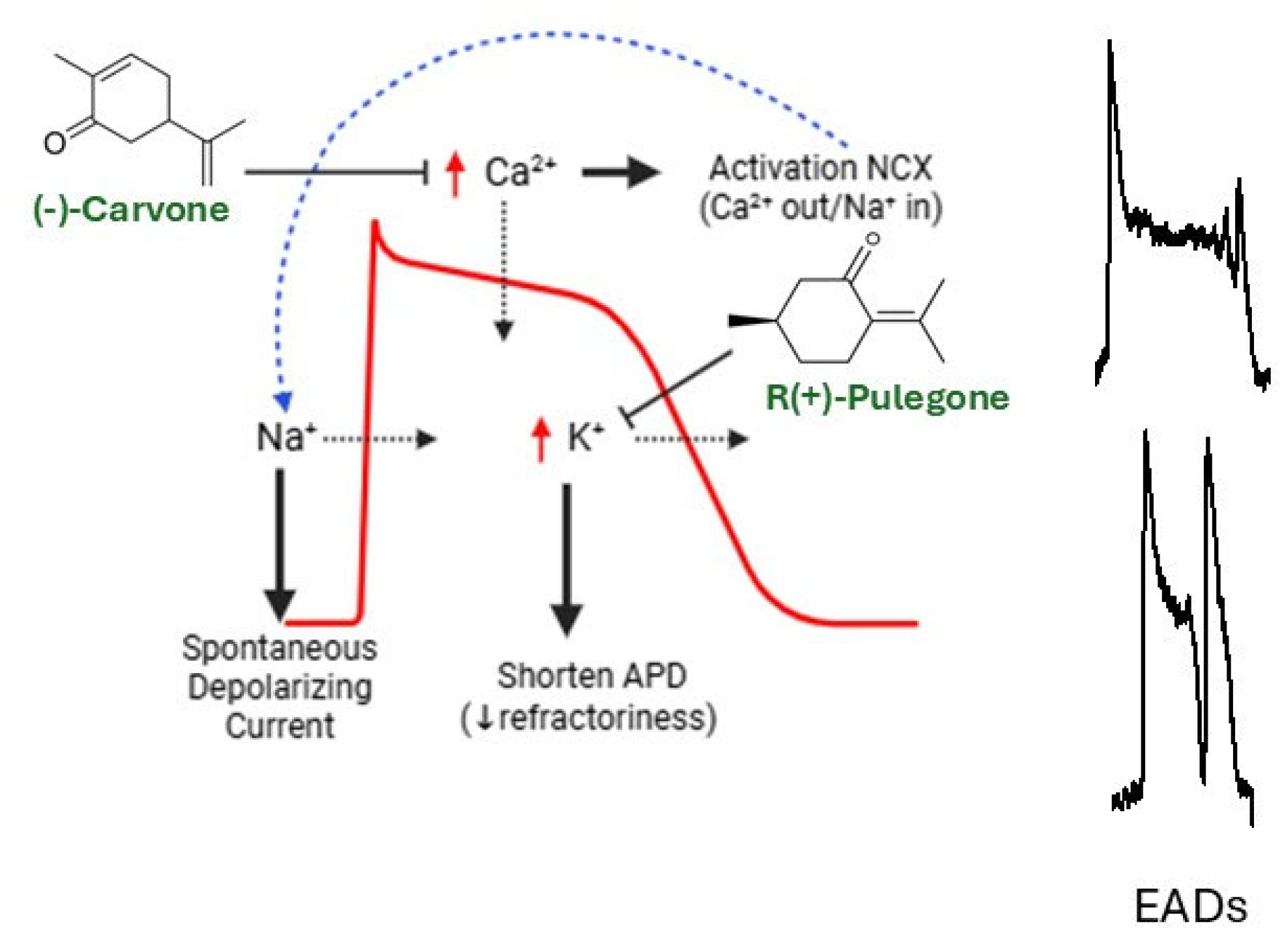
Disclaimer/Publisher’s Note: The statements, opinions and data contained in all publications are solely those of the individual author(s) and contributor(s) and not of MDPI and/or the editor(s). MDPI and/or the editor(s) disclaim responsibility for any injury to people or property resulting from any ideas, methods, instructions or products referred to in the content. |
© 2026 by the authors. Licensee MDPI, Basel, Switzerland. This article is an open access article distributed under the terms and conditions of the Creative Commons Attribution (CC BY) license.
Share and Cite
Ríos-López, J.L.; Blanco-Salas, J.; Cumplido-Laso, G.; Hortigón-Vinagre, M.P. Plant Terpenoids in Cardioprotection: An Overview of Their Therapeutic Potential. Curr. Issues Mol. Biol. 2026, 48, 479. https://doi.org/10.3390/cimb48050479
Ríos-López JL, Blanco-Salas J, Cumplido-Laso G, Hortigón-Vinagre MP. Plant Terpenoids in Cardioprotection: An Overview of Their Therapeutic Potential. Current Issues in Molecular Biology. 2026; 48(5):479. https://doi.org/10.3390/cimb48050479
Chicago/Turabian StyleRíos-López, José L., José Blanco-Salas, Guadalupe Cumplido-Laso, and María P. Hortigón-Vinagre. 2026. "Plant Terpenoids in Cardioprotection: An Overview of Their Therapeutic Potential" Current Issues in Molecular Biology 48, no. 5: 479. https://doi.org/10.3390/cimb48050479
APA StyleRíos-López, J. L., Blanco-Salas, J., Cumplido-Laso, G., & Hortigón-Vinagre, M. P. (2026). Plant Terpenoids in Cardioprotection: An Overview of Their Therapeutic Potential. Current Issues in Molecular Biology, 48(5), 479. https://doi.org/10.3390/cimb48050479