Article

Restoration of Autophagy and Apoptosis in Myelodysplastic Syndromes: The Effect of Azacitidine in Disease Pathogenesis

by
Georgia Tsekoura
1,†,
Andreas Agathangelidis
1,*,†,
Christina-Nefeli Kontandreopoulou
2,
Eirini Sofia Fasouli
3,
Eleni Katsantoni
3,
Vaia Pliaka
4,
Leonidas Alexopoulos
4,5,
Eleni Katana
1,
Myrto Papaioannou
6,
Georgia Taktikou
1,
Maria Eleftheria Strataki
1,
Angeliki Taliouraki
1,
Marina Mantzourani
2,
Nora-Athina Viniou
2,
Panagiotis T. Diamantopoulos
2 and
Panagoula Kollia
1,*
1
Division of Genetics & Biotechnology, Department of Biology, National and Kapodistrian University of Athens, 15772 Athens, Greece
2
First Department of Internal Medicine, National and Kapodistrian University of Athens, 11527 Athens, Greece
3
Basic Research Center, Biomedical Research Foundation, Academy of Athens, 11527 Athens, Greece
4
Protavio Ltd., Demokritos Science Park, 15341 Athens, Greece
5
Department of Mechanical Engineering, National Technical University of Athens, 15773 Athens, Greece
6
Department of Biomedical Sciences, School of Health Sciences, University of West Attica, 12243 Athens, Greece
*
Authors to whom correspondence should be addressed.
These authors contributed equally to this work.
Curr. Issues Mol. Biol. 2025, 47(7), 520; https://doi.org/10.3390/cimb47070520
Submission received: 30 April 2025 / Revised: 27 June 2025 / Accepted: 2 July 2025 / Published: 4 July 2025
(This article belongs to the Section Molecular Medicine)

Abstract

Myelodysplastic syndromes (MDSs) comprise a diverse group of clonal hematopoietic stem cell disorders characterized by ineffective hematopoiesis, cytopenia in the peripheral blood, and an increased risk of transformation into acute myeloid leukemia (AML). Despite extensive research, the mechanisms underlying MDS pathogenesis remain unclear. In the present study, we explored the role of autophagy and apoptosis in the development of MDS and assessed the impact of azacitidine on these processes in vitro. First, we assessed the expression of proteins involved in both autophagic and apoptotic pathways in MDS patients with different prognoses. Furthermore, using the MDS-L cell line as a model, we investigated the in vitro effects of azacitidine treatment on these processes. We report that MDS, irrespective of risk classification, is associated with the dysregulation of autophagy and apoptosis. Notably, azacitidine treatment restored these cellular processes, accompanied by modulation of key signaling phosphoproteins. Overall, these findings provide evidence that impaired autophagy and apoptosis contribute to MDS pathogenesis and that azacitidine helps restore cellular homeostasis by activating both processes. Furthermore, our study highlights the potential therapeutic benefits of targeting these mechanisms and suggests that combining azacitidine with agents that modulate autophagy and apoptosis could enhance the treatment efficacy for MDS patients.

1. Introduction

Myelodysplastic syndromes (MDSs) are clonal hematological malignancies characterized by ineffective hematopoiesis, cytopenia, and an increased risk of progression to acute myeloid leukemia (AML) [1]. MDS predominantly affects the elderly, with an incidence of approximately 20 per 100,000 individuals [2]. Despite extensive research, the mechanisms underlying MDS pathogenesis remain unclear. A prominent feature of MDS is the dysregulation of fundamental cellular processes, particularly autophagy [3] and apoptosis [4].
Autophagy is closely associated with MDS development. In this context, impairment of the PI3K/AKT pathway often results in cytopenia, dysplasia [5], and anemia in patients with MDS [6]. Furthermore, certain autophagy genes serve as prognostic markers, categorizing patients into high- and low-risk groups [7]. Apoptosis plays a dual role in MDS: it contributes to ineffective hematopoiesis in the early stages, while its inhibition is associated with disease progression [8]. As MDS advances, increased expression of anti-apoptotic proteins like BCL2 is observed, which can facilitate transformation into AML [9]. Consequently, therapeutic strategies that modulate apoptosis, such as the use of the BCL2 inhibitor venetoclax, are under investigation in MDS treatment [10].
The interplay between autophagy and apoptosis is complex, with both processes exhibiting cooperative and antagonistic relationships in the context of cell death [11]. Recent evidence, including our own previous work, has shown that MDS patients exhibit significant downregulation of genes associated with both autophagy and apoptosis, with this effect being more pronounced effect in higher-risk patients [12], as defined by the IPSS-R scoring system [13].
Currently, the therapeutic management of MDS involves a range of treatment modalities. Among these, azacitidine is one of the most well-established disease-modifying drugs [14,15,16], shown to improve survival rates mostly in higher-risk patients [17]. However, the precise downstream effects of azacitidine on autophagy and apoptosis remain largely unknown, limiting our understanding of its full therapeutic potential and its impact on disease pathogenesis.
To address this issue, we assessed the expression of proteins involved in autophagy and apoptosis in MDS patients with different disease risks. Subsequently, using the MDS-L cell line as a model, we investigated the in vitro effects of azacitidine on these processes. We report that MDS, irrespective of risk classification, is associated with the dysregulation of autophagy and apoptosis. Notably, azacitidine treatment restored these cellular processes, accompanied by the modulation of key signaling phosphoproteins. In summary, our findings corroborate the notion that the impairment of apoptosis and autophagy is crucial to MDS pathogenesis, while their restoration using existing or novel therapeutic drugs could offer important therapeutic benefits to patients with MDS.

2. Materials and Methods

2.1. The Study Group

The study group comprised 20 untreated patients with MDS and 14 healthy donors. The MDS patients were divided into lower-risk (LR-MDS, n = 12) and higher-risk groups (HR-MDS, n = 8), according to the IPSS-R scoring system [13]. More information regarding the clinical and biological characteristics of the MDS patients is given in the Supplementary Materials and Supplementary Tables S1 and S2.

2.2. Primary Cell Separation from MDS Patients

Mononuclear cells were isolated using Ficoll–Paque medium (GE Healthcare, Bio-Sciences AB, Uppsala, Sweden) and pelleted after erythrocyte lysis and two washes in phosphate-buffered saline (PBS tablets) (Calbiochem, Merck KGaA, Darmstadt, Germany).

2.3. The MDS-L Cell Line Culture

The MDS-L cell line was provided by Dr. Kaoru Tohyama (Kawasaki Medical School, Okayama, Japan). The MDS-L cells were cultured in RPMI 1640 medium with 10% FBS, 1% penicillin–streptomycin, and 10 ng/mL human recombinant IL-3 (Stemcell Technologies, Vancouver, BC, Canada).

2.4. Azacitidine Preparation

Azacitidine Vidaza (25mg/mL) (Celgene Europe B.V., Utrecht, The Netherlands) was freshly prepared using DEPC water.

2.5. MDS-L Cell Viability After Azacitidine Treatment

The MDS-L cell line was cultured with different azacitidine concentrations (namely 0.5, 1, 3, and 5 μM) for various time intervals (namely 24, 48, and 72 h), and cell apoptosis was assessed. More information is provided in the Supplementary Materials.

2.6. RNA and Protein Extraction from the MDS-L Cells After Azacitidine Treatment

RNA and proteins were extracted from the MDS-L cell samples containing 106 cells/mL after treatment with 0.5 μM of azacitidine for 48 h. Total RNA was extracted using TRIZOL (Invitrogen, Carlsbad, CA, USA). Proteins were extracted using a radioimmunoprecipitation assay buffer and quantified using a Bicinchoninic Acid (BCA) Protein Assay Kit (Thermo Fisher Scientific, Waltham, MA, USA).

2.7. The Gene Expression Analysis Using Quantitative Real-Time PCR

cDNA synthesis and PCR amplification were performed as described previously [12]. The expression of genes related to autophagy and apoptosis was assessed before and after azacitidine treatment. All experiments were run in triplicate. Supplementary Table S3 summarizes the 16 target genes and the respective primer sequences.

2.8. The Protein Expression Analysis Using Western Blotting

Proteins were separated and incubated with specific primary antibodies. Blots were probed with secondary goat anti-mouse or anti-rabbit antibodies and visualized. The analysis of the expression of relevant proteins was conducted in primary cells from the MDS patients, as well as in the MDS-L cells before and after azacitidine treatment. The experiments were performed three times for each sample in both analyses. Subsequently, the mean values were calculated, and the standard deviation (SD) or standard error (SE) was employed to indicate the data distribution. The presence of the GAPDH protein was assessed in all cases as a reference. Supplementary Table S4 summarizes all of the primary and secondary antibodies utilized.

2.9. The Phosphoprotein Expression Analysis Using a Multiplex ELISA Assay

The MDS-L cells were lysed, and phosphoprotein levels were assessed using a custom-developed phosphoprotein 21-plex panel (ProtATonce, Athens, Greece) (Supplementary Table S5). These experiments were performed in three treated and three untreated samples.

2.10. Statistical Approach

The data were analyzed using SPSS version 13.0 software (SPSS Inc., Chicago, IL, USA). The significance of the differences between groups was assessed using the Student’s t-test, while a one-way ANOVA and Tukey’s post hoc test were used for multiple comparisons. Statistical significance was defined as p < 0.05.

3. Results

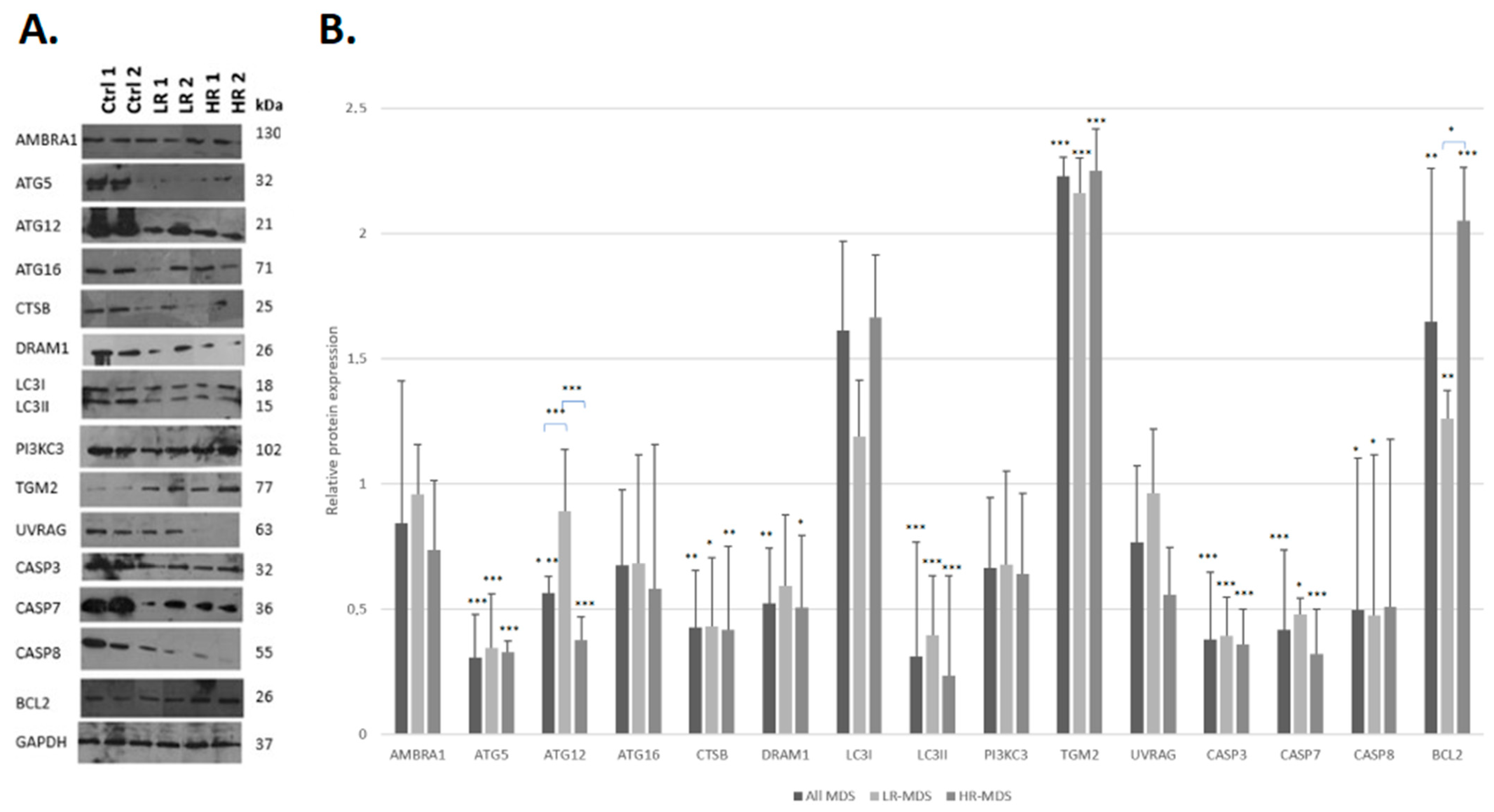
3.1. MDS Is Characterized by the Downregulation of Autophagy and Apoptosis Proteins

To assess the role of autophagy in MDS pathogenesis, we analyzed the expression of 11 relevant autophagic proteins in all MDS groups compared to that in healthy donors (Table 1). The expression levels of the pro-apoptotic proteins CASP3, CASP7, and CASP8, as well as the BCL2 protein, were also investigated. The analysis was performed in the following groups: (a) healthy donors, (b) all MDS patients, (c) the LR-MDS group, and (d) the HR-MDS group; comparisons made performed between healthy donors and each MDS group.
Overall, we observed lower expression levels for most proteins (12/15, 80%) in the patients with MDS compared to those in the healthy controls (Table 1). In terms of autophagy, significantly lower levels of 3/11 proteins (27.3%), namely ATG5, CTSB, and LC3II, were exhibited in all of the MDS groups (namely all MDS patients, LR-MDS, and HR-MDS) versus those in healthy donors. In addition, ATG12 and DRAM1 exhibited the same expression pattern, yet their downregulation was significant only in the all MDS patient and HR-MDS groups. Finally, the downregulation of AMBRA1, ATG16, PI3KC3, and UVRAG in the MDS groups compared to healthy individuals was not significant. On the other hand, TGM2 and LC3I were upregulated in the MDS patients compared to healthy donors, which was significant only for TGM2.
At the level of apoptosis, significantly lower levels of all three caspases were found in all MDS groups versus those in healthy donors, with the exception of CASP8 in HR-MDS. In contrast, BCL2 was significantly upregulated in all of the MDS groups versus healthy donors. The protein expression levels in all MDS groups as well as the group of healthy donors are given in Figure 1.
Lastly, the comparisons among the MDS groups showed that both autophagy and apoptosis were downregulated in HR-MDS compared to LR-MDS, as evidenced by the lower expression levels for 9/11 autophagy-related proteins. In contrast, LC3I and TGM2 were upregulated in HR-MDS compared to LR-MDS. In terms of apoptosis, HR-MDS was characterized by lower expression levels of CASP3 and CASP7, as well as the significant upregulation of BCL2 (Supplementary Table S6).

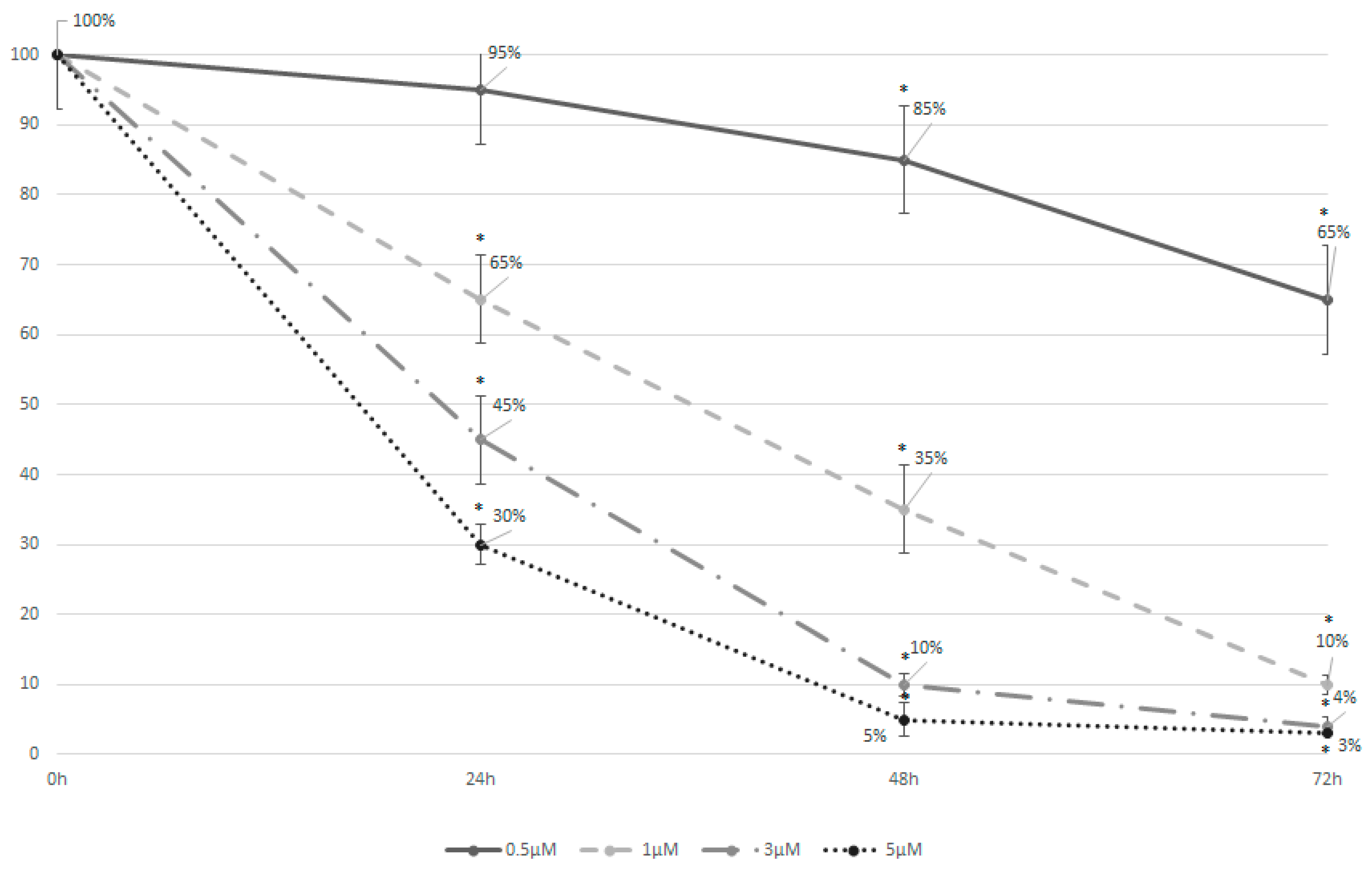
3.2. Azacytidine Induces Effective Cell Apoptosis in the MDS-L Cell Line

Higher concentrations of azacitidine exhibited a stronger cytotoxic effect on the cells, with the extent of cytotoxicity increasing continuously over time irrespective of the concentration (Figure 2). In particular, azacitidine concentrations of 3 and 5 μM achieved an IC50 within only 24 h of treatment. On the other hand, a concentration of 0.5 μM was toxic to only 5% of the MDS-L cells after 24 h, reaching 15% and 35% at the timepoints of 48 h and 72 h, respectively. Based on these findings, all subsequent experiments were performed in MDS-L cells treated with 0.5 μM of azacitidine for 48 h.

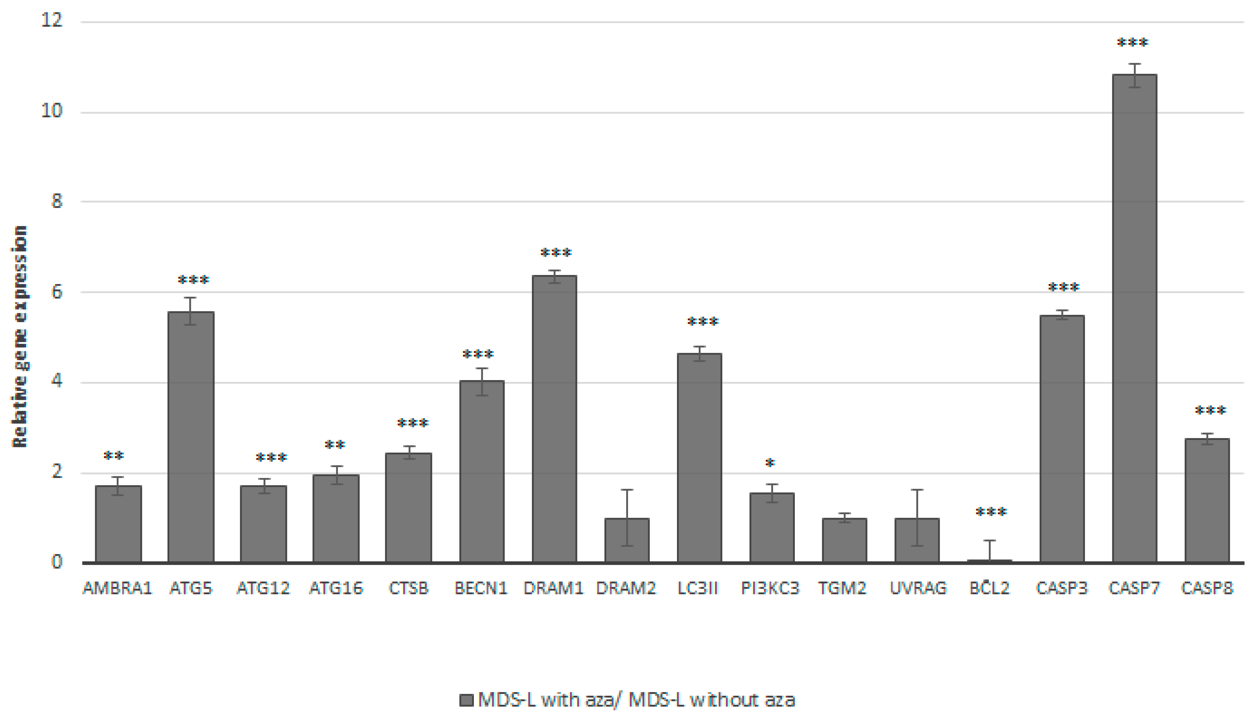
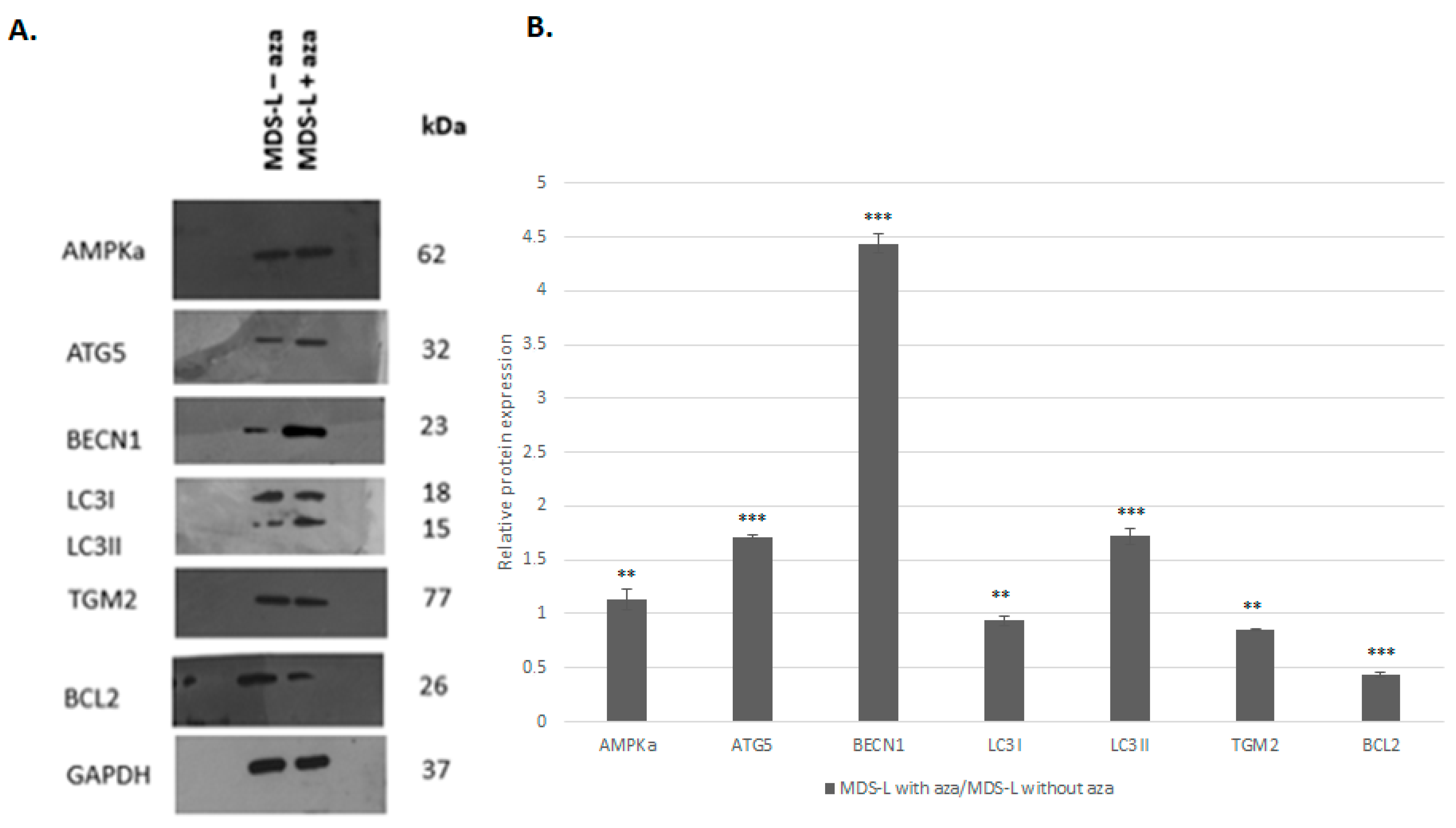
3.3. Treatment of the MDS-L Cells with Azacitidine Restored Autophagy and Apoptosis at the mRNA and Protein Levels

Our gene expression analysis showed that the majority of the analyzed genes (12/16, 75%) exhibited a significant increase in their expression levels after azacitidine treatment. Specifically, we observed (i) a < 2-fold change in the autophagy genes AMBRA1, ATG12, ATG16, and PI3KC3 and (ii) a > 2-fold change in the autophagy genes ATG5, BECN1, CTSB, DRAM1, and LC3II genes, as well as in the apoptotic genes CASP3, CASP7, and CASP8. The BCL2 gene displayed a statistically significant 18-fold decrease in its expression after azacitidine treatment. Detailed information regarding the effect of azacitidine on the gene expression levels is given in Figure 3 and Supplementary Table S7.
To validate these findings, we assessed the effect of azacitidine treatment on critical proteins, namely AMPKα, ATG5, BECN1, LC3I/II, TGM2, and BCL2 (Figure 4). In detail, azacitidine treatment led to a statistically significant increase in AMPKα, ATG5, BECN1, and LC3II. Conversely, the TGM2 and BCL2 levels decreased significantly 1.2-fold and 2-fold after the azacitidine treatment.
Of importance, the effect of the azacitidine treatment on the expression of relevant genes at the mRNA and protein levels was highly consistent for ATG5, BECN1, LC3II, and BCL2 (Table 2). TGM2 was the only gene that exhibited an unaffected state at the mRNA level, accompanied by a significant decrease at the protein level.

3.4. Azacitidine May Also Affect Phosphoproteins Related to Autophagy and Apoptosis

Finally, we investigated the effects of azacitidine by analyzing the expression of 21 relevant phosphoproteins using an ELISA. Table 3 depicts the effect of azacitidine on the phosphoprotein expression levels in the MDS-L cells compared to those in the untreated MDS-L cells. Our results indicate that azacitidine significantly modulated the majority of these key signaling proteins (12/21, 57.1%), yet with different effects.
Specifically, CHK2, c-JUN, ERK1, and P53 displayed a statistically significant increase in their expression patterns. In contrast, azacitidine treatment resulted in a significant decrease in the expression levels of eight key regulators of pro-survival pathways, namely AKT, CREB1, EGFR, MARCKS, mTOR, NFKB, RSK1, and STAT3. The phosphorylation status of the remaining nine phosphoproteins was unaffected by azacitidine treatment, indicating a level of specificity.

4. Discussion

The pathogenesis of MDS is a complex and multifactorial process, involving deregulation in various cellular pathways, including autophagy [5,6] and apoptosis [8]. We recently demonstrated the significant downregulation of genes implicated in autophagy and apoptosis in MDS, which was more evident in HR-MDS patients [12]. Here, we validated this notion by examining the status of relevant proteins, as well as the effect of azacitidine [16] on their regulation. Azacitidine is a hypomethylating agent primarily recommended for HR-MDS patients who are not eligible for intensive therapies, as well as LR-MDS patients who have significant cytopenia or anemia [18]. Overall, we report the downregulation of autophagy- and apoptosis-related proteins in MDS compared to healthy individuals, with this phenomenon being more evident in HR-MDS patients. This finding further corroborates our previous claim [12] that the deregulation of these processes becomes more pronounced and thus relevant as the disease progresses.
Focusing on autophagy, our findings indicate the widespread downregulation of ATG proteins in MDS, namely ATG5, ATG12, and ATG16. This is consistent with previous research in a murine model of myelodysplasia [19] showing a significant decrease in the expression of p-ATG1, p-ATG6, ATG7, and ATG12. We also observed the significant downregulation of key autophagy regulators such as PI3KC3 and AMBRA1. Of relevance, PI3K deletion promoted myelodysplasia in a triple knockout (TKO) mouse model [20]. In the same context, CD34+ cells were characterized by downregulation of the PI3K signaling expression signature in MDS patients compared to that in healthy donors [21]. On the other hand, AMBRA1 has been shown, under mTOR inhibition, to lead to a reduction in the cell division rate [22]; of relevance, we observed that azacitidine treatment led to significant downregulation of the mTOR protein and the upregulation of AMBRA1 at the mRNA level.
Our findings are also in line with the impairment of apoptosis in MDS, as evidenced by the reduced expression of caspases 3, 7, and 8 in both MDS risk groups compared to that in the healthy donors. This downregulation suggests that MDS cells may evade programmed cell death, leading to increased survival of abnormal progenitor cells [23]. Of particular interest was the downregulation of caspase 8, a master regulator of PANOptosis, a form of cell death that is highly relevant in cancer [24]. Moreover, the observation of reduced caspase 3 activity specifically in MDS progenitor cells indicates cell-type-specific apoptosis resistance [25]. Furthermore, a study in CASP8 knockout mice demonstrated that CASP8 deficiency results in an MDS-like phenotype [26].
Finally, our study showed the upregulation of TGM2 in both MDS risk groups, highlighting both its relevance to MDS pathogenesis by dysregulating autophagy and apoptosis and its potential role as a therapeutic target [27], as we previously reported [12]. A possible explanation for this upregulation is that TGM2 stabilizes pro-apoptotic BAX in an inactive conformation, thereby suppressing apoptosis [28]. Additionally, TGM2’s activity can promote autophagosome formation under stress, serving as a compensatory survival mechanism when the canonical autophagy pathways (e.g., the ATG5-ATG12-ATG16 axis) are impaired [29]. In line with this, BCL2 overexpression may inhibit both autophagy and apoptosis, highlighting its potential role—alongside TGM2—as a therapeutic target in MDS [12,30]. Upregulation of LC3I was also found in both MDS risk groups, which could reflect autophagy arrest at the lysosomal stage. Of relevance, studies in PI3K-deficient hematopoietic stem cells (HSCs) revealed reduced autophagic flux due to impaired lysosomal degradation, leading to LC3I buildup despite decreased LC3II [20].
Altogether, the general downregulation of both autophagic and apoptotic processes suggests the profound impairment of cellular homeostasis in MDS [31,32], allowing malignant cells to evade both programmed cell death and quality control mechanisms. Notably, previous studies have highlighted that defective autophagy can impair apoptotic signaling and vice versa, further supporting the notion that coordinated regulation of these pathways is essential for the prevention of malignant transformation [33]. Thus, our findings underscore the importance of restoring both autophagy and apoptosis to re-establish cellular equilibrium and suggest that therapeutic strategies targeting both processes may offer enhanced clinical benefit in MDS.
The second aim of our project was to investigate the impact of azacitidine on the processes of autophagy and apoptosis. In this context, we utilized the MDS-L cell line, which is considered a valuable model for testing therapeutic targets like azacitidine [34]. Overall, treatment with azacitidine led to restoration in the expression of autophagy- and apoptosis-related proteins in the MDS-L cells.
In terms of autophagy, we observed significant upregulation of a series of genes, including ATG5, BECN1, CTSB, DRAM1, and LC3II, among others, further supported by the upregulation of the ATG5, BECN1, and LC3I/II proteins. Of relevance, exposure of BM-derived mast cells (BMMCs) from MDS patients to azacitidine resulted in increased levels of ATG5, BECN1, and LC3II, indicating the induction of autophagy [35]. We also observed a significant decrease in the expression levels of AKT, mTOR, and RSK1, components of the PI3K/AKT/mTOR signaling pathway. This pathway acts as a negative regulator of autophagy in various diseases [36], frequently being hyperactivated in MDS and AML [37].
Regarding apoptosis, azacitidine treatment of the MDS-L cells resulted in a significant increase in the mRNA expression of CASP3, CASP7, and CASP8. Most notable was the tenfold increase in the CASP7 gene, which is consistent with previous studies demonstrating CASP7 activation in the SKM1 [38] as well as the HL-60 and K562 cell lines [39] in response to treatment with decitabine, which shares a similar mechanism of action with azacitidine [40]. Another prominent effect of the azacitidine treatment was the significant suppression of BCL2, both at the mRNA and protein levels, which has previously been reported as a key mechanism underlying azacitidine-mediated cytotoxicity in various cancer cell lines (namely P39, HL60, and Jurkat) [41]. Mechanistically, this could be due to the reduced levels of NF-κB and CREB, as observed in the present and previous studies [42,43]. At the therapeutic level, the downregulation of BCL2 in the MDS-L cell line suggests that azacitidine may directly influence apoptosis. A first link for this interaction in MDS was provided through a significant association between azacitidine resistance and the percentage of malignant cells expressing BCL2L10 [44]. In contrast, the BCL2 expression levels were relatively stable, showing no significant differences between azacitidine-sensitive and azacitidine-resistant patients. This discrepancy may indicate that while azacitidine can effectively downregulate BCL2 in the MDS-L line, the response in primary patient samples may be influenced by additional factors, such as the presence of BCL2L10. Understanding these dynamics can help refine the therapeutic approaches, especially when combining azacitidine with BCL2 inhibitors like venetoclax in the treatment of AML and MDS [45].
Another effect of azacitidine treatment on apoptosis was evidenced through the upregulation of key proteins, including p53, CHK2, and JUN; this suggests that azacitidine may enhance apoptosis through the activation of DNA damage response pathways [46,47] and transcriptional regulation [48]. In particular, p53 activation may be triggered by the ERK pathway [49], which is known to play a complex role in cell survival, autophagy, and programmed cell death [50]. Relevant evidence in other types of cancer includes the ERK1-mediated phosphorylation of BCL2 and its subsequent removal by Beclin-1 leading to the promotion of autophagy in a human lung cancer cell line [49] and the enhancement in the activity of the MEK/ERK pathway after azacitidine treatment in gastric cancer [51].
A possible explanation for the combined effect of azacitidine on autophagy and apoptosis in the context of MDS is that it may induce cellular stress, leading to both processes being triggered, as has been reported in other hematological malignancies [52] and solid tumors [53]. Even though autophagy is generally considered an antagonist of apoptosis, prolonged or intense cellular stress may force autophagy to facilitate or coexist with apoptosis [11]. In fact, several molecules, such as BECN1 and CASP8, may serve as points of crosstalk between these two pathways, enabling a dynamic balance between survival and cell death [54]. Thus, the concurrent activation of autophagy and apoptosis observed in our experiments likely reflects the complex interplay between these processes in MDS cells under therapeutic pressure.
Our findings are also consistent with azacitidine having additional effects, as evidenced by the downregulation of EGFR and MARCKS, two proteins involved in cell proliferation [55,56]. The EGFR downregulation suggests that azacitidine may suppress MDS cell proliferation by inhibiting EGFR-mediated MAPK signaling [57], while the MARCKS downregulation may also prevent PI3K activation [58]. Finally, the azacitidine treatment led to a reduction in CREB1 levels, potentially suppressing its role in promoting cell proliferation and survival in AML and MDS [59,60,61]. Of note, CREB1 may enhance apoptosis, suggesting a mechanism for azacitidine’s anti-proliferative effects in CREB1-driven leukemias [62]. Along these lines, the decreased levels of p38 MAPK after the azacitidine treatment corroborates the finding that p38α silencing led to enhanced hematopoiesis in MDS BM progenitors in vitro [63].
Another critical effect of azacitidine involves the inhibition of NF-κB and STAT3 signaling, both of which are associated with inflammation and cell survival in hematological malignancies [64]. Constitutive activation of NF-κB is frequently observed in many cancers, and suppressing NF-κB limits the proliferation of cancer cells [65]. In this context, persistently activated STAT3 maintains constitutive NF-κB activity [66]. Moreover, STAT3 is significantly upregulated in HSPCs derived from MDS and AML patients, being associated with an increased percentage of blasts, an adverse prognosis, and lower overall survival in MDS [67].
Our study has possible limitations, such as the comparison between BM samples from MDS patients and PB samples from healthy donors. In this context, we recently showed that a comparative analysis of the mRNA expression of key autophagy-related genes in BM and PB samples from MDS patients revealed no significant differences, supporting the feasibility of using the latter in these analyses [12]. Furthermore, given the relatively small size of the present cohort, further validation of our findings in larger series of primary samples from MDS patients would be beneficial, especially regarding characterization of the effect of azacitidine on autophagy and apoptosis. Nevertheless, our research is pioneering in systematically analyzing the expression patterns of a sizeable fraction of proteins implicated in autophagy and apoptosis in MDS with different prognoses. We report evidence of the downregulation of these critical processes in MDS, particularly HR-MDS, and that azacitidine treatment may crucially affect both processes to restore cell homeostasis. These findings reinforce the efficacy of azacitidine in treating MDS and suggest that further exploration of combination therapies targeting critical molecular pathways could improve the outcomes for MDS patients.

Supplementary Materials

The following supporting information can be downloaded at https://www.mdpi.com/article/10.3390/cimb47070520/s1.

Author Contributions

G.T. (Georgia Tsekoura) performed the research; P.K. designed the research study; C.-N.K., E.S.F., E.K. (Eleni Katsantoni), V.P., L.A., E.K. (Eleni Katana), M.P., G.T. (Georgia Taktikou), M.E.S., M.M., N.-A.V. and P.T.D. contributed essential samples, reagents, tools, or protocols; G.T. (Georgia Tsekoura), A.T. and A.A. analyzed the data; G.T. (Georgia Tsekoura), A.A., P.T.D. and P.K. wrote this paper. All authors have read and agreed to the published version of the manuscript.

Funding

This research work was supported by the Hellenic Foundation for Research and Innovation (HFRI) under the HFRI PhD Fellowship grant (Fellowship Number: 1594).

Institutional Review Board Statement

Ethical approval was obtained from the Ethics Committee of the Department of Biology (approval code: 2; approval date: 7 February 2022) and the Ethics Committee of the First Department (approval code: 2; approval date: 2 February 2022) of the National and Kapodistrian University of Athens.

Informed Consent Statement

Informed consent to participation was obtained from all subjects involved in this study.

Data Availability Statement

The data presented in this study can be made available by the corresponding author on request due to privacy issues.

Conflicts of Interest

Authors Vaia Pliaka and Leonidas Alexopoulos were employed by the company Protavio Ltd. The remaining authors declare that the research was conducted in the absence of any commercial or financial relationships that could be construed as a potential conflict of interest.

References

  1. Garcia-Manero, G. Myelodysplastic syndromes: 2023 update on diagnosis, risk-stratification, and management. Am. J. Hematol. 2023, 98, 1307–1325. [Google Scholar] [CrossRef]
  2. McDonald, L.; McCarthy, P.; Khan, M.; Hogan, P.; Kelleher, E.C.; Murphy, P.; Quinn, J.; Desmond, R.; McHugh, J.; Strickland, M.; et al. Should Myelodysplastic Syndromes in Very Old Patients be More Actively Managed? Clinical Characteristics, Management and Outcomes for Patients 85 Years and Older. Blood 2018, 132 (Suppl. 1), 5515. [Google Scholar] [CrossRef]
  3. Watson, A.S.; Mortensen, M.; Simon, A.K. Autophagy in the pathogenesis of myelodysplastic syndrome and acute myeloid leukemia. Cell Cycle 2011, 10, 1719–1725. [Google Scholar] [CrossRef]
  4. Parker, J.E.; Mufti, G.J. The Role of Apoptosis in the Pathogenesis of the Myelodysplastic Syndromes. Int. J. Hematol. 2001, 73, 416–428. [Google Scholar] [CrossRef]
  5. Ames, K.; Gritsman, K. Unraveling the Link between Class 1A PI3-Kinase, Autophagy, and Myelodysplasia. Autophagy 2024, 20, 952–954. [Google Scholar] [CrossRef]
  6. Jiang, H.; Yang, L.; Guo, L.; Cui, N.; Zhang, G.; Liu, C.; Xing, L.; Shao, Z.; Wang, H. Impaired Mitophagy of Nucleated Erythroid Cells Leads to Anemia in Patients with Myelodysplastic Syndromes. Oxidative Med. Cell. Longev. 2018, 2018, 6328051. [Google Scholar] [CrossRef]
  7. Wang, M.J.; Liu, W.Y.; Wang, X.Y.; Li, Y.M.; Xiao, H.Y.; Quan, R.C.; Huang, G.; Hu, X.M. Autophagy Gene Panel-Based Prognostic Model in Myelodysplastic Syndrome. Front. Oncol. 2021, 10, 606928. [Google Scholar] [CrossRef]
  8. Kerbauy, D.B.; Deeg, H.J. Apoptosis and antiapoptotic mechanisms in the progression of myelodysplastic syndrome. Exp. Hematol. 2007, 35, 1739–1746. [Google Scholar] [CrossRef]
  9. McBride, A.; Houtmann, S.; Wilde, L.; Vigil, C.; Eischen, C.M.; Kasner, M.; Palmisiano, N. The Role of Inhibition of Apoptosis in Acute Leukemias and Myelodysplastic Syndrome. Front. Oncol. 2019, 9, 192. [Google Scholar] [CrossRef]
  10. Garcia, J.S. Prospects for Venetoclax in Myelodysplastic Syndromes. Hematol. Oncol. Clin. N. Am. 2020, 34, 441–448. [Google Scholar] [CrossRef]
  11. Mariño, G.; Niso-Santano, M.; Baehrecke, E.H.; Kroemer, G. Self-consumption: The interplay of autophagy and apoptosis. Nat. Rev. Mol. Cell Biol. 2014, 15, 81–94. [Google Scholar] [CrossRef]
  12. Tsekoura, G.; Agathangelidis, A.; Kontandreopoulou, C.-N.; Taliouraki, A.; Mporonikola, G.; Stavropoulou, M.; Diamantopoulos, P.T.; Viniou, N.-A.; Aleporou, V.; Papassideri, I.; et al. Deregulation of Autophagy and Apoptosis in Patients with Myelodysplastic Syndromes: Implications for Disease Development and Progression. Curr. Issues Mol. Biol. 2023, 45, 4135–4150. [Google Scholar] [CrossRef]
  13. Greenberg, P.L.; Tuechler, H.; Schanz, J.; Sanz, G.; Garcia-Manero, G.; Solé, F.; Bennett, J.M.; Bowen, D.; Fenaux, P.; Dreyfus, F.; et al. Revised International Prognostic Scoring System for Myelodysplastic Syndromes. Blood 2012, 120, 2454–2465. [Google Scholar] [CrossRef]
  14. Raj, K.; Mufti, G.J. Azacytidine (Vidaza®) in the treatment of myelodysplastic syndromes. Ther. Clin. Risk Manag. 2006, 2, 377–388. [Google Scholar] [CrossRef]
  15. Buckstein, R.; Yee, K.; Wells, R.A. 5-Azacytidine in myelodysplastic syndromes: A clinical practice guideline. Cancer Treat. Rev. 2011, 37, 160–167. [Google Scholar] [CrossRef]
  16. Campelo, M.D.; Delgado, R.G.; Molias, A.C.G.; Sanchez, J.F. Azacytidine for the treatment of myelodysplastic syndromes in the elderly. Adv. Ther. 2011, 28 (Suppl. 2), 10–15. [Google Scholar] [CrossRef]
  17. Zugasti, I.; Castaño-Díez, S.; Esteban, D.; Pita, A.A.; Pomares, H.; González, A.P.; Garcia-Avila, S.; Padilla-Conejo, I.; de la Fuente, C.; Martínez-Roca, A.; et al. Venetoclax and Azacytidine Treatment for High Risk Myelodysplastic Syndromes and Chronic Myelomonocytic Leukemia As a Bridge Therapy to Transplant. a GESMD Study. Blood 2023, 142 (Suppl. 1), 1858. [Google Scholar] [CrossRef]
  18. Kim, Y.J.; Jang, J.H.; Kwak, J.Y.; Lee, J.H.; Kim, H.J. Use of azacitidine for myelodysplastic syndromes: Controversial issues and practical recommendations. Blood Res. 2013, 48, 87–98. [Google Scholar] [CrossRef]
  19. Daw, S.; Law, S. Quercetin induces autophagy in myelodysplastic bone marrow including hematopoietic stem/progenitor compartment. Environ. Toxicol. 2021, 36, 149–167. [Google Scholar] [CrossRef]
  20. Ames, K.; Kaur, I.; Shi, Y.; Tong, M.M.; Sinclair, T.; Hemmati, S.; Glushakow-Smith, S.G.; Tein, E.; Gurska, L.; Steidl, U.; et al. PI3-kinase deletion promotes myelodysplasia by dysregulating autophagy in hematopoietic stem cells. Sci. Adv. 2023, 9, eade8222. [Google Scholar] [CrossRef]
  21. Im, H.; Rao, V.; Sridhar, K.; Bentley, J.; Mishra, T.; Chen, R.; Hall, J.; Graber, A.; Zhang, Y.; Li, X.; et al. Distinct transcriptomic and exomic abnormalities within myelodysplastic syndrome marrow cells. Leuk. Lymphoma 2018, 59, 2952–2962. [Google Scholar] [CrossRef]
  22. Cianfanelli, V.; Fuoco, C.; Lorente, M.; Salazar, M.; Quondamatteo, F.; Gherardini, P.F.; De Zio, D.; Nazio, F.; Antonioli, M.; D’Orazio, M.; et al. AMBRA1 links autophagy to cell proliferation and tumorigenesis by promoting c-Myc dephosphorylation and degradation. Nat. Cell Biol. 2015, 17, 20–30. [Google Scholar] [CrossRef]
  23. Zang, D.Y.; Goodwin, R.G.; Loken, M.R.; Bryant, E.; Deeg, H.J. Expression of tumor necrosis factor–related apoptosis-inducing ligand, Apo2L, and its receptors in myelodysplastic syndrome: Effects on in vitro hemopoiesis. Blood 2001, 98, 3058–3065. [Google Scholar] [CrossRef]
  24. Jiang, M.; Qi, L.; Li, L.; Wu, Y.; Song, D.; Li, Y. Caspase-8: A key protein of cross-talk signal way in “PANoptosis” in cancer. Int. J. Cancer 2021, 149, 1408–1420. [Google Scholar] [CrossRef]
  25. Iriani, A.; Rachman, A.; Setiabudy, R.D.; Kresno, S.B.; Sudoyo, A.W.; Arief, M.; Harahap, A.R.; Fatina, M.K. TNFα induces Caspase-3 activity in hematopoietic progenitor cells CD34+, CD33+, and CD41 + of myelodysplastic syndromes. BMC Mol. Cell Biol. 2023, 24, 33. [Google Scholar] [CrossRef]
  26. Liu, S.; Joshi, K.; Zhang, L.; Li, W.; Mack, R.; Runde, A.; Hagen, P.A.; Barton, K.; Breslin, P.; Ji, H.-L.; et al. Caspase 8 deletion causes infection/inflammation-induced bone marrow failure and MDS-like disease in mice. Cell Death Dis. 2024, 15, 278. [Google Scholar] [CrossRef]
  27. Gillson, J.; El-Aziz, Y.S.A.; Leck, L.Y.W.; Jansson, P.J.; Pavlakis, N.; Samra, J.S.; Mittal, A.; Sahni, S. Autophagy: A Key Player in Pancreatic Cancer Progression and a Potential Drug Target. Cancers 2022, 14, 3528. [Google Scholar] [CrossRef]
  28. Wang, W.; Li, X.; Han, X.-Z.; Meng, F.-B.; Wang, Z.-X.; Zhai, Y.-Q.; Zhou, D.-S. Transglutaminase-2 is Involved in Cell Apoptosis of Osteosarcoma Cell Line U2OS Under Hypoxia Condition. Cell Biochem. Biophys. 2015, 72, 283–288. [Google Scholar] [CrossRef]
  29. Rossin, F.; D’Eletto, M.; Macdonald, D.; Farrace, M.G.; Piacentini, M. TG2 transamidating activity acts as a reostat controlling the interplay between apoptosis and autophagy. Amino Acids 2012, 42, 1793–1802. [Google Scholar] [CrossRef]
  30. Mukhopadhyay, S.; Panda, P.K.; Sinha, N.; Das, D.N.; Bhutia, S.K. Autophagy and apoptosis: Where do they meet? Apoptosis 2014, 19, 555–566. [Google Scholar] [CrossRef]
  31. Mortensen, M.; Ferguson, D.; Edelmann, M.; Kessler, B.; Morten, K.; Komatsu, M.; Simon, A. Loss of autophagy in erythroid cells leads to defective removal of mitochondria and severe anemia in vivo. Proc. Natl. Acad. Sci. USA 2010, 107, 832–837. [Google Scholar] [CrossRef]
  32. Watson, A.; Riffelmacher, T.; Stranks, A.; Williams, O.; De Boer, J.; Cain, K.; MacFarlane, M.; McGouran, J.; Kessler, B.; Khandwala, S.; et al. Autophagy limits proliferation and glycolytic metabolism in acute myeloid leukemia. Cell Death Discov. 2015, 1, 15008. [Google Scholar] [CrossRef]
  33. Liu, F.; Lee, J.Y.; Wei, H.; Tanabe, O.; Engel, J.D.; Morrison, S.J.; Guan, J.-L. FIP200 is required for the cell-autonomous maintenance of fetal hematopoietic stem cells. Blood 2010, 116, 4806–4814. [Google Scholar] [CrossRef]
  34. Rai, R.; Patel, F.; Feld, J.; Melana, S.; Navada, S.C.; Odchimar-Reissig, R.; Demakos, E.P.; Reddy, E.P.; Silverman, L.R. Combination of Ras Modulator and Azacitidine Impacts Innate Immune Signaling Pathway in MDS-L Cell Line. Blood 2021, 138 (Suppl. 1), 4325. [Google Scholar] [CrossRef]
  35. Romano, A.; Giallongo, C.; La Cava, P.; Parrinello, N.L.; Chiechi, A.; Vetro, C.; Tibullo, D.; Di Raimondo, F.; Liotta, L.A.; Espina, V.; et al. Proteomic Analysis Reveals Autophagy as Pro-Survival Pathway Elicited by Long-Term Exposure with 5-Azacitidine in High-Risk Myelodysplasia. Front. Pharmacol. 2017, 8, 204. [Google Scholar] [CrossRef]
  36. Wu, N.; Zhu, Y.; Xu, X.; Zhu, Y.; Song, Y.; Pang, L.; Chen, Z. The anti-tumor effects of dual PI3K/mTOR inhibitor BEZ235 and histone deacetylase inhibitor Trichostatin A on inducing autophagy in esophageal squamous cell carcinoma. J. Cancer 2018, 9, 987–997. [Google Scholar] [CrossRef]
  37. Liang, S.; Zhou, X.; Cai, D.; Rodrigues-Lima, F.; Chi, J.; Wang, L. Network Pharmacology and Experimental Validation Reveal the Effects of Chidamide Combined With Aspirin on Acute Myeloid Leukemia-Myelodysplastic Syndrome Cells Through PI3K/AKT Pathway. Front. Cell Dev. Biol. 2021, 9, 685954. [Google Scholar] [CrossRef]
  38. Zeng, W.; Dai, H.; Yan, M.; Cai, X.; Luo, H.; Ke, M.; Liu, Z. Decitabine-Induced Changes in Human Myelodysplastic Syndrome Cell Line SKM-1 Are Mediated by FOXO3A Activation. J. Immunol. Res. 2017, 2017, 4302320. [Google Scholar] [CrossRef]
  39. Li, L.; Liu, W.; Sun, Q.; Zhu, H.; Hong, M.; Qian, S. Decitabine Downregulates TIGAR to Induce Apoptosis and Autophagy in Myeloid Leukemia Cells. Oxidative Med. Cell. Longev. 2021, 2021, 8877460. [Google Scholar] [CrossRef]
  40. Saba, H.I. Decitabine in the treatment of myelodysplastic syndromes. Ther. Clin. Risk Manag. 2007, 3, 807–817. [Google Scholar]
  41. Khan, R.; Schmidt-Mende, J.; Karimi, M.; Gogvadze, V.; Hassan, M.; Ekström, T.J.; Zhivotovsky, B.; Hellström-Lindberg, E. Hypomethylation and apoptosis in 5-azacytidine–treated myeloid cells. Exp. Hematol. 2008, 36, 149–157. [Google Scholar] [CrossRef]
  42. Galante, J.M.; Mortenson, M.M.; Schlieman, M.G.; Virudachalam, S.; Bold, R.J. Targeting NF-kB/BCL-2 pathway increases apoptotic susceptibility to chemotherapy in pancreatic cancer. J. Surg. Res. 2004, 121, 306–307. [Google Scholar] [CrossRef]
  43. Wen, A.Y.; Sakamoto, K.M.; Miller, L.S. The Role of the Transcription Factor CREB in Immune Function. J. Immunol. 2010, 185, 6413–6419. [Google Scholar] [CrossRef]
  44. Cluzeau, T.; Robert, G.; Mounier, N.; Karsenti, J.M.; Dufies, M.; Puissant, A.; Jacquel, A.; Renneville, A.; Preudhomme, C.; Cassuto, J.-P.; et al. BCL2L10 is a predictive factor for resistance to Azacitidine in MDS and AML patients. Oncotarget 2012, 3, 490–501. [Google Scholar] [CrossRef]
  45. Du, Y.; Li, C.; Yan, J. The efficacy and safety of venetoclax and azacytidine combination treatment in patients with acute myeloid leukemia and myelodysplastic syndrome: Systematic review and meta-analysis. Hematology 2023, 28, 2198098. [Google Scholar] [CrossRef]
  46. Abuetabh, Y.; Wu, H.H.; Chai, C.; Al Yousef, H.; Persad, S.; Sergi, C.M.; Leng, R. DNA damage response revisited: The p53 family and its regulators provide endless cancer therapy opportunities. Exp. Mol. Med. 2022, 54, 1658–1669. [Google Scholar] [CrossRef]
  47. Hirao, A.; Kong, Y.-Y.; Matsuoka, S.; Wakeham, A.; Ruland, J.; Yoshida, H.; Liu, D.; Elledge, S.J.; Mak, T.W. DNA Damage-Induced Activation of p53 by the Checkpoint Kinase Chk2. Science 2000, 287, 1824–1827. [Google Scholar] [CrossRef]
  48. Bossy-Wetzel, E.; Bakiri, L.; Yaniv, M. Induction of apoptosis by the transcription factor c-Jun. EMBO J. 1997, 16, 1695–1709. [Google Scholar] [CrossRef]
  49. Liu, Y.; Yang, Y.; Ye, Y.-C.; Shi, Q.-F.; Chai, K.; Tashiro, S.-I.; Onodera, S.; Ikejima, T. Activation of ERK–p53 and ERK-Mediated Phosphorylation of Bcl-2 Are Involved in Autophagic Cell Death Induced by the c-Met Inhibitor SU11274 in Human Lung Cancer A549 Cells. J. Pharmacol. Sci. 2012, 118, 423–432. [Google Scholar] [CrossRef]
  50. Cagnol, S.; Chambard, J. ERK and cell death: Mechanisms of ERK-induced cell death—Apoptosis, autophagy and senescence. FEBS J. 2010, 277, 2–21. [Google Scholar] [CrossRef]
  51. Chen, Z.; Zhang, L.; Yang, Y.; Liu, H.; Kang, X.; Nie, Y.; Fan, D. DNMT1 expression partially dictates 5-Azacytidine sensitivity and correlates with RAS/MEK/ERK activity in gastric cancer cells. Epigenetics 2023, 18, 2254976. [Google Scholar] [CrossRef]
  52. Carew, J.S.; Nawrocki, S.T.; Kahue, C.N.; Zhang, H.; Yang, C.; Chung, L.; Houghton, J.A.; Huang, P.; Giles, F.J.; Cleveland, J.L. Targeting autophagy augments the anticancer activity of the histone deacetylase inhibitor SAHA to overcome Bcr-Abl–mediated drug resistance. Blood 2007, 110, 313–322. [Google Scholar] [CrossRef]
  53. Parks, M.; Tillhon, M.; Donà, F.; Prosperi, E.; Scovassi, A.I. 2-Methoxyestradiol: New perspectives in colon carcinoma treatment. Mol. Cell Endocrinol. 2011, 331, 119–128. [Google Scholar] [CrossRef]
  54. Maiuri, M.C.; Zalckvar, E.; Kimchi, A.; Kroemer, G. Self-eating and self-killing: Crosstalk between autophagy and apoptosis. Nat. Rev. Mol. Cell Biol. 2007, 8, 741–752. [Google Scholar] [CrossRef]
  55. Harari, P.M.; Allen, G.W.; Bonner, J.A. Biology of Interactions: Antiepidermal Growth Factor Receptor Agents. J. Clin. Oncol. 2007, 25, 4057–4065. [Google Scholar] [CrossRef]
  56. El Amri, M.; Fitzgerald, U.; Schlosser, G. MARCKS and MARCKS-like proteins in development and regeneration. J. Biomed. Sci. 2018, 25, 43. [Google Scholar] [CrossRef]
  57. Daw, S.; Law, A.; Law, S. Myelodysplastic Syndrome related alterations of MAPK signaling in the bone marrow of experimental mice including stem/progenitor compartment. Acta Histochem. 2019, 121, 330–343. [Google Scholar] [CrossRef]
  58. Ziemba, B.P.; Burke, J.E.; Masson, G.; Williams, R.L.; Falke, J.J. Regulation of PI3K by PKC and MARCKS: Single-Molecule Analysis of a Reconstituted Signaling Pathway. Biophys. J. 2016, 110, 1811–1825. [Google Scholar] [CrossRef]
  59. Sandoval, S.; Pigazzi, M.; Sakamoto, K.M. CREB: A Key Regulator of Normal and Neoplastic Hematopoiesis. Adv. Hematol. 2009, 2009, 634292. [Google Scholar] [CrossRef]
  60. Cheng, J.C.; Kinjo, K.; Judelson, D.R.; Chang, J.; Wu, W.S.; Schmid, I.; Shankar, D.B.; Kasahara, N.; Stripecke, R.; Bhatia, R.; et al. CREB is a critical regulator of normal hematopoiesis and leukemogenesis. Blood 2008, 111, 1182–1192. [Google Scholar] [CrossRef]
  61. Kinjo, K.; Sandoval, S.; Sakamoto, K.M.; Shankar, D.B. The Role of CREB as a Proto-oncogene in Hematopoiesis. Cell Cycle 2005, 4, 1134–1135. [Google Scholar] [CrossRef]
  62. LeBlanc, F.; Bennett, J.; Choi, K.; Starczynowski, D.T. Targeting CREB-Binding Protein (CREBBP) Overcomes Resistance to Azacitidine and Venetoclax Therapy in Acute Myeloid Leukemia (AML). Blood 2023, 142 (Suppl. 1), 5765. [Google Scholar] [CrossRef]
  63. Navas, T.A.; Mohindru, M.; Estes, M.; Ma, J.Y.; Sokol, L.; Pahanish, P.; Parmar, S.; Haghnazari, E.; Zhou, L.; Collins, R.; et al. Inhibition of overactivated p38 MAPK can restore hematopoiesis in myelodysplastic syndrome progenitors. Blood 2006, 108, 4170–4177. [Google Scholar] [CrossRef]
  64. Zhang, T.; Ma, C.; Zhang, Z.; Zhang, H.; Hu, H. NF-κB signaling in inflammation and cancer. MedComm 2021, 2, 618–653. [Google Scholar] [CrossRef]
  65. Park, M.; Hong, J. Roles of NF-κB in Cancer and Inflammatory Diseases and Their Therapeutic Approaches. Cells 2016, 5, 15. [Google Scholar] [CrossRef]
  66. Saluja, S.; Bansal, I.; Bhardwaj, R.; Beg, M.S.; Palanichamy, J.K. Inflammation as a driver of hematological malignancies. Front. Oncol. 2024, 14, 1347402. [Google Scholar] [CrossRef]
  67. Shastri, A.; Choudhary, G.; Teixeira, M.; Gordon-Mitchell, S.; Ramachandra, N.; Bernard, L.; Bhattacharyya, S.; Lopez, R.; Pradhan, K.; Giricz, O.; et al. Antisense STAT3 inhibitor decreases viability of myelodysplastic and leukemic stem cells. J. Clin. Investig. 2018, 128, 5479–5488. [Google Scholar] [CrossRef]
Figure 1. Lower expression levels of proteins implicated in the processes of autophagy and apoptosis in MDS patients compared to those in healthy donors. (A) Western blot experiments in representative samples from the groups of healthy donors (Ctrl1, Ctrl2) and MDS patients of different classifications (HR-MDS: HR1, HR2; LR-MDS: LR1, LR2). The intensity of the bands was quantified using the ImageJ 1.x software, while the expression ratio of each protein was estimated relative to that for the GAPDH protein. (B) Relative expression levels in the group of all MDS patients as well as in each of the MDS risk groups compared to those in the healthy controls. The statistical examination was performed using a one-way ANOVA and Tukey’s post hoc test. Asterisks represent statistically significant differences; one asterisk (*) corresponds to p < 0.05, two asterisks (**) correspond to p < 0.02, and three asterisks (***) correspond to p< 0.001. Error bars correspond to the standard deviation (SD).
Cimb 47 00520 g001
Figure 2. Treatment with azacitidine can induce strong cell apoptosis in MDS-L cells even after 24 h of culture. The graph depicts the cell viability curves for MDS-L cells treated with different concentrations of azacitidine (0.5 μΜ, 1 μΜ, 3 μΜ, or 5 μM) after 24, 48, and 72 h. Higher concentrations of azacitidine demonstrated an enhanced cytotoxic effect on the cells, with the degree of cytotoxicity progressively increasing over time, independent of the concentration. Asterisks represent statistically significant differences corresponding to p < 0.05. Error bars correspond to the standard errors (SEs).
Cimb 47 00520 g002
Figure 3. Treatment of MDS-L cells with a low concentration of azacitidine may lead to the restoration of autophagy and apoptosis. The expression patterns for a selected panel of 16 genes related to the processes of autophagy and apoptosis in MDS-L cells. Bars represent the relative gene expression levels in the MDS-L cells after treatment with azacitidine compared to those under the baseline status (i.e., without azacitidine treatment). Azacitidine treatment led to upregulation of all of the genes, besides downregulating the anti-autophagic and anti-apoptotic BCL2 gene. The HPRT1 gene was used as a reference. Asterisks represent statistically significant differences; one asterisk (*) corresponds to p < 0.05, two asterisks (**) correspond to p < 0.02, and three asterisks (***) correspond to p< 0.001. Error bars correspond to the standard errors (SEs).
Cimb 47 00520 g003
Figure 4. Azacitidine treatment can promote the activation of autophagy by upregulating promoting proteins and downregulating inhibitory proteins. (A) The immunoblot analysis of the expression of a series of proteins implicated in the processes of autophagy and apoptosis in MDS-L cells with and without treatment with azacitidine (MDS-L with aza and MDS-L without aza, respectively). Overall, azacitidine treatment was associated with higher expression levels of the AMPKA, ATG5, BECN1, and LC3II proteins, as well as lower expression levels of BCL2. (B) A bar graph depicting the relative expression levels of all proteins in the treated MDS-L cells compared to those in the baseline (untreated) MDS-L cells. Asterisks represent statistically significant differences; one asterisk (*) corresponds to p < 0.05, two asterisks (**) correspond to p < 0.02, and three asterisks (***) correspond to p < 0.001. Error bars correspond to standard errors (SEs).
Cimb 47 00520 g004
Table 1. The expression differences in the form of the fold changes in 15 proteins related to the processes of autophagy and apoptosis in all MDS patients, as well as in each MDS risk group, compared to a group of healthy donors. Statistically significant differences between any MDS group and the group of healthy donors are given in the form of p-values.
All MDSp-ValueLR-MDSp-ValueHR-MDSp-Value
AMBRA1−1.190165 −1.042547 −1.361363
ATG5−3.274832<0.001−2.911969<0.001−3.066072<0.001
ATG12−1.774992<0.001−1.126027 −2.659288<0.001
ATG16−1.483484 −1.470517 −1.720712
CTSB−2.335516<0.02−2.318447<0.05−2.393512<0.02
DRAM1−1.916758<0.02−1.692008 −1.984616<0.05
LC3I1.612848 1.188060 1.664416
LC3II−3.231055<0.001−2.523108<0.001−4.274320<0.001
PI3KC3−1.509030 −1.478196 −1.566015
TGM22.229709<0.0012.162048<0.0012.249299<0.001
UVRAG−1.307206 −1.042216 −1.797932
CASP3−2.650488<0.001−2.542133<0.001−2.799367<0.001
CASP7−2.410310<0.001−2.095823<0.05−3.116006<0.001
CASP8−2.013422<0.05−2.109531<0.05−1.961725
BCL21.647007<0.021.257670<0.022.051875<0.001
Table 2. Gene and protein expression differences (in the form of fold changes) in 6 proteins implicated in autophagy and apoptosis in MDS-L cells cultured in the presence of azacitidine compared to MDS-L cells without any type of treatment, which served as the controls. Asterisks (*) correspond to statistically significant differences (p ˂ 0.05).
Gene/ProteinGene ExpressionProtein Expression
AMPKα-1.32 *
ATG55.58 *1.72 *
BECN14.03 *4.44 *
LC3II4.63 *1.72 *
TGM21−1.19 *
BCL2−18.54 *−2.28 *
Table 3. Expression differences (in the form of fold changes) in 21 phosphoproteins implicated in autophagy and apoptosis in MDS-L cells cultured in the presence of azacitidine compared to those in MDS-L cells without any type of treatment, which served as the controls. Asterisks correspond to statistically significant differences (p < 0.05).
PhosphoproteinFold Change
AKT−2 *
AKT1S1−1.25
CHK21.17 *
c-JUN1.13 *
CREB1−2.2 *
EGFR−1.62 *
ERK11.76 *
FAK11.1
GSK3a/β−1.15
HSPB1−1.21
IKBA1.18
MARCKS−1.65 *
MEK11.2
MTOR−1.2 *
NFKB−1.97 *
P38 MAPK−1.12
P532.3 *
PTN11−1.21
RSK1−1.16 *
SMAD3−1.12
STAT3−1.23 *
Disclaimer/Publisher’s Note: The statements, opinions and data contained in all publications are solely those of the individual author(s) and contributor(s) and not of MDPI and/or the editor(s). MDPI and/or the editor(s) disclaim responsibility for any injury to people or property resulting from any ideas, methods, instructions or products referred to in the content.

