Article

In Silico Analysis of s-DAPK-1: From Structure to Function and Regulation

1
Department of Biochemistry, Microbiology and Biotechnology, University of Limpopo, Private Bag X1106, Sovenga 0727, South Africa
2
Department of Life and Consumer Sciences, University of South Africa, Private Bag X06, Florida 1710, South Africa
*
Author to whom correspondence should be addressed.
Curr. Issues Mol. Biol. 2025, 47(6), 416; https://doi.org/10.3390/cimb47060416
Submission received: 17 April 2025 / Revised: 21 May 2025 / Accepted: 27 May 2025 / Published: 4 June 2025
(This article belongs to the Special Issue Protein Domains: Structure and Molecular Function)

Abstract

:
The existence of s-DAPK-1, an alternatively spliced variant of DAPK-1, adds complexity to our understanding of the proteins involved in the regulation of cell survival, apoptosis, and autophagy. DAPK-1 has been implicated in the regulation of these processes; however, it remains unclear whether s-DAPK-1 also plays a similar role or a separate function; thus, determining its involvement in these processes is challenging due to the limited understanding of its regulation, interacting partners, function, and three-dimensional (3D) structure. Hence, this study was aimed at (1) understanding the regulation of s-DAPK-1 by predicting its microRNA targets, (2) predicting the 3D structure of s-DAPK-1, (3) its physicochemical and thermodynamic properties, (4) its interacting partners, and (5) molecular functions using computational methods. To achieve this aim, various bioinformatics tools and in silico webservers, such as ProteinPrompt, ProtParam, ProtScale, ScooP, Hawkdock, Phyre2, I-TASSER, PSIPRED, SAVES, and PROCHECK, along with user-friendly databases, such as NCBI, TarBase, and Protein Data Bank (PDB), were employed. For miRNA prediction, we used TarBase, and identified the specific microRNAs targeting s-DAPK-1. Furthermore, the Phyre2 database demonstrated that s-DAPK-1 possesses 40% alpha helices and 4% beta strands, forming a stable 3D structure. Additionally, s-DAPK-1 demonstrated stability to withstand high temperatures, suggesting that it is a thermostable protein. Moreover, s-DAPK-1 was found to interact with a variety of proteins involved in tumor progression and gene regulation, including a prion protein and histone H2B type 2-E (H2B2E). This suggests that s-DAPK-1 may perform diverse molecular functions such as regulation of metabolic processes, nucleic acid binding, and mRNA splicing by interacting with different proteins.

1. Introduction

Progressively, substantial bioinformatics databases continue to provide new knowledge on biology, and it has emerged as a discipline to interpret experimental data in order to better understand biological processes using the information contained within databases [1,2]. Creating and answering biological questions that were previously considered too complex has been made possible by the enormous amount of data contained within these databases. Specifically, in the field of proteomics, bioinformatics plays an important role in all aspects of protein analysis, including sequence analysis, structure analysis, and evolution analysis [3,4]. For example, bioinformatics tools predicted death-associated protein kinase 1 (DAPK-1) as the most promising Alzheimer’s disease (AD)-related target for quercetin [5]. Moreover, bioinformatics tools have shed light on potential DAPK-1 inhibitors [6,7], DAPK-1’s functional role [6], and the proteins it interacts with [8]. This demonstrates the ability of bioinformatics tools to resolve questions in the field of proteomics. However, despite the availability of datasets retrieved from bioinformatics tools for various less-studied proteins, including s-DAPK-1, a comprehensive summary of these datasets remains lacking.
DAPK-1, a protein kinase with multiple domains, plays roles in various cellular processes, including autophagy, apoptosis, and cell survival signaling pathways [9]. Additionally, the DAPK-1 gene undergoes alternative splicing to produce a short isoform referred to as s-DAPK-1 [10]. The regulation of DAPK-1 protein levels is intricately managed through a combination of transcriptional and post-transcriptional processes. Notably, transcription factors such as p53 and cAMP response element-binding protein 1 (CREB1) play significant roles in modulating the transcription of DAPK-1 [11]. DAPK-1 also has many other downstream targets, including N-methyl-D-aspartate receptor 2B (NR2B) [12], peptidyl–prolyl cis–trans isomerase NIMA-interacting 1 (Pin1) [13], and DEAD-box helicase 20 (DDX20) [14]. Targeting these downstream targets of DAPK-1 can alleviate stroke damage, obstruct the ability of Pin1 to activate oncogenic transcription factors, and suppress cell proliferation and tumor development [12,13,14].
Moreover, DAPK-1 has been shown to be post-transcriptionally regulated by a group of noncoding RNA molecules known as microRNAs (miRs) [15,16,17]. These miRs bind to their target mRNAs through sequence complementarity, leading to cleavage of mRNA and translation repression [18]. Several microRNAs have been identified as regulators of DAPK1, including miR-143-3p in AD [19], miR-191 in endometriosis-associated ovarian cancer [20], and miR-483-5p in nasopharyngeal carcinoma [21]. In addition, bioinformatics tools have also been applied to predict microRNAs targeting DAPK-1, such as miR-141-3p in polycystic ovary syndrome [22]. However, there is currently no research study that has examined how microRNAs regulate s-DAPK-1, a variant produced by the alternative splicing of DAPK-1.
Alternative splicing (AS) is a key process in generating proteome diversity, with projections suggesting that 95% of genes are alternatively spliced in humans [23,24,25]. Alternative splicing allows a single gene to generate multiple unique mRNA transcripts, which can subsequently be translated into different protein isoforms. These isoforms may vary in their structures, activities, and interactions, contributing to a variety of functional roles within a cell or tissue [26,27]. According to the Ensembl website, there are 20 splice variants of DAPK-1 gene, with 7 of them potentially coding for proteins and 13 lacking coding capacity [28]. To date, laboratory experiments have validated the existence of only the s-DAPK-1 spliced variant [10]. An examination of multiple sequence alignments indicates that approximately 22% of the amino acids in s-DAPK-1 are identical to those making up the DAPK-1 isoform.
Expression of the s-DAPK-1 mRNA starts on introns 13–14 within the DAPK-1 gene and ends on introns 20–21 within the DAPK-1 gene (Figure 1). The start codon, AUG, for this mRNA is located between the 10–12 bases of exon 15 within the DAPK-1 gene, making its translation in-frame with DAPK-1 mRNA [10]. Further, s-DAPK-1 shares exons 15, 16, 17, 18, 19, and 20 with the full-length DAPK-1 mRNA, and its stop codon is located within base pairs 124–126 of intron 20–21; s-DAPK-1 mRNA encodes a 337 amino acid polypeptide encompassing part of the ankyrin-repeat domain, the P-loop motifs, part of the cytoskeletal binding domain of DAPK-1, and a unique C-terminal “tail” extension of 42 amino acids that is absent in DAPK-1 (Figure 1). Based on the Blastp results, this 42-amino acid query sequence was only mapped on s-DAPK-1 but not to any other motif or domain found in the Homo sapiens database. Interestingly, Lin et al. [10] showed that this s-DAPK-1 C-terminal tail features an internal proteolytic processing site, and the deletion of this site increased the s-DAPK-1 stability and augmented the membrane-blebbing effect of s-DAPK-1. However, there is still limited literature on the structure, protein–protein interactions, cellular function, and expression of s-DAPK-1 in health and disease.
Lin et al. [10] documented that when comparing the expression of s-DAPK-1 mRNA in cancerous cells (HCT116 and A375) and non-cancerous cells (HEK-293), s-DAPK-1 was highly expressed in HEK-293 (5.517-fold change) relative to HCT116 (0.000-fold change) and A375 (0.455-fold change) cells, indicating that s-DAPK-1 could potentially serve as an antitumor protein, particularly in the cases of skin and colorectal cancer. Given that s-DAPK-1 is an isoform of DAPK-1, it has the potential to contribute to various biological processes. Therefore, there is a significant interest in understanding its 3D structure, function, and regulation. The 3D structure, function, and regulation of DAPK-1 are extensively documented in both physiological and pathological conditions. However, knowledge regarding the 3D structure, function, and regulation of s-DAPK-1, an isoform of DAPK-1, remains unexplored. Even studies on the application of bioinformatics to predict s-DAPK-1’s 3D structure, function, and regulatory mechanism are yet to be conducted. The question of whether s-DAPK-1 mimics DAPK-1’s 3D structure, function, and regulation remains unanswered. The application of bioinformatics holds the potential to address all the critical questions regarding s-DAPK-1.

2. Materials and Methods

2.1. Prediction of microRNAs (miRs) Targeting s-DAPK-1 mRNA

The TarBase (https://dianalab.e-ce.uth.gr/tarbasev9, accessed on the 24 April 2024) database was used to search for microRNAs targeting s-DAPK-1 [29]. The Tarbase database was searched using “DAPK1” under the “interactions” option. The results were filtered to display only high-confidence miRNAs (validated through experimental data), with a microT prediction score of 0.8 and higher and expressed within Homo sapiens. Further confirmation of these miRNAs as targets of DAPK1 was through the use of miRWalk (http://mirwalk.umm.uni-heidelberg.de/, accessed on 24 April 2024) and TargetScan (https://www.targetscan.org/vert_80/, accessed on 24 April 2024). These microRNAs were mapped onto the s-DAPK-1 mRNA sequence to identify miRNAs targeting s-DAPK-1.

2.2. Protein Sequence Retrieval

The s-DAPK-1 protein sequence was retrieved from the NCBI database (https://www.ncbi.nlm.nih.gov/, accessed on the 9 March 2024), which is a freely accessible database containing data on protein sequences and their functional annotation [30]. The s-DAPK-1 protein sequence was retrieved using its accession number AK127855.1 in the FASTA format. The protein sequence was then used to predict s-DAPK-1’s hydropathy plot, physicochemical parameters, interacting partners, molecular functions, secondary and tertiary structures.

2.3. Modeling and Validation of s-DAPK-1’s 3D Structure

The s-DAPK-1 protein sequence in the FASTA format was then deposited in the Phyre2 database (http://www.sbg.bio.ic.ac.uk, accessed on 15 April 2024) [31] to generate the secondary structure of s-DAPK-1 using the normal modeling mode. The 3D structure of s-DAPK-1 was modeled using the I-TASSER On-line Server (https://zhanggroup.org/I-TASSER/, accessed on 15 April 2024) [32,33]. The quality of the resultant model was assessed using the C-score and the TM-score, and its validation was conducted through the Ramachandran plot on PROCHECK under the SAVES server (https://saves.mbi.ucla.edu/, accessed on 19 April 2024) [34].

2.4. Prediction of Physical and Chemical Parameters of s-DAPK-1

The ProtParam tool (https://web.expasy.org/protparam/, accessed on 11 May 2024) [35] was used to estimate physicochemical parameters of s-DAPK-1. The raw protein sequence of s-DAPK-1 was used to query the server. The server provides directly calculated values of pI (isoelectric point), molecular weight, percentage of each amino acid, extinction coefficient, instability index, aliphatic index, and grand average of hydropathicity (GRAVY).

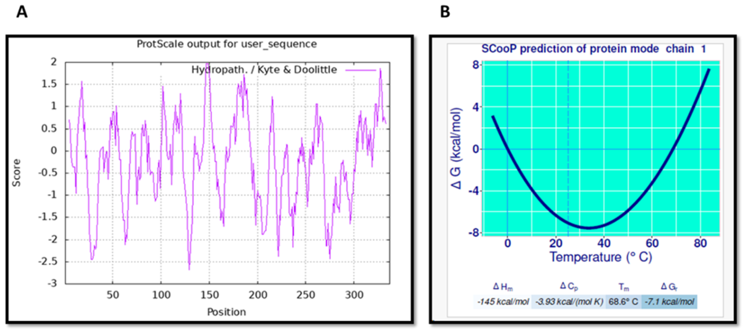
2.5. Prediction of s-DAPK-1’s Hydrophobicity and Thermodynamic Parameters

The hydropathy plot of s-DAPK-1 was generated using the default settings of ProtScale (https://web.expasy.org/protscale/, accessed on 17 May 2024) [36]. This tool allowed for a quantitative analysis of either the hydrophobic or hydrophilic nature of the amino acids present in the protein sequence of s-DAPK-1. Furthermore, the ScooP tool (http://babylone.3bio.ulb.ac.be/SCooP/k_query.php, accessed on 17 May 2024) [37] was used to predict the thermodynamic characteristics of s-DAPK-1 under varying temperature conditions. To query the ScooP tool, the predicted 3D structure of s-DAPK-1 was used.

2.6. Prediction of Protein-Protein Interactions Involving s-DAPK-1

The ProteinPrompt (https://proteinformatics.uni-leipzig.de/protein_prompt/, accessed on 10 April 2024) online sequence-based approach was used to predict proteins interacting with s-DAPK-1. This webserver uses binding scores to indicate high confidence in binding. The quality and speed of this system make it a suitable high-throughput method for scanning sequence libraries [38].

2.7. Protein–Protein Docking

The ProteinPrompt results were confirmed through the use of HawkDock (http://cadd.zju.edu.cn/hawkdock/, accessed on 16 April 2024), a template-free blind protein–protein docking tool. HawkDock was used in order to predict the binding affinity between the proteins identified by ProteinPrompt and the s-DAPK-1 protein. HawkDock is a useful tool for predicting the structures of protein complexes that are yet to be experimentally determined [39]. In order to carry out the HawkDock analysis, the ProteinPrompt results were refined to include solely proteins with confirmed 3D structures stored in the Protein Data Bank (https://www.rcsb.org/, accessed on 27 April 2024). These proteins were cleaned using the Discovery Studio visualizer. The optimal pose was determined by considering the free binding energy, where a more negative score indicated a higher binding energy strength. The ideal conformation was submitted to the Protein Interaction Analysis tool of BioLuminate to visualize the amino acids and bonds participating in the interactions between the two proteins.

2.8. Prediction of the Functions of the Modeled s-DAPK-1 Structure

The PSIPRED (http://bioinf.cs.ucl.ac.uk/psipred/, accessed on 16 May 2024) webserver has access to FFPred 3, a tool to predict protein function. To make predictions, this webserver uses a probability score and support vector machine (SVM) reliability. SVMs within FFPred 3 were used to scan the s-DAPK-1 input sequences in order to make function predictions. Each SVM examines the relationship between protein function and biophysical properties [40]. The results were refined to display the first 25 molecular functions and biological processes related to s-DAPK-1, ranked by reliability and probability score. The confidence in assigning the function to the protein is determined by the posterior probability. A higher-reliability SVM model, denoted by “H”, is associated with greater confidence. In the default view of the FFPred results tab, the predictions of functions from higher-reliability SVM models (“H”) are prioritized and listed first. A lower-reliability SVM model, denoted by “L”, is associated with a low confidence score; the predictions of functions from lower-reliability SVM models (‘L’) are listed last under the FFPred results tab.

3. Results

3.1. Proteins s-DAPK-1 and DAPK-1 Are Regulated by miRs

The expression and regulation of DAPK-1 are well-documented in different diseases, including cancer; however, there is a notable lack of information regarding the expression and regulation of s-DAPK-1 in various diseases, particularly in cancer. Consequently, it is crucial to investigate the molecular mechanism that could potentially regulate the expression of s-DAPK-1. Therefore, the TarBase tool was used to predict microRNAs that can regulate s-DAPK-1. Currently, there are no available data on s-DAPK-1 regulation by microRNAs; thus, “DAPK-1” was used to search various webservers. Only high-confidence miRNAs with a microT prediction score of 0.8 and higher were considered.
In the analysis of TarBase data (Supplementary Figure S1), the microT prediction score serves as an indicator of the accuracy of the prediction, with a score close to 1 indicating a higher level of confidence. A total of 4 miRs featured in Supplementary Figure S1 were identified to target DAPK-1. The mapping of the microRNA binding site was conducted on the mRNA sequence of s-DAPK-1. Interestingly, the analysis revealed that certain DAPK-1 microRNAs may also target s-DAPK-1 (Table 1). Additionally, it can be inferred from these findings that microRNAs exhibit binding not only to the 3′ UTR of the mRNA, as the identified microRNAs only formed complementary bases with the nucleotides present in the 5′ end of s-DAPK-1 (Table 1). The sequences of these miRs, together with their target sequence on s-DAPK-1 and DAPK-1, are illustrated in Supplementary Figure S2.

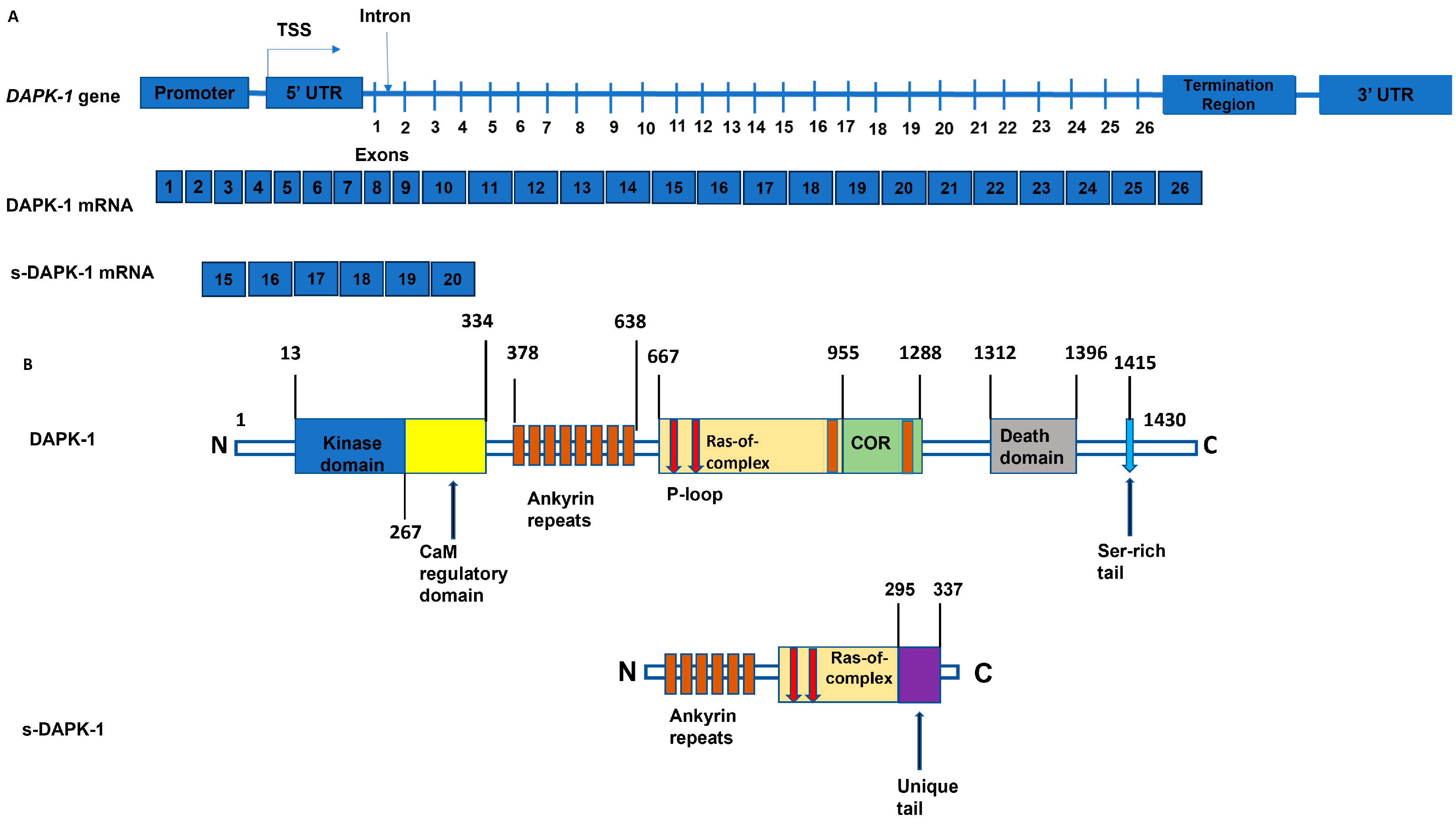
3.2. The 3D Structure of s-DAPK-1 Contains Ankyrin Repeats

Once the structure of a protein is comprehended, it is easy to understand its function and its interaction partners, thus gaining insights into various biological interactions and cellular functions. Therefore, the Phyre2 webserver was used to predict the s-DAPK-1 secondary structure (Supplementary Figure S3), followed by the prediction of its tertiary structure using I-TASSER. The I-TASSER webserver is ranked as the top method in the community-wide Critical Assessment of Protein Structure Prediction (CASP) experiments for accurate 3D structure prediction [16]. I-TASSER modeled five (5) s-DAPK-1 structures (Supplementary Figure S4), which differ in their C-scores (0.54, −2.17, −1.20, −2.03, −3.96). The C-score quantitatively measures the confidence of each model, and it is determined by calculating the significance of threading template alignments and the convergence parameters of structure assembly simulations. The C-score should fall within the range from −5 to 2, where a higher C-score indicates a model with greater confidence, while a lower C-score suggests the opposite. Therefore, model 1 (Figure 2) qualified to be the best-modeled 3D structure of s-DAPK-1 due to its highest confidence score of 0.54. Based on Figure 2, s-DAPK-1 does not seem to resemble characteristics of a globular protein, which are generally spherical in shape, resulting from the folding of a polypeptide chain into a compact structure. In contrast, s-DAPK-1 is made of two antiparallel α-helices and a β-loop, which are arranged in a manner that creates an L-shaped domain that resembles a cupped hand. Such structural characteristics are typical of ankyrin repeat-containing proteins, which are considered fibrous proteins.
Validation of the predicted model was carried out using the Ramachandran plot, which identifies amino acid distribution in different secondary structure regions. Based on Supplementary Figure S5, 72.3% of amino acids are found in favorable regions, while 21.8% are in additional allowed regions, 3.1% are in generously allowed regions, and only 2.8% are in disallowed regions. Based on these results, most residues fall within the favored regions.

3.3. Physicochemical Characterization of s-DAPK-1

The three-dimensional structure and biological activity of proteins are influenced by the physicochemical characteristics of the amino acids they are composed of. Therefore, it was deemed necessary to assess the physicochemical properties of s-DAPK-1. The physicochemical parameters of s-DAPK-1 were computed using Expasy’s ProtParam tool. As depicted in Table 2 (physicochemical properties of s-DAPK-1), the calculated isoelectric point (8.96 > 7) for s-DAPK-1 demonstrated its basic nature, while the extinction coefficient at 280 nm varied from 27,680 to 26,930 M−1 cm−1, suggesting a higher concentration of tyrosine (Tyr) and tryptophan (Trp) amino acids. These findings suggest that UV spectral methods can be utilized to quantify the s-DAPK-1 protein because it has aromatic tryptophan and tyrosine residues, which exhibit strong UV light absorption at 280 nm. Additionally, the stability of s-DAPK-1 was also assessed using the instability index value, an estimation of the stability of a protein in a test tube. Stable proteins are predicted to have an instability index below 40; therefore, s-DAPK-1 is considered unstable because its instability index is predicted to be 40.67.
Furthermore, the aliphatic index of s-DAPK-1, representing the proportion of aliphatic side chains (alanine, valine, isoleucine, and leucine), was measured to be 87.74 for s-DAPK-1. An exceptionally high aliphatic index of 87.74 suggests that s-DAPK-1 could remain stable across various temperature ranges. This is because the aliphatic index reflects the relative volume occupied by aliphatic amino acid side chains, and hydrophobic interactions between these residues help to stabilize the protein structure, making it more resistant to unfolding or denaturation at higher temperatures, thus contributing to protein stability. The GRAVY value represents the ratio of hydropathy values of amino acids to the number of residues in the sequence. For s-DAPK-1, the predicted score was −0.216, suggesting its potential classification as a hydrophilic protein.
To detect the patches of hydrophobic amino acid residues in s-DAPK-1, a hydropathy plot was generated using the ProtScale tool. The plot provided insight into the degree of hydrophobicity or hydrophilicity of s-DAPK-1 amino acids. The graph in Figure 3A shows numerous prominent negative peaks (hydrophilic) and a few strong positive peaks (hydrophobic). Additionally, the thermodynamic properties associated with the folding transition of the s-DAPK-1 protein were assessed. The calculated values included a melting temperature (Tm) of 68.6 °C, a standard folding enthalpy (ΔHm) of −145 kcal/mol, and a standard folding heat capacity (ΔCp) of −3.93 kcal/(mol K). All these data suggest that s-DAPK-1 may exhibit thermodynamic stability. The stability profile of the s-DAPK-1 protein, represented by the change in Gibbs free energy (ΔG) in relation to temperature (Figure 3B), demonstrated its potential to remain stable even at higher temperatures, as evidenced by a negative ΔG value.

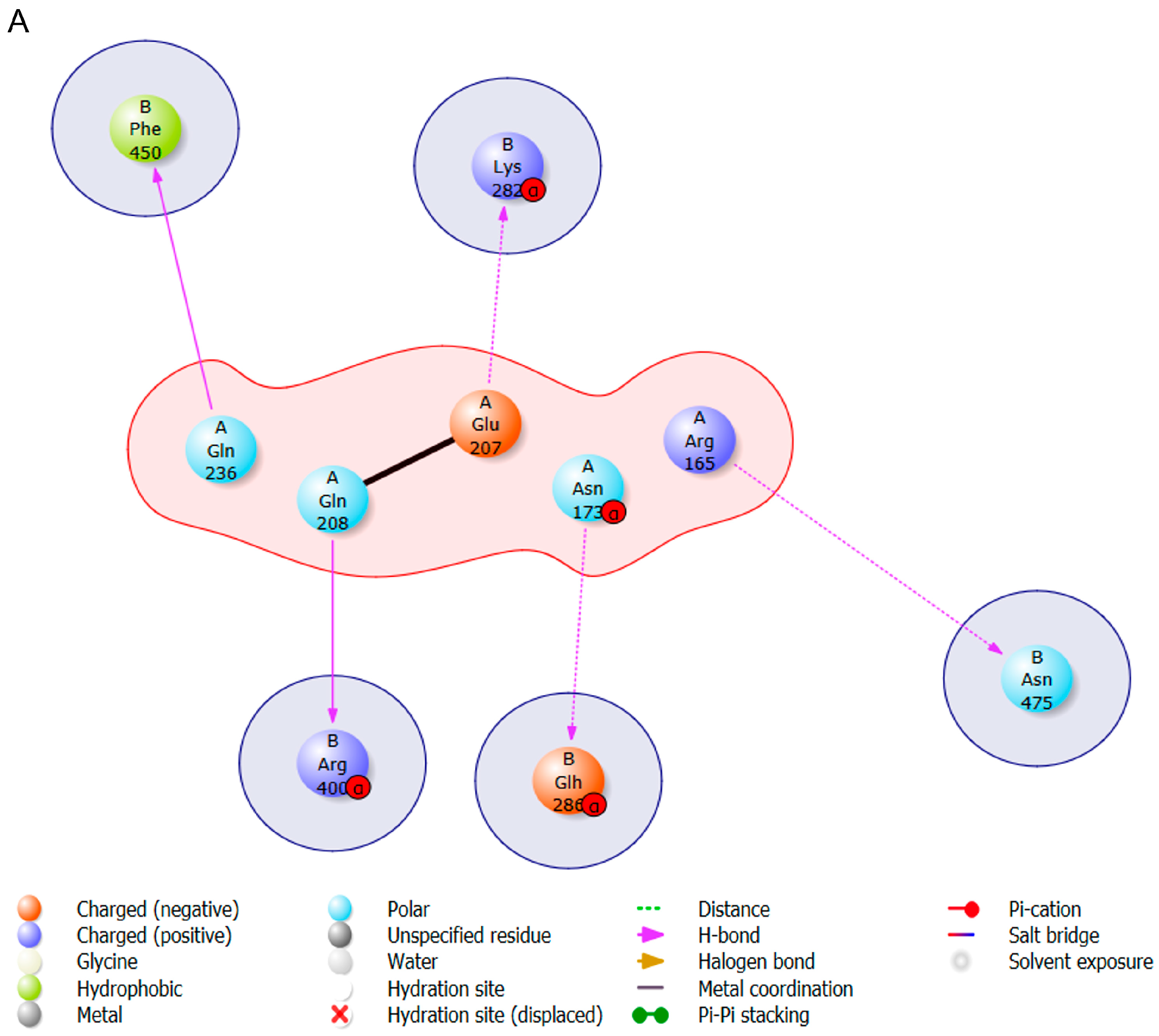
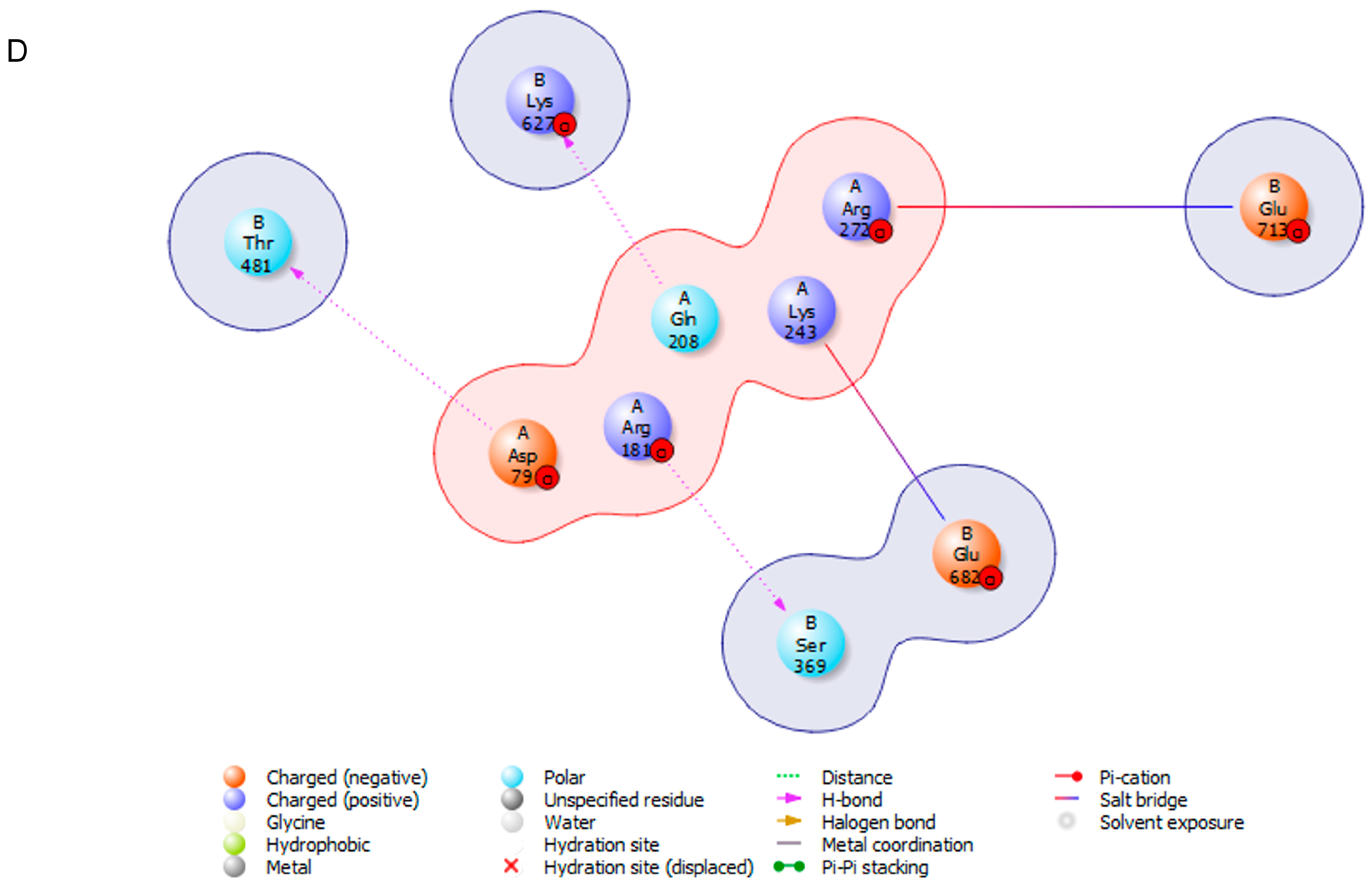
3.4. Protein s-DAPK-1 Interacts with Other Proteins

To further understand the role of s-DAPK-1, the proteins that interact with s-DAPK-1 were investigated using the ProteinPrompt tool, utilizing only the s-DAPK-1 protein sequence to query the server. The first 10 binding partners of s-DAPK-1 with high binding scores were analyzed (Table 3). A significant number of proteins identified in Table 3 are implicated in gene regulation and cancer progression. Notable examples of these proteins include histone H2B type 2-E, prion protein, nuclear protein, polyubiquitin-B, and epididymis secretory protein Li 50. It is possible that s-DAPK-1 interacts with proteins identified in Table 3 proteins using ankyrin repeats, because variations within the surface exposed residues in ankyrin repeat motifs/domains enable specific protein binding.
In order to verify whether the proteins predicted by ProteinPrompt exhibit any binding affinity to s-DAPK-1, blind protein–protein docking (Figure 4) was conducted. Ten binding poses were predicted for each protein docked on s-DAPK-1, and subsequently, the pose with the highest binding energy was chosen from these predictions. It is important to highlight that there is no universally accepted standard value for evaluating the docking score. The score can only be deemed as favorable or unfavorable based on another reference value obtained from the same protein. Some of the proteins identified by ProteinPrompt exhibited a binding affinity to s-DAPK-1 as shown by negative Gibbs free energy. This statement is made because we did not dock all the proteins, but rather focused on those with solved structures and smaller sizes that can function as ligands. The docked proteins were polyubiquitin-B, dynein light chain 1, microtubule-associated proteins 1A/1B light chain 3B and histone H2B type 2-E. The functions of these proteins are already highlighted in Table 3; all these proteins may have the potential to bind to s-DAPK-1. Upon examining the binding energies (Figure 4), it became apparent that microtubule-associated protein 1A/1B light chain 3B exhibited the highest binding affinity, with the binding free energy of −7625.15 kcal/mol. Following this, histone H2B type 2-E demonstrated the next highest affinity, with the binding energy of −7383.30 kcal/mol, succeeded by polyubiquitin-B with −6837.03 kcal/mol. Lastly, dynein light chain 1 is identified as the protein with the lowest binding affinity towards s-DAPK-1 because of its binding energy of −4944.96 kcal/mol.
The next step was to visualize the amino acids involved in protein–protein interactions as well as the bonds implicated. The presence of hydrogen bonds is crucial for the stability of protein–ligand complexes. As ligands interact and form more hydrogen bonds with the protein, the overall stability of the protein–ligand complex is enhanced. Figure 4 provides evidence of specific amino acids that play a role in the binding process through hydrogen bonds. In thes-DAPK-1 and polyubiquitin-B complex, 5 hydrogen bonds were identified between specific residues; namely, Gln208–Arg400, Glu207–Lys282, Asn173–Glu286, Arg165–Asn475, and Gln236–Phe450. The dynein light chain 1 and s-DAPK-1 complex exhibited the formation of one hydrogen bond between Arg271 and Arg3. Furthermore, the microtubule-associated proteins 1A/1B light chain 3B and s-DAPK-1 complex displayed 8 hydrogen bonds; the amino acids involved in these bonds are shown in Figure 4. Lastly, the histone H2B type 2-E and s-DAPK-1 complex formed three hydrogen bonds involving residues Asp79–Thr481, Arg181–Ser369, and Gln208–Lys627. To sum up, the 8 hydrogen bonds found in the microtubule-associated proteins 1A/1B light chain 3B and s-DAPK-1 complex establish it as the most stable complex among others.

3.5. Protein s-DAPK-1 Performs Various Cellular Functions

Currently, there is limited information on s-DAPK-1, both structurally and functionally. The protein–protein interactions in this study demonstrated that s-DAPK-1 may interact with numerous proteins (Table 3 and Figure 4). Therefore, it was fitting to further investigate probable s-DAPK-1 functions, and this was achieved utilizing the FFPred server. Upon examining the proteins that interact with s-DAPK-1 and their impact on its function, it was discovered that s-DAPK-1 may form interactions with H2B2E (Table 3), a crucial component of the nucleosome structure. Therefore, it is unsurprising that the predicted functions of s-DAPK-1 include nucleic acid binding, RNA binding, regulation of the RNA biosynthetic process, regulation of the metabolic process, and adenyl nucleotide binding, as presented in Table 4a. Based on the information that is shown in Table 4a,b, it is evident that s-DAPK-1 and DAPK-1 may have comparable functions such as the regulation of nitrogen compounds’ metabolic processes, catalytic activity, and adenyl nucleotide binding activity. Contrary to s-DAPK-1, DAPK1 plays a crucial role in regulating various activities linked to the central dogma. Among the functions of DAPK-1 presented in Table 4b, its role in the regulation of metabolic processes is particularly noteworthy because it is likely to be through this function that DAPK-1 facilitates different forms of cell death, such as apoptosis and autophagy.

4. Discussion

Several complete and draft genomes are now available, and these include the human genome; however, the biggest task and gap is assigning gene product functions, interactions, regulation, and structures [60,61,62]. To analyze the protein sequence, function, structure, motif/domain, regulation, and interaction network, several bioinformatics tools have been developed and made publicly available [29,63,64,65].
Data regarding the regulation of s-DAPK-1 in cancer cells is unavailable, particularly in relation to its posttranscriptional regulation. In contrast to s-DAPK-1, there is considerable evidence in the literature on microRNAs targeting DAPK-1; for example, DAPK-1 has been reported to be modulated by miR-26b-5p and miR-632 microRNAs in the contexts of intestinal ischemia and gastric cancer, respectively [65,66,67]. Interestingly, this study successfully predicted microRNAs targeting s-DAPK-1 (Table 1) using the TarBase tool; these results were obtained following a query performed on the server using the DAPK-1 gene. “DAPK-1” but not “s-DAPK-1” was used to query the microRNA databases because the identification and characterization of miRNA targets rely on computational prediction methods that often use the full gene sequence, rather than specific splice variants.
In addition, s-DAPK-1 starts in intron 13–14 of DAPK-1, thus classifying it as an intron-retained transcript. Generally, intron-retained transcripts have unique 3′ and 5′ UTRs that differ from those found in the main gene’s spliced transcript [68,69]. However, despite changes in the 3′ UTR and 5′ UTR regions of intron-retained transcripts, these transcripts can still feature miRNA binding sites that are also present in the main gene’s transcript [70,71,72]. Therefore, if the miRNA binding site located within the retained intron is also present in the 5′ or 3′ UTR of the main transcript, both variants could be targeted by the same miRNA [70,71,72]. This is the reason why it became feasible to use the TarBase tool to identify DAPK-1-related microRNAs that could also modulate s-DAPK-1. The TarBase tool has also been used by other researchers, deeming it a valuable tool in the literature for predicting microRNAs targeting different genes [73,74]. In line with the objective of this study, the TarBase tool predicted that hsa-miR-30a-5p and hsa-miR-26a-5p could target mitogen-activated protein kinase 10 (MAPK10), protein phosphatase, Mg2+/Mn2+-dependent 1J (PPM1J), and RB1 inducible coiled-coil 1 (RB1CC1) autophagy-related genes [73]. Additionally, this tool effectively predicted connective tissue growth factor (CTGF) and periostin (POSTN) as direct targets of miR-30a-5p in heart failure conditions [74].
MiRNAs primarily operate by binding to the 3′ untranslated region (UTR) on the messenger RNA post-transcription, thus suppressing the expression of their gene targets, thereby, controlling a wide range of biological and pathological processes, such as the initiation and progression of cancer [75,76]. The scientific literature has extensively documented miR-26a-5p and miR-26b-5p identified in Table 1 as potential regulators of s-DAPK-1 for their tumor-suppressing activities in hepatocellular carcinoma and gastric cancer, respectively [42,43,44]. Therefore, their potential binding to s-DAPK-1 mRNA will influence its translation and function.
Given that a protein’s function is related to its structure [77,78,79], upon discovering the potential of microRNAs to regulate s-DAPK-1, curiosity led us to explore the three-dimensional structure of this protein. Firstly, the prediction of s-DAPK-1’s secondary structures was conducted, and the results showed significant prevalence of helical configurations linked by coils; these helix-turn-helix motifs detected in s-DAPK-1’s secondary structure primarily function in DNA binding and sequence-specific recognition, often acting as transcription factors to modulate gene expression [80,81]. This implies that s-DAPK-1 is likely to be involved in some or all these biological processes as predicted in Table 4a.
The helical configurations dominating s-DAPK-1 initially suggested that s-DAPK-1 may be classified as a globular protein, because most globular proteins have been reported to have at least 30% alpha helices in their globular structure [82]. However, further analysis of the data showed that the detected alpha helices are succeeded by coils. These secondary structures can sequentially arrange themselves, giving rise to repeated sequence motifs [83], which might result in a long, elongated tertiary structure that differs significantly from the compact and spherical forms typically seen in globular proteins.
Indeed, the 3D structure of s-DAPK-1 modeled using I-TASSER proved that s-DAPK-1 does not resemble the characteristics of a globular protein because it is made of two antiparallel α-helices and a β-loop, which are packed together to form a helix-turn-helix bundle known as ankyrin repeats. I-TASSER was used to model the 3D structure of s-DAPK-1 because it has been proven to generate models that closely match experimental ones. A study conducted by Kemege et al. [84] serves as an example, where I-TASSER was employed to model the CT296 structure. Subsequently, the structure was experimentally resolved using X-ray crystallography [84]. Impressively, the model generated by I-TASSER exhibited a close match (RMSD: 2.72 Å) to the high-resolution (1.8 Å) crystal structure of CT296. As per the findings from the I-TASSER webserver, the modeled structure of s-DAPK-1 prominently featured ankyrin repeats, mirroring the ankyrin repeats identified in the DAPK-1 structure [10]. It is noteworthy that these ankyrin repeats in both isoforms consist of the same amino acids. Ankyrin repeats have been identified in a wide range of proteins, serving various functions, such as cell-to-cell communication, maintenance of the cytoskeleton structure, regulation of transcription and cell cycle, inflammatory response, development, and diverse transport processes [83,85,86]. Interestingly, some of these activities are predicted to be the functions of s-DAPK-1 (Table 4a).
Prediction of s-DAPK-1’s 3D structure enabled us to predict its physical, chemical, and thermodynamic parameters. The physicochemical properties revealed that s-DAPK-1 is an unstable protein. This is not surprising as Lin et al. [10] demonstrated that s-DAPK-1′s instability is a consequence of its unique tail that is not found in DAPK-1. This tail is responsible for regulating the protein’s localization and stability, and can be cleaved by proteases within cells. Similar to DAPK-1, s-DAPK-1 demonstrates a more precise localization within the cytoplasm. Notably, when the tail of s-DAPK-1 is deleted, it predominantly localizes in the vicinity of the nucleus, ultimately enhancing its stability and membrane blebbing activity [10,87]. Nevertheless, the cleavage of the tail region of s-DAPK-1 in vivo is significantly diminished when s-DAPK-1 is inserted into GFP vectors and expressed in cells. This observation indicates that protein tags possess the ability to alter the conformational structures of their fusion proteins [88,89].
When observing the predicted 3D structure of s-DAPK-1, it is evident that the s-DAPK-1 structure is dominated by ankyrin repeats that play a major role in mediating protein–protein interactions [83,85,90]. Therefore, it is not surprising to see s-DAPK-1 interacting with other proteins as presented in Table 3. These interactions potentially play a crucial role in enabling s-DAPK-1 to carry out diverse functions, as depicted in Table 4a, because for a protein to effectively perform its designated function, it typically needs to interact with various other molecules, and this interaction is directly influenced by its 3D structure [91]. This is feasible since most proteins, although capable of independent function, often rely on interactions with other proteins for optimal biological activity [92]. Among the proteins that may interact with s-DAPK-1 (Table 3), H2B2E stands out due to its association with nucleosomes. Nucleosomes play a crucial role in regulating the accessibility of the transcription machinery to DNA and serve as a fundamental component in the intricate regulation of gene expression [93]. In the realm of apoptosis, it has been widely proposed that histone modifications play a significant role in influencing the function and structure of chromatin during cell death [94]. Considering the possible affinity of s-DAPK-1 towards H2B2E, it is plausible to suggest that epigenetic alterations of H2B2E may also contribute to the initiation of apoptosis, which is one of the functions of DAPK-1. Consequently, if s-DAPK-1 is capable of inducing apoptosis through the epigenetic modulation of H2B2E, it can be inferred that both s-DAPK-1 and DAPK-1 share the common function of promoting apoptosis.
In summary, while experimental validation (“wet lab experiments”) remain essential for confirming bioinformatics-derived findings, such data retain considerable value in their own right. Specifically, we envision that the insights generated from these bioinformatics analyses can drive renewed interest in s-DAPK-1 and inform the development of more experimental designs and optimization strategies, particularly for the investigation of less-studied proteins, such as s-DAPK-1. This is further supported by numerous studies in which researchers employed exclusively in silico approaches to study protein structure and function, underscoring the growing credibility and utility of advanced computational methods in molecular biology [95,96,97]. For example, Arshad et al. [98] used the Phyre2 and I-TASSER homology modeling tools to generate 3D models of wild-type T-cell activation RhoGTPase-activating protein (TAGAP) and TAGAP mutated with high-risk single nucleotide polymorphisms (SNPs). The research conducted by Khan et al. [99] also relied entirely on in silico data to predict the structure and function of S. typhi hypothetical protein (HP) R_27 using only the protein sequence as a basis. Clearly, investigations to understand protein structure and function based solely on in silico data continue to gain momentum [95,96,97].

5. Conclusions

In an effort to gain a more comprehensive understanding of the biological complexity of s-DAPK-1, bioinformatics tools facilitated the prediction of s-DAPK-1’s physical, chemical, and thermodynamic parameters, 3D structure, functionality, and protein–protein interactions. The findings of this study lay the groundwork for future studies aimed at discovering the potential drug targets associated with s-DAPK-1, regulating its expression, and exploring its various functions. Moreover, given that s-DAPK-1 is less studied, these computational results are likely to stimulate interest in s-DAPK-1, thus fostering ongoing studies on s-DAPK-1 to solidify the in silico findings.

Supplementary Materials

The following supporting information can be downloaded at: https://www.mdpi.com/article/10.3390/cimb47060416/s1.

Author Contributions

L.M. and S.M. conducted the in silico analysis and compiled the article, while Z.M., the research team supervisor, developed the project and edited the article. All authors have read and agreed to the published version of the manuscript.

Funding

This research received no external funding.

Institutional Review Board Statement

Not applicable.

Informed Consent Statement

Not applicable.

Data Availability Statement

All data generated or analyzed throughout this study are contained in this article and the Supplementary Information Files.

Acknowledgments

The authors wish to acknowledge the Department of Biochemistry, Microbiology and Biotechnology (University of Limpopo) and the Department of Life and Consumer Sciences (University of South Africa) for support.

Conflicts of Interest

The authors declare no conflicts of interest.

References

  1. Gauthier, J.; Vincent, A.T.; Charette, S.J.; Derome, N. A brief history of bioinformatics. Brief. Bioinform. 2019, 20, 1981–1996. [Google Scholar] [CrossRef] [PubMed]
  2. Baxevanis, A.D.; Bateman, A. The Importance of Biological Databases in Biological Discovery. Curr. Protoc. Bioinform. 2015, 50, 1.1.1–1.1.8. [Google Scholar] [CrossRef] [PubMed]
  3. Chen, C.; Huang, H.; Wu, C.H. Protein Bioinformatics Databases and Resources. Methods Mol. Biol. 2017, 1558, 3–39. [Google Scholar] [CrossRef]
  4. Gromiha, M.M. (Ed.) Chapter 6—Protein Stability. In Protein Bioinformatics; Academic Press: Singapore, 2010; pp. 209–245. [Google Scholar] [CrossRef]
  5. Sun, Y.; Zhao, J.; Lu, Y.; Ngo, F.Y.; Shuai, B.; Zhang, Z.-J.; Feng, Y.; Rong, J. In Silico Prediction of Quercetin Analogs for Targeting Death-Associated Protein Kinase 1 (DAPK1) Against Alzheimer’s Disease. Curr. Neuropharmacol. 2024, 22, 2353–2367. [Google Scholar] [CrossRef]
  6. Firoz, A.; Talwar, P. Role of death-associated protein kinase 1 (DAPK1) in retinal degenerative diseases: An in-silico approach towards therapeutic intervention. J. Biomol. Struct. Dyn. 2023, 42, 5686–5698. [Google Scholar] [CrossRef]
  7. Singh, P.; Talwar, P. Exploring putative inhibitors of Death Associated Protein Kinase 1 (DAPK1) via targeting Gly- Glu -Leu (G E L) and Pro- Glu -Asn (P E N) substrate recognition motifs. J. Mol. Graph. Model. 2017, 77, 153–167. [Google Scholar] [CrossRef]
  8. Tu, G.; Fu, T.; Yang, F.; Yao, L.; Xue, W.; Zhu, F. Prediction of GluN2B-CT1290–1310/DAPK1 Interaction by Protein−Peptide Docking and Molecular Dynamics Simulation. Molecules 2018, 23, 3018. [Google Scholar] [CrossRef] [PubMed]
  9. Makgoo, L.; Mosebi, S.; Mbita, Z. The Role of Death-Associated Protein Kinase-1 in Cell Homeostasis-Related Processes. Genes 2023, 14, 1274. [Google Scholar] [CrossRef]
  10. Lin, Y.; Stevens, C.; Hrstka, R.; Harrison, B.; Fourtouna, A.; Pathuri, S.; Vojtesek, B.; Hupp, T. An alternative transcript from the death-associated protein kinase 1 locus encoding a small protein selectively mediates membrane blebbing. FEBS J. 2008, 275, 2574–2584. [Google Scholar] [CrossRef]
  11. Zhang, T.; Kim, B.M.; Lee, T.H. Death-associated protein kinase 1 as a therapeutic target for Alzheimer’s disease. Transl. Neurodegener. 2024, 13, 4. [Google Scholar] [CrossRef]
  12. Tu, W.; Xu, X.; Peng, L.; Zhong, X.; Zhang, W.; Soundarapandian, M.M.; Belal, C.; Wang, M.; Jia, N.; Zhang, W.; et al. DAPK1 Interaction with NMDA Receptor NR2B Subunits Mediates Brain Damage in Stroke. Cell 2010, 140, 222–234. [Google Scholar] [CrossRef] [PubMed]
  13. Lee, T.; Chen, C.H.; Suizu, F.; Huang, P.; Schiene-Fischer, C.; Daum, S.; Zhang, Y.J.; Goate, A.; Chen, R.-H.; Zhou, X.Z.; et al. Death-Associated Protein Kinase 1 Phosphorylates Pin1 and Inhibits Its Prolyl Isomerase Activity and Cellular Function. Mol. Cell 2011, 42, 147–159. [Google Scholar] [CrossRef] [PubMed]
  14. Huang, Y.; Wang, C.; Li, K.; Ye, Y.; Shen, A.; Guo, L.; Chen, P.; Meng, C.; Wang, Q.; Yang, X.; et al. Death-associated protein kinase 1 suppresses hepatocellular carcinoma cell migration and invasion by upregulation of DEAD-box helicase 20. Cancer Sci. 2020, 111, 2803–2813. [Google Scholar] [CrossRef] [PubMed]
  15. Luo, Q.; Tian, Y.; Qu, G.; Huang, K.; Hu, P.; Xue, Y.; Hu, B.; Luo, S. The targeting of DAPK1 with miR-190a-3p promotes autophagy in trophoblast cells. Mol. Reprod. Dev. 2024, 91, e23724. [Google Scholar] [CrossRef]
  16. Wang, Y.; Shi, M.; Hong, Z.; Kang, J.; Pan, H.; Yan, C. MiR-130a-3p Has Protective Effects in Alzheimer’s Disease via Targeting DAPK1. Am. J. Alzheimer’s Dis. Other Dement. 2021, 36, 15333175211020572. [Google Scholar] [CrossRef]
  17. Zhai, C.; Tang, G.; Qian, G.; Hu, H.; Wang, S.; Yin, D.; Zhang, S. MicroRNA-98 attenuates cardiac ischemia-reperfusion injury through inhibiting DAPK1 expression. IUBMB Life 2018, 71, 166–176. [Google Scholar] [CrossRef]
  18. Rani, V.; Sengar, R.S. Biogenesis and mechanisms of microRNA-mediated gene regulation. Biotechnol. Bioeng. 2022, 119, 685–692. [Google Scholar] [CrossRef]
  19. Wang, L.; Shui, X.; Mei, Y.; Xia, Y.; Lan, G.; Hu, L.; Zhang, M.; Gan, C.-L.; Li, R.; Tian, Y.; et al. miR-143-3p Inhibits Aberrant Tau Phosphorylation and Amyloidogenic Processing of APP by Directly Targeting DAPK1 in Alzheimer’s Disease. Int. J. Mol. Sci. 2022, 23, 7992. [Google Scholar] [CrossRef]
  20. Tian, X.; Xu, L.; Wang, P. MiR-191 inhibits TNF-α induced apoptosis of ovarian endometriosis and endometrioid carcinoma cells by targeting DAPK1. Int. J. Clin. Exp. Pathol. 2015, 8, 4933–4942. [Google Scholar]
  21. Tian, Y.; Yan, M.; Zheng, J.; Li, R.; Lin, J.; Xu, A.; Liang, Y.; Zheng, R.; Yuan, Y. miR-483-5p decreases the radiosensitivity of nasopharyngeal carcinoma cells by targeting DAPK1. Lab. Investig. 2019, 99, 602–611. [Google Scholar] [CrossRef]
  22. Li, D.; Xu, D.; Xu, Y.; Chen, L.; Li, C.; Dai, X.; Zhang, L.; Zheng, L. MicroRNA-141-3p targets DAPK1 and inhibits apoptosis in rat ovarian granulosa cells. Cell Biochem. Funct. 2017, 35, 197–201. [Google Scholar] [CrossRef] [PubMed]
  23. Jiang, W.; Chen, L. Alternative splicing: Human disease and quantitative analysis from high-throughput sequencing. Comput. Struct. Biotechnol. J. 2020, 19, 183–195. [Google Scholar] [CrossRef]
  24. Pan, Q.; Shai, O.; Lee, L.J.; Frey, B.J.; Blencowe, B.J. Deep surveying of alternative splicing complexity in the human transcriptome by high-throughput sequencing. Nat. Genet. 2008, 40, 1413–1415. [Google Scholar] [CrossRef] [PubMed]
  25. Wang, E.T.; Sandberg, R.; Luo, S.; Khrebtukova, I.; Zhang, L.; Mayr, C.; Kingsmore, S.F.; Schroth, G.P.; Burge, C.B. Alternative isoform regulation in human tissue transcriptomes. Nature 2008, 456, 470–476. [Google Scholar] [CrossRef]
  26. Liu, Q.; Fang, L.; Wu, C. Alternative Splicing and Isoforms: From Mechanisms to Diseases. Genes 2022, 13, 401. [Google Scholar] [CrossRef]
  27. Cieply, B.; Carstens, R.P. Functional roles of alternative splicing factors in human disease. Wiley Interdiscip. Rev. RNA 2015, 6, 311–326. [Google Scholar] [CrossRef]
  28. Wang, Q.; Weng, S.; Zhong, W.; Lin, Y.; Yu, Y.; Huang, Y.; Ge, L.; Zhang, X.; Xue, F.; Assaraf, Y.G.; et al. Modulation of DAPK1 expression by its alternative splice variant DAPK1-215 in cancer. J. Transl. Med. 2025, 23, 85. [Google Scholar] [CrossRef]
  29. Skoufos, G.; Kakoulidis, P.; Tastsoglou, S.; Zacharopoulou, E.; Kotsira, V.; Miliotis, M.; Mavromati, G.; Grigoriadis, D.; Zioga, M.; Velli, A.; et al. TarBase-v9.0 extends experimentally supported miRNA–Gene interactions to cell-types and virally encoded miRNAs. Nucleic Acids Res. 2023, 52, D304–D310. [Google Scholar] [CrossRef] [PubMed]
  30. Yang, M.; Derbyshire, M.K.; Yamashita, R.A.; Marchler-Bauer, A. NCBI’s Conserved Domain Database and Tools for Protein Domain Analysis. Curr. Protoc. Bioinform. 2020, 69, e90. [Google Scholar] [CrossRef]
  31. Kelley, L.A.; Mezulis, S.; Yates, C.M.; Wass, M.N.; Sternberg, M.J. The Phyre2 web portal for protein modeling, prediction and analysis. Nat. Protoc. 2015, 10, 845–858. [Google Scholar] [CrossRef]
  32. Zhou, X.; Zheng, W.; Li, Y.; Pearce, R.; Zhang, C.; Bell, E.W.; Zhang, G.; Zhang, Y. I-TASSER-MTD: A deep-learning-based platform for multi-domain protein structure and function prediction. Nat. Protoc. 2022, 17, 2326–2353. [Google Scholar] [CrossRef] [PubMed]
  33. Yang, J.; Zhang, Y. Protein Structure and Function Prediction Using I-TASSER. Curr. Protoc. Bioinform. 2015, 52, 5.8.1–5.8.15. [Google Scholar] [CrossRef] [PubMed]
  34. Zhang, Y. I-TASSER server for protein 3D structure prediction. BMC Bioinform. 2008, 9, 40. [Google Scholar] [CrossRef]
  35. Laskowski, R.A.; MacArthur, M.W.; Moss, D.S.; Thornton, J.M. PROCHECK: A program to check the stereochemical quality of protein structures. J. Appl. Crystallogr. 1993, 26, 283–291. [Google Scholar] [CrossRef]
  36. Gasteiger, E.; Hoogland, C.; Gattiker, A.; Duvaud, S.E.; Wilkins, M.R.; Appel, R.D.; Bairoch, A. Protein identification and analysis tools on the ExPASy server. In The Proteomics Protocols Handbook; Humana Press: Totowa, NJ, USA, 2005. [Google Scholar] [CrossRef]
  37. Pucci, F.; Kwasigroch, J.M.; Rooman, M. SCooP: An accurate and fast predictor of protein stability curves as a function of temperature. Bioinformatics 2017, 33, 3415–3422. [Google Scholar] [CrossRef] [PubMed]
  38. Canzler, S.; Fischer, M.; Ulbricht, D.; Ristic, N.; Hildebrand, P.W.; Staritzbichler, R. ProteinPrompt: A webserver for predicting protein-protein interactions. Bioinform. Adv. 2022, 2, vbac059. [Google Scholar] [CrossRef]
  39. Weng, G.; Wang, E.; Wang, Z.; Liu, H.; Zhu, F.; Li, D.; Hou, T. HawkDock: A web server to predict and analyze the protein-protein complex based on computational docking and MM/GBSA. Nucleic Acids Res. 2019, 47, W322–W330. [Google Scholar] [CrossRef]
  40. Cozzetto, D.; Minneci, F.; Currant, H.; Jones, D.T. FFPred 3: Feature-based function prediction for all Gene Ontology domains. Sci. Rep. 2016, 6, 31865. [Google Scholar] [CrossRef]
  41. Wu, K.; Mu, X.Y.; Jiang, J.; Tan, M.; Wang, R.; Zhou, W.; Wang, X.; He, Y.; Li, M.; Liu, Z. miRNA 26a 5p and miR 26b 5p inhibit the proliferation of bladder cancer cells by regulating PDCD10. Oncol. Rep. 2018, 40, 3523–3532. [Google Scholar] [CrossRef]
  42. Liang, L.; Zeng, J.-H.; Wang, J.-Y.; He, R.-Q.; Ma, J.; Chen, G.; Cai, X.-Y.; Hu, X.-H. Down-regulation of miR-26a-5p in hepatocellular carcinoma: A qRT-PCR and bioinformatics study. Pathol. Res. Pract. 2017, 213, 1494–1509. [Google Scholar] [CrossRef]
  43. Gasparini, P.; Cascione, L.; Landi, L.; Carasi, S.; Lovat, F.; Tibaldi, C.; Alì, G.; D’incecco, A.; Minuti, G.; Chella, A.; et al. microRNA classifiers are powerful diagnostic/prognostic tools in ALK-, EGFR-, and KRAS-driven lung cancers. Proc. Natl. Acad. Sci. USA 2015, 112, 14924–14929. [Google Scholar] [CrossRef] [PubMed]
  44. Xu, T.; Xie, M.; Jing, X.; Jiang, H.; Wu, X.; Wang, X.; Shu, Y. Loss of miR-26b-5p promotes gastric cancer progression via miR-26b-5p-PDE4B/CDK8-STAT3 feedback loop. J. Transl. Med. 2023, 21, 77. [Google Scholar] [CrossRef] [PubMed]
  45. Heise, R.; Skazik, C.; Marquardt, Y.; Czaja, K.; Sebastian, K.; Kurschat, P.; Gan, L.; Denecke, B.; Ekanayake-Bohlig, S.; Wilhelm, K.-P.; et al. Dexpanthenol modulates gene expression in skin wound healing in vivo. Ski. Pharmacol. Physiol. 2012, 25, 241–248. [Google Scholar] [CrossRef]
  46. Shimomura, Y.; Ito, M. Human hair keratin-associated proteins. J. Investig. Dermatol. Symp. Proc. 2005, 10, 230–233. [Google Scholar] [CrossRef] [PubMed]
  47. Moiana, M.; Aranda, F.; de Larrañaga, G. A focus on the roles of histones in health and diseases. Clin. Biochem. 2021, 94, 12–19. [Google Scholar] [CrossRef] [PubMed]
  48. Pusarla, R.H.; Bhargava, P. Histones in functional diversification. Core histone variants. FEBS J. 2005, 272, 5149–5168. [Google Scholar] [CrossRef]
  49. Ryskalin, L.; Biagioni, F.; Busceti, C.L.; Giambelluca, M.A.; Morelli, L.; Frati, A.; Fornai, F. The Role of Cellular Prion Protein in Promoting Stemness and Differentiation in Cancer. Cancers 2021, 13, 170. [Google Scholar] [CrossRef]
  50. Martin, T.A.; Li, A.X.; Sanders, A.J.; Ye, L.; Frewer, K.; Hargest, R.; Jiang, W.G. NUPR1 and its potential role in cancer and pathological conditions (Review). Int. J. Oncol. 2021, 58, 21. [Google Scholar] [CrossRef]
  51. Vadlamudi, R.K.; Bagheri-Yarmand, R.; Yang, Z.; Balasenthil, S.; Nguyen, D.; Sahin, A.A.; Hollander, P.D.; Kumar, R. Dynein light chain 1, a p21-activated kinase 1-interacting substrate, promotes cancerous phenotypes. Cancer Cell. 2004, 5, 575–585, Erratum in Cancer Cell. 2004, 6, 101; Erratum in Cancer Cell. 2013, 23, 421–422. [Google Scholar] [CrossRef]
  52. Puthalakath, H.; Huang, D.C.; O’Reilly, L.A.; King, S.M.; Strasser, A. The proapoptotic activity of the Bcl-2 family member Bim is regulated by interaction with the dynein motor complex. Mol. Cell. 1999, 3, 287–296. [Google Scholar] [CrossRef]
  53. Kang, H.M.; Noh, K.H.; Chang, T.K.; Park, D.; Cho, H.-S.; Lim, J.H.; Jung, C.-R. Ubiquitination of MAP1LC3B by pVHL is associated with autophagy and cell death in renal cell carcinoma. Cell Death Dis. 2019, 10, 279. [Google Scholar] [CrossRef] [PubMed]
  54. Chen, Z.H.; Lam, H.C.; Jin, Y.; Kim, H.-P.; Cao, J.; Lee, S.-J.; Ifedigbo, E.; Parameswaran, H.; Ryter, S.W.; Choi, A.M.K. Autophagy protein microtubule-associated protein 1 light chain-3B (LC3B) activates extrinsic apoptosis during cigarette smoke-induced emphysema. Proc. Natl. Acad. Sci. USA 2010, 107, 18880–18885. [Google Scholar] [CrossRef] [PubMed]
  55. Yu, Y.; Zheng, Q.; Erramilli, S.K.; Pan, M.; Park, S.; Xie, Y.; Li, J.; Fei, J.; Kossiakoff, A.A.; Liu, L.; et al. K29-linked ubiquitin signaling regulates proteotoxic stress response and cell cycle. Nat. Chem. Biol. 2021, 17, 896–905. [Google Scholar] [CrossRef] [PubMed]
  56. Huang, F.; Kirkpatrick, D.; Jiang, X.; Gygi, S.; Sorkin, A. Differential regulation of EGF receptor internalization and degradation by multiubiquitination within the kinase domain. Mol. Cell 2006, 21, 737–748. [Google Scholar] [CrossRef]
  57. Kedves, A.T.; Gleim, S.; Liang, X.; Bonal, D.M.; Sigoillot, F.; Harbinski, F.; Sanghavi, S.; Benander, C.; George, E.; Gokhale, P.C.; et al. Recurrent ubiquitin B silencing in gynecological cancers establishes dependence on ubiquitin C. J. Clin. Investig. 2017, 127, 4554–4568. [Google Scholar] [CrossRef]
  58. Compton, S.L.; Behrend, E.N. PRAF1: A Golgi complex transmembrane protein that interacts with viruses. Biochem. Cell Biol. 2006, 84, 940–948. [Google Scholar] [CrossRef]
  59. Gougeon, P.Y.; Prosser, D.C.; Da-Silva, L.F.; Ngsee, J.K. Disruption of Golgi morphology and trafficking in cells expressing mutant prenylated rab acceptor-1. J. Biol. Chem. 2002, 277, 36408–36414. [Google Scholar] [CrossRef]
  60. Zheng, W.; Wuyun, Q.; Li, Y.; Zhang, C.; Freddolino, P.L.; Zhang, Y. Improving deep learning protein monomer and complex structure prediction using DeepMSA2 with huge metagenomics data. Nat. Methods 2024, 21, 279–289. [Google Scholar] [CrossRef]
  61. Kustatscher, G.; Collins, T.; Gingras, A.-C.; Guo, T.; Hermjakob, H.; Ideker, T.; Lilley, K.S.; Lundberg, E.; Marcotte, E.M.; Ralser, M.; et al. Understudied proteins: Opportunities and challenges for functional proteomics. Nat. Methods 2022, 19, 774–779. [Google Scholar] [CrossRef]
  62. Forslund, K.; Sonnhammer, E.L. Predicting protein function from domain content. Bioinformatics 2008, 24, 1681–1687, Erratum in Bioinformatics 2009, 25, 1214. [Google Scholar] [CrossRef]
  63. Yoodee, S.; Thongboonkerd, V. Bioinformatics and computational analyses of kidney stone modulatory proteins lead to solid experimental evidence and therapeutic potential. Biomed. Pharmacother. 2023, 159, 114217. [Google Scholar] [CrossRef] [PubMed]
  64. Rahmatbakhsh, M.; Gagarinova, A.; Babu, M. Bioinformatic Analysis of Temporal and Spatial Proteome Alternations During Infections. Front. Genet. 2021, 12, 667936. [Google Scholar] [CrossRef]
  65. Calderón-González, K.G.; Hernández-Monge, J.; Herrera-Aguirre, M.E.; Luna-Arias, J.P. Bioinformatics Tools for Proteomics Data Interpretation. Adv. Exp. Med. Biol. 2016, 919, 281–341. [Google Scholar] [CrossRef] [PubMed]
  66. Zhou, B.; Zhang, W.; Yan, Z.; Zhao, B.; Zhao, J.; Feng, W.; Chen, X.; Li, C.; Liu, K.-X. MicroRNA-26b-5p Targets DAPK1 to Reduce Intestinal Ischemia/Reperfusion Injury via Inhibition of Intestinal Mucosal Cell Apoptosis. Dig. Dis. Sci. 2021, 67, 1794–1805. [Google Scholar] [CrossRef] [PubMed]
  67. Fu, M.; Gu, J.; Yu, D.; Wang, M.; Zhang, J.; Ji, R.; Jiang, P.; Zhang, X. Circ1811 suppresses gastric cancer progression by regulating the miR-632/DAPK1 axis. Gene 2024, 910, 148331. [Google Scholar] [CrossRef]
  68. Ner-Gaon, H.; Halachmi, R.; Savaldi-Goldstein, S.; Rubin, E.; Ophir, R.; Fluhr, R. Intron retention is a major phenomenon in alternative splicing inArabidopsis. Plant J. 2004, 39, 877–885. [Google Scholar] [CrossRef]
  69. Jacob, A.G.; Smith, C.W.J. Intron retention as a component of regulated gene expression programs. Hum. Genet. 2017, 136, 1043–1057. [Google Scholar] [CrossRef]
  70. Tan, S.; Guo, J.; Huang, Q.; Chen, X.; Li-Ling, J.; Li, Q.; Ma, F. Retained introns increase putative microRNA targets within 3′ UTRs of human mRNA. FEBS Lett. 2007, 581, 1081–1086. [Google Scholar] [CrossRef]
  71. Schmitz, U.; Pinello, N.; Jia, F.; Alasmari, S.; Ritchie, W.; Keightley, M.-C.; Shini, S.; Lieschke, G.J.; Wong, J.J.; Rasko, J.E.J. Intron retention enhances gene regulatory complexity in vertebrates. Genome Biol. 2017, 18, 216. [Google Scholar] [CrossRef]
  72. Navarro, E.; Mallén, A.; Hueso, M. Dynamic Variations of 3′UTR Length Reprogram the mRNA Regulatory Landscape. Biomedicines 2021, 9, 1560. [Google Scholar] [CrossRef]
  73. Chen, P.-A. The LncRNA TPT1-AS1 Promotes the Survival of Neuroendocrine Prostate Cancer Cells by Facilitating Autophagy. Am. J. Cancer Res. 2024, 14, 2103–2123. [Google Scholar] [CrossRef] [PubMed]
  74. Bibi, A.; Madè, A.; Greco, S.; Garcia-Manteiga, J.M.; Tascini, A.S.; Tastsoglou, S.; Zaccagnini, G.; Leszek, P.; Gaetano, C.; Martelli, F. Circular PVT1 Promotes Cardiac Fibroblast Activation Interacting with MiR-30a-5p and MiR-125b-5p. Cell Death Dis. 2025, 16, 325. [Google Scholar] [CrossRef] [PubMed]
  75. Ali Syeda, Z.; Langden, S.S.S.; Munkhzul, C.; Lee, M.; Song, S.J. Regulatory Mechanism of MicroRNA Expression in Cancer. Int. J. Mol. Sci. 2020, 21, 1723. [Google Scholar] [CrossRef] [PubMed]
  76. Si, W.; Shen, J.; Zheng, H.; Fan, W. The role and mechanisms of action of microRNAs in cancer drug resistance. Clin. Epigenetics 2019, 11, 25. [Google Scholar] [CrossRef]
  77. Gurzau, A.D.; Blewitt, M.E.; Czabotar, P.E.; Murphy, J.M.; Birkinshaw, R.W. Relating SMCHD1 structure to its function in epigenetic silencing. Biochem. Soc. Trans. 2020, 48, 1751–1763. [Google Scholar] [CrossRef]
  78. Berezovsky, I.N.; Guarnera, E.; Zheng, Z. Basic units of protein structure, folding, and function. Prog. Biophys. Mol. Biol. 2017, 128, 85–99. [Google Scholar] [CrossRef]
  79. Alberts, B.; Johnson, A.; Lewis, J.; Raff, M.; Roberts, K.; Walter, P. Analyzing protein structure and function. In Molecular Biology of the Cell, 4th ed.; Garland Science: New York, NY, USA, 2002. [Google Scholar]
  80. Aravind, L.; Anantharaman, V.; Balaji, S.; Babu, M.; Iyer, L. The many faces of the helix-turn-helix domain: Transcription regulation and beyond. FEMS Microbiol. Rev. 2005, 29, 231–262. [Google Scholar] [CrossRef]
  81. Hao, Y.; Zong, X.; Ren, P.; Qian, Y.; Fu, A. Basic Helix-Loop-Helix (bHLH) Transcription Factors Regulate a Wide Range of Functions in Arabidopsis. Int. J. Mol. Sci. 2021, 22, 7152. [Google Scholar] [CrossRef]
  82. Pace, C.N.; Scholtz, J.M. A helix propensity scale based on experimental studies of peptides and proteins. Biophys. J. 1998, 75, 422–427. [Google Scholar] [CrossRef]
  83. Kumar, A.; Balbach, J. Folding and Stability of Ankyrin Repeats Control Biological Protein Function. Biomolecules 2021, 11, 840. [Google Scholar] [CrossRef]
  84. Kemege, K.E.; Hickey, J.M.; Lovell, S.; Battaile, K.P.; Zhang, Y.; Hefty, P.S. Ab initio structural modeling of and experimental validation for Chlamydia trachomatis protein CT296 reveal structural similarity to Fe(II) 2-oxoglutarate-dependent enzymes. J. Bacteriol. 2011, 193, 6517–6528. [Google Scholar] [CrossRef] [PubMed]
  85. Mosavi, L.K.; Cammett, T.J.; Desrosiers, D.C.; Peng, Z.Y. The ankyrin repeat as molecular architecture for protein recognition. Protein Sci. 2004, 13, 1435–1448. [Google Scholar] [CrossRef]
  86. Li, J.; Mahajan, A.; Tsai, M.D. Ankyrin repeat: A unique motif mediating protein-protein interactions. Biochemistry 2006, 45, 15168–15178. [Google Scholar] [CrossRef]
  87. Lin, Y.; Stevens, C.; Harrison, B.; Pathuri, S.; Amin, E.; Hupp, T.R. The alternative splice variant of DAPK-1, s-DAPK-1, induces proteasome-independent DAPK-1 destabilization. Mol. Cell Biochem. 2009, 328, 101–107. [Google Scholar] [CrossRef]
  88. Klose, J.; Wendt, N.; Kubald, S.; Krause, E.; Fechner, K.; Beyermann, M.; Bienert, M.; Rudolph, R.; Rothemund, S. Hexa-histidin tag position influences disulfide structure but not binding behavior of in vitro folded N-terminal domain of rat corticotropin-releasing factor receptor type 2a. Protein Sci. 2004, 13, 2470–2475. [Google Scholar] [CrossRef] [PubMed]
  89. Smyth, D.R.; Mrozkiewicz, M.K.; McGrath, W.J.; Listwan, P.; Kobe, B. Crystal structures of fusion proteins with large-affinity tags. Protein Sci. 2003, 12, 1313–1322. [Google Scholar] [CrossRef] [PubMed]
  90. Guharoy, M.; Chakrabarti, P. Secondary structure based analysis and classification of biological interfaces: Identification of binding motifs in protein-protein interactions. Bioinformatics 2007, 23, 1909–1918. [Google Scholar] [CrossRef]
  91. Morris, R.; Black, K.A.; Stollar, E.J. Uncovering protein function: From classification to complexes. Essays Biochem. 2022, 66, 255–285. [Google Scholar] [CrossRef]
  92. Stevers, L.M.; Sijbesma, E.; Botta, M.; MacKintosh, C.; Obsil, T.; Landrieu, I.; Cau, Y.; Wilson, A.J.; Karawajczyk, A.; Eickhoff, J.; et al. Modulators of 14-3-3 Protein-Protein Interactions. J. Med. Chem. 2018, 61, 3755–3778. [Google Scholar] [CrossRef]
  93. Rudnizky, S.; Malik, O.; Bavly, A.; Pnueli, L.; Melamed, P.; Kaplan, A. Nucleosome mobility and the regulation of gene expression: Insights from single-molecule studies. Protein Sci. 2017, 26, 1266–1277. [Google Scholar] [CrossRef]
  94. Damiescu, R.; Efferth, T.; Dawood, M. Dysregulation of different modes of programmed cell death by epigenetic modifications and their role in cancer. Cancer Lett. 2024, 584, 216623. [Google Scholar] [CrossRef] [PubMed]
  95. Paramkusham, S.; Mamidala, E.; Madhukar, J. Structural and Functional Insights into Riboflavin-Binding Proteins in Whooping crane (Grus americana) and Other Avian Species: An In Silico Approach. J. Neonatal Surg. 2025, 14, 563–569. [Google Scholar]
  96. Jeba, S.H.; Hasan, M.A.; Faisal, A.; Khatun, M.U.S.; Chowdhury, A.H.; Islam, N.; Miah, M. In silico approach of exploring the structural and functional characteristics of a hypothetical protein from monkeypox virus: A potential insight for antiviral therapeutics. Microbes Infect. Dis./Microbes Infect. Dis. 2024, 29, 13596535241255199. [Google Scholar] [CrossRef]
  97. Saikia, A.; Palherkar, D.A.; Hiremath, L. In silico Analysis and Structural Prediction of a Hypothetical Protein from Leishmania Major. Biomed. Biotechnol. Res. J. (BBRJ) 2021, 5, 320–326. [Google Scholar] [CrossRef]
  98. Arshad, M.; Bhatti, A.; John, P. Identification and in Silico Analysis of Functional SNPs of Human TAGAP Protein: A Comprehensive Study. PLoS ONE 2018, 13, e0188143. [Google Scholar] [CrossRef]
  99. Khan, A.; Ahmed, H.; Jahan, N.; Ali, S.R.; Amin, A.; Morshed, M.N. An in silico Approach for Structural and Functional Annotation of Salmonella enterica serovar typhimurium Hypothetical Protein R_27. Dir. Open Access J. 2016, 20, 31–42. Available online: https://doaj.org/article/2368937bffb84fb0bc5c2843e7d2b2aa (accessed on 16 April 2025).
Figure 1. Illustration of the DAPK-1 gene. (A) shows transcribed mRNAs consisting of exons numbered 1 to 26 for the wildtype DAPK-1 transcript and 15 to 20 exons for the s-DAPK-1 transcript. (B) shows various domains and motifs found in wildtype DAPK-1 (amino acids 1 to 1430) and the s-DAPK-1 isoform, which has 337 amino acids, yielding ankyrin repeats, Ras-of complex domain and a unique C-terminal tail, lacking the Serine-rich tail, which is indicated by a blue arrow.
Cimb 47 00416 g001
Figure 2. Modeled 3D structure of s-DAPK-1 predicted using the I-TASSER tool. The structure of the model exhibits few beta strands and antiparallel α-helices, which are separated by loops and arranged in a nearly parallel manner. On the predicted protein structure, the red secondary structures are at the c-terminus of s-DAPK-1 while the blue structures are at N-terminus. The structure is represented in a ribbon model.
Cimb 47 00416 g002
Figure 3. (A) The hydropathy plot of s-DAPK-1 displays peaks with a positive score representing hydrophobic regions within the protein sequence, while peaks with a negative score signify hydrophilic regions; (B) s-DAPK-1 protein stability curve defined by ΔG as a function of temperature. The solid line on the protein stability curve indicates the transition of s-DAPK-1 from stable to unfolded state.
Cimb 47 00416 g003
Figure 4. Binding energies and visualization of amino acids involved in protein–protein interactions. The s-DAPK-1 and polyubiquitin-B pose (A) has a binding energy of −6837.03 kcal/mol. The s-DAPK-1 and microtubule-associated proteins 1A/1B light chain 3B pose (B) has a binding energy of −7625.15 kcal/mol. The s-DAPK-1 and dynein light chain 1 pose (C) with a binding energy of −4944.96 kcal/mol. The s-DAPK-1 and histone H2B type 2-E pose (D) with a binding energy of −7383.30 kcal/mol.
Cimb 47 00416 g004aCimb 47 00416 g004bCimb 47 00416 g004c
Table 1. Predicted microRNAs targeting s-DAPK-1 mRNA.
Predicted microRNAFunctionBinding SiteReferences
hsa-miR-26a-5pTumor suppressor in hepatocellular carcinoma and lung cancer5′ UTR[41,42,43]
hsa-miR-26b-5pTumor suppressor in gastric cancer5′ UTR[44]
Table 2. Physicochemical properties of s-DAPK-1.
ParametersValues
Number of amino acids337
Molecular weight (Da)36,739.07
Theoretical pI8.96
Extinction coefficients27,680 a
26,930 b
Half-life30 h (mammalian reticulocytes, in vitro)
>20 h (yeast, in vivo)
>10 h (Escherichia coli, in vivo)
Instability index40.67
Aliphatic index87.74
Grand average of hydropathicity (GRAVY)−0.221
Note: a value predicted on the basis that all pairs of Cys residues form cystines; b value predicted on the basis that all Cys residues are reduced.
Table 3. Proteins predicted to interact with s-DAPK-1.
Protein NameUniprot IDScore (1 = Binding)Function
Keratin-associated protein 4–12Q9BQ660.8173Important for making hair strong, this happens when disulfide bonds connect cysteines of hair keratins [45,46]
Histone H2B type 2-EQ167780.7173Responsible for wrapping and compacting DNA into chromatin, thus limiting DNA accessibility to cellular machinery that uses DNA as a template [47,48]
Prion proteinQ53YK70.7093Promotes tumor progression [49]
Nuclear protein 1O603560.7107Promotes cancer progression [50]
Dynein light chain 1, cytoplasmicP631670.7200Regulates apoptosis by sequestering BCL2L11 in microtubules [51,52]
Microtubule-associated protein 1 light chain 3 beta, isoform CRA_cQ658J60.7200Regulates autophagy [53]
Microtubule-associated proteins 1A/1B light chain 3BQ9GZQ80.7200Involved in the formation of autophagosomes [54]
Polyubiquitin-BP0CG470.7133Regulates protein ubiquitination [55,56]
Epididymis secretory protein Li 50Q5U5U60.7133Targets cellular proteins for degradation by the 26S proteosome [57]
Prenylated Rab acceptor protein 1Q9UI140.7133Necessary for the vesicle formation from the Golgi complex [58,59]
Table 4. (a) Predicted functions of s-DAPK-1. (b) Predicted functions of DAPK-1.
(a)
Biological Process Predictions
GO TermNameProbability ScoreSVM Reliability
GO:1903506regulation of nucleic acid-templated transcription0.829H
GO:2001141regulation of RNA biosynthetic process0.813H
GO:0051252regulation of RNA metabolic process0.808H
GO:0051171regulation of nitrogen compound metabolic process0.769H
GO:0034645cellular macromolecule biosynthetic process0.767H
GO:0006355regulation of transcription, DNA-templated0.760H
GO:0009116nucleoside metabolic process0.719H
GO:0006810transport0.718H
GO:0019222regulation of metabolic process0.710H
GO:0006796phosphate-containing compound metabolic process0.704H
GO:0055086nucleobase-containing small molecule metabolic process0.684H
GO:0044281small molecule metabolic process0.675H
GO:0009059macromolecule biosynthetic process0.646H
GO:0010468regulation of gene expression0.638H
GO:0006163purine nucleotide metabolic process0.634H
GO:0045184establishment of protein localization0.608H
GO:0019637organophosphate metabolic process0.605H
GO:0045333cellular respiration0.577H
GO:0051641cellular localization0.571H
GO:0006091generation of precursor metabolites and energy0.551H
GO:0055114oxidation-reduction process0.547H
GO:0009259ribonucleotide metabolic process0.534H
GO:0051649establishment of localization in cell0.531H
GO:0006082organic acid metabolic process0.523H
GO:0006412translation0.510H
Molecular function predictions
GO:0008270zinc ion binding0.932H
GO:0003723RNA binding0.754H
GO:0003824catalytic activity0.715H
GO:0015631tubulin binding0.713H
GO:0003676nucleic acid binding0.708H
GO:0000166nucleotide binding0.687H
GO:0035639purine ribonucleoside triphosphate binding0.679H
GO:0017076purine nucleotide binding0.669H
GO:0001883purine nucleoside binding0.659H
GO:0008092cytoskeletal protein binding0.658H
GO:0016491oxidoreductase activity0.611H
GO:0001882nucleoside binding0.609H
GO:0032549ribonucleoside binding0.601H
GO:0030554adenyl nucleotide binding0.552H
GO:0003779actin binding0.541H
GO:0016740transferase activity0.533H
GO:0019901protein kinase binding0.530H
GO:0003677DNA binding0.520H
GO:0046872metal ion binding0.741L
GO:0005102receptor binding0.691L
GO:0036094small molecule binding0.666L
GO:0043169cation binding0.595L
GO:0032403protein complex binding0.554L
GO:0019904protein domain specific binding0.539L
(b)
Biological Process Predictions
GO TermNameProbability ScoreSVM Reliability
GO:0006796phosphate-containing compound metabolic process0.813H
GO:0008380RNA splicing0.807H
GO:0006811ion transport0.753H
GO:0019222regulation of metabolic process0.735H
GO:0006810transport0.672H
GO:0016310phosphorylation0.648H
GO:0000398mRNA splicing, via spliceosome0.638H
GO:0044281small molecule metabolic process0.614H
GO:0051171regulation of nitrogen compound metabolic process0.598H
GO:0006396RNA processing0.566H
GO:0019637organophosphate metabolic process0.557H
GO:1903506regulation of nucleic acid-templated transcription0.552H
GO:2001141regulation of RNA biosynthetic process0.551H
GO:0009059macromolecule biosynthetic process0.546H
GO:0006397mRNA processing0.539H
GO:0071345cellular response to cytokine stimulus0.527H
GO:0006082organic acid metabolic process0.511H
GO:0055086nucleobase-containing small molecule metabolic process0.510H
GO:0051252regulation of RNA metabolic process0.510H
GO:0006355regulation of transcription, DNA-templated0.505H
GO:0055114oxidation-reduction process0.503H
GO:0008152metabolic process0.925L
GO:0050896response to stimulus0.869L
GO:0051716cellular response to stimulus0.836L
GO:0044237cellular metabolic process0.791L
Molecular Function Predictions
GO:0003824catalytic activity0.973H
GO:0035639purine ribonucleoside triphosphate binding0.939H
GO:0017076purine nucleotide binding0.938H
GO:0030554adenyl nucleotide binding0.925H
GO:0032549ribonucleoside binding0.921H
GO:0000166nucleotide binding0.914H
GO:0001883purine nucleoside binding0.911H
GO:0005524ATP binding0.899H
GO:0001882nucleoside binding0.844H
GO:0016817hydrolase activity, acting on acid anhydrides0.766H
GO:0016773phosphotransferase activity, alcohol group as acceptor0.759H
GO:0044822poly(A) RNA binding0.758H
GO:0016301kinase activity0.751H
GO:0017111nucleoside-triphosphatase activity0.740H
GO:0004386helicase activity0.666H
GO:0003676nucleic acid binding0.650H
GO:0016462pyrophosphatase activity0.636H
GO:0003723RNA binding0.633H
GO:0016818hydrolase activity, acting on acid anhydrides, in phosphorus-containing anhydrides0.596H
GO:0016740transferase activity0.593H
GO:0016491oxidoreductase activity0.523H
GO:0008092cytoskeletal protein binding0.517H
GO:0046872metal ion binding0.887L
GO:0097159organic cyclic compound binding0.869L
GO:0016772transferase activity, transferring phosphorus-containing groups0.862L
Disclaimer/Publisher’s Note: The statements, opinions and data contained in all publications are solely those of the individual author(s) and contributor(s) and not of MDPI and/or the editor(s). MDPI and/or the editor(s) disclaim responsibility for any injury to people or property resulting from any ideas, methods, instructions or products referred to in the content.

