
Recent Advances in Fluorescence Resonance Energy Transfer (FRET) Biosensors for Exosomes
 ,
,
Abstract
1. Introduction
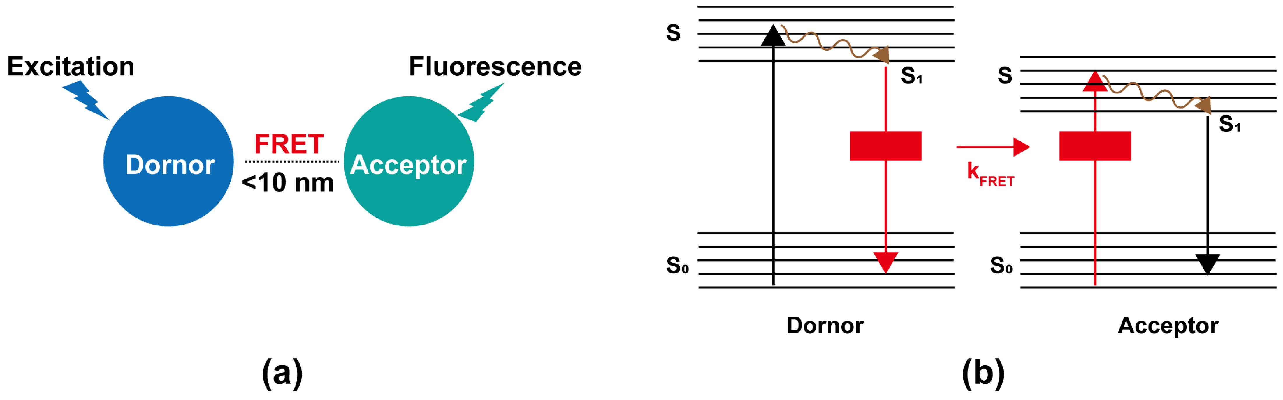
2. Principle of FRET
3. Progress of Exosome Biosensors Based on FRET
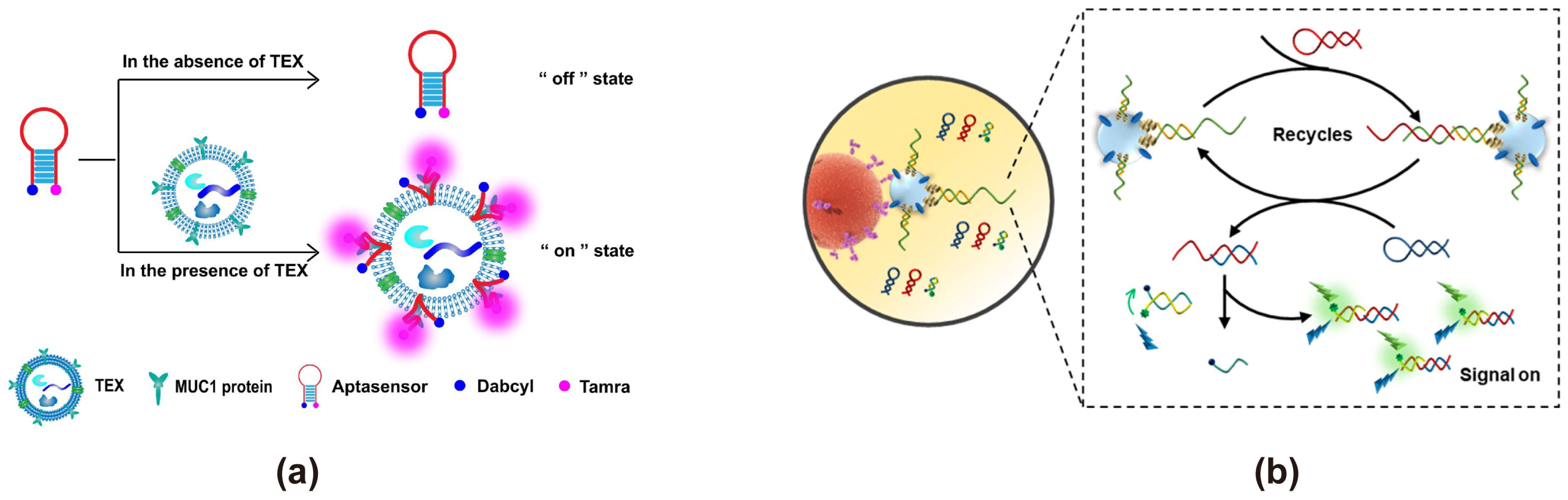
3.1. Based on Organic Fluorescent Dyes and Quenching Groups
3.2. Based on Carbon Nanomaterials
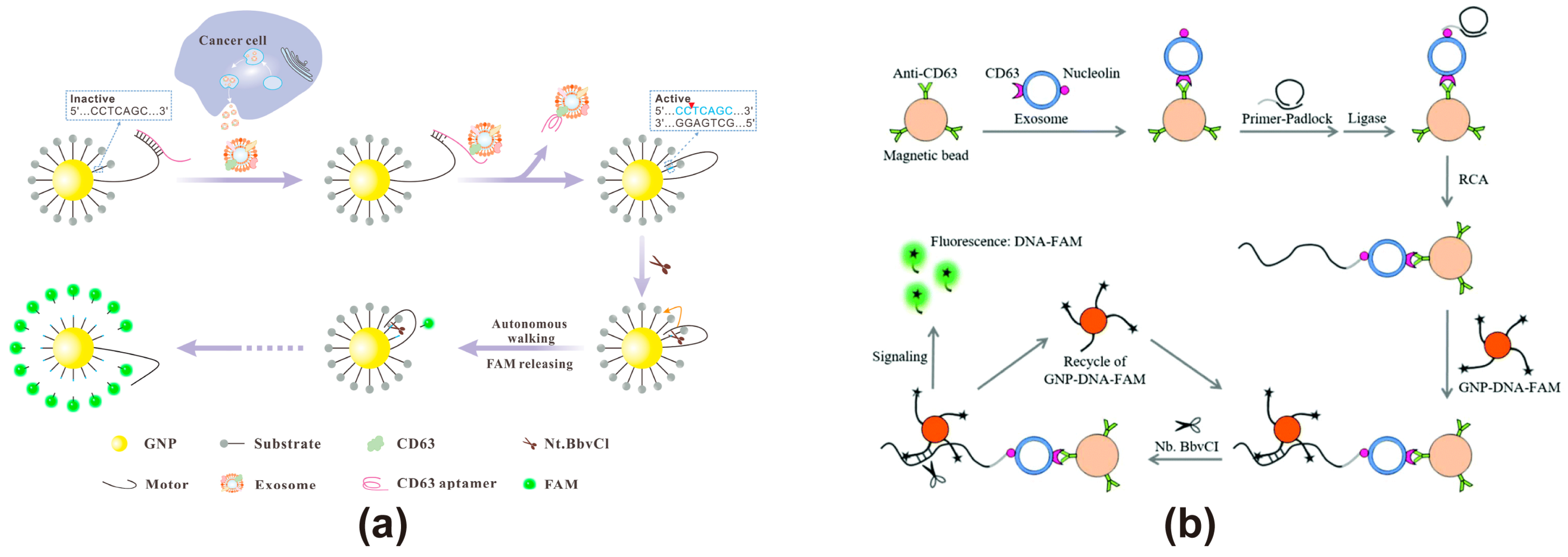
3.3. Based on AuNPs
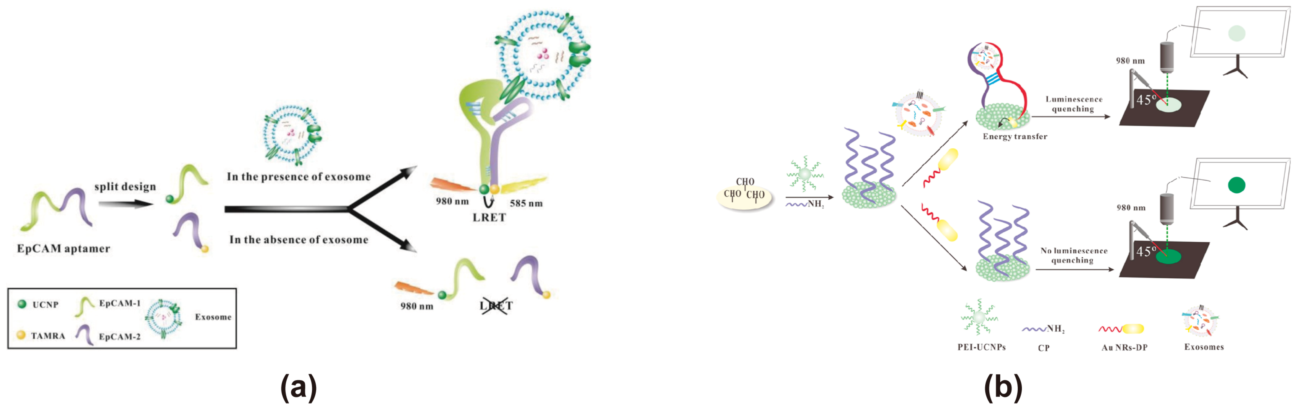
3.4. Based on UCNPs
3.5. Based on 2D Materials
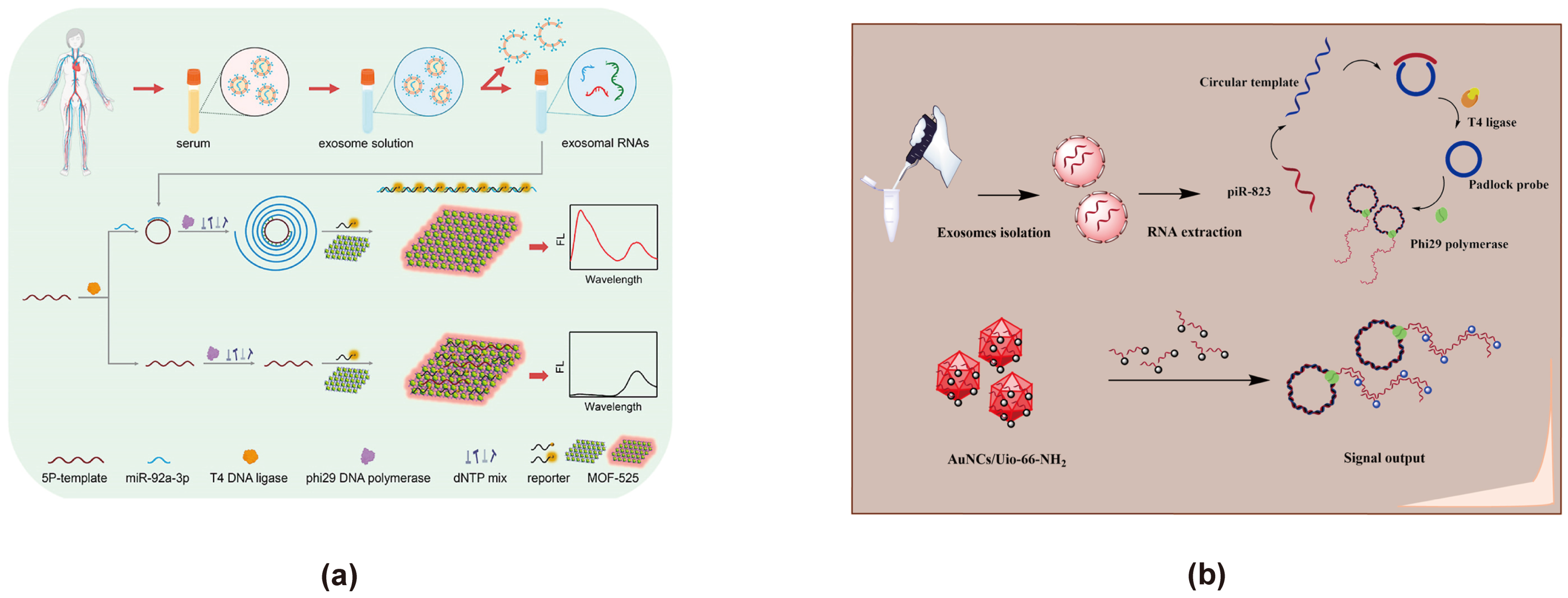
3.6. Based on MOFs
3.7. Others
4. Challenges and Prospects
Author Contributions
Funding
Conflicts of Interest
References
- Siegel, R.L.; Miller, K.D.; Wagle, N.S.; Jemal, A. Cancer statistics, 2023. CA A Cancer J. Clin. 2023, 73, 17–48. [Google Scholar] [CrossRef]
- Hsieh, J.C.-H.; Liao, C.-T.; Wang, H.-M.; Lien, M.-Y.; Lin, Y.-C.; Shen, S.-C.; Kang, S.-T.; Chen, W.-M.; Hsieh, Y.-S.; Fan, Y.-C.; et al. Evaluation of circulating miRNAs for earlier cancer detection through machine-learning expression profiling. J. Clin. Oncol. 2020, 38, 1559. [Google Scholar] [CrossRef]
- Bao, Y.; Zhang, D.; Guo, H.; Ma, W. Beyond blood: Advancing the frontiers of liquid biopsy in oncology and personalized medicine. Cancer Sci. 2024, 115, 1060–1072. [Google Scholar] [CrossRef]
- Zieren, R.C.; Zondervan, P.J.; Pienta, K.J.; Bex, A.; de Reijke, T.M.; Bins, A.D. Diagnostic liquid biopsy biomarkers in renal cell cancer. Nat. Rev. Urol. 2024, 21, 133–157. [Google Scholar] [CrossRef] [PubMed]
- Jiang, L.; Lin, X.; Chen, F.; Qin, X.; Yan, Y.; Ren, L.; Yu, H.; Chang, L.; Wang, Y. Current research status of tumor cell biomarker detection. Microsyst. Nanoeng. 2023, 9, 123. [Google Scholar] [CrossRef]
- Zhou, B.; Xu, K.; Zheng, X.; Chen, T.; Wang, J.; Song, Y.; Shao, Y.; Zheng, S. Application of exosomes as liquid biopsy in clinical diagnosis. Signal Transduct. Target. Ther. 2020, 5, 144. [Google Scholar] [CrossRef]
- Yu, D.; Li, Y.; Wang, M.; Gu, J.; Xu, W.; Cai, H.; Fang, X.; Zhang, X. Exosomes as a new frontier of cancer liquid biopsy. Mol. Cancer 2022, 21, 56. [Google Scholar] [CrossRef]
- Halvaei, S.; Daryani, S.; Eslami-S, Z.; Samadi, T.; Jafarbeik-Iravani, N.; Bakhshayesh, T.O.; Majidzadeh-A, K.; Esmaeili, R. Exosomes in Cancer Liquid Biopsy: A Focus on Breast Cancer. Mol. Ther. Nucleic Acids 2018, 10, 131–141. [Google Scholar] [CrossRef]
- Li, S.; Yi, M.; Dong, B.; Tan, X.; Luo, S.; Wu, K. The role of exosomes in liquid biopsy for cancer diagnosis and prognosis prediction. Int. J. Cancer 2021, 148, 2640–2651. [Google Scholar] [CrossRef]
- Rayamajhi, S.; Sipes, J.; Tetlow, A.L.; Saha, S.; Bansal, A.; Godwin, A.K. Extracellular Vesicles as Liquid Biopsy Biomarkers across the Cancer Journey: From Early Detection to Recurrence. Clin. Chem. 2024, 70, 206–219. [Google Scholar] [CrossRef]
- Kalluri, R.; LeBleu, V.S. The biology, function, and biomedical applications of exosomes. Science 2020, 367, eaau6977. [Google Scholar] [CrossRef]
- Ariston Gabriel, A.N.; Wang, F.; Jiao, Q.; Yvette, U.; Yang, X.; Al-Ameri, S.A.; Du, L.; Wang, Y.-s.; Wang, C. The involvement of exosomes in the diagnosis and treatment of pancreatic cancer. Mol. Cancer 2020, 19, 132. [Google Scholar] [CrossRef]
- Qiao, Y.; Tang, X.; Liu, Z.; Ocansey, D.K.W.; Zhou, M.; Shang, A.; Mao, F. Therapeutic Prospects of Mesenchymal Stem Cell and Their Derived Exosomes in the Regulation of the Gut Microbiota in Inflammatory Bowel Disease. Pharmaceuticals 2024, 17, 607. [Google Scholar] [CrossRef]
- Isaac, R.; Reis, F.C.G.; Ying, W.; Olefsky, J.M. Exosomes as mediators of intercellular crosstalk in metabolism. Cell Metab. 2021, 33, 1744–1762. [Google Scholar] [CrossRef] [PubMed]
- Jeon, H.; Seo, S.M.; Kim, T.W.; Ryu, J.; Kong, H.; Jang, S.H.; Jang, Y.S.; Kim, K.S.; Kim, J.H.; Ryu, S.; et al. Circulating Exosomal miR-1290 for Diagnosis of Epithelial Ovarian Cancer. Curr. Issues Mol. Biol. 2022, 44, 288–300. [Google Scholar] [CrossRef]
- Salciccia, S.; Frisenda, M.; Bevilacqua, G.; Gobbi, L.; Bucca, B.; Moriconi, M.; Viscuso, P.; Gentilucci, A.; Mariotti, G.; Cattarino, S.; et al. Exosome Analysis in Prostate Cancer: How They Can Improve Biomarkers’ Performance. Curr. Issues Mol. Biol. 2023, 45, 6085–6096. [Google Scholar] [CrossRef] [PubMed]
- Gao, W.-Y.; Boonyarat, C.; Samar, N.; Sethabouppha, B.; Waiwut, P. Multiomics Analysis of Molecules Associated with Cancer in Mesenchymal-Stem-Cell-(MSC)-Derived Exosome-Treated Hepatocellular Carcinoma Cells. Curr. Issues Mol. Biol. 2024, 46, 13296–13310. [Google Scholar] [CrossRef]
- Zhang, X.; Yuan, X.; Shi, H.; Wu, L.; Qian, H.; Xu, W. Exosomes in cancer: Small particle, big player. J. Hematol. Oncol. 2015, 8, 83. [Google Scholar] [CrossRef]
- Zhang, L.; Yu, D. Exosomes in cancer development, metastasis, and immunity. Biochimica et biophysica acta. Rev. Cancer 2019, 1871, 455–468. [Google Scholar] [CrossRef]
- Das, A.; Nyahatkar, S.; Sonar, S.; Kalele, K.; Subramaniyan, V. Unlocking the potential of exosomes: A new frontier in liver cancer liquid biopsy. J. Liq. Biopsy 2024, 6, 100166. [Google Scholar] [CrossRef]
- Hsu, C.C.; Wu, Y. Recent advances in nanotechnology-enabled biosensors for detection of exosomes as new cancer liquid biopsy. Exp. Biol. Med. 2022, 247, 2152–2172. [Google Scholar] [CrossRef] [PubMed]
- Liu, Z.; Zhang, W.; Zhang, X.; Wang, S.; Xia, Z.; Guo, X.; Zhao, Y.; Wang, P.; Wang, X.-H. Microstructured Optical Fiber-Enhanced Light–Matter Interaction Enables Highly Sensitive Exosome-Based Liquid Biopsy of Breast Cancer. Anal. Chem. 2023, 95, 1095–1105. [Google Scholar] [CrossRef] [PubMed]
- Uyar, R.; Özçelikay-Akyıldız, G.; Kaya, S.İ.; Bereketoğlu Nergis, S.; Beşbinar, Ö.; Ünal, M.A.; Yilmazer, A.; Özkan, S.A. Early cancer detection based on exosome biosensors in biological samples. Sens. Actuators B Chem. 2024, 400, 134886. [Google Scholar] [CrossRef]
- He, W.-M.; Zhou, Z.; Han, Z.; Li, S.; Zhou, Z.; Ma, L.-F.; Zang, S.-Q. Ultrafast Size Expansion and Turn-On Luminescence of Atomically Precise Silver Clusters by Hydrogen Sulfide. Angew. Chem. Int. Ed. 2021, 60, 8505–8509. [Google Scholar] [CrossRef]
- Vaidya, M.; Kimura, A.; Bajaj, A.; Sugaya, K. 3′-UTR Sequence of Exosomal NANOGP8 DNA as an Extracellular Vesicle-Localization Signal. Int. J. Mol. Sci. 2024, 25, 7294. [Google Scholar] [CrossRef] [PubMed]
- Zhu, X.; Chen, H.; Zhou, Y.; Wu, J.; Ramakrishna, S.; Peng, X.; Nanda, H.S.; Zhou, Y. Recent advances in biosensors for detection of exosomes. Curr. Opin. Biomed. Eng. 2021, 18, 100280. [Google Scholar] [CrossRef]
- Forster, T. Energiewanderung und Fluoreszenz. Naturwissenschaften 1946, 33, 166–175. [Google Scholar] [CrossRef]
- Algar, W.R.; Hildebrandt, N.; Vogel, S.S.; Medintz, I.L. FRET as a biomolecular research tool—Understanding its potential while avoiding pitfalls. Nat. Methods 2019, 16, 815–829. [Google Scholar] [CrossRef]
- Szöllősi, J.; Damjanovich, S.; Nagy, P.; Vereb, G.; Mátyus, L. Principles of Resonance Energy Transfer. Curr. Protoc. Cytom. 2006, 38, 1.12.1–1.12.16. [Google Scholar] [CrossRef]
- Wu, Y.; Jiang, T. Developments in FRET- and BRET-Based Biosensors. Micromachines 2022, 13, 1789. [Google Scholar] [CrossRef]
- Verma, A.K.; Noumani, A.; Yadav, A.K.; Solanki, P.R. FRET Based Biosensor: Principle Applications Recent Advances and Challenges. Biosens. Bioelectron. 2023, 13, 1375. [Google Scholar] [CrossRef]
- Peng, X.-X.; Qin, X.; Qin, Y.; Xiang, Y.; Zhang, G.-J.; Yang, F. Bioprobes-regulated precision biosensing of exosomes: From the nanovesicle surface to the inside. Coord. Chem. Rev. 2022, 463, 214538. [Google Scholar] [CrossRef]
- Imani, M.; Mohajeri, N.; Rastegar, M.; Zarghami, N. Recent advances in FRET-Based biosensors for biomedical applications. Anal. Biochem. 2021, 630, 114323. [Google Scholar] [CrossRef]
- Zhou, Y.; Wu, Y.; Pokholenko, O.; Papper, V.; Marks, R.S.; Steele, T.W.J. Design and optimisation of Photochrome Aptamer Switch Assay (PHASA). Anal. Chim. Acta 2019, 1061, 134–141. [Google Scholar] [CrossRef] [PubMed]
- Wang, X.; Shang, H.; Ma, C.; Chen, L. A Fluorescence Assay for Exosome Detection Based on Bivalent Cholesterol Anchor Triggered Target Conversion and Enzyme-Free Signal Amplification. Anal. Chem. 2021, 93, 8493–8500. [Google Scholar] [CrossRef]
- Zhang, J.; Shi, J.; Liu, W.; Zhang, K.; Zhao, H.; Zhang, H.; Zhang, Z. A simple, specific and “on-off” type MUC1 fluorescence aptasensor based on exosomes for detection of breast cancer. Sens. Actuators B Chem. 2018, 276, 552–559. [Google Scholar] [CrossRef]
- Xiong, Y.; He, L.; Yang, M.; Wang, Y.; Liu, X.; Ma, S.; Yang, B.; Guan, F. Proximity hybridization-mediated fluorescence resonance energy transfer for highly specific detection of tumor-derived exosomes: Combining multiple exosomal surface markers. Sens. Actuators B Chem. 2022, 353, 131126. [Google Scholar] [CrossRef]
- Li, Y.; Tang, X.; Deng, R.; Feng, L.; Xie, S.; Chen, M.; Zheng, J.; Chang, K. Dumbbell Dual-Hairpin Triggered DNA Nanonet Assembly for Cascade-Amplified Sensing of Exosomal MicroRNA. ACS Omega 2024, 9, 19723–19731. [Google Scholar] [CrossRef]
- Park, C.; Chung, S.; Kim, H.; Kim, N.; Son, H.Y.; Kim, R.; Lee, S.; Park, G.; Rho, H.W.; Park, M.; et al. All-in-One Fusogenic Nanoreactor for the Rapid Detection of Exosomal MicroRNAs for Breast Cancer Diagnosis. ACS Nano 2024, 18, 26297–26314. [Google Scholar] [CrossRef]
- Xia, Y.; Huang, Z.; Chen, T.; Xu, L.; Zhu, G.; Chen, W.; Chen, G.; Wu, S.; Lan, J.; Lin, X.; et al. Sensitive fluorescent detection of exosomal microRNA based on enzymes-assisted dual-signal amplification. Biosens. Bioelectron. 2022, 209, 114259. [Google Scholar] [CrossRef]
- Zhu, L.; Xu, Y.; Wei, X.; Lin, H.; Huang, M.; Lin, B.; Song, Y.; Yang, C. Coupling Aptamer-based Protein Tagging with Metabolic Glycan Labeling for In Situ Visualization and Biological Function Study of Exosomal Protein-Specific Glycosylation. Angew. Chem. Int. Ed. 2021, 60, 18111–18115. [Google Scholar] [CrossRef] [PubMed]
- Xia, Y.; Chen, T.; Chen, G.; Weng, Y.; Zeng, L.; Liao, Y.; Chen, W.; Lan, J.; Zhang, J.; Chen, J. A nature-inspired colorimetric and fluorescent dual-modal biosensor for exosomes detection. Talanta 2020, 214, 120851. [Google Scholar] [CrossRef]
- Wang, H.; He, D.; Wan, K.; Sheng, X.; Cheng, H.; Huang, J.; Zhou, X.; He, X.; Wang, K. In situ multiplex detection of serum exosomal microRNAs using an all-in-one biosensor for breast cancer diagnosis. Analyst 2020, 145, 3289–3296. [Google Scholar] [CrossRef]
- Xu, Y.; Li, X.; Niu, C.; Wu, H.; Yong, Y.; Qi, C.; Gong, W.; Bai, H.; Chen, Y.; Ding, S.; et al. Janus wireframe DNA cube-based 3D nanomachine for rapid and stable fluorescence detection of exosomal microRNA. Biosens. Bioelectron. 2022, 212, 114405. [Google Scholar] [CrossRef] [PubMed]
- Mao, D.; Zheng, M.; Li, W.; Xu, Y.; Wang, C.; Qian, Q.; Li, S.; Chen, G.; Zhu, X.; Mi, X. Cubic DNA nanocage-based three-dimensional molecular beacon for accurate detection of exosomal miRNAs in confined spaces. Biosens. Bioelectron. 2022, 204, 114077. [Google Scholar] [CrossRef] [PubMed]
- Zhang, D.; Qiao, L.; Xu, S.; Peng, L.; Yang, Y.; Zhang, P.; Song, Z.-l.; Chen, J.; Zhang, C.-H. Accurate identification of exosomes based on proximity-induced autonomous assembly of DNAzyme wires. Sens. Actuators B Chem. 2023, 383, 133581. [Google Scholar] [CrossRef]
- Wu, T.; Liu, X.; Chen, H.; Liu, Y.; Cao, Y. An in situ exosomal miRNA sensing biochip based on multi-branched localized catalytic hairpin assembly and photonic crystals. Biosens. Bioelectron. 2023, 222, 115013. [Google Scholar] [CrossRef]
- Li, P.; Yu, X.; Han, W.; Kong, Y.; Bao, W.; Zhang, J.; Zhang, W.; Gu, Y. Ultrasensitive and Reversible Nanoplatform of Urinary Exosomes for Prostate Cancer Diagnosis. ACS Sens. 2019, 4, 1433–1441. [Google Scholar] [CrossRef]
- Li, Q.; Wang, Y.; Ling, L.; Qiao, L.; Chen, H.; Ding, C.; Yu, S. Rapid and specific detection nanoplatform of serum exosomes for prostate cancer diagnosis. Microchim. Acta 2021, 188, 283. [Google Scholar] [CrossRef]
- Feng, D.; Ren, M.; Miao, Y.; Liao, Z.; Zhang, T.; Chen, S.; Ye, K.; Zhang, P.; Ma, X.; Ni, J.; et al. Dual selective sensor for exosomes in serum using magnetic imprinted polymer isolation sandwiched with aptamer/graphene oxide based FRET fluorescent ignition. Biosens. Bioelectron. 2022, 207, 114112. [Google Scholar] [CrossRef]
- Li, B.; Pan, W.; Liu, C.; Guo, J.; Shen, J.; Feng, J.; Luo, T.; Situ, B.; Zhang, Y.; An, T.; et al. Homogenous Magneto-Fluorescent Nanosensor for Tumor-Derived Exosome Isolation and Analysis. ACS Sens. 2020, 5, 2052–2060. [Google Scholar] [CrossRef]
- Wang, H.; Chen, H.; Huang, Z.; Li, T.; Deng, A.; Kong, J. DNase I enzyme-aided fluorescence signal amplification based on graphene oxide-DNA aptamer interactions for colorectal cancer exosome detection. Talanta 2018, 184, 219–226. [Google Scholar] [CrossRef] [PubMed]
- Sun, Z.; Li, J.; Tong, Y.; Zhao, L.; Zhou, X.; Li, H.; Wang, C.; Du, L.; Jiang, Y. Ratiometric Fluorescence Detection of Colorectal Cancer-Associated Exosomal miR-92a-3p with DSN-Assisted Signal Amplification by a MWCNTs@Au NCs Nanoplatform. Biosensors 2022, 12, 533. [Google Scholar] [CrossRef] [PubMed]
- Zhang, X.; Wei, X.; Qi, J.; Shen, J.; Xu, J.; Gong, G.; Wei, Y.; Yang, J.; Zhu, Q.; Bai, T.; et al. Simultaneous Detection of Bladder Cancer Exosomal MicroRNAs Based on Inorganic Nanoflare and DNAzyme Walker. Anal. Chem. 2022, 94, 4787–4793. [Google Scholar] [CrossRef]
- Xia, Y.; Wang, L.; Li, J.; Chen, X.; Lan, J.; Yan, A.; Lei, Y.; Yang, S.; Yang, H.; Chen, J. A Ratiometric Fluorescent Bioprobe Based on Carbon Dots and Acridone Derivate for Signal Amplification Detection Exosomal microRNA. Anal. Chem. 2018, 90, 8969–8976. [Google Scholar] [CrossRef] [PubMed]
- Chinnappan, R.; Ramadan, Q.; Zourob, M. An integrated lab-on-a-chip platform for pre-concentration and detection of colorectal cancer exosomes using anti-CD63 aptamer as a recognition element. Biosens. Bioelectron. 2023, 220, 114856. [Google Scholar] [CrossRef]
- Li, B.; Liu, C.; Pan, W.; Shen, J.; Guo, J.; Luo, T.; Feng, J.; Situ, B.; An, T.; Zhang, Y.; et al. Facile fluorescent aptasensor using aggregation-induced emission luminogens for exosomal proteins profiling towards liquid biopsy. Biosens. Bioelectron. 2020, 168, 112520. [Google Scholar] [CrossRef]
- Jin, D.; Yang, F.; Zhang, Y.; Liu, L.; Zhou, Y.; Wang, F.; Zhang, G.-J. ExoAPP: Exosome-Oriented, Aptamer Nanoprobe-Enabled Surface Proteins Profiling and Detection. Anal. Chem. 2018, 90, 14402–14411. [Google Scholar] [CrossRef]
- Zhou, J.; Meng, L.; Ye, W.; Wang, Q.; Geng, S.; Sun, C. A sensitive detection assay based on signal amplification technology for Alzheimer’s disease’s early biomarker in exosome. Anal. Chim. Acta 2018, 1022, 124–130. [Google Scholar] [CrossRef]
- Oh, H.J.; Kim, J.; Park, H.; Chung, S.; Hwang, D.W.; Lee, D.S. Graphene-oxide quenching-based molecular beacon imaging of exosome-mediated transfer of neurogenic miR-193a on microfluidic platform. Biosens. Bioelectron. 2019, 126, 647–656. [Google Scholar] [CrossRef]
- Chinnappan, R.; Ramadan, Q.; Zourob, M. Isolation and Detection of Exosomal Mir210 Using Carbon Nanomaterial-Coated Magnetic Beads. J. Funct. Biomater. 2023, 14, 441. [Google Scholar] [CrossRef]
- Yu, Y.; Zhang, W.S.; Guo, Y.; Peng, H.; Zhu, M.; Miao, D.; Su, G. Engineering of exosome-triggered enzyme-powered DNA motors for highly sensitive fluorescence detection of tumor-derived exosomes. Biosens. Bioelectron. 2020, 167, 112482. [Google Scholar] [CrossRef]
- Huang, L.; Wang, D.-B.; Singh, N.; Yang, F.; Gu, N.; Zhang, X.-E. A dual-signal amplification platform for sensitive fluorescence biosensing of leukemia-derived exosomes. Nanoscale 2018, 10, 20289–20295. [Google Scholar] [CrossRef] [PubMed]
- Zhu, N.; Li, G.; Zhou, J.; Zhang, Y.; Kang, K.; Ying, B.; Yi, Q.; Wu, Y. A light-up fluorescence resonance energy transfer magnetic aptamer-sensor for ultra-sensitive lung cancer exosome detection. J. Mater. Chem. B 2021, 9, 2483–2493. [Google Scholar] [CrossRef]
- Zhai, L.-Y.; Li, M.-X.; Pan, W.-L.; Chen, Y.; Li, M.-M.; Pang, J.-X.; Zheng, L.; Chen, J.-X.; Duan, W.-J. In Situ Detection of Plasma Exosomal MicroRNA-1246 for Breast Cancer Diagnostics by a Au Nanoflare Probe. ACS Appl. Mater. Interfaces 2018, 10, 39478–39486. [Google Scholar] [CrossRef] [PubMed]
- Sun, Z.; Li, J.; Yang, Y.; Tong, Y.; Li, H.; Wang, C.; Du, L.; Jiang, Y. Ratiometric Fluorescent Biosensor Based on Self-Assembled Fluorescent Gold Nanoparticles and Duplex-Specific Nuclease-Assisted Signal Amplification for Sensitive Detection of Exosomal miRNA. Bioconjugate Chem. 2022, 33, 1698–1706. [Google Scholar] [CrossRef]
- Gu, M.; Zhang, H.; Liu, Y.; Li, X.; Lv, M.; Zhao, J.; Zhang, J. Accurate and highly sensitive detection of Alzheimer’s disease-related extracellular vesicles via förster resonance energy transfer. Anal. Chim. Acta 2024, 1314, 342779. [Google Scholar] [CrossRef]
- Gao, M.-L.; He, F.; Yin, B.-C.; Ye, B.-C. A dual signal amplification method for exosome detection based on DNA dendrimer self-assembly. Analyst 2019, 144, 1995–2002. [Google Scholar] [CrossRef]
- Zhang, X.-W.; Du, L.; Liu, M.-X.; Wang, J.-H.; Chen, S.; Yu, Y.-L. All-in-one nanoflare biosensor combined with catalyzed hairpin assembly amplification for in situ and sensitive exosomal miRNA detection and cancer classification. Talanta 2024, 266, 125145. [Google Scholar] [CrossRef]
- Qin, X.; Xiang, Y.; Li, N.; Wei, B.; Chen, Y.; Fang, D.; Fang, M.; Li, Q.; Liu, J.; Tang, Y.; et al. Simultaneous detection of cancerous exosomal miRNA-21 and PD-L1 with a sensitive dual-cycling nanoprobe. Biosens. Bioelectron. 2022, 216, 114636. [Google Scholar] [CrossRef]
- Wang, Y.; Luo, D.; Fang, Y.; Wu, W.; Wang, Y.; Xia, Y.; Wu, F.; Li, C.; Lan, J.; Chen, J. An aptasensor based on upconversion nanoparticles as LRET donors for the detection of exosomes. Sens. Actuators B Chem. 2019, 298, 126900. [Google Scholar] [CrossRef]
- Chen, X.; Lan, J.; Liu, Y.; Li, L.; Yan, L.; Xia, Y.; Wu, F.; Li, C.; Li, S.; Chen, J. A paper-supported aptasensor based on upconversion luminescence resonance energy transfer for the accessible determination of exosomes. Biosens. Bioelectron. 2018, 102, 582–588. [Google Scholar] [CrossRef] [PubMed]
- Zeng, Y.; Wang, X.; Zhu, N.; Yu, Y.; Wang, X.; Kang, K.; Wu, Y.; Yi, Q. Magnetic lanthanide sensor with self-ratiometric time-resolved luminescence for accurate detection of epithelial cancerous exosomes. J. Mater. Chem. B 2024, 12, 7203–7214. [Google Scholar] [CrossRef] [PubMed]
- Wei, J.; He, S.; Mao, Y.; Wu, L.; Liu, X.; Effah, C.Y.; Guo, H.; Wu, Y. A simple “signal off–on” fluorescence nanoplatform for the label-free quantification of exosome-derived microRNA-21 in lung cancer plasma. Microchim. Acta 2021, 188, 397. [Google Scholar] [CrossRef]
- Zhang, Q.; Wang, F.; Zhang, H.; Zhang, Y.; Liu, M.; Liu, Y. Universal Ti3C2 MXenes Based Self-Standard Ratiometric Fluorescence Resonance Energy Transfer Platform for Highly Sensitive Detection of Exosomes. Anal. Chem. 2018, 90, 12737–12744. [Google Scholar] [CrossRef]
- Tayebi, M.; Tavakkoli Yaraki, M.; Yang, H.Y.; Ai, Y. A MoS2–MWCNT based fluorometric nanosensor for exosome detection and quantification. Nanoscale Adv. 2019, 1, 2866–2872. [Google Scholar] [CrossRef]
- Gao, Z.; Yuan, H.; Mao, Y.; Ding, L.; Effah, C.Y.; He, S.; He, L.; Liu, L.-e.; Yu, S.; Wang, Y.; et al. In situ detection of plasma exosomal microRNA for lung cancer diagnosis using duplex-specific nuclease and MoS2 nanosheets. Analyst 2021, 146, 1924–1931. [Google Scholar] [CrossRef]
- Cui, H.; Zheng, T.; Qian, N.; Fu, X.; Li, A.; Xing, S.; Wang, X.-F. Aptamer-Functionalized Magnetic Ti3C2 Based Nanoplatform for Simultaneous Enrichment and Detection of Exosomes. Small 2024, 20, 2402434. [Google Scholar] [CrossRef]
- Xia, Q.; Zheng, J.; Bu, J.; Li, R.; Li, X.; Fan, S.; Ling, K.; Jiang, H. Mn2+-modified black phosphorus nanosensor for detection of exosomal microRNAs and exosomes. Microchim. Acta 2023, 190, 295. [Google Scholar] [CrossRef]
- Sun, Z.; Li, J.; Tong, Y.; Han, H.; Yang, Y.; Wang, C.; Li, H.; Du, L.; Jiang, Y. A ratiometric fluorescent biosensor based on self-fluorescent MOF and target-triggered rolling circle amplification for sensitive detection of exosome-derived miRNA. Anal. Chim. Acta 2022, 1221, 340136. [Google Scholar] [CrossRef]
- Tong, Y.; Guan, B.; Sun, Z.; Dong, X.; Chen, Y.; Li, Y.; Jiang, Y.; Li, J. Ratiometric fluorescent detection of exosomal piRNA-823 based on Au NCs/UiO-66-NH2 and target-triggered rolling circle amplification. Talanta 2023, 257, 124307. [Google Scholar] [CrossRef] [PubMed]
- Zhao, X.; Zhang, W.; Qiu, X.; Mei, Q.; Luo, Y.; Fu, W. Rapid and sensitive exosome detection with CRISPR/Cas12a. Anal. Bioanal. Chem. 2020, 412, 601–609. [Google Scholar] [CrossRef]
- Zhang, J.; Guan, M.; Ma, C.; Liu, Y.; Lv, M.; Zhang, Z.; Gao, H.; Zhang, K. Highly Effective Detection of Exosomal miRNAs in Plasma Using Liposome-Mediated Transfection CRISPR/Cas13a. ACS Sens. 2023, 8, 565–575. [Google Scholar] [CrossRef] [PubMed]
- Lu, Z.; Ni, W.; Liu, N.; Jin, D.; Li, T.; Li, K.; Zhang, Y.; Yao, Q.; Zhang, G.-J. CRISPR/Cas12a-based fluorescence biosensor for detection of exosomal miR-21 derived from lung cancer. Microchem. J. 2023, 187, 108370. [Google Scholar] [CrossRef]
- Liu, Q.; Liu, J.; He, N.; Zhang, M.; Wu, L.; Chen, X.; Zhu, J.; Ran, F.; Chen, Q.; Zhang, H. CRISPR/Cas12a Coupling with Magnetic Nanoparticles and Cascaded Strand Displacement Reaction for Ultrasensitive Fluorescence Determination of Exosomal miR-21. Molecules 2022, 27, 5338. [Google Scholar] [CrossRef] [PubMed]
- Bano, R.; Soleja, N.; Mohsin, M. Genetically Encoded FRET-Based Nanosensor for Real-Time Monitoring of A549 Exosomes: Early Diagnosis of Cancer. Anal. Chem. 2023, 95, 5738–5746. [Google Scholar] [CrossRef]
- Lim, J.; Kang, B.; Son, H.Y.; Mun, B.; Huh, Y.-M.; Rho, H.W.; Kang, T.; Moon, J.; Lee, J.-J.; Seo, S.B.; et al. Microfluidic device for one-step detection of breast cancer-derived exosomal mRNA in blood using signal-amplifiable 3D nanostructure. Biosens. Bioelectron. 2022, 197, 113753. [Google Scholar] [CrossRef]
- Chen, J.; Guo, J.; Hu, M.; Wang, Y.; Hua, F.; Meng, H.-M.; Jin, S. Accurate and portable tumor exosomes detection based on manganese dioxide and aptamer-functionalized fluorescent microspheres mediated dual-mode lateral flow assay. Sens. Actuators B Chem. 2024, 409, 135614. [Google Scholar] [CrossRef]
- Ellington, A.D.; Szostak, J.W. In vitro selection of RNA molecules that bind specific ligands. Nature 1990, 346, 818–822. [Google Scholar] [CrossRef]
- Song, S.; Wang, L.; Li, J.; Fan, C.; Zhao, J. Aptamer-based biosensors. TrAC Trends Anal. Chem. 2008, 27, 108–117. [Google Scholar] [CrossRef]
- Burgess, L.; Jones, A.R.; Hay, S.; Natrajan, L.S. Evaluating spectral overlap with the degree of quenching in UCP luminescence energy transfer systems. Methods Appl. Fluoresc. 2019, 7, 034003. [Google Scholar] [CrossRef] [PubMed]
- Hwang, H.S.; Jeong, J.W.; Kim, Y.A.; Chang, M. Carbon Nanomaterials as Versatile Platforms for Biosensing Applications. Micromachines 2020, 11, 814. [Google Scholar] [CrossRef] [PubMed]
- Wang, Y.; Bao, L.; Liu, Z.; Pang, D.-W. Aptamer Biosensor Based on Fluorescence Resonance Energy Transfer from Upconverting Phosphors to Carbon Nanoparticles for Thrombin Detection in Human Plasma. Anal. Chem. 2011, 83, 8130–8137. [Google Scholar] [CrossRef] [PubMed]
- Salama, A.M.; Yasin, G.; Zourob, M.; Lu, J. Fluorescent Biosensors for the Detection of Viruses Using Graphene and Two-Dimensional Carbon Nanomaterials. Biosensors 2022, 12, 460. [Google Scholar] [CrossRef]
- Li, C.; Shi, G. Carbon nanotube-based fluorescence sensors. J. Photochem. Photobiol. C Photochem. Rev. 2014, 19, 20–34. [Google Scholar] [CrossRef]
- Dai, H.; Shi, Y.; Wang, Y.; Sun, Y.; Hu, J.; Ni, P.; Li, Z. A carbon dot based biosensor for melamine detection by fluorescence resonance energy transfer. Sens. Actuators B Chem. 2014, 202, 201–208. [Google Scholar] [CrossRef]
- Upadhyayula, V.K.K.; Ghoshroy, S.; Nair, V.S.; Smith, G.B.; Mitchell, M.C.; Deng, S. Single-Walled Carbon Nanotubes as Fluorescence Biosensors for Pathogen Recognition in Water Systems. J. Nanotechnol. 2008, 2008, 156358. [Google Scholar] [CrossRef]
- Su, D.; Li, H.; Yan, X.; Lin, Y.; Lu, G. Biosensors based on fluorescence carbon nanomaterials for detection of pesticides. TrAC Trends Anal. Chem. 2021, 134, 116126. [Google Scholar] [CrossRef]
- Barua, S.; Gogoi, S.; Khan, R. Fluorescence biosensor based on gold-carbon dot probe for efficient detection of cholesterol. Synth. Met. 2018, 244, 92–98. [Google Scholar] [CrossRef]
- Yu, Y.; Li, Y.-T.; Jin, D.; Yang, F.; Wu, D.; Xiao, M.-M.; Zhang, H.; Zhang, Z.-Y.; Zhang, G.-J. Electrical and Label-Free Quantification of Exosomes with a Reduced Graphene Oxide Field Effect Transistor Biosensor. Anal. Chem. 2019, 91, 10679–10686. [Google Scholar] [CrossRef]
- Lee, J.; Kim, J.; Kim, S.; Min, D.-H. Biosensors based on graphene oxide and its biomedical application. Adv. Drug Deliv. Rev. 2016, 105, 275–287. [Google Scholar] [CrossRef] [PubMed]
- Gutiérrez-Cruz, A.; Ruiz-Hernández, A.R.; Vega-Clemente, J.F.; Luna-Gazcón, D.G.; Campos-Delgado, J. A review of top-down and bottom-up synthesis methods for the production of graphene, graphene oxide and reduced graphene oxide. J. Mater. Sci. 2022, 57, 14543–14578. [Google Scholar] [CrossRef]
- Shi, J.; Tian, F.; Lyu, J.; Yang, M. Nanoparticle based fluorescence resonance energy transfer (FRET) for biosensing applications. J. Mater. Chem. B 2015, 3, 6989–7005. [Google Scholar] [CrossRef] [PubMed]
- Ferrier, D.C.; Honeychurch, K.C. Carbon Nanotube (CNT)-Based Biosensors. Biosensors 2021, 11, 486. [Google Scholar] [CrossRef]
- Wang, W.; Impundu, J.; Jin, J.; Peng, Z.; Liu, H.; Wei, Z.; Xu, Y.; Wang, Y.; You, J.; Fan, W.; et al. Ferromagnetism in sp2 carbon. Nano Res. 2023, 16, 12883–12900. [Google Scholar] [CrossRef]
- Liu, X.; Luo, Y.; Zhang, Y.; Xie, Z.; Xu, C. Gold nanoparticle-mediated fluorescence resonance energy transfer for analytical applications in the fields of life health and safety. Talanta 2025, 282, 127023. [Google Scholar] [CrossRef] [PubMed]
- Priyadarshini, E.; Pradhan, N. Gold nanoparticles as efficient sensors in colorimetric detection of toxic metal ions: A review. Sens. Actuators B Chem. 2017, 238, 888–902. [Google Scholar] [CrossRef]
- Yang, L.; Wei, H.; Qing, Z.; Wu, L. AuNP@DNA nanoflares: Preparation and application in bioanalysis and biomedicine. Chin. Chem. Lett. 2024, 110524, in press. [Google Scholar] [CrossRef]
- Kong, J.; Zhang, W.; Wu, Y.; Zhou, M. Optical properties of gold nanoclusters constructed from Au13 units. Aggregate 2022, 3, e207. [Google Scholar] [CrossRef]
- Lee, E.S.; Cha, B.S.; Kim, S.; Park, K.S. Synthesis of Exosome-Based Fluorescent Gold Nanoclusters for Cellular Imaging Applications. Int. J. Mol. Sci. 2021, 22, 4433. [Google Scholar] [CrossRef]
- Lin, M.; Gao, Y.; Hornicek, F.; Xu, F.; Lu, T.J.; Amiji, M.; Duan, Z. Near-infrared light activated delivery platform for cancer therapy. Adv. Colloid Interface Sci. 2015, 226, 123–137. [Google Scholar] [CrossRef] [PubMed]
- Naher, H.S.; Al-Turaihi, B.A.H.; Mohammed, S.H.; Naser, S.M.; Albark, M.A.; Madlool, H.A.; Al- Marzoog, H.A.M.; Turki Jalil, A. Upconversion nanoparticles (UCNPs): Synthesis methods, imaging and cancer therapy. J. Drug Deliv. Sci. Technol. 2023, 80, 104175. [Google Scholar] [CrossRef]
- Ma, Y.; Song, M.; Li, L.; Lao, X.; Liu, Y.; Wong, M.-c.; Yang, M.; Chen, H.; Hao, J. Attomolar-level detection of respiratory virus long-chain oligonucleotides based on FRET biosensor with upconversion nanoparticles and Au–Au dimer. Biosens. Bioelectron. 2024, 243, 115778. [Google Scholar] [CrossRef]
- Oliveira, H.; Bednarkiewicz, A.; Falk, A.; Fröhlich, E.; Lisjak, D.; Prina-Mello, A.; Resch, S.; Schimpel, C.; Vrček, I.V.; Wysokińska, E.; et al. Critical Considerations on the Clinical Translation of Upconversion Nanoparticles (UCNPs): Recommendations from the European Upconversion Network (COST Action CM1403). Adv. Healthc. Mater. 2019, 8, 1801233. [Google Scholar] [CrossRef]
- Niu, Y.; Bao, Z.; Gao, Y.; Guo, M.; Liu, J.; Shao, J.; Lu, M.; Yuan, Z.; Xie, X. Brightening heavily doped upconversion nanoparticles by tuning characteristics of core-shell structures. J. Rare Earths 2024, 42, 947–954. [Google Scholar] [CrossRef]
- Zahra, T.; Javeria, U.; Jamal, H.; Baig, M.M.; Akhtar, F.; Kamran, U. A review of biocompatible polymer-functionalized two-dimensional materials: Emerging contenders for biosensors and bioelectronics applications. Anal. Chim. Acta 2024, 1316, 342880. [Google Scholar] [CrossRef] [PubMed]
- Zhai, K.; Wang, H.; Ding, Q.; Wu, Z.; Ding, M.; Tao, K.; Yang, B.-R.; Xie, X.; Li, C.; Wu, J. High-Performance Strain Sensors Based on Organohydrogel Microsphere Film for Wearable Human–Computer Interfacing. Adv. Sci. 2023, 10, 2205632. [Google Scholar] [CrossRef]
- Ding, H.; Wu, Z.; Wang, H.; Zhou, Z.; Wei, Y.; Tao, K.; Xie, X.; Wu, J. An ultrastretchable, high-performance, and crosstalk-free proximity and pressure bimodal sensor based on ionic hydrogel fibers for human-machine interfaces. Mater. Horiz. 2022, 9, 1935–1946. [Google Scholar] [CrossRef]
- Ding, Q.; Wu, Z.; Tao, K.; Wei, Y.; Wang, W.; Yang, B.-R.; Xie, X.; Wu, J. Environment tolerant, adaptable and stretchable organohydrogels: Preparation, optimization, and applications. Mater. Horiz. 2022, 9, 1356–1386. [Google Scholar] [CrossRef]
- Li, S.; Chen, Z.; Yang, F.; Yue, W. Emerging two-dimensional materials for analytical lab-on-chip platforms: A review of electrochemical and optical biosensor. Microchem. J. 2023, 194, 109247. [Google Scholar] [CrossRef]
- Zhao, L.; Kong, D.; Wu, Z.; Liu, G.; Gao, Y.; Yan, X.; Liu, F.; Liu, X.; Wang, C.; Cui, J.; et al. Interface interaction of MoS2 nanosheets with DNA based aptameric biosensor for carbohydrate antigen 15–3 detection. Microchem. J. 2020, 155, 104675. [Google Scholar] [CrossRef]
- Wang, L.; Huang, Z.; Liu, Y.; Wu, J.; Liu, J. Fluorescent DNA Probing Nanoscale MnO2: Adsorption, Dissolution by Thiol, and Nanozyme Activity. Langmuir 2018, 34, 3094–3101. [Google Scholar] [CrossRef] [PubMed]
- Tayo, B.O.; Walkup, M.A.; Caliskan, S. Adsorption of DNA nucleobases on single-layer Ti3C2 MXene and graphene: vdW-corrected DFT and NEGF studies. AIP Adv. 2023, 13, 085213. [Google Scholar] [CrossRef]
- Dou, X.; Xu, S.; Jiang, Y.; Ding, Z.; Xie, J. Aptamers-functionalized nanoscale MOFs for saxitoxin and tetrodotoxin sensing in sea foods through FRET. Spectrochim. Acta Part. A Mol. Biomol. Spectrosc. 2023, 284, 121827. [Google Scholar] [CrossRef]
- Reuven, N.; Shaul, Y. Selecting for CRISPR-Edited Knock-In Cells. Int. J. Mol. Sci. 2022, 23, 11919. [Google Scholar] [CrossRef]
- Park, J.; Kim, J. CRISPR/Cas9 Technology Providing the Therapeutic Landscape of Metastatic Prostate Cancer. Pharmaceuticals 2024, 17, 1589. [Google Scholar] [CrossRef]
- Kalligosfyri, P.M.; Lamprou, E.; Kalogianni, D.P. Emerging Sensing Technologies for Liquid Biopsy Applications: Steps Closer to Personalized Medicine. Sens. Actuators B Chem. 2024, 24, 7902. [Google Scholar] [CrossRef] [PubMed]
- Li, J.; Wang, H.; Luo, Y.; Zhou, Z.; Zhang, H.; Chen, H.; Tao, K.; Liu, C.; Zeng, L.; Huo, F.; et al. Design of AI-Enhanced and Hardware-Supported Multimodal E-Skin for Environmental Object Recognition and Wireless Toxic Gas Alarm. Nano-Micro Lett. 2024, 16, 256. [Google Scholar] [CrossRef]


| Category | Donor/Acceptor | Exosome Target | References |
|---|---|---|---|
| Based on organic fluorescent dyes and quenching groups | FAM/Dabcyl | CD63 | [35] |
| TAMRA/Dabcyl | MUC1 | [36] | |
| FAM/TAMRA | CD63, EGFR, EpCAM | [37] | |
| FAM/BHQ1 | miR-141 | [38] | |
| CY3/CY5 | miR-200c-3p, miR-222-3p, miR-375-3p | [39] | |
| FAM/BHQ1 | miR-21 | [40] | |
| CY3/CY5 | PD-L1 | [41] | |
| Fluorescein/PPDox | CD63 | [42] | |
| FAM, Cy3, Cy5/BHQ1, BHQ2 | miR-21, miR-27a, miR-375 | [43] | |
| FAM/BHQ1 | miRNA-21 | [44] | |
| FAM/BHQ1 | miRNA-21 | [45] | |
| Cy3/BHQ2 | CD63 | [46] | |
| FAM/BHQ1 | miRNA-27a | [47] | |
| FITC/BHQ1 | PSMA | [48] | |
| TAMRA/Dabcyl | PSMA | [49] | |
| Based on carbon nanomaterials | FAM/GO | CD63, MUC1 | [50] |
| FITC/GO | CD63 | [51] | |
| CY3, FAM/GO | CD63, EpCAM | [52] | |
| Atto-425/MWCNTs@Au NCs | miR-92a-3p | [53] | |
| CDs/AuNPs | miR-133b, miR-135b | [54] | |
| CDs/DSA | miRNA-21 | [55] | |
| FAM/GO | CD63 | [56] | |
| TPE-TA/GO | PSMA | [57] | |
| FAM/GO | PSMA | [58] | |
| FAM-GO | Aβ42 | [59] | |
| FAM/GO | miR-193a | [60] | |
| FAM/CCM | Mir210 | [61] | |
| Based on AuNPs | FAM/AuNPs | CD63 | [62] |
| FAM/AuNPs | CD63 | [63] | |
| QD/AuNPs | EpCAM | [64] | |
| Cy5/Au nanoflare | MicroRNA-1246 | [65] | |
| Atto-425/AuNPs | miR-92a-3p | [66] | |
| AuNCs/PDANS | Aβ42, CD63 | [67] | |
| FAM/AuNPs | CD63 | [68] | |
| FAM, HEX, Cy5/Au nanoflare | miR-21, 122, 375 | [69] | |
| FAM, Cy5/AuNPs | PD-L1, ExomiR-21 | [70] | |
| Based onUCNPs | UCNPs/TAMRA | EpCAM | [71] |
| UCNPs/AuNRs | CD63 | [72] | |
| NaYF4: Tb/BHQ1 | EpCAM | [73] | |
| Based on 2D materials | FAM/MoS2 | microRNA-21 | [74] |
| Cy3/Ti3C2 Mxenes | CD63 | [75] | |
| PE/MoS2-MWCNT | CD63 | [76] | |
| FAM/MoS2 | miRNA-21 | [77] | |
| FAM/Ti3C2 | CD63 | [78] | |
| Alexa Fluor 647/BP@Mn2+ | miR-21 | [79] | |
| Based on MOFs | FAM/MOF-525 | miR-92a-3p | [80] |
| Au NCs/UiO-66-NH | piR-823 | [81] | |
| Others | Cy3/BHQ | CD63 | [82] |
| Cy5 | miR-21 | [83] | |
| FAM | miRNA-21 | [84] | |
| FAM/BHQ1 | miRNA-21 | [85] | |
| ECFPs/Venus | EGFR | [86] | |
| ERBB2 | [87] | ||
| FMs/MnO2 | MUC1 | [88] |
Disclaimer/Publisher’s Note: The statements, opinions and data contained in all publications are solely those of the individual author(s) and contributor(s) and not of MDPI and/or the editor(s). MDPI and/or the editor(s) disclaim responsibility for any injury to people or property resulting from any ideas, methods, instructions or products referred to in the content. |
© 2025 by the authors. Licensee MDPI, Basel, Switzerland. This article is an open access article distributed under the terms and conditions of the Creative Commons Attribution (CC BY) license (https://creativecommons.org/licenses/by/4.0/).
Share and Cite
Huang, F.; Xie, Z.; Zhang, Q.; Zada, S.; Lin, R.; Deng, Y.; Liu, Q.; Chen, H.; Zhou, H.; Miao, H.; et al. Recent Advances in Fluorescence Resonance Energy Transfer (FRET) Biosensors for Exosomes. Curr. Issues Mol. Biol. 2025, 47, 235. https://doi.org/10.3390/cimb47040235
Huang F, Xie Z, Zhang Q, Zada S, Lin R, Deng Y, Liu Q, Chen H, Zhou H, Miao H, et al. Recent Advances in Fluorescence Resonance Energy Transfer (FRET) Biosensors for Exosomes. Current Issues in Molecular Biology. 2025; 47(4):235. https://doi.org/10.3390/cimb47040235
Chicago/Turabian StyleHuang, Feng, Zhenyu Xie, Qianjiao Zhang, Shah Zada, Ruhan Lin, Yanmei Deng, Qifeng Liu, Huizhi Chen, Hui Zhou, Huilai Miao, and et al. 2025. "Recent Advances in Fluorescence Resonance Energy Transfer (FRET) Biosensors for Exosomes" Current Issues in Molecular Biology 47, no. 4: 235. https://doi.org/10.3390/cimb47040235
APA StyleHuang, F., Xie, Z., Zhang, Q., Zada, S., Lin, R., Deng, Y., Liu, Q., Chen, H., Zhou, H., Miao, H., & Zhou, Y. (2025). Recent Advances in Fluorescence Resonance Energy Transfer (FRET) Biosensors for Exosomes. Current Issues in Molecular Biology, 47(4), 235. https://doi.org/10.3390/cimb47040235