Article

CNPY3 Promotes Human Breast Cancer Progression and Metastasis via Modulation of the Tumor Microenvironment

1
Department of Microbiology & Immunology, Hollings Cancer Center, Medical University of South Carolina, 86 Jonathan Lucas Street, Charleston, SC 29425, USA
2
Department of Pathology, The Ohio State University Wexner Medical Center, Columbus, OH 43210, USA
3
Division of Medical Oncology, Department of Internal Medicine, The Ohio State University Comprehensive Cancer Center, Columbus, OH 43210, USA
4
The Pelotonia Institute for Immuno-Oncology, The Ohio State University Comprehensive Cancer Center, Columbus, OH 43210, USA
5
Division of Hematology, Department of Internal Medicine, The Ohio State University Comprehensive Cancer Center, Columbus, OH 43210, USA
*
Author to whom correspondence should be addressed.
Current address: Department of Minimally Invasive Esophageal Surgery, Tianjin Medical University Cancer Institute and Hospital, Tianjin 300060, China.
These authors contributed equally to this work.
§
Current address: Department of Clinical Laboratory, Tianjin Medical University Cancer Institute and Hospital, Tianjin 300060, China.
Current address: ALLERDIA Inc., Torrance, CA 90502, USA.
Curr. Issues Mol. Biol. 2025, 47(11), 883; https://doi.org/10.3390/cimb47110883
Submission received: 6 September 2025 / Revised: 16 October 2025 / Accepted: 21 October 2025 / Published: 24 October 2025
(This article belongs to the Special Issue Tumorigenesis and Tumor Microenvironment)

Abstract

Canopy FGF signaling regulator 3 (CNPY3) is a cochaperone of the molecular chaperone GRP94. CNPY3 is critical for the post-translational maturation of toll-like receptors and for regulating inflammasome signaling. However, the role of CNPY3 in cancer development and progression is still not fully understood. In this study, we aimed to investigate the role of CNPY3 in human breast cancer progression and metastasis. We used genomic and clinical information from multiple databases to profile CNPY3 and GRP94 in human cancers. We found that CNPY3 and GRP94 were elevated in human breast cancers compared to normal tissue. Higher expression of CNPY3 correlated with cancer progression and poor clinical outcomes in breast cancers. We confirmed these findings using a human breast cancer tissue array. We silenced CNPY3 in human breast cancer cells using a CRISPR/Cas9 system. For the first time, we found that deletion of CNPY3 significantly reduced tumor growth and metastasis in vitro and in vivo. Additionally, network and enrichment analyses revealed that changes in the unfolded protein response pathway and immune-related genes were significantly dependent on alterations in CNPY3 and GRP94. This study suggests that CNPY3 is a potential biomarker and novel therapeutic target for cancers.

1. Introduction

A broad range of stressful conditions caused by intrinsic and extrinsic factors may exist with the tumor development and progression, including hypoxia, changes in redox homeostasis, disrupted cellular metabolism, acidosis, and genetic lesions, leading to the production of mutated proteins, fast cell proliferation, and abnormal protein synthesis, which then induce endoplasmic reticulum (ER) stress and trigger the unfolded protein response (UPR) [1,2,3,4]. Then, UPR subsequently regulates downstream gene expression related to protein folding, such as molecular chaperones, which are specialized proteins that assist in the proper folding, assembly, and stability of other proteins (termed client proteins), preventing their misfolding or aggregation under cellular stress. Co-chaperones act as regulators that enhance chaperone specificity and efficiency by targeting specific client proteins or modulating chaperone activity. In the ER, chaperone–co-chaperone complexes are essential for maintaining proteostasis, and their dysfunction is linked to diseases, including cancer [2,5].
Previous studies have demonstrated that glucose-regulated protein 94 (GRP94, encoded by HSP90B1 gene), also known as GP96, a stress-inducible molecular chaperone, is upregulated in various stress conditions that disrupt ER functions and homeostasis, including glucose starvation, oxidative stress, ER calcium store depletion, and the accumulation of misfolded proteins [6]. Hence, GRP94 may play a key role in regulating the balance between cancer cell viability and apoptosis through maintaining ER protein folding capacity and activating UPR sensors and ER-associated pro-apoptotic machinery. Moreover, previous studies have demonstrated that GRP94 is an essential molecular chaperone for toll-like receptors (TLRs), integrins, LRP6, GARP, and other vital innate receptors [7,8,9]. GRP94 and its client proteins are critical in oncogenesis and tumor development [10,11,12,13,14,15,16].
Previously, we discovered that canopy FGF signaling regulator 3 (CNPY3), also known as a protein associated with TLR4 (PRAT4A) [17], is a cochaperone of GRP94, which is critical for the post-translational maturation of TLRs [18]. CNPY3 is an ER resident protein initially involved in regulating TLR4 expression on the cell surface [19,20] and is required for multiple TLR trafficking and responses to TLR ligands except for the TLR3 ligand [17,21,22]. Interestingly, a recent study demonstrated that loss of CNPY3 in macrophages decreased activation of caspase-1 and processing of IL-1β and IL-18, which is independent of TLR signaling [23]. The deletion of CNPY3 altered the inflammasome structure and impaired ASC/caspase-1 colocalization [23], suggesting that CNPY3 might interact with ER molecules involved in ER stress responses and are critical in regulating the inflammasome. More recent studies showed that CNPY3 was upregulated in colon adenocarcinoma, and a higher level of CNPY3 was correlated with worse clinical outcomes. Knockdown of CNPY3 in colon tumor cell lines decreased tumor cell growth and migration in vitro [24]. However, the role of CNPY3 in cancer development and progression is still not completely understood.
Breast cancer is the second leading cause of malignant death in women. Despite the introduction of new early detection and treatments, breast cancer remains the development of multidrug resistance and metastasis. About 316,950 new cases of female breast cancer will be diagnosed in 2025 in the United States, with 14,600 deaths. Therefore, identifying targets for the development of novel therapeutics is still a major challenge. In this study, we demonstrated that expression levels of CNPY3 and GRP94 were elevated in human breast cancers, and a high level of CNPY3 was associated with an aggressive phenotype and poor clinical outcome. Furthermore, targeting CNPY3 in human breast cancer cells by CRISPR/Cas9 significantly reduced tumor growth and metastasis in vitro and in vivo. Mechanistically, we found that GRP94 and its co-chaperone CNPY3 regulate UPR pathway genes and immune-related genes in the tumor microenvironment. For the first time, our study uncovers the vital role of CNPY3 in the progression and metastasis of breast cancer, suggesting that CNPY3 may be a novel biomarker and therapeutic target for cancers.

2. Materials and Methods

2.1. Mice and Cell Lines

NSG mice were purchased from JAX Laboratory (Bar Harbor, ME, USA) and were bred and maintained according to the established guidelines and a protocol approved by the Medical University of South Carolina Institutional Animal Care and Use Committee. MCF-7 and MDA-MB-231 human breast cancer cell lines were kindly provided by Beichu Guo (Medical University of South Carolina). Human breast cancer cells were cultured in Eagle’s minimum essential medium (Sigma-Aldrich, St. Louis, MO, USA) supplemented with 10% heat-inactivated fetal calf serum (FCS, Atlas Biologicals, Fort Collins, CO, USA) and penicillin-streptomycin (Gibco, Waltham, MA, USA). All cells were cultured in a 5% CO2 incubator.

2.2. Reagents

Antibodies against CNPY3 were purchased from Sigma-Aldrich, and the GRP94 antibody was purchased from Enzo Life Sciences, Inc. (Farmingdale, NY, USA). The ABC-HPR kit and DAB substrate kit were obtained from Vector Laboratories (Newark, CA, USA). All other chemicals were obtained from Sigma-Aldrich and Fisher Scientific (Waltham, MA, USA).

2.3. Oncomine Database

The Oncomine Platform (Thermo Fisher, Ann Arbor, MI, USA; http://www.oncomine.org, accessed on 15 October 2025) provides translational bioinformatics solutions and multinational comparisons, along with clusters, gene expression signatures, and gene-set modules, as well as extracts biological insights from the data [25,26]. The Oncomine contains 715 datasets and 86,733 samples. Through an Oncomine Research Premium Edition Upgrade, we downloaded the raw datasets, including mRNA expression, clinical and pathological information, and survival data.

2.4. The cBioPortal for Cancer Genomics

cBioPortal (http://cbioportal.org, accessed on 15 October 2025) provides a resource with multidimensional cancer genomics datasets for cancer research, currently providing access to data from more than 48,511 tumor samples from 168 cancer studies [27].

2.5. Kaplan–Meier Plotter

Kaplan-Meier plotter (http://www.kmplot.com, accessed on 15 October 2025), an online database, including gene expression data and clinical data (breast cancer) [25], was further used to confirm the prognostic significance of the CNPY3 and HSP90B1 mRNA expression in breast cancers. Briefly, the CNPY3 and HSP90B1 genes were uploaded into the database to obtain Kaplan–Meier survival plots. Log-rank p-values and hazard ratios (HRs) with 95% confidence intervals (Cis) were calculated and displayed. The desired Affymetrix IDs of the CNPY3 and HSP90B1 genes were 217931_at and 200599_s_at, respectively. In this study, the patients were split by the best cutoff value of the gene expression level. We furt”er a’alyzed the prognostic value of CNPY3 and HSP90B1 gene expression in cancer subtypes and selected cohorts.

2.6. Tumor Tissue Array and Patient Data

Tissue microarrays (TMAs) for breast cancer specimens were obtained from US Biomax, Inc. (Rockville, MD, USA). Breast cancer cohort 1 (BC081120b) consisted of tumors and adjacent normal tissue from 110 patients, including tumor-node-metastasis (TNM) stage and pathology grade, estrogen receptor (ER), progesterone receptor (PR), and human epidermal growth factor receptor 2 (HER2) immunohistochemistry results. Breast cancer cohort 2 (BRM961) contained 48 cases of breast cancer with matched metastatic lymph nodes and adjacent normal tissue, including histological grade and TNM stage information.

2.7. Immunohistochemistry (IHC)

TMA series (5 μm thick) were deparaffinized and hydrated under the standard protocol. Then, we performed antigen retrieval using sodium citrate buffer and permeabilized slides with cold methanol at −20 °C for 5 min. For blocking endogenous peroxidase activity, the slides were incubated for 5 min at room temperature (RT) in 0.3% H2O2 PBS solution plus 0.3% normal goat serum. Tissues were blocked with 2% bovine serum albumin (BSA) and 10% normal goat serum in PBS at RT for 2 h. The slides were incubated in 1% BSA and 1% NGS/PBS for 1 h at RT with a rabbit anti-human CNPY3 antibody (Atlas Antibodies, Inc., Stockholm, Sweden) and a rat anti-GRP94 antibody (9G10, Enzo Life Sciences, Inc.), followed by blotting with biotin-anti-rabbit or biotin-anti-rat IgG antibody for 30 min (ABC kit, Vector Lab, Inc.) at RT. After washing, the staining signal was developed by 3,3′-diaminobenzidine tetrahydrochloride (DAB Kit, Vector Lab, Inc.) and counterstained with hematoxylin. The slides were scored blindly by Dr. Shaoli Sun, an experienced pathologist. The criteria are: 0 (negative staining), 1 (weak staining), 2 (moderate staining), 3 (strong staining), or 4 (very intense staining). The TMA slides were scanned with an Olympus BX61 microscope.

2.8. CNPY3 CRIPR/Cas9 Viral Vectors and Transduction

The human CNPY3 CRISPR/Cas9 lentiviral vector and control vector were purchased from Open Biosystems (Huntsville, AL, USA). CNPY3-CRISPR/Cas9 lentivirus was packaged in the 293FT cells. For deletion of CNPY3, human breast cancer cells (1 × 10⁵/well) were seeded in a 12-well plate, then spin-infected with recombinant lentivirus, and centrifuged at 3000 rpm at 32 °C for 90 min.

2.9. Protein Extraction and Western Blot

Protein extraction and immunoblotting were performed as described previously [10]. Briefly, cells were washed three times with ice-cold PBS and lysed in radioimmunoprecipitation assay (RIPA) lysis buffer (0.01 M sodium phosphate, pH 7.2, 150 mM NaCl, 2 mM EDTA, 1% NP-40, 1% sodium deoxycholate, 0.1% SDS, and protease inhibitor cocktail). Total cell lysates were separated by 10% to 12% SDS-PAGE and transferred onto Immobilon-P membranes. Then, the membrane was blocked with 5% nonfat milk in PBS and blotted with different antibodies. Protein bands were visualized by using enhanced chemiluminescent (ECL) substrate (Thermo Scientific, Waltham, MA, USA).

2.10. Colony Formation Assay

Colony formation assays were performed as described previously. Briefly, Seed 2 × 10³ WT control and pool CNPY3 knockout (KO) MCF-7 and MDA-MB-231 human breast cancer cells per well in a 6-well plate and changed the medium the next day. Until the clones were visible, the cells were rinsed twice with PBS, fixed with methanol for 5 min, and washed twice with PBS. Then, the cells were stained with 0.5% crystal violet for 10 min and washed twice with PBS. The colonies were counted.

2.11. Human Breast Cancer Xenograft Model

Eight- to twelve-week-old NSG mice were implanted with 5 × 10⁶ WT control and CNPY3 KO MDA-MB-231 human breast cancer cells into the fourth mammary gland. The tumor growth was measured every three days using digital calipers [measuring the length (mm) and width (mm)]. Tumor volume {V = [(width)2 × length]/2, mm3} was plotted. We also monitored endpoint survival and general health indicators, including overall behavior, feeding, neuromuscular tone, body weight, and appearance of fur. At the endpoint, the primary tumor was excised and weighed, and metastatic tumors in the liver and lymph nodes were examined under a dissecting microscope after the mice were sacrificed.

2.12. Statistical Analysis

All statistical analyses were performed using the SPSS 24.0 (SPSS Inc., Chicago, IL, USA) and GraphPad Prism 9 (GraphPad Software, Inc., San Diego, CA, USA) statistical software packages. An unpaired two-tailed Student’s t-test or Mann–Whitney test was used to compare the differences between groups. Survival curves were plotted using the Kaplan–Meier method. The receiver operating characteristic (ROC) curve was used to determine the optimal cutoff point of gene expression in low-risk and high-risk patients. A p-value of less than 0.05 was considered to be statistically significant (* p < 0.05; ** p < 0.01; *** p < 0.001; **** p < 0.0001).

3. Results

3.1. The Expression of CNPY3 and GRP94 Is Upregulated in Human Cancers

Previous studies have shown that GRP94 is highly expressed in tumor cells [9,10,13]. However, it is unclear whether its co-chaperone, CNPY3, plays a critical role in tumor progression. We first investigated the CNPY3 and GRP94 expressions in 20 human cancers by the Human Protein Atlas. CNPY3 and GRP94 were overexpressed in most human cancers compared with normal tissue (Figure S1). Additionally, we found that the most common cancer types with increased CNPY3 and GRP94 were breast, colon, and gastric cancers (Figure S1). These results indicate that CNPY3 may play a crucial role in cancer development in the stressful TME.

3.2. CNPY3 and GRP94 Are Hallmarks of Human Breast Cancer Development and Progression

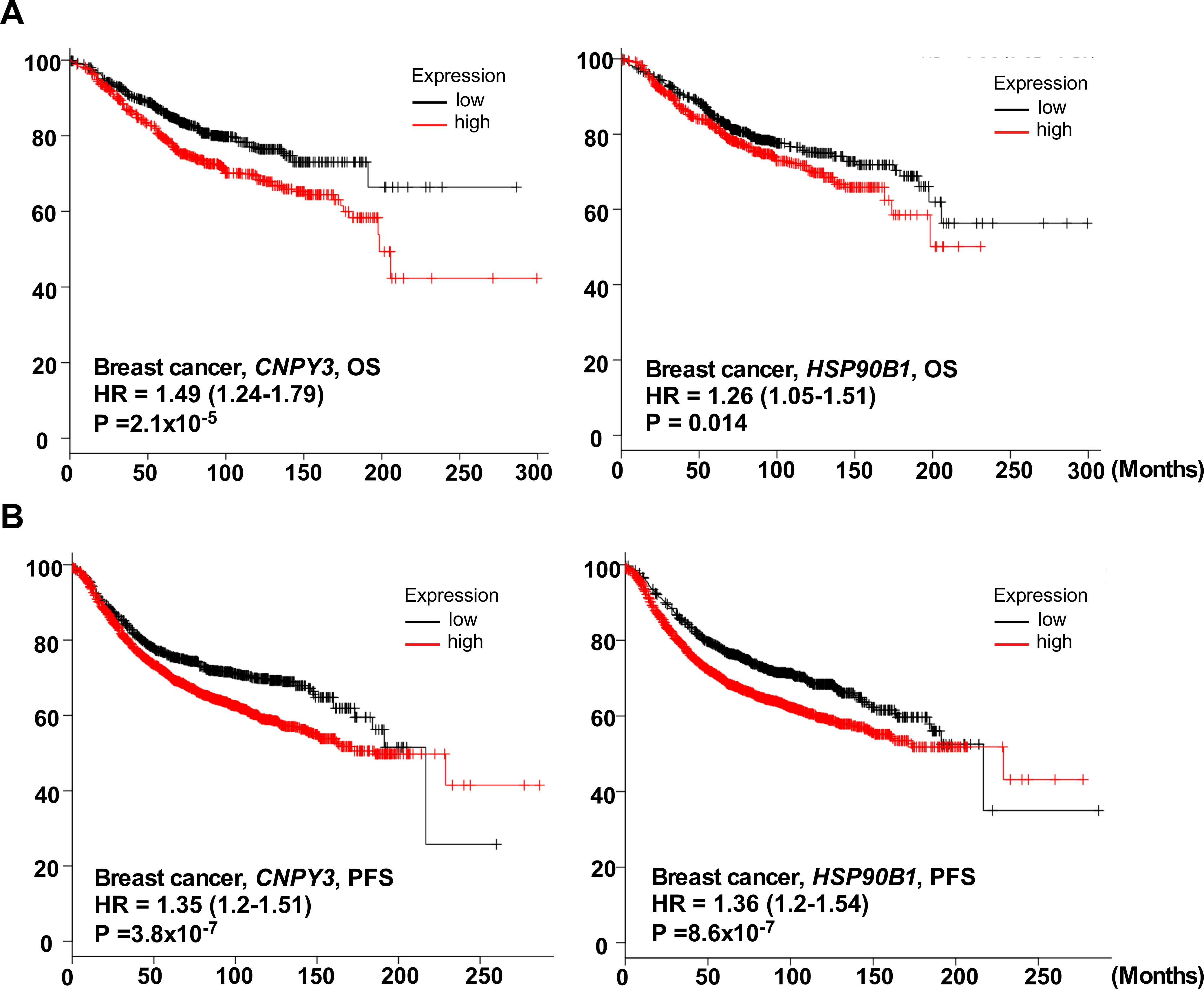
To validate the coordination of CNPY3 and HSP90B1 to predict an aggressive phenotype, we next determined the correlation of these two genes with clinical parameters based on the Curtis breast dataset (n = 2136) downloaded from the Oncomine database [28] (Table S1). Both CNPY3 and HSP90B1 were overexpressed in breast cancers (Figure 1A). Also, we confirmed the consistency in the TCGA breast cancer dataset (n = 593) and Ki colon dataset (n = 123) (Figure S2, Table S1). Furthermore, high expression of CNPY3 and HSP90B1 correlated with poor differentiation (Figure 1B), PR negative status (Figure 1C), triple-negative breast cancer (TNBC) (Figure 1D), a high Nottingham Prognostic Index (NPI) (Figure 1E), and an aggressive basal subtype (Figure 1F). Additionally, we used the Kaplan–Meier plotter database to confirm the consistent predictive value for clinical outcomes. In a large group of breast cancer patients (n = 1907), patients with high levels of CNPY3 and HSP90B1 expression had shorten overall survival (Figure 2A) and progression-free survival (Figure 2B).
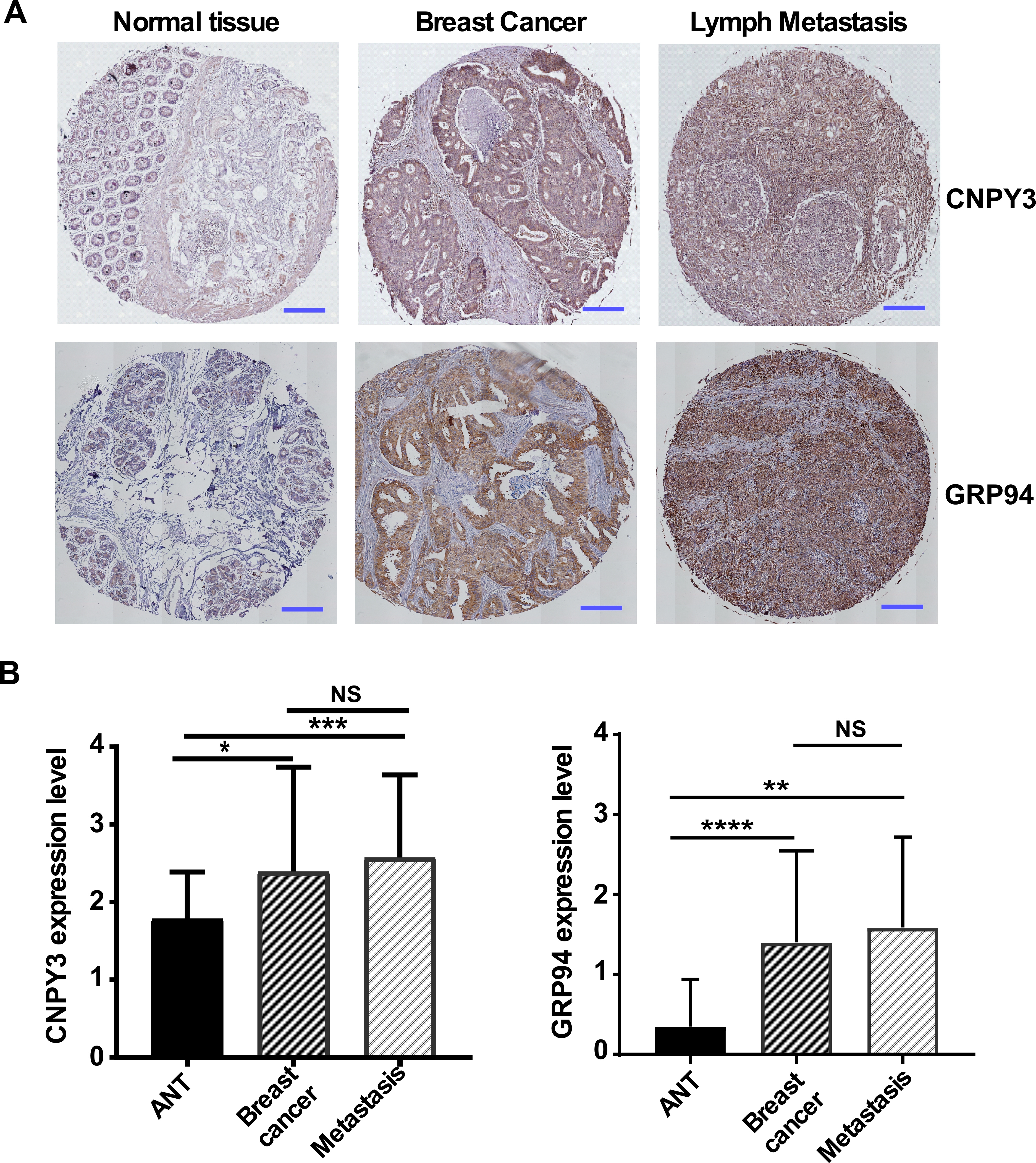
To determine whether protein expression levels of CNPY3 and GRP94 are consistent with gene expression levels and associated with clinical features in human breast cancers, we used the tissue microarray (TMA) approach, which included breast cancer, their corresponding adjacent normal tissue (ANT), and metastasis tissue (n = 158). For the first time, we found that CNPY3 was significantly overexpressed in cancer and metastatic tissues but not in ANT (Figure 3A, top and Figure 3B, left). Consistent with CNPY3, GRP94 was highly expressed in cancer and metastasis tissues compared with ANT (Figure 3A, bottom and Figure 3B, right). However, there is no significant difference in CNPY3 and GRP94 expression between primary and metastatic cancers. These results suggested that CNPY3 may be critical in human cancer development and progression.

3.3. Deletion of CNPY3 Suppresses Human Breast Tumor Growth and METASTASIS In Vitro and In Vivo

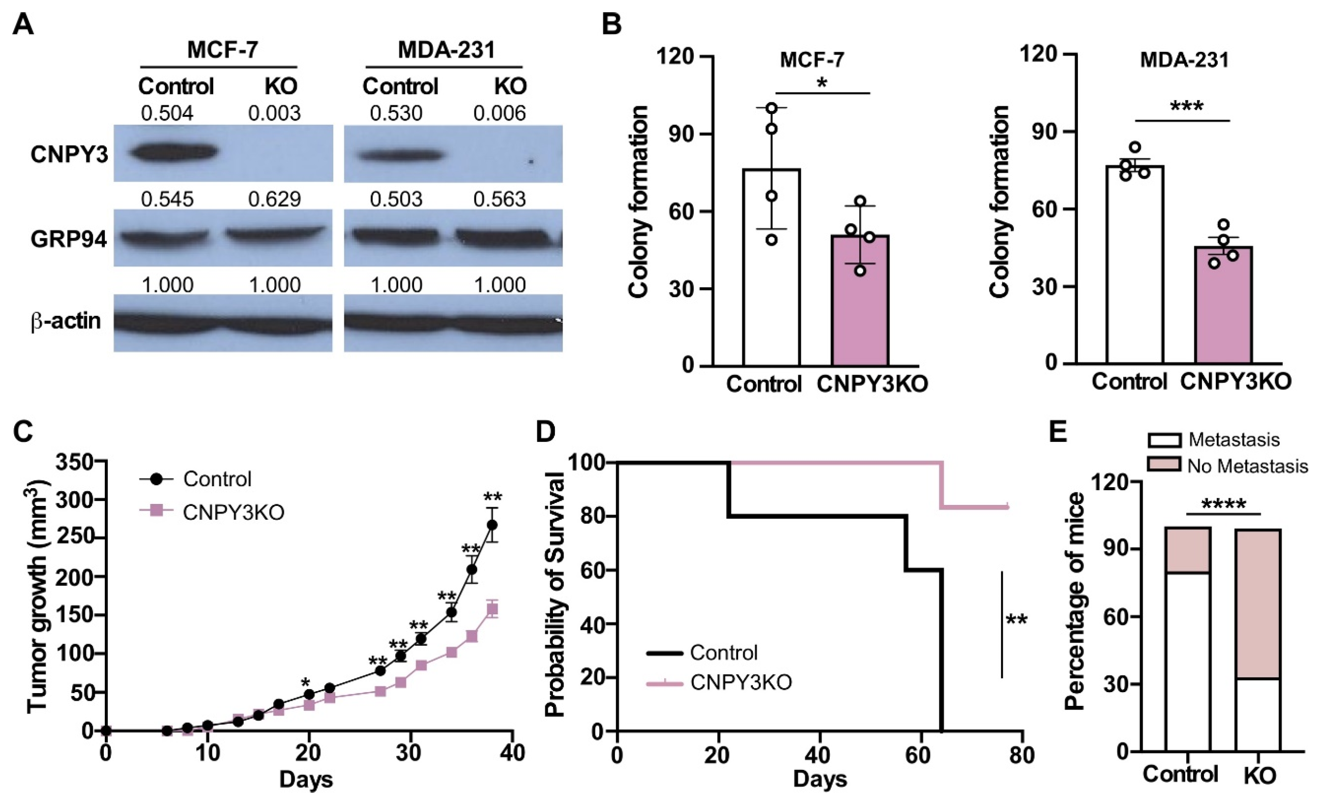
To further investigate the role of CNPY3 in breast cancer progression and metastasis, we next silenced CNPY3 in human breast cancer cell lines MCF-7 (ER+PR+BC) and MDA-MB-231 (TNBC) using a CRISPR/Cas9 lentiviral system. We confirmed that CNPY3 was completely knocked out in both MCF-7 and MDA-MB-231 tumor cells from different passages (Figure 4A). First, we performed tumor cell colony formation in vitro. We found that silencing of CNPY3 causes significantly compromised MCF-7 and MDA-MB-231 tumor cell growth compared with control tumor cells (Figure 4B). The MDA-MB-231 cell line is a poorly differentiated triple-negative breast cancer and has a stronger invasive ability than MCF-7, and it is a well-known metastatic breast cancer model. To determine whether CNPY3 is required for human breast cancer cell growth and metastasis in vivo, we performed a human breast cancer xenograft model by implanting WT control and CNPY3 KO MDA-MB-231 breast cancer cells into the fourth mammary gland in NSG mice and monitored tumor growth, endpoint survival, and metastasis. We found that deletion of CNPY3 in human breast cancer cells significantly decreased tumor growth and prolonged survival (Figure 4C,D) and reduced metastasis (Figure 4E), suggesting that CNPY3 may be a potential therapeutic target for cancers.

3.4. CNPY3 and GRP94 Alterations Regulate UPR Pathway Genes and Immune-Related Genes in the Tumor Microenvironment

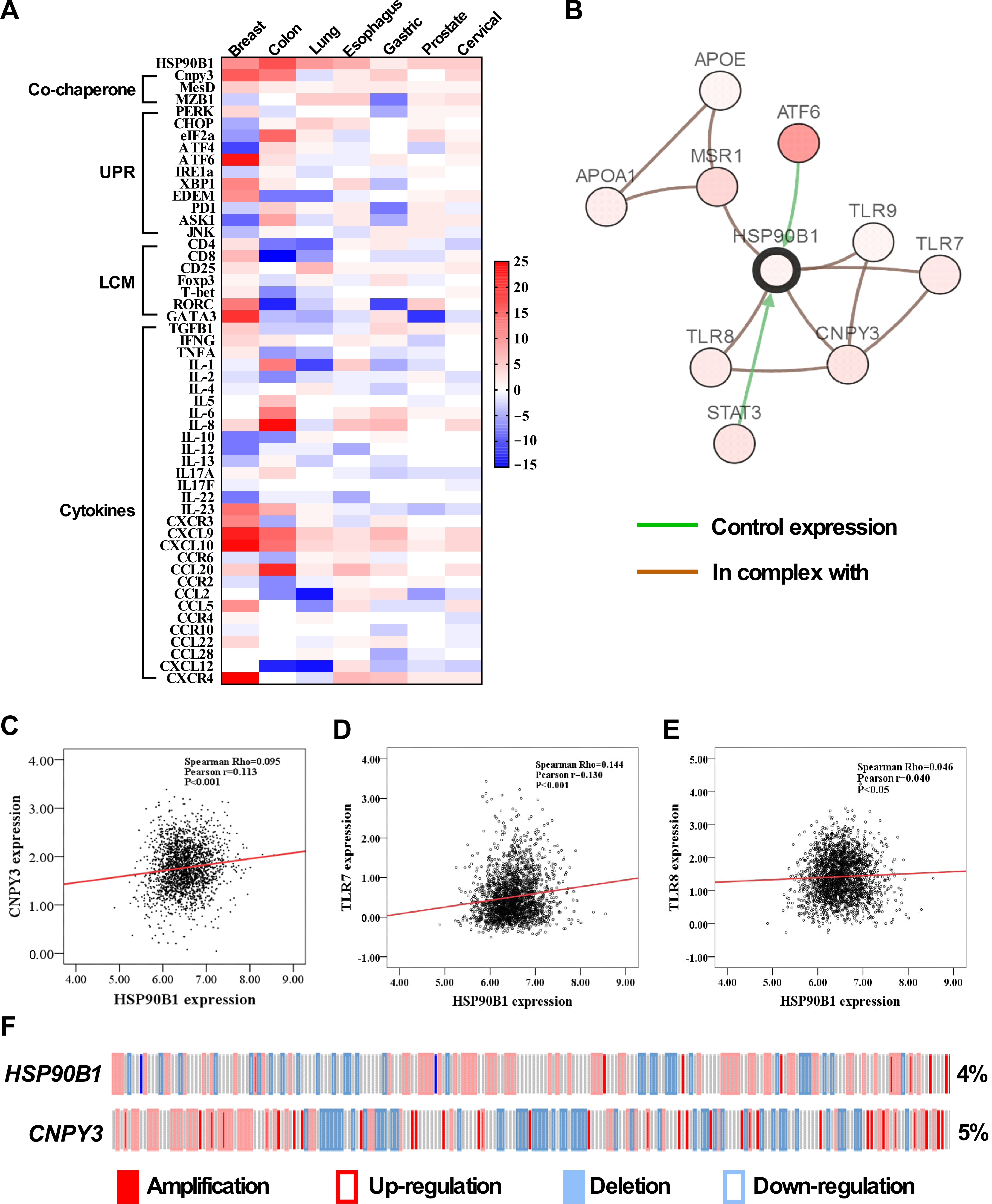
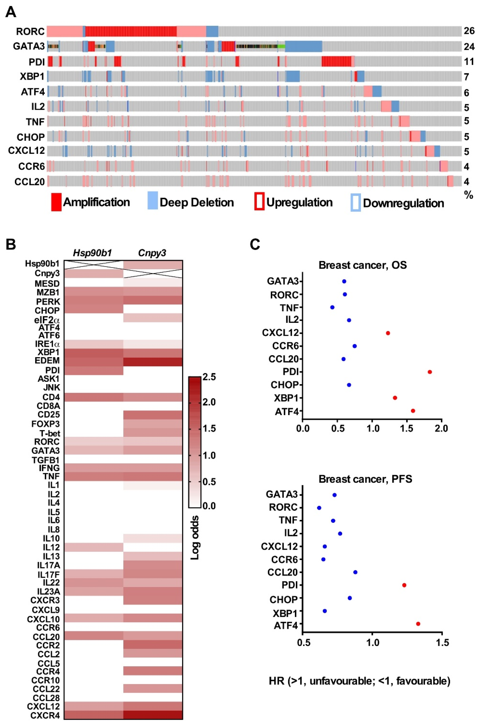
GRP94 is a downstream molecule in the ER to mediate UPR [29]. It also plays a key immunological role through variety of client proteins [6]. A recent study showed that GARP, one of GRP94 client protein, promotes oncogenesis in breast cancer [11]. To understand the functional significance of CNPY3, a co-chaperone of GRP94, in the tumor microenvironment, we examined the co-expression of HSP90B1 with co-chaperones, UPR pathway genes, lymphocyte markers, and cytokines using the Oncomine database and found that higher expression of HSP90B1, co-chaperone CNPY3, UPR pathway, and immune-related genes in common solid tumors (Figure 5A). In addition, we generated the network of HSP90B1 and found that it is associated with 9 most frequently altered neighbor genes, including CNPY3, ATF4, STAT3, TLR7, TLR8 and TLR9 based on the cBioportal database (Figure 5B). However, we found a weak correlation between HSP90B1 and CNPY3 (Figure 5C) and TLR7 (Figure 5D). There is no correlation of HSP90B1 with TLR8 (Figure 5E) based on the breast cancer microarray datasets [28]. The gene levels of CNPY3 and GRP94 might not be the same as protein levels. Whether CNPY3’s oncogenic effects are dependent on GRP94 and whether CNPY3 is correlated with GRP94 still need further investigation. To determine whether HSP90B1 and CNPY3 gene mutations impact the UPR, lymphocyte markers, and cytokines, we assessed gene mutations of HSP90B1 and CNPY3 with mutation rates of 4% and 5%, most of which were amplified and upregulated (Figure 5F). Next, based on the presence or absence of the HSP90B1 mutation, we evaluated the mRNA expression of targeted genes and found that the HSP90B1 gene mutation was associated with alterations in the expression of various genes, including genes in the UPR pathway (ATF4, XBP1, CHOP, PDI); Th17 and Th2 T cell markers (RORC and GATA3); and cytokines (CCR6, CCL20, CXCL12, IL2, TNF). Additionally, we found that these genes had a relatively high mutation rate of 4-26% (Figure 6A). Furthermore, we pursued the pairwise association analysis for HSP90B1, CNPY3 and UPR and immune-related genes and confirmed the co-occurrence of the genomic alterations (Figure 6B). These genes were found to be significant prognostic factors based on Kmplot data (Figure 6C). Our study suggested that CNPY3, a co-chaperone of GRP94, may play important roles in regulating the tumor microenvironment and anti-tumor immune response.

4. Discussion

As an ER chaperone involved in protein quality control, stress, and inflammation, GRP94 promotes tumor progression through its role in folding a variety of clients [11]. Using a genetic strategy, our group has discovered that GRP94 is an essential chaperone for folding the Wnt co-receptor LRP6 [30] and that it is required for multiple myeloma cell survival, which is mediated in part by the Wnt target survivin [10]. Our group also demonstrated that GRP94 chaperones GARP [9], and the GARP-TGFβ axis plays an important role in platelets, which inhibit anti-tumor immunity. Platelet-specific deletion of the GARP blunted TGFβ activity in the TME and enhanced protective immunity against both melanoma and colon cancer [31]. Recent studies have shown that human epidermal growth factor receptor-2 (HER2) expression is elevated in approximately 18–25% of breast cancers [32], and GRP94 is required for both the proper intracellular trafficking and stability of HER2 protein [33]. Also, GRP94-specific inhibitors affected high-density HER2 formations at the plasma membrane, which is critical for proper HER2 signaling in breast cancers [34]. These GRP94 clients have been described to function at various stages of cancer development. Beyond its known function as a TLR-specific co-chaperone for GRP94 in the ER [17,18], CNPY3 regulates inflammasome activation by promoting ASC/caspase-1 colocalization independently of TLR signaling [23]. Furthermore, CNPY3 is upregulated in colon adenocarcinoma, where its expression correlates with poor clinical outcomes and promotes tumor cell proliferation and migration in vitro [24].
However, the role of CNPY3 in human cancers is still not fully understood. In this study, we pursued comprehensive molecular profiling of CNPY3 and GRP94 and found that both CNPY3 and GRP94 are upregulated in different human cancers, including breast, liver, colon, pancreatic, ovarian cancers, etc., suggesting that CNPY3 may be a potential biomarker and therapeutic target for pan cancers (Figures S1 and S2). Human breast cancer is a heterogeneous cancer with different types based on origins, molecular features, and responses to therapeutics. We found that the elevated expression of both CNPY3 and GRP94 was associated with an aggressive phenotype and poor clinical outcome (Figure 1, Figure 2 and Figure 3). Both CNPY3 and GRP94 overexpression were correlated with TNBC, advanced stage, high NPI, and poorly differentiated breast cancers (Figure 1). Additionally, both CNPY3 and GRP94 had consistent prognostic value for overall survival and progression-free survival in breast cancer (Figure 2). Intriguingly, targeting CNPY3 in human ER+PR+ and TNBC cells by CRISPR/Cas9 significantly decreased tumor growth, prolonged survival, and reduced metastasis in vitro and in vivo (Figure 4). We have shown that the expression level of CNPY3 is strongly associated with TNBC and basal subtypes (Figure 1), suggesting CNPY3 subtype-specific roles. Further studies comparing CNPY3 dependency across a broader panel of breast cancer subtypes will be a focus of our ongoing research. Clinically, TNBC is an aggressive breast cancer with a high rate of recurrence and is ineffective in responding to hormone and HER2-targeted therapies, as well as checkpoint blockade immunotherapy. This study may provide a foundation for the development of CNPY3-targeted therapy to overcome drug resistance and enhance immunotherapy. However, there are no small molecule inhibitors and antibodies available for targeting CNPY3. Further development of CNPY3-specific inhibitors for preclinical study is needed.
To identify potential oncogenic mechanisms underlying the elevated expression of CNPY3 and GRP94, we performed network analysis and found the nine most frequently altered neighboring genes, including STAT3, ATF6, CNPY3, TLR7, TLR8, and TLR9 (Figure 5). As a protective factor, ATF6 orchestrates cellular adaptation to ER stress by fine-tuning the expression of UPR target genes, such as GRP78 and GRP94. A wide range of stressful conditions exist in tumor development and progression, and chaperones are needed to regulate the balance between cancer cell viability and apoptosis. Prolonged activation of ER stress sensors enables malignant cells to acquire enhanced tumorigenic, metastatic, and therapy-resistant capabilities [35]. STAT3 is a member of the STAT protein family. Triggered by diverse cytokines and growth factors, STAT3 modulates the expression of stimulus-responsive genes, thereby exerting a pivotal regulatory influence on cellular processes, including cell growth and apoptosis, invasion, and immunosuppression [36].
In this study, we also demonstrate that CNPY3 and GRP94 are associated with the UPR pathway and immunity-related genes, including lymphocyte cell markers and cytokines (Figure 5). After enrichment and pairwise co-occurrence analysis, we found that GRP94 and CNPY3 mediated changes in the expression of genes, including the UPR pathway genes ATF4, XBP1, CHOP, and PDI, and the immune-related genes RORC, GATA3, IL2, TNF, CCR6, CXCL12, and CCL20 (Figure 6). Upon antigen stimulation through their T cell receptor, naive CD4+ T cells differentiate into distinct effector lineages, including Th1, Th2, and IL-17-producing Th17 cells. The commitment to distinct T helper (Th) lineages is orchestrated by lineage-defining transcription factors: T-bet governs Th1 differentiation, GATA3 dictates Th2 differentiation, and RORC drives Th17 cell fate specification [37,38]. Human Th17 cells express high levels of CCR6 and CXCR4. These homing receptors likely orchestrate Th17 cell migration and retention in inflammatory foci and tumor sites [39]. For instance, human tumor microenvironments exhibit elevated expression of CXCL12 (CXCR4 ligand) and CCL20 (CCR6 ligand), creating a chemokine gradient that potentiates Th17 cell infiltration into tumors. [39]. Furthermore, Th17 cells are established as pivotal regulators across multiple immune-mediated inflammatory diseases and cancer contexts [40]. Patients with high expression of the Th17 cluster had a poor prognosis [41]. CXCL12-CXCR4 signaling is implicated in immune cell tumor trafficking and tumor cell biology. CXCL12-CXCR4 signaling mediates plasmacytoid DC trafficking into tumors and Treg cell homing to the bone marrow microenvironment and is involved in tumor cell proliferation, metastasis, and tumor vascularization [39]. Hence, on the one hand, CNPY3 and GRP94 may promote prosurvival signaling through the UPR pathway to promote tumor cell survival under endogenous and exogenous stress conditions; on the other hand, they also play important immunosuppressive roles through cytokines to misbalance immune cells in the tumor microenvironment. However, how the CNPY3-UPR signaling pathway regulates the tumor microenvironment is still not clear. Further study is needed to understand how CNPY3 regulates the tumor microenvironment and anti-tumor immunity for the development of combination therapies for cancers.

5. Conclusions

Our study demonstrated that CNPY3, a cochaperone of immune chaperone GRP94, was elevated in the majority of human cancers, and the overexpression of CNPY3 and GRP94 predicts an aggressive phenotype and poor clinical outcome. Targeting CNPY3 in human breast cancer cells reduced tumor growth and metastasis. In addition, the changes in the UPR pathway and immune-related genes in the tumor microenvironment were significantly dependent on alterations in CNPY3 and GRP94. For the first time, our study uncovers the critical roles of CNPY3 in the progression and metastasis of breast cancer, suggesting that CNPY3 may be a novel biomarker and therapeutic target for cancers. However, the underlying mechanism of CNPY3 in regulating the tumor microenvironment and anti-tumor immunity needs to be further investigated.

Supplementary Materials

The following supporting information can be downloaded at: https://www.mdpi.com/article/10.3390/cimb47110883/s1.

Author Contributions

Conceptualization, Z.L. and B.L.; Methodology, X.D., R.Z., S.S. and B.L.; Formal Analysis, X.D., R.Z., S.S. and B.L.; Investigation, X.D., R.Z. and B.L.; Resources, B.G. and Z.L.; Writing—Original Draft, X.D., R.Z. and B.L.; Writing—Review & Editing, X.D., R.Z., S.S., B.G., Z.L. and B.L.; Supervision, B.L.; Funding Acquisition, B.L. All authors have read and agreed to the published version of the manuscript.

Funding

This study was partially supported by NIH grants CA193939 and AI125859 (to B.L.), and internal research funds from OSUCCC-James Cancer Center to B.L.

Institutional Review Board Statement

All mice were bred and maintained according to the guidelines and a protocol approved by the Medical University of South Carolina Institutional Animal Care and Use Committee (approve number: IACUC-2018-00375, 19 September 2018).

Informed Consent Statement

Not applicable.

Data Availability Statement

The original contributions presented in this study are included in the article/Supplementary Material. Further inquiries can be directed to the corresponding author.

Acknowledgments

We thank our laboratory members (Soo Ngoi, Stephen Iwanowycz, Yingqi Li, and Jennifer Gutierrez) for stimulating discussions and suggestions.

Conflicts of Interest

The authors, Xiaofeng Duan, Ran Zhao, Shaoli Sun, Zihai Li, Bei Liu declare no conflicts of interest. The author, Beichu, Guo, is currently an employee of ALLERDIA Inc. and declares that ALLERDIA Inc. was not involved in the study design, collection, analysis, interpretation of data, funding support. the writing of this article or the decision to submit it for publication.

References

  1. Rozpedek, W.; Pytel, D.; Mucha, B.; Leszczynska, H.; Diehl, J.A.; Majsterek, I. The Role of the PERK/eIF2alpha/ATF4/CHOP Signaling Pathway in Tumor Progression During Endoplasmic Reticulum Stress. Curr. Mol. Med. 2016, 16, 533–544. [Google Scholar] [CrossRef]
  2. Heritz, J.A.; Backe, S.J.; Mollapour, M. Molecular chaperones: Guardians of tumor suppressor stability and function. Oncotarget 2024, 15, 679–696. [Google Scholar] [CrossRef]
  3. Hwang, S.M.; Chang, S.; Rodriguez, P.C.; Cubillos-Ruiz, J.R. Endoplasmic reticulum stress responses in anticancer immunity. Nat. Rev. Cancer 2025, 25, 684–702. [Google Scholar] [CrossRef]
  4. Chen, X.; Cubillos-Ruiz, J.R. Endoplasmic reticulum stress signals in the tumour and its microenvironment. Nat. Rev. Cancer 2021, 21, 71–88. [Google Scholar] [CrossRef]
  5. Singh, M.K.; Han, S.; Ju, S.; Ranbhise, J.S.; Ha, J.; Yeo, S.G.; Kim, S.S.; Kang, I. Hsp70: A Multifunctional Chaperone in Maintaining Proteostasis and Its Implications in Human Disease. Cells 2025, 14, 509. [Google Scholar] [CrossRef] [PubMed]
  6. Ansa-Addo, E.A.; Thaxton, J.; Hong, F.; Wu, B.X.; Zhang, Y.; Fugle, C.W.; Metelli, A.; Riesenberg, B.; Williams, K.; Gewirth, D.T.; et al. Clients and Oncogenic Roles of Molecular Chaperone gp96/grp94. Curr. Top. Med. Chem. 2016, 16, 2765–2778. [Google Scholar] [CrossRef] [PubMed]
  7. Staron, M.; Yang, Y.; Liu, B.; Li, J.; Shen, Y.; Zuniga-Pflucker, J.C.; Aguila, H.L.; Goldschneider, I.; Li, Z. gp96, an endoplasmic reticulum master chaperone for integrins and Toll-like receptors, selectively regulates early T and B lymphopoiesis. Blood 2010, 115, 2380–2390. [Google Scholar] [CrossRef] [PubMed]
  8. Weekes, M.P.; Antrobus, R.; Talbot, S.; Hor, S.; Simecek, N.; Smith, D.L.; Bloor, S.; Randow, F.; Lehner, P.J. Proteomic plasma membrane profiling reveals an essential role for gp96 in the cell surface expression of LDLR family members, including the LDL receptor and LRP6. J. Proteome Res. 2012, 11, 1475–1484. [Google Scholar] [CrossRef] [PubMed]
  9. Duan, X.; Iwanowycz, S.; Ngoi, S.; Hill, M.; Zhao, Q.; Liu, B. Molecular Chaperone GRP94/GP96 in Cancers: Oncogenesis and Therapeutic Target. Front. Oncol. 2021, 11, 629846. [Google Scholar] [CrossRef]
  10. Hua, Y.; White-Gilbertson, S.; Kellner, J.; Rachidi, S.; Usmani, S.Z.; Chiosis, G.; Depinho, R.; Li, Z.; Liu, B. Molecular chaperone gp96 is a novel therapeutic target of multiple myeloma. Clin. Cancer Res. 2013, 19, 6242–6251. [Google Scholar] [CrossRef]
  11. Metelli, A.; Wu, B.X.; Fugle, C.W.; Rachidi, S.; Sun, S.; Zhang, Y.; Wu, J.; Tomlinson, S.; Howe, P.H.; Yang, Y.; et al. Surface Expression of TGFbeta Docking Receptor GARP Promotes Oncogenesis and Immune Tolerance in Breast Cancer. Cancer Res. 2016, 76, 7106–7117. [Google Scholar] [CrossRef]
  12. Cho, Y.B.; Kim, J.W.; Heo, K.; Kim, H.J.; Yun, S.; Lee, H.S.; Shin, H.G.; Shim, H.; Yu, H.; Kim, Y.H.; et al. An internalizing antibody targeting of cell surface GRP94 effectively suppresses tumor angiogenesis of colorectal cancer. Biomed. Pharmacother. 2022, 150, 113051. [Google Scholar] [CrossRef]
  13. Wang, T.; Yang, L.; Li, C.; Wang, J.; Zhang, J.; Zhou, Y.; Sun, F.; Wang, H.; Ma, F.; Qian, H. Comprehensive analysis reveals GRP94 is associated with worse prognosis of breast cancer. Transl. Cancer Res. 2021, 10, 298–309. [Google Scholar] [CrossRef]
  14. Li, C.; Wang, Y.; Long, L.; Zhang, P.; Zhang, Y.; Ji, N. gp96 Expression in Gliomas and Its Association with Tumor Malignancy and T Cell Infiltrating Level. J. Oncol. 2022, 2022, 9575867. [Google Scholar] [CrossRef]
  15. Klarica Gembic, T.; Grebic, D.; Gulic, T.; Golemac, M.; Avirovic, M. Predictive and Prognostic Values of Glycoprotein 96, Androgen Receptors, and Extranodal Extension in Sentinel Lymph Node-Positive Breast Cancer: An Immunohistochemical Retrospective Study. J. Clin. Med. 2024, 13, 7665. [Google Scholar] [CrossRef] [PubMed]
  16. Tian, T.; Han, J.; Huang, J.; Li, S.; Pang, H. Hypoxia-Induced Intracellular and Extracellular Heat Shock Protein gp96 Increases Paclitaxel-Resistance and Facilitates Immune Evasion in Breast Cancer. Front. Oncol. 2021, 11, 784777. [Google Scholar] [CrossRef]
  17. Takahashi, K.; Shibata, T.; Akashi-Takamura, S.; Kiyokawa, T.; Wakabayashi, Y.; Tanimura, N.; Kobayashi, T.; Matsumoto, F.; Fukui, R.; Kouro, T.; et al. A protein associated with Toll-like receptor (TLR) 4 (PRAT4A) is required for TLR-dependent immune responses. J. Exp. Med. 2007, 204, 2963–2976. [Google Scholar] [CrossRef] [PubMed]
  18. Miyake, K.; Saitoh, S.I.; Fukui, R.; Shibata, T.; Sato, R.; Murakami, Y. Dynamic control of nucleic-acid-sensing Toll-like receptors by the endosomal compartment. Int. Immunol. 2021, 33, 835–840. [Google Scholar] [CrossRef]
  19. Saitoh, S.; Miyake, K. Mechanism regulating cell surface expression and activation of Toll-like receptor 4. Chem. Rec. 2006, 6, 311–319. [Google Scholar] [CrossRef] [PubMed]
  20. Wakabayashi, Y.; Kobayashi, M.; Akashi-Takamura, S.; Tanimura, N.; Konno, K.; Takahashi, K.; Ishii, T.; Mizutani, T.; Iba, H.; Kouro, T.; et al. A protein associated with toll-like receptor 4 (PRAT4A) regulates cell surface expression of TLR4. J. Immunol. 2006, 177, 1772–1779. [Google Scholar] [CrossRef]
  21. McGettrick, A.F.; O’Neill, L.A. Localisation and trafficking of Toll-like receptors: An important mode of regulation. Curr. Opin. Immunol. 2010, 22, 20–27. [Google Scholar] [CrossRef] [PubMed]
  22. Hart, B.E.; Tapping, R.I. Cell surface trafficking of TLR1 is differentially regulated by the chaperones PRAT4A and PRAT4B. J. Biol. Chem. 2012, 287, 16550–16562. [Google Scholar] [CrossRef]
  23. Ghait, M.; Husain, R.A.; Duduskar, S.N.; Haack, T.B.; Rooney, M.; Gohrig, B.; Bauer, M.; Rubio, I.; Deshmukh, S.D. The TLR-chaperone CNPY3 is a critical regulator of NLRP3-inflammasome activation. Eur. J. Immunol. 2022, 52, 907–923. [Google Scholar] [CrossRef]
  24. Gao, X.C.; Zhou, B.H.; Ji, Z.X.; Li, Q.; Liu, H.N. Canopy FGF signaling regulator 3 affects prognosis, immune infiltration, and PI3K/AKT pathway in colon adenocarcinoma. World J. Gastrointest. Oncol. 2024, 16, 3284–3298. [Google Scholar] [CrossRef] [PubMed]
  25. Hou, G.X.; Liu, P.; Yang, J.; Wen, S. Mining expression and prognosis of topoisomerase isoforms in non-small-cell lung cancer by using Oncomine and Kaplan-Meier plotter. PLoS ONE 2017, 12, e0174515. [Google Scholar] [CrossRef]
  26. Yan, H.; Hu, K.; Wu, W.; Li, Y.; Tian, H.; Chu, Z.; Koeffler, H.P.; Yin, D. Low Expression of DYRK2 (Dual Specificity Tyrosine Phosphorylation Regulated Kinase 2) Correlates with Poor Prognosis in Colorectal Cancer. PLoS ONE 2016, 11, e0159954. [Google Scholar] [CrossRef]
  27. Gao, J.; Aksoy, B.A.; Dogrusoz, U.; Dresdner, G.; Gross, B.; Sumer, S.O.; Sun, Y.; Jacobsen, A.; Sinha, R.; Larsson, E.; et al. Integrative analysis of complex cancer genomics and clinical profiles using the cBioPortal. Sci. Signal. 2013, 6, pl1. [Google Scholar] [CrossRef] [PubMed]
  28. Curtis, C.; Shah, S.P.; Chin, S.F.; Turashvili, G.; Rueda, O.M.; Dunning, M.J.; Speed, D.; Lynch, A.G.; Samarajiwa, S.; Yuan, Y.; et al. The genomic and transcriptomic architecture of 2,000 breast tumours reveals novel subgroups. Nature 2012, 486, 346–352. [Google Scholar] [CrossRef]
  29. Zhu, G.; Lee, A.S. Role of the unfolded protein response, GRP78 and GRP94 in organ homeostasis. J. Cell. Physiol. 2015, 230, 1413–1420. [Google Scholar] [CrossRef]
  30. Liu, B.; Staron, M.; Hong, F.; Wu, B.X.; Sun, S.; Morales, C.; Crosson, C.E.; Tomlinson, S.; Kim, I.; Wu, D.; et al. Essential roles of grp94 in gut homeostasis via chaperoning canonical Wnt pathway. Proc. Natl. Acad. Sci. USA 2013, 110, 6877–6882. [Google Scholar] [CrossRef]
  31. Rachidi, S.; Metelli, A.; Riesenberg, B.; Wu, B.X.; Nelson, M.H.; Wallace, C.; Paulos, C.M.; Rubinstein, M.P.; Garrett-Mayer, E.; Hennig, M.; et al. Platelets subvert T cell immunity against cancer via GARP-TGFbeta axis. Sci. Immunol. 2017, 2, eaai7911. [Google Scholar] [CrossRef] [PubMed]
  32. Baselga, J.; Swain, S.M. Novel anticancer targets: Revisiting ERBB2 and discovering ERBB3. Nat. Rev. Cancer 2009, 9, 463–475. [Google Scholar] [CrossRef]
  33. Chavany, C.; Mimnaugh, E.; Miller, P.; Bitton, R.; Nguyen, P.; Trepel, J.; Whitesell, L.; Schnur, R.; Moyer, J.; Neckers, L. p185erbB2 binds to GRP94 in vivo. Dissociation of the p185erbB2/GRP94 heterocomplex by benzoquinone ansamycins precedes depletion of p185erbB2. J. Biol. Chem. 1996, 271, 4974–4977. [Google Scholar] [CrossRef]
  34. Patel, P.D.; Yan, P.; Seidler, P.M.; Patel, H.J.; Sun, W.; Yang, C.; Que, N.S.; Taldone, T.; Finotti, P.; Stephani, R.A.; et al. Paralog-selective Hsp90 inhibitors define tumor-specific regulation of HER2. Nat. Chem. Biol. 2013, 9, 677–684. [Google Scholar] [CrossRef]
  35. Cubillos-Ruiz, J.R.; Bettigole, S.E.; Glimcher, L.H. Tumorigenic and Immunosuppressive Effects of Endoplasmic Reticulum Stress in Cancer. Cell 2017, 168, 692–706. [Google Scholar] [CrossRef] [PubMed]
  36. Yu, H.; Lee, H.; Herrmann, A.; Buettner, R.; Jove, R. Revisiting STAT3 signalling in cancer: New and unexpected biological functions. Nat. Rev. Cancer 2014, 14, 736–746. [Google Scholar] [CrossRef] [PubMed]
  37. Yang, X.O.; Pappu, B.P.; Nurieva, R.; Akimzhanov, A.; Kang, H.S.; Chung, Y.; Ma, L.; Shah, B.; Panopoulos, A.D.; Schluns, K.S.; et al. T helper 17 lineage differentiation is programmed by orphan nuclear receptors ROR alpha and ROR gamma. Immunity 2008, 28, 29–39. [Google Scholar] [CrossRef]
  38. Yu, F.; Sharma, S.; Edwards, J.; Feigenbaum, L.; Zhu, J. Dynamic expression of transcription factors T-bet and GATA-3 by regulatory T cells maintains immunotolerance. Nat. Immunol. 2015, 16, 197–206. [Google Scholar] [CrossRef]
  39. Nagarsheth, N.; Wicha, M.S.; Zou, W. Chemokines in the cancer microenvironment and their relevance in cancer immunotherapy. Nat. Rev. Immunol. 2017, 17, 559–572. [Google Scholar] [CrossRef]
  40. Sundrud, M.S.; Trivigno, C. Identity crisis of Th17 cells: Many forms, many functions, many questions. Semin. Immunol. 2013, 25, 263–272. [Google Scholar] [CrossRef]
  41. Yoshida, N.; Kinugasa, T.; Miyoshi, H.; Sato, K.; Yuge, K.; Ohchi, T.; Fujino, S.; Shiraiwa, S.; Katagiri, M.; Akagi, Y.; et al. A High RORgammaT/CD3 Ratio is a Strong Prognostic Factor for Postoperative Survival in Advanced Colorectal Cancer: Analysis of Helper T Cell Lymphocytes (Th1, Th2, Th17 and Regulatory T Cells). Ann. Surg. Oncol. 2016, 23, 919–927. [Google Scholar] [CrossRef] [PubMed]
Figure 1. High expression ofCNPY3 and HSP90B1are correlated with disease progression phenotype in human breast cancers. CNPY3 and HSP90B1 predicts aggressive behavior in breast cancer based on 1980 breast cancer samples and 144 normal breast samples in Curtis et al. Nature dataset. (A) CNPY3 and HSP90B1 mRNA were both overexpressed in breast cancers compared with adjacent normal tissues. (BF) High mRNA levels of CNPY3 and HSP90B1 correlate with poor pathological differentiation breast cancers (B), progesterone receptor (PR)-negative breast cancers (C), triple-negative breast cancers (D), high Nottingham Prognostic Index (NPI) breast cancers (E), and PAM50 luminal A, luminal B and basal subtype breast cancers (F). * p < 0.05, ** p < 0.01, *** p < 0.001, **** p < 0.0001, NS/ns: not significant.The red dot line represnts mean.
Cimb 47 00883 g001
Figure 2. Upregulation ofCNPY3 and HSP90B1is associated with poor prognosis in human breast cancers. (A) Breast cancer patients with high expression of CNPY3 (left) and HSP90B1 (right) have significant poor overall survival (OS). (B) Breast cancer patients with high expression of CNPY3 (left) and HSP90B1 (right) have significant poor progression-free survival (PFS). HR: hazard ratio.
Cimb 47 00883 g002
Figure 3. CNPY3 and GRP94 are hallmarks of human breast cancer development and progression. (A) Representative CNPY3 (top) and GRP94 (bottom) staining in adjacent normal tissue (ANT), breast cancer and lymph node metastasis tissue by immunohistochemistry. Scale bar: 25 μm. (B) Quantification of CNPY3 (left) and GRP94 (right) expression in breast cancer. n = 158 for ANT, tumor, and metastasis. NS: not significant. Data are shown as mean ± SD. * p < 0.05, ** p < 0.01, *** p < 0.001, **** p < 0.0001, NS: Not Significant.
Cimb 47 00883 g003
Figure 4. Deletion of CNPY3 decreased human breast cancer cell growth and metastasis in vitro and in vivo. (A) Immunoblot analysis of CNPY3, GRP94, and β-actin in empty vector (EV) control and CNPY3 knockout (KO) MCF-7 and MDA-231 human breast cancer cells. (B) Colony formation assays were performed to assess the control and CNPY3 KO MCF-7 and MDA-231 tumor cell proliferation. (C) Tumor growth of WT control and CNPY3 knockdown MDA-231 cells implanted into the fourth mammary gland in NSG mice (n = 6). (D) Kaplan–Meier curves for the endpoint survival after implantation of control and CNPY3 KO MDA-231 cells in NSG mice (n = 6). (E) Percentage of mice with metastasis and without metastasis. Error bars indicate SD. * p < 0.05, ** p < 0.01, *** p < 0.001, **** p < 0.0001.
Cimb 47 00883 g004
Figure 5. Heatmap and network ofHSP90B1andCNPY3in human cancers. (A) Heatmap of mRNA expression for HSP90B1, co-chaperone, unfolded protein response (UPR) pathway, lymphocyte marker (LCM), and immune-related genes in common solid tumors. The red square indicates upregulation, and blue indicates downregulation, and p value < 0.05 (Oncomine Platform, Thermo Fisher, Ann Arbor, MI, USA). (B) HSP90B1 and CNPY3 gene network in cBioPortal for Cancer Genomics (http://www.cbioportal.org, accessed on 15 October 2025). The network contains 10 nodes, including HSP90B1 and the 9 most frequently altered neighbor genes. (C) The weak correlation of HSP90B1 and CNPY3 mRNA levels in the Curtis et al., Nature 2012 dataset [28] (n = 2136, r = 0.113, p value < 0.001). (D) The weak correlation of HSP90B1 and TLR7 mRNA levels in the Curtis et al., Nature 2012 dataset (n = 2136, r= 0.130, p value < 0.001). (E) There was no correlation of HSP90B1 and TLR8 mRNA levels in the Curtis et al., Nature 2012 dataset (n = 2136, r = 0.040, p value < 0.001). (F) The gene alternation of HSP90B1 and CNPY3 in the Curtis et al., Nature 2012 dataset. Red solid bars indicate gene amplifications, blue solid bars are deletions, and red blank bars indicate upregulation, and blue blank bars indicate downregulation.
Cimb 47 00883 g005
Figure 6. CNPY3 and GRP94 alterations regulate UPR pathway and immune-related genes in the tumor microenvironment. (A) cBioportal data show the gene alternation of enrichment genes based on the HSP90B1 alternation, including the UPR pathway ATF4, CHOP, XBP1 and PDI genes; Th17 and Th2 cell markers RORC and GATA3; and cytokines CCL20, CCR6, CXCL12, IL2 and TNF. Red solid bars indicate gene amplifications, blue solid bars are deletions, and red blank bars indicate upregulation, and blue blank bars indicate downregulation. (B) Pair wise association plot for HSP90B1, CNPY3, the UPR pathway and immune-related genes based on the Curtis breast cancer dataset. This map provides summary statistics on mutual exclusivity and co-occurrence of genomic alterations in each pair of genes. The color scale represents the magnitude of the association (log odds). (C) Kaplan–Meier plotter data (http://kmplot.com, accessed on 15 October 2025) show the prognostic correlation of enrichment genes in breast cancer. Red dot (hazard ratio, HR > 1, p < 0.05), high expression is an unfavorable factor; blue dot (hazard ratio, HR < 1, p < 0.05), high expression is a favorable factor. OS, overall survival; PFS, progression-free survival.
Cimb 47 00883 g006
Disclaimer/Publisher’s Note: The statements, opinions and data contained in all publications are solely those of the individual author(s) and contributor(s) and not of MDPI and/or the editor(s). MDPI and/or the editor(s) disclaim responsibility for any injury to people or property resulting from any ideas, methods, instructions or products referred to in the content.