Article

Integrating Morphological, Pathogenic, and Molecular Approaches to Characterize Fusarium Root Rot Pathogens of Common Bean in Egypt

by
Taghrid A. Kamel
1,
Manal M. Yasser
2,
Naglaa A. Taha
1,
Dalal Hussien M. Alkhalifah
3,
Marym A. Marzouk
2,
Wael N. Hozzien
2 and
Walaa R. Abdelghany
1,*
1
Plant Pathology Research Institute, Agricultural Research Center, Giza 12619, Egypt
2
Botany and Microbiology Department, Faculty of Science, Beni-Suef University, Beni-Suef 62511, Egypt
3
Department of Biology, College of Science, Princess Nourahbint Abdulrahman University, Riyadh 11671, Saudi Arabia
*
Author to whom correspondence should be addressed.
Curr. Issues Mol. Biol. 2025, 47(10), 803; https://doi.org/10.3390/cimb47100803
Submission received: 18 August 2025 / Revised: 13 September 2025 / Accepted: 25 September 2025 / Published: 29 September 2025
(This article belongs to the Section Molecular Plant Sciences)

Abstract

Fusarium root rot (FRR) is a major disease affecting common bean (Phaseolus vulgaris L.) production worldwide. In Egypt, FRR has recently become more prevalent, threatening crop yields. Fusarium species are considered the primary causal agents of this disease. To identify the pathogens associated with FRR and evaluate host resistance, Fusarium isolates were obtained from diseased common bean plants collected in different Egyptian governorates. Morphological identification, pathogenicity assays on six cultivars (Alpha, Samantha, Giza 6, Giza 12, Cambo, and Nebraska), and molecular identification using TEF-1α gene sequencing were conducted. Thirteen isolates exhibited pathogenicity, and five isolates (FP33, FP24, FP26, FP21, and FP11) were classified as highly aggressive. Isolate FP33 caused the highest disease severity, reaching 90% on Giza 6 and 80% on Cambo, while Nebraska showed the highest resistance (30% disease severity). Similarly, FP24 led to 85% and 75% severity on Giza 6 and Cambo, respectively. Nebraska and Giza 12 showed the greatest resistance, while Giza 6 and Cambo were most susceptible. Molecular analysis identified FP33 and FP24 as F. equiseti, FP26 and FP21 as F. oxysporum, and FP11 as F. solani. The study demonstrates the genetic and pathogenic variability among Fusarium isolates causing root rot in common bean. Nebraska and Giza 12 were identified as the most resistant cultivars, while Giza 6 and Cambo were highly susceptible. These findings highlight the importance of selecting resistant cultivars and implementing integrated disease management strategies to mitigate FRR in Egypt. The results also contribute valuable data for breeding programs aimed at developing durable resistance. The integration of morphological, molecular, and pathogenicity data provides a framework for future epidemiological studies and sustainable disease management strategies.

Graphical Abstract

1. Introduction

Among species in the Phaseolus genus, the common bean (Phaseolus vulgaris L.) holds the greatest economic significance, being extensively cultivated and considered the most significant leguminous crop, particularly for direct human consumption. Common bean is a vital nutritional crop, valued for its high protein content, which contributes to its status as one of the most significant pulse crops globally. In addition to protein, it serves as a rich source of dietary fiber, iron, essential vitamins, minerals, and complex carbohydrates [1]. As a result, it is a vital crop for global food security, especially in developing countries, where it contributes to combating malnutrition among millions of smallholder farmers [2]. It accounts for 85% of global bean production [3], with 37.8 million hectares harvested annually, yielding approximately 28.5 million tons in 2023 and feeding over 300 million people [4]. In Egypt, the total cultivated area reaches 28,291 hectares for dry beans, producing around 174,178 tons per year [4], while green beans are grown on 28,390.32 hectares, yielding approximately 2.82 million tons annually [5].
Fungal diseases can lead to severe yield and quality losses, ranging from 20% to 100%, depending on factors such as the specific diseases affecting the crop, the severity of the infections, environmental conditions, and agricultural management practices [6]. These pathogens are also known to affect common beans as foliar and seed-borne diseases [6,7,8]. Despite its significance, common bean cultivation is hindered by numerous challenges, including pests and diseases caused by various bacteria [9,10], fungi [11], nematodes, and insects [12], along with abiotic stresses such as salinity [13], drought [14], and heat [15]. These biotic and abiotic factors can lead to substantial reductions in yield and quality, with losses often reaching up to 60% [14]. Phaseolus vulgaris L. plants attacked by numerous pathogens cause various diseases, such as root rot, stem rot, rust, and anthracnose. Among the most damaging diseases is root rot, which is caused by fungi like Fusarium, Pythium, Macrophomina, Sclerotium, and Rhizoctonia species [16]. Fungal pathogens are responsible for significant yield losses in common bean production, with root rot alone accounting for an estimated 221,000 metric tons of annual losses in sub-Saharan Africa [16]. Fusarium wilt was initially reported on common bean in the United States in 1929 [17]. Since then, this pathogen species has been classified as Fusarium oxysporum f. sp. phaseoli (Fop). Fungal pathogens such as F. oxysporum influence the production of common beans with a loss of up to 60% [18]. The pathogen invades the plant by penetrating root tissues and then spreading through the vascular system of the roots, stems, and sometimes the entire plant. This infection results in phloem blockage, internal stem discoloration, and eventual wilting. Common symptoms of (Fop) infection in common beans include stunted growth, wilting, yellowing (chlorosis), tissue death (necrosis), and, ultimately, plant death [19].
Given the substantial yield losses caused by these pathogens and their persistence in soil, the deployment of resistant cultivars remains one of the most practical and cost-effective elements of integrated disease management. Planting certified seeds of Fusarium-tolerant or resistant cultivars can markedly reduce disease pressure and improve productivity [20]. Nevertheless, host resistance alone is insufficient; it must be combined with classical approaches such as crop rotation, soil health improvement, sanitation, and biological or chemical interventions to achieve durable control under field conditions. Historically, common bean breeding programs have focused primarily on foliar diseases, while systematic efforts to enhance resistance against the root rot complex have been limited. Therefore, accurate identification of root rot pathogens, coupled with the screening of resistant cultivars, provides a critical foundation for sustainable management [21]. For instance, [22] evaluated 100 bean cultivars against Fusarium root rot and identified seven with high or moderate resistance, whereas no resistant mung bean cultivars were found, underscoring the urgent need for targeted breeding initiatives.
Fusarium oxysporum, documented as a vascular wilt pathogen in more than 100 plant species, has been found to cause root rot in certain cases of common bean [23,24]. Fusarium root rot of common bean has been severe in major production areas, and the pathogens of FRR were generally regarded as F. solani and F. oxysporum using basic morphological characteristics in previous studies [22,25]. Recent progress in molecular tools has greatly improved the early detection and accurate identification of Fusarium species. PCR-based assays and species-specific primers, in particular, have enhanced sensitivity and reliability compared with traditional morphology alone, thus contributing to earlier disease diagnosis and more effective management strategies [26,27]. However, the application of multilocus molecular phylogenetic analysis has led to the identification of numerous cryptic species in the F. solani species complex and F. oxysporum species complex, which have been resolved and described as new species [27,28]. Therefore, the objective of this study was to isolate the fungi associated with diseased common bean and identify the pathogenic fungal isolates using a pathogenicity test, morphological characterization, molecular phylogenetic analysis, and screening for resistance cultivars.

2. Materials and Methods

2.1. Isolation and Morphological Characterization of Fusarium spp.

A total of 50 naturally infected common bean plants showing typical root rot symptoms were collected from fields in Beni Suef, Qalyubia, Kafr El-Sheikh, and Beheira governorates in Egypt. The infected root samples were first cut into small pieces of 5 mm and washed under running tap water. They were then air-dried, surface-disinfected by dipping in a 3% sodium hypochlorite solution for 2 min, rinsed twice in sterile distilled water, and dried using sterilized filter papers. The sterilized plant pieces were transferred under aseptic conditions to 9-cm Petri dishes containing potato dextrose agar (PDA) medium. The plates were incubated at 25 ± 2 °C and visually inspected every day for 1 week. Emerging fungi were purified using single-spore isolation, as described by [29]. In which single spore suspensions were diluted, and individual germinated spores were transferred under a stereomicroscope to fresh PDA plates to establish pure cultures.
Morphological identification of Fusarium isolates was performed based on standard taxonomic keys of Leslie and Summerell [29], considering colony characteristics such as appearance on the upper and lower surfaces of culture media, pigmentation, and growth rate, as well as spore morphology, including the size and shape of macroconidia and microconidia and the presence or absence of chlamydospores.

2.2. Pathogenicity Test

Pathogenicity of the recovered Fusarium isolates was tested under greenhouse conditions at the Vegetables Disease Research Department, Plant Pathology Research Institute, Giza, Egypt. Six cultivars, Alpha, Samantha, Giza 6, Giza 12, Cambo, and Nebraska, were chosen for their agronomic importance, prevalence in Egyptian production systems, and known or suspected differences in resistance to root rot. Certified pathogen-free seeds were obtained from the Department of Vegetables Production Research, Horticultural Research Institute, Agricultural Research Center, Egypt.
Isolates were grown on potato dextrose agar for 7 days at 25 ± 2 °C. Mycelial plugs of 5 mm were transferred into bottles containing sterilized sand–sorghum medium prepared from 25 g washed sand, 75 g sorghum grains, and 80 mL distilled water. Bottles were sealed with cotton wool and aluminum foil and incubated at 25 ± 2 °C for 14 days with regular shaking to promote fungal colonization [30]. Thereafter, the colonized inoculum was mixed thoroughly into sterilized sandy loam soil at a concentration of 2.5% (w/w), which is the standard rate used for Fusarium–common bean pathogenicity studies [31]. Plastic pots of 20 cm diameter were filled with 1 kg of infested soil. Nine healthy, surface-sterilized seeds of each of six common bean cultivars were sown per pot. Each treatment was replicated three times. Non-infested pots, filled with sterilized soil and sown with the same cultivars, served as the negative control. The experiment was arranged in a completely randomized block design with three replicates, each consisting of three pots per cultivar. Greenhouse conditions were maintained at 25–28 °C with 60–70% relative humidity, natural photoperiod of approximately 12–13 h light per day, and regular irrigation.

2.3. Disease Assessment

Disease assessment of the cultivated common bean plants was evaluated at three stages:
Pre-emergence damping-off (15 days after sowing), post-emergence damping-off (30 days after sowing), and root rot severity at the flowering stage (approximately 45 days after sowing).
The pre-and post-emergence damping-off percentages were assessed as described by the method of [32] according to the following formula:
P r e - e m e r g e n c e   d a m p i n g   o f f % = n B × 100
where n = the number of non-emerged seeds and B = the total number of seeds sown.
P o s t - e m e r g e n c e   d a m p i n g   o f f % = n E × 100
where n = the number of dead plants and E = the total number of emerging plants.
The percentages of root rot disease incidence and the efficacy of treatments were assessed 45 days after the sowing date using the following formula:
D i s e a s e   I n c i d e n c e = N u m b e r   o f   i n f e c t e d   p l a n t s t o t a l   n u m b e r   o f   p l a n t s   × 100
The disease severity was evaluated 45 days after sowing, depending on the progress of symptoms, according to the method described by [33]. Lesions on roots and hypocotyls were scored on a 0–5 scale: 0 = no visible symptoms; 1 = slight brown discoloration affecting up to 20% of tissue; 2 = 21–40% of tissue showing moderate discoloration; 3 = 41–60% affected, with pronounced discoloration, root pruning, and hypocotyls collapsing under pressure; 4 = 61–80% affected, with darkly discolored hypocotyls and roots severely pruned or completely collapsed; and 5 = more than 80% of tissue affected or plants completely dead. The percentage of disease severity (DS%) was calculated according to the formula:
D i s e a s e   S e v e r i t y   % = n × r T × M × 100
where n = number of plants in each numerical rate, r = rating category, T = total number of plants, M = the maximum numerical rate.

2.4. DNA Extraction

Genomic DNA was extracted from the five most aggressive Fusarium isolates selected for molecular identification. Fresh mycelial plugs (7 mm diameter) were transferred into 250 mL Erlenmeyer flasks containing 50 mL of potato dextrose broth (PDB) and incubated for 5 days at 25 °C on a rotary shaker at 100 rpm. Fungal mats were harvested by filtration through sterile cheesecloth, rinsed with sterile distilled water, and blotted dry. Approximately 1–2 g of fungal biomass was ground in liquid nitrogen, and DNA was extracted following the Dellaporta method [34] with minor modifications. In brief, powdered tissue was suspended in extraction buffer (100 mM Tris-HCl, pH 8.0, 50 mM EDTA, 500 mM NaCl, 1% SDS) and incubated at 65 °C for 10 min. An equal volume of chloroform–isoamyl alcohol (24:1) was added, mixed gently, and centrifuged to separate phases. The aqueous phase was transferred to a fresh tube, and DNA was precipitated with cold isopropanol, washed with 70% ethanol, air-dried, and dissolved in TE buffer. Subsequently, DNA concentration was measured using a NanoDrop One spectrophotometer (Thermo Fisher Scientific, Waltham, MA, USA), and integrity was confirmed by electrophoresis on 1.5% agarose gels.

2.5. Molecular and Phylogenetic Analysis

Molecular identification of the five most aggressive Fusarium isolates was performed by amplifying the translation elongation factor 1-alpha (TEF1-α) gene using the protocol described by [35] with slight modifications. The TEF1-α marker was selected for molecular identification because it is widely recognized as a standard taxonomic locus, offering high discriminatory power and reliable single-locus resolution for distinguishing closely related Fusarium species. The primer pair used for amplification was EF1 (5′-ATGGGTAAGGA(A/G) GACAAGAC-3′; forward) and EF2 (5′-GGA(G/A) GTACCAGT(G/C) ATCATGTT-3′; reverse), as reported by [36], which amplifies an approximately 700 bp fragment.
Polymerase chain reaction (PCR) was performed in a final volume of 25 µL consisting of 12.5 µL EmeraldAmp GT PCR Master Mix (Takara Bio Inc., Shiga, Japan), 1 µL of each primer, 2.5 µL genomic DNA template, and sterile distilled water to make up the volume. Reactions were run in an Applied Biosystems 2720 thermal cycler (Applied Biosystems, Foster City, CA, USA). The amplification program consisted of an initial denaturation at 95 °C for 2 min, followed by 35 cycles at 95 °C for 30 s, 56 °C for 40 s, and 72 °C for 1 min, with a final extension at 72 °C for 5 min. PCR products were visualized on a 1.5% agarose gel in 1× TBE buffer stained with EZ-Vision® DNA dye (AMRESCO, Solon, OH, USA). Band sizes were estimated using the GeneRuler 1 kb DNA ladder (Thermo Fisher Scientific, Waltham, MA, USA).
The PCR products were purified and sequenced by Macrogen Inc. (Seoul, Republic of Korea). Raw sequence chromatograms were processed using BioEdit v7.2.5 [37] to trim low-quality regions at the 5′ and 3′ ends. The cleaned nucleotide sequences were translated using the NCBI ORF Finder to confirm the absence of internal stop codons and ensure the correctness of the open reading frame. Only high-quality, curated sequences were used in downstream analyses.
Each sequence was then submitted to the NCBI GenBank database, and corresponding accession numbers were obtained. Final sequences were deposited in the GenBank database under accession numbers PV595127–PV595131. To determine species identity, the sequences were aligned and compared to GenBank sequences using the BLASTn algorithm. Multiple sequence alignment was carried out with ClustalW v2.1, and phylogenetic analysis was performed in MEGA X v10.2.6 [38] using the neighbor-joining method with 1000 bootstrap replicates.

2.6. Statistical Analysis

Data were analyzed statistically using the Social Sciences (SPSS) software for Windows version 25.0. All comparisons were first subjected to a one-way analysis of variance (ANOVA) test. The significant differences among treatments means were determined with Duncan’s Multiple Range test at p ≤ 0.05. All values represent means of three replicates ± standard error.

3. Results

3.1. Frequency and Morphological Characterization of Fusarium Isolates

A total of 35 Fusarium isolates were recovered from diseased common bean plants collected from four Egyptian governorates. Qalyubia showed the highest number of isolates (20), followed by Kafr El-Sheikh (7), Beni-Suef (5), and Beheira (3) (Table 1). All isolates were purified, morphologically identified, and evaluated for their pathogenicity against six common bean cultivars (Alpha, Samantha, Giza 6, Giza 12, Cambo, and Nebraska).
The Fusarium isolates were morphologically characterized based on distinctive features, including colony appearance on both the upper and lower surfaces of PDA medium, pigmentation, and radial growth rate. Spore morphology, specifically the size and shape of macroconidia and microconidia, as well as the presence or absence of chlamydospores. Results indicated that colony diameters on PDA after 7 days at 25 °C ranged from 45 to 72 mm. Macroconidia length varied between 18 and 40 μm with 3–5 septa, with species differing in curvature and wall thickness: F. equiseti produced strongly curved, thick-walled spores with frequent chlamydospores, whereas F. oxysporum produced shorter, slightly curved spores. Based on these characteristics, the isolates were classified into nine Fusarium species: F. proliferatum, F. solani, F. equiseti, F. verticillioides, F. semitectum, F. subglutinans, F. oxysporum, F. anthophilum, and F. tricinctum (Table 1 and Figure 1).
The most frequent were F. proliferatum (8 isolates), F. solani (7 isolates), and F. equiseti (6 isolates), together representing over 60% of the total. Pathogenicity tests revealed that 13 isolates were pathogenic, including 4 F. proliferatum, 2 F. oxysporum, 2 F. equiseti, 2 F. verticillioides, 1 F. solani, 1 F. subglutinans, and 1 F. semitectum, while the remaining isolates were non-pathogenic (Table 1).

3.2. Pre-Emergence (%) Damping-Off of Fusarium Species with Different Cultivars of Common Beans

The pre-emergence damping-off varied notably among Fusarium isolates and common bean cultivars (Table 2). Isolate FP33 caused the highest damping-off percentages across all cultivars, reaching up to 44.44% in Alpha, Samantha, Giza 6, and Cambo, and 22.22% in Nebraska and Giza 12. Other highly pathogenic isolates included FP24, FP26, and FP21, which also produced high damping-off levels (33.33–44.44%) in Alpha, Samantha, and Giza 6, and moderate values (11.11–22.22%) in Nebraska and Giza 12. FP11 and FP17 showed moderate effects, causing up to 33.33% damping-off in Giza 6 and Cambo. In contrast, isolates such as FP1, FP18, FP2, FP8, FP10, FP14, and FP15 caused only low damping-off (≤22.22%) across most cultivars. The uninfected control plants showed no damping-off (0%).

3.3. Post-Emergence (%) Damping-Off of Fusarium Species with Different Cultivars of Common Beans

The post-emergence damping-off caused by Fusarium isolates varied among isolates and cultivars (Table 3). Isolate FP33 was the most aggressive, causing the highest damping-off percentages across all cultivars, with values ranging from 16.67% in Nebraska and Giza 12 to 25% in Alpha, Samantha, Giza 6, and Cambo. Isolates FP24, FP26, and FP21 also caused significant damping-off (14.29–25%), particularly in Alpha, Samantha, Giza 6, and Cambo. FP11 produced moderate effects (14.29–20%) across several cultivars, especially Samantha, Giza 6, and Cambo. By contrast, most isolates (FP1, FP2, FP8, FP10, FP14, FP15, FP17, and FP18) did not cause any post-emergence damping-off (0%) in any cultivar. Control plants also showed no damping-off (0%).

3.4. Disease Incidence Percentage on Fusarium Isolates with Different Cultivars of Common Beans

The results in Table 4 showed clear differences in disease incidence depending on both the Fusarium isolate and the common bean cultivar tested. The most aggressive isolates (FP33, FP24, FP26, FP21, and FP11) caused 100% disease incidence in the highly susceptible cultivars Alpha, Samantha, Giza 6, and Cambo. On the other hand, the same isolates produced lower values ranging from 25 to 33.3% on Nebraska and Giza 12, indicating partial resistance.
By contrast, isolates FP2, FP8, FP10, and FP14 showed very low incidence, ranging from 0 to 14.3%, and in some cases, no disease symptoms were observed, such as FP8 and FP14 in Nebraska. Moderate incidence, ranging from 25% to 33.3%, was recorded for FP18, FP1, FP15, and FP17, suggesting intermediate virulence. Overall, cultivar susceptibility could be ranked as follows: Giza 6, Alpha, Samantha, and Cambo were the most susceptible, while Giza 12 showed moderate resistance, and Nebraska was the most resistant cultivar.

3.5. Disease Severity Percentage on Fusarium Isolates with Different Cultivars of Common Beans

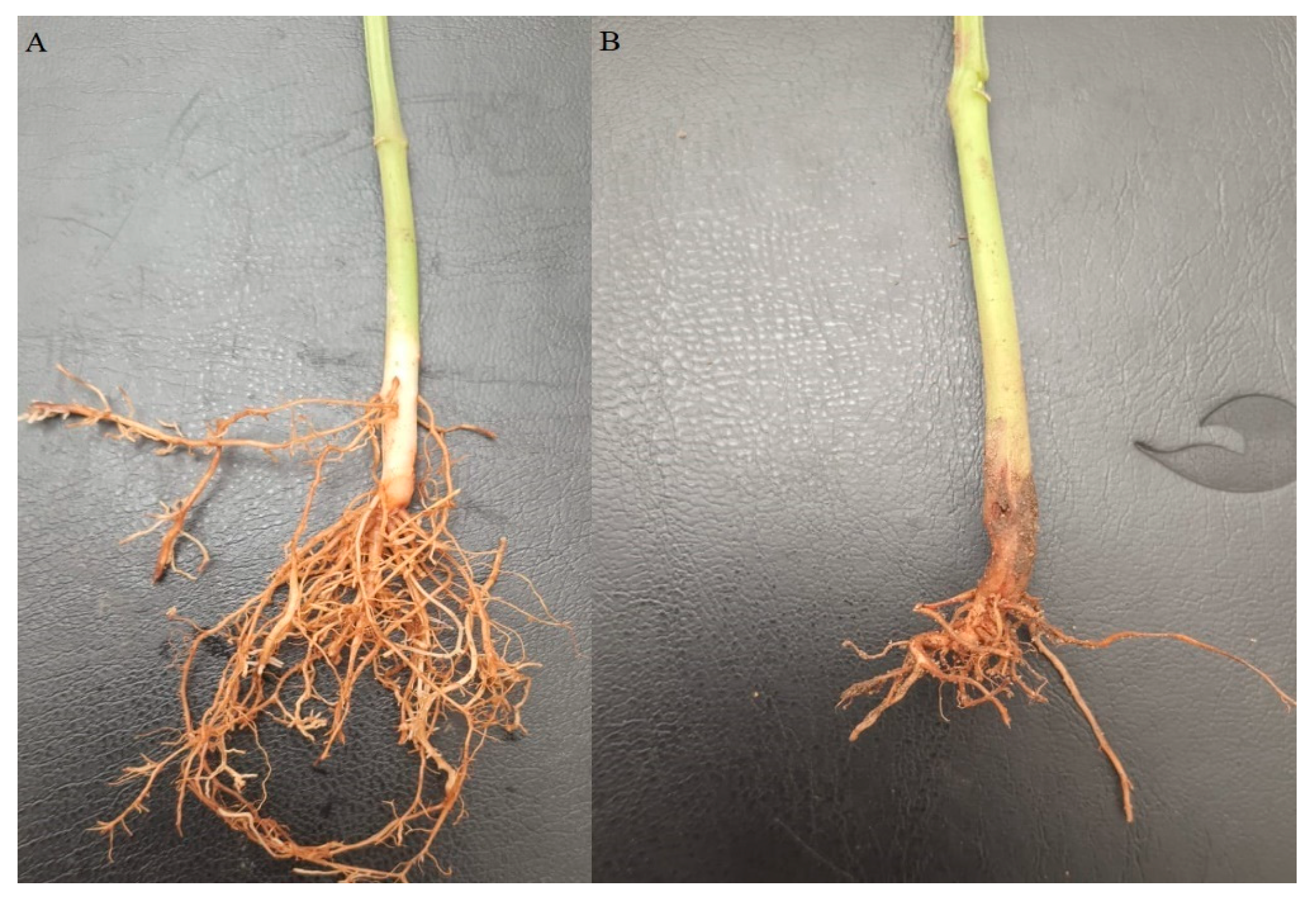
Disease severity results presented in Table 5 confirmed the aggressiveness of some Fusarium isolates. FP33 was the most virulent, causing severity values up to 90% in Giza 6 and 80% in Cambo, followed by 65% in Alpha and 70% in Samantha (Figure 2). However, its effect was much lower on Nebraska (30%) and Giza 12 (33%), which appeared as the most resistant cultivars. Similar results were observed for FP24, FP26, FP21, and FP11, which also caused high severity ranging from 60 to 85% on Alpha, Samantha, Giza 6, and Cambo, but significantly lower values of 20 to 33% on Nebraska and Giza 12.
Moderate disease severity was observed with isolates FP18, FP1, FP15, and FP17, which ranged from 25 to 33%. In contrast, the least aggressive isolates (FP2, FP8, FP10, and FP14) produced very low severity levels of 0 to 14% and, in some cases, did not induce any visible disease symptoms on certain cultivars. Overall, Giza 6 emerged as the most susceptible cultivar, followed by Alpha, Samantha, and Cambo, while Nebraska and Giza 12 demonstrated the greatest resistance.

3.6. Molecular Identification and Phylogenetic Analysis

The TEF1-α gene region (~700 bp) was successfully amplified from the five most aggressive Fusarium isolates associated with common bean root rot. Clear single bands were obtained for all isolates on agarose gel (Figure 3), confirming successful PCR amplification. The purified PCR products were then sequenced, and the resulting nucleotide sequences were analyzed using the BLASTn algorithm against the NCBI GenBank database.
Specifically, FP33 (Kafr El-Sheikh) and FP24 (Beni-Suef) were identified as Fusarium equiseti, FP26 (Kafr El-Sheikh) and FP21 (Beni-Suef) as Fusarium oxysporum, and FP11 (Qalyubia) as Fusarium solani. The percentage identity ranged from 96.45% to 99.69%, query coverage from 89% to 94%, and all E-values were 0.0 (Table 6). All sequences were deposited in GenBank under accession numbers PV595127–PV595131.
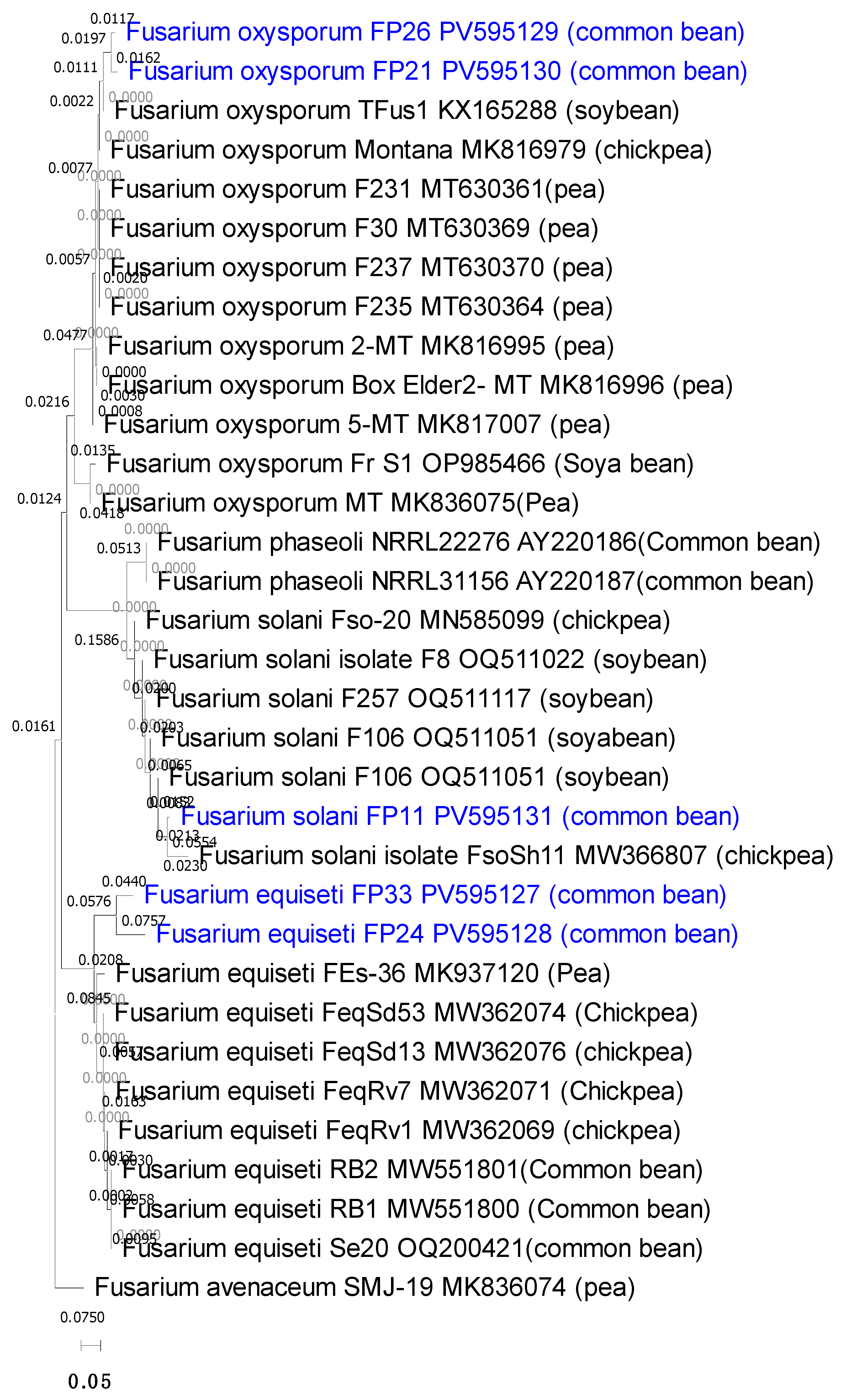
Multiple sequence alignment using ClustalW and a neighbor-joining phylogenetic tree generated in MEGA X with 1000 bootstrap replicates grouped the isolates into three well-supported clades corresponding to F. equiseti, F. oxysporum, and F. solani (Figure 4). The common bean associated isolates clustered closely with Fusarium sequences previously reported from other legumes, including soybean, chickpea, and pea (Table 7). Specifically, the F. oxysporum isolates formed a clade with sequences from pea and soybean roots and stems, F. solani clustered with chickpea and soybean isolates from the USA, and F. equiseti clustered with sequences from chickpea, pea, and previously reported common bean isolates from Iran. confirming their taxonomic placement and suggesting potential cross-host pathogenic relationships.

4. Discussion

Fusarium species are among the most important soil-borne pathogens responsible for severe losses in common bean production worldwide. The present study provides an integrated assessment of the diversity, pathogenicity, and genetic variability of Fusarium isolates associated with common bean root rot in Egypt, combining morphological characterization, greenhouse pathogenicity assays, and molecular identification. This multifaceted approach allowed not only the confirmation of species identity but also the exploration of their pathogenic variability and implications for disease management.
Out of 35 isolates recovered from symptomatic roots, nine Fusarium species were identified: F. proliferatum, F. solani, F. equiseti, F. verticillioides, F. semitectum, F. subglutinans, F. oxysporum, F. anthophilum, and F. tricinctum. This high diversity indicates a complex disease etiology in Egyptian fields, consistent with previous reports documenting the occurrence of multiple Fusarium species in common bean production systems worldwide [18,26,47]. For instance, several Fusarium taxa were isolated from beans in Iran [47], in Michigan, United States [48], and with specific species such as F. cuneirostrum confirmed in Canada [49], highlighting their global prevalence and adaptability.
Greenhouse assays revealed significant heterogeneity in pathogenicity. Isolates FP33, FP24, FP26, FP21, and FP11 were highly virulent, inducing severe pre- and post-emergence damping-off, high disease incidence, and extensive root rot lesions. By contrast, FP8, FP14, FP2, and FP10 were either non-pathogenic or weakly virulent, while FP18, FP1, FP15, and FP17 displayed intermediate aggressiveness. This gradient of pathogenicity reflects the well-documented heterogeneity of Fusarium populations, where aggressive and weakly pathogenic strains coexist within the same field [50,51,52]. Such variability represents an adaptive advantage, enabling persistence under diverse soil and climatic conditions.
The observed pathogenic spectrum (37% of isolates being highly aggressive) aligns with previous studies reporting approximately 40% of Fusarium strains as strongly pathogenic on bean seedlings [21]. These findings emphasize that effective disease management strategies must account for the coexistence of diverse pathogenic races, as single-gene resistance is unlikely to provide durable protection.
Marked differences in cultivar susceptibility were detected. Giza 6 and Cambo were highly susceptible, exhibiting complete mortality under infection with the most aggressive isolates, while Alpha and Samantha displayed moderate susceptibility. In contrast, Nebraska and Giza 12 consistently showed high resistance, with significantly reduced disease incidence and severity. Typical symptoms included seedling yellowing, wilting, root necrosis, and stunted growth, consistent with descriptions of FRR symptomatology [25]. These results corroborate earlier reports that resistance to FRR is quantitative rather than absolute, relying on polygenic defense mechanisms [53,54,55]. Proposed mechanisms include lignification of root tissues, enhanced peroxidase and polyphenol oxidase activity, and accumulation of antifungal metabolites, all of which limit pathogen colonization [56]. The partial resistance demonstrated by Nebraska and Giza 12 thus represents an exploitable trait in breeding programs. Importantly, the strong susceptibility of Giza 6 highlights the vulnerability of widely cultivated Egyptian varieties, underscoring the urgent need for resistant alternatives.
Disease incidence and severity data provided complementary insights into isolate–host interactions. FP33 and FP24 consistently produced the highest values, causing up to 100% incidence and 90% severity in Giza 6 and Cambo. Conversely, Nebraska and Giza 12 showed strong resistance, with incidence below 35% and severity under 30% even when inoculated with the most aggressive isolates. Such variability underscores the importance of host genotype in modulating disease outcomes. Similar quantitative resistance patterns were reported by [53], where resistant cultivars restricted lesion expansion, and by [54], who emphasized resistance in reducing disease progression.
Molecular characterization using the TEF1-α gene marker provided reliable confirmation of isolate identities. BLASTn analysis showed high sequence homology with reference Fusarium species, with identities ranging from 96.45% to 99.69%, query coverage between 89% and 94%, and all E-values equal to 0.0, confirming the robustness of the molecular approach. The five most virulent isolates were identified as F. equiseti (FP33, FP24), F. oxysporum (FP26, FP21), and F. solani (FP11), consistent with their pathogenic profiles. TEF1-α is considered a highly informative marker for Fusarium taxonomy due to its discriminatory power at the species level [57,58]. Ref. [59] reported that Fusarium species involved in bean root rot in Zanjan province are F. solani, F. equiseti, F. oxysporum, F. crookwellense, F. acuminatum, and F. sambucinum. However, the predominance of F. equiseti in our collection is noteworthy, as this species is often reported as secondary compared with the more widely studied F. solani and F. oxysporum [52,60]. This novel finding emphasizes the shifting dynamics of pathogen populations under Egyptian agroecological conditions and warrants further investigation.
Phylogenetic analysis clustered the isolates into three distinct clades corresponding to F. equiseti, F. oxysporum, and F. solani, with strong bootstrap support. Importantly, Egyptian isolates grouped with Fusarium strains previously reported from other legumes, including soybean, chickpea, and pea. For example, F. oxysporum isolates clustered with pea and soybean sequences from Europe and the USA, while F. equiseti grouped with chickpea and common bean isolates from Iran and Morocco. This clustering highlights intra-specific diversity and suggests potential cross-host pathogenicity, raising epidemiological concerns about the capacity of Fusarium strains to adapt to multiple legume hosts. Similar observations of cross-host clustering and intra-specific variability have been reported previously, highlighting the adaptability of these pathogens across different legumes [61].
The predominance of F. equiseti in Egypt may be linked to specific agroecological conditions. Egypt’s hot, dry climate interspersed with periods of irrigation, creates fluctuating soil moisture and temperature regimes favorable for diverse Fusarium species. F. equiseti, in particular, has been reported to thrive in semi-arid environments and may outcompete other Fusarium taxa under such conditions [62]. Moreover, continuous bean cultivation and limited crop rotation in Egyptian fields could contribute to pathogen build-up and selection of highly aggressive strains. Understanding these ecological drivers is critical for designing targeted disease management practices.
The genetic variability observed within and among Fusarium species also has epidemiological implications. The coexistence of diverse lineages within Egyptian fields suggests multiple sources of inoculum and possible introduction through seed trade. This diversity enhances the adaptive potential of pathogen populations, increasing the risk of resistance breakdown if single-gene resistance is deployed [63].
Looking ahead, the findings of this study provide a foundation for designing sustainable management strategies against Fusarium root rot in common bean. Breeding programs should prioritize the incorporation of quantitative resistance traits, as demonstrated by Nebraska and Giza 12, to achieve durable resistance across multiple Fusarium species. Molecular tools such as TEF1-α should be further exploited for rapid pathogen surveillance and to monitor the emergence of new virulent strains. Additionally, biological control using antagonistic microorganisms and plant growth-promoting rhizobacteria represents an eco-friendly alternative that can complement host resistance. From an agroecological perspective, integrating crop rotation, soil amendments, and optimized irrigation practices could suppress Fusarium inoculum in fields. Finally, mechanistic studies focusing on host defense pathways, including secondary metabolite biosynthesis and systemic resistance induction, are needed to clarify the molecular basis of partial resistance. Together, these strategies will enhance integrated disease management, safeguard bean productivity, and contribute to sustainable legume cultivation under Egyptian and global conditions.

5. Conclusions

This study highlights the significant role of Fusarium species in causing root rot in common bean, identifying F. equiseti, F. oxysporum, and F. solani as the primary pathogens responsible for disease development. The findings demonstrated considerable genetic variability among the isolates, emphasizing the complexity of root rot etiology in common bean fields in Egypt. The high virulence of specific isolates, particularly F. equiseti (FP33 and FP24), underscores the need for targeted disease management strategies. The results also highlight the varying resistance levels among common bean cultivars, suggesting that breeding for genetic resistance remains a crucial approach to mitigate yield losses. Future studies should focus on exploring host–pathogen interactions, screening for resistant genotypes, and developing integrated disease management strategies that combine resistant cultivars. Overall, this research contributes to a better understanding of Fusarium diversity and its impact on common bean production. The integration of molecular tools in pathogen identification can enhance disease diagnosis and guide the development of effective and sustainable control measures to improve common bean productivity and food security.

Author Contributions

Conceptualization, T.A.K.; Software, N.A.T. and W.R.A.; Formal analysis, W.N.H.; Resources, T.A.K. and N.A.T.; Data curation, M.A.M., W.N.H. and W.R.A.; Writing—original draft, T.A.K.; Writing—review & editing, M.M.Y., N.A.T., M.A.M., W.N.H. and W.R.A.; Visualization, T.A.K., M.M.Y. and W.R.A.; Supervision, M.M.Y., N.A.T., W.N.H. and W.R.A.; Project administration, W.N.H.; Funding acquisition, D.H.M.A. All authors have read and agreed to the published version of the manuscript.

Funding

This research received no external funding.

Institutional Review Board Statement

Not applicable.

Informed Consent Statement

Not applicable.

Data Availability Statement

The original contributions presented in this study are included in the article. Further inquiries can be directed to the corresponding authors.

Acknowledgments

The authors acknowledge the support from Princess Nourah bint Abdulrahman University Researchers Supporting Project number (PNURSP2025R15), Princess Nourah bint Abdulrahman University, Riyadh, Saudi Arabia.

Conflicts of Interest

The authors declare no conflict of interest.

References

  1. Celmeli, T.; Sari, H.; Canci, H.; Sari, D.; Adak, A.; Eker, T.; Toker, C. The nutritional content of common bean (Phaseolus vulgaris L.) landraces in comparison to modern varieties. Agronomy 2018, 8, 166. [Google Scholar] [CrossRef]
  2. Meziadi, C.; Richard, M.M.S.; Derquennes, A.; Thareau, V.; Blanch Romero, A. Development of molecular markers linked to disease resistance genes in common bean based on whole genome sequence. Plant Sci. 2016, 242, 351–357. [Google Scholar] [CrossRef] [PubMed]
  3. Machiani, M.A.; Rezaei-Chiyaneh, E.; Javanmard, A.; Maggi, F.; Morshedloo, M.R. Evaluation of common bean (Phaseolus vulgaris L.) seed yield and quali-quantitative production of the essential oils from fennel (Foeniculum vulgare Mill.) and dragonhead (Dracocephalum moldavica L.) in intercropping system under humic acid application. J. Clean. Prod. 2019, 235, 112–122. [Google Scholar] [CrossRef]
  4. FAOSTAT. FAOSTAT Statistics Database. Available online: http://www.fao.org/faostat (accessed on 21 April 2025).
  5. Mostafa, F.W. Integrated Control for White Rot Caused by Sclerotinia sclerotiorum (Lib.) de Bary and Rust Caused by Uromyces appendiculatus (Pers.) Unger of Bean (Phaseolus vulgaris L.) Plant. Ph.D. Thesis, Faculty of Agriculture, Fayoum University, Fayoum, Egypt, 2014; 184p. [Google Scholar]
  6. Singh, S.P.; Schwartz, H.F. Breeding common bean for resistance to diseases: A review. Crop Sci. 2010, 50, 2199–2223. [Google Scholar] [CrossRef]
  7. Vural, C.; Soylu, S.O. Prevalence and incidence of fungal disease agents affecting bean (Phaseolus vulgaris L.) plants. Res. Crops 2012, 13, 634–640. [Google Scholar]
  8. Marcenaro, D.; Valkonen, J.P. Seedborne pathogenic fungi in common bean (Phaseolus vulgaris cv. INTA Rojo) in Nicaragua. PLoS ONE 2016, 11, e0168662. [Google Scholar] [CrossRef]
  9. Girma, F.; Fininsa, C.; Terefe, H.; Amsalu, B. Evaluation of common bean (Phaseolus vulgaris) genotypes for resistance to common bacterial blight and angular leaf spot diseases, and agronomic performances. Heliyon 2022, 8, e10425. [Google Scholar] [CrossRef]
  10. Rahmanzadeh, A.; Khahani, B.S.; Taghavi, M.; Khojasteh, M.; Osdaghi, E. Genome-wide meta-QTL analyses provide novel insight into disease resistance repertoires in common bean. BMC Genom. 2022, 23, 680. [Google Scholar] [CrossRef]
  11. Félix-Pablos, C.M.; Parra-Cota, F.I.; Santoyo, G.; Orozco-Mosqueda, M.C.; Santos-Villalobos, S.D. Draft genome sequence of Bacillus sp. strain FSQ1, a biological control agent against white mold in common bean (Phaseolus vulgaris L.). Curr. Res. Microb. Sci. 2022, 3, 100138. [Google Scholar] [CrossRef]
  12. Adomako, J.S.; Yeboah, J.F.; Asamoah, P.; Amankwaa-Yeboah, E.A.; Adjei, E.A.; Obeng, B.; Sakyiamah, M.; Lamptey, L.; Asibuo, B.J.Y. Survey of plant parasitic nematodes and disease severity of common bean lines evaluated for reaction to root knot nematodes infestation. Afr. Crop Sci. J. 2022, 30, 147–154. [Google Scholar] [CrossRef]
  13. Rady, M.M.; Elrys, A.S.; Selem, E.; Mohsen, A.A.A.; Arnaout, S.M.A.I.; El-Sappah, A.H.; El-Tarabily, K.A.; Desoky, E.M. Spirulina platensis extract improves the production and defenses of the common bean grown in a heavy metals-contaminated saline soil. J. Environ. Sci. 2023, 129, 240–257. [Google Scholar] [CrossRef]
  14. Papathanasiou, F.; Ninou, E.; Mylonas, I.; Baxevanos, D.; Papadopoulou, F.; Avdikos, I.; Sistanis, I.; Koskosidis, A.; Vlachostergios, D.N.; Stefanou, S. The evaluation of common bean (Phaseolus vulgaris L.) genotypes under water stress based on physiological and agronomic parameters. Plants 2022, 11, 2432. [Google Scholar] [CrossRef]
  15. Zhang, Q.; Geng, J.; Du, Y.; Zhao, Q.; Zhang, W.; Fang, Q.; Yin, Z.; Li, J.; Yuan, X.; Fan, Y. Heat shock transcription factor (Hsf) gene family in common bean (Phaseolus vulgaris): Genome-wide identification, phylogeny, evolutionary expansion and expression analyses at the sprout stage under abiotic stress. BMC Plant Biol. 2022, 22, 33. [Google Scholar] [CrossRef] [PubMed]
  16. Paparu, P.; Acur, A.; Kato, F.; Acam, C.; Nakibuule, J.; Musoke, S.; Mukankusi, C. Prevalence and incidence of four common bean root rots in Uganda. J. Exp. Agric. Int. 2018, 54, 888–900. [Google Scholar] [CrossRef] [PubMed]
  17. Harter, L. A Fusarium disease of beans. Phytopathology 1929, 19, 84. [Google Scholar]
  18. Sampaio, A.M.; de Araújo, S.S.; Rubiales, D.; Vaz Patto, M.C. Fusarium wilt management in legume crops. Agronomy 2020, 10, 1073. [Google Scholar] [CrossRef]
  19. Ampaire, E. Farmers’ Indigenous Technical Knowledge of Bean Diseases Management and Communication Systems in Southwestern Uganda. Master’s Thesis, Makerere University, Kampala, Uganda, 2003; 127p. [Google Scholar]
  20. Opio, F.; Ugen, M.; Namayanja, A.; Mugagga, I.; Mawejje, D. Improving food security in southwestern Uganda by transferring and promoting resistant varieties and integrated management packages for BRR. In Biotechnology, Breeding, and Seed Systems for African Crops Conference; National Institute for Agriculture Research (IIAM): Maputo, Mozambique, 2007. [Google Scholar]
  21. Usmael, A.; Dejene, M.; Ayena, G. Assessment of root rot pathogens of common bean (Phaseolus vulgaris L.) and reaction of genotypes to the pathogens in West Hararghe Zone, Ethiopia. Open J. Plant Sci. 2023, 8, 037–055. [Google Scholar] [CrossRef]
  22. Deng, D.; Wu, W.; Duan, C.; Sun, S.; Zhu, Z. A novel pathogen Fusarium cuneirostrum causing common bean (Phaseolus vulgaris) root rot in China. J. Integr. Agric. 2024, 23, 166–176. [Google Scholar] [CrossRef]
  23. Adesemoye, A.O.; Orrell, T.; Kodati, S. Effect of virulence of root rot pathogens and cultivar resistance on disease occurrence in dry beans. Plant Health Prog. 2018, 19, 237–241. [Google Scholar] [CrossRef]
  24. Husaini, A.M.; Sakina, A.; Cambay, S.R. Host–pathogen interaction in Fusarium oxysporum infections: Where do we stand? Mol. Plant Microbe Interact. 2018, 31, 889–898. [Google Scholar] [CrossRef]
  25. Wang, C.H.; Sun, C.; Liu, S.Y. Preliminary study on pathogen of root rot of kidney bean and its control in Xinjiang. Arid Zone Res. 2010, 27, 380–384. (In Chinese) [Google Scholar]
  26. Liu, T.; Shen, Y.Q.; Liu, Z.; Han, D.; Cui, J.; Zuo, Y.H. Control effect of three kinds of seed-coating formulations on the root rot of kidney beans. Plant Prot. 2017, 43, 216–219. [Google Scholar]
  27. Aoki, T.; O’Donnell, K.; Geiser, D.M. Systematics of key phytopathogenic Fusarium species: Current status and future challenges. J. Gen. Plant Pathol. 2014, 80, 189–201. [Google Scholar] [CrossRef]
  28. Lombard, L.; Sandoval-Denis, M.; Lamprecht, S.C.; Crous, P.W. Epitypification of Fusarium oxysporum—Clearing the taxonomic chaos. Persoonia 2019, 43, 1–47. [Google Scholar] [CrossRef] [PubMed]
  29. Leslie, J.F.; Summerell, B.A. The Fusarium Laboratory Manual; John Wiley & Sons: Hoboken, NJ, USA, 2008; ISBN 978-0-470-27646-4. [Google Scholar]
  30. Sennoi, R.; Jogloy, S.; Saksirirat, W.; Patanothai, A. Pathogenicity test of Sclerotium rolfsii, a causal agent of Jerusalem artichoke (Helianthus tuberosus L.) stem rot. Asian J. Plant Sci. 2010, 9, 281–284. [Google Scholar] [CrossRef]
  31. Ali, M.A.; Abd El Gwad, T.I.; Isamail, M.E.; Galal, A.A. Eco-friendly control traits of common bean root rot caused by Macrophomina phaseolina (Tossi) Goid and Fusarium equiseti using fungicide alternatives. New Val. J. Agric. Sci. 2022, 2, 530–543. [Google Scholar] [CrossRef]
  32. Shaban, W.I.; El-Bramawy, M.A. Impact of dual inoculation with Rhizobium and Trichoderma on damping-off, root rot diseases and plant growth parameters of some legumes field crop under greenhouse conditions. Int. Res. J. Agric. Sci. Soil Sci. 2011, 1, 98–108. [Google Scholar]
  33. Kator, L.; Hosea, Z.Y.; Oche, O.D. Sclerotium rolfsii: Causative organism of southern blight, stem rot, white mold and sclerotia rot disease. Ann. Biol. Res. 2015, 6, 78–89. [Google Scholar]
  34. Dellaporta, S.L.; Wood, J.; Hicks, J.B. A plant DNA minipreparation: Version II. Plant Mol. Biol. Rep. 1983, 1, 19–21. [Google Scholar] [CrossRef]
  35. O’Donnell, K.; Kistler, H.C.; Cigelnik, E.; Ploetz, R.C. Multiple evolutionary origins of the fungus causing Panama disease of banana: Concordant evidence from nuclear and mitochondrial gene genealogies. Proc. Natl. Acad. Sci. USA 1998, 95, 2044–2049. [Google Scholar] [CrossRef]
  36. Geiser, D.M.; Jiménez-Gasco, M.M.; Kang, S.; Makalowska, I.; Veeraraghavan, N.; Ward, T.J.; O’Donnell, K. FUSARIUM-ID v.1.0: A DNA sequence database for identifying Fusarium. Eur. J. Plant Pathol. 2004, 110, 473–479. [Google Scholar] [CrossRef]
  37. Hall, T.A. BioEdit: A user-friendly biological sequence alignment editor and analysis program for Windows 95/98/NT. Nucleic Acids Symp. Ser. 1999, 41, 95–98. [Google Scholar]
  38. Kumar, S.; Stecher, G.; Li, M.; Knyaz, C.; Tamura, K. MEGA X: Molecular evolutionary genetics analysis across computing platforms. Mol. Biol. Evol. 2018, 35, 1547–1549. [Google Scholar] [CrossRef]
  39. Jenkins, S.; Labanska, M.; Amsterdam, S.; Clarkson, J.; Covington, J. Preliminary studies on detection of Fusarium basal rot infection in onions and shallots using electronic nose. Sensors 2021, 22, 5453. [Google Scholar] [CrossRef]
  40. Duvnjak, T.; Sudaric, A.; Matosa Kocar, M.; Cosic, J.; Vrandecic, K. First report of soybean fusarium wilt caused by Fusarium oxysporum in Croatia. Plant Dis. 2017, 101, 249. [Google Scholar] [CrossRef]
  41. Moparthi, S.; Agindotan, B.O.; Burrows, M.E. Identification and characterization of Fusarium spp. associated with root rot of dry pea in Montana. Plant Health Prog. 2019, 20, 215–219. [Google Scholar] [CrossRef]
  42. Olszak-Przybyś, H.; Korbecka-Glinka, G.; Patkowska, E. Identification and pathogenicity of Fusarium isolated from soybean in Poland. Pathogens 2023, 12, 1162. [Google Scholar] [CrossRef]
  43. Aoki, T.; O’Donnell, K.; Homma, Y.; Lattanzi, A.R. Sudden-death syndrome of soybean is caused by two morphologically and phylogenetically distinct species within the Fusarium solani species complex F. virguliforme in North America and F. tucumaniae in South America. Mycologia 2003, 95, 660–684. [Google Scholar] [CrossRef]
  44. Weerasooriya, S.; Bovill, M.S.; Benson, A.; Musick, A.M.; Ricotti, M. Devouring the Milky Way satellites: Modeling dwarf galaxies with Galacticus. Astrophys. J. 2023, 948, 87. [Google Scholar] [CrossRef]
  45. Moparthi, S.; Peluola, C.; Agindotan, B.; McPhee, K.; Burrows, M. First report of gray mold of chickpea caused by Botrytis euroamericana in the USA. Crop Prot. 2020, 137, 105297. [Google Scholar] [CrossRef]
  46. Mojerlou, S.; Sepehri, G.; Shahbaz, S. Identification of bean root rot causal and associated fungal agents in Khomein County, Markazi Province, Iran. Crop Prot. 2021, 10, 633–646. [Google Scholar]
  47. Darvishnia, M.; Hasanvand, E.; Pakbaz, S. Morphological and molecular identification of Fusarium associated with beans in Selseleh County. J. Genet. Res. 2023, 9, 222–231. [Google Scholar]
  48. Oudman, L. Identification, Characterization, and Management of Fusarium Root Rot Pathogens of Dry Beans in Michigan. Master’s Thesis, Michigan State University, East Lansing, MI, USA, 2018. [Google Scholar]
  49. Henriquez, M.A.; Conner, R.L.; Hou, A.; Balasubramanian, P.; McLaren, D.L.; Chang, K.F.; McRae, K.B. Reaction of dry bean cultivars grown in western Canada to root rot inoculation. Can. J. Plant Sci. 2014, 94, 1219–1230. [Google Scholar] [CrossRef]
  50. Zitnick-Anderson, K.; Oladzadabbasabadi, A.; Jain, S.; Modderman, C.; Osorno, J.M.; McClean, P.E.; Pasche, J.S. Sources of resistance to Fusarium solani and associated genomic regions in common bean diversity panels. Front. Genet. 2020, 11, 475. [Google Scholar] [CrossRef]
  51. Abdelaziz, A.M.; El-Wakil, D.A.; Hashem, A.H.; Al-Askar, A.A.; AbdElgawad, H.; Attia, M.S. Efficient role of endophytic Aspergillus terreus in biocontrol of Rhizoctonia solani causing damping-off disease of Phaseolus vulgaris and Vicia faba. Microorganisms 2023, 11, 1487. [Google Scholar] [CrossRef] [PubMed]
  52. Naseri, B.; Mousavi, S.S. Root rot pathogens in field soil, roots and seeds in relation to common bean (Phaseolus vulgaris), disease and seed production. Int. J. Pest Manag. 2015, 61, 60–67. [Google Scholar] [CrossRef]
  53. Diaz, L.M.; Arredondo, V.; Ariza-Suarez, D.; Aparicio, J.; Buendia, H.F.; Cajiao, C.; Raatz, B. Genetic analyses and genomic predictions of root rot resistance in common bean across trials and populations. Front. Plant Sci. 2021, 12, 629221. [Google Scholar] [CrossRef] [PubMed]
  54. Shahiba, A.M.; Pranay, G.; Sonaniya, P.; Kushwaha, J.S.; Aware, S.A. Modern approaches in plant breeding enhancing crop genetics. In Modern Approaches in Plant Breeding Enhancing Crop Genetics; Elite Publishing House: Delhi, India, 2023; pp. 15–33. [Google Scholar]
  55. Maina, P.K.; Wachira, P.M.; Okoth, S.A.; Kimenju, J.W. Cultural, morphological and pathogenic variability among Fusarium oxysporum f. sp. phaseoli causing wilt in French bean (Phaseolus vulgaris L.). J. Adv. Microbiol. 2017, 2, 1–9. [Google Scholar] [CrossRef]
  56. Singh, H.B.; Singh, B.N.; Singh, S.P.; Nautiyal, C.S. Solid-state cultivation of Trichoderma harzianum NBRI-1055 for modulating natural antioxidants in soybean seed matrix. Bioresour. Technol. 2010, 101, 6444–6453. [Google Scholar] [CrossRef]
  57. Moparthi, S.; Burrows, M.; Mgbechi-Ezeri, J.; Agindotan, B. Fusarium spp. associated with root rot of pulse crops and their cross-pathogenicity to cereal crops in Montana. Plant Dis. 2021, 105, 548–557. [Google Scholar] [CrossRef]
  58. Olarte, R.A.; Hall, R.; Tabima, J.F.; Malvick, D.; Bushley, K. Genetic diversity and aggressiveness of Fusarium virguliforme isolates across the midwestern United States. Phytopathology 2022, 112, 1273–1283. [Google Scholar] [CrossRef] [PubMed]
  59. Safarloo, Z.; Hemmati, R. Identification and study of pathogenicity of Fusarium species involved in bean root rot in Zanjan province. Appl. Res. Phytomed. 2014, 3, 77–92. [Google Scholar]
  60. Ahari Mostafavi, H.; Mirmajlessi, S.M.; Safaie, N.; Minassyan, V.; Fathollahi, H.; Dorri, H.R.; Mansouripour, S.M. The use of a gamma-irradiated mutant of Fusarium solani f. sp. phaseoli with reduced pathogenicity for the biological control of Fusarium root rot of bean (Phaseolus vulgaris) in field conditions. J. Agric. Sci. Technol. 2012, 14, 1415–1423. [Google Scholar]
  61. Naseri, B.; Marefat, A. Large-scale assessment of agricultural practices affecting Fusarium root rot and common bean yield. Eur. J. Plant Pathol. 2011, 131, 179–195. [Google Scholar] [CrossRef]
  62. El Hazzat, N.; Adnani, M.; Msairi, S.; El Alaoui, M.A.; Mouden, N.; Chliyeh, M.; Douira, A. Fusarium equiseti as one of the main Fusarium species causing wilt and root rot of chickpeas in Morocco. Acta Mycol. 2023, 57, 576. [Google Scholar] [CrossRef]
  63. Zhou, T.; DallaSanta, K.; Nazarenko, L.; Schmidt, G.A.; Jin, Z. The impact of increasing stratospheric radiative damping on the quasi-biennial oscillation period. Atmos. Chem. Phys. 2021, 21, 7395–7407. [Google Scholar] [CrossRef]
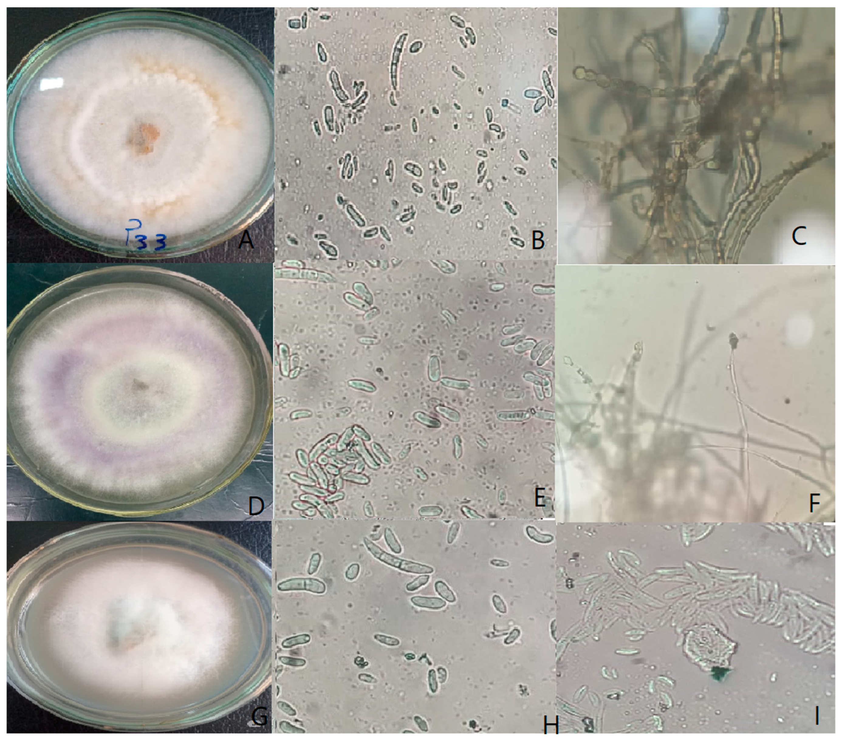
Figure 1. Morphological characters of the Fusarium spp. Isolated from common bean plants. (AC) F. equiseti (isolate FP33) growth on PDA; macroconidia and microconidia; chlamydospores. (DF) F. oxysporum (isolate FP26) growth on PDA; macroconidia and microconidia; microconidiophores (monophialides). (GI) F. solani (isolate FP11) growth on PDA; macroconidia and microconidia; microconidia produced in sporodochia. ِAll microscopic observations were taken at 40× magnification.
Cimb 47 00803 g001
Figure 2. Typical symptoms of common bean root rot (A) uninfected control; (B) root of Samantha cultivar infected with Fusarium equiseti isolate FP33.
Cimb 47 00803 g002
Figure 3. Agarose gel electrophoresis of PCR-amplified TEF1-α gene (~700 bp) from the five most aggressive Fusarium isolates associated with common bean root rot. Lane M: GeneRuler 1 kb DNA marker (Thermo Fisher Scientific); lanes 1–5: FP11, FP21, FP24, FP26, and FP33: respective Fusarium isolates.
Cimb 47 00803 g003
Figure 4. A neighbor-joining phylogenetic tree based on TEF-1α gene sequences illustrating the relationships between the five most potent Fusarium isolates and their closely related members from the NCBI database. The tree was constructed using 1000 replications of resampled datasets. Fusarium avenaceum included as outgroup.
Cimb 47 00803 g004
Table 1. Fusarium species isolated from common bean and their pathogenicity on common bean plants.
Isolate CodeGovernorateFusarium spp.Pathogenic Reaction
 FP1 QalyubiaF. proliferatumPathogenic
FP2QalyubiaF. proliferatumPathogenic
FP3QalyubiaF. equisetiNon-pathogenic
FP4QalyubiaF. verticillioidesNon-pathogenic
FP5QalyubiaF. semitectumNon-pathogenic
FP6QalyubiaF. solaniNon-pathogenic
FP7QalyubiaF. proliferatumNon-pathogenic
FP8QalyubiaF. proliferatumPathogenic
FP9QalyubiaF. solaniNon-pathogenic
FP10QalyubiaF. proliferatumPathogenic
FP11QalyubiaF. solaniPathogenic
FP12QalyubiaF. equisetiNon-pathogenic
FP13QalyubiaF. verticillioidesNon-pathogenic
FP14QalyubiaF. subglutinansPathogenic
FP15QalyubiaF. semitectumPathogenic
FP16QalyubiaF. solaniNon-pathogenic
FP17QalyubiaF. verticillioidesPathogenic
FP18Kafr-elsheikhF. verticillioidesPathogenic
FP19Kafr-elsheikhF. subglutinansNon-pathogenic
FP20Kafr-elsheikhF. semitectumNon-pathogenic
FP21Beni-SuefF. oxysporumPathogenic
FP22Beni-SuefF. subglutinansNon-pathogenic
FP23Beni-SuefF. solaniNon-pathogenic
FP24Beni-SuefF. equisetiPathogenic
FP25Beni-SuefF. tricinctumNon-pathogenic
FP26Kafr-elsheikhF. oxysporumPathogenic
FP27QalyubiaF. anthophilumNon-pathogenic
FP28QalyubiaF. semitectumNon-pathogenic
FP29QalyubiaF. equisetiNon-pathogenic
FP30BaheiraF. oxysporumNon-pathogenic
FP31Kafr-elsheikhF. solaniNon-pathogenic
FP32Kafr-elsheikhF. verticillioidesNon-pathogenic
FP33Kafr-elsheikhF. equisetiPathogenic
FP34BeheiraF. solaniNon-pathogenic
FP35BaheiraF. equisetiNon-pathogenic
Table 2. Pre-emergence (%) damping-off of common bean cultivars inoculated with different Fusarium isolates.
Common Bean Cultivars
 Isolate code AlphaSamanthaNebraskaGiza6Giza 12Cambo
FP1811.11 ± 0.64 d11.11 ± 1.28 d11.11 ± 1.28 d22.22 ± 1.28 c11.11 ± 1.92 d22.22 ± 2.95 c
FP3344.44 ± 2.57 a44.44 ± 5.13 a22.22 ± 2.57 c44.44 ± 2.57 a22.22 ± 3.85 c44.44 ± 5.9 a
FP2433.33 ± 1.92 b33.33 ± 3.85 b11.11 ± 1.28 d44.44 ± 2.57 a22.22 ± 3.85 c44.44 ± 5.9 a
FP2633.33 ± 1.92 b33.33 ± 3.85 b11.11 ± 1.28 d44.44 ± 2.57 a22.22 ± 3.85 c44.44 ± 5.9 a
FP111.11 ± 0.64 d22.22 ± 2.57 c11.11 ± 1.28 d22.22 ± 1.28 c11.11 ± 1.92 d22.22 ± 2.95 c
FP2133.33 ± 1.92 b33.33 ± 3.85 b11.11 ± 1.28 d44.44 ± 2.57 a22.22 ± 3.85 c44.44 ± 5.9 a
FP211.11 ± 0.64 d11.11 ± 1.28 d11.11 ± 1.28 d22.22 ± 1.28 c11.11 ± 1.92 d22.22 ± 2.95 c
FP811.11 ± 0.64 d11.11 ± 1.28 d11.11 ± 1.28 d22.22 ± 1.28 c11.11 ± 1.92 d22.22 ± 2.95 c
FP1011.11 ± 0.64 d11.11 ± 1.28 d11.11 ± 1.28 d22.22 ± 1.28 c11.11 ± 1.92 d22.22 ± 2.95 c
FP1122.22 ± 1.28 c22.22 ± 2.57 c11.11 ± 1.28 d33.33 ± 1.92 b11.11 ± 1.92 d33.33 ± 4.43 b
FP1722.22 ± 1.28 c22.22 ± 2.57 c11.11 ± 1.28 d33.33 ± 1.92 b11.11 ± 1.92 d33.33 ± 4.43 b
FP1511.11 ± 0.64 d22.22 ± 2.57 c11.11 ± 1.28 d22.22 ± 1.28 c11.11 ± 1.92 d22.22 ± 2.95 c
FP1411.11 ± 0.64 d11.11 ± 1.28 d11.11 ± 1.28 d22.22 ± 1.28 c11.11 ± 1.92 d22.22 ± 2.95 c
Control0 ± 0 e0 ± 0 e0 ± 0 e0 ± 0 e0 ± 0 e0 ± 0 e
Means within a column ± standard error followed by different letters (a, b, c, d, e) indicate significant differences among treatments according to Duncan’s multiple range tests at p ≤ 0.05. Values were means of three replicates for each treatment as well as the control.
Table 3. Post-emergence (%) damping-off of common bean cultivars inoculated with different Fusarium isolates.
Common Bean Cultivars
Isolate CodeAlphaSamanthaNebraskaGiza6Giza 12Cambo
FP180 ± 0 d0 ± 0 d0 ± 0 d0 ± 0 d0 ± 0 d0 ± 0 d
FP3325 ± 1.44 a25 ± 2.89 a16.67 ± 1.92 b25 ± 1.44 a16.67 ± 2.89 b25 ± 3.32 a
FP2420 ± 1.15 b20 ± 2.31 b14.29 ± 1.65 c25 ± 1.44 a16.67 ± 2.89 b25 ± 3.32 a
FP2620 ± 1.15 b20 ± 2.31 b14.29 ± 1.65 c25 ± 1.44 a16.67 ± 2.89 b25 ± 3.32 a
FP10 ± 0 d0 ± 0 d0 ± 0 d0 ± 0 d0 ± 0 d0 ± 0 d
FP2120 ± 1.15 b20 ± 2.31 b14.29 ± 1.65 c25 ± 1.44 a16.67 ± 2.89 b25 ± 3.32 a
FP20 ± 0 d0 ± 0 d0 ± 0 d0 ± 0 d0 ± 0 d0 ± 0 d
FP80 ± 0 d0 ± 0 d0 ± 0 d0 ± 0 d0 ± 0 d0 ± 0 d
FP100 ± 0 d0 ± 0 d0 ± 0 d0 ± 0 d0 ± 0 d0 ± 0 d
FP1116.67 ± 0.96 b16.67 ± 1.92 b14.29 ± 1.65 c20 ± 1.15 b14.29 ± 2.48 c20 ± 2.66 b
FP170 ± 0 d0 ± 0 d0 ± 0 d0 ± 0 d0 ± 0 d0 ± 0 d
FP150 ± 0 d0 ± 0 d0 ± 0 d0 ± 0 d0 ± 0 d0 ± 0 d
FP140 ± 0 d0 ± 0 d0 ± 0 d0 ± 0 d0 ± 0 d0 ± 0 d
Control0 ± 0 d0 ± 0 d0 ± 0 d0 ± 0 d0 ± 0 d0 ± 0 d
Means within a column ± standard error followed by different letters (a, b, c, d) indicate significant differences among treatments according to Duncan’s multiple range tests at p ≤ 0.05. Values were means of three replicates for each treatment as well as the control.
Table 4. Disease incidence % of Fusarium isolates against different cultivars of common beans.
Common Bean Cultivars
Isolate CodeAlphaSamanthaNebraskaGiza6Giza 12Cambo
FP1825 ± 0.72 c25 ± 0.43 c25 ± 1.37 c28.57 ± 2.47 b25 ± 2.89 c28.57 ± 1.65 b
FP33100 ± 0 a100 ± 0 a33.33 ± 1.83 b100 ± 0 a33.33 ± 3.85 b100 ± 0 a
FP24100 ± 0 a100 ± 0 a28.57 ± 1.57 b100 ± 0 a33.33 ± 3.85 b100 ± 0 a
FP26100 ± 0 a100 ± 0 a28.57 ± 1.57 b100 ± 0 a33.33 ± 3.85 b100 ± 0 a
FP125 ± 0.72 c28.57 ± 0.49 b25 ± 1.37 c28.57 ± 2.47 b25 ± 2.89 c28.57 ± 1.65 b
FP21100 ± 0 a100 ± 0 a28.57 ± 1.57 b100 ± 0 a33.33 ± 3.85 b100 ± 0 a
FP212.5 ± 0.36 c12.5 ± 0.22 c12.5 ± 0.69 c14.29 ± 1.24 c12.5 ± 1.44 c14.29 ± 0.83 c
FP812.5 ± 0.36 c12.5 ± 0.22 c0 ± 0 d14.29 ± 1.24 c0 ± 0 d14.29 ± 0.83 c
FP1012.5 ± 0.36 c12.5 ± 0.22 c12.5 ± 0.69 c14.29 ± 1.24 c12.5 ± 1.44 c14.29 ± 0.83 c
FP11100 ± 0 a100 ± 0 a25 ± 1.37 c100 ± 0 a25 ± 2.89 c100 ± 0 a
FP1728.57 ± 0.82 b28.57 ± 0.49 b25 ± 1.37 c33.33 ± 2.89 b25 ± 2.89 c33.33 ± 1.92 b
FP1525 ± 0.72 c28.57 ± 0.49 b25 ± 1.37 c28.57 ± 2.47 b25 ± 2.89 c28.57 ± 1.65 b
FP1412.5 ± 0.36 c12.5 ± 0.22 c0 ± 0 d14.29 ± 1.24 c12.5 ± 1.44 c14.29 ± 0.83 c
Control0 ± 0 d0 ± 0 d0 ± 0 d0 ± 0 d0 ± 0 d0 ± 0 d
Means within a column ± standard error followed by different letters (a, b, c, d) indicate significant differences among treatments according to Duncan’s multiple range tests at p ≤ 0.05. Values were means of three replicates for each treatment as well as the control.
Table 5. Disease severity % of Fusarium isolates against different cultivars of common beans.
Common Bean Cultivars
Isolate CodeAlphaSamanthaNebraskaGiza6Giza 12Cambo
FP1825 ± 2.89 c25 ± 1.15 c20 ± 1.27 d25.71 ± 3.27 c22.5 ± 1.69 d25.71 ± 1.48 c
FP3365 ± 7.51 a70 ± 3.23 a30 ± 1.91 c90 ± 11.43 a33.33 ± 2.5 c80 ± 4.62 a
FP2464 ± 7.39 a68 ± 3.14 a28.57 ± 1.81 c85 ± 10.8 a33.33 ± 2.5 c75 ± 4.33 a
FP2660 ± 6.93 b64 ± 2.96 a25.71 ± 1.63 c80 ± 10.16 a25.71 ± 1.93 c70 ± 4.04 a
FP125 ± 2.89 c28.57 ± 1.32 c22.5 ± 1.43 d28.57 ± 3.63 c25 ± 1.88 c25.71 ± 1.48 c
FP2156 ± 6.47 b60 ± 2.77 b22.86 ± 1.45 d75 ± 9.53 a25.71 ± 1.93 c65 ± 3.75 a
FP210 ± 1.15 d12.5 ± 0.58 d7.5 ± 0.48 e14.29 ± 1.82 d10 ± 0.75 d11.43 ± 0.66 d
FP87.5 ± 0.87 e10 ± 0.46 d0 ± 0 e11.43 ± 1.45 d0 ± 0 e8.57 ± 0.49 c
FP1012.5 ± 1.44 d12.5 ± 0.58 d10 ± 0.64 d14.29 ± 1.82 d10 ± 0.75 d14.29 ± 0.83 d
FP1151.43 ± 5.94 b60 ± 2.77 b22.5 ± 1.43 d70 ± 8.89 a25 ± 1.88 c63.33 ± 3.66 a
FP1728.57 ± 3.3 c28.57 ± 1.32 c22.5 ± 1.43 d30 ± 3.81 c25 ± 1.88 c30 ± 1.73 c
FP1525 ± 2.89 c28.57 ± 1.32 c22.5 ± 1.43 d28.57 ± 3.63 c25 ± 1.88 c28.57 ± 1.65 c
FP1410 ± 1.15 d10 ± 0.46 d0 ± 0 e11.43 ± 1.45 d7.5 ± 0.56 e11.43 ± 0.66 d
Control0 ± 0 e0 ± 0 e0 ± 0 e0 ± 0 e0 ± 0 e0 ± 0 e
Means within a column ± standard error followed by different letters (a, b, c, d, e) indicate significant differences among treatments according to Duncan’s multiple range tests at p ≤ 0.05. Values were means of three replicates for each treatment as well as the control.
Table 6. The accession number of the most aggressive isolates that were submitted to and retrieved from NCBI database.
Isolate CodeFusarium SpeciesGovernorateClosest GenBank MatchIdentity (%)Query Coverage (%)E-ValueGenBank
Accession
Number
FP33Fusarium equisetiKafr-elsheikhMK93712096.4589%0.0PV595127
FP24Fusarium equisetiBeni-SuefMW36207697.1394%0.0PV595128
FP26Fusarium oxysporumKafr-elsheikhKX16528898.7993%0.0PV595129
FP21Fusarium oxysporumBeni-SuefMT 63036499.6993%0.0PV595130
FP11Fusarium solaniQalyubiaOQ51105199.4291%0.0PV595131
Table 7. Fusarium species and their genBank accession numbers related to other research used in this study.
Species from IsolateOrigin PlantIsolation SourceReferencesCountryGenBank
Accession Number
Fusarium oxysporumpeaRoot[39]United KingdomMT 630361
Fusarium oxysporumpeaRoot[39]United KingdomMT 630369
Fusarium oxysporumpeaRoot[39]United KingdomMT 630370
Fusarium oxysporumpeaRoot[39]United KingdomMT 630364
Fusarium oxysporumsoybeanStem[40]CroatiaKX165288
Fusarium oxysporumchickpeaRoot[41]Gallatin, MontanaMK816979
Fusarium oxysporumpeaRoot[41]Daniels, Montana
USA
MK816995
Fusarium oxysporumpeaRoot[41]Sheridan, USAMK816996
Fusarium oxysporumpeaRoot[41]Daniels, Montana
USA
MK817007
Fusarium oxysporumsoybeanSeed[42]PolandOP985466
Fusarium oxysporumpeaRoot[41]Daniels, Montana
USA
MK836075
Fusarium phaseoliCommon beanRoot[43]USAAY220186
Fusarium phaseoliCommon beanRoot[43]USAAY220187
Fusarium solanichickpeaRoot[43]Gallatin, Montana
USA
MN585099
Fusarium solanisoybeanRoot[44]Pennsylvania
USA
OQ511117
Fusarium solanisoybeanRoot[44]Pennsylvania
USA
OQ511022
Fusarium solanisoybeanRoot[44]Pennsylvania, USAOQ511051
Fusarium solanichickpeaRoot[45]Montana, USAMW366807
Fusarium equisetipeaseed[41]USAMK937120
Fusarium equisetichickpeaRoot[45]USAMW362071
Fusarium equisetichickpeaRoot[45]USAMW362074
Fusarium equisetichickpeaRoot[45]USAMW362076
Fusarium equisetichickpeaRoot[45]USAMW362069
Fusarium equisetiCommon beanRoot[46]Khomein, IranMW551801
Fusarium equisetiCommon beanRoot[46]Khomein, IranMW551800
Fusarium equisetiCommon beanRoot[47]SelselehOQ200421
Disclaimer/Publisher’s Note: The statements, opinions and data contained in all publications are solely those of the individual author(s) and contributor(s) and not of MDPI and/or the editor(s). MDPI and/or the editor(s) disclaim responsibility for any injury to people or property resulting from any ideas, methods, instructions or products referred to in the content.