Article

Anti-Staphylococcal, Anti-Candida, and Free-Radical Scavenging Potential of Soil Fungal Metabolites: A Study Supported by Phenolic Characterization and Molecular Docking Analysis

by
Amal A. Al Mousa
1,*,
Mohamed E. Abouelela
2,
Nadaa S. Al Ghamidi
1,
Youssef Abo-Dahab
3,
Hassan Mohamed
4,
Nageh F. Abo-Dahab
4 and
Abdallah M. A. Hassane
4,*
1
Department of Botany and Microbiology, College of Science, King Saud University, P.O. Box 145111, Riyadh 4545, Saudi Arabia
2
Department of Pharmacognosy, Faculty of Pharmacy (Boys), Al-Azhar University, Cairo P.O. Box 11884, Egypt
3
Ministry of Health and Populations, Cairo P.O. Box 11516, Egypt
4
Botany and Microbiology Department, Faculty of Science, Al-Azhar University, Assiut 71524, Egypt
*
Authors to whom correspondence should be addressed.
Curr. Issues Mol. Biol. 2024, 46(1), 221-243; https://doi.org/10.3390/cimb46010016
Submission received: 4 December 2023 / Revised: 22 December 2023 / Accepted: 25 December 2023 / Published: 28 December 2023
(This article belongs to the Section Bioorganic Chemistry and Medicinal Chemistry)

Abstract

:
Staphylococcus and Candida are recognized as causative agents in numerous diseases, and the rise of multidrug-resistant strains emphasizes the need to explore natural sources, such as fungi, for effective antimicrobial agents. This study aims to assess the in vitro anti-staphylococcal and anti-candidal potential of ethyl acetate extracts from various soil-derived fungal isolates. The investigation includes isolating and identifying fungal strains as well as determining their antioxidative activities, characterizing their phenolic substances through HPLC analysis, and conducting in silico molecular docking assessments of the phenolics’ binding affinities to the target proteins, Staphylococcus aureus tyrosyl-tRNA synthetase and Candida albicans secreted aspartic protease 2. Out of nine fungal species tested, two highly potent isolates were identified through ITS ribosomal gene sequencing: Aspergillus terreus AUMC 15447 and A. nidulans AUMC 15444. Results indicated that A. terreus AUMC 15447 and A. nidulans AUMC 15444 extracts effectively inhibited S. aureus (concentration range: 25–0.39 mg/mL), with the A. nidulans AUMC 15444 extract demonstrating significant suppression of Candida spp. (concentration range: 3.125–0.39 mg/mL). The A. terreus AUMC 15447 extract exhibited an IC50 of 0.47 mg/mL toward DPPH radical-scavenging activity. HPLC analysis of the fungal extracts, employing 18 standards, revealed varying degrees of detected phenolics in terms of their presence and quantities. Docking investigations highlighted rutin as a potent inhibitor, showing high affinity (−16.43 kcal/mol and −12.35 kcal/mol) for S. aureus tyrosyl-tRNA synthetase and C. albicans secreted aspartic protease 2, respectively. The findings suggest that fungal metabolites, particularly phenolics, hold significant promise for the development of safe medications to combat pathogenic infections.

1. Introduction

Soil contains a diverse range of microorganisms, including fungi, which are the most abundant producers of potentially beneficial biologically active metabolites [1]. Out of all the bioactive compounds that have been isolated from biological sources, more than 38% of the metabolites that are biologically active come from fungi [2]. It has been reported that fungi are potent producer of diverse bioactive metabolites, with their secondary metabolism comprising polysaccharides [3], enzymes [4,5], lipids and fatty acids [6], and low molecular-weight byproducts that are produced as an adaptation for specific functions in nature [7]. These metabolites may also include alkaloids, flavonoids, benzopyranones, phenolic acids, tannins, quinones, saponins, terpenoids, steroids, tetralones, xanthones, and many others [8]. The identified metabolites showed varied bioactivities with anticancer [9], antioxidative [10,11], antifungal [12,13], immunosuppressive [14], antiprotozoal [15], antiviral [16], and antibacterial efficiencies [17]. However, harmful influences can be caused by mycotoxins produced by some types of fungi [18,19]. Due to their unique metabolism, fungi are able to produce a wide range of useful secondary metabolites, which increases their potential for drug development [20].
Phenolics are compounds that include one or more phenol units and are primarily found in plants, although they can also be found in bacteria and fungi [21]. For example, tyrosol and p-hydroxyphenylacetamide isolated from the extract of the endophytic fungus Coriolopsis rigida showed potent antioxidative activity [22]. The significance of phenolic compounds like vanillic and caffeic acids in potentially overcoming resistance in multidrug-resistant bacteria like methicillin-resistant Staphylococcus aureus (MRSA) has been highlighted in a numerous research studies [23,24] elucidating the hopeful impact of phenolic acids in suppressing the emergence of new resistant strains [25]. Flavonoids and isoflavonoids are other promising antimicrobial mediators that target many microbial cells and reduce the virulence particularity of drug-resistant strains [26].
The ability of Candida species to cause infections ranging from superficial to potentially life-threatening is well documented [27,28]. This variability in infection severity reflects the complexity and adaptability of these pathogenic organisms. Candida albicans is the most widespread species, while the ratio of non-albicans species has grown over the past few decades, comprising C. glabrata, C. krusei, C. parapsilosis, and C. tropicalis [29,30,31]. Candida albicans is a polymorphic and pathogenic fungal species causing enormous damage to humans, including biofilm formation, and oral, vaginal, and skin infections in immune-deficient patients [32,33]. Candida albicans can grow together with S. aureus [34] and other diverse bacteria or fungi in oral as well as in skin/wound infections [35]. Staphylococcus aureus serves as a commensal bacterium in a significant portion (approximately 30%) of the human population [36]. It commonly colonizes the skin and is associated with infections in wounds and deep tissues [37]. The bacterium has demonstrated remarkable genetic adaptability, leading to the emergence of various drug-resistant strains that display resistance to existing antimicrobial medications. This resistance poses a challenge to current treatment options [38].
Staphylococcus aureus is frequently found alongside C. albicans in systemic bloodstream infections, with over 25% of candidemia cases estimated to be polymicrobial, whereas systemic Staphylococcus infections without Candida are less prevalent [39,40]. The emergence of azole-resistant C. albicans isolates has prompted various suggested approaches to address this issue. Similarly, effectively treating drug-resistant S. aureus poses a significant challenge in the field of medicine [41]. Growing fungal resistance limits their convenient therapeutic efficacies, thus making the treatment of fungal infection disease more intractable [42]. MRSA strains are intrinsically resistant to β-lactams and rapidly develop resistance to multiple antimicrobial drug classes [43]. The widespread use of antibiotics has led to the prominent problem of microbial pathogens developing drug resistance, underscoring the urgent need to explore new antibacterial and antifungal agents that are both safe for patients and capable of efficiently eliminating pathogens to promote wound healing [44].
The genetic plasticity of C. albicans is directly reflected in its metabolic diversity and its ability to produce a wide range of virulence factors, such as secreted aspartyl proteases (SAPs), which cleave the extracellular matrix and fluid-phase proteins to induce tissue damage and promote infection [33,45,46]. Secreted aspartic proteinase 2 (SAP2) represents an important virulence factor for vaginal infection, which is released by the yeast cells and causes damage to the reconstituted human epithelium [47,48]. As a result, inhibiting the enzyme’s active center using phytochemicals would reduce the severity of the enzyme’s virulence [32]. Staphylococcus aureus tyrosyl-tRNA synthetase belongs to the group of aminoacyl-tRNA synthetases, which are responsible for catalyzing the covalent binding of amino acids to their corresponding tRNA by generating charged tRNAs and which play an important role in the production of proteins [49]. The structural differences between bacterial and eukaryotic aaRSs are a unique feature that allows the consideration of tyrosyl-tRNA synthetase as a good therapeutic target for the prevention of bacterial infection [50,51]. Thus, inhibition of this enzyme influences cell proliferation due to its vital role in the biosynthesis of proteins [52].
Progress in analytical chemistry, computational tools, and drug discovery research has facilitated the creation of fungal-derived antimicrobial compounds. These compounds exhibit potential therapeutic effects and can be utilized independently or in combination therapies to manage challenging pathogens that are resistant to traditional treatments [53]. Moreover, structure-based drug design plays a pivotal role [54]. Molecular docking is performed to check the reliability of compounds in protein-binding sites [55]. The present study focused on the in vitro antimicrobial potency of soil fungal extracts, which are rich in phenolic constituents, from different S. aureus and Candida species isolates. HPLC analysis of phenolic compounds as well as their in silico analysis in comparison with S. aureus tyrosyl-tRNA synthetase and C. albicans secreted aspartic protease 2 will be conducted to evaluate the interactions of these bioactive compounds and to provide clues for the therapeutic targeting of the enzymes’ active sites.

2. Materials and Methods

2.1. Sample Collection and Fungal Isolation

Seven soil samples were randomly collected from Haʾil, (27.8942° N, 42.6832° E), Saudi Arabia, during December 2022, from the superficial layer of the soil with a maximum depth of 10 cm and transported in sterile plastic bags to the laboratory. Mycological analysis was achieved by the dilution-plate method [56] and Czapek’s agar medium was utilized for the isolation and purification of fungal isolates.

2.2. Identification of Fungal Isolates

The most bioactive fungal isolates were identified based on their macromorphology and microscopic features using the key references of Raper and Fennell [57], Moubasher [58] and Domsch et al. [59]; these were then deposited into the Assiut University Mycological Center (AUMC) culture collection with an institutional number.

2.3. Fermentation and Extraction of Fungal Metabolites

The fermentation process for the fungal isolates was performed in autoclaved flasks containing solid rice inoculated with 1 mL of the fungal spore suspension (107 spore/mL), and then incubated at 28 ± 1 °C for thirty days [60]. The fermented medium was extracted twice by ethyl acetate (EtOAc) (Analytical Grade, Alpha Chemika, Mumbai, India) [61], filtered over Na2SO4 anhydrous (AL-Nasr Chemicals Co., Cairo, Egypt), and concentrated by a vacu-rotavapor [62].

2.4. Antimicrobial Susceptibility Testing

2.4.1. Test Microorganisms

Test microbial strains and isolates were obtained from the culture collection at the Botany and Microbiology Department, Faculty of Science, Assiut, Al-Azhar University, Egypt. The tested Staphylococcus aureus strains included S. aureus ATCC 6538, enterotoxigenic S. aureus AZHAR2 (MF563554) [63], S. aureus AUHL123 [64], S. aureus BLBM 112, and S. aureus MLBM 117, which were grown on nutrient agar medium (NA) at 37 °C for 24 h. The Candida spp. comprised C. albicans ATCC 10231, C. albicans MLBM 73 (ON430508) from urine samples, C. albicans C-13 (MN419371), C. glabrata C-4 (MN419362), and C. krusei C-30 (MN419388) from vaginal swab specimens [65], and these were grown on Sabouraud dextrose agar medium (SDA) for 2 days at 25 °C.

2.4.2. Agar Well-Diffusion Method

The preliminary anti-staphylococcal and anti-candidal activities of fungal EtOAc crude extracts were screened by the well-diffusion method [66] on NA and SDA, respectively. One hundred mg of each fungal extract was dissolved in 1 mL of dimethyl sulfoxide (DMSO) (MilliporeSigma—St. Louis, MI, USA). Wells of 8 mm diameter were made in the media plates, pre-inoculated with the staphylococcal and candidal spore suspension (105 CFU/mL), filled with 100 µL of the extract (10% w/v), and then incubated for 24 h at 37 °C (bacteria) or and at 28 °C for 2 days (Candida). The diameter of the inhibition zone around the well was measured in millimeters (mm) using vernier calipers. Chloramphenicol (Chemical Industries Development Company, Cairo, Egypt) (0.1% w/v) was utilized in the anti-staphylococcal test as a positive control, while 0.1% w/v fluconazole (Pfizer, Egypt) was used for candida. DMSO was used as a negative control, and all experiments were performed three times. Extracts that showed positive activities were subjected to minimum inhibitory concentration (MIC) determination.

2.4.3. Determination of Minimum Inhibitory Concentration

A microdilution assay combined with p-iodonitrotetrazolium chloride (INT) (Merck, Darmstadt, Germany) at 0.2 mg/mL was used to determine the MIC values of the active EtOAc fungal extracts on the tested S. aureus. A 96-well microtiter plate was filled with a final volume of 200 μL in each well, comprising 100 μL of 105 CFU/mL and 100 μL of 2-fold serially diluted fungal extract in nutrient broth, which was incubated at 37 °C for 24 h. The final fungal extract concentration, using two-fold serial dilution, in each well was 100, 50, 25, 12.5, 6.25, 3.125, 1.56, 0.78, 0.39, 0.195, and 0.098 mg/mL, respectively. Positive and negative controls (wells with and without chloramphenicol, 2-fold serially diluted) and blank (non-inoculated wells) were included. After incubation, 40 μL of INT was added to the wells and set aside for 30 min at 37 °C, and then the MICs of the extracts were determined. Lall et al. defined the MIC as the lowest concentration at which INT was reduced to formazan due to mitochondrial dehydrogenase in the bacterial cell, which results in a color change from yellow to purple [67]. The minimum bactericidal concentration (MBC) was assessed by streaking 50 µL from each well of the determined MIC and higher concentrations to NA plates, and these were then examined after incubation for one day at 37 °C.
Candida spp. cell suspension was adjusted to 105 cells/mL in sterile Sabouraud dextrose broth, and then 100 μL of this was added to a 96-well microtiter plate containing 100 μL aliquots of 2-fold serially diluted fungal extract in Sabouraud dextrose broth. Positive (wells with fluconazole, 2-fold serially diluted) and negative (wells without any treatment) controls as well as blank (wells with uninoculated medium) were established. The plates were incubated at 30 °C for 48 h, and then the MIC values were recorded visually, based on the growth of the microorganism. The MIC was defined as the lowest concentration at which no visible growth was established [68]. The minimum fungicidal concentration (MFC) was confirmed by transferring 50 µL from the wells of the previously assessed MIC and of higher concentrations to SDA plates and then observed after incubation for two days at 30 °C.

2.5. Free-Radical-Scavenging Assay

The radical-scavenging assay was performed by using 1,1-diphenyl-2-picryl-hydrazyl (DPPH) (MilliporeSigma—St. Louis, USA) based on the procedures described in [69]. Several concentrations of fungal extracts ranging between 10 and 0.1 mg/mL were prepared, and 0.2 mL of each extract was added to 1.8 mL of 0.1 mM methanolic DPPH (negative control). These were measured spectrophotometrically after 30 min 517 nm and compared with the blank. To determine the scavenging capacity, the following formula was carried out:
% A n t i o x i d a t i v e   c a p a c i t y = A 0 A A 0 × 100
where A0 represents the absorbance of the negative control and A is the absorbance of the tested extract. The IC50 was derived by intercalation from the linear regression exploration.

2.6. Brine-Shrimp Lethality Assay

Fungal extract cytotoxicity was assessed on Artemia salina larvae by the addition of 100 µL from each extract dilution to tubes containing 0.9 mL seawater with 10 living larvae for 1 day. After that, the number of vital larvae in each tube was counted and the average mortality percentage for each extract was calculated along with the LC50 (extract concentration which caused 50% larval mortality) [70].

2.7. Phylogenetic Analysis of the Most Potent Isolates

2.7.1. Isolation of Genomic DNA

Total DNA was extracted from the pure fungal culture, which had been cultivated in Czapeks’ broth for 5 days at 28 °C, using the Norgen Plant/Fungi DNA Isolation Kit (Sigma, Thorold, ON, Canada) and preserved at −20 °C [71].

2.7.2. PCR Amplification and Nucleotide Sequence Analysis

As described by Mohamed et al. [72], the internal transcribed spacer (ITS) region of the tested strain was amplified using the specific universal primers ITS-1 (5′-TCC GTA GGT GAA CCT GCG G-3′) and ITS-4 (5′-TCC TCC GCT TAT TGA TAT GC-3′), while the PCR was performed as described previously by Hassan et al. [71]. The obtained sequences were aligned with the BLAST search tool from NCBI to reveal any similarities. The BioEdit software program (version No. 7.2.5) was employed to check and analyze the ITS sequences, and the search for homology was achieved by comparison with strains from sequencing databases using the BLAST search algorithm of GenBank (http://www.ncbi.nlm.nih.gov/BLAST/ (accessed on 13 May 2023)).

2.8. Determination of Total Phenolic and Flavonoid Content

The total phenolic content (TPC) was assessed spectrophotometrically three times in accordance with Suleria et al. [73], with the procedure modified by adding a 0.5 mL volume (1% w/v) of the extract to Folin–Ciocalteu’s phenol reagent (MilliporeSigma—St. Louis, USA) (0.5 mL) and 1 mL of sodium carbonate (10% w/v) and measuring at 750 nm after 1 h against the blank. The TPC was expressed as gallic acid equivalents (mg/g) through the standardization curve equation. Assessment of the total flavonoid content (TFC) was conducted spectrophotometrically as reported by Quettier-Deleu et al. [74] by mixing a 0.5 mLextract (1% w/v) with 1 mL of ethanolic aluminum chloride (2% w/v) (AL-Nasr Chemicals Co., Egypt) and measured after 10 min at 430 nm against the blank. The TFC was calculated as quercetin equivalents (mg/g) by employing the standardization curve equation.

2.9. High-Performance Liquid Chromatography Analysis

The analysis employed an Agilent 1260 HPLC-DAD system (Agilent Technologies, Waldbronn, Germany) for high-performance liquid chromatography (HPLC). A separation process was conducted using an Eclipse C18 column (4.6 mm × 250 mm i.d., 5 μm), with 10 μL of each fungal extract sample injected. The mobile phase comprising water and 0.05% trifluoroacetic acid in acetonitrile (HPLC grade, Alpha Chemika, Mumbai, India), flowed at a rate of 1 mL/min, following a linear gradient. The column temperature was approximately 35 °C, and a multi-wavelength detector performed the monitoring at 280 nm [75]. Cinnamic, caffeic, ferulic, syringic, gallic, ellagic, p-coumaric, and chlorogenic acids, in addition to hesperetin, kaempferol, rutin, catechin, quercetin, apigenin, methyl gallate, vanillin, daidzein, and naringenin were purchased from Merck, Darmstadt, Germany, and utilized as standard phenolics.

2.10. Molecular Docking Simulations with Target Proteins

The docking experiments were carried out using the PyRx software version 0.9 [76]. The inhibition of S. aureus tyrosyl-tRNA synthetase (PDB ID: 1JIJ) [77] and C. albicans secreted aspartic protease 2 (PDB ID: 3PVK) [78] by the eighteen detected compounds were assessed by evaluating their ligand–protein binding patterns and interactions with enzymes retrieved from the Protein Data Bank (http://www.rcsb.org/pdb (accessed on 20 June 2023)). The protein targets were prepared for docking by removing unnecessary water molecules. The active site for interactions was selected as the complex inhibitor ligand site. Further, the detected compounds were justified, and a virtual ligand database was generated. The docking scores were recorded by a rigid receptor–flexible ligand-docking procedure and 2D and 3D interaction figures were generated by BIOVIA Discovery Studio (v21.1.0.20298) [76,79].

2.11. Data Analysis

All experiments were carried out three times. Data were presented as the mean ± SD and established by analysis of variance (one-way ANOVA) using the SPSS software, version 16 (IBM, Armonk, NY, USA), as being below the 0.05 level of significance.

3. Results

3.1. Identification of Fungal Isolates

Forty-five fungal isolates were derived from different collected soil samples; among these isolates, nine fungal ethyl acetate extracts exhibited anti-staphylococcal and anti-Candida activities. These potent isolates were identified based on their macro- and microscopic characteristics as Trichoderma harzianum AUMC 15440 and AUMC 15443, Aspergillus aureolatus AUMC 15441 and AUMC 15446, A. nidulans AUMC 15444, A. terreus AUMC 15447 and AUMC 15448, Penicillium crustosum AUMC 15445, and P. novae-zeelandiae AUMC 15442 (Table 1).

3.2. Antimicrobial Activity

As depicted in Table 2, the investigation into the anti-staphylococcal effects of diverse fungal extracts at a concentration of 100 mg/mL demonstrated notable efficacy against the tested S. aureus. Notably, among the S. aureus isolates, including ATCC 6538, AUHL123, and MLBM 117, the ethyl acetate (EtOAc) extracts derived from A. terreus AUMC 15447 and A. nidulans AUMC 15444 exhibited significant potency. For AUMC 15447, the inhibition zone diameters were 22.33 mm, 24.67 mm, and 23.00 mm, respectively, compared with the control. Similarly, for AUMC 15444, the inhibition zone diameters were 23.33 mm, 19.67 mm, and 20.33 mm, respectively, demonstrating substantial inhibitory activity compared with the control. On the contrary, some extracts exhibited diverse levels of activity, including instances of no inhibitory effects.
Regarding the anti-candidal effects of the fungal extracts, the EtOAc extract from A. nidulans AUMC 15444 stood out as the most effective, demonstrating inhibition zone diameters of 20.67 mm, 19.67 mm, 22.33 mm, 20.00 mm, and 24.00 mm against various tested Candida species. Additionally, T. harzianum AUMC 15440 showed significant activity against both C. albicans ATCC 10231 and C. albicans MLBM 73 compared with the control (Table 3). In contrast, the remaining extracts displayed varying or no inhibition against the tested Candida species. It is noteworthy that certain fungal extracts exhibited antimicrobial efficacy against both Staphylococcus and Candida species, while others were effective against only one of these pathogens.

Determination of the Minimum Inhibitory Concentration (MIC) of Extracts

The MIC, minimum bactericidal concentration (MBC), and minimum fungicidal concentration (MFC) of the bioactive fungal extracts were assessed utilizing the microdilution assay. For S. aureus, the MIC and MBC were determined employing INT, and the results showed a great variation among the values in the range of the tested concentrations (100 to 0.39 mg/mL). Both A. terreus AUMC 15447 and A. nidulans AUMC 15444 extracts showed the most significant MIC values against Staphylococcus isolates in the range between 25 and 0.39 mg/mL, followed by extracts from T. harzianum AUMC 15440 and A. terreus AUMC 15448 (Table 4). It was noted that the obtained MFC values were represented by the concentrations preceding the MIC. The determined MIC and MFC of extracts against the Candida spp. revealed the high potency of the A. nidulans AUMC 15444 extracts, where the MIC range was between 3.125 and 0.39 mg/mL, while the MFC was in the range between 6.25 and 0.78 mg/mL (Table 5).

3.3. Antioxidative and Cytotoxic Activities of Extracts

The results illustrated that the lowest IC50 value (0.47 mg/mL) in the DPPH antioxidant assay was afforded by A. terreus AUMC 15447 followed by that of the A. terreus AUMC 15448 EtOAc extract (0.58 mg/mL), while the highest IC50 value (42.87 mg/mL) was scored by T. harzianum AUMC 15443. Regarding the Artemia cytotoxicity assay, the best (lowest LC50 value) of 1.18 mg/mL was recorded by the T. harzianum AUMC 15440 extract followed by the A. terreus AUMC 15448 extract (1.29 mg/mL), whereas the highest LC50 values (2.01 and 2.0 mg/mL) were presented by extracts from T. harzianum AUMC 15443 and A. terreus AUMC 15447, respectively (Table 6).

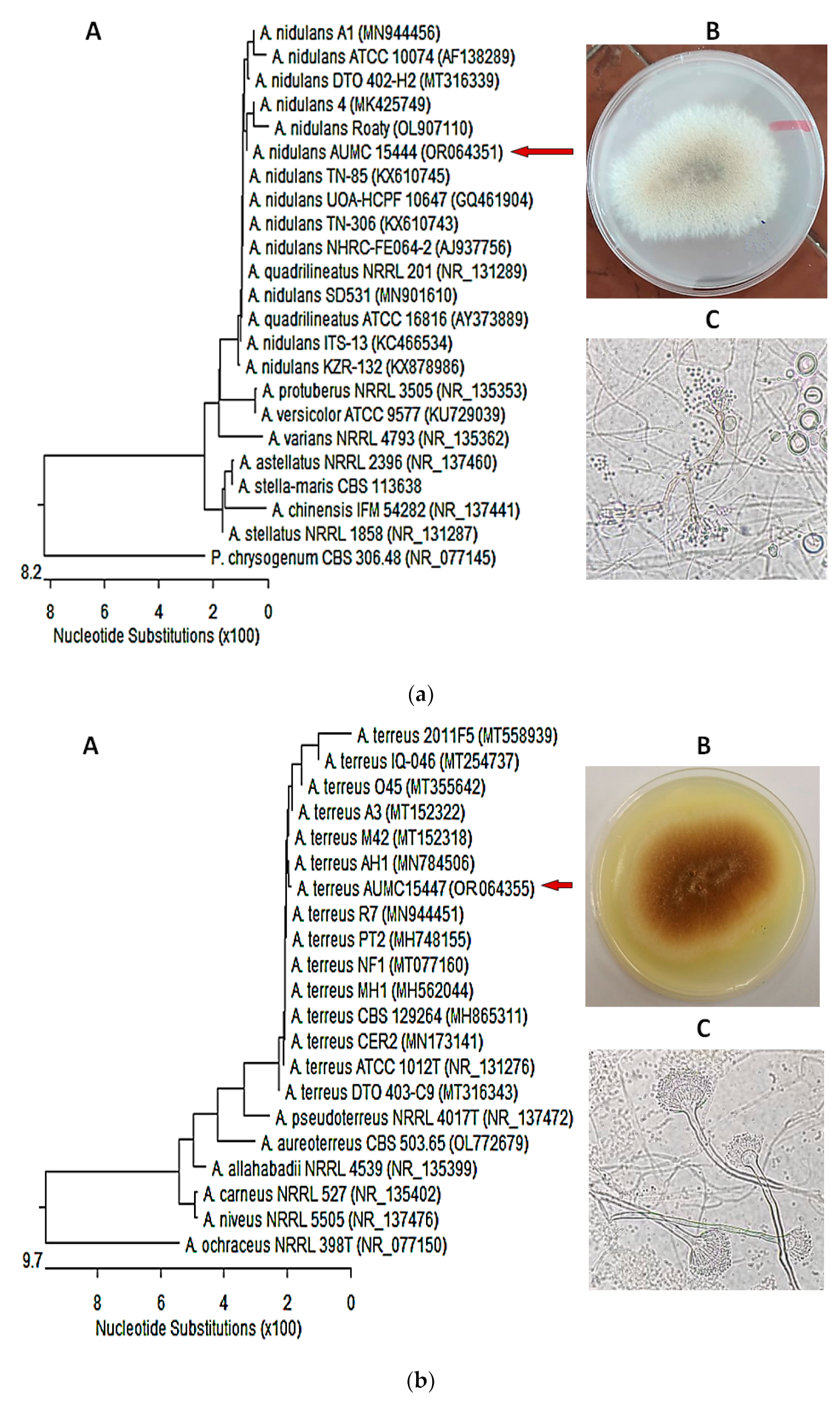
3.4. Molecular Identification of the of Most Potent Isolates

Identification of the most biopotent isolates was performed molecularly by sequencing the ITS region, and then sequences were subjected to a BLAST search of the NCBI database. The isolates were confirmed as Aspergillus nidulans AUMC 15444 (GenBank accession no. OR064351) and Aspergillus terreus AUMC 15447 (GenBank accession no. OR064355). Nucleotide comparison of the ITS regions among A. nidulans AUMC 15444 strain and other similar strains recaptured from the NCBI showed 99.62–99.81% identity and 99–100% coverage with several strains of the same species. Penicillium chrysogenum was included as an outgroup strain (Figure 1a). Meanwhile, A. terreus AUMC 15447 showed 99.67–100% identity and 98–100% coverage with numerous strains of the same species comprising the type strain A. terreus ATCC1012 with GenBank accession no. NR_131276. Aspergillus ochraceus was included as an outgroup strain (Figure 1b).

3.5. Evaluation of Total Phenolics and Flavonoids

The results proved that the A. terreus AUMC 15447 EtOAc extract had the highest content of phenolics (138.30 mg/g) and flavonoids (72.09 mg/g), while the A. terreus AUMC 15448 EtOAc extract showed good phenolic and flavonoid content (116.54 and 53.60 mg/g, respectively). The lowest content of phenolics was noted in the extracts of P. novae-zeelandiae AUMC 15442 and T. harzianum AUMC 15443 (20.60 and 25.81 mg/g, respectively), and the lowest flavonoid content in A. aureolatus AUMC 15441 and P. novae-zeelandiae AUMC 15442 (17.43 and 18.48 mg/g, respectively) (Table 7).

3.6. Flavonoid and Phenolic HPLC Profile of Extracts

HPLC analysis for profiling the flavonoid and phenolic chemical constituents of EtOAc extracts of the derived fungal isolates was performed. Standards utilized in the analysis, according to their retention time, included chlorogenic acid, gallic acid, catechin, methyl gallate, caffeic acid, syringic acid, rutin, ellagic acid, p-coumaric acid, vanillin, cinnamic acid, ferulic acid, daidzein, naringenin, quercetin, apigenin, kaempferol, and hesperetin, as shown in Table 8 and Supplementary Figures S1–S9.
Ellagic acid, vanillin, and apigenin were present at high levels in T. harzianum AUMC 15440 (12.34, 0.91, and 15.30%, respectively) in comparison with other isolate extracts, with apigenin being a major compound. The Trichoderma harzianum AUMC 15443 extract contained gallic acid (3.62%), catechin (0.26%), methyl gallate (19.89%), rutin (8.17%), and p-coumaric acid (2.73%) in high concentrations compared with other extracts, with methyl gallate being a major constituent. Chlorogenic acid (66.39%) was detected at high levels and was a major phenolic in A. aureolatus AUMC 15441, while syringic acid and daidzein, as major chemical constituents, were present in large quantities (14.49 and 28.24%) in the A. aureolatus AUMC 15441 extract. Meanwhile, elevated quantities of caffeic acid (15.43%) and hesperetin (29.24%) as the major components in the A. nidulans AUMC 15444 extract, quercetin (26.30%) in A. terreus AUMC 15447, as well as ferulic acid and naringenin (14.19% and 7.50%, respectively) in the A. terreus AUMC 15448 EtOAc extract were detected. Among all the extracts, the highest concentrations of kaempferol (9.34%) and cinnamic acid (64.83%) as the major phenolics were detected in extracts derived from P. crustosum AUMC 15445 and P. novae-zeelandiae AUMC 15442, respectively. The results of the detected phenolics revealed that cinnamic acid (41.65 and 36.58%) and chlorogenic acid (40.99%) are the major phenolics quantified in A. terreus AUMC 15447 and AUMC 15448 and P. crustosum AUMC 15445 extracts, respectively. On the other hand, neither vanillin nor apigenin were detected in A. terreus AUMC 15447 and P. novae-zeelandiae AUMC 15442 extracts, while kaempferol was not detected in A. aureolatus AUMC 15441and AUMC 15446 and P. novae-zeelandiae AUMC 15442 extracts. Catechin was detected only in T. harzianum AUMC 15443 and A. nidulans AUMC 15444 extracts.

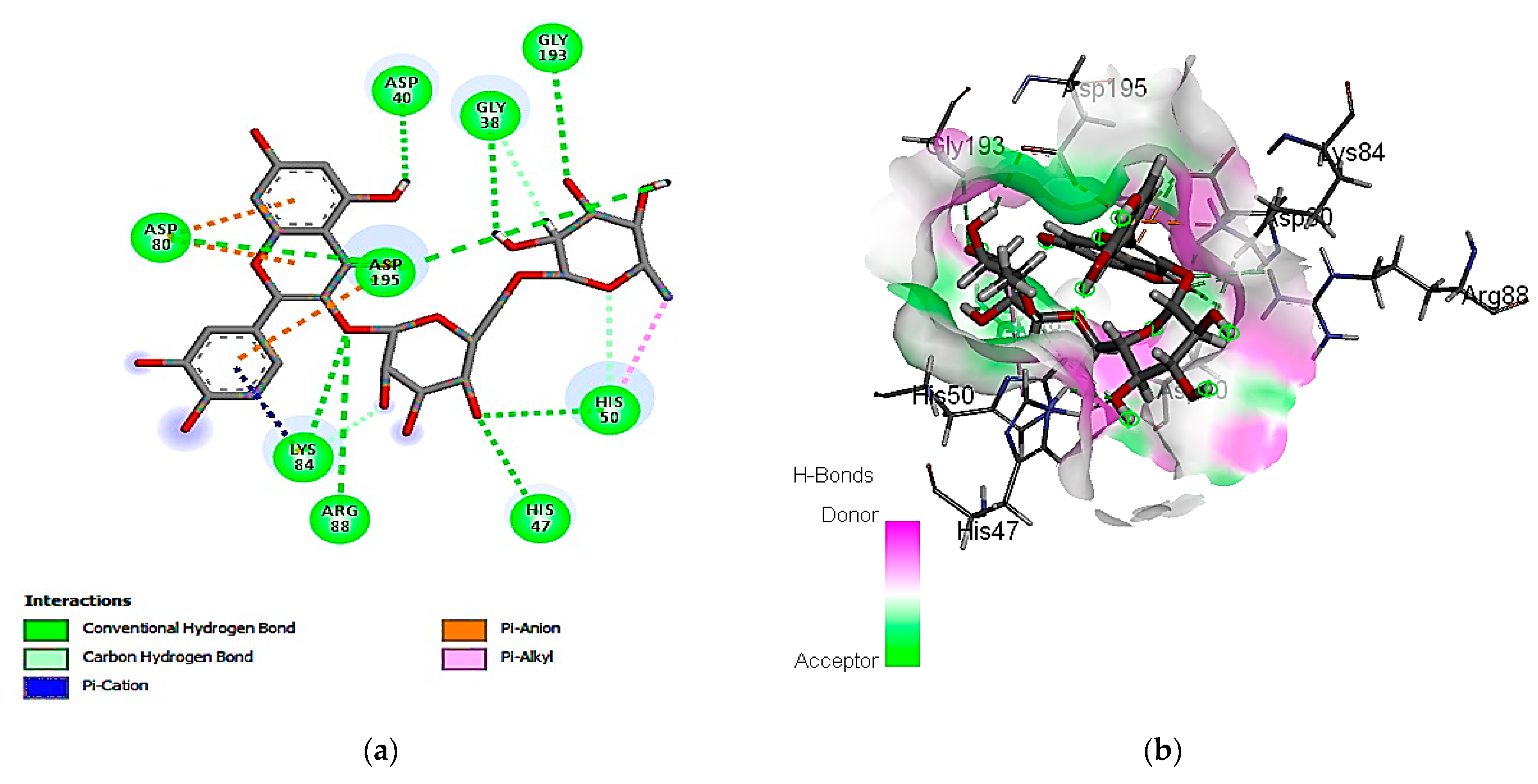
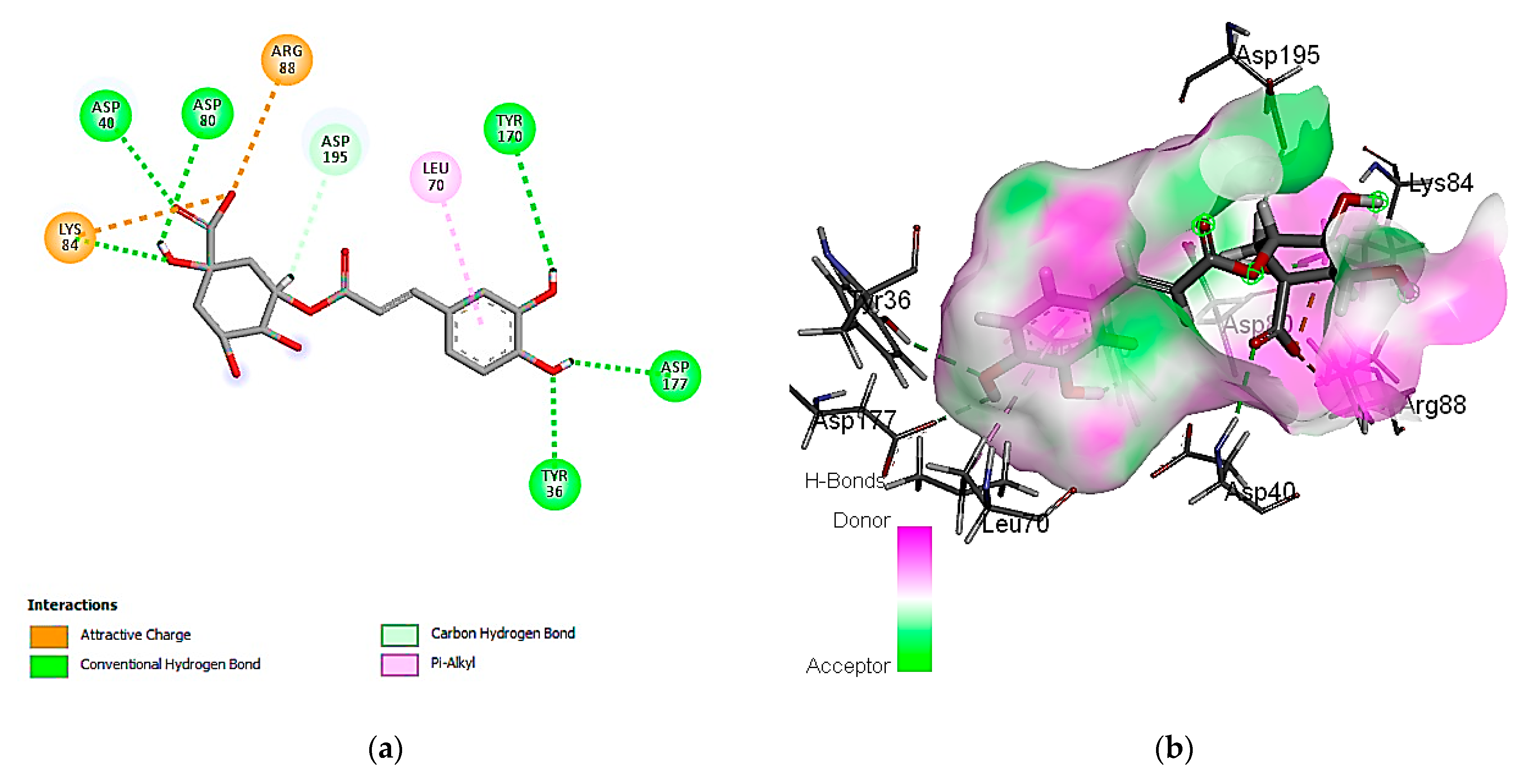
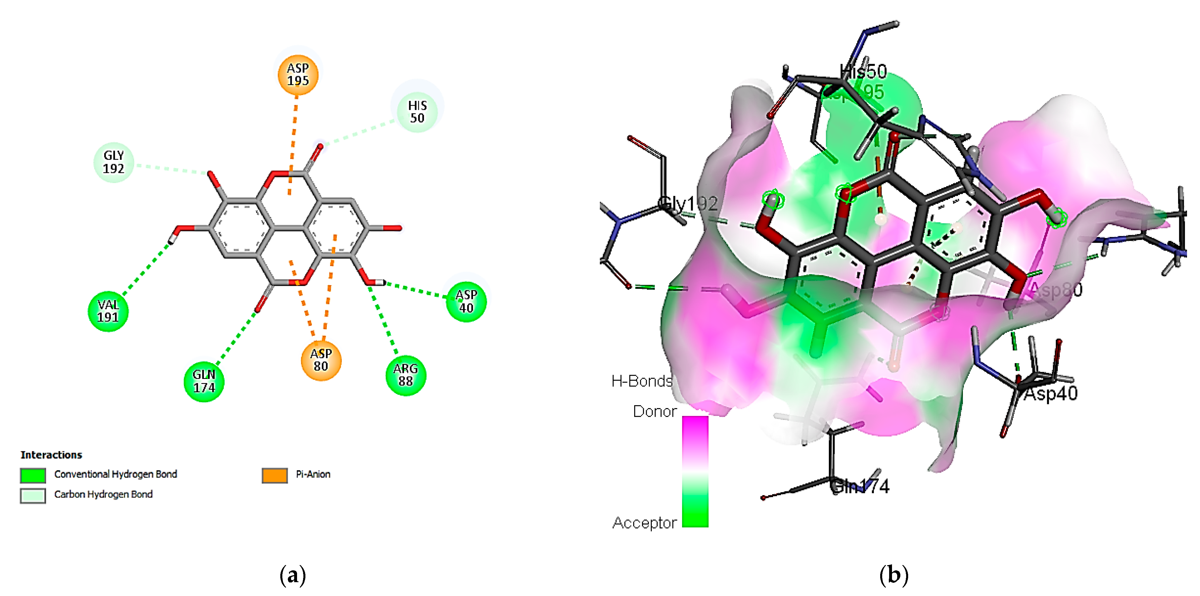
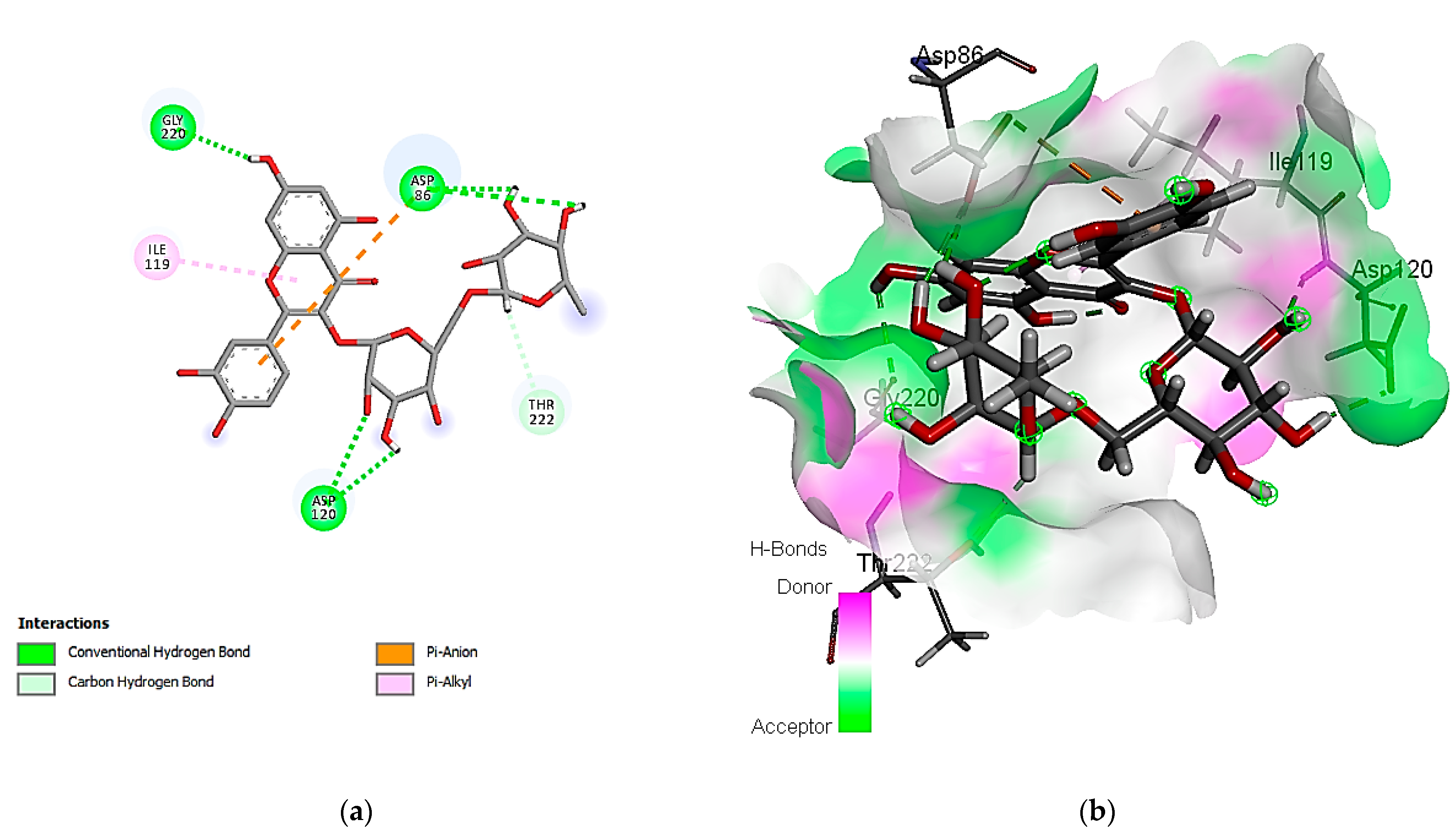
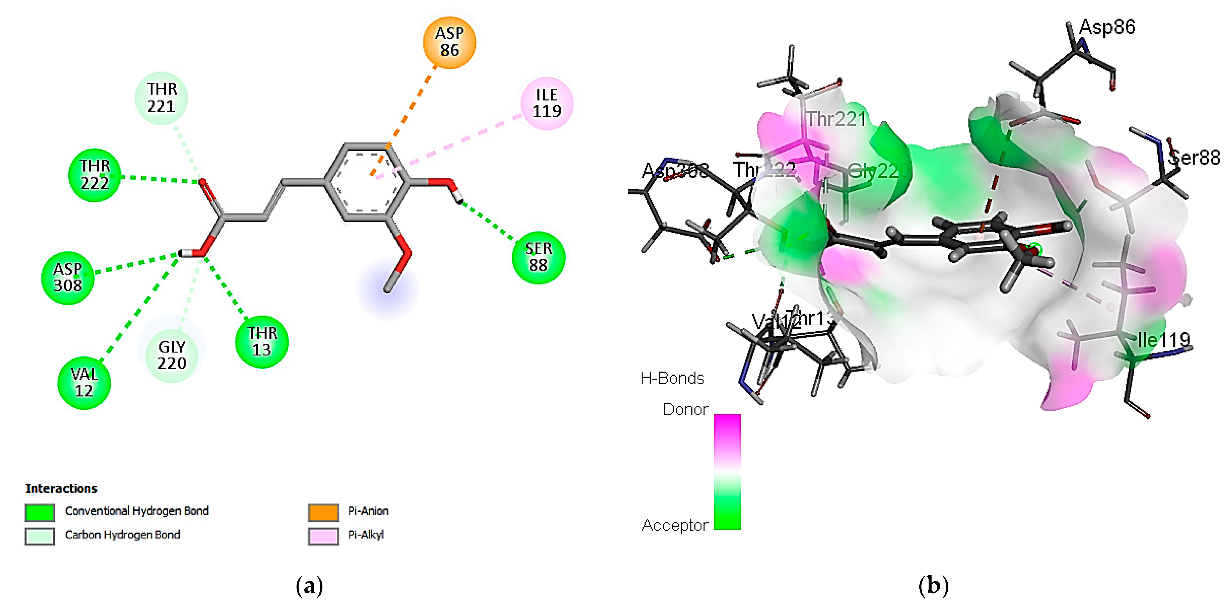
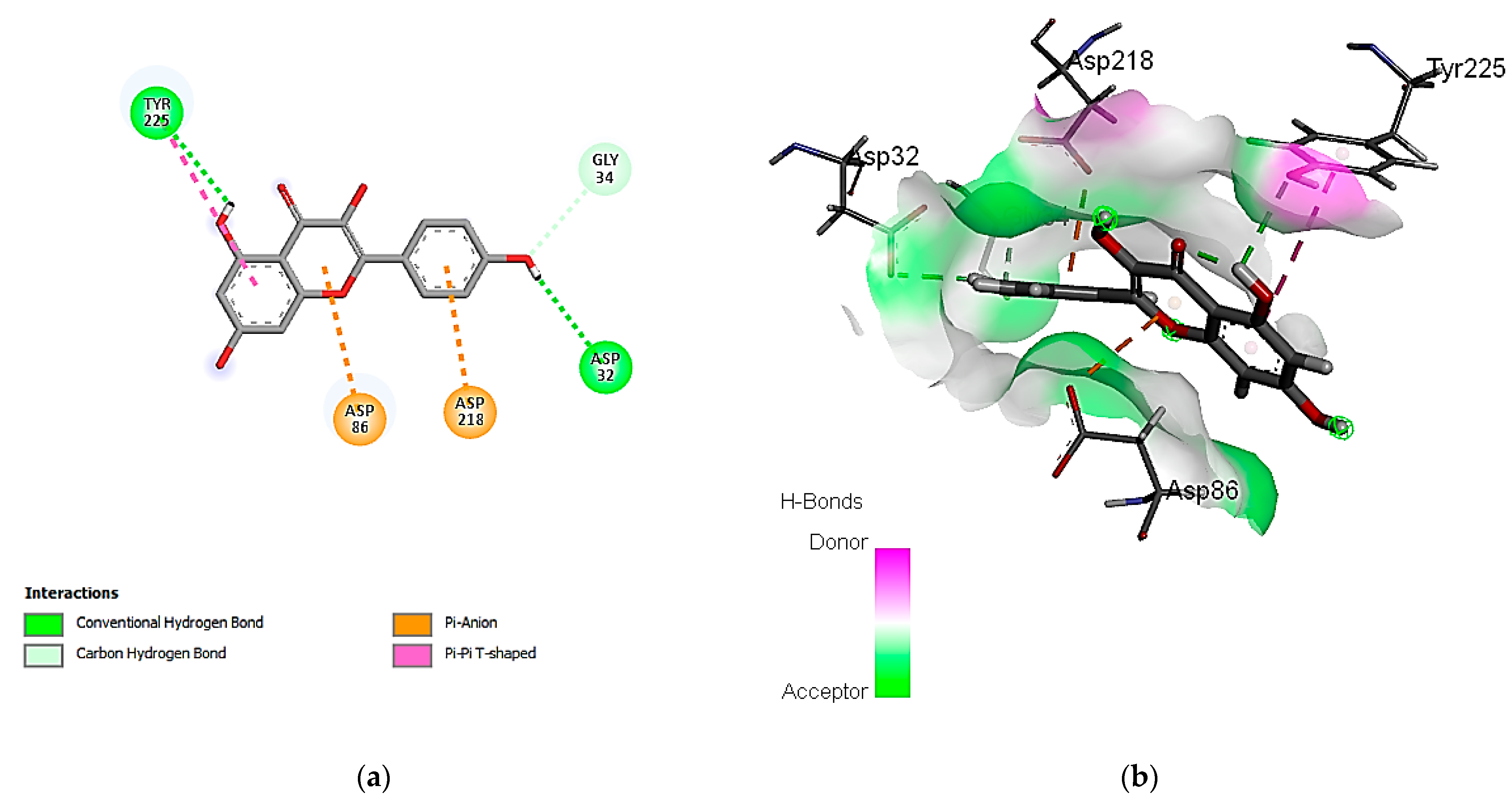
3.7. In Silico Molecular Docking of Identified Compounds

According to the HPLC analysis, the diverse extracts were found to contain approximately 18 phenolic compounds, as detailed in Table 8. Docking studies were then conducted to assess the binding affinities of these compounds as potential antimicrobials against both S. aureus and C. albicans. The ranking of binding interactions for each compound with the respective target proteins was based on factors such as the lowest energy and lowest RMSD (Root-Mean-Square Deviation). It is noteworthy that a lower binding energy score indicates better stability in the protein–ligand binding interaction [80].

3.7.1. Molecular Docking Simulation against S. aureus Tyrosyl-tRNA Synthetase

The exploration of molecular simulation outcomes for the identified molecules against S. aureus tyrosyl-tRNA synthetase indicated diverse affinities for the enzyme, ranging from −16.43 kcal/mol (rutin) to −7.30 kcal/mol (cinnamic acid), suggesting their potential as inhibitors (Table 9). Rutin, chlorogenic acid, and ellagic acid emerged as the top-scoring compounds, exhibiting pose scores of −16.43 (RMSD = 1.32 Å), −13.94 (RMSD = 1.40 Å), and −13.89 (RMSD = 0.55 Å) kcal/mol, respectively. Notably, the highest-affinity compound, rutin, demonstrated hydrogen-bond interactions with Cys 37 and Asp 40 amino acid residues, as revealed in Figure 2. Additionally, the 2D and 3D interaction models illustrated the involvement of hydrophobic interactions with the Ala 39, Pro 53, and Phe 54 amino acid residues. Also, the interactions of rutin with the enzyme include an acidic interaction with Asp 40, 80, and 195 as well as basic interactions with the Lys 84 and Arg 88 amino acid residues. Other polar interactions for chlorogenic and ellagic acids are shown in Figure 3 and Figure 4.

3.7.2. Molecular Docking Simulation with the C. albicans Secreted Aspartic Protease 2

Concerning the binding affinity of the investigated compounds against the C. albicans secreted aspartic protease 2 (Table 9), the compounds rutin, ferulic acid, and kaempferol achieved the highest binding energy scores of −12.3505, −11.1575, and −10.9706 kcal/mol, respectively. The binding mode of rutin showed HB binding with the Asp 86, Asp 120, and Thr 222 amino acid residues (Figure 5). It is noteworthy that hydrophobic interactions are involved in the binding of rutin with the Ile 30, 119, 123, and Val 12 amino acid residues. Other polar interactions for ferulic acid and kaempferol are shown in Figure 6 and Figure 7.

4. Discussion

Fungi are a prolific source of bio-effective secondary metabolites, including those with antimicrobial efficiency, which have been developed into significant pharmaceuticals. Endophytic fungi have been documented as having the capacity to generate novel antibacterial, antifungal, antiviral, anti-inflammatory, antitumor, and antimalarial compounds. These bioactive natural products are eliciting substantial interest from biologists and natural product chemists [81]. Numerous diseases caused by S. aureus, ranging from mild skin infections to more serious and potentially fatal diseases, accompanied by the emergence of multi-resistant strains, have increased the demand for new antibacterial agents [53]. Limited availability, in addition to the side effects of efficient antifungal agents, are some of the issues faced with C. albicans infections [82]. Consequently, it is imperative to develop new drugs by exploring alternate sources such as terrestrial and marine environments for novel medications [17,83].
In the current investigation, nine EtOAc extracts were obtained from soil fungal isolates belonging to six different fungal species, namely T. harzianum, A. aureolatus, A. nidulans, A. terreus, P. crustosum, and P. novae-zeelandiae, which exerted anti-staphylococcal and anti-candidal influences against tested species of Staphylococcus and Candida. The resultant anti-staphylococcal activities of the different fungal extracts at 100 mg/mL revealed that most of these extracts exhibited efficiency against the tested S. aureus species. Aspergillus terreus, A. nidulans, and T. harzianum extracts exhibited the highest activity, with MIC values ranging between 25 and 0.39 mg/mL. On the other hand, the A. nidulans extract revealed a high potency, with a MIC of 3.125–0.39 mg/mL against Candida spp. The Aspergillus terreus MK-1 ethyl acetate extract showed antagonistic activity against S. aureus and C. albicans, with inhibition zones of 45 and 30 mm, respectively [84]. Phupiewkham et al. [85] reported that T. harzianum TS3 and TS12 filtrates exert a positive control effect against S. aureus ATCC25923. A crude extract from T. harzianum exhibited the highest zone of inhibition of 26 mm against methicillin-resistant S. aureus [86]. A Trichoderma harzianum extract had an MIC of 50 μg/mL against S. aureus [87]. An Aspergillus nidulans ethyl acetate extract against an oral isolate of C. albicans (CA 09) showed a growth inhibitory effect, with an 18.4 mm diameter inhibition zone and an MIC value of 250 μg/mL [88]. External secondary metabolites in an n-butanol extract from Aspergillus terreus var. africans exhibited a 22 and 19 mm diameter inhibition zone against C. albicans and C. glabrata, respectively [12]. The ability of C. albicans to transform into a hyphal state and adhere to tissue surfaces is one of the main causes for its severe pathogenicity [88].
Staphylococcus aureus exerted resistance to gentamicin and chloramphenicol while presenting susceptibility to amoxicillin and vancomycin, with MICs of 0.1 and 0.015 mg/mL, respectively [89]. Antibacterial agents such as ampicillin, amoxicillin, chloramphenicol, and cefotaxime had MIC values of 39, 39, 16, and 1 µg/mL, respectively, against S. aureus ATCC 11632 [90]. However, ampicillin exhibited MICs of 2.5 and 10 µg/mL on S. aureus ATCC 25923 and S. aureus TN2A, respectively [52]. Al Halteet et al. [65], in their study to determine the antifungal susceptibility of 24 Candida species, indicated that C. krusei MN419370 exhibited resistance to all the tested antifungal agents (fluconazole, voriconazole, caspofungin, micafungin, amphotericin B, and flucytosine), while C. krusei MN419388 was resistant only to fluconazole and flucytosine. Meanwhile, 2 out of 18 C. albicans strains showed resistance to all antifungal agents. On the other hand, three C. glabrata strains were susceptible to all tested antifungal agents.
The utilization of the microbroth dilution method using microtiter plates has emerged as the technique of choice for assessing drug susceptibility testing due to its low sample needs, affordability, and high throughput rate [91]. The p-iodonitrotetrazolium (INT) was chosen as an indicator because of its preparation in ethanol that, as an antiseptic, makes it appropriate for reducing the contamination risk during test procedures [92]. Violet INT is a tetrazolium dye precursor that, upon reduction, constitutes a purple formazan whose intensity is directly proportional to the number of live cells. The reduction of INT is due to the mitochondrial enzymatic activity within living cells [67].
According to the results, the A. terreus AUMC 15447 extract exhibited the highest phenolic content of 138.30 mg/g, a flavonoid content of 72.09 mg/g, and an IC50 of 0.47 mg/mL by the anti-DPPH radical-scavenging assay, followed by the A. terreus AUMC 15448 extract. The ethyl acetate extract fraction from A. terreus LS01 showed an IC50 of 19.91 μg/mL in the in vitro anti-DPPH radical-scavenging assay [93]. A total phenolic content of 122.475 mg/g was detected in an A. terreus-18 EtOAc extract with the highest DPPH scavenging activity of 80.4% inhibition [94]. Chandra and Arora [95] determined a phenolic content (20.4 and 16.7 mg/mL) and DPPH scavenging efficiency (86.8 and 65.8%) in A. terreus-1 and -2 soil isolates, respectively, after fermentation on sugarcane bagasse. Aspergillus nidulans ST22 showed total phenolic and flavonoid amounts of 0.1413 and 0.01162 mg/mL, respectively, and a 34.17% antioxidative capacity by a hydroxyl radical-scavenging assay [96]. Goncalves and Pombeiro-Sponchiado [97] reported the ability of melanin from A. nidulans MEL1 to scavenge the oxidants HOCl and H2O2. Al-Askar [98] established total phenols of 53.30 μg/mL in a T. harzianum 1-SSR filtrate. Tavares et al. [99] evaluated a TPC of 8 mg/g and an EC50 value of 25.41 μg/mL for free radical DPPH in an ethyl acetate extract of A. aureolatus CML2964 isolated from caves, while Ahmed and Al-Shamary [100] determined a phenolic content of 51 μg/mL in the soil A. niger B1b strain. In our results, T. harzianum AUMC 15440 recorded an LC50 of 1.18 mg/mL in the brine- shrimp cytotoxicity assay, while the A. terreus AUMC 15448 extract showed an LC50 of 1.29 mg/mL. The biotoxicity of the A. terreus var. africanus crude ethyl acetate extract showed a slight toxicity against Artemia salina (LC50 = 1500 µg/mL) [101].
The most effective fungal isolates were subjected to phylogenetic analysis by ITS region sequencing, which molecularly identified them as A. nidulans AUMC 15444 and A. terreus AUMC 15447. The ITS regions represent important efficient markers for confirming fungal strain identification at the species level [102]. Molecular identification of Aspergillus strains at the species level using ITS region sequencing was presented as an efficient substitute approach for their accurate identification [103].
HPLC analysis for the detection of phenolic compounds in fungal EtOAc extracts with reference standards showed great variation in the types and quantities of detected constituents among the fungal extracts. In the same context, Hassane et al. [10,11] detected a variety of phenolic and flavonoid compounds in fungal extracts belonging to different genera of Aspergillus, Penicillium, Chaetomium, and Rhizopus. Abdel-Wareth and Ghareeb [104] identified 22 phenolic and flavonoid compounds (cinnamic acid, 3,4,5-methoxy cinnamic acid, α-coumaric acid, p-coumaric acid, coumarin, pyrogallol, gallic acid, methyl gallate, ellagic acid, benzoic acid, 4-amino-benzoic acid, catechin, epicatechin, protocatechuic acid, vanillic acid, ferulic acid, isoferulic acid, chlorogenic acid, caffeic acid, resveratrol, and salicylic acid) in Penicillium implicatum and Aspergillus niveus extracts from fresh-water isolates by RP-HPLC/DAD analysis, where pyrogallol and e-vanillic were the major phenolics detected in the extracts, respectively. Abdel-Aty et al. [105] optimized the production of p-coumaric, apigenin, and kaempferol using SSF of Trichoderma reesei with a TPC of 30 mg/g. The diorcinol phenolic was detected and isolated from A. nidulans MCCC 3A00050 [106]. Shevelev et al. [44] reported the in vivo efficacy of polyphenols, including resveratrol, dihydroquercetin, and dihydromyricetin, in the treatment of infected wounds with S. aureus ATCC 25923 and C. albicans NCTC 2625. Kepa et al. [107] demonstrated the promised efficiency of caffeic acid with MICs (256–1024 µg/mL) against MR S. aureus, clinical isolates, and reference strains derived from wound infections.
Moreover, advancements in computational chemistry have enabled the development of some fungal-derived anti-pathogenic compounds with potential therapeutic influences in controlling health-threatening microbes [53]. The docking results of detected phenolics against the S. aureus tyrosyl-tRNA synthetase and the C. albicans secreted aspartic protease 2 found that rutin had the lower binding energy score, which conferred on it better protein–ligand binding stability [108]. Rutin and rutin–gentamicin act as pro-oxidants against Pseudomonas aeruginosa through oxidative stress by inducing reactive oxygen species generation, which leads to cell death [109]. Docking of flavonoids—saltillin, taxifolin, and 6-methoxyflavone—from the A. nidulans chloroform extract displayed good binding interactions with the C. albicans growth regulator N-myristoyltransferase [88].
Naturally derived substances are a prime contender for treating microbial infections and restricting the spread of drug-resistant strains because of their capacity to interact with the microbial cell through a variety of antimicrobial processes [110,111]. Phenolic acids and their derivatives showed antimicrobial effects by influencing the solubility of microbial membranes, rupturing the cellular membranes and allowing vital intracellular components to outflow, thus resulting in death of the microbial cell [23,112,113]. The compounds’ capacity to pass through the cytoplasmic membrane and acidify the cytoplasm is dependent on the number of hydroxyl, methoxy, and carboxyl groups in addition to the saturation state of the alkyl side chain [114]. Lou et al. [115] evaluated p-coumaric acid’s antimicrobial efficacy in triggering bacterial cell death by disturbing the cell membrane and the interaction with genomic DNA. Wu et al. [26] reported that flavonoids and their isomers represent a valuable anti-pathogenic mediator by targeting a variety of pathogens and suppressing virulence properties in drug-resistant microbial strains. Their capacity to impede microbial cell energy metabolism, nucleic acid synthesis, complex formation with the bacterial cell wall, and cytoplasmic membrane function, are the mechanisms linked to their bacteriostatic actions [25,116].

5. Conclusions

Ethyl acetate (EtOAc) extracts from the fungal isolates exhibited notable anti-staphylococcal and anti-candidal efficacy, with A. terreus AUMC 15447 and A. nidulans AUMC 15444 strains demonstrating particularly high efficiency, as determined through microdilution assays combined with INT for assessing antimicrobial efficacy. Additionally, the assayed extracts displayed considerable phenolic and flavonoid contents, along with significant IC50 values for antioxidative activity and LC50 values in Artemia cytotoxicity assays. HPLC analysis revealed the presence of various phenolics in different concentrations within the fungal EtOAc extracts. Molecular simulations of these detected phenolics highlighted the high affinity of rutin with the S. aureus tyrosyl-tRNA synthetase and the C. albicans secreted aspartic protease 2, indicating inhibitory properties. This research suggests the potential for bioprospecting anti-staphylococcal and anti-candidal bio-active metabolites from fungi, providing a sustainable source for safe medication.

Supplementary Materials

The following supporting information can be downloaded at: https://www.mdpi.com/article/10.3390/cimb46010016/s1, HPLC chromatograms of phenolic components in EtOAc extracts (Figures S1–S9).

Author Contributions

Conceptualization, A.M.A.H. and A.A.A.M.; methodology, M.E.A., A.M.A.H. and Y.A.-D.; software, M.E.A. and Y.A.-D.; validation, A.A.A.M., M.E.A. and N.S.A.G.; formal analysis, H.M.; investigation, M.E.A. and H.M.; resources, M.E.A. and A.M.A.H.; data curation, H.M.; writing—original draft preparation, H.M., N.S.A.G. and Y.A.-D.; writing—review and editing, M.E.A., A.A.A.M. and A.M.A.H.; visualization, A.M.A.H. and N.F.A.-D.; supervision, A.A.A.M. and A.M.A.H.; project administration, A.A.A.M. and N.F.A.-D.; funding acquisition, A.A.A.M. All authors have read and agreed to the published version of the manuscript.

Funding

This research was funded by the Distinguished Scientist Fellowship Program through the Researchers Supporting Project (RSP2023R227), King Saud University, Riyadh, Saudi Arabia.

Institutional Review Board Statement

Not applicable.

Informed Consent Statement

Not applicable.

Data Availability Statement

Data are contained within the article and Supplementary Materials.

Conflicts of Interest

The authors declare no conflicts of interest.

References

  1. Mohammed, A.E.; Sonbol, H.; Alwakeel, S.S.; Alotaibi, M.O.; Alotaibi, S.; Alothman, N.; Suliman, R.S.; Ahmedah, H.T.; Ali, R. Investigation of biological activity of soil fungal extracts and LC/MS-QTOF based metabolite profiling. Sci. Rep. 2021, 11, 4760. [Google Scholar] [CrossRef] [PubMed]
  2. Higginbotham, S.J.; Arnold, A.E.; Ibanez, A.; Spadafora, C.; Coley, P.D.; Kursar, T.A. Bioactivity of fungal endophytes as a function of endophyte taxonomy and the taxonomy and distribution of their host plants. PLoS ONE 2013, 8, e73192. [Google Scholar] [CrossRef] [PubMed]
  3. Giavasis, I. Bioactive fungal polysaccharides as potential functional ingredients in food and nutraceuticals. Curr. Opin. Biotechnol. 2014, 26, 162–173. [Google Scholar] [CrossRef] [PubMed]
  4. Al Mousa, A.A.; Abo-Dahab, N.F.; Hassane, A.M.A.; Gomaa, A.F.; Aljuriss, J.A.; Dahmash, N.D. Harnessing Mucor spp. for xylanase production: Statistical optimization in submerged fermentation using agro-industrial wastes. BioMed Res. Int. 2022, 2022, 3816010. [Google Scholar] [CrossRef] [PubMed]
  5. Al Mousa, A.A.; Hassane, A.M.A.; Gomaa, A.F.; Aljuriss, J.A.; Dahmash, N.D.; Abo-Dahab, N.F. Response-surface statistical optimization of submerged fermentation for pectinase and cellulase production by Mucor circinelloides and M. hiemalis. Fermentation 2022, 8, 205. [Google Scholar] [CrossRef]
  6. Mohamed, H.; Awad, M.F.; Shah, A.M.; Nazir, Y.; Naz, T.; Hassane, A.; Nosheen, S.; Song, Y. Evaluation of different standard amino acids to enhance the biomass, lipid, fatty acid, and γ -linolenic acid production in Rhizomucor pusillus and Mucor circinelloides. Front. Nutr. 2022, 9, 876817. [Google Scholar] [CrossRef]
  7. Rajamanikyam, M.; Vadlapudi, V.; Amanchy, R.; Upadhyayula, S.M. Endophytic fungi as novel resources of natural therapeutics. Braz. Arch. Biol. Technol. 2017, 60, e17160542. [Google Scholar] [CrossRef]
  8. Pimentel, M.R.; Molina, G.; Dionisio, A.P.; Maróstica, M.R.; Pastore, G.M. Use of endophytes to obtain bioactive compounds and their application in biotransformation process. Biotechnol. Res. Int. 2011, 2011, 576286. [Google Scholar] [CrossRef]
  9. Al Mousa, A.A.; Abouelela, M.E.; Hassane, A.M.A.; Al-Khattaf, F.S.; Hatamleh, A.A.; Alabdulhadi, H.S.; Dahmash, N.D.; Abo-Dahab, N.F. Cytotoxic potential of Alternaria tenuissima AUMC14342 mycoendophyte extract: A study combined with LC-MS/MS metabolic profiling and molecular docking simulation. Curr. Issues Mol. Biol. 2022, 44, 5067–5085. [Google Scholar] [CrossRef]
  10. Hassane, A.M.A.; Taha, T.M.; Awad, M.F.; Mohamed, H.; Melebari, M. Radical scavenging potency, HPLC profiling and phylogenetic analysis of endophytic fungi isolated from selected medicinal plants of Saudi Arabia. Electron. J. Biotechnol. 2022, 58, 37–45. [Google Scholar] [CrossRef]
  11. Hassane, A.M.A.; Hussien, S.M.; Abouelela, M.E.; Taha, T.M.; Awad, M.F.; Mohamed, H.; Hassan, M.M.; Hassan, M.H.A.; Abo-Dahab, N.F.; El-Shanawany, A.A. In vitro and in silico antioxidant efficiency of bio-potent secondary metabolites from different taxa of black seed-producing plants and their derived mycoendophytes. Front. Bioeng. Biotechnol. 2022, 10, 930161. [Google Scholar] [CrossRef] [PubMed]
  12. Alkhulaifi, M.M.; Awaad, A.S.; AL-Mudhayyif, H.A.; Alothman, M.R.; Alqasoumi, S.I.; Zain, S.M. Evaluation of antimicrobial activity of secondary metabolites of fungi isolated from Sultanate Oman soil. Saudi Pharm. J. 2019, 27, 401–405. [Google Scholar] [CrossRef] [PubMed]
  13. Abdelrahem, M.M.M.; Hassane, A.M.A.; Abouelela, M.E.; Abo-Dahab, N.F. Comparative bioactivity and metabolites produced by fungal co-culture system against myco-phytopathogens. J. Environ. Stud. 2023, 31, 1–15. [Google Scholar] [CrossRef]
  14. Ujam, N.T.; Ajaghaku, D.L.; Okoye, F.B.C.; Esimone, C.O. Antioxidant and immunosuppressive activities of extracts of endophytic fungi isolated from Psidium guajava and Newbouldia laevis. Phytomed. Plus 2021, 1, 100028. [Google Scholar] [CrossRef]
  15. Tawfike, A.F.; Romli, M.; Clements, C.; Abbott, G.; Young, L.; Schumacher, M.; Diederich, M.; Farag, M.; Edrada-Ebel, R. Isolation of anticancer and anti-trypanosome secondary metabolites from the endophytic fungus Aspergillus flocculus via bioactivity guided isolation and MS based metabolomics. J. Chromatogr. B 2019, 1106–1107, 71–83. [Google Scholar] [CrossRef] [PubMed]
  16. El-Kassem, L.A.; Hawas, U.W.; El-Souda, S.; Ahmed, E.F.; El-Khateeb, W.; Fayad, W. Anti-HCV protease potential of endophytic fungi and cytotoxic activity. Biocatal. Agric. Biotechnol. 2019, 19, 101170. [Google Scholar] [CrossRef]
  17. Al-Fakih, A.A.; Almaqtri, W.Q.A. Overview on antibacterial metabolites from terrestrial Aspergillus spp. Mycology 2019, 10, 191–209. [Google Scholar] [CrossRef]
  18. Saber, S.M.; Youssef, M.S.; Arafa, R.F.; Hassane, A.M.A. Mycotoxins production by Aspergillus ostianus Wehmer and using phytochemicals as control agent. J. Sci. Eng. Res. 2016, 3, 198–213. [Google Scholar]
  19. Hassane, A.M.A.; El-Shanawany, A.A.; Abo-Dahab, N.F.; Abdel-Hadi, A.M.; Abdul-Raouf, U.M.; Mwanza, M. Cultural and analytical assays for aflatoxin B production by Aspergillus flavus isolates. J. Nat. Prod. Chem. 2017, 1, 17–23. [Google Scholar] [CrossRef]
  20. Zhong, J.J.; Xiao, J.H. Secondary metabolites from higher fungi: Discovery, bioactivity, and bioproduction. Adv. Biochem. Eng. Biotechnol. 2009, 113, 79–150. [Google Scholar] [CrossRef]
  21. Oliver, S.; Vittorio, O.; Cirillo, G.; Boyer, C. Enhancing the therapeutic effects of polyphenols with macromolecules. Polym. Chem. 2016, 7, 1529–1544. [Google Scholar] [CrossRef]
  22. Dantas, S.B.S.; Moraes, G.K.A.; Araujo, A.R.; Chapla, V.M. Phenolic compounds and bioactive extract produced by endophytic fungus Coriolopsis rigida. Nat. Prod. Res. 2023, 37, 2037–2042. [Google Scholar] [CrossRef] [PubMed]
  23. Sánchez-Maldonado, A.F.; Schieber, A.; Gänzle, M.G. Structure–function relationships of the antibacterial activity of phenolic acids and their metabolism by lactic acid bacteria. J. Appl. Microbiol. 2011, 111, 1176–1184. [Google Scholar] [CrossRef] [PubMed]
  24. Keman, D.; Soyer, F. Antibiotic-resistant Staphylococcus aureus does not develop resistance to vanillic acid and 2-hydroxycinnamic acid after continuous exposure in vitro. ACS Omega 2019, 4, 15393–15400. [Google Scholar] [CrossRef] [PubMed]
  25. Ecevit, K.; Barros, A.A.; Silva, J.M.; Reis, R.L. Preventing microbial infections with natural phenolic compounds. Future Pharmacol. 2022, 2, 460–498. [Google Scholar] [CrossRef]
  26. Wu, S.-C.; Liu, F.; Zhu, K.; Shen, J.-Z. Natural products that target virulence factors in antibiotic-resistant Staphylococcus aureus. J. Agric. Food Chem. 2019, 67, 13195–13211. [Google Scholar] [CrossRef] [PubMed]
  27. Nobile, C.J.; Johnson, A.D. Candida albicans biofilms and human disease. Annu. Rev. Microbiol. 2015, 69, 71–92. [Google Scholar] [CrossRef]
  28. Patil, S.; Rao, R.S.; Majumdar, B.; Anil, S. Clinical appearance of oral candida infection and therapeutic strategies. Front. Microbiol. 2015, 6, 1391. [Google Scholar] [CrossRef]
  29. Silva, S.; Negri, M.; Henriques, M.; Oliveira, R.; Williams, D.W.; Azeredo, J. Candida glabrata, Candida parapsilosis and Candida tropicalis: Biology, epidemiology, pathogenicity and antifungal resistance. FEMS Microbial. Rev. 2012, 36, 288–305. [Google Scholar] [CrossRef]
  30. Ng, K.P.; Kuan, C.S.; Kaur, H.; Na, S.L.; Atiya, N.; Velayuthan, R.D. Candida species epidemiology 2000-2013: A laboratory-based report. Trop. Med. Int. Health 2015, 20, 1447–1453. [Google Scholar] [CrossRef]
  31. De Bernardis, F.; Graziani, S.; Tirelli, F.; Antonopoulou, S. Candida vaginitis: Virulence, host response and vaccine prospects. Med. Mycol. 2018, 56, 26–31. [Google Scholar] [CrossRef] [PubMed]
  32. Meenambiga, S.S.; Venkataraghavan, R.; Biswal, A. In silico analysis of plant phytochemicals against secreted aspartic proteinase enzyme of Candida albicans. J. Appl. Pharm. Sci. 2018, 8, 140–150. [Google Scholar] [CrossRef]
  33. Bhattacharya, S.; Sae-Tia, S.; Fries, B.C. Candidiasis and mechanisms of antifungal resistance. Antibiotics 2020, 9, 312. [Google Scholar] [CrossRef] [PubMed]
  34. Harriott, M.M.; Noverr, M.C. Candida albicans and Staphylococcus aureus form polymicrobial biofilms: Effects on antimicrobial resistance. Antimicrob. Agents Chemother. 2009, 53, 3914–3922. [Google Scholar] [CrossRef] [PubMed]
  35. Harriott, M.M.; Noverr, M.C. Ability of Candida albicans mutants to induce Staphylococcus aureus vancomycin resistance during polymicrobial biofilm formation. Antimicrob. Agents Chemother. 2010, 54, 3746–3755. [Google Scholar] [CrossRef] [PubMed]
  36. Tong, S.Y.C.; Davis, J.S.; Eichenberger, E.; Holland, T.L.; Fowler, V.G., Jr. Staphylococcus aureus infections: Epidemiology, pathophysiology, clinical manifestations, and management. Clin. Microbiol. Rev. 2015, 28, 603–661. [Google Scholar] [CrossRef] [PubMed]
  37. Stark, L. Staphylococcus aureus: Aspects of Pathogenesis and Molecular Epidemiology. Ph.D. Thesis, Linköping University Electronic Press, Linköping, Sweden, 2013; p. 1371. [Google Scholar]
  38. Saxena, S.; Gomber, C. Surmounting antimicrobial resistance in the Millennium Superbug: Staphylococcus aureus. Open Med. 2010, 5, 12–29. [Google Scholar] [CrossRef]
  39. Klotz, S.A.; Chasin, B.S.; Powell, B.; Gaur, N.K.; Lipke, P.N. Polymicrobial bloodstream infections involving Candida species: Analysis of patients and review of the literature. Diagn. Microbiol. Infect. Dis. 2007, 59, 401–406. [Google Scholar] [CrossRef]
  40. Carolus, H.; Van Dyck, K.; Van Dijck, P. Candida albicans and Staphylococcus species: A threatening twosome. Front. Microbiol. 2019, 10, 2162. [Google Scholar] [CrossRef]
  41. Budzynska, A.; Rozalska, S.; Sadowska, B.; Rozalska, B. Candida albicans/Staphylococcus aureus dual-species biofilm as a target for the combination of essential oils and fluconazole or mupirocin. Mycopathologia 2017, 182, 989–995. [Google Scholar] [CrossRef]
  42. Sanguinetti, M.; Posteraro, B.; Lass-Florl, C. Antifungal drug resistance among Candida species: Mechanisms and clinical impact. Mycoses 2015, 58, 2–13. [Google Scholar] [CrossRef] [PubMed]
  43. Guignard, B.; Entenza, J.M.; Moreillon, P. β-Lactams against methicillin-resistant Staphylococcus aureus. Curr. Opin. Pharmacol. 2005, 5, 479–489. [Google Scholar] [CrossRef] [PubMed]
  44. Shevelev, A.B.; La Porta, N.; Isakova, E.P.; Martens, S.; Biryukova, Y.K.; Belous, A.S.; Sivokhin, D.A.; Trubnikova, E.V.; Zylkova, M.V.; Belyakova, A.V.; et al. In vivo antimicrobial and wound-healing activity of resveratrol, dihydroquercetin, and dihydromyricetin against Staphylococcus aureus, Pseudomonas aeruginosa, and Candida albicans. Pathogens 2020, 9, 296. [Google Scholar] [CrossRef] [PubMed]
  45. Schaller, M.; Borelli, C.; Korting, H.C.; Hube, B. Hydrolytic enzymes as virulence factors of Candida albicans. Mycoses 2005, 48, 365–377. [Google Scholar] [CrossRef]
  46. Selmecki, A.; Forche, A.; Berman, J. Genomic plasticity of the human fungal pathogen Candida albicans. Eukaryot. Cell 2010, 9, 991–1008. [Google Scholar] [CrossRef]
  47. Naglik, J.; Albrecht, A.; Bader, O.; Hube, B. Candida albicans proteinases and host/pathogen interactions. Cell. Microbiol. 2004, 6, 915–926. [Google Scholar] [CrossRef]
  48. Talapko, J.; Juzbašić, M.; Matijević, T.; Pustijanac, E.; Bekić, S.; Kotris, I.; Škrlec, I. Candida albicans-the virulence factors and clinical manifestations of infection. J. Fungi 2021, 7, 79. [Google Scholar] [CrossRef]
  49. Mseddi, K.; Alimi, F.; Noumi, E.; Veettil, V.N.; Deshpande, S.; Adnan, M.; Hamdi, A.; Elkahoui, S.; Ahmed, A.; Kadri, A.; et al. Thymus musilii Velen. as a promising source of potent bioactive compounds with its pharmacological properties: In vitro and in silico analysis. Arab. J. Chem. 2020, 13, 6782–6801. [Google Scholar] [CrossRef]
  50. Pohlmann, J.; Brotz-Oesterhelt, H. New aminoacyltRNA synthetase inhibitors as antibacterial agents. Curr. Drug Targets Infect. Disord. 2004, 4, 261–272. [Google Scholar] [CrossRef]
  51. Farshadfar, C.; Mollica, A.; Rafii, F.; Noorbakhsh, A.; Nikzad, M.; Seyedi, S.H.; Abdi, F.; Verki, S.A.; Mirzaie, S. Novel potential inhibitor discovery against tyrosyl-tRNA synthetase from Staphylococcus aureus by virtual screening, molecular dynamics, MMPBSA and QMMM simulations. Mol. Simul. 2020, 46, 507–520. [Google Scholar] [CrossRef]
  52. Pisano, M.B.; Kumar, A.; Medda, R.; Gatto, G.; Pal, R.; Fais, A.; Era, B.; Cosentino, S.; Uriarte, E.; Santana, L.; et al. Antibacterial activity and molecular docking studies of a selected series of hydroxy-3-arylcoumarins. Molecules 2019, 24, 2815. [Google Scholar] [CrossRef] [PubMed]
  53. Vallavan, V.; Krishnasamy, G.; Zin, N.M.; Abdul Latif, M. A review on antistaphylococcal secondary metabolites from Basidiomycetes. Molecules 2020, 25, 5848. [Google Scholar] [CrossRef] [PubMed]
  54. Chopade, A.R.; Sayyad, F.J.; Pore, Y.V. Molecular docking studies of phytocompounds fom Phyllanthus species as potential chronic pain modulators. Sci. Pharm. 2015, 83, 243–267. [Google Scholar] [CrossRef] [PubMed]
  55. Othman, I.M.M.; Gad-Elkareem, M.A.M.; Anouar, E.; Aouadi, K.; Snoussi, M.; Kadri, A. New substituted pyrazolones and dipyrazolotriazines as promising tyrosyl-tRNA synthetase and peroxiredoxin-5 inhibitors: Design, synthesis, molecular docking and structure-activity relationship (SAR) analysis. Bioorg. Chem. 2021, 109, 104704. [Google Scholar] [CrossRef] [PubMed]
  56. Moubasher, A.H.; El-Naghy, M.A.; Abdel-Hafez, S.I.I. Studies on the fungus flora of three grains in Egypt. Mycopathol. Mycol. Appl. 1972, 47, 261–274. [Google Scholar] [CrossRef]
  57. Raper, K.B.; Fennell, D.I. The Genus Aspergillus; Williams & Wilkins Company: Baltimore, MD, USA, 1965. [Google Scholar]
  58. Moubasher, A.H. Soil Fungi in Qatar and Other Arab Countries; University of Qatar, Center of Scientific and Applied Research: Doha, Qatar, 1993; p. 566. [Google Scholar]
  59. Domsch, K.H.; Gams, W.; Anderson, T.H. Compendium of Soil Fungi, 2nd ed.; IHW Verlag: Eching, Germany, 2007; p. 672. [Google Scholar]
  60. Al Mousa, A.A.; Mohamed, H.; Hassane, A.M.; Abo-Dahab, N.F. Antimicrobial and cytotoxic potential of an endophytic fungus Alternaria tenuissima AUMC14342 isolated from Artemisia judaica L. growing in Saudi Arabia. J. King Saud Univ. Sci. 2021, 33, 101462. [Google Scholar] [CrossRef]
  61. Mohamed, H.; Hassane, A.; Atta, O.; Song, Y. Deep learning strategies for active secondary metabolites biosynthesis from fungi: Harnessing artificial manipulation and application. Biocatal. Agric. Biotechnol. 2021, 38, 102195. [Google Scholar] [CrossRef]
  62. Pretsch, A.; Nagl, M.; Schwendinger, K.; Kreiseder, B.; Wiederstein, M.; Pretsch, D.; Genov, M.; Hollaus, R.; Zinssmeister, D.; Debbab, A.; et al. Antimicrobial and anti-inflammatory activities of endophytic fungi Talaromyces wortmannii extracts against acne-inducing bacteria. PLoS ONE 2014, 9, e97929. [Google Scholar] [CrossRef]
  63. Baz, A.A.; Bakhiet, E.K.; Abdul-Raouf, U.; Abdelkhalek, A. Prevalence of enterotoxin genes (SEA to SEE) and antibacterial resistant pattern of Staphylococcus aureus isolated from clinical specimens in Assiut city of Egypt. Egypt. J. Med. Hum. Genet. 2021, 22, 84. [Google Scholar] [CrossRef]
  64. Mohamed, H.; Hassane, A.; Rawway, M.; El-Sayed, M.; Gomaa, A.; Abdul-Raouf, U.; Shah, A.M.; Abdelmotaal, H.; Song, Y. Antibacterial and cytotoxic potency of thermophilic Streptomyces werraensis MI-S.24-3 isolated from an Egyptian extreme environment. Arch. Microbiol. 2021, 203, 4961–4972. [Google Scholar] [CrossRef]
  65. Al Halteet, S.; Abdel-Hadi, A.; Hassan, M.; Awad, M. Prevalence and antifungal susceptibility profile of clinically relevant Candida species in postmenopausal women with diabetes. BioMed Res. Int. 2020, 2020, 7042490. [Google Scholar] [CrossRef] [PubMed]
  66. Jahangirian, H.; Haron, M.; Ismail, M.H.S.; Rafiee-Moghaddam, R.; Afsah-Hejri, L.; Abdollahi, Y.; Rezayi, M.; Vafaei, N. Well diffusion method for evaluation of antibacterial activity of copper phenyl fatty hydroxamate synthesized from canola and palm kernel oils. Dig. J. Nanomater. Biostructures 2013, 8, 1263–1270. [Google Scholar]
  67. Lall, N.; Henley-Smith, C.J.; De Canha, M.N.; Oosthuizen, C.B.; Berrington, D. Viability reagent, PrestoBlue, in comparison with other available reagents, utilized in cytotoxicity and antimicrobial assays. Inter. J. Microbiol. 2013, 2013, 420601. [Google Scholar] [CrossRef] [PubMed]
  68. John, C.N.; Abrantes, P.M.D.S.; Prusty, B.K.; Ablashi, D.V.; Africa, C.W.J. K21 compound, a potent antifungal agent: Implications for the treatment of fluconazole-resistant HIV-associated Candida species. Front. Microbiol. 2019, 10, 1021. [Google Scholar] [CrossRef] [PubMed]
  69. Brand-Williams, W.; Cuvelier, M.E.; Berset, C. Use of a free radical method to evaluate antioxidant activity. LWT Food Sci. Technol. 1995, 28, 25–30. [Google Scholar] [CrossRef]
  70. Moshi, M.J.; Van den Beukel, C.J.; Hamza, O.J.M.; Mbwambo, Z.H.; Nondo, R.O.S.; Masimba, P.J.; Matee, M.I.; Kapingu, M.C.; Mikx, F.; Verweije, P.J. Brine shrimp toxicity evaluation of some Tanzanian plants used traditionally for the treatment of fungal infections. Afr. J. Tradit. Complement. Altern. Med. 2007, 4, 219–225. [Google Scholar] [CrossRef] [PubMed]
  71. Hassan, M.M.; Farid, M.A.; Gaber, A. Rapid identification of Trichoderma koningiopsis and Trichoderma longibrachiatum using sequence characterized amplified region markers. Egypt. J. Biol. Pest Control 2019, 29, 13. [Google Scholar] [CrossRef]
  72. Mohamed, H.; El-Shanawany, A.; Shah, A.M.; Nazir, Y.; Naz, T.; Ullah, S.; Mustafa, K.; Song, Y. Comparative analysis of different isolated oleaginous Mucoromycota fungi for their γ-linolenic acid and carotenoid production. BioMed Res. Int. 2020, 2020, 3621543. [Google Scholar] [CrossRef]
  73. Suleria, H.A.; Butt, M.S.; Anjum, F.M.; Saeed, F.; Khalid, N. Onion: Nature protection against physiological threats. Crit. Rev. Food Sci. Nutr. 2015, 55, 50–66. [Google Scholar] [CrossRef]
  74. Quettier-Deleu, C.; Gressier, B.; Vasseur, J.; Dine, T.; Brunet, C.; Luyckx, M.; Cazin, M.; Cazin, J.; Bailleul, F.; Trotin, F. Phenolic compounds and antioxidant activities of buckwheat (Fagopyrum esculentum Moench) hulls and flour. J. Ethnopharm. 2002, 72, 35–42. [Google Scholar] [CrossRef]
  75. Akpotu, M.O.; Eze, P.M.; Abba, C.C.; Umeokoli, B.O.; Nwachukwu, C.U.; Okoye, F.B.C.; Esimone, C.O. Antimicrobial activities of secondary metabolites of endophytic fungi isolated from Catharanthus roseus. J. Health Sci. 2017, 7, 15–22. [Google Scholar] [CrossRef]
  76. Orabi, M.A.A.; Alshahrani, M.M.; Sayed, A.M.; Abouelela, M.E.; Shaaban, K.A.; Abdel-Sattar, E.-S. Identification of potential Leishmania N-myristoyltransferase inhibitors from Withania somnifera (L.) Dunal: A molecular docking and molecular dynamics investigation. Metabolites 2023, 13, 93. [Google Scholar] [CrossRef] [PubMed]
  77. Abdel-Kader, N.S.; Moustafa, H.; El-Ansary, A.L.; Farghaly, A.M. Theoretical calculations for new coumarin Schiff base complexes as candidates for in vitro and in silico biological applications. Appl. Organomet. Chem. 2022, 36, e6840. [Google Scholar] [CrossRef]
  78. Bhairavi, V.A.; Vidya, S.L.; Sathishkumar, R. Identification of effective plant extracts against candidiasis: An in silico and in vitro approach. Future J. Pharm. Sci. 2023, 9, 38. [Google Scholar] [CrossRef]
  79. Sharma, S.; Sharma, A.; Gupta, U. Molecular docking studies on the anti-fungal activity of Allium sativum (garlic) against mucormycosis (black fungus) by BIOVIA discovery studio visualizer 21.1.0. Ann. Antivir. Antiretrovir. 2021, 5, 28–32. [Google Scholar]
  80. Chugh, A.; Sehgal, I.; Khurana, N.; Verma, K.; Rolta, R.; Vats, P.; Salaria, D.; Fadare, O.A.; Awofisayo, O.; Verma, A.; et al. Comparative docking studies of drugs and phytocompounds for emerging variants of SARS-CoV-2. 3 Biotech 2023, 13, 36. [Google Scholar] [CrossRef]
  81. Nisa, H.; Kamili, A.N.; Nawchoo, I.A.; Shafi, S.; Shameem, N.; Bandh, S.A. Fungal endophytes as prolific source of phytochemicals and other bioactive natural products: A review. Microb. Pathog. 2015, 82, 50–59. [Google Scholar] [CrossRef]
  82. Ibrahim, D.; Nurhaida; Hong, L.S. Anti-candidal activity of Aspergillus flavus IBRL-C8, an endophytic fungus isolated from Cassia siamea Lamk leaf. J. App. Pharm. Sci. 2018, 8, 83–87. [Google Scholar] [CrossRef]
  83. Nath, A.; Joshi, S. Anti-candidal effect of endophytic fungi isolated from Calotropis gigantean. Rev. Biol. Trop. 2017, 65, 1437–1447. [Google Scholar] [CrossRef]
  84. Jawaid, K.; Shafique, M.; Versiani, A.; Muhammed, H.; Naz, S.A.; Jabeen, N. Antimicrobial potential of newly isolated Aspergillus terreus MK-1: An approach towards new antibiotics. J. Pak. Med. Assoc. 2019, 69, 18. [Google Scholar]
  85. Phupiewkham, W.; Sirithorn, P.; Saksirirat, W.; Thammasirirak, S. Antibacterial agents from Trichoderma harzianum strain T9 against pathogenic bacteria. Chiang Mai J. Sci. 2015, 42, 304–316. [Google Scholar]
  86. Bekoe, E.O.; Wiafe-Kwagyan, M.; Gaysi, J. Antibacterial activity of the metabolites of Aspergillus chevalieri and Trichoderma harzianum. Int. J. Pharm. Pharm. Sci. 2021, 13, 67–70. [Google Scholar] [CrossRef]
  87. Leelavathi, M.S.; Vani, L.; Reena, P. Antimicrobial activity of Trichoderma harzianum against bacteria and fungi. Int. J. Curr. Microbiol. App. Sci. 2014, 3, 96–103. [Google Scholar]
  88. Meenambiga, S.S.; Rajagopal, K. Antibiofilm activity and molecular docking studies of bioactive secondary metabolites from endophytic fungus Aspergillus nidulans on oral Candida albicans. J. Appl. Pharm. Sci. 2018, 8, 037–045. [Google Scholar] [CrossRef]
  89. Perumal, S.; Pillai, S.; Cai, L.W.; Mahmud, R.; Ramanathan, S. Determination of minimum inhibitory concentration of Euphorbia hirta (L) extracts by tetrazolium microplate assay. J. Nat. Prod. 2012, 5, 68–76. [Google Scholar]
  90. Chukwujekwu, J.C.; van Staden, J. In vitro antibacterial activity of Combretum edwardsii, Combretum krausii, and Maytenus nemorosa and their synergistic effects in combination with antibiotics. Front. Pharmacol. 2016, 7, 208. [Google Scholar] [CrossRef]
  91. Pauli, G.F.; Case, R.J.; Inui, T.; Wang, Y.; Cho, S.; Fischer, N.H.; Franzblau, S.G. New perspectives on natural products in TB drug research. Life Sci. 2005, 78, 485–494. [Google Scholar] [CrossRef]
  92. Valgas, C.; de Souza, S.M.; Smania, E.F.A.; Junior, A.S. Screening methods to determine antibacterial activity of natural products. Braz. J. Microbiol. 2007, 38, 369–380. [Google Scholar] [CrossRef]
  93. Dewi, R.T.; Tachibana, S.; Itoh, K.; Ilyas, M. Isolation of antioxidant compounds from Aspergillus terreus LSJ. Microbial Biochem. Technol. 2012, 4, 1. [Google Scholar] [CrossRef]
  94. Saleh, A.M.; El-Refai, H.A.; Hashem, A.M.; El-Menoufy, H.A.; Mansour, N.M.; El-Beih, A.A. Optimization studies and chemical investigations of Aspergillus terreus-18 showing antioxidant activity. Egypt. J. Chem. 2019, 62, 215–230. [Google Scholar] [CrossRef]
  95. Chandra, P.; Arora, D.S. Production of antioxidant bioactive phenolic compounds by solid-state fermentation on agro-residues using various fungi isolated from soil. Asian J. Biotechnol. 2016, 8, 8–15. [Google Scholar] [CrossRef]
  96. Qiu, M.; Xie, R.; Shi, Y.; Zhang, H.; Chen, H. Isolation and identification of two flavonoid-producing endophytic fungi from Ginkgo biloba L. Ann. Microbiol. 2010, 60, 143–150. [Google Scholar] [CrossRef]
  97. Goncalves, R.C.R.; Pombeiro-Sponchiado, S.R. Antioxidant activity of the melanin pigment extracted from Aspergillus nidulans. Biol. Pharm. Bull. 2005, 28, 1129–1131. [Google Scholar] [CrossRef] [PubMed]
  98. Al-Askar, A.A. Bioactive compounds produced by Trichoderma harzianum 1-SSR for controlling Fusarium verticillioides (Sacc.) Nirenberg and growth promotion of Sorghum vulgare. Egypt. J. Biol. Pest Control 2016, 26, 379–386. [Google Scholar]
  99. Tavares, D.G.; Barbosa, B.V.L.; Ferreira, R.L.; Duarte, W.F.; Cardoso, P.G. Antioxidant activity and phenolic compounds of the extract from pigment producing fungi isolated from Brazilian caves. Biocatal. Agric. Biotechnol. 2018, 16, 148–154. [Google Scholar] [CrossRef]
  100. Ahmed, N.S.; Al-Shamary, E.I. Optimization of phenolic compound production by local Aspergillus niger B1b isolate. IOP Conf. Ser.: Earth Environ. Sci. 2021, 761, 012119. [Google Scholar] [CrossRef]
  101. Barakat, K.M.; Gohar, Y.M. Antimicrobial agents produced by marine Aspergillus terreus var. africanus against some virulent fish pathogens. Indian J. Microbiol. 2012, 52, 366–372. [Google Scholar] [CrossRef]
  102. Mohamed, H.; Awad, M.F.; Shah, A.M.; Sadaqat, B.; Nazir, Y.; Naz, T.; Yang, W.; Song, Y. Coculturing of Mucor plumbeus and Bacillus subtilis bacterium as an efficient fermentation strategy to enhance fungal lipid and gamma-linolenic acid (GLA) production. Sci. Rep. 2022, 12, 13111. [Google Scholar] [CrossRef]
  103. Mazrou, Y.S.; Makhlouf, A.H.; Elbealy, E.R.; Salem, M.A.; Awad, M.F.; Hassan, M.M.; Ismail, M. Molecular characterization of phosphate solubilizing fungi Aspergillus niger and its correlation to sustainable agriculture. J. Environ. Biol. 2020, 41, 592–599. [Google Scholar] [CrossRef]
  104. Abdel-Wareth, M.T.A.; Ghareeb, M.A. Bioprospecting certain freshwater-derived fungi for phenolic compounds with special emphasis on antimicrobial and larvicidal activity of methyl gallate and p-coumaric acid. Egypt. J. Chem. 2018, 61, 773–784. [Google Scholar] [CrossRef]
  105. Abdel-Aty, A.M.; Barakat, A.Z.; Bassuiny, R.I.; Mohamed, S.A. Improved production of antioxidant-phenolic compounds and certain fungal phenolic-associated enzymes under solid-state fermentation of chia seeds with Trichoderma reesei: Response surface methodology-based optimization. J. Food Meas. Charact. 2022, 16, 3488–3500. [Google Scholar] [CrossRef]
  106. Xing, C.; Wu, J.; Xia, J.; Fan, S.; Yang, X. Steroids and anthraquinones from the deep-sea-derived fungus Aspergillus nidulans MCCC 3ABiochem. Syst. Ecol. 2019, 83, 103–105. [Google Scholar] [CrossRef]
  107. Kepa, M.; Miklasinska-Majdanik, M.; Wojtyczka, R.D.; Idzik, D.; Korzeniowski, K.; Smolen-Dzirba, J.; Wasik, T.J. Antimicrobial potential of caffeic acid against Staphylococcus aureus clinical strains. BioMed Res. Int. 2018, 2018, e7413504. [Google Scholar] [CrossRef] [PubMed]
  108. Imana, S.N.; Ningsih, E.G.; Tambunan, U.S.F. In silico identification of peptide as epidermal growth factor receptor tyrosine kinase inhibitors in lung cancer treatment. Pak. J. Biol. Sci. 2020, 23, 567–574. [Google Scholar] [CrossRef]
  109. Deepika, M.S.; Thangam, R.; Sakthidhasan, P.; Arun, S.; Sivasubramanian, S.; Thirumurugan, R. Combined effect of a natural flavonoid rutin from Citrus sinensis and conventional antibiotic gentamicin on Pseudomonas aeruginosa biofilm formation. Food Control 2018, 90, 282–294. [Google Scholar] [CrossRef]
  110. Bautista-Baños, S.; Hernández-Lauzardo, A.N.; Velázquez-del Valle, M.G.; Hernández-López, M.; Ait Barka, E.; Bosquez-Molina, E.; Wilson, C.L. Chitosan as a potential natural compound to control pre and postharvest diseases of horticultural commodities. Crop Prot. 2006, 25, 108–118. [Google Scholar] [CrossRef]
  111. Chew, J.; Peh, S.-C.; Sin Yeang, T. Non-microbial natural products that inhibit drug-resistant Staphylococcus aureus. In Staphylococcus aureus; Hemeg, H., Ozbak, H., Afrin, F., Eds.; IntechOpen: London, UK, 2019; pp. 1–30. [Google Scholar] [CrossRef]
  112. Campos, F.M.; Couto, J.A.; Figueiredo, A.R.; Tóth, I.V.; Rangel, A.O.S.S.; Hogg, T.A. Cell membrane damage induced by phenolic acids on wine lactic acid bacteria. Int. J. Food Microbiol. 2009, 135, 144–151. [Google Scholar] [CrossRef]
  113. Bouarab-Chibane, L.; Forquet, V.; Lantéri, P.; Clément, Y.; Léonard-Akkari, L.; Oulahal, N.; Degraeve, P.; Bordes, C. Antibacterial properties of polyphenols: Characterization and QSAR (Quantitative Structure–Activity Relationship) models. Front. Microbiol. 2019, 10, 829. [Google Scholar] [CrossRef]
  114. Kumar, N.; Goel, N. Phenolic acids: Natural versatile molecules with promising therapeutic applications. Biotechnol. Rep. 2019, 24, e00370. [Google Scholar] [CrossRef]
  115. Lou, Z.; Wang, H.; Rao, S.; Sun, J.; Ma, C.; Li, J. p-Coumaric acid kills bacteria through dual damage mechanisms. Food Control 2012, 25, 550–554. [Google Scholar] [CrossRef]
  116. Cushnie, T.P.T.; Lamb, A.J. Antimicrobial activity of flavonoids. Int. J. Antimicrob. Agents 2005, 26, 343–356. [Google Scholar] [CrossRef] [PubMed]
Figure 1. (a) Aspergillus nidulans AUMC 15444: (A) Phylogenetic tree based on the ITS sequencing of rDNA; (B,C) Culture characteristics, microscope’s magnification 400×. (b) Aspergillus terreus AUMC 15447: (A) Phylogenetic tree based on the ITS sequencing of rDNA; (B,C) Culture characteristics, microscope’s magnification 400×.
Cimb 46 00016 g001
Figure 2. Predicted binding-pose interactions of rutin with residues of the S. aureus tyrosyl-tRNA synthetase target. (a) 2D interaction model; (b) 3D interaction model.
Cimb 46 00016 g002
Figure 3. Predicted binding-pose interactions of chlorogenic acid with residues of the S. aureus tyrosyl-tRNA synthetase target. (a) 2D interaction model; (b) 3D interaction model.
Cimb 46 00016 g003
Figure 4. Predicted binding-pose interactions of ellagic acid with residues of the S. aureus tyrosyl-tRNA synthetase target. (a) 2D interaction model; (b) 3D interaction model.
Cimb 46 00016 g004
Figure 5. Predicted binding-pose interactions of rutin with residues of the C. albicans secreted aspartic protease 2. (a) 2D interaction model; (b) 3D interaction model.
Cimb 46 00016 g005
Figure 6. Predicted binding-pose interactions of ferulic acid with residues of the C. albicans secreted aspartic protease 2. (a) 2D interaction model; (b) 3D interaction model.
Cimb 46 00016 g006
Figure 7. Predicted binding-pose interactions of kaempferol with residues of the C. albicans secreted aspartic protease 2. (a) 2D interaction model; (b) 3D interaction model.
Cimb 46 00016 g007
Table 1. Bioactive fungal species isolated from different soil samples.
AUMC No.Fungal Isolate IdentificationFungal Phylum; Class; Order
15440Trichoderma harzianum RifaiAscomycota;
Sordariomycetes;
Hypocreales
15443T. harzianum Rifai
15441Aspergillus aureolatus Munt.-Cvetk. & BataAscomycota;
Eurotiomycetes; Eurotiales
15446A. aureolatus Munt.-Cvetk. & Bata
15444A. nidulans (Eidam) Wint.
15447A. terreus Thom
15448A. terreus Thom
15445Penicillium crustosum Thom
15442P. novae-zeelandiae Van Beyma
Table 2. Anti-staphylococcal activity of the fungal extracts at a concentration of 100 mg/mL.
Fungal Extract
AUMC No.
Diameter of Inhibition Zone (mm)
S. aureus
ATCC 6538
S. aureus
AZHAR2
S. aureus
BLBM 112
S. aureus
AUHL 123
S. aureus
MLBM 117
1544019.67 ± 0.33 c11.67 ± 0.33 c13.67 ± 0.33 e17.33 ± 0.33 b,c16.33 ± 0.88 c,d
1544311.00 ± 0.58 d14.33 ± 0.67 b,c17.33 ± 0.67 b,c,d14.33 ± 0.33 c14.00 ± 0.58 d
15441RRR10.00 ± 0.58 dR
1544610.00 ± 0.00 d14.00 ± 0.58 b,c14.67 ± 0.33 d,e15.00 ± 0.00 c12.67 ± 0.67 e
1544423.33 ± 0.33 b16.33 ± 0.88 b18.00 ± 0.58 b19.67 ± 0.88 b20.33 ± 0.33 b
1544722.33 ± 0.67 b16.33 ± 0.33 b17.67 ± 0.67 b,c24.67 ± 0.33 a23.00 ± 0.58 a,b
1544817.67 ± 0.33 c14.67 ± 0.33 b15.00 ± 0.00 c,d,e19.33 ± 0.88 b15.33 ± 0.33 c,d
154459.67 ± 0.33 d13.67 ± 0.88 b,c12.67 ± 0.33 e16.67 ± 0.88 b,c17.00 ± 0.00 c
15442RRRRR
Chloramphenicol26.00 ± 0.58 a27.33 ± 0.33 a27.67 ± 1.20 a26.00 ± 1.53 a25.33 ± 0.88 a
R, resistant. Data are presented as the mean ± SD, and values associated with superscripts differ significantly, with p ˂ 0.05.
Table 3. Anti-candidal activity of the fungal extracts at a concentration of 100 mg/mL.
Fungal Extract
AUMC No.
Diameter of Inhibition Zone (mm)
C. albicans
ATCC 10231
C. albicans
MLBM 73
C. albicans
C-13
C. glabrata
C-4
C. krusei
C-30
1544020.33 ± 0.88 b19.33 ± 0.33 b13.67 ± 0.33 b13.00 ± 0.58 bR
154439.67 ± 0.33 d,eRRRR
154419.00 ± 0.00 e9.33 ± 0.33 cR10.00 ± 0.00 cR
1544612.00 ± 0.00 c,dRRRR
1544420.67 ± 0.67 b19.67 ± 0.33 b22.33 ± 0.88 a20.00 ± 0.58 a24.00 ± 0.58 a
1544713.00 ± 0.00 cR12.00 ± 0.58 bRR
1544813.33 ± 0.33 cRRR11.00 ± 0.58 b
15445RRRRR
1544212.33 ± 0.33 c,d11.00 ± 0.58 c10.67 ± 0.33 b10.00 ± 0.58 cR
Fluconazole24.33 ± 1.20 a26.00 ± 0.58 a24.33 ± 0.88 a21.67 ± 0.88 a22.67 ± 0.33 a
R, resistant. Data are represented as the mean ± SD, and values associated with superscripts differ significantly, with p ˂ 0.05.
Table 4. Anti-staphylococcal activity assay and MIC and MBC (mg/mL) determination of the tested fungal extracts.
Fungal Extract
AUMC No.
MIC and MBC (mg/mL)
ATCC 6538AZHAR2BLBM 112AUHL 123MLBM 117
MICMBCMICMBCMICMBCMICMBCMICMBC
154403.1256.255010025503.1256.253.1256.25
15443100R12.5253.1256.2525502550
15441RRRRRR100RRR
15446100R25502550255050100
154440.781.56255012.5250.781.560.781.56
154470.781.5612.5256.2512.50.390.780.390.78
154486.2512.512.52512.5253.1256.2512.525
15445100R2550255012.5256.2512.5
15442RRRRRRRRRR
Chloramphenicol0.390.780.781.560.781.560.1950.390.1950.39
R, resistant.
Table 5. Anti-candidal activity assay and MIC and MBC (mg/mL) determination of the tested fungal extracts.
Fungal Extract
AUMC No.
MIC and MBC (mg/mL)
ATCC 10231MLBM 73C-13C-4C-30
MICMFCMICMFCMICMFCMICMFCMICMFC
154401.563.1253.1256.256.2512.512.525RR
15443100RRRRRRRRR
15441100R100RRR100RRR
1544650100RRRRRRRR
154441.563.1251.563.1250.781.563.1256.250.390.78
154472550RR12.525RRRR
1544812.525RRRRRR50100
15445RRRRRRRRRR
154422550100R100R100RRR
Fluconazole0.781.561.563.1250.781.560.390.780.781.56
R, resistant.
Table 6. Antioxidative activities (IC50 values) by the DPPH assay and the brine-shrimp lethality assay (LC50 values) of ethyl acetate extracts from the investigated fungal isolates.
Fungal Isolate AUMC No.IC50 Value (mg/mL)LC50 Value (mg/mL)
154403.51 ± 0.02 b1.18
1544342.87 ± 1.73 f2.01
1544117.69 ± 0.07 e1.45
154469.73 ± 0.01 d1.34
154446.06 ± 0.10 c1.39
154470.47 ± 0.00 a2.00
154480.58 ± 0.01 a1.29
1544511.09 ± 0.01 d1.56
1544217.12 ± 0.19 e1.71
BHT3.38 ± 0.04 b-
Data are presented as the mean ± SD, and values associated with superscripts differ significantly at p ˂ 0.05.
Table 7. Total phenolic and flavonoid content of ethyl acetate extracts from the investigated fungal isolates.
Fungal Isolate AUMC No.Total Phenolics
Gallic Acid Equivalent (mg/g)
Total Flavonoids
Quercetin Equivalent
(mg/g)
1544038.95 ± 1.45 e32.14 ± 0.40 c
1544325.81 ± 0.52 g22.94 ± 0.65 e
1544131.87 ± 0.70 f17.43 ± 0.40 g
15446103.11 ± 0.71 c29.09 ± 0.50 d
1544451.86 ± 1.01 d21.66 ± 0.39 e,f
15447138.30 ± 1.14 a72.09 ± 0.71 a
15448116.54 ± 1.04 b53.60 ± 0.49 b
1544528.04 ± 0.85 g20.42 ± 0.42 f
1544220.60 ± 0.26 h18.48 ± 0.17 g
Data are presented as the mean ± SD, and values associated with superscripts differ significantly at p ˂ 0.05.
Table 8. Retention time (Rt), area percentage, and concentration (µg/g) of HPLC-detected phenolic compounds from EtOAc fungal isolate extracts.
PeakRT
(min)
NameFungal isolate extracts
AUMC
15440
AUMC
15443
AUMC
15441
AUMC
15446
AUMC
15444
AUMC
15447
AUMC
15448
AUMC
15445
AUMC
15442
Area %Conc. (µg/g)Area %Conc. (µg/g)Area %Conc. (µg/g)Area %Conc. (µg/g)Area %Conc. (µg/g)Area %Conc. (µg/g)Area %Conc. (µg/g)Area %Conc. (µg/g)Area %Conc. (µg/g)
13.49Gallic acid2.07410.403.62142.750.80115.120.56635.790.4642.130.24140.542.18680.121.3185.81.76108.74
24.38Chlorogenic acid6.261937.1627.441685.5366.3914,915.6223.3641,284.663.64522.120.36331.109.494614.2640.994167.34.50432.88
34.74Catechin0.00ND0.2617.900.00ND0.00ND0.1524.370.00ND0.00ND0.00ND0.00ND
45.68Methyl gallate13.90372.1219.89105.730.234.490.1420.713.4042.190.2620.580.2510.680.433.80.241.99
56.17Caffeic acid13.261969.9011.10327.341.11119.962.782359.2415.431062.600.0315.073.02704.8810.18497.01.2758.73
66.66Syringic acid0.0810.231.5439.779.20867.3314.4910,746.512.85171.220.0520.853.74762.560.8234.91.4357.79
78.21Rutin0.0310.648.17553.190.78194.255.119946.401.88297.560.33338.603.641949.474.52506.10.4749.32
89.15Ellagic acid12.343586.824.17240.922.31486.812.403977.621.26169.330.1086.161.60731.221.07102.50.1917.20
99.24p-Coumaric acid1.0256.502.7330.130.4116.410.026.561.0827.730.046.180.6455.790.8615.70.376.31
109.76Vanillin0.9169.330.243.590.4725.870.45196.280.103.510.00ND0.5262.810.092.40.429.92
1110.32Ferulic acid11.371544.835.14138.742.25222.142.271763.413.89245.231.47596.6814.193029.111.9687.63.15133.19
1210.58Naringenin0.73167.461.3963.461.04173.171.632145.881.19127.382.161490.007.502713.250.3929.72.73195.45
1312.33Daidzein2.12226.284.5296.056.96540.7228.2417,248.318.05398.8923.187426.395.66950.921.8364.37.36244.70
1412.81Quercetin0.72121.880.6522.050.7997.933.103010.9414.781165.7726.3013,411.961.12300.740.2715.23.54187.42
1514.03Cinnamic acid0.3411.145.1333.211.6839.680.3769.201.4922.5141.654070.0536.581874.540.818.764.83657.33
1614.54Apigenin15.302678.130.5318.574.18531.598.678669.6210.41844.853.381770.674.061116.3213.52777.50.00ND
1715.03Kaempferol0.046.851.6159.460.00ND0.00ND0.6959.400.28153.691.24360.209.34569.50.00ND
1815.55Hesperetin0.3831.351.8530.251.4083.566.413014.1929.241116.630.1947.994.56590.4311.60314.07.74198.38
ND, Not detected.
Table 9. Pose score results of detected compound interactions with target proteins.
Seq.CompoundS. aureus Tyrosyl-tRNA SynthetaseC. albicans Aspartic Protease 2
Score (kcal/mol)RMSD (Å)Score (kcal/mol)RMSD (Å)
1Caffeic acid−12.570.58−7.711.12
2Catechin−11.100.96−9.931.03
3Chlorogenic acid−13.941.40−9.571.34
4Cinnamic acid−7.300.98−9.080.84
5Ellagic acid−13.890.55−9.090.67
6Ferulic acid−9.040.60−11.150.60
7Gallic acid−10.060.70−9.640.52
8Hesperetin−11.170.80−8.470.92
9Kaempferol−11.760.89−10.970.72
10Methyl gallate−11.470.66−10.760.77
11Naringenin−11.450.68−9.210.67
12p-Coumaric acid−7.971.00−7.821.03
13Querectin−12.760.78−9.301.03
14Rutin−16.431.32−12.351.05
15Syringic acid−10.080.63−8.911.25
16Vanillin−8.030.65−6.331.06
17Apigenin−12.440.85−8.710.90
18Daidzein−10.530.64−7.861.00
Disclaimer/Publisher’s Note: The statements, opinions and data contained in all publications are solely those of the individual author(s) and contributor(s) and not of MDPI and/or the editor(s). MDPI and/or the editor(s) disclaim responsibility for any injury to people or property resulting from any ideas, methods, instructions or products referred to in the content.

