Article

Characterization of a Bacillus subtilis S-16 Thiazole-Synthesis-Related Gene thiS Knockout and Antimicrobial Activity Analysis

1
College of Horticulture and Plant Protection, Inner Mongolia Agricultural University, Hohhot 010020, China
2
Institute of Plant Protection, Hebei Academy of Agricultural and Forestry Sciences, Baoding 071000, China
3
Inner Mongolia Cold and Arid Region Crop Protection Engineering Technology Center, Hohhot 010020, China
*
Authors to whom correspondence should be addressed.
Curr. Issues Mol. Biol. 2023, 45(6), 4600-4611; https://doi.org/10.3390/cimb45060292
Submission received: 27 April 2023 / Revised: 23 May 2023 / Accepted: 23 May 2023 / Published: 26 May 2023

Abstract

:
Bacillus subtilis S-16 isolated from sunflower-rhizosphere soil is an effective biocontrol agent for preventing soilborne diseases in plants. Previous research revealed that the volatile organic compounds (VOCs) produced by the S-16 strain have strong inhibitory effects on Sclerotinia sclerotiorum. The identification of the VOCs of S-16 using gas chromatography-tandem mass spectrometry (GC-MS/MS) revealed 35 compounds. Technical-grade formulations of four of these compounds were chosen for further study: 2-pentadecanone, 6,10,14-trimethyl-2-octanone, 2-methyl benzothiazole (2-MBTH), and heptadecane. The major constituent, 2-MBTH, plays an important role in the antifungal activity of the VOCs of S-16 against the growth of Sclerotinia sclerotiorum. The purpose of this study was to determine the impact of the thiS gene’s deletion on the 2-MBTH production and to conduct an antimicrobial activity analysis of the Bacillus subtilis S-16. The thiazole-biosynthesis gene was deleted via homologous recombination, after which the contents of 2-MBTH in the wild-type and mutant S-16 strains were analyzed using GC-MS. The antifungal effects of the VOCs were determined using a dual-culture technique. The morphological characteristics of the Sclerotinia sclerotiorum mycelia were examined via scanning-electron microscopy (SEM). Additionally, the lesion areas on the sunflower leaves with and without treatment with the VOCs from the wild-type and mutant strains were measured to explore the effects of the VOCs on the virulence of the Sclerotinia sclerotiorum. Moreover, the effects of the VOCs on the sclerotial production were assessed. We showed that the mutant strain produced less 2-MBTH. The ability of the VOCs produced by the mutant strain to inhibit the growth of the mycelia was also reduced. The SEM observation showed that the VOCs released by the mutant strain also caused more flaccid and gapped hyphae in the Sclerotinia sclerotiorum. The Sclerotinia sclerotiorum treated by the VOCs produced by the mutant strains caused more damage to the leaves than that treated by the VOCs produced by the wild type and the mutant-strain-produced VOCs inhibited sclerotia formation less. The production of 2-MBTH and its antimicrobial activities were adversely affected to varying degrees by the deletion of thiS.

1. Introduction

Sclerotinia wilt in sunflowers caused by Sclerotinia sclerotiorum is one of the main airborne and soilborne diseases of sunflowers, and it causes substantial global economic losses [1]. Sclerotia, which are the main resting structures of S. sclerotiorum resting in the soil [2], germinate under suitable conditions to produce asci in the following year. Mature asci release many ascospores, which are dispersed by airflow or rain and subsequently germinate to produce mycelia that can infect sunflower plants [3]. Because the sclerotia remain viable in the soil for long periods and there is currently a lack of resistant sunflower varieties, crop rotation and the cultivation of disease-resistant varieties are relatively ineffective for controlling Sclerotinia wilt in sunflowers [4]. Although chemical fungicides have been used to control Sclerotinia in sunflowers, the excessive application of these chemicals results in polluted environments and has detrimental effects on soil ecology [5]. Therefore, there is an urgent need for the development of alternative methods for suppressing Sclerotinia in sunflowers. Biological control measures involving microbes have been confirmed as efficient and environmentally friendly methods for s-suppressing soilborne diseases in plants [6].
Bacillus subtilis and its close relatives are important resources for the development of microbial fungicides, partly because they produce stress-resistant spores that may be incorporated into products with long shelflives. Previous research revealed that B. subtilis suppresses soilborne diseases by producing antifungal compounds that induce systemic resistance in plants, and by competing with phytopathogens for nutrients and niches [7]. The production of antifungal compounds is a common characteristic among biocontrol agents, including B. subtilis [8,9]. Lipopeptide antibiotics are the main antifungal compounds produced by B. subtilis [10,11]; however, the volatile organic compounds (VOCs) produced by some biocontrol strains of B. subtilis also have strong inhibitory effects on phytopathogens, making them important for protecting plants from soil-borne diseases [12]. More specifically, VOCs disrupt the growth of phytopathogen growth, either directly or through microbial interactions [13]. Additionally, volatile compounds can diffuse easily in the air contained in soil pores, enabling them to disperse farther in soil than bacterial soluble compounds [14]. Bacterial VOCs have antimicrobial activities and can increase plant growth, interact with the host, and function as signaling molecules in bacterial communities. For example, n-decanal inhibits the growth of S. sclerotiorum growth [5], allyl alcohol positively affects beneficial bacterial populations, and 2,3-butadienol increases plant growth [15]. Phenol is a useful antiseptic in clinical applications because of its cytotoxic properties [16]. Some studies revealed the antifungal activities of certain volatile compounds produced by antagonistic fungi and bacteria [5,17,18]. Moreover, the utility of antifungal volatile compounds from fungal strains for the biocontrol of plant diseases was investigated under greenhouse conditions [19,20]. However, these earlier studies were insufficient for exploiting the diverse microorganisms in the soil. There are relatively few reports describing the precise repressive effects of antifungal compounds on soilborne plant diseases.
The B. subtilis strain S-16 can suppress Sclerotinia wilt in sunflowers because its VOCs inhibit mycelial growth and the formation of sclerotia by S. sclerotiorum fungi [21]. A previous study demonstrated that 2-methyl benzothiazole (2-MBTH), which is the main component in the volatile compounds produced by this strain, has strong inhibitory effects on the mycelial growth and sclerotial germination of S. sclerotiorum fungi [22]. Additionally, 2-MBTH is a derivative of benzothiazole, which has promising inhibitory activities against S. sclerotiorum [5,23,24]. The whole genome of Bacillus subtilis S-16 was analyzed (GenBank: CP063150.1), and the thiS gene was identified in the genome and functionally annotated as a thiazole-synthesis-related gene. The objective of this study was to determine the impact of the thiS gene’s deletion on the 2-MBTH production and to conduct an antimicrobial activity analysis of the B. subtilis S-16.

2. Materials and Methods

2.1. Bacterial Strains, Fungal Pathogens, and Growth Conditions

The bacterial strains and plasmids used in this study are listed in Table 1. The B. subtilis strain S-16 was isolated from the soil surrounding sunflower roots in Salazi, Baotou, Inner Mongolia [25]. The B. subtilis strains were stored at −80 °C in Luria–Bertani (LB) broth containing 30% glycerol. Fresh bacterial cultures were routinely retrieved from frozen stocks before each experiment and grown at 37 °C on LB agar medium. The Escherichia coli DH5α cells used as the hosts for constructing recombinant plasmids were cultured at 37 °C in LB medium. The X-8 strain used for testing was collected from diseased sunflower plants in Wuyuan County, Bayannur, Inner Mongolia, by staff from the Laboratory of Molecular Plant Pathology of Inner Mongolia Agricultural University. After surface disinfection and purification on PDA medium, the strain was identified as S. sclerotiorum via sequencing [26]. The strain was maintained on PDA slants, which were supplemented with 20% glycerol and kept at −80 °C for long-term storage.

2.2. Strain Construction

The analysis of the genome sequence of B. subtilis S-16 (GenBank: CP063150) revealed that the thiS gene encodes a sulfur-carrier protein (protein ID: BSn5_17755). To functionally characterize this gene in terms of its effects on the production of 2-MBTH and its role in inhibiting the growth of S. sclerotiorum, a mutant S-16 strain was produced with an in-frame deletion of thiS. Specifically, the temperature-sensitive vector pKSV7 [27] was used to remove an internal fragment of the thiS gene in strain S-16. The deletion of thiS in the S-16 strain was performed as previously described using homologous recombination [28]. Specifically, the upstream region was amplified by PCR using the primer pair thiS-1 and thiS-2, whereas the downstream region was amplified by PCR using the primer pair thiS-3 and thiS-4. The resulting PCR products were linked using the primer pair thiS-1 and thiS-4, which included the EcoRI and BamHI restriction sites, respectively. This PCR fragment was introduced into pKSV7, and then the recombinant plasmid was inserted into strain S-16 via electroporation. The knockout mutant was confirmed by PCR amplification and sequencing using the primer pair thiS-1 and thiS-4. To produce a complementary strain, a fragment containing the intact thiS gene and the 500-bp upstream sequence was amplified from B. subtilis S-16 chromosomal DNA using the primer pair thiS-comF (with the BamHI restriction site) and thiS-comR (with the HindIII restriction site). The PCR product was digested using EcoRI and HindIII, and cloned into the EcoRI and HindIII sites of pHY300PLK [29]. The recombinant plasmid was introduced into the mutant via electroporation.

2.3. Measurements of the Growth Curve

To analyze the growth patterns of the wild-type strain S-16, as well as the thiS deletion mutant and its complementary strain, the B. subtilis strains were grown overnight at 37 °C in LB medium. The overnight cultures were used to inoculate fresh LB medium at an optical density at 600 nm (OD600) of 1. The B. subtilis strains were cultured at 37 °C with shaking (180 rpm). The growth of the B. subtilis strains was monitored by measuring the OD600 at 3-h intervals for 36 h. This experiment was completed using three biological replicates. The growth curves were plotted on the basis of the mean values and standard deviations.

2.4. In Vitro Antifungal Activities of the VOCs Produced by B. subtilis S-16 and Its Mutants

Cultures of Bacillus subtilis S-16 and its mutants were used to inoculate 5 mL of LB broth and grown overnight. An aliquot (1%) of the overnight culture was used to inoculate 50 mL of fresh LB broth, which was then incubated at 30 °C with shaking (180 rpm) for 12 h. Next, 200 µL samples of B. subtilis S-16 and its mutants were transferred to one compartment of a divided Petri dish and spread evenly on the surface of the LB agar medium. The other compartment containing PDA medium was inoculated by placing a S. sclerotiorum disc (5-mm diameter) at the center. Furthermore, LB agar plates without B. subtilis S-16 or its mutants were used as controls. The Petri dishes were sealed using parafilm and incubated at 25 °C. After 4–7 days, the diameters of the fungal colonies were measured and the rate of mycelial growth inhibition was determined. After 7 days of incubation at 20 °C, the growth of the sclerotia was evaluated visually. The number of sclerotia per treatment was recorded and the weight of the sclerotia was measured.

2.5. Scanning Electron Microscopy

The morphological characteristics of the mycelia of S. sclerotiorum treated with the VOCs from the wild-type and mutant B. subtilis S-16 strains were visualized using scanning-electron microscopy. Briefly, S. sclerotiorum was co-cultured with the wild-type and mutant B. subtilis S-16 strains for 7 days at 25 °C using divided Petri dishes. The treated mycelia were collected and fixed in 2% glutaraldehyde at 4 °C and then dehydrated using an ethanol gradient (30%, 50%, 80%, 90%, and 100%). The mycelia were lyophilized, coated with gold, and examined using an S-3500N field-emission scanning-electron microscope (Hitachi, Tokyo, Japan). The experiment was repeated three times.

2.6. In Vivo Antifungal Activities of the VOCs Produced by B. subtilis S-16 and Its Mutants

The utility of the VOCs from the wild-type S-16, the mutant strain ΔthiS, and the complementary strain ΔthiS for the in vivo control of S. sclerotiorum was evaluated using detached sunflower leaves. The wild-type and mutant B. subtilis S-16 strains were cultured overnight in LB broth at 30 °C with shaking (180 rpm). A 200 µL aliquot of each overnight culture was transferred to one compartment of a divided Petri dish and spread evenly over the surface of the LB agar medium. Fresh sunflower leaves (LD 5009) were collected and gently rinsed with sterile water. After air-drying on a clean bench, the leaves were inoculated with a 6-mm agar plug containing S. sclerotiorum and then placed on wet filter paper in the other compartment of the divided Petri dishes. The leaves were incubated at 25 °C under a 12 h-light: 12 h-dark photoperiod for 5 days. The lesion areas were measured using the crossover method and the inhibitory efficiency was calculated as inhibitory efficiency = (control lesion diameter–treatment lesion diameter)/control lesion diameter × 100%. The control lesion diameter represents the diameters of the lesions without the VOC treatment, treatment lesion diameter represents the diameters of lesions after treatment with VOCs produced by the wild-type S-16, the mutant strain ΔthiS, or the complementary strain ΔthiS.

2.7. Collection of VOCs via SPME and GC-MS Analysis

To extract the VOCs, 20 mL of LB medium in 100-mL vials was inoculated with the wild-type S-16, the mutant strain ΔthiS, or the complementary strain ΔthiS. The vials were incubated at 37 °C for 3 days and then used for the subsequent analyses. To ensure the results of the experiment were reproducible, four samples were prepared for each strain. Additionally, non-inoculated LB medium was used as the control. The volatile organic compounds were analyzed via solid-phase microextraction (SPME) coupled with gas chromatography-tandem mass spectrometry (GC-MS). A SPME fiber (65 µm divinylbenzene/carboxen/polydimethylsiloxane fiber) was inserted into the headspace of the flask, which was incubated at 37 °C for 7 h. The compounds were then desorbed for 20 min in the injection port of the gas chromatograph at 220 °C with the purge valve turned off (splitless mode). An HP-5 capillary column (30.0 m × 0.25 mm × 0.25 µm, Thermo, Waltham, MA, USA) with helium as the carrier gas was used for the GC-MS/MS analysis. A Thermo Trace 1300 ISQ MS system was used for separating and detecting the peaks. Each run was 45 min long. The initial oven temperature (40 °C) was held for 4 min. The temperature was then increased at a rate of 5 °C/min to 150 °C. After holding for 1 min, the temperature was increased at a rate of 10 °C/min to 280 °C and held for 5 min. The mass spectrometer was operated in electron-ionization mode at 70 eV with a source temperature of 280 °C for a continuous scan (35–400 m/z). The analysis was performed in the full-scan mode. The mass spectral data of the VOCs were compared with the data in the National Institute of Standards and Technology Mass Spectrum Library.

2.8. Statistical Analyses

Three independent experiments were conducted for each assay. A two-way ANOVA was performed for the statistical analyses, which were carried out using SPSS 18.0 software (SPSS, Chicago, IL, USA), with p ≤ 0.05 set as the threshold for determining the significance of any differences.

3. Results

3.1. Knockout of thiS from the Genome of B. subtilis S-16

The thiS gene encodes a 7310-Da sulfur carrier that participates in the thiamin-biosynthesis pathway of B. subtilis. The full-length gene sequence contains 203 bp. Its functions include the processing of genetic information and the folding, sorting, and degradation of proteins. To explore the impact of the thiS gene’s deletion on the 2-MBTH production and antimicrobial activity analysis of the B. subtilis S-16, the gene was deleted via in-frame mutagenesis involving the temperature-sensitive vector pKSV7. The PCR amplification and sequencing with the primers thiS-1 and thiS-4 confirmed that the thiS sequence was disrupted in the mutant (Table 2). Five transformants that had undergone homologous recombination were obtained and one was selected for further study (Figure 1A), which was named ΔthiS in this study. To generate the complementary strain, the intact gene was introduced into the ΔthiS mutant strain using the shuttle vector pHY300PLK. Four complementary strains were obtained and the selected complementary strain was named ΔthiS-c. There were no significant differences between the growth curves of the wild-type and mutant S-16 strains (Figure 1B).

3.2. In Vitro Antifungal Activities of the VOCs from B. subtilis S-16 and Its Mutants

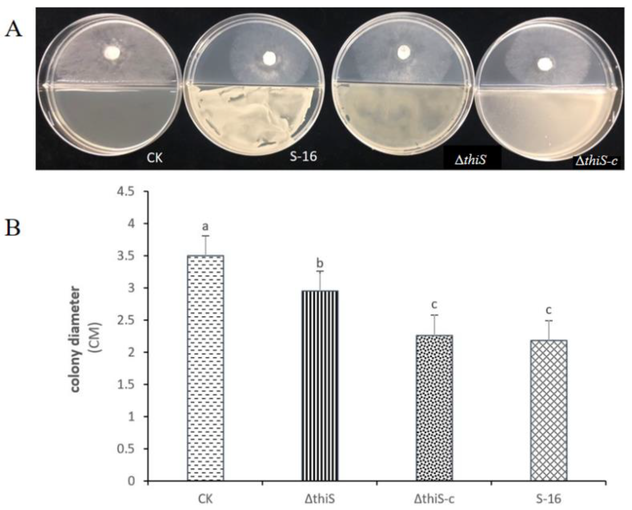
The antifungal activities of the VOCs from the wild-type and mutant strains of S-16 were compared using divided Petri dishes. The mycelia of the S. sclerotiorum in the untreated control grew rapidly and covered the whole dish at 3 days post-inoculation. The mycelial growth of the S. sclerotiorum was significantly inhibited by the wild-type S-16 strain, but the co-culturing with the complementary strain increased the mycelial growth of S. sclerotiorum. Accordingly, the thiS gene appears to have affected the antifungal activities of the VOCs produced by the strain S-16 (Figure 2).

3.3. Determination of the Morphological Characteristics of the Fungal Hyphae Treated with the VOCs from Mutant Strains

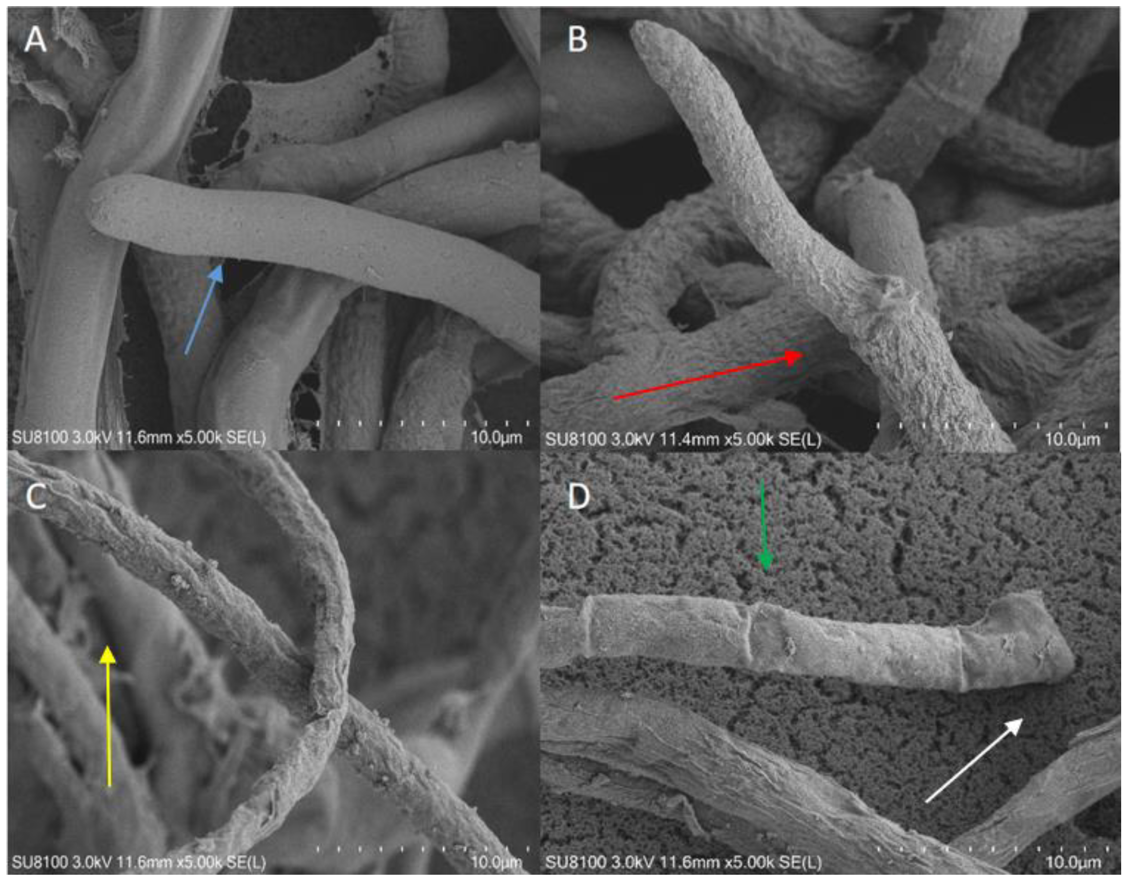
The hyphal structures of S. sclerotiorum following treatment with the VOCs from the wild-type and mutant S-16 strains were observed using a scanning-electron microscope. The hyphae of the S. sclerotiorum in the untreated control were intact and smooth (Figure 3A), whereas the hyphal structure was extensively damaged by the treatment with the VOCs from the wild-type, ΔthiS, and ΔthiS-c strains. More specifically, some hyphal expansion was detected (Figure 3B, red arrow). Additionally, the hyphae of the treated mycelia appeared to be relatively flaccid, with uneven surfaces (Figure 3C, yellow arrow). Moreover, empty segments formed (Figure 3D, white arrow), and thin structures or structures with gaps representing a retracted protoplasm were detected (Figure 3D, green arrow). This structural damage may have led to the leakage of cytoplasmic components. Thus, the VOCs produced by the S-16 strain substantially altered the morphology of the hyphae of S. sclerotiorum and degraded the cell membrane and wall. However, the mycelial damage caused by the ΔthiS treatment was not as extensive as that caused by the ΔthiS-c and wild-type S-16 treatments.

3.4. Effects of Volatile Compounds from the Mutant Strains on the Formation of Sclerotia

The formation of sclerotia following the treatment with the VOCs from the wild-type and mutant S-16 strains was examined. The untreated S. sclerotiorum control produced four sclerotia per Petri dish. In contrast, the S. sclerotiorum treated with the wild-type S-16 strain produced only 1.5 sclerotia per Petri dish. The treatment with the ΔthiS mutant strain resulted in 3.25 sclerotia per Petri dish. The treatment with the complementary strain produced 1.75 sclerotia per Petri dish. Mutations in thiS gene affect the ability of volatiles produced by B. subtilis to inhibit sclerotia formation in S. sclerotiorum (Table 3).

3.5. Test on Detached Sunflower Leaves

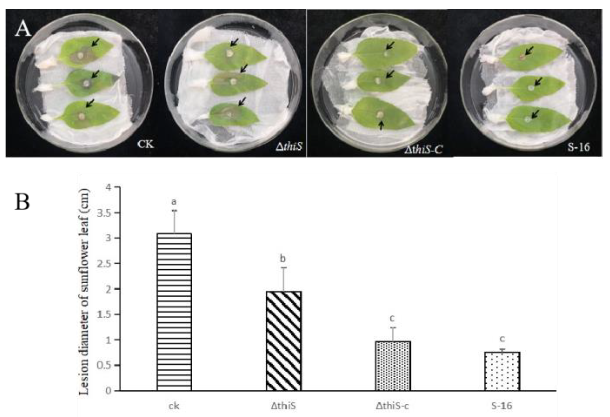
The antifungal activities of the VOCs from the wild-type and mutant S-16 strains were compared using detached sunflower leaves. The lesions on the control leaves were relatively large, with diameters of about 3.08 cm. The lesions were smaller on the leaves treated with the VOCs from the wild-type S-16 strain and the derived mutants. After the treatment with the VOCs from the wild-type S-16, the lesion diameters were 0.76 cm, which were smaller than the diameters of the lesions that formed after the treatment with the VOCs from the deletion mutant (1.95 cm). There were no significant differences in the diameters of the S. sclerotiorum lesions between the leaves treated with the VOCs from the wild-type and the complementary strains (Figure 4).

3.6. Analysis of the 2-MBTH Content in the Mutant Strains

The retention time for the 2-MBTH standard during the gas chromatography–tandem mass spectrometry (GC-MS/MS) analysis was 22.40 min. The contents of 2-MBTH in the wild-type and mutant S-16 strains were compared according to the area of the GC-MS peaks. The peak area was 8,715,422 for the wild-type S-16 strain, but it was only 6,381,940 for the deletion mutant. However, the peak area was partially restored to 7,939,165 for the complementary strain. Thus, these results indicate that the thiS gene partly affects the synthesis of 2-MBTH in S-16 (Table 4).

4. Discussion

The use of microorganisms and their metabolites is a promising and environmentally friendly alternative to chemical treatments for preventing or controlling plant diseases [30]. A previous study confirmed that the VOCs from the B. subtilis S-16 strain have antimicrobial activities that are useful for controlling fungal pathogens [25]. The main VOC from this strain (2-MBTH) can effectively control Sclerotinia in sunflowers.
Thiazole-ring-bearing compounds constitute an important class of heterocycles that possess a wide variety of pharmacological activities and have notable pharmaceutical value because of their potential chemotherapeutic properties [31]. The importance of the antimicrobial activity of thiazoles is related to the inclusion of the -S-C=N group as a toxophoric unit in the molecular frame [32]. In addition, benzothiazole has been reported in several studies in terms of its microbiological importance [33,34,35,36]. The potential antimicrobial activity of benzothiazole derivatives was illustrated previously [31,37,38,39,40,41]; thus, the continued search for new antibacterial and antifungal drugs within this group is strongly encouraged. The VOC 2-MBTH is a derivative of benzothiazole. Thiazole moiety 8 is biosynthesized in B. subtilis and most of the other bacteria from 1-deoxy-D-xylulose-5-phosphate (1, DXP), glycine, and cysteine in a complex oxidative condensation reaction [42]. This reaction requires five different proteins, namely ThiO, ThiG, thiS, ThiF, and a cysteine desulfurase. Glycine oxidase (ThiO) catalyzes the oxidation of glycine to the corresponding imine 7, the sulfur-carrier protein adenylyl transferase (ThiF) catalyzes the adenylation of the carboxy terminus of the sulfur carrier protein (thiS-carboxylate), and cysteine desulfurase catalyzes the transfer of sulfur from cysteine to the thiS-acyl adenylate to produce thiS-thiocarboxylate 6 [41,42,43,44]. Furthermore, ThiG is a thiazole synthase and catalyzes the formation of thiazole from dehydroglycine 7, DXP 1, and thiS-thiocarboxylate 6. The early steps of the formation of thiazole have been elucidated [45]. The formation of imine between lysine 96 on ThiG and DXP, followed by tautomerization, produces aminoketone 5, which is then thought to react with thiS-thiocarboxylate 6 and dehydroglycine 7 to yield thiazole phosphate 8. The mechanism of the formation of thiazole-phosphate moiety 8 of thiamin in vitro starts with the sulfur-transfer reaction from thiS-thiocarboxylate to amino ketone 5, during which a hydroxyl group from 1-deoxy-D-xylulose-5-phosphate (1) (DXP) is transferred to the C-terminal end of the sulfur carrier protein thiS-carboxylate [46]. In a previous study, to determine whether thiS influenced the synthesis of thiazole in B. subtilis S-16, a series of mutant strains was generated. The related gene was functionally verified by comparing the basic biological characteristics of the wild-type and mutant strains.
As expected, compared with the wild-type control, the ∆thiS gene knockout strain grew more slowly. In contrast, the growth rate of the ∆thiS-c complementary strain was not significantly different from that of the wild-type strain. Hence, thiS influences the growth of B. subtilis S-16. The growth curves of cultured bacteria can be divided into distinct phases [47]. During the logarithmic growth phase, the bacteria divide vigorously, with a short and stable generation time, resulting in an exponential increase in the number of cells. Thus, the logarithmic phase is often used for microbial fermentation and culturing. During the stationary phase, many microbial metabolites are produced and accumulated. Accordingly, the production of useful metabolites may be enhanced by prolonging the stationary phase [48]. In the current study, the in-frame deletion of thiS resulted in delayed growth and the accumulation of secondary metabolites was affected, ultimately leading to weakened antimicrobial activity.
The contents of 2-MBTH in the three bacterial strains were quantitatively analyzed using a GC-MS system. The lack of thiS expression affected the production of 2-MBTH. Accordingly, we determined that thiS was involved in the synthesis of 2-MBTH in the B. subtilis. Furthermore, the volatile compounds produced by the wild-type control, the deletion mutant, and the complementary strain inhibited the mycelial growth and sclerotial formation of the S. sclerotiorum. The antimicrobial activities of S-16 may involve additional compounds containing thiazole and its derivatives. Thiazole synthase can catalyze the synthesis of thiazole, which has antibacterial effects. Moreover, thiazole can be converted by certain enzymes to thiazole derivatives with antibacterial activities. Because the thiazoles in S-16 cells flow into metabolic pathways that synthesize antimicrobial substances, the knockdown of thiazole synthase genes can affect antimicrobial activity.
The antimicrobial properties and their effects on the formation of sclerotia varied among the B. subtilis strains analyzed. The S. sclerotiorum hyphae treated with VOCs were observed using a scanning-electron microscope, which revealed that the VOCs produced by the three examined strains adversely affected the morphological structures of the S. sclerotiorum hyphae to a certain extent. The differences in the degree of damage to the mycelial morphology of S. sclerotiorum may not have been caused only by the diversity in the VOCs produced by the B. subtilis strains. The S. sclerotiorum discs treated with the VOCs produced by the three B. subtilis strains were used to inoculate fresh and healthy sunflower leaves to clarify the effect of the thiS on the S. sclerotiorum’s fungal pathogenicity. The results indicated that the deletion of the thiS weakened the inhibitory effects of the VOCs against the S. sclerotiorum’s fungal pathogenicity. This was in contrast to the considerable decrease in the S. sclerotiorum’s fungal pathogenicity following the treatments with the VOCs from the complementary strain and the wild-type control.
The volatiles produced by B. subtilis include γ-patchoulene, 3-methylbutanal, 1-octen3-ol, 2-undecanone, 2-nonanone, 3-methylbutanoate, 2-methylbutan-1-ol, 4-methyl-2-heptanone, ethanethioic acid, and dimethyltrisulfide-2,3,6-trimethylphenol. The antifungal activities of several of these chemicals have already been evaluated. These compounds were detected during the analysis of the gaseous volatiles produced by S-16. In this study, the gaseous volatiles produced by the deletion mutant still had antimicrobial effects. These findings may reflect the antimicrobial activities of other gaseous volatile components.
The VOC-based bacteriostatic effects of the deletion mutant differed significantly from those of the wild-type and complementary strains. Therefore, thiS is involved in the synthesis of VOCs, which are important for the use of B. subtilis S-16 as a biocontrol agent. The results of the current study confirmed the reliability of metabonomic and whole-genome analyses of B. subtilis S-16. Furthermore, our results suggest that the thiS was involved in the synthesis of 2-MBTH in the B. subtilis, which contributed to the biocontrol effects of the latter. The data generated in this study regarding the VOCs of B. subtilis S-16 may be useful for enhancing biocontrol measures to protect sunflowers and related species. In the further work, we will continue to study and analyze the inhibitory mechanism through which 2-MBTH inhibits the growth of S. sclerotiorum and controls Sclerotinia wilt in sunflowers by combining transcriptomics and proteomics. Furthermore, the biological role of thiS gene has been extensively investigated in most other bacteria. However, the function of this gene in the synthesis of volatile compounds by B. subtilis is unknown. Here, we present the effect of thiS on the synthesis of 2-MBTH in B. subtilis through knockout-mutant construction and phenotypical characterization. The deletion of the thiS gene in B. subtilis results in decreased 2-MBTH content and decreased antibacterial activity. The gene-expression-pattern levels of other thiazole synthases may constitute a key regulatory mechanism controlling the synthesis of 2-MBTH in Bacillus subtilis. This mechanism is of interest, and we will explore it in our future work.

5. Conclusions

The goal of the present study was to determine the impact of the thiS gene’s deletion on the 2-MBTH production and antimicrobial activity of Bacillus subtilis S-16. We showed that the mutant strain produced less 2-MBTH. The ability of the VOCs produced by the mutant strain to inhibit the growth of the mycelia was also reduced. The production of 2-MBTH and its antimicrobial activities were adversely affected to varying degrees by the deletion of the thiS. Accordingly, we determined that the thiS was involved in the synthesis of the 2-MBTH in the B. subtilis. The functions of other regulatory genes that may interact with thiazole synthesis are yet to be verified, and the idea of modifying Bacillus subtilis to make it a more effective biological control agent requires further investigation.

Author Contributions

J.H. performed the experimental works, investigated and interpreted the results, and wrote the manuscript. Z.S., X.L. and Q.G. supervised, provided technical support, and edited the manuscript. B.D., D.W., H.M. and H.Z. were responsible for the primary conception of the study. All authors have read and agreed to the published version of the manuscript.

Funding

This study was supported by the National Natural Science Foundation of China (31260445 and 31960533) and the earmarked fund for National Key R & D Projects (2017YFD0201101).

Institutional Review Board Statement

Not applicable.

Informed Consent Statement

Not applicable.

Data Availability Statement

The authors confirm that the data supporting the findings of this study are available within the article.

Conflicts of Interest

The authors declare that they have no known competing financial interest or personal relationships that could have appeared to influence the work reported in this paper.

References

  1. Jing, W.; Jianru, Z.; Chaomin, C.; Xia, C.; Zhiyong, C.; Xiaoshuo, L.; Xi, C.; Shuyin, W. Research progress of sunflower Sclerotinia sclerotiorum. J. North. Agric. 2006, 25–28. [Google Scholar]
  2. Bloomfield, B.J.; Alexander, M. Melanins and Resistance of Fungi to Lysis. J. Bacteriol. 1967, 93, 1276. [Google Scholar] [CrossRef]
  3. Dean, R.; Kan, J.; Pretorius, Z.A.; Hammond-Kosack, K.E.; Pietro, A.D.; Spanu, P.D.; Rudd, J.J.; Dickman, M.; Kahmann, R.; Ellis, J. The Top 10 fungal pathogens in molecular plant pathology. Mol. Plant Pathol. 2012, 13, 414–430. [Google Scholar] [CrossRef]
  4. Guoqing, L.; Hongzhang, H. Study on diversity of sclerotia germination of Sclerotia of Sclerotiorum sclerotiorum. J. Plant Prot. 1997, 24, 59–64. [Google Scholar] [CrossRef]
  5. Fernando, W.G.D.; Ramarathnam, R.; Krishnamoorthy, A.S.; Savchuk, S.C. Identification and use of potential bacterial organic antifungal volatiles in biocontrol. Soil Biol. Biochem. 2005, 37, 955–964. [Google Scholar] [CrossRef]
  6. Minuto, A.; Spadaro, D.; Garibaldi, A.; Gullino, M.L. Control of soilborne pathogens of tomato using a commercial formulation of Streptomyces griseoviridis and solarization. Crop Prot. 2006, 25, 468–475. [Google Scholar] [CrossRef]
  7. Quansheng, L.; Zongming, X.; Zheng, L.; Guoli, Z.; Dongmei, W.; Ying, T. Screening and identification of antagonistic bacterium H14 against Verticillium dahliae Kleb. and its antagonistic mechanisms. J. Plant Prot. 2018, 45, 1204–1211. [Google Scholar]
  8. Afzal, S. Plant beneficial endophytic bacteria: Mechanisms, diversity, host range and genetic determinants. Microbiol. Res. 2019, 221, 36–49. [Google Scholar] [CrossRef]
  9. Arbi, A.E.; Rochex, A.; Chataigne, G.; Bechet, M.; Lecouturier, D.; Arnauld, S.; Gharsallah, N.; Jacques, P. The Tunisian oasis ecosystem is a source of antagonistic Bacillus spp. producing diverse antifungal lipopeptides. Res. Microbiol. 2016, 167, 46–57. [Google Scholar] [CrossRef]
  10. Cochrane, S.A.; Vederas, J.C. Lipopeptides from Bacillus and Paenibacillus spp.: A Gold Mine of Antibiotic Candidates. Med. Res. Rev. 2016, 36, 4–31. [Google Scholar] [CrossRef]
  11. Olishevska, S.; Nickzad, A.; Déziel, E. Bacillus and Paenibacillus secreted polyketides and peptides involved in controlling human and plant pathogens. Appl. Microbiol. Biotechnol. 2019, 103, 1189–1215. [Google Scholar] [CrossRef]
  12. Minerdi, D.; Bossi, S.; Gullino, M.L.; Garibaldi, A. Volatile organic compounds: A potential direct long-distance mechanism for antagonistic action of Fusarium oxysporum strain MSA 35. Environ. Microbiol. 2010, 11, 844–854. [Google Scholar] [CrossRef] [PubMed]
  13. Asghar, H.; Mohammad, P. A Review on Biological Control of Fungal Plant Pathogens Using Microbial Antagonists. J. Biol. Sci. 2010, 10, 273–290. [Google Scholar]
  14. Tyc, O.; Garbeva, P. Poster OlafTyc FEMS2017 The Ecological Role of Volatile and Soluble Secondary Metabolites produced by Soil Bacteria. Trends Microbiol. 2017, 25, 280–292. [Google Scholar] [CrossRef] [PubMed]
  15. Ryu, C.M.; Farag, M.A.; Hu, C.H.; Reddy, M.S.; Kloepper, J.W. Bacterial volatiles promote growth in Arabidopsis. Proc. Natl. Acad. Sci. USA 2003, 100, 4927–4932. [Google Scholar] [CrossRef]
  16. Breinig, S.; Schiltz, E.; Fuchs, G. Genes Involved in Anaerobic Metabolism of Phenol in the Bacterium Thauera aromatica. J. Bacteriol. 2000, 182, 5849–5863. [Google Scholar] [CrossRef]
  17. Zou, C.S.; Mo, M.H.; Gu, Y.Q.; Zhou, J.P.; Zhang, K.Q. Possible contributions of volatile-producing bacteria to soil fungistasis. Soil Biol. Biochem. 2007, 39, 2371–2379. [Google Scholar] [CrossRef]
  18. Alström, S. Characteristics of Bacteria from Oilseed Rape in Relation to their Biocontrol Activity against Verticillium dahliae. J. Phytopathol. 2010, 149, 57–64. [Google Scholar] [CrossRef]
  19. Koitabashi, M. New biocontrol method for parsley powdery mildew by the antifungal volatiles-producing fungus Kyu-W63. J. Gen. Plant Pathol. 2005, 71, 280–284. [Google Scholar] [CrossRef]
  20. Guevara-Avendano, E.; Carrillo, J.D.; Ndinga-Muniania, C.; Moreno, K.; Mendez-Bravo, A.; Guerrero-Analco, J.A.; Eskalen, A.; Reverchon, F. Antifungal activity of avocado rhizobacteria against Fusarium euwallaceae and Graphium spp., associated with Euwallacea spp. nr. fornicatus, and Phytophthora cinnamomi. Antonie Van Leeuwenhoek 2018, 111, 563–572. [Google Scholar] [CrossRef]
  21. Qi, W. Cloning of the Bacteriostat Related Gene and Identification of Antifungal Substances from Bacillus Subtilis S-16; Inner Mongolia Agricultural University: Hohhot, China, 2014. [Google Scholar]
  22. Jinghan, H.; Dong, W.; Baozhu, D.; Huanwen, M.; Jun, Z.; Hongyou, Z. The inhibitory effect of the volatile compound 2-methyl benzothiazole on pathogenic fungi Sclerotinia sclerotiorum and Botrytis cinerea. J. Plant Prot. 2021, 48, 781–788. [Google Scholar]
  23. Lijun, C.; Yanan, Z.; Chunsheng, W.; Jinzhu, C.; Yinli, J.; Yuehua, C. Analysis of the fumigation inhibition mechanism of essential oil from Chenopodium ambrosioides against the Botrytis cinerea. J. Plant Prot. 2020, v.47, 221–222. [Google Scholar]
  24. Ginsberg, G.; Toal, B.; Kurland, T. Benzothiazole Toxicity Assessment in Support of Synthetic Turf Field Human Health Risk Assessment. J. Toxicol. Environ. Health Part A 2011, 74, 1175–1183. [Google Scholar] [CrossRef] [PubMed]
  25. Yiming, Z. Isolation and Identification of a Biocontrol Bacterium against Sclerotinia Sclerotinia of Sunflower and Preliminary Study on Its Antibacterial Mechanism; Inner Mongolia Agricultural University: Hohhot, China, 2010. [Google Scholar]
  26. Yaguang, H. The Study on the Pathogenicity Differentiation and Sclerotial Formation Mechanism of Sunflower Sclerotinia sclerotiorum; Inner Mongolia Agricultural University: Hohhot, China, 2016. [Google Scholar]
  27. Smith, K.; Youngman, P. Use of a new integrational vector to investigate compartment-specific expression of the Bacillus subtilis spoIIM gene. Biochimie 1992, 74, 705–711. [Google Scholar] [CrossRef]
  28. Xu, Y.B.; Mai, C.; Ying, Z.; Wang, M.; Ying, W.; Qiu-Bin, H.; Xue, W.; Wang, G. The phosphotransferase system gene ptsI in the endophytic bacterium Bacillus cereus is required for biofilm formation, colonization, and biocontrol against wheat sharp eyespot. Fems Microbiol. Lett. 2014, 354, 142–152. [Google Scholar] [CrossRef] [PubMed]
  29. Ishiwa, H.; Shibahara, H. New shuttle vectors for Escherichia coli and Bacillus subtilis. II: Plasmid pHY300PLK, a multipurpose cloning vector with a polylinker, derived from pHY460. Jpn. J. Genet. 1985, 60, 235–243. [Google Scholar] [CrossRef]
  30. Sahni, S.; Sarma, B.K.; Singh, K.P. Management of Sclerotium rolfsii with integration of non-conventional chemicals, vermicompost and Pseudomonas syringae. World J. Microbiol. Biotechnol. 2008, 24, 517–522. [Google Scholar] [CrossRef]
  31. Yurttaş, L.; Özkay, Y.; Duran, M.; Turan-Zitouni, G.; Özdemir, A.; Cantürk, Z.; Küçükoğlu, K.; Kaplancıklı, Z.A. Synthesis and antimicrobial activity evaluation of new dithiocarbamate derivatives bearing thiazole/benzothiazole rings. Phosphorus Sulfur Silicon Relat. Elem. 2016, 191, 1166–1173. [Google Scholar] [CrossRef]
  32. Taori, K.; Paul, V.J.; Luesch, H. Structure and Activity of Largazole, a Potent Antiproliferative Agent from the Floridian Marine Cyanobacterium Symploca sp. J. Am. Chem. Soc. 2008, 130, 13506. [Google Scholar] [CrossRef]
  33. Soni, B.; Ranawat, M.S.; Sharma, R.; Bhandari, A.; Sharma, S. Synthesis and evaluation of some new benzothiazole derivatives as potential antimicrobial agents. Eur. J. Med. Chem. 2010, 45, 2938–2942. [Google Scholar] [CrossRef]
  34. Sharma, P.C.; Sinhmar, A.; Sharma, A.; Rajak, H.; Pathak, D.P. Medicinal significance of benzothiazole scaffold: An insight view. J. Enzym. Inhib. Med. Chem. 2013, 28, 240–266. [Google Scholar] [CrossRef] [PubMed]
  35. Franchini, C.; Muraglia, M.; Corbo, F.; Florio, M.A.; Mola, A.D.; Rosato, A.; Matucci, R.; Nesi, M.; Bambeke, F.V.; Vitali, C. Synthesis and biological evaluation of 2-mercapto-1,3-benzothiazole derivatives with potential antimicrobial activity. Archiv. Pharm. 2010, 342, 605–613. [Google Scholar] [CrossRef] [PubMed]
  36. Facchinetti, V.; Reis, R.; Gomes, C.; Vasconcelos, T. Chemistry and biological activities of 1,3-benzothiazoles. Mini-Rev. Org. Chem. 2012, 9, 44–53. [Google Scholar] [CrossRef]
  37. Lad, N.P.; Manohar, Y.; Mascarenhas, M.; Pandit, Y.B.; Pandit, S.S. Methylsulfonyl benzothiazoles (MSBT) derivatives: Search for new potential antimicrobial and anticancer agents. Bioorganic Med. Chem. Lett. 2017, 27, 1319–1324. [Google Scholar] [CrossRef]
  38. Padalkar, V.S.; Borse, B.N.; Gupta, V.D.; Phatangare, K.R.; Patil, V.S.; Umape, P.G.; Sekar, N. Synthesis and antimicrobial activity of novel 2-substituted benzimidazole, benzoxazole and benzothiazole derivatives. Arab. J. Chem. 2016, 9, S1125–S1130. [Google Scholar] [CrossRef]
  39. Trapani, A.; Catalano, A.; Carocci, A.; Carrieri, A.; Mercurio, A.; Rosato, A.; Mandracchia, D.; Tripodo, G.; Schiavone, B.I.P.; Franchini, C.; et al. Effect of Methyl-β-Cyclodextrin on the antimicrobial activity of a new series of poorly water-soluble benzothiazoles. Carbohydr. Polym. 2019, 207, 720–728. [Google Scholar] [CrossRef]
  40. Al-Talib, M.; Al-Soud, Y.A.; Abussaud, M.; Khshashneh, S. Synthesis and biological evaluation of new benzothiazoles as antimicrobial agents. Arab. J. Chem. 2011, 62, S926–S930. [Google Scholar] [CrossRef]
  41. Park, J.H.; Dorrestein, P.C.; Zhai, H.; Kinsland, C.; Mclafferty, F.W.; Begley, T.P. Biosynthesis of the Thiazole Moiety of Thiamin Pyrophosphate (Vitamin B1). Biochemistry 2003, 42, 12430–12438. [Google Scholar] [CrossRef]
  42. Wang, C.; Xi, J.; Begley, T.P.; Nicholson, L.K. Solution structure of thiS and implications for the evolutionary roots of ubiquitin. Nat. Struct. Biol. 2001, 8, 47–51. [Google Scholar]
  43. Settembre, E.C.; Dorrestein, P.C.; Park, J.; Augustine, A.; Begley, T.P.; Ealick, S.E. Structural and mechanistic studies on ThiO, a glycine oxidase essential for thiamin biosynthesis in Bacillus subtilis. Biochemistry 2003, 42, 2971–2981. [Google Scholar] [CrossRef]
  44. Lauhon, C.T.; Kambampati, R. The iscS Gene in Escherichia coli Is Required for the Biosynthesis of 4-Thiouridine, Thiamin, and NAD. J. Biol. Chem. 2000, 275, 20096–20103. [Google Scholar] [CrossRef] [PubMed]
  45. Dorrestein, P.C.; Zhai, H.; Taylor, S.V.; Mclafferty, F.W.; Begley, T.P. The biosynthesis of the thiazole phosphate moiety of thiamin (vitamin B1): The early steps catalyzed by thiazole synthase. J. Am. Chem. Soc. 2004, 126, 3091–3096. [Google Scholar] [CrossRef]
  46. Dorrestein, P.C.; Zhai, H.; Mclafferty, F.W.; Begley, T.P. The Biosynthesis of the Thiazole Phosphate Moiety of Thiamin: The Sulfur Transfer Mediated by the Sulfur Carrier Protein thiS. Cell Chem. Biol. 2004, 11, 1373–1381. [Google Scholar]
  47. Yanlei, Z. Study on experimental method of measuring bacterial growth curve. J. Microbiol. 2016, 36, 108–112. [Google Scholar]
  48. Jixian, M.; Zhigang, W.; Changhe, W. Comparison of common microbe number determination methods. Biol. Teach. 2012, 37, 42–43. [Google Scholar]
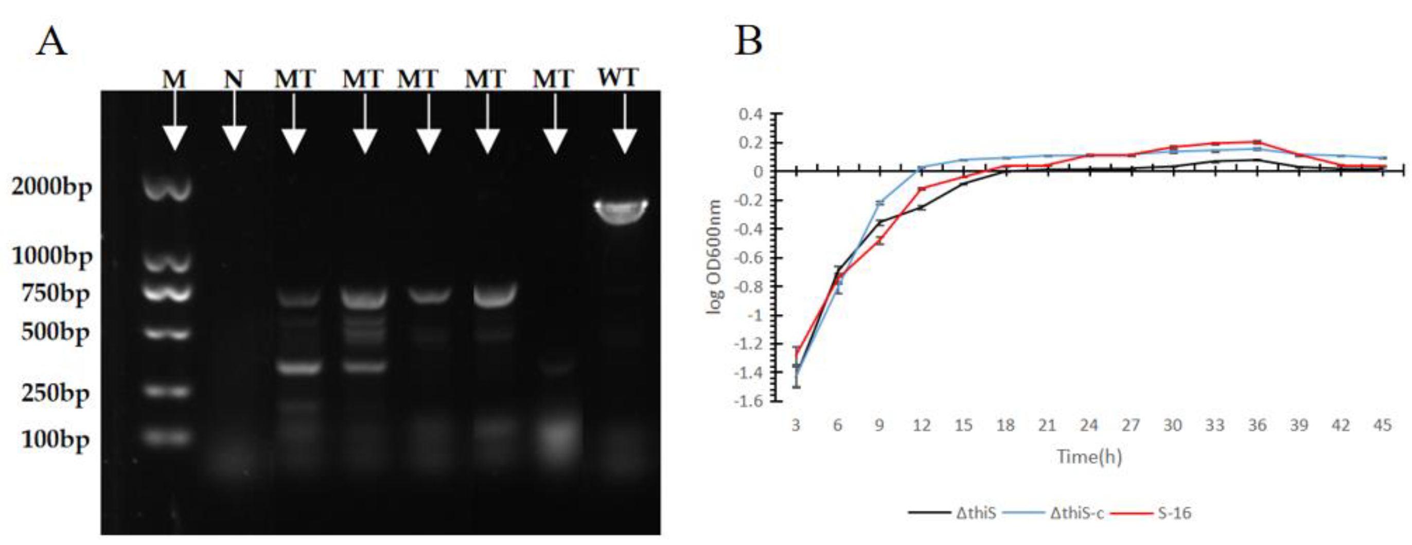
Figure 1. (A) Confirmation of the knockout of thiS. PCR amplification and sequencing were carried out with the primers thiS-1 and thiS-4, and the PCR products were analyzed using agarose-gel electrophoresis. Lane M (the first lane): DNA marker (this marker was purchased from Takara. The size of the tape was known and fixed. The operation was carried out according to the product instructions); Lane N (the second lane): no template control; lane WT (the eighth lane): genomic DNA of wild-type S-16 as the template; Lane MT (three-to-seven lane): mutant strains. (B) Growth curves of the wild-type S-16, the mutant strain ΔthiS, and the complementary strain ΔthiS-c.
Cimb 45 00292 g001
Figure 2. Effects of the wild-type S-16, the mutant ΔthiS, and the complementary strain ΔthiS-c on mycelial growth. (A) Effect of VOCs produced by the wild-type S-16, the mutant strain ∆thiS, and the complementary strain ∆thiS-c on the mycelial growth of S. sclerotiorum. (B) Mycelial growth with and without treatment with the volatile compounds of wild-type S-16, the mutant strain ΔthiS, and the complementary strain ΔthiS-c. The data are the mean ± s.d. (n = 3). Matching letters on the bars for each column indicate no significant difference according to the LSD test at p = 0.05.
Cimb 45 00292 g002
Figure 3. Effects of the volatile compounds from the mutant strains on the hyphal morphology of Sclerotinia sclerotiorum. (A) Control (blue arrow: smooth hyphae). (B)The ΔthiS treatment (red arrow: swollen hyphae). (C) The ΔthiS-c treatment (yellow arrow: jagged hyphae). (D) Wild-type S-16 treatment (white arrow: empty segments; green arrow: broken hyphae).
Cimb 45 00292 g003
Figure 4. Effects of the volatile compounds from the wild-type S-16, the mutant strain ΔthiS, and the complementary strain ΔthiS-c on the pathogenicity of Sclerotinia sclerotiorum. (A) Effect of VOCs produced by wild-type S-16, the mutant strain ΔthiS, and the complementary strain ΔthiS-c on the development of S. sclerotiorum symptoms on sunflower leaves (black arrows indicate the location of the edge of the lesion). (B) Lesion areas of sunflower leaves with or without treatment with volatile compounds of wild-type S-16, the mutant strain ΔthiS, and the complementary strain ΔthiS-c. The data are the mean ± s.d (n = 3). Matching letters on the bars for each column indicate no significant difference according to the LSD test at p = 0.05.
Cimb 45 00292 g004
Table 1. Tested strains and plasmids used in this experiment.
Strain or PlasmidProperties and ApplicationSource or Reference
B. subtilis
Bacillus subtilis S-16Wild-type strain in this studyKept in our laboratory, isolated from
sunflower root.
ΔthiSThe This gene’s defect mutant, ΔthiSConstruct in this study
ΔthiS-cΔthiS supplemented with its native thiS gene by allelic exchangeConstruct in this study
E. coli
E. coli DH5αPlasmid propagationPurchased from Takara, Dalian
plasmid
pKSV7For gene knockoutPurchased from Takara, Dalian
pHY300PLKFor gene knockoutPurchased from Takara, Dalian
pKSV7-thiSFor gene knockout, Amp+; cm+Construct in this study
pHY300PLK-thiSFor gene complementation, Cm+Construct in this study
Sclerotinia sclerotiorum X-8For testing the inhibition of mycelial growth by VOCsKept in our laboratory, isolated from
sunflower plants.
Table 2. The sequence of the primers of mutation in the thiS gene.
Gene NamePrimer (5′-3′)Restriction Sites
thiS-1CGG AAT TCG ATG GAC GAT TTG GAC TCT GEcoRI
thiS-2CGG GGT ACC ATA TCT GAA CCG CCT CCT TGKpnI
thiS-3CGG GTA CCA GGC GGA TGA GCA TGT TAA CKpnI
thiS-4CGG GAT CCG ACT GCT TGC CGT TCC GTA CBamHI
thiS-promoter-FCGGAATTCCTGCAAGCCGGTAGAAGAGEcoRI
thiS-promoter-RCGGGATCCGCACGCGGTGAATAGTAGAGBamHI
thiS-comFCGGGATCCAGGCGGTTCAGATATGATGCBamHI
thiS-comRCCCAAGCTTCTCATCCGCCTCCTACHindIII
Table 3. Effects of volatile compounds from the mutant strains on the formation of sclerotia *.
StrainThe Number of Sclerotia (Single-Digit)Sclerotium Weight (g)
ck4 ± 0.82 a0.22 ± 0.06 a
ΔthiS3.25 ± 0.82 a0.20 ± 0.03 a
ΔthiS-c1.75 ± 0.5 b0.17 ± 0.02 a
S-161.5 ± 0.58 b0.15 ± 0.07 a
* Data are average area ± the standard deviation of three replicates. Means in the third column of with different letters (a and b) are significant different (p < 0.05) according to Duncan’s multiple range tests.
Table 4. Analysis of the 2-MBTH contents in the mutant strains.
Active VOCsRetention Time (Min)HeightAreaArea (%)Compound
2-methyl benzothiazole22.55136,015,8723,001,261,21194.852-methyl benzothiazole
S-1622.394258,6768,715,4220.4052-methyl benzothiazole
ΔthiS22.400211,8876,381,9400.2152-methyl benzothiazole
ΔthiS-c22.402248,1327,939,1650.4032-methyl benzothiazole
Disclaimer/Publisher’s Note: The statements, opinions and data contained in all publications are solely those of the individual author(s) and contributor(s) and not of MDPI and/or the editor(s). MDPI and/or the editor(s) disclaim responsibility for any injury to people or property resulting from any ideas, methods, instructions or products referred to in the content.