Article

Ulcerative Colitis Seems to Imply Oral Microbiome Dysbiosis

1
Institute of Food Science Research (CIAL), CSIC-UAM, Campus de Cantoblanco, Nicolás Cabrera 9, 28049 Madrid, Spain
2
Department of Genetics, Physiology and Microbiology, Complutense University of Madrid, 28040 Madrid, Spain
3
Center for Advanced Research in Public Health, Department of Health and Genomics, FISABIO Foundation, 46020 Valencia, Spain
*
Author to whom correspondence should be addressed.
Curr. Issues Mol. Biol. 2022, 44(4), 1513-1527; https://doi.org/10.3390/cimb44040103
Submission received: 23 February 2022 / Revised: 19 March 2022 / Accepted: 29 March 2022 / Published: 30 March 2022
(This article belongs to the Special Issue Advances in Research on Molecular Oral Microorganisms)

Abstract

:
Ulcerative colitis (UC) is a recurrent pathology of complex etiology that has been occasionally associated with oral lesions, but the overall composition of the oral microbiome in UC patients and its role in the pathogenesis of the disease are still poorly understood. In this study, the oral microbiome of UC patients and healthy individuals was compared to ascertain the possible changes in the oral microbial communities associated with UC. For this, the salivary microbiota of 10 patients diagnosed with an active phase of UC and 11 healthy controls was analyzed by 16S rRNA gene sequencing (trial ref. ISRCTN39987). Metataxonomic analysis revealed a decrease in the alpha diversity and an imbalance in the relative proportions of some key members of the oral core microbiome in UC patients. Additionally, Staphylococcus members and four differential species or phylotypes were only present in UC patients, not being detected in healthy subjects. This study provides a global snapshot of the existence of oral dysbiosis associated with UC, and the possible presence of potential oral biomarkers.

1. Introduction

Inflammatory bowel diseases (IBDs) represent a group of chronic, idiopathic, relapsing disorders of unclear etiology affecting millions of people worldwide. IBDs are caused by an inappropriate and persistent inflammatory response to commensal gut microbiota and other environmental factors in genetically susceptible individuals, leading to a deterioration of the gastrointestinal structure and function [1,2,3]. IBDs are categorized into two clinical types: Crohn’s disease (CD), and ulcerative colitis (UC), the latter being characterized by continuous, diffuse, and superficial mucosal inflammation that starts in the rectum and extends to proximal segments of the colon [3]. Several studies have been focused on characterizing the dysbiotic state present in the gut microbiome of UC patients, reporting a decrease in bacterial diversity accompanied by a decrease in relative proportions of Enterococcus and Bacteroides, and in members of Clostridium subcluster XIVa, as well as increases in several opportunistic pathogens [4]. However, IBDs—both CD and UC—have also been associated with several extraintestinal manifestations, such as oral lesions, which can occur either concomitantly with intestinal symptoms or before the presentation of disease, and may even be the primary presenting sign in some cases [5,6]. Oral lesions are more common in CD compared to UC, and are more prevalent in children compared to adults [7]. The main oral manifestations of IBD are cobble-stoning of the oral mucosa, labial swellings with vertical fissures, pyostomatitis vegetans, angular cheilitis, perioral erythema, and glossitis, with pyostomatitis vegetans being the only condition that is more prevalent in UC patients [7]. Although the oral mucosa lesions and oral symptoms can be more severe during the disease activity period, up to 30% of affected patients continue suffering active oral manifestations despite remission [5,8]. However, despite the existence of different oral manifestations in patients with IBD, the pathogenesis of the disease remains unclear. It has been proposed that specific oral manifestations could result from the direct extension of intestinal inflammation—a hypothesis supported by the elevated levels of proinflammatory cytokines present in the saliva of IBD patients [8]. This general inflammatory state associated with the IBD condition could affect the oral mucosa and lead to an oral dysbiosis that could contribute to the worsening of the inflammatory state and play a crucial role in the oral manifestations of these patients [5]. However, only a few studies have focused on analyzing changes in the oral microbiome of IBD patients—most of them performed in the CD phenotype [9,10,11,12,13,14,15]. Regarding UC, most of the data come from pediatric populations or animal models [2,3,16,17,18], and only one study has focused on the analysis of the oral microbiome of a specific UC cohort, performed in a murine model of colitis [16]. Hence, we still lack a comprehensive understanding of the oral microbiota composition in UC patients in particular, and of the possible correlation between oral dysbiosis and the pathogenesis of UC.
In this context, the aim of this work was to study and characterize the oral microbiome of a selected group of UC patients in an active phase of the disease, and to compare their profiles with those of similar unaffected populations, in order to determine whether—as found in the gut—bacterial dysbiosis also exists in the oral cavity. We also aimed to determine the presence of potential oral microbial biomarkers associated with the onset of the disease. Our findings should provide insight into the association between the human microbiome and UC, and the development of new precautionary or diagnostic methods for patients with UC.

2. Material and Methods

2.1. Study Design

The study lasted from April 2018 to October 2019, and involved UC patients and healthy donors. UC patients were selected from the patient cohort of the Digestive System Department of the “Infanta Sofia Hospital” (Madrid, Spain) (ISRCTN39987), whereas healthy donors were recruited in the Primary Care Centre “Paseo de la Chopera” (Madrid, Spain) (ISRCTN13100451). As inclusion criteria for patients with UC, participants had no oral manifestations at the time of sampling, did not take any medication that could alter the results obtained, and were in an active phase of the disease. As exclusion criteria for both groups, participants could not have received antibiotics for at least 6 months before the study, suffer from type I diabetes, have severe cardiac, endocrine, or gastrointestinal disorders, have a previous history of alcohol or drug abuse, or follow exclusive diets (e.g., vegan or vegetarian). The study protocol was approved by the Ethics Committee of the Hospital Infanta Sofia and Hospital La Paz (Madrid, Spain) and the Bioethics Committee of The Spanish National Research Council (CSIC), and it was conducted according to the guidelines laid down in the Declaration of Helsinki. All participants were informed about the study, and they signed written informed consent.
For all participants, serum biochemical parameters were measured in plasma using an automated biochemical autoanalyzer. The blood tests included the measurement of glucose levels and lipid profiles, among others. For UC patients, fecal calprotectin was determined by quantitative enzyme-linked immunosorbent assay (ELISA) immediately after fecal sample collection. To analyze the oral microbiome from a global point of view (tongue, teeth, palate, mucosa, etc.), saliva samples were collected after awakening, and after brushing teeth with water (without using toothpaste), but before drinking or eating any type of food. Unstimulated saliva (3–5 mL) was expectorated naturally by drooling—that is, without forcing its production. All of the saliva samples were kept in an ice bath during collection, and then were kept at −80 °C until analysis.

2.2. DNA Extraction and Sequencing

Saliva samples were used for DNA extraction using the MasterPure™ Complete DNA and RNA Purification Kit (Epicentre, Madison, WI, USA), following the manufacturer’s instructions, with a prior lysis with lysostaphin from Staphylococcus staphylolyticus (5000 U/mL), mutanolysin from Streptomyces globisporus ATCC 21553 (2500 U/mL), and lysozyme from chicken egg white (50,000 U/mL) (Sigma-Aldrich, San Luis, MO, USA). The V3–V4 region of the 16S ribosomal RNA gene was amplified using the primers used by Klindworth et al., which produce a PCR product of 460 bp [19]. Primer sequences were as follows: 5′-CCTACGGGNBGCASCAG-3′ and reverse 5′-GACTACNVGGGTATCTAATCC-3′. The two-step Illumina® PCR protocol was followed to prepare the libraries, and samples were submitted to 2 × 300 bp paired-end sequencing by means of an Illumina® MiSeq instrument (Illumina®, San Diego, CA, USA). Raw files are available in the National Center for Biotechnology (NCBI) repository under the project code PRJNA749643.

2.3. Sequence Processing

To process raw reads (FASTQ files) from the Illumina® instrument, RStudio software v. 4.03 was used. The FASTQC files were filtered for reads with low quality and the presence of alien DNA using DADA2 (v.1.18.0). The DADA2 algorithm was also employed to denoise, join paired-end reads, and filter out chimeras in the raw data [20,21]. This algorithm can allow the differentiation of a single nucleotide, leading to the formation of amplicon sequence variants (ASVs). The taxonomic assignment was performed using the naïve Bayesian classifier implemented in DADA2, using Silva v.138 as a reference database [22], with a bootstrap cutoff of 80%. A total of 1,283,321 complete good-quality reads were used for the analysis. All of the information about the sequencing process, as well as error rates and rarefaction curves to assess sequencing depth, are available in Table S4 and Figures S1 and S2, respectively.

2.4. Statistical Analysis

Statistical analyses were conducted in the RStudio environment. Bacterial diversity, expressed in terms of alpha diversity, was estimated using the ASVs by calculating the observed, Shannon, and Simpson indices through the “phyloseq” package. ANOVA analysis was employed to detect significant differences between alpha diversity values. Compositional differences between samples (beta diversity) were obtained by employing a Bray–Curtis dissimilarity matrix represented by non-metric multidimensional scaling (NMDS) through the “vegan” package. Permutational multivariate analysis (PERMANOVA) belonging to the “vegan” package was conducted to find statistical differences between experimental groups, using Holm’s multiple comparison testing correction by default. Differences in the relative abundances of taxa and ASVs were assessed using the Mann–Whitney U test, and subsequently applying Benjamini–Hochberg post hoc correction, with a false discovery rate (FDR) threshold of 0.25. Sequences of the differential ASVs as well as the absolute total reads per group are available in Table S3. The microbial taxonomic features (i.e., families or genera) most likely to explain differences between healthy subjects and patients suffering from UC were also determined by LEfSe (linear discriminant analysis effect size), using the packages “microbiomeMarker” and “yingtools2”.

3. Results

3.1. Clinical Characteristics of the Participants

Initially, we were able to recruit 20 UC patients and 11 healthy volunteers who agreed to participate in the study. After applying the strict inclusion and exclusion criteria, 10 UC patients dropped out of the study for various reasons (worsening of the disease or its symptoms, other diseases, antibiotics, or lack of interest in the study). We finally enrolled 10 UC patients in the active phase of the disease, categorized as “mild or moderate” by the medical personnel of the Hospital Digestive Service—a fact that was supported by the high fecal calprotectin levels detected, as clinical indicators of their disease activity (Table S1). Moreover, none of the participants showed or manifested oral symptoms or oral diseases at the time of sampling. The data of all participants, including the clinical characteristics collected from medical records, are shown in Table S1. No significant differences existed between the groups in terms of age, gender, body mass index (BMI), and main clinical parameters. Values for fecal calprotectin and partial Mayo score are specifically reported for UC patients, as clinical indicators of their disease activity [23] (Table S1).

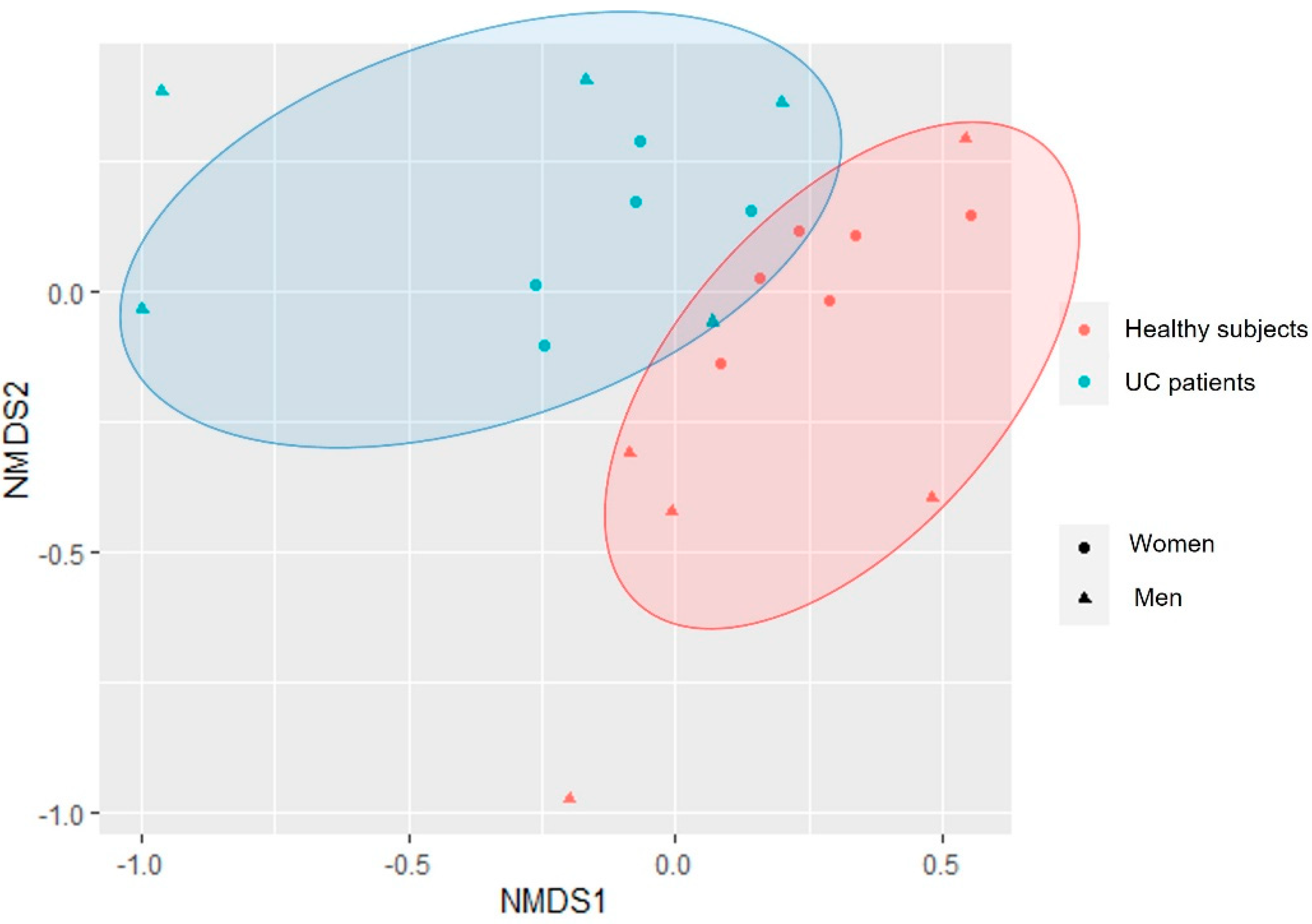
3.2. Changes in the Biodiversity Indices between UC Patients and Healthy Subjects

Analysis of raw reads revealed a total of 3270 ASVs (amplicon sequence variants). Bacterial diversity analysis between groups showed a decrease in the alpha diversity indices in the UC patients, revealing statistically significant differences for the observed ASVs (p-value = 0.0454) and Shannon indices (p-value = 0.0314) (Figure 1), but not for the Simpson indices (p-value = 0.174). The analysis of the beta diversity showed two different groups (PERMANOVA, R2 = 0.06523, p-value = 0.08791): one for the healthy subjects, and another for the UC patients (Figure 2), with the female participants showing more similar microbial composition profiles, and the male participants a more scattered distribution, although this result did not show statistical significance (PERMANOVA, R2 = 0.06178, p-value = 0.14985).

3.3. Taxonomic Profiles of the UC Patients and Healthy Subjects

Regarding the taxonomic profiles, results revealed differences in the relative abundance of some taxa between the group of healthy subjects and the UC patients (see Table S2 for all taxa, Table 1 for taxa with an average relative abundance > 0.5%, and Figure 3 for taxonomic biomarkers for each group calculated by LDA). At the phylum level, results revealed a tendency for higher relative proportions of Proteobacteria in the oral cavities of the UC patients (p-value = 0.072). In relation to the family level, the results revealed statistically significant differences (p-value < 0.05) in the proportions of Family XIII and Peptostreptococcaceae, which showed lower values in their relative abundance in the UC patients, and in Neisseriaceae, whose values were higher in the UC group. These remarks were corroborated by LDA analysis (Figure 3). Furthermore, a trend (p-value < 0.1) was detected for lower levels of Atopobiaceae, Lachnospiraceae and Ruminococcaceae members in UC subjects.
At the genus level, results showed higher relative proportions of Neisseria genus members (p-value < 0.05) in the oral cavities of the UC subjects, which were also detected in higher proportions in this group by LDA approach (Figure 3). On the other hand, saliva samples of the UC group presented lower levels of Filifactor, Parvimonas, Lachnoanaerobaculum, and Stomatobaculum (p-value < 0.05), together with a trend of lower levels of Atopobium, Peptostreptococcus, and Ruminococcaceae-UCG_014 genera (p-value < 0.1) (Table 1 and Table S2). Additionally, although with a low relative abundance (mean 0.005%), our results highlighted the exclusive presence of Staphylococcus members in the saliva samples of five of the UC patients (p-value = 0.011), with members of this genus not being detected in any of the healthy subjects (Table S2, Figure 3). LDA analysis revealed differences in the same taxa and bacterial groups, among other several unidentified species, supporting the statistical power of the results.
Finally, the analysis of the differences at the ASV level revealed 57 differential ASVs (p-value < 0.05), most of them only detected in the oral microbiomes of healthy donors (Table 2). In this regard, it is interesting to note the absence of different species of Streptococcus, Oribacterium, Rothia, Prevotella, and Porphyromonas, among others, in the saliva samples of the UC group. In particular, some species that were only detected in the saliva samples of the healthy donors’ group were ASVs assigned with high confidence/similarity to Haemophilus parainfluenzae and Porphyromonas pasteri, as well as Veillonella atypica and different species of the Rothia genus (e.g., Rothia dentocariosa and Rothia mucilaginosa)—many of them well-known members of the healthy oral core microbiome [24]. Nevertheless, on the other hand, four ASVs were only detected in the oral cavities of the UC group, highlighting ASV4, ASV14, ASV39, and ASV219. BLAST analysis of these ASVs showed that ASV4 and ASV14 had the highest similarity with Veillonella parvula (99.78% and 100% identity, respectively), ASV39 with Fusobacterium nucleatum (100% identity), and ASV219 with Prevotella sp. (100% identity).

4. Discussion

Currently, there are several studies describing the gut microbiome changes and a dysbiotic state in UC patients [4,25]. However, UC can be related to other extraintestinal symptoms, such as oral lesions or oral diseases, which can occur together with intestinal symptoms or even before they occur [5,8]. Various studies have reported oral manifestations in IBD patients [5,6,7,8,26,27,28,29,30], whose findings have led to the proposal of a “gum–gut” axis, revealing the importance of the balance in the oral microbial communities not only for oral health, but also for overall health status [27]. Moreover, it has been suggested that UC-related oral manifestations could be due to dysbiosis in the oral microbiome associated with this pathology. To the best of our knowledge, this is the first study characterizing the oral microbiota present in adult UC patients in terms of both composition and diversity. Additionally, and bearing in mind the intrinsic variability associated with oral microbiota, this study considered a quite homogeneous patient cohort, all with an active phase of UC, categorized as mild or moderate.
As expected, we found an important interindividual variability between the salivary microbial profiles of the participants of the study. However, our results indicated a decrease in alpha diversity indices in UC patients, in agreement with the observations of Zhang et al. during the active phase of CD, and the results of Docktor et al. and Elmaghrawy et al. in pediatric cohorts [2,13,15]. In the same line, Lucas López et al. described a decrease in alpha diversity indices in the gut microbiome of UC patients [3], so it can be hypothesized that UC could be associated with a loss of both gut and oral bacterial diversity. The analysis of the beta diversity showed two clearly differentiated groups, revealing that the taxonomic composition of the oral microbiomes of UC patients and healthy subjects is different. Potential sex differences in the oral microbial profiles, consistent with the pattern previously described in the gut microbiome of IBD subjects [31]—including some studies in murine models [32,33]—need to be tested with larger sample sizes.
At the phylum level, we observed a trend towards a higher relative abundance of Proteobacteria in the oral cavities of the UC patients, as previously reported in the gut microbiome of IBD patients [18,34,35]. At the family level, also consistent with our results, depletion in the levels of Peptostreptococcaceae and Lachnospiraceae has been reported in the oral cavities of UC or CD patients [13,18] and, in the case of Lachnospiraceae and Ruminococcaceae bacteria, also in the gut microbiome [36,37]. However, contrary to what has been described in previous studies [13,18,38], our results showed significantly higher values for Neisseriaceae family members and Neisseria—dominant commensal members in the healthy oral cavity [24,39], but which can also act as opportunistic pathogens [40,41]. Conversely, we detected lower proportions of Atopobium, Lachnoanaerobaculum, Peptostreptococcus, and Stomatobaculum members in the saliva samples of the UC patients. The majority of these members form part of the oral core microbiome [39,42], and some species—for example, Stomatobaculum longum and Lachnoanaerobaculum saburreum—have been related to oral health conditions [43,44,45]. Moreover, the depletion in Peptostreptococcus members as well as other oral commensals in the saliva samples of UC patients was previously reported by Xun et al. in the oral cavities of IBD patients [18], while lower proportions of Peptostreptococcus members were also reported in the gut microbiome of UC patients [46]. The tryptophan metabolite indoleacrylic acid—produced by gut commensal Peptostreptococcus species—has shown the ability to promote intestinal epithelial barrier function and mitigate inflammatory responses in a murine model [47]. Thus, in line with this, a possible hypothesis could be that the depletion in Peptostreptococcus members, together with the lower proportions of other key members in the guts and oral cavities of the UC patients—such as Lachnospiraceae and Ruminococcaceae—could be related to lower anti-inflammatory activities, which could promote or contribute to the characteristic inflammatory profile of these patients.
Interestingly, our results highlighted the exclusive presence of Staphylococcus members in the saliva samples in 5 of the 10 UC patients, but not in any of the healthy controls. Staphylococcus members are not part of the normal oral microbiota, but they have been found to occur in immunocompromised patients who can suffer from oral inflammatory conditions (e.g., gingivitis caused by Staphylococcus epidermidis) [48]. Although further evidence is needed due to the small sample size of this study, which could limit these results, the presence of members of the Staphylococcus genus in the oral cavities of UC patients could be proposed as an oral biomarker of the disease in a non-invasive sample.
Analysis of the differences at the ASV level between healthy donors and UC patients revealed the absence of different species/phylotypes belonging to key members of the oral core microbiome, such as members of Prevotella (i.e., Prevotella salivae, Prevotella histicola, and Prevotella melaninogenica), Porphyromonas (i.e., Porphyromonas pasteri), and Rothia (i.e., Rothia dentocariosa), in the saliva samples of UC patients—a finding that has been also reported in IBD patients [13,18]. Moreover, some species that were only detected in the saliva samples of the healthy donors’ group—such as H. parainfluenzae, P. pasteri, V. atypica R. dentocariosa, and R. mucilaginosa—have been correlated in other works with a healthy oral state [24,49,50,51]. On the other hand, four ASVs were only detected in the saliva samples of the UC patients, corresponding to V. parvula, F. nucleatum, and Prevotella sp. Members belonging to Veillonella are among the most abundant members of the oral microbiome [52], and their co-aggregation properties play a crucial role affecting the colonization site and the ecology of oral microbial biofilms [53]. Although V. parvula plays an essential role as a commensal in the oral microbiome, and is considered to be non-pathogenic, rarely causing serious infections, it is present at very high levels in dental carious lesions, being predominant at all stages of caries progression and contributing to caries development [54]. In this sense, Veillonella species mainly utilize lactate as their sole carbon source, producing acetate and propionate, so it has been proposed that their levels may serve as a sensitive biological indicator and early warning sign of acid production [55]. Taking this into account, a possible explanation for the higher levels of V. parvula in the UC patients could be that lactate is one of the most enriched byproducts of cellular metabolism in inflamed tissues, and its accumulation leads to the exacerbation of the inflammatory response [56]. The chronic inflammatory state present in UC could promote lactate production in the oral cavity, which, together with the higher levels of other acid-producing bacteria, could provide the conditions for the improved growth of lactate-utilizing bacteria such as V. parvula and Neisseria [57].
Regarding F. nucleatum, it is a common member of the oral core microbiome; however, because of its proinflammatory potential, it has been associated with various periodontal diseases, with its increase also related to the severity of disease and the progression of inflammation [58]. Furthermore, F. nucleatum has been detected as being significantly enriched in the gut in IBD and UC subjects [4,59,60], being related even to the promotion of colitis, its aggravation, and the impediment of the remission [61,62,63], as well as having been found to cause opportunistic infections, and to be associated with colorectal cancer and IBD [64].
Some of these differential oral species have been reported in higher proportions in the gut microbiome of UC patients, highlighting the previously mentioned F. nucleatum and V. parvula, which have been reported in increased levels in IBD patients [60,65]. Considering that in our study these bacterial species/phylotypes were only observed in the saliva samples of the UC patients, together with the exclusive detection of Staphylococcus members in this group, our results suggest that the presence or the increase in the proportions of these bacterial members could be used as possible oral biomarkers associated with UC. However, we are aware that the small sample size could limit the detection of these species in the control group, so these trends need to be corroborated in studies with larger numbers of participants.
By comparing our data to gut microbiome profiles previously described in UC patients, we observed that several of the trends observed in some oral microorganisms in our study were consistent with changes detected in the guts of the UC subjects, highlighting the slight increase in Streptococcus members in the oral cavity, as also reported in the gut microbiome with UC [66], as well as the previously mentioned higher proportions of Gemella and Haemophilus members [67], and the depletion of Lachnospiraceae, Ruminococcaceae, and Prevotella in UC [4,18,68,69]. Furthermore, some of the specific species detected in the oral microbiome of the UC patients’ group of our study were reported in higher levels in the gut microbiome of UC patients. Thus, a plausible explanation for our results is that the changes in the bacterial communities causing or caused by UC could be similar in the oral cavity and the gut, revealing a possible association between the microbial communities of the two environments and the risk of the disease. In general, oral bacteria are poor colonizers of the healthy intestine, although it is known that transmission and colonization of the intestine by oral microorganisms is common and extensive among healthy individuals [70]. Considering that we ingest about 1 L of saliva per day—containing an enormous amount of oral bacteria—it is possible that some members of the oral microbiome can tolerate the harsh pH of the stomach or can translocate to the gut, facilitated by gum inflammation, and then colonize and proliferate in the gastrointestinal tract, promoting gut dysbiosis. This is the case of P. gingivalis—which can resist acid pH, and can reach the gut by descending from the oral cavity, promoting a modulation of the gut microbiome in animals and humans, which is thought to have a role in orodigestive cancers [71]—and the colon-cancer-associated bacterium F. nucleatum, which reaches the colon from the oral cavity via the circulatory system [72,73]. Atarashi et al. revealed increased amounts of different typically oral bacteria in the fecal microbiota of UC patients, highlighting species belonging to the genera Fusobacterium, Veillonella, Streptococcus, Neisseria, Prevotella, and Gemella [74]. Some members belonging to these genera were observed in slightly higher proportions in the saliva samples of the UC participants of our study, so our results could suggest—supporting the previously hypothesized [74,75,76]—that some potential pathogenic members of an imbalanced oral microbiota could reach and colonize the gut and, consequently, promote a change in the gut microbial communities, leading to an aberrant activation of the immune system, and promoting the chronic gut inflammatory state associated with UC. On the other hand, our results reflect the possible changes in the saliva microbiome associated with UC, from an overall point of view. However, more frequent UC oral manifestations normally occur at specific mucosal sites; therefore, the analysis of possible changes in the microbiome of specific mucosal surfaces could provide more detailed information on the existence of a relationship between oral bacteria, UC oral manifestations, and UC itself.

5. Conclusions

In conclusion, our study suggests the presence of oral dysbiosis associated with UC, and supports the hypothesis that UC could be associated not only with gut dysbiosis, as previously observed, but also with an imbalance in the oral microbial communities, with the proportions of some commensals and opportunistic-pathogen-related groups altered in the oral cavity, and even with the possible presence of specific bacterial species/phylotypes that could be used, after corroboration in future studies with larger sample sizes, as oral biomarkers of UC. Taken together, these results provide a glimpse into the distinctive oral microbiome in ulcerative colitis, and allow for a deeper understanding of the oral health trends therein. Future large-scale studies are needed to corroborate the trends observed and provide insight into the possible role of the oral microbiome in the risk of ulcerative colitis.

Supplementary Materials

The following are available online at https://www.mdpi.com/article/10.3390/cimb44040103/s1, Figure S1: Plot showing error rates for each possible transition (A→C, A→G, …) (Points are the observed error rates for each consensus quality score). The black line shows the estimated error rates after convergence of the machine-learning algorithm. The red line shows the error rates expected under the nominal definition of the Q-score. Here the estimated error rates (black line) are a good fit to the observed rates (points), and the error rates drop with increased quality as expected, Figure S2: Rarefaction curves per sample that show the sequencing depth of the study, Table S1: Clinical information (mean ± SD or range) of the participants in this study, Table S2: Comparison of the oral taxonomic profiles between UC participants of the study and a group of healthy donors, Table S3: Sequence of the differential ASVs detected between UC patients and healthy volunteers. Absolute number of reads detected per sample (number codes) in each group is also shown, Table S4: Description of the sequencing run and processing.

Author Contributions

Conceptualization, M.V.M.-A.; Methodology, D.T., I.Z.-P. and N.M.; Software, N.M.; Validation, N.M.; Formal Analysis, N.M., D.T. and A.M.; Investigation, N.M.; Resources, N.M. and D.T.; Data Curation, N.M.; Writing—Original Draft Preparation, N.M. and M.V.M.-A.; Writing—Review and Editing, N.M., A.M., M.d.C., I.B., B.B. and M.V.M.-A.; Visualization, N.M. and M.V.M.-A.; Supervision, M.V.M.-A.; Project Administration, M.V.M.-A.; Funding Acquisition, M.V.M.-A. All authors have read and agreed to the published version of the manuscript.

Funding

This work was carried out within the framework of the URJC’s “WINE MICROBIAL ECOLOGY AND BIOTECHNOLOGY” unit with the CSIC. Research in our labs was funded by grants AGL2015-64522-C2-R and PID2019-108851RB-C21 (Spanish Ministry of Science and Innovation), and ALIBIRD-CM 2020 P2018/BAA-4343 (Comunidad de Madrid, Spain). I.Z.-P. is the recipient of a fellowship from the FPI-MCIN program.

Institutional Review Board Statement

This study was conducted according to the guidelines of the Declaration of Helsinki, and approved by the Ethics Committees of the Hospital Infanta Sofia (Madrid, Spain) (ISRCTN39987) and the Hospital La Paz (Madrid, Spain) (ISRCTN13100451), as well as the Bioethics Committee of The Spanish National Research Council (CSIC).

Informed Consent Statement

Informed consent was obtained from all subjects involved in the study.

Data Availability Statement

The FASTQ files for the samples used for the analyses in this study were uploaded to the Sequence Read Archive (SRA) with the BioProject accession number PRJNA749643, and can be found here: http://www.ncbi.nlm.nih.gov/bioproject/749643, last accessed on 28 July 2021.

Conflicts of Interest

The authors declare no conflict of interest.

References

  1. Miyoshi, J.; Chang, E.B. The gut microbiota and inflammatory bowel diseases. Transl. Res. 2017, 179, 38–48. [Google Scholar] [CrossRef] [PubMed] [Green Version]
  2. Docktor, M.J.; Paster, B.J.; Abramowicz, S.; Ingram, J.; Wang, Y.E.; Correll, M.; Jiang, H.; Cotton, S.L.; Kokaras, A.S.; Bousvaros, A. Alterations in diversity of the oral microbiome in pediatric inflammatory bowel disease. Inflamm. Bowel Dis. 2012, 18, 935–942. [Google Scholar] [CrossRef]
  3. Lucas López, R.; Grande Burgos, M.J.; Gálvez, A.; Pérez Pulido, R. The human gastrointestinal tract and oral microbiota in inflammatory bowel disease: A state of the science review. APMIS 2017, 125, 3–10. [Google Scholar] [CrossRef]
  4. Guo, X.Y.; Liu, X.J.; Hao, J.Y. Gut microbiota in ulcerative colitis: Insights on pathogenesis and treatment. J. Dig. Dis. 2020, 21, 147–159. [Google Scholar] [CrossRef] [PubMed]
  5. Ribaldone, D.G.; Brigo, S.; Mangia, M.; Saracco, G.M.; Astegiano, M.; Pellicano, R. Oral manifestations of inflammatory bowel disease and the role of non-invasive surrogate markers of disease activity. Medicines 2020, 7, 33. [Google Scholar] [CrossRef] [PubMed]
  6. Vavricka, S.R.; Schoepfer, A.; Scharl, M.; Lakatos, P.L.M.D.; Navarini, A.; Rogler, G. Extraintestinal Manifestations of Inflammatory Bowel Disease. Inflamm. Bowel Dis. 2015, 21, 1982–1992. [Google Scholar] [CrossRef] [PubMed] [Green Version]
  7. Lankarani, K.B.; Sivandzadeh, G.R.; Hassanpour, S. Oral manifestation in inflammatory bowel disease: A review. World J Gastroenterol. 2013, 19, 8571–8579. [Google Scholar] [CrossRef] [PubMed]
  8. Vasovic, M.; Gajovic, N.; Brajkovic, D.; Jovanovic, M.; Zdravkovaic, N.; Kanjevac, T. The relationship between the immune system and oral manifestations of inflammatory bowel disease: A review. Cent. Eur. J. Immunol. 2016, 41, 302–310. [Google Scholar] [CrossRef] [PubMed]
  9. Elmaghrawy, K.; Hussey, S.; Moran, G.P. The oral microbiome in pediatric IBD: A source of pathobionts or biomarkers? Front. Pediatr. 2021, 8, 620254. [Google Scholar] [CrossRef]
  10. Rowland, M.; Fleming, P.; Bourke, B. Looking in the mouth for Crohn’s disease. Inflamm. Bowel Dis. 2010, 16, 332–337. [Google Scholar] [CrossRef]
  11. Harty, S.; Fleming, P.; Rowland, M.; Crushell, E.; McDermott, M.; Drumm, B.; Bourke, B. A prospective study of the oral manifestations of Crohn’s disease. Clin. Gastroenterol. Hepatol. 2005, 3, 886–891. [Google Scholar] [CrossRef]
  12. Pittock, S.; Drumm, B.; Fleming, P.; McDermott, M.; Imrie, C.; Flint, S.; Bourke, B. The oral cavity in Crohn’s disease. J. Pediatr. 2001, 138, 767–771. [Google Scholar] [CrossRef] [PubMed]
  13. Zhang, T.; Kayani, M.U.R.; Hong, L.; Zhang, C.; Zhong, J.; Wang, Z.; Chen, L. Dynamics of the salivary microbiome during different phases of Crohn’s Disease. Front. Cell. Infect. Microbiol. 2020, 10, 544704. [Google Scholar] [CrossRef]
  14. Kelsen, J.; Bittinger, K.; Pauly-Hubbard, H.; Posivak, L.; Grunberg, S.; Baldassano, R.; Lewis, J.D.; Wu, G.D.; Bushman, F.D. Alterations of the subgingival microbiota in pediatric Crohn’s disease studied longitudinally in discovery and validation cohorts. Inflamm. Bowel Dis. 2015, 21, 2797–2805. [Google Scholar] [CrossRef] [Green Version]
  15. Elmaghrawy, K. The oral mucosal microbiome in children with Crohn’s disease exhibits reduced biodiversity compared to healthy children, revealed by 16s profiling. J. Oral Microbiol. 2017, 9, 1325254. [Google Scholar] [CrossRef]
  16. Rautava, J.; Pinnell, L.J.; Vong, L.; Akseer, N.; Assa, A.; Sherman, P.M. Oral microbiome composition changes in mouse models of colitis. J. Gastroenterol. Hepatol. 2015, 30, 521–527. [Google Scholar] [CrossRef] [PubMed]
  17. Szczeklik, K.; Owczarek, D.; Cibor, D.; Cześnikiewicz-Guzik, M.; Krzyściak, P.; Krawczyk, A.; Mach, T.; Karczewska, E.; Krzyściak, W. Relative homogeneity of oral bacterial flora in Crohn’s disease compared to ulcerative colitis and its connections with antioxidant defense-preliminary report. Folia Med. Cracov. 2019, LIX, 15–35. [Google Scholar]
  18. Xun, Z.; Zhang, Q.; Xu, T.; Chen, N.; Chen, F. Dysbiosis and ecotypes of the salivary microbiome associated with inflammatory bowel diseases and the assistance in diagnosis of diseases using oral bacterial profiles. Front. Microbiol. 2018, 9, 1136. [Google Scholar] [CrossRef] [Green Version]
  19. Klindworth, A.; Pruesse, E.; Schweer, T.; Peplies, J.; Quast, C.; Horn, M.; Glöckner, F.O. Evaluation of general 16S ribosomal RNA gene PCR primers for classical and next-generation sequencing-based diversity studies. Nucleic Acids Res. 2013, 41, e1. [Google Scholar] [CrossRef]
  20. Callahan, B.J.; McMurdie, P.J.; Rosen, M.J.; Han, A.W.; Johnson, A.J.A.; Holmes, S.P. DADA2: High-resolution sample inference from Illumina amplicon data. Nat. Methods 2016, 13, 581–583. [Google Scholar] [CrossRef] [Green Version]
  21. Callahan, B.J.; Sankaran, K.; Fukuyama, J.A.; McMurdie, P.J.; Holmes, S.P. Bioconductor workflow for microbiome data analysis: From raw reads to community analyses. F1000Research 2016, 5, 1492. [Google Scholar] [CrossRef]
  22. Quast, C.; Pruesse, E.; Yilmaz, P.; Gerken, J.; Schweer, T.; Yarza, P.; Peplies, J.; Glöckner, F.O. The SILVA ribosomal RNA gene database project: Improved data processing and web-based tools. Nucleic Acids Res. 2013, 41, D590–D596. [Google Scholar] [CrossRef] [PubMed]
  23. Lewis, J.D.; Chuai, S.; Nessel, L.; Lichtenstein, G.R.; Aberra, F.N.; Ellenberg, J.H. Use of the non-invasive components of the Mayo score to assess clinical response in ulcerative colitis. Inflamm. Bowel Dis. 2008, 14, 1660–1666. [Google Scholar] [CrossRef] [PubMed] [Green Version]
  24. Zaura, E.; Keijser, B.J.; Huse, S.M.; Crielaard, W. Defining the healthy ‘core microbiome’ of oral microbial communities. BMC Microbiol. 2009, 9, 259. [Google Scholar] [CrossRef] [PubMed] [Green Version]
  25. Zakerska-Banaszak, O.; Tomczak, H.; Gabryel, M.; Baturo, A.; Wolko, L.; Michalak, M.; Malinska, N.; Mankowska-Wierzbicka, D.; Eder, P.; Dobrowolska, A.; et al. Dysbiosis of gut microbiota in Polish patients with ulcerative colitis: A pilot study. Sci. Rep. 2021, 11, 2166. [Google Scholar] [CrossRef]
  26. Byrd, K.; Gulati, A. The “gum–gut” axis in inflammatory bowel diseases: A hypothesis-driven review of associations and advances. Front. Immunol. 2021, 12, 620124. [Google Scholar] [CrossRef] [PubMed]
  27. Chandan, J.S.; Thomas, T. The impact of inflammatory bowel disease on oral health. Br. Dent. J. 2017, 222, 549–553. [Google Scholar] [CrossRef] [PubMed]
  28. Greuter, T.; Bertoldo, F.; Rechner, R.; Straumann, A.; Biedermann, L.; Zeitz, J.; Misselwitz, B.; Scharl, M.; Rogler, G.; Safroneeva, E.; et al. Extraintestinal manifestations of pediatric inflammatory bowel disease: Prevalence, presentation, and anti-TNF treatment. J. Pediatr. Gastroenterol. Nutr. 2017, 65, 200–206. [Google Scholar] [CrossRef] [Green Version]
  29. Muhvić-Urek, M.; Tomac-Stojmenović, M.; Mijandrusic-Sincic, B. Oral pathology in inflammatory bowel disease. World J. Gastroenterol. 2016, 22, 5655–5667. [Google Scholar] [CrossRef]
  30. Rodrigues, E.; Laranjeira, N.; Nunes, G.; Roque-Ramos, L.; Vieira, A.; Fonseca, J. Are cariogenic bacteria the major risk factor to dental caries in patients with ulcerative colitis? Arq. Gastroenterol. 2019, 56, 118–123. [Google Scholar] [CrossRef] [PubMed]
  31. Kim, Y.S.; Unno, T.; Kim, B.Y.; Park, M.S. Sex differences in gut microbiota. World J. Men’s Health 2020, 38, 48. [Google Scholar] [CrossRef] [PubMed]
  32. Son, H.J.; Kim, N.; Song, C.-H.; Nam, R.H.; Choi, S.I.; Kim, J.S.; Lee, D.H. Sex-related alterations of gut microbiota in the C57BL/6 mouse model of inflammatory bowel disease. J. Cancer Prev. 2019, 24, 173–182. [Google Scholar] [CrossRef] [PubMed]
  33. Kozik, A.J.; Nakatsu, C.H.; Chun, H.; Jones-Hall, Y.L. Age, sex, and TNF associated differences in the gut microbiota of mice and their impact on acute TNBS colitis. Exp. Mol. Pathol. 2017, 103, 311–319. [Google Scholar] [CrossRef] [PubMed]
  34. Ott, S.J.; Musfeldt, M.; Wenderoth, D.F.; Hampe, J.; Brant, O.; Fölsch, U.R.; Timmis, K.N.; Schreiber, S. Reduction in diversity of the colonic mucosa associated bacterial microflora in patients with active inflammatory bowel disease. Gut 2004, 53, 685–693. [Google Scholar] [CrossRef] [PubMed] [Green Version]
  35. Walker, A.W.; Sanderson, J.D.; Churcher, C.; Parkes, G.C.; Hudspith, B.N.; Rayment, N.; Brostoff, J.; Parkhill, J.; Dougan, G.; Petrovska, L. High-throughput clone library analysis of the mucosa-associated microbiota reveals dysbiosis and differences between inflamed and non-inflamed regions of the intestine in inflammatory bowel disease. BMC Microbiol. 2011, 11, 7. [Google Scholar] [CrossRef] [PubMed] [Green Version]
  36. Pei, L.; Ke, Y.; Zhao, H.; Wang, L.; Jia, C.; Liu, W.; Fu, Q.; Shi, M.; Cui, J.; Li, S. Role of colonic microbiota in the pathogenesis of ulcerative colitis. BMC Gastroenterol. 2019, 19, 10. [Google Scholar] [CrossRef] [PubMed] [Green Version]
  37. Vatn, S.; Carstens, A.; Kristoffersen, A.B.; Bergemalm, D.; Casén, C.; Moen, A.E.F.; Tannaes, T.M.; Lindstrøm, J.; Detlie, T.E.; Olbjørn, C.; et al. Faecal microbiota signatures of IBD and their relation to diagnosis, disease phenotype, inflammation, treatment escalation and anti-TNF response in a European Multicentre Study (IBD-Character). Scand. J. Gastroenterol. 2020, 55, 1146–1156. [Google Scholar] [CrossRef] [PubMed]
  38. Said, H.S.; Suda, W.; Nakagome, S.; Chinen, H.; Oshima, K.; Kim, S.; Kimura, R.; Iraha, A.; Ishida, H.; Fujita, J.; et al. Dysbiosis of salivary microbiota in inflammatory bowel disease and its association with oral immunological biomarkers. DNA Res. 2014, 21, 15–25. [Google Scholar] [CrossRef] [PubMed] [Green Version]
  39. Deo, P.N.; Deshmukh, R. Oral microbiome: Unveiling the fundamentals. J. Oral Maxillofac. Pathol. 2019, 23, 122–128. [Google Scholar]
  40. Chen, H.; Jiang, W. Application of high-throughput sequencing in understanding human oral microbiome related with health and disease. Front. Microbiol. 2014, 5, 508. [Google Scholar] [CrossRef] [PubMed] [Green Version]
  41. Liu, G.; Tang, C.M.; Exley, R.M. Non-pathogenic Neisseria: Members of an abundant, multi-habitat, diverse genus. Microbiology 2015, 161, 1297–1312. [Google Scholar] [CrossRef] [PubMed]
  42. Verma, D.; Garg, P.K.; Dubey, A.K. Insights into the human oral microbiome. Arch. Microbiol. 2018, 200, 525–540. [Google Scholar] [CrossRef]
  43. Mok, S.F.; Chinna, K.A.; Cheah, Y.K.; Ngeow, W.C.; Zain, R.B.; Yap, S.F.; Ong, H.K. The oral microbiome community variations associated with normal, potentially malignant disorders and malignant lesions of the oral cavity. Malays. J. Pathol. 2017, 39, 1–15. [Google Scholar] [PubMed]
  44. Xiao, C.; Ran, S.; Huang, Z.; Liang, J. Bacterial diversity and community structure of supragingival plaques in adults with dental health or caries revealed by 16S Pyrosequencing. Front. Microbiol. 2016, 7, 1145. [Google Scholar] [CrossRef] [PubMed]
  45. Chattopadhyay, I.; Verma, M.; Panda, M. Role of oral microbiome signatures in diagnosis and prognosis of oral cancer. Technol. Cancer Res. Treat. 2019, 18. [Google Scholar] [CrossRef] [PubMed] [Green Version]
  46. Al-Bayati, L.; Fasaei, B.N.; Merat, S.; Bahonar, A. Longitudinal analyses of gut-associated bacterial microbiota in ulcerative colitis patients. Arch. Iran. Med. 2018, 21, 578–584. [Google Scholar] [PubMed]
  47. Wlodarska, M.; Luo, C.; Kolde, R.; d’Hennezel, E.; Annand, J.W.; Heim, C.E.; Krastel, P.; Schmitt, E.K.; Omar, A.S.; Creasey, E.A.; et al. Indoleacrylic acid produced by commensal Peptostreptococcus species suppresses inflammation. Cell Host Microbe 2017, 22, 25–37.e6. [Google Scholar] [CrossRef] [PubMed] [Green Version]
  48. Zawadzki, P.J.; Perkowski, K.; Starościak, B.; Baltaza, W.; Padzik, M.; Pionkowski, K.; Chomicz, L. Identification of infectious microbiota from oral cavity environment of various population group patients as a preventive approach to human health risk factors. Ann. Agric. Environ. Med. 2016, 23, 566–569. [Google Scholar] [CrossRef] [PubMed] [Green Version]
  49. Yang, C.Y.; Yeh, Y.M.; Yu, H.Y.; Chin, C.Y.; Hsu, C.W.; Liu, H.; Huang, P.J.; Hu, S.N.; Liao, C.T.; Chang, K.P.; et al. Oral microbiota community dynamics associated with oral squamous cell carcinoma staging. Front. Microbiol. 2018, 9, 862. [Google Scholar] [CrossRef] [PubMed] [Green Version]
  50. Kirst, M.E.; Li, E.C.; Alfant, B.; Chi, Y.Y.; Walker, C.; Magnusson, I.; Wanga, G.P. Dysbiosis and alterations in predicted functions of the subgingival microbiome in chronic periodontitis. Appl. Environ. Microbiol. 2015, 81, 783–793. [Google Scholar] [CrossRef] [PubMed] [Green Version]
  51. Dewhirst, F.E.; Chen, T.; Izard, J.; Paster, B.J.; Tanner, A.C.R.; Yu, W.-H.; Lakshmanan, A.; Wade, W.G. The Human Oral Microbiome. J. Bacteriol. 2010, 192, 5002–5017. [Google Scholar] [CrossRef] [PubMed] [Green Version]
  52. Knapp, S.; Brodal, C.; Peterson, J.; Qi, F.; Kreth, J.; Merritt, J. Natural competence is common among clinical isolates of Veillonella parvula and is useful for genetic manipulation of this key member of the oral microbiome. Front. Cell. Infect. Microbiol. 2017, 7, 139. [Google Scholar] [CrossRef] [PubMed] [Green Version]
  53. Mashima, I.; Nakazawa, F. The influence of oral Veillonella species on biofilms formed by Streptococcus species. Anaerobe 2014, 28, 54–61. [Google Scholar] [CrossRef] [PubMed]
  54. Belda-Ferre, P.; Alcaraz, L.D.; Cabrera-Rubio, R.R.; Romero, H.; Simón-Soro, A.; Pignatelli, M.; Mira, A. The oral metagenome in health and disease. ISME J. 2012, 6, 46–56. [Google Scholar] [CrossRef] [PubMed] [Green Version]
  55. Mashima, I.; Nakazawa, F. Interaction between Streptococcus spp. and Veillonella tobetsuensis in the early stages of oral biofilm formation. J. Bacteriol. 2015, 197, 2104. [Google Scholar] [CrossRef] [Green Version]
  56. Pucino, V.; Certo, M.; Bulusu, V.; Cucchi, D.; Goldmann, K.; Pontarini, E.; Haas, R.; Smith, J.; Headland, S.E.; Blighe, K.; et al. Lactate buildup at the site of chronic inflammation promotes disease by inducing CD4+ T cell metabolic rewiring. Cell Metab. 2019, 30, 1055–1074.e8. [Google Scholar] [CrossRef] [Green Version]
  57. McLean, J.S.; Fansler, S.J.; Majors, P.D.; McAteer, K.; Allen, L.Z.; Shirtliff, M.E.; Lux, R.; Shi, W. Identifying low ph active and lactate-utilizing taxa within oral microbiome communities from healthy children using stable isotope probing techniques. PLoS ONE 2012, 7, e32219. [Google Scholar] [CrossRef] [Green Version]
  58. Han, Y.W. Fusobacterium nucleatum: A commensal-turned pathogen. Curr. Opin. Microbiol. 2015, 23, 141–147. [Google Scholar] [CrossRef] [Green Version]
  59. Huh, J.W.; Roh, T.Y. Opportunistic detection of Fusobacterium nucleatum as a marker for the early gut microbial dysbiosis. BMC Microbiol. 2020, 20, 208. [Google Scholar] [CrossRef]
  60. Schirmer, M.; Denson, L.; Vlamakis, H.; Franzosa, E.A.; Thomas, S.; Gotman, N.M.; Rufo, P.; Baker, S.S.; Sauer, C.; Markowitz, J.; et al. Compositional and temporal changes in the gut microbiome of pediatric ulcerative colitis patients are linked to disease course. Cell Host Microbe 2018, 24, 600–610.e4. [Google Scholar] [CrossRef] [Green Version]
  61. Liu, H.; Hong, X.L.; Sun, T.T.; Huang, X.W.; Wang, J.L.; Xiong, H. Fusobacterium nucleatum exacerbates colitis by damaging epithelial barriers and inducing aberrant inflammation. J. Dig. Dis. 2020, 21, 385–398. [Google Scholar] [CrossRef]
  62. Chen, Y.; Chen, Y.; Cao, P.; Su, W.; Zhan, N.; Dong, W. Fusobacterium nucleatum facilitates ulcerative colitis through activating IL-17F signaling to NF-κB via the upregulation of CARD3 expression. J. Pathol. 2020, 250, 170–182. [Google Scholar] [CrossRef] [PubMed]
  63. Yamashita, T.; Tai, S.; Tsukahara, T.; Inoue, R. Fusobacterium nucleatum impedes remission of colitis in a mouse model. Biosci. Biotechnol. Biochem. 2021, 85, 1235–1242. [Google Scholar] [CrossRef] [PubMed]
  64. Brennan, C.A.; Garrett, W.S. Fusobacterium nucleatum—Symbiont, opportunist and oncobacterium. Nat. Rev. Microbiol. 2019, 17, 156–166. [Google Scholar] [CrossRef]
  65. Bajer, L.; Kverka, M.; Kostovcik, M.; Macinga, P.; Dvorak, J.; Stehlikova, Z.; Brezina, J.; Wohl, P.; Spicak, J.; Drastich, P. Distinct gut microbiota profiles in patients with primary sclerosing cholangitis and ulcerative colitis. World J. Gastroenterol. 2017, 23, 4548–4558. [Google Scholar] [CrossRef] [PubMed]
  66. Mar, J.S.; Lamere, B.J.; Lin, D.L.; Levan, S.; Nazareth, M.; Mahadevan, U.; Lynch, S.V. Disease severity and immune activity relate to distinct interkingdom gut microbiome states in ethnically distinct ulcerative colitis patients. MBio 2016, 7, e01072-16. [Google Scholar] [CrossRef] [PubMed] [Green Version]
  67. Dinakaran, V.; Mandape, S.N.; Shuba, K.; Pratap, S.; Sakhare, S.S.; Tabatabai, M.A.; Smoot, D.T.; Farmer-Dixon, C.M.; Kesavalu, L.N.; Adunyah, S.E.; et al. Identification of specific oral and gut pathogens in full thickness colon of colitis patients: Implications for colon motility. Front. Microbiol. 2019, 9, 3220. [Google Scholar] [CrossRef] [PubMed]
  68. Frank, D.N.; Amand, A.L.S.; Feldman, R.A.; Boedeker, E.C.; Harpaz, N.; Pace, N.R. Molecular-phylogenetic characterization of microbial community imbalances in human inflammatory bowel diseases. Proc. Natl. Acad. Sci. USA 2007, 104, 13780–13785. [Google Scholar] [CrossRef] [PubMed] [Green Version]
  69. Khan, I.; Ullah, N.; Zha, L.; Bai, Y.; Khan, A.; Zhao, T.; Che, T.; Zhang, C. Alteration of gut microbiota in inflammatory bowel disease (IBD): Cause or consequence? IBD treatment targeting the gut microbiome. Pathogens 2019, 8, 126. [Google Scholar] [CrossRef] [Green Version]
  70. Schmidt, T.S.; Hayward, M.R.; Coelho, L.P.; Li, S.S.; Costea, P.I.; Voigt, A.Y.; Wirbel, J.; Maistrenko, O.M.; Alves, R.J.; Bergsten, E.; et al. Extensive transmission of microbes along the gastrointestinal tract. Elife 2019, 12, e42693. [Google Scholar] [CrossRef]
  71. Olsen, I.; Yamazaki, K. Can oral bacteria affect the microbiome of the gut? J. Oral Microbiol. 2019, 11, 1586422. [Google Scholar] [CrossRef] [PubMed] [Green Version]
  72. Abed, J.; Maalouf, N.; Manson, A.L.; Earl, A.M.; Parhi, L.; Emgård, J.; Klutstein, M.; Tayeb, S.; Almogy, G.; Atlan, K.A.; et al. Colon cancer-associated Fusobacterium nucleatum may originate from the oral cavity and reach colon tumors via the circulatory system. Front. Cell. Infect. Microbiol. 2020, 10, 400. [Google Scholar] [CrossRef] [PubMed]
  73. Cueva, C.; Silva, M.; Pinillos, I.; Bartolomé, B.; Moreno-Arribas, M.V. Interplay between dietary polyphenols and oral and gut microbiota in the development of colorectal cancer. Nutrients 2020, 12, 625. [Google Scholar] [CrossRef] [PubMed] [Green Version]
  74. Atarashi, K.; Suda, W.; Luo, C.; Kawaguchi, T.; Motoo, I.; Narushima, S.; Kiguchi, Y.; Yasuma, K.; Watanabe, E.; Tanoue, T.; et al. Ectopic colonization of oral bacteria in the intestine drives TH1 cell induction and inflammation. Science 2017, 358, 359–365. [Google Scholar] [CrossRef]
  75. Kobayashi, R.; Ogawa, Y.; Hashizume-Takizawa, T.; Kurita-Ochiai, T. Oral bacteria affect the gut microbiome and intestinal immunity. Pathog. Dis. 2020, 78, ftaa024. [Google Scholar] [CrossRef] [PubMed]
  76. Khor, B.; Snow, M.; Herrman, E.; Ray, N.; Mansukhani, K.; Patel, K.A.; Said-al-naief, N.; Maier, T.; Machida, C.A. Interconnections between the oral and gut microbiomes: Reversal of microbial dysbiosis and the balance between systemic health and disease. Microorganisms 2021, 9, 496. [Google Scholar] [CrossRef]
Figure 1. Boxplots representing the different alpha diversity indices (observed, Shannon, and Simpson) detected in the saliva samples of the UC patients and healthy subjects. Alpha indices that showed statistically differences (* p-value < 0.05) following the ANOVA test are marked with an asterisk.
Cimb 44 00103 g001
Figure 2. Beta diversity analysis following the NMDS Bray–Curtis method for the UC patients and healthy subjects.
Cimb 44 00103 g002
Figure 3. Linear discriminant analysis (LDA) scores (log10) for the significant taxonomic biomarkers obtained from the LEfSe analysis between the groups of healthy subjects and UC patients. Color code: green—taxa in higher proportions in the healthy group; red—taxa in higher proportions in UC participants.
Cimb 44 00103 g003
Table 1. Comparison of the oral taxonomic profiles between UC patients and healthy subjects. Only taxa with an average relative abundance > 0.5% are represented.
TaxonomyMean ± SD Healthy SubjectsMean ± SD UC
Patients
p-Value
Phylum
Bacteroidetes31.46 ± 10.1833.88 ± 5.460.704
Firmicutes41.90 ± 5.5644.89 ± 6.880.511
Proteobacteria11.49 ± 7.476.24 ± 4.350.072 #
Patescibacteria2.30 ± 1.693.75 ± 2.550.314
Fusobacteria6.56 ± 3.685.29 ± 3.260.468
Actinobacteria5.64 ± 3.574.77 ± 2.200.704
Epsilonbacteraeota0.50 ± 0.330.69 ± 0.350.426
Family
Actinomycetaceae1.41 ± 0.711.3 ± 1.220.349
Atopobiaceae1.08 ± 0.560.67 ± 0.660.084 #
Campylobacteraceae0.69 ± 0.350.50 ± 0.330.426
Carnobacteriaceae1.27 ± 0.691.22 ± 0.550.972
Erysipelotrichaceae0.54 ± 0.230.50 ± 0.530.217
Family_XI1.06 ± 0.421.90 ± 1.300.132
Family_XIII1.16 ± 0.890.35 ± 0.310.024 *
Flavobacteriaceae0.68 ± 0.451.35 ± 1.310.511
Fusobacteriaceae2.48 ± 1.472.96 ± 2.490.972
Lachnospiraceae3.38 ± 1.452.00 ± 1.330.061 #
Leptotrichiaceae2.81 ± 2.183.60 ± 2.580.511
Micrococcaceae2.20 ± 1.653.30 ± 2.140.251
Neisseriaceae2.19 ± 1.586.29 ± 5.140.019 *
Pasteurellaceae3.90 ± 2.835.00 ± 2.940.349
Peptostreptococcaceae2.54 ± 4.020.38 ± 0.460.044 *
Porphyromonadaceae5.92 ± 5.152.93 ± 2.830.223
Prevotellaceae26.75 ± 8.2426.76 ± 10.431
Ruminococcaceae0.59 ± 0.420.24 ± 0.220.066 #
Saccharimonadaceae2.84 ± 1.982.02 ± 1.770.426
Streptococcaceae11.77 ± 4.9812.29 ± 6.200.863
Veillonellaceae21.86 ± 7.2622.24 ± 7.520.917
Genus
Actinomyces1.40 ± 0.721.28 ± 1.200.387
Alloprevotella3.00 ± 1.533.15 ± 2.100.918
Atopobium1.07 ± 0.560.67 ± 0.660.084 #
Campylobacter0.69 ± 0.350.50 ± 0.330.426
Capnocytophaga0.68 ± 0.451.35 ± 1.310.511
Fusobacterium2.48 ± 1.472.96 ± 2.490.972
Gemella1.06 ± 0.421.90 ± 1.300.132
Granulicatella1.27 ± 0.691.22 ± 0.550.972
Haemophilus3.73 ± 2.784.66 ± 2.740.386
Lachnoanaerobaculum0.95 ± 0.600.33 ± 0.190.018 *
Leptotrichia2.78 ± 2.153.60 ± 2.580.511
Megasphaera1.65 ± 1.571.06 ± 1.020.573
Neisseria2.01 ± 1.545.66 ± 5.150.034 *
Oribacterium1.25 ± 0.730.96 ± 0.690.511
Peptostreptococcus2.19 ± 3.850.34 ± 0.430.084 #
Porphyromonas5.92 ± 5.152.93 ± 2.830.223
Prevotella4.18 ± 2.393.48 ± 2.270.756
Prevotella_61.20 ± 0.750.93 ± 0.860.314
Prevotella_718.1 ± 8.5418.83 ± 9.340.917
Rothia2.20 ± 1.653.30 ± 2.140.251
Ruminococcaceae_UCG-0140.58 ± 0.420.24 ± 0.220.066 #
Selenomonas_30.88 ± 0.840.67 ± 0.710.459
Solobacterium0.52 ± 0.240.50 ± 0.530.245
Stomatobaculum0.59 ± 0.370.24 ± 0.180.035 *
Streptococcus11.77 ± 4.9812.29 ± 6.20.863
Veillonella18.89 ± 7.4120.18 ± 7.80.704
* p-value < 0.05, # p-value < 0.1.
Table 2. Analysis of the differential relative abundance (%) at the ASV level: Only ASVs with statistically significant differences (p-value < 0.05) based on the Mann–Whitney U test for non-related samples are shown. The ASVs that were detected only in the group of UC patients are marked in red, and the ASVs that were detected exclusively in the group of healthy donors are marked in green.
ASV CodeSpeciesMean ± SD
Healthy Subjects
Mean ± SD UC Patientsp-Value
ASV4Veillonella parvulaND0.136 ± 0.296-
ASV14Veillonella parvulaND0.152 ± 0.316-
ASV39Fusobacterium nucleatumND0.068 ± 0.118-
ASV219Prevotella NAND0.01 ± 0.019-
ASV754Haemophilus parainfluenzae0.771 ± 0.896ND-
ASV756Saccharimonadaceae family NA0.742 ± 0.591ND-
ASV758Rothia mucilaginosa0.567 ± 0.6260.162 ± 0.3263.65 × 10−2
ASV762Veillonella rogosae0.704 ± 1.004ND-
ASV763Porphyromonas NA0.530 ± 0.823ND-
ASV765Veillonella atypica0.690 ± 0.469ND-
ASV773Prevotella histicola0.519 ± 0.573ND-
ASV778Veillonella rogosae0.356 ± 0.440ND-
ASV781Prevotella melaninogenica0.390 ± 0.978ND-
ASV787Rothia mucilaginosa0.246 ± 0.4341.419 ± 1.3042.72 × 10−2
ASV792Porphyromonas NA0.270 ± 0.271ND-
ASV795Peptostreptococcus stomatis0.793 ± 1.565ND-
ASV797Streptococcus salivarius0.453 ± 0.736ND-
ASV809Prevotella salivae0.245 ± 0.225ND-
ASV814Ruminococcaceae_UCG-014 NA0.247 ± 0.2240.071 ± 0.1033.43 × 10−2
ASV816Prevotella jejuni0.269 ± 0.381ND-
ASV821Oribacterium sinus0.199 ± 0.225ND-
ASV834Eubacterium sulci0.264 ± 0.270ND-
ASV842Prevotella nanceiensis0.139 ± 0.323ND-
ASV852Gemella sanguinis0.121 ± 0.101ND-
ASV855Porphyromonas pasteri0.170 ± 0.290ND-
ASV857Alloprevotella NA0.302 ± 0.687ND-
ASV859Prevotella nanceiensis0.121 ± 0.216ND-
ASV861Bergeyella NA0.093 ± 0.0420.18 ± 0.1152.42 × 10−2
ASV865Absconditabacteriales_SR1 order NA0.162 ± 0.223ND-
ASV875Leptotrichia NA0.191 ± 0.378ND-
ASV878Campylobacter concisus0.105 ± 0.071ND-
ASV883Lachnoanaerobaculum gingivalis0.125 ± 0.144ND-
ASV893Oribacterium parvum0.115 ± 0.135ND-
ASV894Granulicatella elegans0.043 ± 0.093ND-
ASV896Parvimonas micra0.059 ± 0.089ND-
ASV901Rothia dentocariosa0.082 ± 0.123ND-
ASV905Ruminococcaceae_UCG-014 NA0.089 ± 0.096ND-
ASV906Leptotrichia NA0.099 ± 0.142ND-
ASV920Streptococcus sanguinis0.068 ± 0.060ND-
ASV964Prevotella melaninogenica0.098 ± 0.204ND-
ASV967Stomatobaculum longum0.097 ± 0.177ND-
ASV979Catonella morbi0.043 ± 0.064ND-
ASV1001Oribacterium asaccharolyticum0.028 ± 0.040ND-
ASV1010Lachnospiraceae family NA0.064 ± 0.0840.013 ± 0.0293.02 × 10−2
ASV1015Treponema_2 NA0.032 ± 0.045ND-
ASV1050Eubacterium yurii0.058 ± 0.137ND-
ASV1101Capnocytophaga NA0.026 ± 0.040ND-
ASV1153Campylobacter gracilis0.043 ± 0.089ND-
ASV1158Capnocytophaga sputigena0.039 ± 0.070ND-
ASV1161Selenomonas_3 NA0.027 ± 0.038ND-
ASV1329Streptococcus sanguinis0.032 ± 0.071ND-
ASV1363Prevotella NA0.003 ± 0.007ND-
ASV1405Treponema refringens0.015 ± 0.025ND-
ASV1414Streptococcus gordonii0.012 ± 0.023ND-
ASV1473Butyrivibrio_2 NA0.015 ± 0.035ND-
ASV1569Candidatus_Saccharimonas NA0.013 ± 0.021ND-
ASV1600Fretibacterium NA0.024 ± 0.052ND-
ND = not detected.
Publisher’s Note: MDPI stays neutral with regard to jurisdictional claims in published maps and institutional affiliations.

