Case Report

The First Homozygote Mutation c.499G>T (Asp167Tyr) in the RPE65 Gene Encoding Retinoid Isomerohydrolase Causing Retinal Dystrophy

by
Mirjana Bjeloš
1,2,3,
Ana Ćurić
1,3,
Benedict Rak
1,
Mladen Bušić
1,2,3,* and
Biljana Kuzmanović Elabjer
1,2,3
1
University Eye Department, Reference Center of the Ministry of Health of the Republic of Croatia for Inherited Retinal Dystrophies, Reference Center of the Ministry of Health of the Republic of Croatia for Pediatric Ophthalmology and Strabismus, University Hospital “Sveti Duh”, 10000 Zagreb, Croatia
2
Faculty of Medicine, Josip Juraj Strossmayer University of Osijek, 31000 Osijek, Croatia
3
Faculty of Dental Medicine and Health Osijek, Josip Juraj Strossmayer University of Osijek, 31000 Osijek, Croatia
*
Author to whom correspondence should be addressed.
Curr. Issues Mol. Biol. 2022, 44(12), 6397-6403; https://doi.org/10.3390/cimb44120436
Submission received: 23 November 2022 / Revised: 8 December 2022 / Accepted: 14 December 2022 / Published: 16 December 2022
(This article belongs to the Special Issue Complex Molecular Mechanism of Monogenic Diseases)

Abstract

:
RPE65, an abundant membrane-associated protein present in the retinal pigment epithelium (RPE), is a vital retinoid isomerase necessary for regenerating 11-cis-retinaldehyde from all-trans retinol in the visual cycle. In patients with inherited retinal dystrophy (IRD), precise genetic diagnosis is an indispensable approach as it is required to establish eligibility for the genetic treatment of RPE65-associated IRDs. This case report aims to report the specific phenotype–genotype correlation of the first patient with a homozygous missense variant RPE65 c.499G>T, p. (Asp167Tyr). We report a case of a 66-year-old male who demonstrated a unique phenotype manifesting less severe functional vision deterioration in childhood and adolescence, and extensive nummular pigment clusters. The underlying causes of the differences in the typical bone spicule and atypical nummular pigment clumping are unknown, but suggest that the variant itself influenced the rate of photoreceptor death. Functional studies are needed to define whether the substitution of aspartate impairs the folding of the tertiary RPE65 structure only and does not lead to the complete abolishment of chromophore production, thus explaining the less severe phenotype in adolescence.

1. Introduction

RPE65, an abundant membrane-associated protein present in the retinal pigment epithelium (RPE), is a vital retinoid isomerase necessary for regenerating 11-cis-retinaldehyde from all-trans retinol in the visual cycle [1]. RPE65 variants account for 2.1% of patients with autosomal recessive inherited retinal dystrophies (IRDs), and the phenotype resulting from RPE65 variants appears to be relatively uniform [2]. More than 240 variants of RPE65 are currently annotated as disease-causing in the HGMD Professional Variant Database (version 2022.2) [3]. Half of the variants are clearly missense variants, and approximately 40% are truncating variants (nonsense, frameshift, variants affecting splicing, and gross deletions). The loss of RPE65 function involves the loss of catalytic activity, a lower expression level of RPE65, or the rapid degradation of the variant protein [4,5].
In IRD patients, precise genetic diagnosis is an indispensable approach, as it is required to establish eligibility for the genetic treatment of RPE65-associated IRDs with voretigene neparvovec: a recombinant adeno-associated viral vector providing a functional RPE65 gene to act in place of a mutated RPE65 gene [6].
This case report aims to report the specific phenotype–genotype correlation of the first patient with a homozygous missense variant RPE65 c.499G>T, p. (Asp167Tyr), namely nummular pigment deposits. We hypothesized the fuel theory to reveal the mechanisms behind the development of typical bone spicule and atypical nummular pigment clumping. We assume that the RPE detachment and migration to perivascular inner retinal sites is caused by the need for fuel intake, as RPE cells cannot regenerate 11-cis retinal from all-trans retinol.

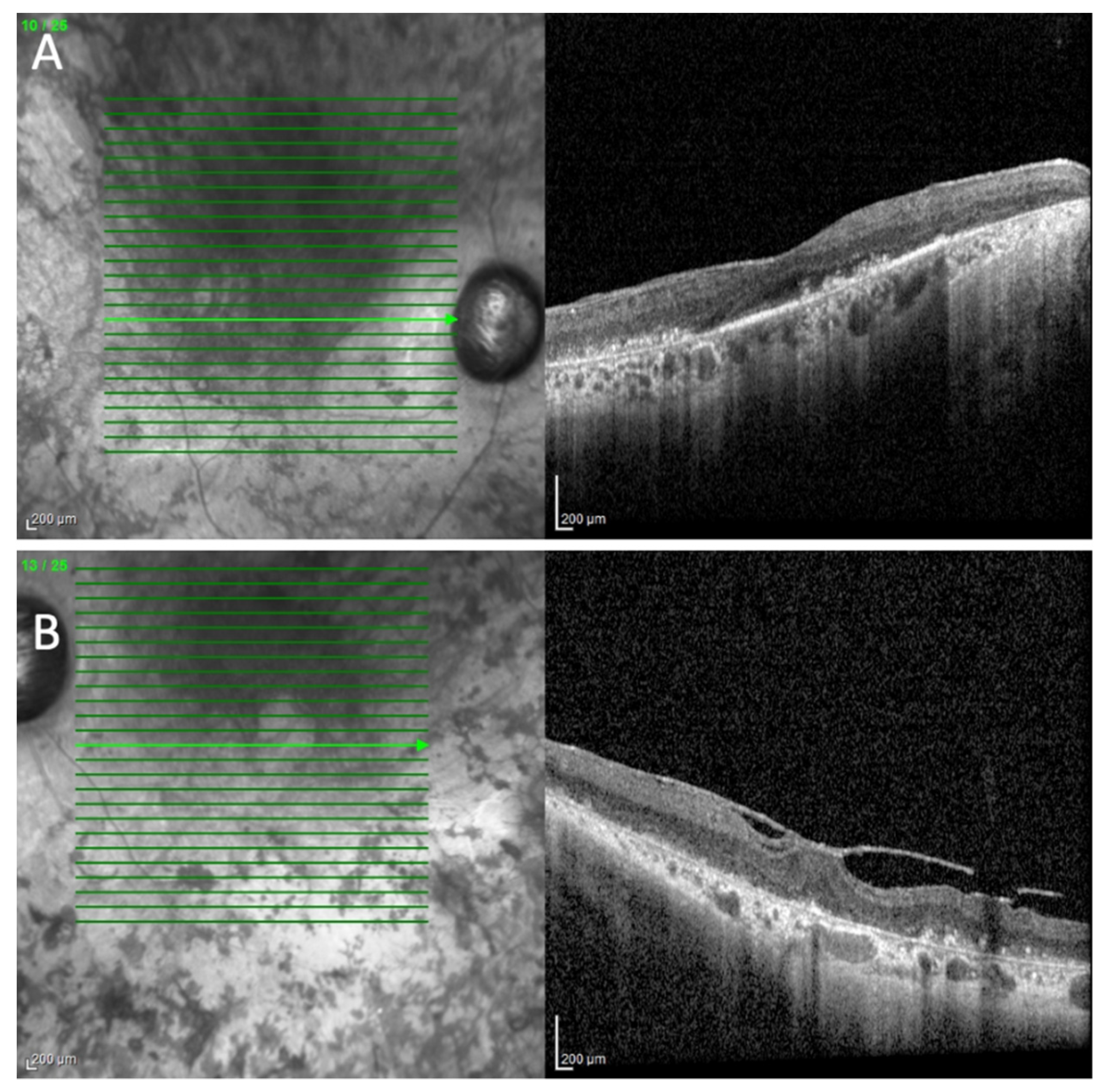
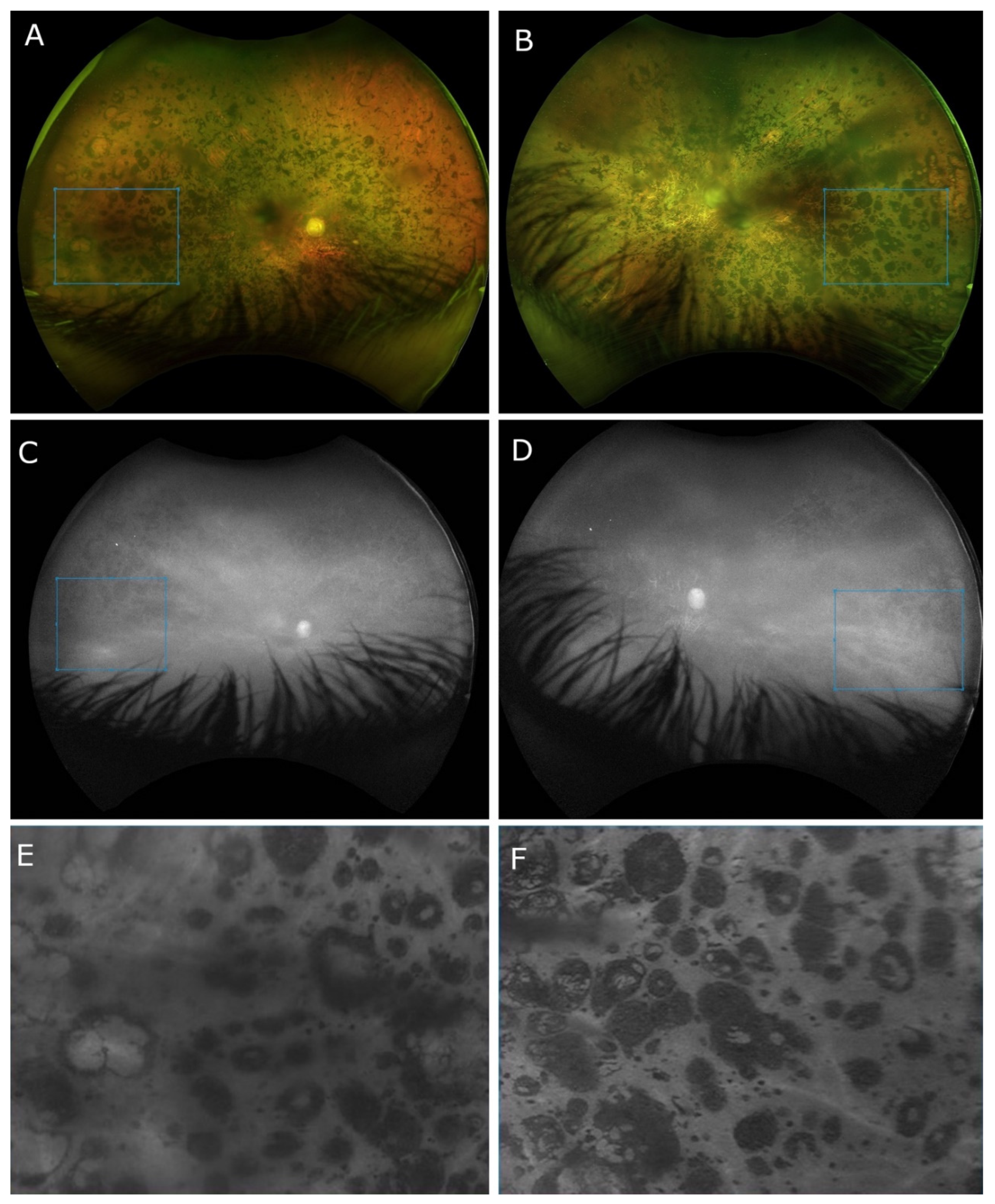
2. Case Presentation

A 66-year-old male with IRD was referred to our Eye Department for clinical examination and genetic testing. The patient manifested nyctalopia since early childhood. However, he attended elementary school, high school, and college according to the regular program. At the age of 17, a more pronounced decline in visual functions ensued. His brother was also visually impaired, but his family history was otherwise unremarkable.
On clinical examination, his best corrected visual acuity (BCVA) was light perception on the right eye (RE) and amaurosis on the left eye (LE). The CSV-1000 contrast sensitivity test, standardized color vision tests (Farnsworth’s D-15 dichotomous test and Lanthony desaturated 15-hue panel), Octopus static and kinetic perimetry (Haag-Streit Inc., Mason, OH, USA), and MAIA microperimetry (iCare Finland Oy, Vantaa, Finland) were not feasible due to the low BCVA.
HRA+ OCT Spectralis® (Heidelberg Engineering, Heidelberg, Germany) depicted prominent sub-RPE deposits and epiretinal membranes on both eyes (BE) with vitreoretinal traction on LE (Figure 1). Central macular thicknesses of 253 and 323 microns were measured on the RE and LE, respectively. An ONL of 32 microns was present on the RE.
The Optos® California (Optos Inc., Marlborough, MA, USA) ultra-widefield imaging depicted a pale and waxy optic nerve head with clear boundaries, more pronounced on his LE. In the posterior pole, extensive chorioretinal atrophy was noted, while in the mid- and far peripheries, clumped pigmentary changes in the nummular appearance were present. Retinal vessels presented obliterative sclerosis, revealing avascular mid- and far peripheries. The Optos® short-wavelength fundus autofluorescence was absent (Figure 2).
The full-field electroretinography testing (Roland Consult, RETI-port/scan 21, Roland Consult Stasche and Finger GmbH–German Engineering, Brandenburg an der Havel, Germany) according to ISCEV standards exhibited extinguished scotopic and photopic responses on BE.
IOLMaster® 700 (Zeiss, Oberkochen, Germany) optical biometry measured axial length of 22.83 mm RE and 22.66 mm LE, while retinoscopy was unremarkable (+0.75 Dsph on RE, and +0.75 Dsph/−1.00 Dcyl ax 115° on LE).
Full-field stimulus testing (Metrovision, Perenchies, France) detected responses of 43 dB, 26 dB, and 53 dB to white, red, and blue light, respectively, on the RE, while on the LE, no response was identified.
Given the clinical diagnosis of IRD, the patient was referred to genetic testing. Sequence analysis using the Blueprint Genetics Retinal Dystrophy Panel identified a homozygous missense variant RPE65 c.499G>T, p. (Asp167Tyr).

3. Discussion

RPE65 is the key enzyme of the classical visual cycle [7], a complex enzymatic pathway of trans-to-cis isomerization, regenerating the visual chromophore [8].
The basic RPE65 structure is a seven-bladed β-propeller with single-strand extensions on blades VI and VII and a two-strand extension on blade III. It seems that a majority of the amino acid substitutions found in LCA and RP patients occur within or adjacent to the blades of the β-propeller, rather than in the connecting loops and helices.
Position 167 resides within strand 11 of blade I, away from the iron ion active site cavity [9]. The iron ion binding site is coupled with four His and three Glu residues, each derived from a single blade. As a hydrophilic residue, Asp167 could serve as a gatekeeper protecting the aggregation-prone regions of proteins [10,11]. Thus, Asp167Tyr replacement could allow the formation of predominantly hydrophobic contacts, as observed with D477G and induced toxic gain-of-function [9].
To the best of our knowledge, this is the first patient reported in the literature to be homozygous for RPE65 c.499G>T, p. (Asp167Tyr) variant. In the Genome Aggregation Database (gnomAD), the p.Asp167Tyr variant was absent from 50 control individuals, but was reported at a frequency of 0.000026 in the European (non-Finnish) population [12]. The variant affects a highly conserved amino acid in the carotenoid oxygenase domain of the protein; there was a large physicochemical difference between asparagine and tyrosine (Grantham score 160, [0–215]), and all of the in silico tools utilized (PolyPhen, SIFT) predicted the alteration to be deleterious [12].
This variant is a missense variant that has been reported in a compound heterozygous state in three individuals with either LCA or RP [2,13,14,15]. Further, the p.Asp167Tyr variant was also identified in a heterozygous state in one additional case in whom a second variant was not identified [16]. Based on the collective evidence, the RPE65 c.499G>T, p. (Asp167Tyr) variant has been classified as pathogenic/likely pathogenic for RPE65-related disorders [17].
Many RPE65 patients have similar phenotypes characterized by poor, but useful, visual function in early life that declines significantly throughout school age. Additionally, some of these patients retain residual, although significantly narrowed, islands of peripheral vision into the third decade of life [2]. As presented by Chung et al., 88.5% of affected individuals have severely reduced or absent fundus autofluorescence [18]. The majority of RPE65 patients have whitish deposits and scarce pigmentary changes in the early stages and typical RP bone spicule-like pigment deposits later in the disease course, with a spectrum of disease severity that ranges from a mild form to a severe one [19,20,21,22]. Almost all of the mutations in RPE65 are inherited in a recessive manner; however, an autosomal dominant form of RPE65-associated RP with choroidal involvement has also been identified. Carriers of the D477G mutation, unlike typical RP patients, exhibit areas of retinal atrophy ranging from the choroid to the photoreceptor layer, and initially present with a central visual defect. Further, choroidal atrophy and RPE hypertrophy are frequently observed and distinctive features associated with this mutation [8].
Our patient manifested a unique genotype and phenotype biallelic RPE65 c.499G>T, p. (Asp167Tyr) variant that presented with less severe functional vision deterioration in childhood and extensive nummular pigment clusters affecting the mid- and far peripheries, reported only in patients with chloridemia, retinitis pigmentosa 86 (RP-86), and retinitis pigmentosa 87 (RP-87) with choroidal involvement [23,24,25,26].
The underlying causes of the differences in the typical bone spicule and atypical nummular pigment clumping are unknown, but suggest that the variant itself influences the rate of photoreceptor death.
The major stimulus that triggers RPE detachment from Bruch’s membrane and the migration of RPE cells to perivascular sites in the inner retina is proposed to be the direct contact of deep capillary retinal vessels and apical side of the RPE due to photoreceptor loss [27]. Bone spicule pigment clusters delineate retinal capillaries and venules at different retinal layers from the RPE to the retinal nerve fiber layer [28]. Relocated RPE cells induce endothelial fenestration, deposit the extracellular matrix, and seal the vessel by forming tight junctions reestablishing the blood–retinal barrier comparable to the choriocapillaris/RPE interface [27,29]. It is hypothesized that RPE cells are attracted to vascular basal lamina due to the endothelial release of cytokines [28,29] and higher intravascular oxygen levels [28], causing a vicious cycle of secondary retinal obliterative sclerosis. However, we assume that the RPE detachment and migration to perivascular inner retinal sites is caused by the need for fuel intake, as RPE cells cannot regenerate 11-cis retinal from all-trans retinol; thus, they need to fuel themselves from outer sources i.e., retinal capillaries. In a healthy retina, all-trans retinol is taken up from the bloodstream of the fenestrated choriocapillaris to the RPE. Thus, to provide supply of the chromophore, the RPE detaches, migrates, and induces the pathologic fenestration of the non-fenestrated retinal capillaries.
In contrast to typical bone spicule clusters of relocated RPE cells containing few melanin granules in the inner retina, the rarer form of rounded nummular flecks demonstrates a different pattern of reactive changes with densely packed melanin granules clustered against Bruch’s membrane [30]. We hypothesize the latter scenario to occur in cases of a lower expression level of RPE65 or the rapid degradation of the variant protein, as observed in D477G RPE65 mutation [8]. The study of missense variant RPE65 c.1430A>G, p. (Asp477Gly) was reported to cause a dominantly inherited, distinctive form of RP, RP-87 with choroidal involvement, evidencing the abnormally slow regeneration of the 11-cis retinal chromophore [31]. This corroborates the hypothesized fuel theory because, in RP-87 induced by D477G RPE65 substitution, toxic gain-of function is induced, the chromophore is slowly renewed, enabling RPE cells to stay in situ, thus presenting with a less severe phenotype in adolescence [31]. Heavily pigmented, they form nummular deposits against Bruch’s membrane, possibly consistent with the hexagonal lobules of choriocapillaris. Although RPE is generally viewed as a postmitotic cell, the concept that melanin turnover is absent in the adult RPE is grounded in the tyrosinase activity identified only in prenatal periods [32] and is absent much before gestation ends [33]. Whether the RPE cells in RP are capable of melanin synthesis is still unknown. It has been evidenced that tyrosinase can be induced in postnatal RPE by the orthodenticle homeobox 2 (OTX2) activation of the human tyrosinase gene promoter [34], overexpression of tyrosinase [35] and the transcriptional repressor Zeb1 [36], and phagocytosis of rod outer segments [37], demonstrating increased pigmentation.
With aging, the RPE melanin, a powerful electron acceptor, may undergo significant chemical modifications induced by oxygen, light, and toxic ions that bind to melanin, converting its antioxidant potential to phototoxic, pro-oxidative potential [38,39]. It seems plausible that this prooxidative potentially advances chorioretinal atrophy later in the disease’s course.
Our patient is homozygous for the RPE65 c.499G>T, p. (Asp167Tyr) variant, consistent with autosomal recessive inheritance. Functional studies are needed to define whether the substitution of Asp impairs the folding of the tertiary RPE65 structure only [40] and does not lead to the complete abolishment of chromophore production.

4. Conclusions

In conclusion, RPE65 c.499G>T, p. (Asp167Tyr) is classified as pathogenic, based on currently available evidence supporting its disease-causing role. Diseases caused by RPE65 c.499G>T, p. (Asp167Tyr) are inherited in an autosomal recessive manner. To the best of our knowledge, this is the first patient reported as homozygous for this variant. The patient demonstrated a unique phenotype not previously described in the literature, confirming that RPE65-associated IRDs may pose special challenges for genotype/phenotype correlations [18].

Author Contributions

Conceptualization: M.B. (Mirjana Bjeloš), A.Ć. and B.R.; methodology: M.B. (Mirjana Bjeloš), A.Ć., B.R., M.B. (Mladen Bušić) and B.K.E.; validation: M.B. (Mirjana Bjeloš), A.Ć., B.R., M.B. (Mladen Bušić) and B.K.E.; formal analysis: M.B.(Mirjana Bjeloš), A.Ć., B.R. and M.B. (Mladen Bušić); investigation, resources, and data curation: M.B. (Mirjana Bjeloš), A.Ć., B.R., M.B. (Mladen Bušić) and B.K.E.; writing—original draft preparation: M.B. (Mirjana Bjeloš), A.Ć. and M.B. (Mladen Bušić); writing—review and editing: M.B. (Mirjana Bjeloš), A.Ć., B.R., M.B. (Mladen Bušić) and B.K.E.; supervision: M.B. (Mirjana Bjeloš), A.Ć., M.B. (Mladen Bušić) and B.K.E. All authors have read and agreed to the published version of the manuscript.

Funding

No funding was received for this research.

Institutional Review Board Statement

Not applicable.

Informed Consent Statement

Prior to testing, written informed consent was obtained from the patient. No compensation or incentive was offered to the subject to participate in the study.

Data Availability Statement

The data presented in this case report are available on request from the corresponding author. The data are not publicity available due to privacy protection.

Conflicts of Interest

The authors declare no conflict of interest.

References

  1. *180069 RETINOID ISOMEROHYDROLASE RPE65; RPE65. Available online: https://omim.org/entry/180069?search=rpe65&highlight=rpe65 (accessed on 31 October 2022).
  2. Thompson, D.A.; Gyürüs, P.; Fleischer, L.L.; Bingham, E.L.; McHenry, C.L.; Apfelstedt-Sylla, E.; Zrenner, E.; Lorenz, B.; Richards, J.E.; Jacobson, S.G.; et al. Genetics and phenotypes of RPE65 mutations in inherited retinal degeneration. Investig. Ophthalmol. Vis. Sci. 2000, 41, 4293–4299. [Google Scholar]
  3. The Human Gene Mutation Database. Available online: http://www.hgmd.cf.ac.uk/ac/index.php (accessed on 31 October 2022).
  4. Redmond, T.M.; Poliakov, E.; Yu, S.; Tsai, J.-Y.; Lu, Z.; Gentleman, S. Mutation of key residues of RPE65 abolishes its enzymatic role as isomerohydrolase in the visual cycle. Proc. Natl. Acad. Sci. USA 2005, 102, 13658–13663. [Google Scholar] [CrossRef] [Green Version]
  5. Chen, Y.; Moiseyev, G.; Takahashi, Y.; Ma, J.-X. Impacts of two point mutations of RPE65 from Leber’s congenital amaurosis on the stability, subcellular localization and isomerohydrolase activity of RPE65. FEBS Lett. 2006, 580, 4200–4204. [Google Scholar] [CrossRef] [Green Version]
  6. German Society of Ophthalmology (Deutsche Ophthalmologische Gesellschaft, DOG); German Retina Society e.V. (Retinologische Gesellschaft e. V., RG); Professional Association of German Ophthalmologists (Berufsverband der Augenärzte Deutschlands e. V., BVA). Statement of the DOG, the RG, and the BVA on the therapeutic use of voretigene neparvovec (LuxturnaTM) in ophthalmology. English version: January 2019. Ophthalmologe 2020, 117, 16–24. [Google Scholar] [CrossRef]
  7. Tsin, A.; Betts-Obregon, B.; Grigsby, J. Visual cycle proteins: Structure, function, and roles in human retinal disease. J. Biol. Chem. 2018, 293, 13016–13021. [Google Scholar] [CrossRef] [PubMed] [Green Version]
  8. Choi, E.H.; Suh, S.; Sander, C.L.; Hernandez, C.J.O.; Bulman, E.R.; Khadka, N.; Dong, Z.; Shi, W.; Palczewski, K.; Kiser, P.D. Insights into the pathogenesis of dominant retinitis pigmentosa associated with a D477G mutation in RPE65. Hum. Mol. Genet. 2018, 27, 2225–2243. [Google Scholar] [CrossRef] [Green Version]
  9. Kiser, P.D.; Golczak, M.; Lodowski, D.T.; Chance, M.R.; Palczewski, K. Crystal structure of native RPE65, the retinoid isomerase of the visual cycle. Proc. Natl. Acad. Sci. USA 2009, 106, 17325–17330. [Google Scholar] [CrossRef] [Green Version]
  10. De Baets, G.; Van Doorn, L.; Rousseau, F.; Schymkowitz, J. Increased Aggregation Is More Frequently Associated to Human Disease-Associated Mutations Than to Neutral Polymorphisms. PLoS Comput. Biol. 2015, 11, e1004374. [Google Scholar] [CrossRef] [Green Version]
  11. Reumers, J.; Maurer-Stroh, S.; Schymkowitz, J.; Rousseau, F. Protein sequences encode safeguards against aggregation. Hum. Mutat. 2009, 30, 431–437. [Google Scholar] [CrossRef] [PubMed]
  12. Genome Aggregation Database (gnomAD). Available online: https://gnomad.broadinstitute.org/variant/1-68906680-C-A?dataset=gnomad_r2_1 (accessed on 1 November 2022).
  13. Simonelli, F.; Ziviello, C.; Testa, F.; Rossi, S.; Fazzi, E.; Bianchi, P.E.; Fossarello, M.; Signorini, S.; Bertone, C.; Galantuomo, S.; et al. Clinical and molecular genetics of Leber’s congenital amaurosis: A multicenter study of Italian patients. Investig. Ophthalmol. Vis. Sci. 2007, 48, 4284–4290. [Google Scholar] [CrossRef] [PubMed]
  14. Bainbridge, J.W.B.; Smith, A.J.; Barker, S.S.; Robbie, S.; Henderson, R.; Balaggan, K.; Viswanathan, A.; Holder, G.E.; Stockman, A.; Tyler, N.; et al. Effect of gene therapy on visual function in Leber’s congenital amaurosis. N. Engl. J. Med. 2008, 358, 2231–2239. [Google Scholar] [CrossRef] [PubMed]
  15. Ripamonti, C.; Henning, G.B.; Ali, R.R.; Bainbridge, J.W.; Robbie, S.J.; Sundaram, V.; Luong, V.A.; van den Born, L.I.; Casteels, I.; de Ravel, T.J.L.; et al. Nature of the visual loss in observers with Leber’s congenital amaurosis caused by specific mutations in RPE65. Investig. Ophthalmol. Vis. Sci. 2014, 55, 6817–6828. [Google Scholar] [CrossRef] [PubMed] [Green Version]
  16. Henderson, R.H.; Waseem, N.; Searle, R.; van der Spuy, J.; Russell-Eggitt, I.; Bhattacharya, S.S.; Thompson, D.A.; Holder, G.E.; Cheetham, M.E.; Webster, A.R.; et al. An assessment of the apex microarray technology in genotyping patients with Leber congenital amaurosis and early-onset severe retinal dystrophy. Investig. Ophthalmol. Vis. Sci. 2007, 48, 5684–5689. [Google Scholar] [CrossRef] [Green Version]
  17. ClinVar. Available online: https://www.ncbi.nlm.nih.gov/clinvar/variation/98873/ (accessed on 1 November 2022).
  18. Chung, D.C.; Bertelsen, M.; Lorenz, B.; Pennesi, M.E.; Leroy, B.P.; Hamel, C.P.; Pierce, E.; Sallum, J.; Larsen, M.; Stieger, K.; et al. The Natural History of Inherited Retinal Dystrophy Due to Biallelic Mutations in the RPE65 Gene. Am. J. Ophthalmol. 2019, 199, 58–70. [Google Scholar] [CrossRef] [PubMed]
  19. Gao, F.-J.; Wang, D.-D.; Li, J.-K.; Hu, F.-Y.; Xu, P.; Chen, F.; Qi, Y.-H.; Liu, W.; Li, W.; Zhang, S.-H.; et al. Frequency and phenotypic characteristics of RPE65 mutations in the Chinese population. Orphanet. J. Rare Dis. 2021, 16, 174. [Google Scholar] [CrossRef]
  20. Cideciyan, A.V. Leber congenital amaurosis due to RPE65 mutations and its treatment with gene therapy. Prog. Retin. Eye Res. 2010, 29, 398–427. [Google Scholar] [CrossRef] [Green Version]
  21. Gardiner, K.L.; Cideciyan, A.V.; Swider, M.; Dufour, V.L.; Sumaroka, A.; Komáromy, A.M.; Hauswirth, W.W.; Iwabe, S.; Jacobson, S.G.; Beltran, W.A.; et al. Long-Term structural outcomes of late-stage RPE65 gene therapy. Mol. Ther. 2020, 28, 266–278. [Google Scholar] [CrossRef]
  22. Maguire, A.M.; Bennett, J.; Aleman, E.M.; Leroy, B.P.; Aleman, T.S. Clinical Perspective: Treating RPE65-Associated Retinal Dystrophy. Mol. Ther. 2021, 29, 442–463. [Google Scholar] [CrossRef]
  23. National Library of Medicine. National Center for Biotechnology Information. Available online: https://www.ncbi.nlm.nih.gov/medgen/892932 (accessed on 30 October 2022).
  24. RETINITIS PIGMENTOSA 87 WITH CHOROIDAL INVOLVEMENT; RP87. Available online: https://www.omim.org/entry/618697 (accessed on 30 October 2022).
  25. Hull, S.; Mukherjee, R.; Holder, G.E.; Moore, A.T.; Webster, A.R. The clinical features of retinal disease due to a dominant mutation in RPE65. Mol. Vis. 2016, 22, 626–635. [Google Scholar]
  26. Jauregui, R.; Park, K.S.; Tsang, S.H. Two-year progression analysis of RPE65 autosomal dominant retinitis pigmentosa. Ophthalmic. Genet. 2018, 39, 544–549. [Google Scholar] [CrossRef]
  27. Jaissle, G.B.; May, C.A.; van de Pavert, S.A.; Wenzel, A.; Claes-May, E.; Giessl, A.; Szurman, P.; Wolfrum, U.; Wijnholds, J.; Fischer, M.D.; et al. Bone spicule pigment formation in retinitis pigmentosa: Insights from a mouse model. Graefe’s Arch. Clin. Exp. Ophthalmol. 2010, 248, 1063–1070. [Google Scholar] [CrossRef]
  28. Milam, A.H.; Li, Z.Y.; Fariss, R.N. Histopathology of the human retina in retinitis pigmentosa. Prog. Retin. Eye Res. 1998, 17, 175–205. [Google Scholar] [PubMed]
  29. Li, Z.Y.; Possin, D.E.; Milam, A.H. Histopathology of bone spicule pigmentation in retinitis pigmentosa. Ophthalmology 1995, 102, 805–816. [Google Scholar] [CrossRef] [PubMed]
  30. To, K.W.; Adamian, M.; Jakobiec, F.A.; Berson, E.L. Clinical and histopathologic findings in clumped pigmentary retinal degeneration. Arch. Ophthalmol. 1996, 114, 950–955. [Google Scholar]
  31. Shin, Y.; Moiseyev, G.; Chakraborty, D.; Ma, J.-X. A Dominant Mutation in Rpe65, D477G, Delays Dark Adaptation and Disturbs the Visual Cycle in the Mutant Knock-In Mice. Am. J. Pathol. 2017, 187, 517–527. [Google Scholar] [CrossRef] [Green Version]
  32. Körner, A.; Pawelek, J. Mammalian tyrosinase catalyzes three reactions in the biosynthesis of melanin. Science 1982, 217, 1163–1165. [Google Scholar] [CrossRef] [PubMed]
  33. Miyamoto, M.; Fitzpatrick, T.B. On the nature of the pigment in retinal pigment epithelium. Science 1957, 126, 449–450. [Google Scholar] [CrossRef]
  34. Reinisalo, M.; Putula, J.; Mannermaa, E.; Urtti, A.; Honkakoski, P. Regulation of the human tyrosinase gene in retinal pigment epithelium cells: The significance of transcription factor orthodenticle homeobox 2 and its polymorphic binding site. Mol. Vis. 2012, 18, 38–54. [Google Scholar]
  35. Biesemeier, A.; Kreppel, F.; Kochanek, S.; Schraermeyer, U. The classical pathway of melanogenesis is not essential for melanin synthesis in the adult retinal pigment epithelium. Cell Tissue Res. 2010, 339, 551–560. [Google Scholar] [CrossRef]
  36. Liu, Y.; Ye, F.; Li, Q.; Tamiya, S.; Darling, D.S.; Kaplan, H.J.; Dean, D.C. Zeb1 represses Mitf and regulates pigment synthesis, cell proliferation, and epithelial morphology. Investig. Ophthalmol. Vis. Sci. 2009, 50, 5080–5088. [Google Scholar] [CrossRef] [Green Version]
  37. Schraermeyer, U.; Kopitz, J.; Peters, S.; Henke-Fahle, S.; Blitgen-Heinecke, P.; Kokkinou, D.; Schwarz, T.; Bartz-Schmidt, K.-U. Tyrosinase biosynthesis in adult mammalian retinal pigment epithelial cells. Exp. Eye Res. 2006, 83, 315–321. [Google Scholar] [CrossRef] [PubMed]
  38. Peters, S.; Lamah, T.; Kokkinou, D.; Bartz-Schmidt, K.-U.; Schraermeyer, U. Melanin protects choroidal blood vessels against light toxicity. Z. Naturforsch. C J. Biosci. 2006, 61, 427–433. [Google Scholar] [CrossRef] [PubMed]
  39. Żądto, A.; Ito, S.; Sarna, M.; Wakamatsu, K.; Mokrzyński, K.; Sarna, T. The role of hydrogen peroxide and singlet oxygen in the photodegradation of melanin. Photochem. Photobiol. Sci. 2020, 19, 654–667. [Google Scholar] [CrossRef] [PubMed]
  40. Bowne, S.J.; Humphries, M.M.; Sullivan, L.S.; Kenna, P.F.; Tam, L.C.S.; Kiang, A.S.; Campbell, M.; Weinstock, G.M.; Koboldt, D.C.; Ding, L.; et al. A dominant mutation in RPE65 identified by whole-exome sequencing causes retinitis pigmentosa with choroidal involvement. Eur. J. Hum. Genet. 2011, 19, 1074–1081. [Google Scholar] [CrossRef] [Green Version]
Figure 1. Spectral domain OCT scan (HRA+ OCT Spectralis®) of the right (A) and left (B) eyes depicting prominent sub-RPE deposits and epiretinal membranes on both eyes with vitreoretinal traction on the left eye.
Cimb 44 00436 g001
Figure 2. Ultra-widefield (UWF) 200° fundus photos of the right eye (A) and left eye (B) showing nummular pigment clumping of variable sizes. Retinal vessels are presented as solid strands of hyaline-like connective tissue with avascular mid- and peripheral retina. Short-wavelength fundus autofluorescence (Optos® California) UWF 200° photos of the right eye (C) and left eye (D) depicting the absence of autofluorescence. Chorioretinal atrophy revealed the hyperautofluorescent attributes of the sclera. Enlarged image of the areas (marked with the blue squares in (AD)) with nummular pigment clumping on RE (E) and LE (F) obtained with a 100% red blend and 4.35× magnification.
Cimb 44 00436 g002
Publisher’s Note: MDPI stays neutral with regard to jurisdictional claims in published maps and institutional affiliations.

