Article

Cannabigerol Modulates Cannabinoid Receptor Type 2 Expression in the Spinal Dorsal Horn and Attenuates Neuropathic Pain Models

by
Bismarck Rezende
1,†,
Gabriel Gripp Fernandes
2,3,†,
Vitória Macario de Simas Gonçalves
1,
Gabriela Guedes Nascimento
1,
Kethely Lima Marques
1,
Barbara Conceição Costa Azeredo de Oliveira
1,
Yure Bazilio dos Santos
1,
Maria Eduarda Barros de Andrade
1,
Karine Simões Calumbi
1,
Eduardo Perdigão Maia
1,
Luisa Menezes Trefilio
1,2,
Fernanda Antunes
4,
Fabrícia Lima Fontes-Dantas
1,2,* and
Guilherme Carneiro Montes
1,*
1
Department of Pharmacology and Psychobiology, Roberto Alcantara Gomes Biology Institute (IBRAG), Rio de Janeiro State University (UERJ), Rio de Janeiro 20551-030, Brazil
2
NeuroPharmacoGenetics Laboratory, Department of Pharmacology and Psychobiology, Rio de Janeiro State University (UERJ), Rio de Janeiro 20551-030, Brazil
3
Department of Biophysics and Physiology, Federal University of Juiz de Fora, Juiz de Fora 36036-900, Brazil
4
Animal Clinic and Surgery Laboratory, Center for Agricultural Sciences and Technologies (CCTA), State University of Northern Fluminense Darcy Ribeiro (UENF), Campos dos Goytacazes 28013-602, Brazil
*
Authors to whom correspondence should be addressed.
These authors contributed equally to this work.
Pharmaceuticals 2025, 18(10), 1508; https://doi.org/10.3390/ph18101508
Submission received: 23 August 2025 / Revised: 23 September 2025 / Accepted: 3 October 2025 / Published: 8 October 2025
(This article belongs to the Topic Research on Natural Products of Medical Plants)

Abstract

Background/Objectives: The expanding focus on novel therapeutic pathways for long-term pain relief has directed interest toward compounds obtained from Cannabis sativa. This study evaluated the antinociceptive potential of cannabigerol-enriched extract (CBG) in models of acute and chronic hypernociception, along with morphological outcomes. Methods: Formalin and hot plate tests were used on male Swiss mice to assess acute oral antinociception. To the chronic pain model, 8-week-old male Wistar rats underwent spinal nerve ligation (SNL), and CBG was administered orally by gavage once daily for 14 days. Results: CBG reduced nociceptive responses in the formalin test and hot plate tests, mainly at a dose of 30 mg/kg, showing antinociceptive activity. CBG attenuated SNL-induced thermal and mechanical hypersensitivity, accompanied by reduced microglial density and spinal morphological changes. Importantly, cannabinoid receptor type 2 (CB2R) signaling contributed to the antinociceptive effects of orally administered CBG, whereas cannabinoid receptor type 1 (CB1R), Brain-Derived Neurotrophic Factor (BDNF), and Tumor Necrosis Factor (TNF) did not appear to play major roles under our experimental conditions. Conclusions: Collectively, these findings support CBG as a promising alternative for chronic pain management.

1. Introduction

Chronic pain is a leading global health problem, with meta-analyses estimating a prevalence of approximately 20–30% among adults, higher in women and older individuals [1,2]. Systematic reviews also highlight its profound impact on disability, quality of life, and work capacity, making it a major contributor to years lived with disability worldwide [3].
A 2023 systematic review estimated that 35.7% of Brazilian adults suffer from chronic pain, rising among older adults [4]. Neuropathic pain affects around 10% of adults and is associated with greater interference in daily activities compared with non-neuropathic chronic pain [5].
Despite significant advances in understanding pain mechanisms, current therapeutic approaches remain insufficient, particularly for chronic and neuropathic pain. Conventional treatments such as nonsteroidal anti-inflammatory drugs (NSAIDs), antidepressants, gabapentinoids, and opioids often fail to provide adequate long-term relief and are associated with undesirable side effects, including gastrointestinal toxicity, tolerance, and dependence [6]. Among available treatments, opioids carry a pronounced risk of dependence and abuse, which has become a pressing global health problem [6,7].
In this context, cannabinoids have attracted growing interest due to their neuroprotective, antioxidant, anti-inflammatory, and analgesic properties demonstrated in both preclinical and clinical studies [8,9,10,11,12]. A systematic review of pharmacological and non-pharmacological treatments for neuropathic pain, along with French clinical recommendations, highlights the therapeutic relevance of cannabinoid-based approaches as part of comprehensive pain management strategies [13]. Recent clinical trials have reinforced these findings: a phase III, 12-week study demonstrated that a cannabis-based product containing tetrahydrocannabinol, cannabidiol, and cannabinol (THC/CBD/CBN) significantly reduced pain in patients with diabetic neuropathy, with a favorable safety profile [14]. Furthermore, meta-analyses and double-blind, randomized trials indicate benefits and manageable adverse effects of THC/CBD extracts in neuropathic pain [15,16]. Our group has been investigating the endocannabinoid system and recently demonstrated that an oral Cannabis sativa extract rich in cannabigerol (CBG) at 50 mg/kg produced significant antinociceptive effects without impairing locomotor performance in animals exposed to prenatal hypoxia-ischemia [17]. In this context, the present work investigates the analgesic potential of CBG-rich extracts from Cannabis sativa, using a reduced oral dose of 30 mg/kg to assess its antinociceptive efficacy in neuropathic pain models and to explore the molecular mechanisms underlying its activity.

2. Results

2.1. Oral Cannabigerol Produces Central and Peripheral Antinociceptive Effects in Rodent Pain Models

Oral administration of CBG at 30 mg/kg produced significant antinociceptive effects in different experimental pain models. In the formalin test, CBG (30 mg/kg) markedly reduced nociceptive behavior in both the neurogenic and inflammatory phases. Figure 1A illustrates the responses observed during the neurogenic and inflammatory phases, following oral administration of vehicle, morphine, ASA, and CBG in the animals. It was observed that the formalin response time during the neurogenic phase in the CBG-treated group was reduced to 41.5 ± 2.7 s compared with 68.6 ± 6.8 s in the vehicle group. During the inflammatory phase, animals treated with CBG exhibited a decrease in formalin response time from 167.5 ± 32.7 s (vehicle) to 45.5 ± 10.7 s. In the hot plate test, CBG (30 mg/kg) produced %MPE values of 30.8 ± 9.2%, 28.4 ± 9.0%, and 28.2 ± 6.7% at 50, 60, and 70 min, respectively, all significantly higher than the vehicle. In the SNL model, thermal and mechanical hypernociception developed 7 days after surgery (Figure 1C,D), while no hypernociception occurred in the sham group. In male rats, paw withdrawal latency (PWL) decreased from 6.1 ± 0.4 s and 201.1 ± 1.9 g (baseline on day 0) to 2.1 ± 0.1 s and 89.3 ± 5.8 g (day 7) in the thermal and mechanical assessments, respectively. In the SNL + vehicle group, PWL values decreased from 5.8 ± 0.3 s and 198.4 ± 3 g to 2.2 ± 0.2 s and 84 ± 2.4 g, confirming the establishment of thermal and mechanical hypernociception. Treatment with CBG significantly increased latency (p < 0.05) in both thermal and mechanical hypernociception assessments when compared to the vehicle-treated group. Reversal of thermal and mechanical hypernociception was observed from the 10th to the 21st day post-surgery.

2.2. Cannabigerol Attenuates Microgliosis but Fails to Modulate Astrogliosis

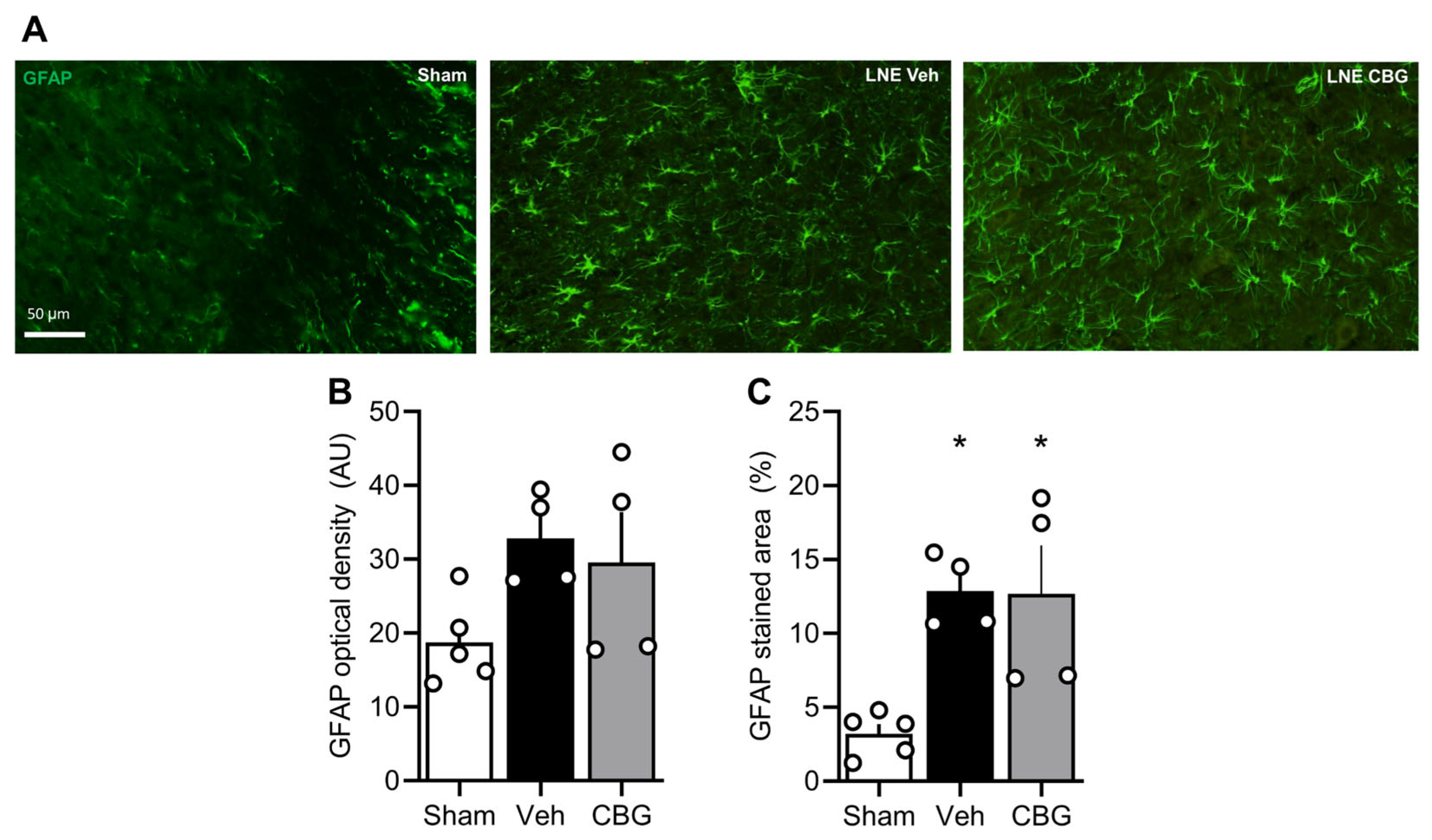
As depicted in Figure 2A–C, CBG was able to block the microgliosis observed in Rexed layers I and II of SNL submitted animals (Figure 2B) associated with a reduction in their soma area, a parameter commonly used to assess microglia reactivity and pro-inflammatory phenotype Figure 2C [18,19,20]. However, CBG did not reverse the elevated TNFα levels detected in the dorsal horn after SNL surgery (E). Additionally, SNL animals displayed marked astrogliosis compared to the sham group, and CBG treatment failed to attenuate this response (Figure 2D). These findings indicate that, in the SNL model, CBG can modulate microglial activation but has limited effects on TNFα upregulation in the spinal dorsal horn.
Among glia cells responsible for the development of neuropathic pain, several reports support the hypothesis that astroglia cells may act in synergy with microglia cells, secreting neurotrophic and pro-inflammatory mediators, ultimately leading to allodynia and hyperalgesia [21]. In our protocol, the SNL submitted rats present astrogliosis compared to the SHAM group, and CBG failed to block this phenomenon (Figure 3), suggesting that microglia cells may be one of the major glia cells responsible for CBG actions.

2.3. Involvement of Cannabinoid 2 Receptors in CBG Effects

In order to investigate the molecular mechanism responsible for CBG action, we initially investigated the role played by brain-derived neurotrophic factors (BDNF) in our model, since some reports support the role of this neurotrophin in the genesis of neuropathic pain [22]. BDNF levels remained unchanged in our animal model (Supplement Figure S1). Next, we assessed the role of CB1 and CB2 in the mechanism of action of CBG (Figure 4). Using the formalin test, we observed that SR144528, a selective CB2R antagonist, but not AM281, a selective CB1R antagonist, blocked the CBG effect in the neurogenic phase of the formalin test (A) but had no effect in the inflammatory phase (B). To further explore this, we measured the optical density of CB1 and CB2 receptors in laminae I/II of the dorsal horn in the spinal cord of SNL animals treated with CBG (C–F). Interestingly, while we did not observe any changes in CB1 receptor expression, SNL reduced CB2R optical density in the spinal cord, and CBG reversed this effect.

3. Discussion

Chronic pain is broadly acknowledged as a critical global health issue, ranking among the foremost causes of disability and generating significant social and economic costs [1,2,3].
In this context, our study demonstrates that oral administration of CBG at 30 mg/kg produces robust antinociceptive effects in both acute and neuropathic pain models. These results reinforce previous findings from our group showing that CBG at 50 mg/kg reduced hyperalgesia induced by prenatal hypoxia–ischemia, without impairing locomotor activity [17]. In that previous study, a higher dose of the same extract was administered, and no signs of sedation or motor impairment were observed in the animals. Pharmacokinetic and safety studies indicate that CBG is well absorbed following oral administration, distributes to the brain, and is tolerated without significant adverse effects [23,24]. These findings support the selection of the 30 mg/kg dose used in the present study and corroborate our observation that CBG does not induce sedation or motor impairment in the tested animals. Therefore, confounding effects related to sedation or motor impairment are unlikely at the dose used in the present study. While the earlier work also reported reductions in TNFα and Nav1.7 expression in the spinal cord, the current data highlight additional effects of CBG in different pain paradigms, including strong inhibition of nociceptive behavior in the formalin test, reversal of thermal and mechanical hypernociception after SNL, and attenuation of microgliosis in the spinal dorsal horn.
Consistent with our observations, preclinical studies in other neuropathic pain models—including chemotherapy-induced peripheral neuropathy—show that chronic administration of CBG maintains efficacy without tolerance. Importantly, combinations of CBG with CBD have been reported to reduce mechanical hypersensitivity more effectively than CBG alone [25,26]. Moreover, systematic reviews [27] corroborate that CBG, whether administered alone, combined with CBD, or as part of CBG-rich extracts, consistently exhibits antinociceptive effects across neuropathic pain models. Collectively, these findings highlight the central and peripheral analgesic potential of CBG, supporting its development as a novel therapeutic agent.
Several lines of evidence suggest an interaction between the endocannabinoid system and the secretion of neurotrophic factors, such as BDNF. In this direction, we investigated whether CBG was able to interact with BDNF. Surprisingly, in our SNL neuropathic pain model, BDNF optical density was unchanged among all groups analyzed. Supporting our observation, Geng and colleagues observed that BDNF decreases to Sham-control levels 14 days after SNL and sustains until 28 days [28]. This effect may account for the lack of differences described in the present study. Nonetheless, the role of CBG in modulating BDNF at other points was not evaluated in the present study, necessitating further experiments to better delineate the interaction between CBG and neurotrophins and their role in generating neuropathic pain.
Beyond our observations, the endocannabinoid system has been described to interact with BDNF in order to promote synaptic plasticity [29]. Specifically, it has been shown that in the striatum, long-term depression (LTD) induced by BDNF was blocked after CB1 antagonist administration [30] and BDNF regulates CB1R expression in this region [31]. In the same direction, CB1R blockage disrupts the cortical interneurons’ LTD facilitated by BDNF administration in a subthreshold protocol [32]. Furthermore, synaptic plasticity has been implicated in the development and maintenance of neuropathic pain [33,34], and several studies outline that neurons present in the dorsal horn are capable of both long-term potentiation (LTP) and LTD [35,36,37]. In this sense, our data suggests that it seems unlikely that CBG is acting through CB1R and BDNF systems to facilitate LTD, ultimately leading to an anti-nociceptive phenotype.
Besides CB1R, pharmacological studies suggest that CBG may interact with CB2R in a higher-affinity manner, acting as an agonist, while acting as a partial agonist with CB1R [38,39,40,41]. In accordance with Sepulveda and colleagues [26], we also demonstrated that CBG reduces nociception through CB2 receptors. It is well established that SNL augments the CB2 levels in the spinal cord at least seven days after surgery [42], which may be related to the fact that the endocannabinoid system is responsible for breaking the inflammation process. Nonetheless, in our study, SNL reduced CB2 receptor levels, consistent with the hyperalgesic phenotype observed in these animals. Indeed, in vitro studies using BV2 cells showed that CB2 receptor expression may increase or decrease according to the inflammatory challenge used or the time point analyzed [43,44], corroborating the discrepancy on CB2 levels in the present study and those observed by Zhang and colleagues [42]. Additionally, during inflammatory events, the endogenous CB2 receptor ligands, such as anandamide (AEA) and 2-arachidonoylglycerol (2-AG), are degraded through the COX-2 alternative pathway, enzymes usually upregulated during inflammatory events [45]. This prompts us to suggest that CBG interacts with CB2, preserving its signaling pathway, leading to an antinociceptive phenotype associated with reduced microgliosis.
To explore the mechanism behind CBG’s effect on microgliosis, we investigated whether it involves a reduction in TNF levels, since several pieces of evidence suggest that TNF facilitates nociception through neuronal plasticity and LTP [46,47]. Unexpectedly, we did not observe any significant reduction in TNF levels in the spinal cord dorsal horn of SNL. The divergent result observed may indicate that CBG acts through an unknown molecular mechanism in our animal model. In fact, it has been shown that LPS-stimulated BV2 produced both TNF and NO, and that CBG blockades the secretion of both mediators [48]. Compelling evidence has also shown that NO plays a crucial role in pain [49]. Borrelli et al. (2013) [50] demonstrated that CBG attenuates murine colitis by reducing nitric oxide production and inducible nitric oxide synthase (iNOS) protein expression in macrophages, an effect modulated by CB2R, along with antioxidant and anti-inflammatory actions. In this sense, further studies are needed to investigate whether CBG has its antinociceptive properties through reduction in NO synthesis via iNOS, an isoform upregulated in inflammatory events [51], or through another, yet undescribed, mechanism. In line with previously demonstrated by our group [52], SNL induced an astrogliosis in the dorsal horn, a phenomena that was not revered by CBG. This fact has been already demonstrated in other animal models. For instance, it has been described that either that treatment with omega-3-enriched fish oil or mitochondrial therapy, produced an antinociceptive effect and anti-inflammatory effect associated with none effect on astrogliosis, suggesting that the anti-inflammatory effect of some drugs may be devoid of an astrocytic-dependent mechanism [53,54]. Strengthening the assumption that astrocytes may not have a sole role in neuropathy in SNL, some evidence demonstrated that central nervous system astrocytes, but not microglia, seems to be the source of TNF-α expression [55], this evidence is in accordance with the present report, where CBG had no effect on either TNF or GFAP stain. It is also important to consider sex-dependent factors that may influence pain responses and cannabinoid mechanisms. In a previous study, CBG reduced TNFα expression in males and Nav1.7 expression in females in a model of prenatal hypoxia–ischemia [17], indicating sex-specific mechanisms in nociception. Although the present study focused exclusively on male animals, future investigations should determine whether similar CB2-dependent effects occur in females to enhance the translational relevance of CBG.

4. Materials and Methods

4.1. Laboratory Animals

The experimental procedures applied in this research were approved by the Ethics Committee on the Care and Use of Laboratory Animals of the State University of Rio de Janeiro (CEUA/UERJ, protocol 018/2023). The study involved male Swiss mice (Mus musculus), aged 4–6 weeks and weighing 25–30 g, as well as male Wistar rats (Rattus norvegicus), weighing 180–280 g. All animals were bred and maintained in the animal facility of the Department of Pharmacology and Psychobiology (DFP), Instituto de Biologia Roberto Alcantara Gomes (IBRAG), UERJ. They were kept under standard laboratory conditions: a 12 h light/dark cycle (lights off from 6:00 p.m. to 6:00 a.m.), temperature of 21 ± 1 °C, and relative humidity of 50 ± 2%. Housing was provided in polypropylene cages with sawdust bedding, with groups of four animals randomly assigned. Food in pellet form and drinking water were available ad libitum. Before the beginning of the experiments, the animals were allowed a 30 min acclimatization period at room temperature.

4.2. Drugs

The cannabis extracts used in this study were generously provided by the Association for the Support of Research and Medicinal Cannabis Patients (APEPI, Rio de Janeiro, Brazil). The cannabis extract used in this study was characterized at the Center for Information and Toxicological Assistance (CIATox, University of Campinas, Campinas, Brazil) which provided an analytical certificate of composition. According to the report, the extract was highly enriched in cannabigerol (CBG, 59.0% m/m; 95%), while Δ9-THC was present only at 0.5% m/m (0.9%). Cannabidiolic acid (CBDA), cannabidiol (CBD), tetrahydrocannabinolic acid (THCA) and cannabinol (CBN) were below the detection limit (<0.2%). The study also employed acetylsalicylic acid (ASA, Sigma-Aldrich, St. Louis, MO, USA), SR144528, a selective CB2 receptor antagonist, AM281, a selective CB1 receptor antagonist (Sigma, Saint Louis, MO, USA), dimethyl sulfoxide (DMSO, ≥99.9% purity, Dinâmica, Indaiatuba, Brazil), formaldehyde P.A. 36% (Proquimios, Rio de Janeiro, Brazil); xylazine hydrochloride 2% (Vetecia, Louveira, Brazil), morphine and ketamine hydrochloride (Cristália, Itapira, Brazil), and isoflurane (BioChimico, Itatiaia, Brazil). For experimental procedures, the CBG-rich cannabis extract (comprising 95% of the total plant cannabinoids), morphine, ASA, SR144528, and AM281 were freshly dissolved in DMSO immediately before administration. The vehicle consisted of dimethyl sulfoxide (DMSO, ≥99.9% purity), administered orally without dilution (100% v/v).

4.3. Experimental Model

All animals were randomly allocated to treatment groups using a random number generator. Drug solutions were prepared and coded by an experimenter not involved in behavioral assessments. Oral administration and surgical procedures were performed by one investigator, whereas nociceptive scoring and data collection were carried out by a second investigator blinded to group allocation. Treatment codes were revealed only after completion of data analysis to ensure allocation concealment and minimize bias.

4.3.1. Formalin Test

The formalin test was used as a model of acute peripheral nociception [56]. Mice received a subcutaneous injection of 20 µL of 2.5% formalin into the plantar surface of the right hind paw. Nociceptive behavior was assessed in two distinct phases: phase 1 (0–5 min post-injection), representing neurogenic pain, and phase 2 (15–45 min post-injection), representing inflammatory pain. Animals (n = 10 per group) were treated orally (p.o.) by gavage with vehicle, morphine (30 mg/kg), CBG (1–30 mg/kg), or ASA (300 mg/kg). Fifty minutes after oral treatment, the formalin injection was administered. Nociceptive responses were quantified as the total time (in seconds) spent licking, scratching, or biting the injected paw during the 45 min observation period. To elucidate the involvement of cannabinoid receptors in the antinociceptive activity of CBG, the antagonists AM281 (1 mg/kg) and SR144528 (10 mg/kg) were administered intraperitoneally (i.p.); subsequently, the animals (n = 7) received oral CBG 30 min later, and formalin was injected 30 min thereafter.

4.3.2. Hot Plate Test

To evaluate the analgesic activity (MPE), we employed the hot plate test [57]. Initially, animals were habituated to the hot plate apparatus (51 ± 1 °C). Reaction latency (paw licking, paw shaking, or jumping) was then assessed twice, with a 30 min interval between trials, to establish the control latency. The mice with a reaction time longer than 20 s were excluded. After obtaining control latency, animals (n = 10) received oral administration (by gavage) of either vehicle, morphine (30 mg/kg), or CBG (30 mg/kg). Reaction time was recorded from 0 up to 150 min after compound administration, with a cut-off time of 30 s to prevent paw injury. Antinociceptive efficacy was expressed as the percentage of the maximum possible effect (%MPE), calculated according to Equation (1): % MPE (post-drug latency) − (control latency)/(cut-off time) − (control latency) × 100

4.3.3. Chronic Nociception Model

Chronic nociception was induced via spinal nerve ligation (SNL), in accordance with previously established protocols [58]. Briefly, male Wistar rats were anesthetized with an intraperitoneal injection of xylazine (20 mg/kg) and ketamine (100 mg/kg). Following disinfection of the surgical site with 10% povidone–iodine, a midline dorsal incision was made to expose the lateral laminae of the lower lumbar and upper sacral vertebrae. The right L6 transverse process was carefully removed to allow for exposure and tight ligation of the right L5 and L6 spinal nerves using 6-0 silk sutures. Animals in the sham group underwent the same surgical procedure, excluding nerve ligation. Seven days post-surgery, SNL animals exhibited thermal and mechanical hypernociception. At this point, pharmacological treatment was initiated and continued daily for a duration of 14 days.

4.3.4. Evaluation of Thermal and Mechanical Hypernociception in the SNL Model

Seven days after SNL surgery, animals developed both thermal and mechanical hypernociception. They were then allocated into three experimental groups (n = 7–8 per group): (1) SNL + CBG (30 mg/kg, orally by gavage), (2) SNL + vehicle (orally), and (3) sham surgery + vehicle (orally). Thermal hypernociception was assessed using the paw immersion test, in which the ipsilateral hind paw was immersed in water maintained at 46 °C. The primary outcome was the latency (s) to paw withdrawal, defined as the thermal nociceptive threshold. A cut-off time of 15 s was set to prevent tissue damage [59]. Mechanical hypernociception was evaluated using a digital analgesiometer (Insight, Brazil), which applies a gradually increasing force to the dorsal surface of the hind paw at a constant rate (grams per second). The applied force was continuously recorded and displayed on an electronic panel. Paw withdrawal in response to the stimulus was considered a nociceptive response, and the corresponding force value (in grams) at the moment of withdrawal was recorded as the mechanical nociceptive threshold [60].

4.4. Tissue Collection and Immunofluorescence

A cohort of rats was anesthetized with xylazine (20 mg/kg, i.p.) and ketamine (100 mg/kg i.p.) and transcardially perfused with PBS 0.01M (pH 7.4) followed by PFA 4%. Immediately after, the spinal cord was dissected and transferred to PFA 4% overnight. After, the spinal cords were stored in a 30% sucrose solution for cryoprotection. A week later, brains were frozen in −30 °C isopentane and stored at −80 °C until being processed. Coronal slices, 30 μm thick, were obtained from the lumbar part of the spinal cord using a cryostat at −20 °C (Leica Microsystems, Wetzlar, Germany). The sections were kept in an anti-freezing solution (30% ethylene glycol/20% glycerol) until the immunofluorescence assays were performed.
Briefly, free-floating sections were washed three times with PBS (15 min each) followed by 30 min incubation with citrate buffer 10 mM (pH = 6.0) at 70 °C for antigen retrieval. After three washes with PBS (15 min/wash), the sections were incubated with a blocking solution (TBS + Triton × 0.025% + BSA 3%) for 2 h followed by overnight incubation with anti-Iba1 antibody (rabbit, 1:750; FUJIFILM Wako Pure Chemical Corporation, Osaka, Japan), anti-TNF (rabbit, 1:100; Thermo Fisher Scientific, Waltham, MA, USA), anti-GFAP (mouse, 1:1000; Abcam, Cambridge, UK), anti-BDNF (rabbit, 1:250; Thermo Fisher Scientific, Waltham, MA, USA), anti-CB1R (rabbit, 1:250; Thermo Fisher Scientific, Waltham, MA, USA), and anti-CB2R (rabbit, 1:500; Thermo Fisher Scientific, Waltham, MA, USA). After three washes with PBS, the sections were incubated for 1.5 h with a secondary antibody (anti-rabbit AlexaFluor 488, anti-mouse 555; 1:750). DAPI staining (1:10,000 from 2 μg/mL stock solution) was used to visualize the dorsal horn of the spinal cord and oriented our analysis towards Rexed layers I and II.

4.5. Image Acquisition and Quantification

Photographs were taken epifluorescence microscope, Olympus BX53 (Olympus Corporation, Hachioji, Tokyo, Japan). The IBA-1 cell density was manually counted under a 20× objective and calculated by normalizing the number of cells counted by the area analyzed (measured using a 10× objective). Optical density of TNF, GFAP, BDNF, CB1R, and CB2R was calculated using the software FIJI (version 1.53t), after image acquisition under a 10× objective. The GFAP-stained area was estimated using the FIJI automatic plugin in images acquired using a 20× objective.

4.6. Statistical Analysis

All statistical analyses and graphical representations were performed using GraphPad Prism® version 10 (GraphPad Software Inc., San Diego, CA, USA). Data normality was evaluated with the Shapiro–Wilk test. For datasets showing a parametric distribution, one-way ANOVA followed by Dunnett’s post hoc test was applied for the formalin test, while two-way ANOVA followed by Sidak’s post hoc test was used for the hot-plate test and assessments of thermal and mechanical hypernociception. Experimental groups were compared to their respective controls. Results are expressed as mean ± standard error of the mean (SEM), and differences were considered statistically significant at p < 0.05. Significance levels are indicated by symbols (* or #).

5. Limitations of the Study

Although this study provides important insights into the antinociceptive effects of CBG in models of acute and chronic pain, it presents some limitations. As it was conducted in an animal model, the findings may not be directly extrapolated to humans due to physiological and pharmacokinetic differences. Despite evaluating multiple molecular markers (BDNF, CB1R, CB2R, IBA1, TNF, and GFAP) in the spinal cord, analyses were not performed in other relevant tissues, such as the dorsal root ganglion (DRG), which plays a key role in pain modulation. The experimental design employed a specific route, dose, and treatment duration, which may not reflect other clinically relevant regimens. While CB1 and CB2 antagonists were used in a screening test, the involvement of this system and other cannabinoid or non-cannabinoid targets cannot be excluded due to experimental and ethical limitations. Immunohistochemistry provided data on protein expression but did not include functional assays to confirm receptor activity. In addition, the evaluation at fixed time points may not capture the full dynamics of molecular changes during the transition from acute to chronic pain. Another limitation is that only male animals were included, which precludes conclusions on potential sex-dependent differences. Considering that pain mechanisms and cannabinoid responses may differ between sexes, further studies including female subjects are necessary to strengthen the translational relevance of our findings. Therefore, the translational applicability of the results requires further validation. Future studies should investigate the long-term efficacy and safety of CBG in chronic pain models to expand the understanding of its therapeutic potential.

6. Conclusions

In summary, our study provides robust evidence that CBG exerts potent antinociceptive effects across acute, inflammatory, and neuropathic pain models. Importantly, we demonstrate that a lower oral dose (30 mg/kg) of a CBG-rich extract is sufficient to produce these effects, enhancing translational relevance compared with previous reports at higher doses. Mechanistically, our findings indicate that CB2 receptor signaling mediates the antinociceptive action of CBG, as supported by receptor restoration after spinal nerve ligation and selective blockade by SR144528, whereas CB1R, BDNF, and TNF did not appear to play a major role under our experimental conditions. Additionally, we show that CBG attenuates microgliosis but not astrogliosis in the spinal dorsal horn, providing new cellular insights. Collectively, these results highlight CBG as a promising candidate for pain management and support further translational studies.

Supplementary Materials

The following supporting information can be downloaded at: https://www.mdpi.com/article/10.3390/ph18101508/s1, Figure S1. Representative image of BDNF in laminae I/II of the spinal cord dorsal horn.

Author Contributions

Conceptualization, G.C.M. and F.L.F.-D.; methodology, B.R., G.G.F., F.A., V.M.d.S.G., B.C.C.A.d.O., G.G.N., Y.B.d.S., M.E.B.d.A., K.S.C., E.P.M., K.L.M. and L.M.T.; software, B.R. and G.G.F.; validation, B.R., G.G.F., F.L.F.-D. and G.C.M.; formal analysis, F.L.F.-D. and G.C.M.; investigation, B.R. and G.G.F.; resources, B.R., G.G.F. and F.A.; data curation, B.R., G.G.F. and L.M.T.; writing—original draft preparation, B.R. and G.G.F.; writing—review and editing, F.L.F.-D. and G.C.M.; visualization, B.R., G.G.F., F.L.F.-D. and G.C.M.; supervision, F.L.F.-D. and G.C.M.; project administration, F.L.F.-D. and G.C.M.; funding acquisition, F.L.F.-D. and G.C.M. All authors have read and agreed to the published version of the manuscript.

Funding

This research was supported by the Coordenação de Aperfeiçoamento de Pessoal de Nível Superior (CAPES); The Fundação de Amparo à Pesquisa do Estado do Rio de Janeiro (FAPERJ) to G.C.M. (SEI-260003/004509/2025—APQ1 (E-26/210.559/2025); SEI-260003/013164/2024 (E-26/204.419/2024—BOLSA) and to G.G.F. (E17-200.197/2025; 200.198/2025); and the Conselho Nacional de Desenvolvimento Científico e Tecnológico (CNPq) to G.C.M. (grant number 444648/2023-2). Support was also provided to F.L.F.-D. (SEI-260003/006158/2024; SEI-260003/013218/2024). We also acknowledge the Parliamentary Amendment No. 491, allocated by Deputy Carlos Minc (State of Rio de Janeiro), aimed at fostering research on therapeutic cannabis. The funders had no role in study design, data collection and analysis, decision to publish, or preparation of the manuscript.

Institutional Review Board Statement

The animal study protocol was approved by the Institutional Review Board (or Ethics Committee) of the Instituto de Biologia Roberto Alcantara Gomes-UERJ (protocol code 018/2023 and date of approval 12 April 2023).

Informed Consent Statement

Not applicable.

Data Availability Statement

The data presented in this study are available upon request.

Acknowledgments

We thank the Conselho Nacional de Desenvolvimento Científico e Tecnológico (CNPq), Fundação Carlos Chagas Filho de Amparo à Pesquisa do Estado do Rio de Janeiro (FAPERJ) and Apoio à Pesquisa e Pacientes de Cannabis Medicinal (APEPI-No. 65065921/2023; Processo No. SEI-260007/026216/2023), and State Deputy of Rio de Janeiro Carlos Minc for his support.

Conflicts of Interest

The authors declare no conflicts of interest.

References

  1. Fayaz, A.; Croft, P.; Langford, R.M.; Donaldson, L.J.; Jones, G.T. Prevalence of Chronic Pain in the UK: A Systematic Review and Meta-Analysis of Population Studies. BMJ Open 2016, 6, e010364. [Google Scholar] [CrossRef]
  2. Jackson, T.; Thomas, S.; Stabile, V.; Shotwell, M.; Han, X.; McQueen, K. A Systematic Review and Meta-Analysis of the Global Burden of Chronic Pain Without Clear Etiology in Low- and Middle-Income Countries: Trends in Heterogeneous Data and a Proposal for New Assessment Methods. Anesth. Analg. 2016, 123, 739–748. [Google Scholar] [CrossRef]
  3. Steingrímsdóttir, Ó.A.; Landmark, T.; Macfarlane, G.J.; Nielsen, C.S. Defining Chronic Pain in Epidemiological Studies: A Systematic Review and Meta-Analysis. Pain 2017, 158, 2092–2107. [Google Scholar] [CrossRef]
  4. Santiago, B.V.M.; de Oliveira, A.B.G.; da Silva, G.M.R.; da Silva, M.D.F.; Bergamo, P.E.; Parise, M.; Villela, N.R. Prevalence of Chronic Pain in Brazil: A Systematic Review and Meta-Analysis. Clinics 2023, 78, 100209. [Google Scholar] [CrossRef] [PubMed]
  5. Udall, M.; Kudel, I.; Cappelleri, J.C.; Sadosky, A.; King-Concialdi, K.; Parsons, B.; Hlavacek, P.; Hopps, M.; Salomon, P.A.; DiBonaventura, M.D.; et al. Epidemiology of Physician-Diagnosed Neuropathic Pain in Brazil. J. Pain Res. 2019, 12, 243–253. [Google Scholar] [CrossRef] [PubMed]
  6. Hylands-White, N.; Duarte, R.V.; Raphael, J.H. An Overview of Treatment Approaches for Chronic Pain Management. Rheumatol. Int. 2017, 37, 29–42. [Google Scholar] [CrossRef] [PubMed]
  7. Carroll, C.P.; Brandow, A.M. Chronic Pain: Prevalence and Management. Hematol. Oncol. Clin. N. Am. 2022, 36, 1151–1165. [Google Scholar] [CrossRef]
  8. Pantoja-Ruiz, C.; Restrepo-Jimenez, P.; Castañeda-Cardona, C.; Ferreirós, A.; Rosselli, D. Cannabis and Pain: A Scoping Review. Braz. J. Anesthesiol. 2022, 72, 142–151. [Google Scholar] [CrossRef]
  9. Lessa, M.A.; Cavalcanti, I.L.; Figueiredo, N.V. Cannabinoid Derivatives and the Pharmacological Management of Pain. Rev. Dor 2016, 17, 47–51. [Google Scholar] [CrossRef]
  10. Rezende, B.; Alencar, A.K.; de Bem, G.F.; Fontes-Dantas, F.L.; Montes, G.C. Endocannabinoid System: Chemical Characteristics and Biological Activity. Pharmaceuticals 2023, 16, 148. [Google Scholar] [CrossRef]
  11. Johnson, B.W.; Strand, N.H.; Raynak, J.C.; Jara, C.; Habtegiorgis, K.; Hand, B.A.; Hong, S.; Maloney, J.A. Cannabinoids in Chronic Pain Management: A Review of the History, Efficacy, Applications, and Risks. Biomedicines 2025, 13, 530. [Google Scholar] [CrossRef] [PubMed]
  12. Cásedas, G.; Yarza-Sancho, M.D.; López, V. Cannabidiol (CBD): A Systematic Review of Clinical and Preclinical Evidence in the Treatment of Pain. Pharmaceuticals 2024, 17, 1438. [Google Scholar] [CrossRef] [PubMed]
  13. Moisset, X.; Bouhassira, D.; Avez Couturier, J.; Alchaar, H.; Conradi, S.; Delmotte, M.H.; Lanteri-Minet, M.; Lefaucheur, J.P.; Mick, G.; Piano, V.; et al. Pharmacological and Non-Pharmacological Treatments for Neuropathic Pain: Systematic Review and French Recommendations. Rev. Neurol. 2020, 176, 325–352. [Google Scholar] [CrossRef] [PubMed]
  14. Seevathee, K.; Kessomboon, P.; Manimmanakorn, N.; Luangphimai, S.; Thaneerat, T.; Wanaratna, K.; Plengphanich, S.; Thaenkham, T.; Sena, W. Efficacy and Safety of Transdermal Medical Cannabis (THC:CBD:CBN Formula) to Treat Painful Diabetic Peripheral Neuropathy of Lower Extremities. Med. Cannabis Cannabinoids 2025, 8, 1–14. [Google Scholar] [CrossRef]
  15. Bilbao, A.; Spanagel, R. Medical Cannabinoids: A Pharmacology-Based Systematic Review and Meta-Analysis for All Relevant Medical Indications. BMC Med. 2022, 20, 259. [Google Scholar] [CrossRef]
  16. Serpell, M.; Ratcliffe, S.; Hovorka, J.; Schofield, M.; Taylor, L.; Lauder, H.; Ehler, E. A Double-Blind, Randomized, Placebo-Controlled, Parallel Group Study of THC/CBD Spray in Peripheral Neuropathic Pain Treatment. Eur. J. Pain 2014, 18, 999–1012. [Google Scholar] [CrossRef]
  17. Rezende, B.; Marques, K.L.; de Carvalho, F.E.; Gonçalves, V.M.; de Oliveira, B.C.; Nascimento, G.G.; dos Santos, Y.B.; Antunes, F.; Barradas, P.C.; Fontes-Dantas, F.L.; et al. Cannabigerol Reduces Acute and Chronic Hypernociception in Animals Exposed to Prenatal Hypoxia-Ischemia. Sci. Pharm. 2024, 92, 53. [Google Scholar] [CrossRef]
  18. Diniz, L.P.; Almeida, J.C.; Tortelli, V.; Vargas Lopes, C.; Setti-Perdigão, P.; Stipursky, J.; Kahn, S.A.; Romão, L.F.; de Miranda, J.; Alves-Leon, S.V.; et al. Astrocyte-Induced Synaptogenesis Is Mediated by Transforming Growth Factor β Signaling through Modulation of D-Serine Levels in Cerebral Cortex Neurons. J. Biol. Chem. 2012, 287, 41432–41445. [Google Scholar] [CrossRef]
  19. Fernandes, G.G.; Costa, K.C.M.; Scomparin, D.S.; Freire, J.B.; Guimarães, F.S.; Campos, A.C. Genetic Ablation of the Inducible Form of Nitric Oxide in Male Mice Disrupts Immature Neuron Survival in the Adult Dentate Gyrus. Front. Immunol. 2021, 12, 782831. [Google Scholar] [CrossRef]
  20. Fontes-Dantas, F.L.; Fernandes, G.G.; Gutman, E.G.; De Lima, E.V.; Antonio, L.S.; Hammerle, M.B.; Mota-Araujo, H.P.; Colodeti, L.C.; Araújo, S.M.B.; Froz, G.M.; et al. SARS-CoV-2 Spike Protein Induces TLR4-Mediated Long-Term Cognitive Dysfunction Recapitulating Post-COVID-19 Syndrome in Mice. Cell Rep. 2023, 42, 112189. [Google Scholar] [CrossRef]
  21. Cheng, T.; Xu, Z.; Ma, X. The Role of Astrocytes in Neuropathic Pain. Front. Mol. Neurosci. 2022, 15, 1007889. [Google Scholar] [CrossRef]
  22. Smith, P.A. BDNF in Neuropathic Pain; the Culprit That Cannot Be Apprehended. Neuroscience 2024, 543, 49–64. [Google Scholar] [CrossRef] [PubMed]
  23. Deiana, S.; Watanabe, A.; Yamasaki, Y.; Amada, N.; Arthur, M.; Fleming, S.; Woodcock, H.; Dorward, P.; Pigliacampo, B.; Close, S.; et al. Plasma and Brain Pharmacokinetic Profile of Cannabidiol (CBD), Cannabidivarine (CBDV), Δ9-Tetrahydrocannabivarin (THCV) and Cannabigerol (CBG) in Rats and Mice Following Oral and Intraperitoneal Administration and CBD Action on Obsessive-Compulsive Behaviour. Psychopharmacology 2012, 219, 859–873. [Google Scholar] [CrossRef]
  24. Lacerda, M.; Carona, A.; Castanheira, S.; Falcão, A.; Bicker, J.; Fortuna, A. Pharmacokinetics of Non-Psychotropic Phytocannabinoids. Pharmaceutics 2025, 17, 236. [Google Scholar] [CrossRef] [PubMed]
  25. Nachnani, R.; Sepulveda, D.E.; Booth, J.L.; Zhou, S.; Graziane, N.M.; Raup-Konsavage, W.M.; Vrana, K.E. Chronic Cannabigerol as an Effective Therapeutic for Cisplatin-Induced Neuropathic Pain. Pharmaceuticals 2023, 16, 1442. [Google Scholar] [CrossRef] [PubMed]
  26. Sepulveda, D.E.; Morris, D.P.; Raup-Konsavage, W.M.; Sun, D.; Vrana, K.E.; Graziane, N.M. Cannabigerol (CBG) Attenuates Mechanical Hypersensitivity Elicited by Chemotherapy-Induced Peripheral Neuropathy. Eur. J. Pain 2022, 26, 1950–1966. [Google Scholar] [CrossRef]
  27. Li, S.; Li, W.; Malhi, N.K.; Huang, J.; Li, Q.; Zhou, Z.; Wang, R.; Peng, J.; Yin, T.; Wang, H. Cannabigerol (CBG): A Comprehensive Review of Its Molecular Mechanisms and Therapeutic Potential. Molecules 2024, 29, 5471. [Google Scholar] [CrossRef]
  28. Geng, S.-J.; Liao, F.-F.; Dang, W.-H.; Ding, X.; Liu, X.-D.; Cai, J.; Han, J.-S.; Wan, Y.; Xing, G.-G. Contribution of the Spinal Cord BDNF to the Development of Neuropathic Pain by Activation of the NR2B-Containing NMDA Receptors in Rats with Spinal Nerve Ligation. Exp. Neurol. 2010, 222, 256–266. [Google Scholar] [CrossRef]
  29. Zhao, L.; Levine, E.S. BDNF-Endocannabinoid Interactions at Neocortical Inhibitory Synapses Require Phospholipase C Signaling. J. Neurophysiol. 2014, 111, 1008–1015. [Google Scholar] [CrossRef]
  30. Gangarossa, G.; Perez, S.; Dembitskaya, Y.; Prokin, I.; Berry, H.; Venance, L. BDNF Controls Bidirectional Endocannabinoid Plasticity at Corticostriatal Synapses. Cereb. Cortex 2020, 30, 197–214. [Google Scholar] [CrossRef]
  31. De Chiara, V.; Angelucci, F.; Rossi, S.; Musella, A.; Cavasinni, F.; Cantarella, C.; Mataluni, G.; Sacchetti, L.; Napolitano, F.; Castelli, M.; et al. Brain-Derived Neurotrophic Factor Controls Cannabinoid CB1 Receptor Function in the Striatum. J. Neurosci. Off. J. Soc. Neurosci. 2010, 30, 8127–8137. [Google Scholar] [CrossRef] [PubMed]
  32. Zhao, L.; Yeh, M.L.-W.; Levine, E.S. Role for Endogenous BDNF in Endocannabinoid-Mediated Long-Term Depression at Neocortical Inhibitory Synapses. eNeuro 2015, 2, e0029-14.2015. [Google Scholar] [CrossRef] [PubMed]
  33. Bak, M.S.; Park, H.; Kim, S.K. Neural Plasticity in the Brain during Neuropathic Pain. Biomedicines 2021, 9, 624. [Google Scholar] [CrossRef] [PubMed]
  34. Luo, C.; Kuner, T.; Kuner, R. Synaptic Plasticity in Pathological Pain. Trends Neurosci. 2014, 37, 343–355. [Google Scholar] [CrossRef]
  35. Kim, H.Y.; Jun, J.; Wang, J.; Bittar, A.; Chung, K.; Chung, J.M. Induction of Long-Term Potentiation and Long-Term Depression Is Cell-Type Specific in the Spinal Cord. Pain 2015, 156, 618–625. [Google Scholar] [CrossRef]
  36. Randić, M.; Jiang, M.C.; Cerne, R. Long-Term Potentiation and Long-Term Depression of Primary Afferent Neurotransmission in the Rat Spinal Cord. J. Neurosci. Off. J. Soc. Neurosci. 1993, 13, 5228–5241. [Google Scholar] [CrossRef] [PubMed]
  37. Song, Q.; E, S.; Zhang, Z.; Liang, Y. Neuroplasticity in the Transition from Acute to Chronic Pain. Neurother. J. Am. Soc. Exp. Neurother. 2024, 21, e00464. [Google Scholar] [CrossRef]
  38. Cascio, M.G.; Gauson, L.A.; Stevenson, L.A.; Ross, R.A.; Pertwee, R.G. Evidence That the Plant Cannabinoid Cannabigerol Is a Highly Potent Alpha2-Adrenoceptor Agonist and Moderately Potent 5HT1A Receptor Antagonist. Br. J. Pharmacol. 2010, 159, 129–141. [Google Scholar] [CrossRef] [PubMed]
  39. Navarro, G.; Varani, K.; Reyes-Resina, I.; Sánchez de Medina, V.; Rivas-Santisteban, R.; Sánchez-Carnerero Callado, C.; Vincenzi, F.; Casano, S.; Ferreiro-Vera, C.; Canela, E.I.; et al. Cannabigerol Action at Cannabinoid CB(1) and CB(2) Receptors and at CB(1)-CB(2) Heteroreceptor Complexes. Front. Pharmacol. 2018, 9, 632. [Google Scholar] [CrossRef]
  40. Pollastro, F.; Taglialatela-Scafati, O.; Allarà, M.; Muñoz, E.; Di Marzo, V.; De Petrocellis, L.; Appendino, G. Bioactive Prenylogous Cannabinoid from Fiber Hemp (Cannabis Sativa). J. Nat. Prod. 2011, 74, 2019–2022. [Google Scholar] [CrossRef]
  41. Rosenthaler, S.; Pöhn, B.; Kolmanz, C.; Nguyen Huu, C.; Krewenka, C.; Huber, A.; Kranner, B.; Rausch, W.-D.; Moldzio, R. Differences in Receptor Binding Affinity of Several Phytocannabinoids Do Not Explain Their Effects on Neural Cell Cultures. Neurotoxicol. Teratol. 2014, 46, 49–56. [Google Scholar] [CrossRef]
  42. Zhang, J.; Hoffert, C.; Vu, H.K.; Groblewski, T.; Ahmad, S.; O’Donnell, D. Induction of CB2 Receptor Expression in the Rat Spinal Cord of Neuropathic but Not Inflammatory Chronic Pain Models. Eur. J. Neurosci. 2003, 17, 2750–2754. [Google Scholar] [CrossRef]
  43. Grabon, W.; Ruiz, A.; Gasmi, N.; Degletagne, C.; Georges, B.; Belmeguenai, A.; Bodennec, J.; Rheims, S.; Marcy, G.; Bezin, L. CB2 Expression in Mouse Brain: From Mapping to Regulation in Microglia under Inflammatory Conditions. J. Neuroinflam. 2024, 21, 206. [Google Scholar] [CrossRef]
  44. Maresz, K.; Carrier, E.J.; Ponomarev, E.D.; Hillard, C.J.; Dittel, B.N. Modulation of the Cannabinoid CB2 Receptor in Microglial Cells in Response to Inflammatory Stimuli. J. Neurochem. 2005, 95, 437–445. [Google Scholar] [CrossRef]
  45. Alhouayek, M.; Muccioli, G.G. COX-2-Derived Endocannabinoid Metabolites as Novel Inflammatory Mediators. Trends Pharmacol. Sci. 2014, 35, 284–292. [Google Scholar] [CrossRef] [PubMed]
  46. Leung, L.; Cahill, C.M. TNF-Alpha and Neuropathic Pain—A Review. J. Neuroinflam. 2010, 7, 27. [Google Scholar] [CrossRef]
  47. Liu, Y.-L.; Zhou, L.-J.; Hu, N.-W.; Xu, J.-T.; Wu, C.-Y.; Zhang, T.; Li, Y.-Y.; Liu, X.-G. Tumor Necrosis Factor-Alpha Induces Long-Term Potentiation of C-Fiber Evoked Field Potentials in Spinal Dorsal Horn in Rats with Nerve Injury: The Role of NF-Kappa B, JNK and P38 MAPK. Neuropharmacology 2007, 52, 708–715. [Google Scholar] [CrossRef] [PubMed]
  48. Fleisher-Berkovich, S.; Ventura, Y.; Amoyal, M.; Dahan, A.; Feinshtein, V.; Alfahel, L.; Israelson, A.; Bernstein, N.; Gorelick, J.; Ben-Shabat, S. Therapeutic Potential of Phytocannabinoid Cannabigerol for Multiple Sclerosis: Modulation of Microglial Activation In Vitro and In Vivo. Biomolecules 2023, 13, 376. [Google Scholar] [CrossRef] [PubMed]
  49. Cury, Y.; Picolo, G.; Gutierrez, V.P.; Ferreira, S.H. Pain and Analgesia: The Dual Effect of Nitric Oxide in the Nociceptive System. Nitric Oxide Biol. Chem. 2011, 25, 243–254. [Google Scholar] [CrossRef]
  50. Borrelli, F.; Fasolino, I.; Romano, B.; Capasso, R.; Maiello, F.; Coppola, D.; Orlando, P.; Battista, G.; Pagano, E.; Di Marzo, V.; et al. Beneficial Effect of the Non-Psychotropic Plant Cannabinoid Cannabigerol on Experimental Inflammatory Bowel Disease. Biochem. Pharmacol. 2013, 85, 1306–1316. [Google Scholar] [CrossRef]
  51. Hervera, A.; Negrete, R.; Leánez, S.; Martín-Campos, J.M.; Pol, O. The Spinal Cord Expression of Neuronal and Inducible Nitric Oxide Synthases and Their Contribution in the Maintenance of Neuropathic Pain in Mice. PLoS ONE 2010, 5, e14321. [Google Scholar] [CrossRef] [PubMed]
  52. Vieira, M.C.; Monte, F.B.D.M.; Eduardo Dematte, B.; Montagnoli, T.L.; Montes, G.C.; da Silva, J.S.; Mendez-Otero, R.; Trachez, M.M.; Sudo, R.T.; Zapata-Sudo, G. Antinociceptive Effect of Lodenafil Carbonate in Rodent Models of Inflammatory Pain and Spinal Nerve Ligation-Induced Neuropathic Pain. J. Pain Res. 2021, 14, 857–866. [Google Scholar] [CrossRef]
  53. Maia, J.R.L.C.B.; Machado, L.K.A.; Fernandes, G.G.; Vitorino, L.C.; Antônio, L.S.; Araújo, S.M.B.; Colodeti, L.C.; Fontes-Dantas, F.L.; Zeidler, J.D.; Saraiva, G.N.; et al. Mitotherapy Prevents Peripheral Neuropathy Induced by Oxaliplatin in Mice. Neuropharmacology 2024, 245, 109828. [Google Scholar] [CrossRef]
  54. Melato, J.; Goldoni, F.C.; Benvenutti, L.; Corrêa, T.P.; Remor, A.P.; Varela, K.G.; Stoeberl, L.C.; Fernandes, G.G.; de Lima Rasga, G.; Passos, G.F.; et al. Omega-3-Enriched Fish Oil Reduces the Chemotherapy-Induced Peripheral Neuropathy in Mice. Neuropharmacology 2025, 271, 110384. [Google Scholar] [CrossRef]
  55. Guan, Y.; Wang, R.; Li, X.; Zou, H.; Yu, W.; Liang, Z.; Li, L.; Chen, L.; Zhou, L.; Chen, Z. Astrocytes Constitute the Major TNF-α-Producing Cell Population in the Infarct Cortex in dMCAO Rats Receiving Intravenous MSC Infusion. Biomed. Pharmacother. 2021, 142, 111971. [Google Scholar] [CrossRef]
  56. Choi, S.S.; Lee, J.K.; Suh, H.W. Antinociceptive Profiles of Aspirin and Acetaminophen in Formalin, Substance P and Glutamate Pain Models. Brain Res. 2001, 921, 233–239. [Google Scholar] [CrossRef]
  57. Łuszczki, J.J. Dose-Response Relationship Analysis of Pregabalin Doses and Their Antinociceptive Effects in Hot-Plate Test in Mice. Pharmacol. Rep. 2010, 62, 942–948. [Google Scholar] [CrossRef]
  58. Ho Kim, S.; Mo Chung, J. An Experimental Model for Peripheral Neuropathy Produced by Segmental Spinal Nerve Ligation in the Rat. Pain 1992, 50, 355–363. [Google Scholar] [CrossRef]
  59. Lolignier, S.; Amsalem, M.; Maingret, F.; Padilla, F.; Gabriac, M.; Chapuy, E.; Eschalier, A.; Delmas, P.; Busserolles, J. Nav1.9 Channel Contributes to Mechanical and Heat Pain Hypersensitivity Induced by Subacute and Chronic Inflammation. PLoS ONE 2011, 6, e23083. [Google Scholar] [CrossRef]
  60. Santos-Nogueira, E.; Redondo Castro, E.; Mancuso, R.; Navarro, X. Randall-Selitto Test: A New Approach for the Detection of Neuropathic Pain after Spinal Cord Injury. J. Neurotrauma 2012, 29, 898–904. [Google Scholar] [CrossRef]
Figure 1. Effects of oral gavage administration of vehicle, morphine (30 mg/kg), ASA (300 mg/kg), or high-CBG cannabis extract (30 mg/kg) on the neurogenic and inflammatory phases of the formalin test (A); and on hot-plate test performance (B) in male Swiss mice. The effects of a 14-day oral treatment with vehicle or CBG on thermal (C) and mechanical (D) hypernociception in male Wistar rats, 7 days after SNL surgery. Data are expressed as mean ± SEM. p < 0.05. * p < 0.05 vs. 0 day (before SNL); # p < 0.05 when compared to the vehicle group throughout the treatment.
Pharmaceuticals 18 01508 g001
Figure 2. Representative image of IBA-1 stain (A) and TNF-alpha in (D) laminae I/II of spinal cord dorsal horn. Cannabigerol reduces microglia density (B) and soma area ((C); n = 5, 6 and 4, respectively) without affecting TNF stain ((E); n = 5, 4 and 4, respectively). Graphs are expressed as mean ± SEM. * p < 0.05 compared to the Sham group.
Pharmaceuticals 18 01508 g002
Figure 3. Representative image of GFAP stain (A) in laminae I/II of the spinal cord dorsal horn. Cannabigerol did not reverse the astrogliosis observed after SNL ((B,C); n 5, 4, and 4, respectively). Graphs are expressed as mean ± SEM. * p < 0.05 compared to the Sham group.
Pharmaceuticals 18 01508 g003
Figure 4. The SR144528, but not AM281, blocked the antinociceptive effect of CBG in the neurogenic phase, without affecting the inflammatory phase (A,B). Representative image of CB1R and CB2R (C,E) immunofluorescence in laminae I/II of the spinal cord dorsal horn. Cannabigerol had no effect on CB1R receptor density ((D); n = 4/group) and reversed the reduction in CB2 density observed after SNL ((F); n = 4/group). Graphs are expressed as mean ± SEM. * p < 0.05.
Pharmaceuticals 18 01508 g004
Disclaimer/Publisher’s Note: The statements, opinions and data contained in all publications are solely those of the individual author(s) and contributor(s) and not of MDPI and/or the editor(s). MDPI and/or the editor(s) disclaim responsibility for any injury to people or property resulting from any ideas, methods, instructions or products referred to in the content.