Review

Modulators of the Human Voltage-Gated Proton Channel Hv1

1
Department of Biophysics and Cell Biology, Faculty of Medicine, University of Debrecen, Egyetem ter 1, H-4032 Debrecen, Hungary
2
MTA-DE Cell Biology and Signalling Research Group, Faculty of Medicine, University of Debrecen, Egyetem ter 1, H-4032 Debrecen, Hungary
*
Author to whom correspondence should be addressed.
These authors contributed equally to this work.
Pharmaceuticals 2025, 18(10), 1480; https://doi.org/10.3390/ph18101480
Submission received: 13 August 2025 / Revised: 16 September 2025 / Accepted: 26 September 2025 / Published: 1 October 2025
(This article belongs to the Section Pharmacology)

Abstract

The voltage-gated proton channel (Hv1) selectively transports protons (H+) across biological membranes in response to membrane potential changes. Hv1 is assembled as a dimer, and unlike most voltage-gated ion channels, it lacks a traditional central pore domain; instead, the voltage-sensing domain (VSD) of each monomer facilitates proton conduction via a hydrogen-bond network. Hv1 is widely expressed in various human cell types (e.g., immune cells, sperm, etc.) including tumor cells. In tumor cells, the accumulation of acidic intermediates generated by glycolysis under hypoxic conditions or ROS production leads to significant cytosolic acidification. Hv1 can remove protons from the cytosol rapidly, contributing to the adaptation of the cells to the tumor microenvironment, which may have significant consequences in tumor cell survival, proliferation, and progression. Therefore, Hv1 may be very promising not only as a tumor marker but also as a potential therapeutic target in oncology. Molecules that modulate the proton flux through Hv1 can be divided into two broad groups: inhibitors and activators. Hv1 inhibitors can be simple ions, small molecules, lipids, and peptides. In contrast, fewer Hv1 activators are known, including albumin, NH29, quercetin, and arachidonic acid. The mechanism of action of some inhibitors is well described, but not all. Hv1 modulation has profound effects on cellular physiology, especially under stress or pathological conditions, like cancer and inflammation. The therapeutic application of selective Hv1 inhibitors or activators could be a very promising strategy in the treatment of several serious diseases.

1. Introduction

1.1. Voltage-Gated Proton Channels

Voltage-gated proton channels (Hvs) represent a unique family of voltage-gated ion channels responsible for the selective passage of protons (H+) across biological membranes in response to changes in membrane potential. The first direct electrophysiological evidence of voltage-gated and pH-sensitive proton currents was published in the early 1980s [1]. This pioneering study laid the groundwork for understanding voltage-gated proton channels, whose molecule was later identified in 2006 [2,3]. The voltage-sensing domain (VSD) of Hv channels serves the dual function of sensing voltage changes and facilitating proton permeation [4]. Recently, our understanding of the voltage-gated proton channel family has been expanded with the discovery and characterization of three new proton channel members—AcHv1, AcHv2, and AcHv3—identified in the mollusk “Aplysia californica” [5]. More recently, another Hv channel was announced, namely Hv4, which was identified only in bivalvian mollusks [6].
Hv1 shows a wide tissue distribution pattern and presents diverse functions. It is expressed by several cell types in physiological conditions, like different immune cell types (neutrophils [7], eosinophils [8], macrophages [9], microglia [10], T and B cells [11,12]), human airway epithelium cells [13], human cardiac fibroblasts [14], chorion-derived mesenchymal stem cells [15], pancreatic islet β-cells [16], sperm [17], and oocytes [18]. Hv1 is also expressed in pathological situations, such as by tumor cells: malignant B cells [19], glioblastoma multiforme cells [20], leukemic Jurkat T cells [21], breast cancer cells [22,23], and colorectal cancer cell lines [24].

1.2. Structure of Hv1

Structurally, Hv channels differ from classic voltage-gated ion channels (VGICs), such as sodium (Nav), potassium (Kv), or calcium (Cav) ion channels. Typically, VGICs are composed of either homo- or heterotetrameric structures (Kv) or a single polypeptide chain of four homologous domains (Nav and Cav); each of the four subunits or domains consists of six transmembrane α-helical segments linked by intra- and extracellular loops. Of these, segments S1–S4 form the voltage-sensing domain (VSD) that regulates channel opening upon membrane depolarization, while segments S5 and S6 and the connecting extracellular pore loops (P-loops) constitute the pore domain. However, Hv channels have a unique architecture, as they are homodimers, composed of only two identical subunits, and both subunits contain four transmembrane segments (S1–S4) serving as VSDs (Figure 1). Unlike most VGICs, there is no distinct pore structure in Hv1 channels. Instead, protons permeate through each VSD individually, each with its intrinsic conduction pathway [25,26,27]. However, when monomers assemble into dimer proton channels in the membrane, cooperativity can be observed between them, and the dimer channel only becomes conductive when both monomers are activated [28,29,30]. More specifically, proton conduction in Hv1 channels may be described by the Grotthus mechanism, that is, H+ ions hop along a robust water wire within the channel structure, facilitated by specific water–protein interactions [31]. However, several studies have shown that Hv1 contains a dry region. This region disrupts the water wire such that protonation of one or more amino acids may occur, allowing protons to hop across via a hydrogen-bond chain mechanism [32,33,34]. The S4 segment terminates with a coiled-coil region facing the cell interior (Figure 1A), which contributes to the dimerization of the channel [25,26,35,36]. The voltage-gated proton channels are highly selective for H+ ions, ensured by specific charged residues—the open Hv1 channel requires an aspartate at 112 (Asp112) in the S1 segment and an arginine in the S4 helix, forming a narrow region that conducts protons selectively [37] (Figure 1B). Some studies propose that Asp112 interacts with Arg211 (R3) [38,39], while others propose its interaction with Arg208 (R2). Specifically, truncation of the S4 segment between R2 and R3 [27] or mutation of R3 to alanine [40] did not alter the proton selectivity of Hv1. The voltage-sensing part of the channel contains three positively charged arginine residues, known as gating arginines (Arg205 (R1), Arg208 (R2), and Arg211 (R3)); all are located in the S4 domain (Figure 1). These gating arginines interact with their negatively charged countercharge residues in the S1–S3 domains and “detect” the change in the membrane potential [41]. In the case of the Ci-Hv1 ion channel, attempts were made to isolate and measure the gating charge [42,43]. When the cell membrane is depolarized, or when there is an elevated proton concentration in the cyctosol or the contrary, the extracellular pH is alkaline, and channel activation is triggered [30]. In Hv1 channels, there is no inactivation mechanism; only open or closed states are observed [44]. In response to membrane depolarization, the gating arginines are repelled toward the extracellular space. These changes are mediated by two steps, as described by the three-state model, based on Hv1 current and fluorescence recordings [45,46,47]. According to this model, the structural changes between the different states result in a notable shift in the salt-bridge interaction network formed between S3 and S4. It should also be mentioned that there are articles that assume less or more than three states [48,49]. Channel activation critically influences both water distribution and proton permeability. In the open state, internal water molecules interact with charged residues through hydrogen bonds, often forming a continuous network that can conduct protons from the cytosol to the extracellular space [31]. However, multiple studies indicate the presence of intermittent dry regions within the pore that disrupt the continuity of this water wire. In such cases, proton transport may require transient protonation of amino acid side chains, allowing protons to “hop” across the gap via a hydrogen-bond chain mechanism. This dual mode of conduction highlights the dynamic interplay between water molecules and protein residues in regulating Hv1 channel function [32,33,34]. In addition to voltage gating, the Hv1 channel can be regulated by relative pH changes. Acidification of the cell interior alters the voltage gating. The response of Hv1 proton conductance to changes in pH gradient is approximately 40 mV/ΔpH [47,48,50,51]. This, together with its high proton selectivity, is perhaps the most characteristic feature of the channel. The pH dependence of gating is fundamental to the role of Hv1 in maintaining cellular pH homeostasis. This aspect is critical because it forms the basis for understanding the potential role of the channel in cancer cell survival.

1.3. Functions of Hv1 and Its Role in Cancer

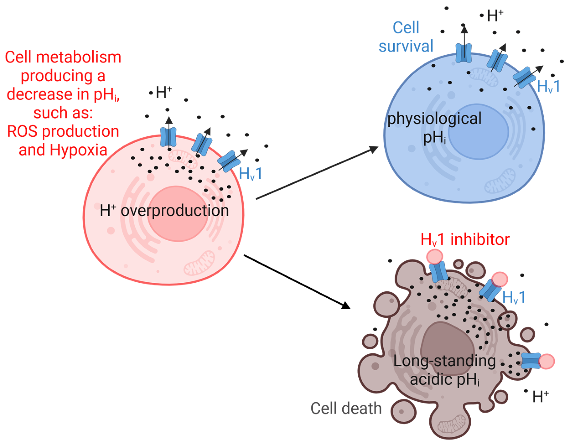
Studies have revealed the diverse roles of Hv1 in cancer cell types, particularly the ability to rapidly and robustly transport protons from the cytosol, thus regulating the intracellular pH (pHi) of cells. Therefore, Hv1 is involved in many processes that can lead to a decrease in pHi, such as the NADPH oxidase-dependent production of ROS by immune cells [52] or the accumulation of acidic intermediates during glycolysis in tumor cells under hypoxic conditions [53]. Inhibition of Hv1 results in at least two things: depolarization of the cell membrane (in a few milliseconds) [54], and a decrease in pHi (on a timescale of seconds to minutes) [55]. However, in many cases the role of Hv1 in different cellular processes has not yet been clarified. Without being exhaustive, the role of Hv1 has been highlighted in cancer cell migration and proliferation, cell survival and apoptosis [10,21], sustained calcium entry [56], neutrophil migration and superoxide production [56], sperm capacitation and motility [17], participation in optimal B-cell receptor signaling and redox control in human B lymphocytes [19], and regulation of insulin secretion [16]. Moreover, Hv1 plays a crucial role in cancer development, progression, and metastasis formation, allowing Hv1 to become a potential target in tumor therapy [23]. During processes producing ROS or under hypoxic conditions, tumor cells produce elevated proton concentrations in the cytosol. In the hypoxic tumor microenvironment, tumor cells have high glycolytic activity, converting glucose to acidic metabolites [57]. When pHi reaches a critical value relative to the extracellular pH, the threshold potential for the channel opening of Hv1 shifts sufficiently towards a more negative membrane potential, releasing protons from the cell, thereby reducing the proton concentration in the cytosol. Proton extrusion consequently affects the extracellular proton concentration, which may contribute to maintaining the acidic tumor microenvironment [57,58]. If, however, the Hv1 channel is inhibited, pHi in tumor cells is expected to remain permanently low, promoting cell death (Figure 2). Furthermore, since Hv1 may contribute to the acidification of the extracellular milieu, which suppresses antitumoral T-cell responses, proton extrusion from the IC to the EC also promotes tumor growth and progression by inhibiting the immune system [57,58,59]. Hv1 may also be important in the normal function and regulation of the nervous system, and accordingly, Hv1 can be responsible for various neurological diseases [60]. Thus, finding or developing a suitable Hv1 inhibitor or activator has been intensively pursued. Functional studies have explored a number of known Hv1 inhibitors or activators, but for most of these, selectivity (whether they affect other ion channels) has not been investigated. Hv1 inhibition by Zn2+ or ClGBI produced a significant acidification of Jurkat cells and induced cell death by apoptosis [21]. ClGBI also decreased the cell viability of tumorigenic breast cells along with a decrease in pHi [22]. Inhibiting Hv1 with Zn2+ significantly reduced pHi, decreasing cell survival and migration of a glioblastoma multiforme cell line [20]. Moreover, Zn2+ markedly decreased the cell invasion and migration of a colorectal cell line (SW620, HT29; [24]). Zn2+ ions also induced apoptosis in human highly metastatic glioma and effectively suppressed cancer growth and metastasis by reducing proton extrusion and downregulating gelatinase activity [61]. Myeloid-derived suppressor cells (MDSCs) also express Hv1 [62]. ClGBI significantly decreased the migration and osteogenic differentiation of chorion-derived mesenchymal stem cells [15]. Corza6 also blocked the acrosome reaction during capacitation of sperm and inhibited ROS production in human white blood cells (WBCs) [63]. Inhibition of neuronal Hv1 by a newly discovered inhibitor, YHV98-4, reduced intracellular alkalization and ROS production in peripheral sensory neurons [64]. Interestingly, macrophages are an important source of arachidonic acid metabolites, which are able to activate Hv1 [65].
Since the inhibitors are not selective for Hv1, it is difficult to estimate to what extent these effects are due to the inhibition of proton currents or due to other reasons. Recently, it has been shown that ClGBI is not a specific inhibitor of Hv1 since it inhibits several other channels [66] on lymphocytes. 2GBI, the precursor of ClGBI, has been shown to bind to NLRP3, which leads to inflammasome assembly and activation. This function of 2GBI is independent of Hv1 since the impact on inflammasome is also detected in bone marrow-derived macrophages where the HVCN1 gene is knocked down [67]. Therefore, the use of Hv1 knockout (KO) mice or the silencing HVCN1 gene is needed in additional pharmacological studies to understand and explore the function of Hv1 in health and disease [23,24,62,68,69]. Targeting the Hv1 proton channel in biological systems is pretty challenging due to its broad tissue distribution pattern. While considering Hv1 as a potential target in cancer therapy, it has to be taken into consideration that Hv1 is also expressed by immune cells that present anti-cancer properties, such as cytotoxic T cells and B cells. However, it is important to note that there are two known isoforms of Hv1; the shorter isoform (Hv1s) is shorter by 20 amino acids at the N-terminal compared to the long isoform. The appearance of Hv1s may have prognostic and therapeutic significance, as the appearance of the shorter isoform may contribute to tumor progression and proliferation, as presented in studies on the MDA-MB-231 breast cancer cell line and malignant B cells [19,22]. The development of Hv1-dependent tumor therapy raises further questions and possibilities, as more than 100 somatic mutations in Hv1 have been described in numerous tumor types (see the COSMIC and ClinVar databases). Without claiming to be exhaustive, these include the following cancer types: Burkitt lymphoma; glioblastoma multiforme; breast ductal carcinoma; colon, lung, and prostate adenocarcinoma; malignant melanoma; etc. The mutations in the S4 segments of Hv1 resulted in different biophysical properties in channel function [70]. The consequences of the mutations in tumor proliferation and progression are still unknown. Thus, while pharmacological tools and KO models are essential, a deeper understanding of Hv1 function will require explicit, testable mechanistic models, analogous to those developed for the respiratory burst in phagocytes, where quantitative evaluation of proton currents, channel kinetics, and oxidase-derived electron fluxes have provided robust functional insights.

2. Hv1 Modulators

The proton current through the Hv1 channel can be modulated by a diverse array of molecules at concentrations ranging from nanomolar (nM) to micromolar (µM) levels (Table 1). These Hv1 modulators are simple ions (e.g., Zn2+), small molecules (e.g., HIF, ClGBI), unsaturated fatty acids (e.g., arachidonic acid), and peptides (e.g., HaTx, GsAF-I). Based on their effect on proton currents, Hv1 modulators can be divided into two groups: inhibitors and activators.

2.1. Hv1 Inhibitors

One of the earliest identified inhibitors of Hv1 was Zn2+ [91]. This divalent cation inhibits Hv1 in a reversible manner by binding to the closed conformation of Hv1, thereby reducing the open probability of the channel and stabilizing its non-conducting state [92]. The binding site of Zn2+ is in the S3–S4 loop, with H140 and H193 playing key roles in binding (Figure 3) [3,93]. Interestingly, dimerization of the hHv1 channel creates novel binding sites for divalent cations by reorienting and juxtaposing key coordinating residues that remain spatially separated in the monomeric state. In dimeric assembly, histidines and acidic side chains contributed by adjacent protomers are positioned in close proximity, establishing unique coordination geometries that are absent in the monomeric channel [93,94]. In addition to Zn2+, several other inhibitors exhibit a similar mechanism of action. For instance, the mutated version of AGAP-W38F (anti-tumor analgesic peptide), isolated from the scorpion Buthus martensii, behaves as an Hv1 inhibitor by trapping the S4 voltage sensor in its deactivated state [76]. In contrast, while Zn2+ inhibition demonstrates a high degree of pH dependency, AGAP exhibits a reduced sensitivity to pH changes. Intriguingly, the binding pocket of AGAP-W38F partially overlaps with that of Zn2+, sharing critical residues H140 and H193 [3,76]. Other molecules that also stabilize the channel in its closed conformation are cholesterol [88], Oxophench [72], PNX61442 [86], a molecule called 13 [80], and NH17 [85]; however, the molecular determinants underlying their interaction with Hv1 are still poorly understood. For example, cholesterol does not directly interact with hHv1 residues but instead inhibits channel activity indirectly by altering the conformational kinetics of the voltage-sensing S4 domain and modifying the biophysical properties of the surrounding membrane [88,95]. In fact, a recent study concluded that cholesterol probably does not affect Hv1 directly, but indirectly. The data obtained can be explained more plausibly by the fact that Hv1 preferentially associates with cholesterol-dependent lipid domains, or “rafts” [96].
Molecules derived from guanidine have also been identified as Hv1 inhibitors [83]. 2-guanidinobenzimidazole (2GBI) has been observed to bind to the voltage-sensing domain (VSD) when the channel is in its open conformation (Figure 3). The binding pocket, accessible from the intracellular side of the membrane, involves amino acids D112, F150, S181, and R211, with F150 being critical for the interaction [83,97]. However, due to its high polarity, 2GBI has low permeability through cell membranes, thereby limiting its usage in pharmacological studies and precluding it from being a drug candidate [84,98]. To address this limitation, a derivative called Cl-guanidinobenzimidazole (ClGBI) was developed, which exhibits enhanced membrane permeability, enabling access to the intracellular domain of the channel and blocking it with higher binding affinity [83]. However, ClGBI has been shown to inhibit not only Hv1 at micromolar concentrations but also other ion channels, which greatly limits its future use as a tool in functional studies or as a potential drug candidate [66].
Another small molecule derivative, HIF (3-(2-amino-5-methyl-1H-imidazol-4-yl)-1-(3,5-difluorophenyl)propan-1-one), exhibits dual mechanisms of action depending on its interaction site within the channel. When HIF binds to “site 1”, its binding mechanism is similar to that of 2GBI, and it was confirmed that mutations of D112 and F150 abolish the inhibitory effect of HIF against Hv1. In contrast, binding to “site 2”, involving residues E171 and D174, leads to a slower recovery from inhibition [82]. Located near the 2GBI pocket, another binding pocket has been identified that accommodates the small molecule modulator of YHV98-4. This pocket is formed by amino acids I155, F161, and S219 (Figure 3). Molecular dynamic (MD) simulations suggest that upon binding to Hv1, YHV98-4 inhibits proton conduction by disrupting the water wires necessary for proton transfer [64]. It is important to note that in the article, the concepts of IC50 and Kd are confused, mostly because the inhibition does not saturate around zero but rather around 0.5. Therefore, IC50 should not be used, but rather Kd.
Certain antidepressant drugs, e.g., imipramine [74], antitussive drugs, e.g., dextromethorphan [87], and antipsychotic drugs, such as chlorpromazine, haloperidol, and clozapine [75,81], have been shown to inhibit the voltage-gated proton currents in BV2 microglial cells. These drugs penetrate the cell membrane in their uncharged, neutral form and subsequently undergo protonation in the cytosol. The charged forms of the drugs then block the proton channel intracellularly. Since these molecules are protonated, they may reduce the pH gradient, potentially leading to the reduction of the proton current. However, no changes in the reversal potential of the current were observed, suggesting that the inhibition mechanism does not directly alter the electrochemical equilibrium. Moreover, a similar inhibitory effect has been reported for other molecules, such as epigallocatechin-3-gallate (EGCG), the principal bioactive constituent of green tea [78]. Further studies are needed to fully explore the mechanism by which these protonated molecules inhibit proton currents.
Besides the peptide inhibitor AGAP-W38F, other peptides have also been reported as Hv1 inhibitors. One of these is the synthetic C6 peptide, which binds with nanomolar affinity to both the S3 and S4 loops of the dimer hHv1 in a cooperative manner. This cooperative binding causes the C6-bound channels to activate at a more positive membrane potential, i.e., C6 slows the activation of Hv1. The most critical residues for this interaction are V187, E192, H193, E196, and L200 (Figure 3) [71]. The venom of the tarantula Grammostola rosea has also been identified as the source of three Hv1 inhibitors. The first of these is Hanatoxin (HaTx), one of the earliest reported peptides that is capable of inhibiting Hv1. Extracellular application of HaTx produced inhibition of Hv1 proton currents, shifting the activation of the channel to more positive voltages [73]. Based on its interaction with Kv1.2, HaTx is assumed to partition into the membrane before interacting with a binding site at the protein–lipid interface. D185 plays a key role in this interaction, as shown by site-directed mutagenesis (D185A), which effectively abolishes the inhibitory effect of HaTx [73]. The other two peptides isolated from Grammostola rosea, Gr1b (GsAF-I) and Gr2c (GsAF-II), showed similar effects to HaTx; they shifted the activation threshold potential of Hv1 towards more positive potentials, reduced the Hv1 current in a membrane potential-dependent manner, and stabilized the channel in its closed state ([77]). However, the molecular determinants responsible for these inhibitory effects are still unknown.
It should also be noted that several of the substances listed in Table 1 can be considered weak base compounds (Fluoxetine, Chlorpromazine, Imipramine, Haloperidol, Clozapine, Desipramine, Dextromethorphan, Olanzapine). After passing through the membrane, these molecules can bind to protons inside the cell, thereby reducing the pHi. This may indicate apparent inhibition of the Hv1 channel, as the increased pHi affects the function of Hv1, reducing its probability of opening [99].

2.2. Hv1 Activators

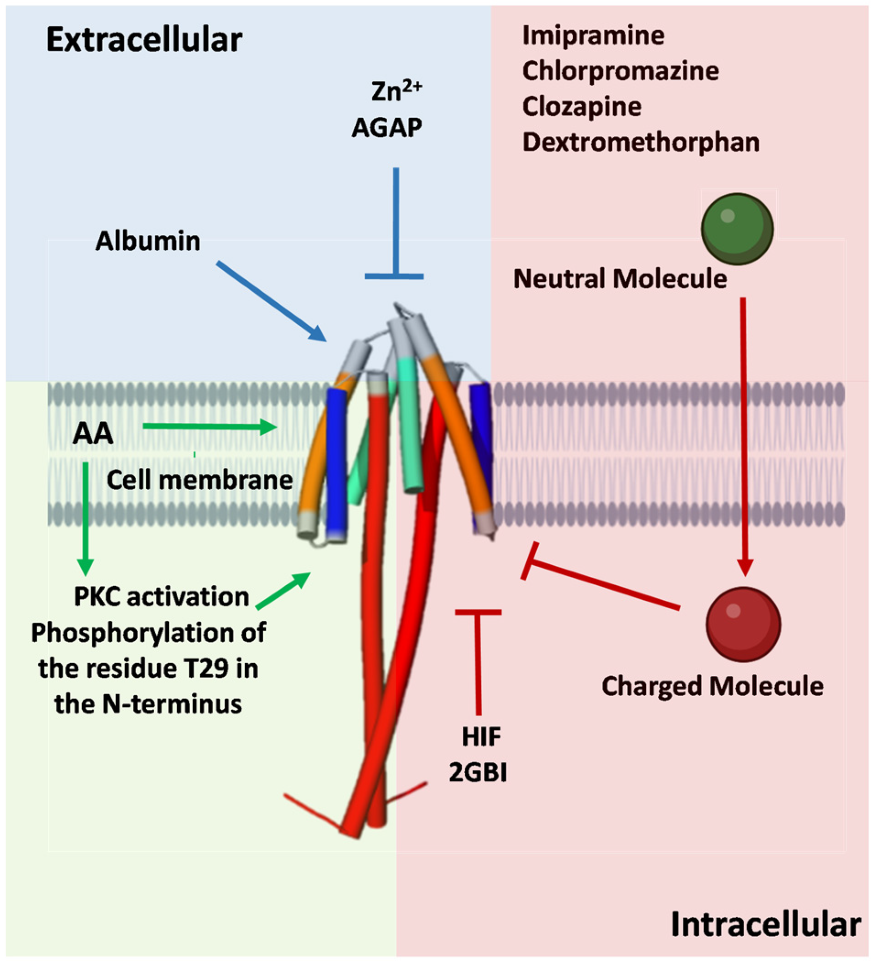
In contrast to the Hv1 inhibitors, only a few Hv1 activators have been described so far. One notable example is albumin (Alb), which has been shown to enhance the opening probability and increase proton currents in Hv1. A single Alb molecule binds to the dimeric hHv1 channel at both voltage-sensor domains (VSDs), specifically to the external S3-S4 loops. Mutations H193C and L200C in Hv1 fully eliminated Alb activation, suggesting that these two residues mediate direct interaction with Alb [89]. Interestingly, these residues are also involved in the inhibitory effects of Zn2+ and the C6 peptide. Similar to Alb, NH29 stabilizes the Hv1 channel in its open state [85]. External application of NH29 increased proton currents at all test potentials, primarily due to a significant hyperpolarizing shift in the conductance–voltage relationship [85]. In some cases, activators can enhance H+ currents through indirect mechanisms. Onion peel extract (OPE), from Allium cepa L., was found to modulate Hv1 channel opening and activates the channel at more negative membrane voltages. Subsequent studies revealed that quercetin, the major active component in OPE, is responsible for this effect. The activation of Hv1 induced by OPE was inhibited by 10 µM Zn2+ and GF109203X (GFX), a specific protein kinase C (PKC) inhibitor. It was concluded that the pro-oxidant effects of quercetin play a significant role in OPE-induced activation of Hv1 as well as its probable involvement in PKC signaling pathways [90]. While the precise interaction between OPE, PKC, and Hv1 remains unclear, similar PKC-related mechanisms have been observed in Hv1 activation by lipopolysaccharides (LPSs). The acute addition of LPSs increased the Hv1 channel activity that was abolished by GFX. However, the activating effect of LPSs on Hv1 disappeared after 24 h incubation with LPSs, instead acting as an inhibitor compound over time [100]. Arachidonic acid (AA) also activates Hv1 channels, enhancing the proton current both by direct interaction and by activating PKC [101]. The phenomenon of phosphorylation is consistent with prior observations of OPE and lipopolysaccharides. However, in the truncated isoform of mHv1, which lacks the N-terminal region that contains the phosphorylation site T29, AA addition still enhanced Hv1 currents. This indicates that AA can act directly on mHv1, with channel modulation occurring after AA incorporates into the membrane rather than through interaction from the extracellular side. The hydrophilic head group of AA is essential for this effect, whereas the molecular charge does not appear to play a critical role [65]. Figure 3 highlights the principal binding sites for Hv1 channel activators and inhibitors, whereas Figure 4 illustrates representative mechanisms by which these modulators influence channel activity.

3. Conclusions

Hv1 has emerged as a pivotal player in numerous physiological and pathological contexts, particularly in cancer and inflammation. The growing repertoire of Hv1 modulators not only deepens our understanding of proton channel function but also opens the door to promising therapeutic strategies. Targeting Hv1 with selective modulators may soon translate into meaningful advances in disease treatment.

Funding

This research was funded by the University of Debrecen Scientific Research Bridging Fund (DETKA) (FP) and also supported by the University of Debrecen Program for Scientific Publication (DETKA) (ZV). This work was also supported by the Ministry of Culture and Innovation of Hungary from the National Research, Development, and Innovation Fund, grant 2024-1.2.3-HU-RIZONT-2024-00099. This research was also funded by the Hungarian National Research, Development and Innovation Office (OTKA K143071, OTKA 132906).

Data Availability Statement

No new data were created or analyzed in this study. Data sharing is not applicable to this article.

Conflicts of Interest

The authors declare no conflicts of interest.

Abbreviations

The following abbreviations are used in this manuscript:
VSDVoltage-sensing domain
ROSReactive oxygen species
VGICVoltage-gated ion channel
icIntracellular
ecExtracellular
membMembrane
NADPHNicotinamide adenine dinucleotide phosphate
SW620A colorectal cell line
HT29A colorectal cell line
MDSCMyeloid-derived suppressor cell
WBCWhite blood cell
NLRP3NLR family pyrin domain-containing 3
KOKnockout
HVCN1The gene that encodes “Voltage-gated hydrogen channel 1”
NOX2NADPH oxidase 2
HIF3-(2-amino-5-methyl-1H-imidazol-4-yl)-1-(3,5-difluorophenyl)propan-1-one
AGAPAnti-tumor analgesic peptide, isolated from the scorpion Buthus martensii
BV2A mouse-derived microglial cell
EGCGEpigallocatechin-3-gallate, the principal bioactive constituent of green tea
OPEOnion peel extract
PKCProtein kinase C
LPSLipopolysaccharide
AAArachidonic acid

References

  1. Thomas, R.C.; Meech, R.W. Hydrogen ion currents and intracellular pH in depolarized voltage-clamped snail neurones. Nature 1982, 299, 826–828. [Google Scholar] [CrossRef]
  2. Sasaki, M.; Takagi, M.; Okamura, Y. A voltage sensor-domain protein is a voltage-gated proton channel. Science 2006, 312, 589–592. [Google Scholar] [CrossRef]
  3. Ramsey, I.S.; Moran, M.M.; Chong, J.A.; Clapham, D.E. A voltage-gated proton-selective channel lacking the pore domain. Nature 2006, 440, 1213–1216. [Google Scholar] [CrossRef] [PubMed]
  4. DeCoursey, T.E. The Voltage-Gated Proton Channel: A Riddle, Wrapped in a Mystery, inside an Enigma. Biochemistry 2015, 54, 3250–3268. [Google Scholar] [CrossRef] [PubMed]
  5. Chaves, G.; Ayuyan, A.G.; Cherny, V.V.; Morgan, D.; Franzen, A.; Fieber, L.; Nausch, L.; Derst, C.; Mahorivska, I.; Jardin, C.; et al. Unexpected expansion of the voltage-gated proton channel family. FEBS J. 2023, 290, 1008–1026. [Google Scholar] [CrossRef] [PubMed]
  6. Chaves, G.; Jardin, C.; Franzen, A.; Mahorivska, I.; Musset, B.; Derst, C. Proton channels in molluscs: A new bivalvian-specific minimal HV4 channel. FEBS J. 2023, 290, 3436–3447. [Google Scholar] [CrossRef]
  7. Okochi, Y.; Okamura, Y. Regulation of Neutrophil Functions by Hv1/VSOP Voltage-Gated Proton Channels. Int. J. Mol. Sci. 2021, 22, 2620. [Google Scholar] [CrossRef]
  8. Petheo, G.L.; Orient, A.; Barath, M.; Kovacs, I.; Rethi, B.; Lanyi, A.; Rajki, A.; Rajnavolgyi, E.; Geiszt, M. Molecular and functional characterization of Hv1 proton channel in human granulocytes. PLoS ONE 2010, 5, e14081. [Google Scholar] [CrossRef]
  9. El Chemaly, A.; Nunes, P.; Jimaja, W.; Castelbou, C.; Demaurex, N. Hv1 proton channels differentially regulate the pH of neutrophil and macrophage phagosomes by sustaining the production of phagosomal ROS that inhibit the delivery of vacuolar ATPases. J. Leukoc. Biol. 2014, 95, 827–839. [Google Scholar] [CrossRef]
  10. Wu, L.J. Voltage-gated proton channel Hv1 in microglia. Neuroscientist 2014, 20, 599–609. [Google Scholar] [CrossRef]
  11. Coe, D.; Poobalasingam, T.; Fu, H.; Bonacina, F.; Wang, G.; Morales, V.; Moregola, A.; Mitro, N.; Cheung, K.C.; Ward, E.J.; et al. Loss of voltage-gated hydrogen channel 1 expression reveals heterogeneous metabolic adaptation to intracellular acidification by T cells. JCI Insight 2022, 7, e147814. [Google Scholar] [CrossRef]
  12. Capasso, M.; Bhamrah, M.K.; Henley, T.; Boyd, R.S.; Langlais, C.; Cain, K.; Dinsdale, D.; Pulford, K.; Khan, M.; Musset, B.; et al. HVCN1 modulates BCR signal strength via regulation of BCR-dependent generation of reactive oxygen species. Nat. Immunol. 2010, 11, 265–272. [Google Scholar] [CrossRef]
  13. Iovannisci, D.; Illek, B.; Fischer, H. Function of the HVCN1 proton channel in airway epithelia and a naturally occurring mutation, M91T. J. Gen. Physiol. 2010, 136, 35–46. [Google Scholar] [CrossRef] [PubMed]
  14. El Chemaly, A.; Guinamard, R.; Demion, M.; Fares, N.; Jebara, V.; Faivre, J.F.; Bois, P. A voltage-activated proton current in human cardiac fibroblasts. Biochem. Biophys. Res. Commun. 2006, 340, 512–516. [Google Scholar] [CrossRef] [PubMed]
  15. Meszaros, B.; Papp, F.; Mocsar, G.; Kokai, E.; Kovacs, K.; Tajti, G.; Panyi, G. The voltage-gated proton channel hHv1 is functionally expressed in human chorion-derived mesenchymal stem cells. Sci. Rep. 2020, 10, 7100. [Google Scholar] [CrossRef] [PubMed]
  16. Zhao, Q.; Che, Y.; Li, Q.; Zhang, S.; Gao, Y.T.; Wang, Y.; Wang, X.; Xi, W.; Zuo, W.; Li, S.J. The voltage-gated proton channel Hv1 is expressed in pancreatic islet beta-cells and regulates insulin secretion. Biochem. Biophys. Res. Commun. 2015, 468, 746–751. [Google Scholar] [CrossRef]
  17. Lishko, P.V.; Botchkina, I.L.; Fedorenko, A.; Kirichok, Y. Acid extrusion from human spermatozoa is mediated by flagellar voltage-gated proton channel. Cell 2010, 140, 327–337. [Google Scholar] [CrossRef]
  18. Smith, R.Y.; Morgan, D.; Sharma, L.; Cherny, V.V.; Tidswell, N.; Molo, M.W.; DeCoursey, T.E. Voltage-gated proton channels exist in the plasma membrane of human oocytes. Hum. Reprod. 2019, 34, 1974–1983. [Google Scholar] [CrossRef]
  19. Hondares, E.; Brown, M.A.; Musset, B.; Morgan, D.; Cherny, V.V.; Taubert, C.; Bhamrah, M.K.; Coe, D.; Marelli-Berg, F.; Gribben, J.G.; et al. Enhanced activation of an amino-terminally truncated isoform of the voltage-gated proton channel HVCN1 enriched in malignant B cells. Proc. Natl. Acad. Sci. USA 2014, 111, 18078–18083. [Google Scholar] [CrossRef]
  20. Ribeiro-Silva, L.; Queiroz, F.O.; da Silva, A.M.; Hirata, A.E.; Arcisio-Miranda, M. Voltage-Gated Proton Channel in Human Glioblastoma Multiforme Cells. ACS Chem. Neurosci. 2016, 7, 864–869. [Google Scholar] [CrossRef]
  21. Asuaje, A.; Smaldini, P.; Martin, P.; Enrique, N.; Orlowski, A.; Aiello, E.A.; Gonzalez Leon, C.; Docena, G.; Milesi, V. The inhibition of voltage-gated H+ channel (HVCN1) induces acidification of leukemic Jurkat T cells promoting cell death by apoptosis. Pflug. Arch. 2017, 469, 251–261. [Google Scholar] [CrossRef]
  22. Ventura, C.; Leon, I.E.; Asuaje, A.; Martin, P.; Enrique, N.; Nunez, M.; Cocca, C.; Milesi, V. Differential expression of the long and truncated Hv1 isoforms in breast-cancer cells. J. Cell. Physiol. 2020, 235, 8757–8767. [Google Scholar] [CrossRef] [PubMed]
  23. Wang, Y.; Li, S.J.; Pan, J.; Che, Y.; Yin, J.; Zhao, Q. Specific expression of the human voltage-gated proton channel Hv1 in highly metastatic breast cancer cells, promotes tumor progression and metastasis. Biochem. Biophys. Res. Commun. 2011, 412, 353–359. [Google Scholar] [CrossRef] [PubMed]
  24. Wang, Y.; Wu, X.; Li, Q.; Zhang, S.; Li, S.J. Human voltage-gated proton channel Hv1: A new potential biomarker for diagnosis and prognosis of colorectal cancer. PLoS ONE 2013, 8, e70550. [Google Scholar] [CrossRef] [PubMed]
  25. Koch, H.P.; Kurokawa, T.; Okochi, Y.; Sasaki, M.; Okamura, Y.; Larsson, H.P. Multimeric nature of voltage-gated proton channels. Proc. Natl. Acad. Sci. USA 2008, 105, 9111–9116. [Google Scholar] [CrossRef]
  26. Lee, S.Y.; Letts, J.A.; Mackinnon, R. Dimeric subunit stoichiometry of the human voltage-dependent proton channel Hv1. Proc. Natl. Acad. Sci. USA 2008, 105, 7692–7695. [Google Scholar] [CrossRef]
  27. Sakata, S.; Kurokawa, T.; Norholm, M.H.; Takagi, M.; Okochi, Y.; von Heijne, G.; Okamura, Y. Functionality of the voltage-gated proton channel truncated in S4. Proc. Natl. Acad. Sci. USA 2010, 107, 2313–2318. [Google Scholar] [CrossRef]
  28. Gonzalez, C.; Koch, H.P.; Drum, B.M.; Larsson, H.P. Strong cooperativity between subunits in voltage-gated proton channels. Nat. Struct. Mol. Biol. 2010, 17, 51–56. [Google Scholar] [CrossRef]
  29. Tombola, F.; Ulbrich, M.H.; Kohout, S.C.; Isacoff, E.Y. The opening of the two pores of the Hv1 voltage-gated proton channel is tuned by cooperativity. Nat. Struct. Mol. Biol. 2010, 17, 44–50. [Google Scholar] [CrossRef]
  30. Musset, B.; Smith, S.M.; Rajan, S.; Cherny, V.V.; Morgan, D.; DeCoursey, T.E. Oligomerization of the voltage-gated proton channel. Channels 2010, 4, 260–265. [Google Scholar] [CrossRef]
  31. Ramsey, I.S.; Mokrab, Y.; Carvacho, I.; Sands, Z.A.; Sansom, M.S.P.; Clapham, D.E. An aqueous H+ permeation pathway in the voltage-gated proton channel Hv1. Nat. Struct. Mol. Biol. 2010, 17, 869–875. [Google Scholar] [CrossRef]
  32. DeCoursey, T.E.; Cherny, V.V. Deuterium isotope effects on permeation and gating of proton channels in rat alveolar epithelium. J. Gen. Physiol. 1997, 109, 415–434. [Google Scholar] [CrossRef] [PubMed]
  33. van Keulen, S.C.; Gianti, E.; Carnevale, V.; Klein, M.L.; Rothlisberger, U.; Delemotte, L. Does Proton Conduction in the Voltage-Gated H+ Channel hHv1 Involve Grotthuss-Like Hopping via Acidic Residues? J. Phys. Chem. B 2017, 121, 3340–3351. [Google Scholar] [CrossRef] [PubMed]
  34. Boytsov, D.; Brescia, S.; Chaves, G.; Koefler, S.; Hannesschlaeger, C.; Siligan, C.; Goessweiner-Mohr, N.; Musset, B.; Pohl, P. Trapped Pore Waters in the Open Proton Channel HV1. Small 2023, 19, e2205968. [Google Scholar] [CrossRef] [PubMed]
  35. Takeshita, K.; Sakata, S.; Yamashita, E.; Fujiwara, Y.; Kawanabe, A.; Kurokawa, T.; Okochi, Y.; Matsuda, M.; Narita, H.; Okamura, Y.; et al. X-ray crystal structure of voltage-gated proton channel. Nat. Struct. Mol. Biol. 2014, 21, 352–357. [Google Scholar] [CrossRef]
  36. Tombola, F.; Ulbrich, M.H.; Isacoff, E.Y. The voltage-gated proton channel Hv1 has two pores, each controlled by one voltage sensor. Neuron 2008, 58, 546–556. [Google Scholar] [CrossRef]
  37. Dudev, T.; Musset, B.; Morgan, D.; Cherny, V.V.; Smith, S.M.; Mazmanian, K.; DeCoursey, T.E.; Lim, C. Selectivity Mechanism of the Voltage-gated Proton Channel, HV1. Sci. Rep. 2015, 5, 10320. [Google Scholar] [CrossRef]
  38. Chamberlin, A.; Qiu, F.; Wang, Y.; Noskov, S.Y.; Larsson, H.P. Mapping the gating and permeation pathways in the voltage-gated proton channel Hv1. J. Mol. Biol. 2015, 427, 131–145. [Google Scholar] [CrossRef]
  39. Berger, T.K.; Isacoff, E.Y. The pore of the voltage-gated proton channel. Neuron 2011, 72, 991–1000. [Google Scholar] [CrossRef]
  40. Chaves, G.; Derst, C.; Franzen, A.; Mashimo, Y.; Machida, R.; Musset, B. Identification of an HV 1 voltage-gated proton channel in insects. FEBS J. 2016, 283, 1453–1464. [Google Scholar] [CrossRef]
  41. DeCoursey, T.E.; Morgan, D.; Musset, B.; Cherny, V.V. Insights into the structure and function of HV1 from a meta-analysis of mutation studies. J. Gen. Physiol. 2016, 148, 97–118. [Google Scholar] [CrossRef]
  42. Carmona, E.M.; Larsson, H.P.; Neely, A.; Alvarez, O.; Latorre, R.; Gonzalez, C. Gating charge displacement in a monomeric voltage-gated proton (Hv1) channel. Proc. Natl. Acad. Sci. USA 2018, 115, 9240–9245. [Google Scholar] [CrossRef]
  43. Fernandez, M.; Alvear-Arias, J.J.; Carmona, E.M.; Carrillo, C.; Pena-Pichicoi, A.; Hernandez-Ochoa, E.O.; Neely, A.; Alvarez, O.; Latorre, R.; Garate, J.A.; et al. Trapping Charge Mechanism in Hv1 Channels (CiHv1). Int. J. Mol. Sci. 2023, 25, 426. [Google Scholar] [CrossRef] [PubMed]
  44. Musset, B.; Decoursey, T. Biophysical properties of the voltage gated proton channel HV1. Wiley Interdiscip. Rev. Membr. Transp. Signal. 2012, 1, 605–620. [Google Scholar] [CrossRef] [PubMed]
  45. Qiu, F.; Rebolledo, S.; Gonzalez, C.; Larsson, H.P. Subunit interactions during cooperative opening of voltage-gated proton channels. Neuron 2013, 77, 288–298. [Google Scholar] [CrossRef] [PubMed]
  46. Papp, F.; Toombes, G.E.S.; Petho, Z.; Bagosi, A.; Feher, A.; Almassy, J.; Borrego, J.; Kuki, A.; Keki, S.; Panyi, G.; et al. Multiple mechanisms contribute to fluorometry signals from the voltage-gated proton channel. Commun. Biol. 2022, 5, 1131. [Google Scholar] [CrossRef]
  47. Cherny, V.V.; Markin, V.S.; DeCoursey, T.E. The voltage-activated hydrogen ion conductance in rat alveolar epithelial cells is determined by the pH gradient. J. Gen. Physiol. 1995, 105, 861–896. [Google Scholar] [CrossRef]
  48. Rangel-Yescas, G.; Cervantes, C.; Cervantes-Rocha, M.A.; Suarez-Delgado, E.; Banaszak, A.T.; Maldonado, E.; Ramsey, I.S.; Rosenbaum, T.; Islas, L.D. Discovery and characterization of Hv1-type proton channels in reef-building corals. eLife 2021, 10, e69248. [Google Scholar] [CrossRef]
  49. Chaves, G.; Bungert-Plumke, S.; Franzen, A.; Mahorivska, I.; Musset, B. Zinc modulation of proton currents in a new voltage-gated proton channel suggests a mechanism of inhibition. FEBS J. 2020, 287, 4996–5018. [Google Scholar] [CrossRef]
  50. Villalba-Galea, C.A. Hv1 proton channel opening is preceded by a voltage-independent transition. Biophys. J. 2014, 107, 1564–1572. [Google Scholar] [CrossRef]
  51. DeCoursey, T.E. Voltage and pH sensing by the voltage-gated proton channel, HV1. J. R. Soc. Interface 2018, 15, 20180108. [Google Scholar] [CrossRef] [PubMed]
  52. Droste, A.; Chaves, G.; Stein, S.; Trzmiel, A.; Schweizer, M.; Karl, H.; Musset, B. Zinc accelerates respiratory burst termination in human PMN. Redox Biol. 2021, 47, 102133. [Google Scholar] [CrossRef] [PubMed]
  53. Mazzio, E.A.; Smith, B.; Soliman, K.F. Evaluation of endogenous acidic metabolic products associated with carbohydrate metabolism in tumor cells. Cell Biol. Toxicol. 2010, 26, 177–188. [Google Scholar] [CrossRef] [PubMed]
  54. DeCoursey, T.E.; Morgan, D.; Cherny, V.V. The voltage dependence of NADPH oxidase reveals why phagocytes need proton channels. Nature 2003, 422, 531–534. [Google Scholar] [CrossRef]
  55. Morgan, D.; Capasso, M.; Musset, B.; Cherny, V.V.; Rios, E.; Dyer, M.J.; DeCoursey, T.E. Voltage-gated proton channels maintain pH in human neutrophils during phagocytosis. Proc. Natl. Acad. Sci. USA 2009, 106, 18022–18027. [Google Scholar] [CrossRef]
  56. El Chemaly, A.; Okochi, Y.; Sasaki, M.; Arnaudeau, S.; Okamura, Y.; Demaurex, N. VSOP/Hv1 proton channels sustain calcium entry, neutrophil migration, and superoxide production by limiting cell depolarization and acidification. J. Exp. Med. 2010, 207, 129–139. [Google Scholar] [CrossRef]
  57. Fernandez, A.; Pupo, A.; Mena-Ulecia, K.; Gonzalez, C. Pharmacological Modulation of Proton Channel Hv1 in Cancer Therapy: Future Perspectives. Mol. Pharmacol. 2016, 90, 385–402. [Google Scholar] [CrossRef]
  58. Alvear-Arias, J.J.; Pena-Pichicoi, A.; Carrillo, C.; Fernandez, M.; Gonzalez, T.; Garate, J.A.; Gonzalez, C. Role of voltage-gated proton channel (Hv1) in cancer biology. Front. Pharmacol. 2023, 14, 1175702. [Google Scholar] [CrossRef]
  59. Navarro, F.; Casares, N.; Martin-Otal, C.; Lasarte-Cia, A.; Gorraiz, M.; Sarrion, P.; Llopiz, D.; Reparaz, D.; Varo, N.; Rodriguez-Madoz, J.R.; et al. Overcoming T cell dysfunction in acidic pH to enhance adoptive T cell transfer immunotherapy. Oncoimmunology 2022, 11, 2070337. [Google Scholar] [CrossRef]
  60. Shen, Y.; Luo, Y.; Liao, P.; Zuo, Y.; Jiang, R. Role of the Voltage-Gated Proton Channel Hv1 in Nervous Systems. Neurosci. Bull. 2023, 39, 1157–1172. [Google Scholar] [CrossRef]
  61. Wang, Y.; Zhang, S.; Li, S.J. Zn2+ induces apoptosis in human highly metastatic SHG-44 glioma cells, through inhibiting activity of the voltage-gated proton channel Hv1. Biochem. Biophys. Res. Commun. 2013, 438, 312–317. [Google Scholar] [CrossRef]
  62. Cozzolino, M.; Gyongyosi, A.; Korpos, E.; Gogolak, P.; Naseem, M.U.; Kallai, J.; Lanyi, A.; Panyi, G. The Voltage-Gated Hv1 H+ Channel Is Expressed in Tumor-Infiltrating Myeloid-Derived Suppressor Cells. Int. J. Mol. Sci. 2023, 24, 6216. [Google Scholar] [CrossRef] [PubMed]
  63. Zhao, R.; Kennedy, K.; De Blas, G.A.; Orta, G.; Pavarotti, M.A.; Arias, R.J.; de la Vega-Beltran, J.L.; Li, Q.; Dai, H.; Perozo, E.; et al. Role of human Hv1 channels in sperm capacitation and white blood cell respiratory burst established by a designed peptide inhibitor. Proc. Natl. Acad. Sci. USA 2018, 115, E11847–E11856. [Google Scholar] [CrossRef] [PubMed]
  64. Zhang, Q.; Ren, Y.; Mo, Y.; Guo, P.; Liao, P.; Luo, Y.; Mu, J.; Chen, Z.; Zhang, Y.; Li, Y.; et al. Inhibiting Hv1 channel in peripheral sensory neurons attenuates chronic inflammatory pain and opioid side effects. Cell Res. 2022, 32, 461–476. [Google Scholar] [CrossRef] [PubMed]
  65. Kawanabe, A.; Okamura, Y. Effects of unsaturated fatty acids on the kinetics of voltage-gated proton channels heterologously expressed in cultured cells. J. Physiol. 2016, 594, 595–610. [Google Scholar] [CrossRef]
  66. Szanto, T.G.; Feher, A.; Korpos, E.; Gyöngyösi, A.; Kállai, J.; Mészáros, B.; Ovari, K.; Lányi, Á.; Panyi, G.; Varga, Z. 5-Chloro-2-Guanidinobenzimidazole (ClGBI) Is a Non-Selective Inhibitor of the Human HV1 Channel. Pharmaceuticals 2023, 16, 656. [Google Scholar] [CrossRef]
  67. Liu, X.; He, H.; Qi, M.; Jiang, Z.; Lin, B.; Wang, X.; Wang, D.; Ma, M.; Jiang, W.; Zhou, R. A small molecule directly targets NLRP3 to promote inflammasome activation and antitumor immunity. Cell Death Dis. 2025, 16, 252. [Google Scholar] [CrossRef]
  68. Bare, D.J.; Cherny, V.V.; DeCoursey, T.E.; Abukhdeir, A.M.; Morgan, D. Expression and function of voltage gated proton channels (Hv1) in MDA-MB-231 cells. PLoS ONE 2020, 15, e0227522. [Google Scholar] [CrossRef]
  69. Wang, Y.; Li, S.J.; Wu, X.; Che, Y.; Li, Q. Clinicopathological and biological significance of human voltage-gated proton channel Hv1 protein overexpression in breast cancer. J. Biol. Chem. 2012, 287, 13877–13888. [Google Scholar] [CrossRef]
  70. Jardin, C.; Derst, C.; Franzen, A.; Mahorivska, I.; DeCoursey, T.E.; Musset, B.; Chaves, G. Biophysical Properties of Somatic Cancer Mutations in the S4 Transmembrane Segment of the Human Voltage-Gated Proton Channel hHV1. Biomolecules 2025, 15, 156. [Google Scholar] [CrossRef]
  71. Zhao, R.; Shen, R.; Dai, H.; Perozo, E.; Goldstein, S.A.N. Molecular determinants of inhibition of the human proton channel hHv1 by the designer peptide C6 and a bivalent derivative. Proc. Natl. Acad. Sci. USA 2022, 119, e2120750119. [Google Scholar] [CrossRef]
  72. Petho, Z.; Pajtas, D.; Piga, M.; Magyar, Z.; Zakany, F.; Kovacs, T.; Zidar, N.; Panyi, G.; Varga, Z.; Papp, F. A synthetic flavonoid derivate in the plasma membrane transforms the voltage-clamp fluorometry signal of CiHv1. FEBS J. 2024, 291, 2354–2371. [Google Scholar] [CrossRef]
  73. Alabi, A.A.; Bahamonde, M.I.; Jung, H.J.; Kim, J.I.; Swartz, K.J. Portability of paddle motif function and pharmacology in voltage sensors. Nature 2007, 450, 370–375. [Google Scholar] [CrossRef] [PubMed]
  74. Song, J.H.; Marszalec, W.; Kai, L.; Yeh, J.Z.; Narahashi, T. Antidepressants inhibit proton currents and tumor necrosis factor-alpha production in BV2 microglial cells. Brain Res. 2012, 1435, 15–23. [Google Scholar] [CrossRef] [PubMed]
  75. Shin, H.; Song, J.H. Antipsychotics, chlorpromazine and haloperidol inhibit voltage-gated proton currents in BV2 microglial cells. Eur. J. Pharmacol. 2014, 738, 256–262. [Google Scholar] [CrossRef] [PubMed]
  76. Tang, D.; Yang, Y.; Xiao, Z.; Xu, J.; Yang, Q.; Dai, H.; Liang, S.; Tang, C.; Dong, H.; Liu, Z. Scorpion toxin inhibits the voltage-gated proton channel using a Zn2+-like long-range conformational coupling mechanism. Br. J. Pharmacol. 2020, 177, 2351–2364. [Google Scholar] [CrossRef]
  77. Rahpeymaei, A.; Mehrabi, S.; Csoti, A.; Borrego, J.; Husain, S.; Teshome, R.T.; Cardoso-Arenas, S.; Clement, H.; Arenas, I.; Panyi, G.; et al. Two peptides from tarantula venom inhibit the human voltage-gated proton channel by a unique mechanism. Authorea 2025. [CrossRef]
  78. Jin, S.; Park, M.; Song, J.H. (−)-Epigallocatechin-3-gallate inhibits voltage-gated proton currents in BV2 microglial cells. Eur. J. Pharmacol. 2013, 698, 154–160. [Google Scholar] [CrossRef]
  79. Alexander, S.P.H.; Mathie, A.A.; Peters, J.A.; Veale, E.L.; Striessnig, J.; Kelly, E.; Armstrong, J.F.; Faccenda, E.; Harding, S.D.; Davies, J.A.; et al. The Concise Guide to PHARMACOLOGY 2023/24: Ion channels. Br. J. Pharmacol. 2023, 180 (Suppl. S2), S145–S222. [Google Scholar] [CrossRef]
  80. Piga, M.; Varga, Z.; Feher, A.; Papp, F.; Korpos, E.; Bangera, K.C.; Frlan, R.; Ilas, J.; Dernovsek, J.; Tomasic, T.; et al. Identification of a Novel Structural Class of HV1 Inhibitors by Structure-Based Virtual Screening. J. Chem. Inf. Model. 2024, 64, 4850–4862. [Google Scholar] [CrossRef]
  81. Shin, H.; Kim, J.; Song, J.H. Clozapine and olanzapine inhibit proton currents in BV2 microglial cells. Eur. J. Pharmacol. 2015, 755, 74–79. [Google Scholar] [CrossRef]
  82. Zhao, C.; Hong, L.; Galpin, J.D.; Riahi, S.; Lim, V.T.; Webster, P.D.; Tobias, D.J.; Ahern, C.A.; Tombola, F. HIFs: New arginine mimic inhibitors of the Hv1 channel with improved VSD-ligand interactions. J. Gen. Physiol. 2021, 153, e202012832. [Google Scholar] [CrossRef] [PubMed]
  83. Hong, L.; Kim, I.H.; Tombola, F. Molecular determinants of Hv1 proton channel inhibition by guanidine derivatives. Proc. Natl. Acad. Sci. USA 2014, 111, 9971–9976. [Google Scholar] [CrossRef] [PubMed]
  84. Hong, L.; Pathak, M.M.; Kim, I.H.; Ta, D.; Tombola, F. Voltage-sensing domain of voltage-gated proton channel Hv1 shares mechanism of block with pore domains. Neuron 2013, 77, 274–287. [Google Scholar] [CrossRef] [PubMed]
  85. Kornilov, P.; Peretz, A.; Lee, Y.; Son, K.; Lee, J.H.; Refaeli, B.; Roz, N.; Rehavi, M.; Choi, S.; Attali, B. Promiscuous gating modifiers target the voltage sensor of Kv7.2, TRPV1, and Hv1 cation channels. FASEB J. 2014, 28, 2591–2602. [Google Scholar] [CrossRef]
  86. El Chemaly, A.; Jaquet, V.; Cambet, Y.; Caillon, A.; Cherpin, O.; Balafa, A.; Krause, K.H.; Demaurex, N. Discovery and validation of new Hv1 proton channel inhibitors with onco-therapeutic potential. Biochim. Biophys. Acta Mol. Cell Res. 2023, 1870, 119415. [Google Scholar] [CrossRef]
  87. Song, J.H.; Yeh, J.Z. Dextromethorphan inhibition of voltage-gated proton currents in BV2 microglial cells. Neurosci. Lett. 2012, 516, 94–98. [Google Scholar] [CrossRef]
  88. Han, S.; Chu, X.P.; Goodson, R.; Gamel, P.; Peng, S.; Vance, J.; Wang, S. Cholesterol inhibits human voltage-gated proton channel hHv1. Proc. Natl. Acad. Sci. USA 2022, 119, e2205420119. [Google Scholar] [CrossRef]
  89. Zhao, R.; Dai, H.; Arias, R.J.; De Blas, G.A.; Orta, G.; Pavarotti, M.A.; Shen, R.; Perozo, E.; Mayorga, L.S.; Darszon, A.; et al. Direct activation of the proton channel by albumin leads to human sperm capacitation and sustained release of inflammatory mediators by neutrophils. Nat. Commun. 2021, 12, 3855. [Google Scholar] [CrossRef]
  90. Chae, M.R.; Kang, S.J.; Lee, K.P.; Choi, B.R.; Kim, H.K.; Park, J.K.; Kim, C.Y.; Lee, S.W. Onion (Allium cepa L.) peel extract (OPE) regulates human sperm motility via protein kinase C-mediated activation of the human voltage-gated proton channel. Andrology 2017, 5, 979–989. [Google Scholar] [CrossRef]
  91. Cherny, V.V.; DeCoursey, T.E. pH-dependent inhibition of voltage-gated H+ currents in rat alveolar epithelial cells by Zn2+ and other divalent cations. J. Gen. Physiol. 1999, 114, 819–838. [Google Scholar] [CrossRef]
  92. Qiu, F.; Chamberlin, A.; Watkins, B.M.; Ionescu, A.; Perez, M.E.; Barro-Soria, R.; González, C.; Noskov, S.Y.; Larsson, H.P. Molecular mechanism of Zn2+ inhibition of a voltage-gated proton channel. Proc. Natl. Acad. Sci. USA 2016, 113, E5962–E5971. [Google Scholar] [CrossRef] [PubMed]
  93. Jardin, C.; Chaves, G.; Musset, B. Assessing Structural Determinants of Zn2+ Binding to Human HV1 via Multiple MD Simulations. Biophys. J. 2020, 118, 1221–1233. [Google Scholar] [CrossRef] [PubMed]
  94. Musset, B.; Smith, S.M.; Rajan, S.; Cherny, V.V.; Sujai, S.; Morgan, D.; DeCoursey, T.E. Zinc inhibition of monomeric and dimeric proton channels suggests cooperative gating. J. Physiol. 2010, 588 Pt 9, 1435–1449. [Google Scholar] [CrossRef] [PubMed]
  95. Han, S.; Applewhite, S.; DeCata, J.; Jones, S.; Cummings, J.; Wang, S. Arachidonic acid reverses cholesterol and zinc inhibition of human voltage-gated proton channels. J. Biol. Chem. 2023, 299, 104918. [Google Scholar] [CrossRef]
  96. Ayuyan, A.G.; Cherny, V.V.; Chaves, G.; Musset, B.; Cohen, F.S.; DeCoursey, T.E. Interaction with stomatin directs human proton channels into cholesterol-dependent membrane domains. Biophys. J. 2024, 123, 4180–4190. [Google Scholar] [CrossRef]
  97. Lim, V.T.; Geragotelis, A.D.; Lim, N.M.; Freites, J.A.; Tombola, F.; Mobley, D.L.; Tobias, D.J. Insights on small molecule binding to the Hv1 proton channel from free energy calculations with molecular dynamics simulations. Sci. Rep. 2020, 10, 13587. [Google Scholar] [CrossRef]
  98. Pupo, A.; Gonzalez León, C. In pursuit of an inhibitory drug for the proton channel. Proc. Natl. Acad. Sci. USA 2014, 111, 9673–9674. [Google Scholar] [CrossRef]
  99. Matsuura, T.; Mori, T.; Hasaka, M.; Kuno, M.; Kawawaki, J.; Nishikawa, K.; Narahashi, T.; Sawada, M.; Asada, A. Inhibition of voltage-gated proton channels by local anaesthetics in GMI-R1 rat microglia. J. Physiol. 2012, 590, 827–844. [Google Scholar] [CrossRef]
  100. Szteyn, K.; Yang, W.; Schmid, E.; Lang, F.; Shumilina, E. Lipopolysaccharide-sensitive H+ current in dendritic cells. Am. J. Physiol. Cell Physiol. 2012, 303, C204–C212. [Google Scholar] [CrossRef]
  101. Morgan, D.; Cherny, V.V.; Finnegan, A.; Bollinger, J.; Gelb, M.H.; DeCoursey, T.E. Sustained activation of proton channels and NADPH oxidase in human eosinophils and murine granulocytes requires PKC but not cPLA2 alpha activity. J. Physiol. 2007, 579 Pt 2, 327–344. [Google Scholar] [CrossRef]
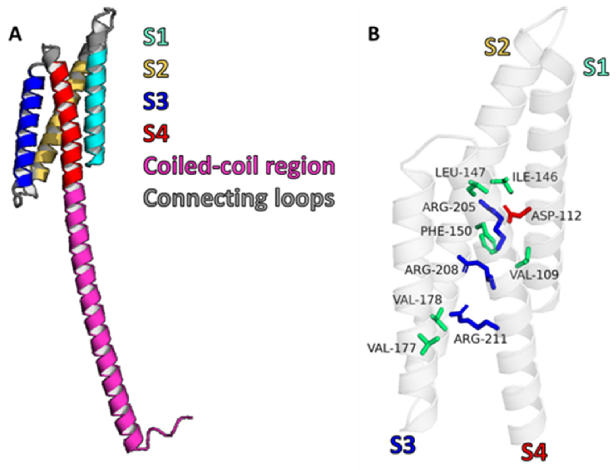
Figure 1. Structural features of the human Hv1 channel. (A) Cartoon representation of truncated hHv1 lacking the N-terminus. Transmembrane helices S1, S2, S3, and S4 are shown in cyan, yellow, blue, and red, respectively. The intracellular C-terminal coiled-coil region is depicted in pink, and connecting loops are shown in gray. (B) Transmembrane helices are displayed as a semi-transparent white cartoon. Residues forming the hydrophobic plug are highlighted as green sticks, the selectivity filter as red sticks, and voltage-sensing arginines as blue sticks. The monomeric AlphaFold-predicted structure AF-Q96D96-F1 was used for figure generation.
Pharmaceuticals 18 01480 g001
Figure 2. Schematic representation of how Hv1 inhibition is responsible for promoting cell death by preventing proton extrusion. (top) The normal mechanism for sustained ROS production or hypoxia elevates intracellular H+, and the action of Hv1 compensates for the accumulation of H+. (bottom) When Hv1 is inhibited, one consequence is that protons accumulate, resulting in a decrease in pHi. This inhibits the action of NOX2, which in turn reduces the production of ROS, maintaining the intracellular acidification that promotes cell death. Created in https://BioRender.com, accessed on 12 September 2025.
Pharmaceuticals 18 01480 g002
Figure 3. Binding sites of Hv1 modulators. The intermembrane region of the human Hv1 channel and its respective loops. The monomeric AlphaFold-predicted structure AF-Q96D96-F1 was used to generate the hHv1 dimer. Transmembrane helices S1, S2, S3, and S4 are shown as transparent cartoons, while residues that are involved in the binding of different Hv1 modulators are illustrated in stick representations.
Pharmaceuticals 18 01480 g003
Figure 4. Different modulation mechanisms of the Hv1 channel. The monomeric AlphaFold-predicted structure (AF-Q96D96-F1) was used to generate the hHv1 dimer embedded in a cell membrane. Hv1 channel modulators influence proton currents through direct or indirect mechanisms acting extracellularly, intracellularly, or within the membrane. Pointed arrows indicate activation pathways (blue and green), whereas flat-headed arrows indicate inhibition (red). AA, arachidonic acid.
Pharmaceuticals 18 01480 g004
Table 1. IC50 of different Hv1 modulators.
CompoundTypeIC50Effect on the ChannelReference
Peptide C6Peptide31 nMInhibitor (ec)[71]
OxopenchSmall molecule819 nMInhibitor[72]
Zn2+Cation1.9 µMInhibitor (ec)[3]
HanatoxinPeptide2 µMInhibitor (memb)[73]
FluoxetineSmall molecule2.1 µMInhibitor (ic)[74]
ChlorpromazineSmall molecule2.2 µMInhibitor (ic)[75]
AGAP/W38FPeptide2.5 µMInhibitor (ec)[76]
Gr1b/GsAF-lPeptide3.2 µMInhibitor (ec)[77]
Gr2c/GsAF-llPeptide3.6 µMInhibitor (ec)[77]
EpigallocatechinSmall molecule3.7 µMInhibitor (ic)[78]
Cd2+Cation5 µMInhibitor (ec)[79]
ImipramineSmall molecule5.7 µMInhibitor (ic)[74]
MitriptylineSmall molecule5.8 µMInhibitor (ic)[74]
HaloperidolSmall molecule8.4 µMInhibitor (ic)[75]
13Small molecule8.5 µMInhibitor (ic)[80]
ClozapineSmall molecule9.8 µMInhibitor (ic)[81]
DesipramineSmall molecule<10 µMInhibitor (ic)[74]
HIFSmall molecule26 µMInhibitor (ic)[82]
ClGBISmall molecule26.3 µMInhibitor (ic)[83]
2GBISmall molecule38 µMInhibitor (ic)[84]
NH17Small molecule>50 µMInhibitor (ic)[85]
PNX52429Small molecule>50 µMInhibitor[86]
PNX61442Small molecule50 µMInhibitor[86]
DextromethorphanSmall molecule51.7 µMInhibitor (ic)[87]
OlanzapineSmall molecule84 µMInhibitor (ic)[81]
CholesterolLipid~10% (wt/wt, to total membrane lipids)Inhibitor (memb)[88]
AlbuminProtein158 µMActivator (ec)[89]
Arachidonic acidLipid10-100 µMActivator (memb)[65]
NH29Small molecule50 µMActivator[85]
OPE (onion peel extract)Organic extract30 µg/mlActivator (ec)[90]
Disclaimer/Publisher’s Note: The statements, opinions and data contained in all publications are solely those of the individual author(s) and contributor(s) and not of MDPI and/or the editor(s). MDPI and/or the editor(s) disclaim responsibility for any injury to people or property resulting from any ideas, methods, instructions or products referred to in the content.