Article

Alleviation of Autophagic Deficits and Neuroinflammation by Histamine H3 Receptor Antagonist E159 Ameliorates Autism-Related Behaviors in BTBR Mice

by
Shilu Deepa Thomas
1,2,
Petrilla Jayaprakash
1,2,
Nurfirzana Z. H. J. Marwan
1,2,
Ezzatul A. B. A. Aziz
1,2,
Kamil Kuder
3,
Dorota Łażewska
3,
Katarzyna Kieć-Kononowicz
3 and
Bassem Sadek
1,2,*
1
Department of Pharmacology & Therapeutics, College of Medicine and Health Sciences, United Arab Emirates University, Al Ain P.O. Box 17666, United Arab Emirates
2
Zayed Center for Health Sciences, United Arab Emirates University, Al Ain P.O. Box 15551, United Arab Emirates
3
Department of Technology and Biotechnology of Drugs, Faculty of Pharmacy, Jagiellonian University Medical College, Medyczna Str. 9, 30-688 Kraków, Poland
*
Author to whom correspondence should be addressed.
Pharmaceuticals 2024, 17(10), 1293; https://doi.org/10.3390/ph17101293
Submission received: 9 August 2024 / Revised: 25 September 2024 / Accepted: 26 September 2024 / Published: 28 September 2024

Abstract

:
Background/Objectives: Autism spectrum disorder (ASD) is a neurodevelopmental condition marked by social interaction difficulties, repetitive behaviors, and immune dysregulation with elevated pro-inflammatory markers. Autophagic deficiency also contributes to social behavior deficits in ASD. Histamine H3 receptor (H3R) antagonism is a potential treatment strategy for brain disorders with features overlapping ASD, such as schizophrenia and Alzheimer’s disease. Methods: This study investigated the effects of sub-chronic systemic treatment with the H3R antagonist E159 on social deficits, repetitive behaviors, neuroinflammation, and autophagic disruption in male BTBR mice. Results: E159 (2.5, 5, and 10 mg/kg, i.p.) improved stereotypic repetitive behavior by reducing self-grooming time and enhancing spontaneous alternation in addition to attenuating social deficits. It also decreased pro-inflammatory cytokines in the cerebellum and hippocampus of treated BTBR mice. In BTBR mice, reduced expression of autophagy-related proteins LC3A/B and Beclin 1 was observed, which was elevated following treatment with E159, attenuating the disruption in autophagy. The co-administration with the H3R agonist MHA (10 mg/kg, i.p.) reversed these effects, highlighting the role of histaminergic neurotransmission in observed behavioral improvements. Conclusions: These preliminary findings suggest the therapeutic potential of H3R antagonists in targeting neuroinflammation and autophagic disruption to improve ASD-like behaviors.

Graphical Abstract

1. Introduction

Autism Spectrum Disorder (ASD) is characterized by challenges in social communication, and stereotypic and repetitive behaviors, affecting learning and development [1]. ASD frequently co-occurs with other conditions, such as depression, epilepsy, tics, attention-deficit hyperactivity disorder, sleep disorders, and gastrointestinal issues [2,3]. Despite its prevalence and impact, effective treatments for core symptoms are lacking, highlighting the need for more precise and efficient therapies [4].
Various factors such as oxidative stress, neuroimmune dysfunction, imbalances in excitatory and inhibitory neurotransmitters, and deficits in neurotrophic factors are implicated in the pathogenesis of ASD [5,6,7,8]. Inflammation and neuroimmune dysregulation are critical in ASD [6,9,10]. Exposure to inflammation during pregnancy leads to disruption of normal fetal neuronal development, resulting in irregular neuronal activity, social behavior changes, and poor cognitive performance in the offspring [11,12]. Neuroinflammation, closely linked to cognitive impairment, involves microglia, the brain’s immune cells, releasing proinflammatory cytokines that are harmful to neurons and perpetuate inflammation [13,14].
Autophagy is a cell process crucial for maintaining internal homeostasis by eliminating damaged proteins, which affects axonal maintenance, synaptic pruning, and neurogenesis. Furthermore, evidence suggests that autophagy can also modulate microglial activation [15]. Autophagy reduces inflammation and prevents cell death [16,17]. It is controlled by the mTOR complex, which is crucial for energy metabolism, protein synthesis, as well as cell growth [18]. According to a study by Lieberman et al. (2020), autophagy is downregulated during postnatal development following the upregulation of mTOR in the mice. They also found that during late postnatal development, impairments in autophagy are associated with deficiencies in synaptic transmission and social behavior [19]. Multiple lines of evidence show disruption in autophagic pathways in ASD animal models suggesting a potential role in the pathogenesis of ASD [20,21].
The histamine H3 receptor (H3R) is a constitutively active receptor primarily located in the brain, functioning as a presynaptic autoreceptor that inhibits histamine synthesis and release [22]. H3Rs also function as heteroreceptors, modulating the release of various neurotransmitters such as acetylcholine (Ach), gamma-aminobutyric acid (GABA), serotonin (5-HT), and dopamine (DA). Histamine has also been shown to influence behaviors in disorders that overlap with ASD, such as Alzheimer’s disease, schizophrenia, and Tourette’s. Multiple studies have connected histaminergic signaling through H3Rs to autism-like repetitive behaviors. This connection is supported by post-mortem brain analyses, genetic association studies, and animal models [23,24,25].
Preclinical and clinical studies suggest that antagonizing H3Rs can alleviate behavioral and cognitive symptoms in Alzheimer’s disease and schizophrenia, both of which share similar behavioral features with ASD, including cognitive impairment [26]. H3R antagonism improved performance in context discrimination tasks in aged mice [27]. It has also been shown to improve cognitive deficits induced by NMDA receptor antagonists like MK-801 and ketamine [28]. In an animal model of schizophrenia, an H3R antagonist improved behavioral deficits as well as spatial working memory [29], which are also observed in patients with ASD [30]. Furthermore, antagonizing H3R mitigated impairment in social behavior in animals exposed to phencyclidine, an outcome that may be relevant for ASD [31]. Notably, H3R antagonist ciproxifan was found to reduce stereotypies and sociability deficits in a rodent model of autism [23]. The non-imidazole-based H3R antagonist DL77 has been reported to exhibit promising effects in improving autism-like features induced by valproic acid in mice [32]. Additionally, dual-action H3R antagonist and acetylcholinesterase (AChE) inhibitor E100 reduced oxidative stress and proinflammatory cytokine levels, while also alleviating social impairments and repetitive stereotypical behaviors in C57BL/6 mice exposed to valproic acid [33]. Several H3R antagonists have shown promise in bringing behavioral improvements in ASD by targeting neuroinflammation [34]. The H3R antagonist ST713 reduced NF-κB activity and cytokine levels (TNF-α, IL-1, and IL-6) in BTBR mice, a model of autism [35]. These studies highlight the therapeutic potential of H3R antagonists in managing both cognitive and social impairments observed in ASD. Notably, H3R antagonism has been linked to enhanced autophagy via the PI3K/AKT/mTOR pathway, offering protection against ischemic injury [36].
The BTBR T+ Itpr3tf/J (BTBR) inbred mouse strain, presents ASD-related behaviors with reduced sociability and increased repetitive behaviors, like marble burying and self-grooming [37,38]. In the current study, the test compound E159 and its docking to human H3R were evaluated using in silico methods. Subsequently, we evaluated the impact of systemic sub-chronic treatment with E159, a potent and selective H3R antagonist with high in vitro specificity for H3Rs (Figure 1), on ASD-related behavioral dysfunctions in BTBR mice [39]. The H3R antagonist E159 was selected based on its previous significant procognitive effects in a model of memory deficit induced by dizocilpine in rodents [40]. Additionally, the effects of the sub-chronic treatment of E159 on neuroinflammation and autophagy in the cerebellum of treated mice were evaluated. Also, the effects of E159 on repetitive and social behavioral deficits were assessed in C57BL/6J (B6) which served as the control strain, displaying normal sociability and a low level of repetitive behaviors [41,42]. The H3R plays a key role in regulating the release of neurotransmitters, particularly histamine. (R)-α-methylhistamine (MHA) is a CNS-penetrant and selective H3R agonist [32]. To further explore the involvement of histaminergic signaling in the behavioral effects observed for H3R antagonist E159, we investigated whether the agonist MHA could counteract the positive effect of H3R antagonist E159. The abrogation study assessed whether sub-chronic systemic co-administration of the CNS-penetrant H3R agonist MHA could reverse the behavioral and biochemical improvements elicited by E159. This approach aimed to clarify the role of brain histaminergic neurotransmission in mediating the beneficial effects of E159.

2. Results

2.1. Docking Studies of E159

For in silico studies, receptor-ligand complexes for three histamine receptors, H1, H3, and H4, were chosen, which are represented by PBD structures 3RZE [43], 7F61 [44], and 7YFC [45], respectively. Docking to the H1R structure resulted in a putative calculated pose (dG Bind = −51.42 kcal/mol), where the protonated nitrogen formed a salt bridge with D1073.32 and cation-π with Y108333. The proton (at nitrogen) was directed toward TM6, and the whole ligand bent in a way that the distal aromatic group was parallel to TM5 with no additional interactions. On the other hand, docking to H4 structures resulted in low-scoring poses with dG Bind = 20.88 kcal/mol, which may explain the low affinity for both targets.
In the case of the H3R complex, the compound exhibited a relatively high binding free energy (dG Bind) of −91.43 kcal/mol. It occupied the H3R binding pocket in a manner similar to the re-solved complex ligand PF03654746, maintaining key interactions typical of histamine H3R antagonists/inverse agonists. These interactions included the formation of a salt bridge and a hydrogen bond between the protonated amine nitrogen and D1143.32. Additional ligand’s west-end stabilization through cation-π interactions with caging, aromatic sidechains of Y1153.33, and F3987.39 was also found, while piperidine 3-methyl substituent occupied the cavity formed by Y3746.51 and W4027.43 at the sides and F3987.36 at the top. The east-end aromatic substituent of E159 was positioned within the space enclosed by the aromatic features of Y189 (ECL2) above and Y912.61 & Y942.64 on the sides, further stabilizing the structure through either hydrogen bonding with the ether oxygen or π-π stacking interactions, respectively (Figure 2).
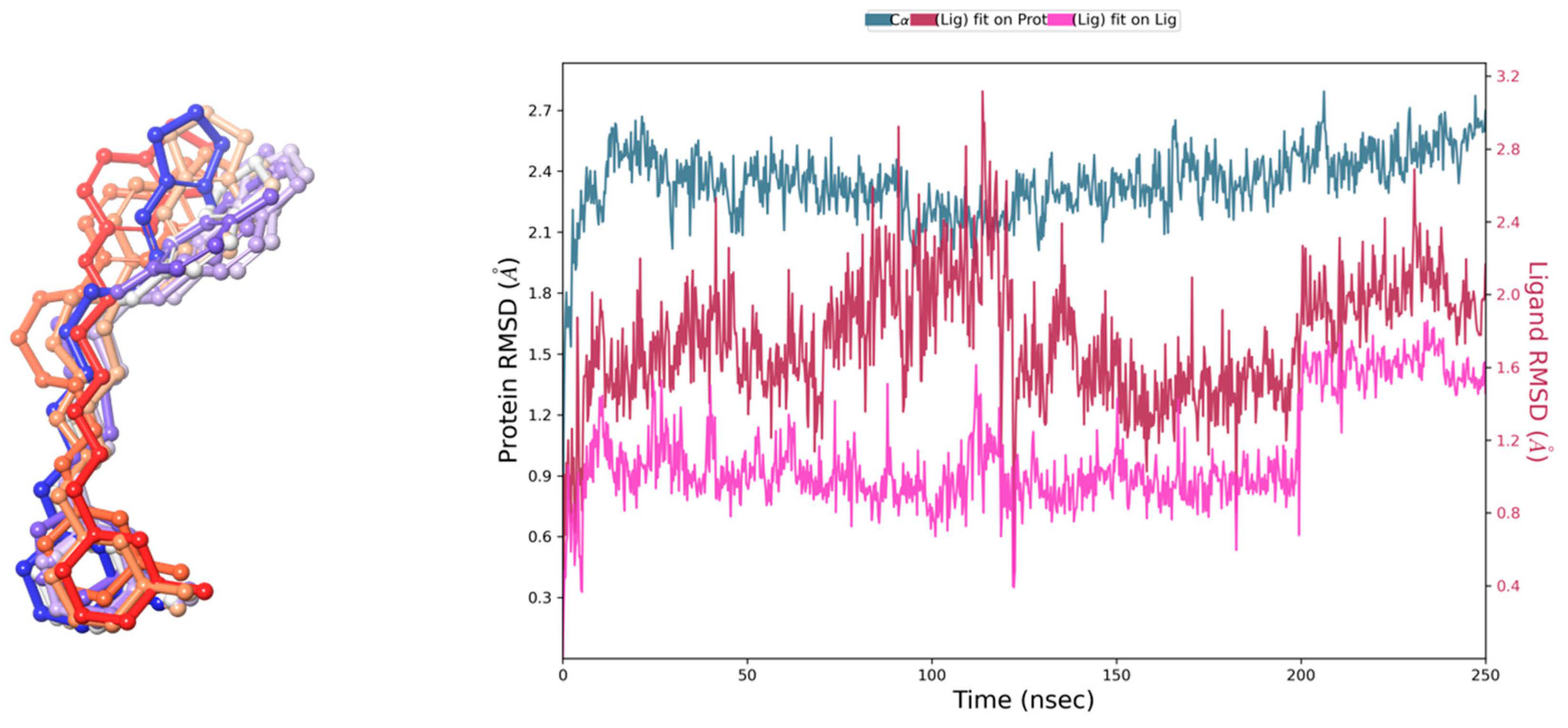
The stability of the calculated pose was assessed through 250 ns molecular dynamics (MD) simulations. Ten snapshots were selected from the simulation starting pose and after each 25 ns (Figure 3). Analysis of the ligand position during the simulation revealed that E159 remained stable throughout most of the recorded trajectory time, maintaining crucial interactions (Figure 2), and consistently engaging with a set of crucial amino acids, although the final conformation was slightly varied from its starting orientation Most of the interactions occur within the 2nd, 3rd and 7th helices. However, the highly flexible nature of the alkyl spacer and the distal aromatic group was also observed which resulted in fluctuations of RMSD value. Around 200 ns the aromatic feature moved closer to Y942.64, losing the support from the other tyrosine moiety (Y912.61) in favor of Y94, followed by a slight drop in complex binding free energy to the level of −80.66 kcal/mol. Yet, overall, the ligand displayed stable key interactions throughout the whole time of the recorded simulation, suggesting that the ligand might stabilize the inactive state of the receptor. Last but not least, the postulated H3R inactive state 3–7 lock between D1143.32 and W4027.43 [46] was observed through the whole recorded trajectory, which may give additional confirmation of the presumed stabilization of the inactive state of the receptor by the tested E159 ligand.

2.2. Effects of E159 on Stereotypical Repetitive Self-Grooming Behaviors in Mice

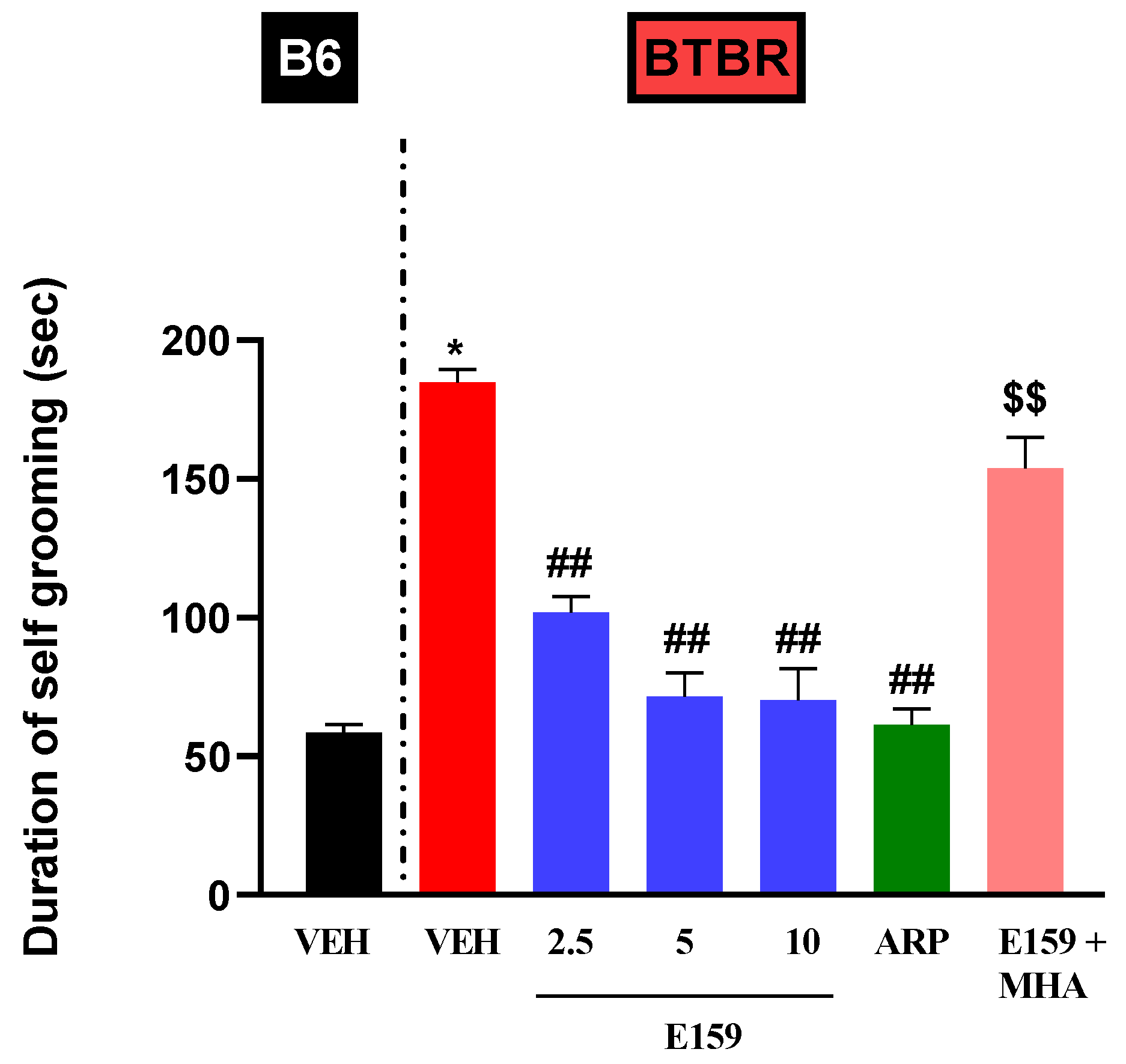
The impact of various treatments on mouse grooming duration is depicted in Figure 4. Statistical analyses showed substantial effects for strain, treatment, and their interaction (p < 0.001). The systemic pretreatments with E159 (2.5, 5, and 10 mg/kg) and ARP (1 mg/kg) did not alter the self-grooming behavior in B6 mice (Table 1). Statistical analyses demonstrated that VEH-treated BTBR mice (184.7 ± 4.752 s) spent considerably more time grooming than B6 mice (58.5 ± 2.89 s) (p < 0.001). The compound E159 at all the tested doses significantly reduced grooming duration (all p’s < 0.001) in autistic mice. ARP (1 mg/kg, i.p.) also displayed a considerable reduction in self-grooming duration in BTBR mice (p < 0.001). As shown in Figure 4, the reduction in self-grooming time induced by E159 (2.5 mg) was prevented upon co-treatment with MHA (10 mg/kg) (p < 0.01), relative to BTBR mice administered 2.5 mg of E159 alone (Figure 4).

2.3. Effects of E159 on Spontaneous Alternation in BTBR Mice

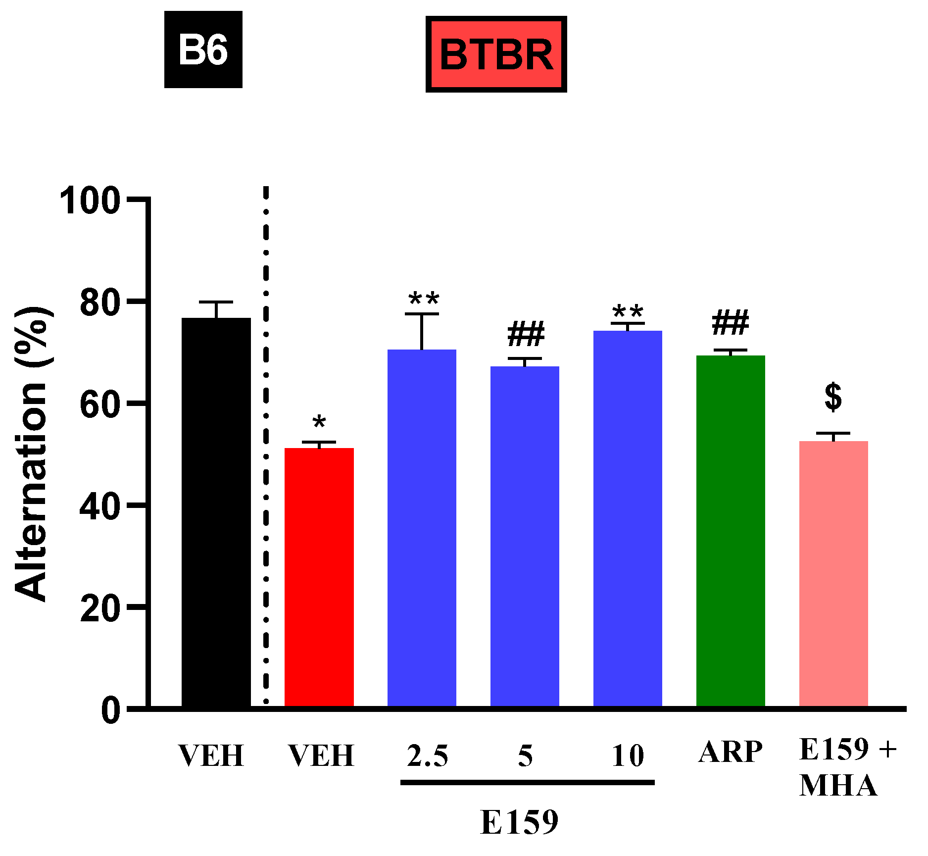
Statistical analyses revealed substantial main effects for strain (F(1,60) = 25.63, p < 0.001), treatment (F(5,60) = 5.30, p < 0.01), and their interaction (p < 0.01). The vehicle-treated BTBR animals showed considerably low spontaneous alternation relative to control B6 mice (p < 0.001) (Figure 5). Treatment with E159 (2.5, 5, and 10 mg/kg, i.p.) and ARP (1 mg/kg, i.p.) significantly increased the spontaneous alternation percentage in BTBR mice, with F-values of (F(1,10) = 7.12, p < 0.01), (F(1,10) = 63.8, p < 0.05), (F(1,10) = 130.96, p < 0.01) and (F(1,10) = 106.4, p < 0.05) respectively. Furthermore, no significant differences were found between the tested doses of E159 (all p > 0.05). In B6 mice, E159 (2.5, 5, and 10 mg/kg, i.p.) and ARP (1 mg/kg, i.p.) did not affect the spontaneous alternation percentage (Table 1). Moreover, the enhancement in alternation seen with E159 (2.5 mg) in autistic mice was completely negated by simultaneous administration with MHA (p < 0.01) in comparison with BTBR animals treated with E159 (2.5 mg) alone (Figure 5).

2.4. Effects of E159 on Sociability and Social Novelty Preference of BTBR Mice in the Three-Chambered Task

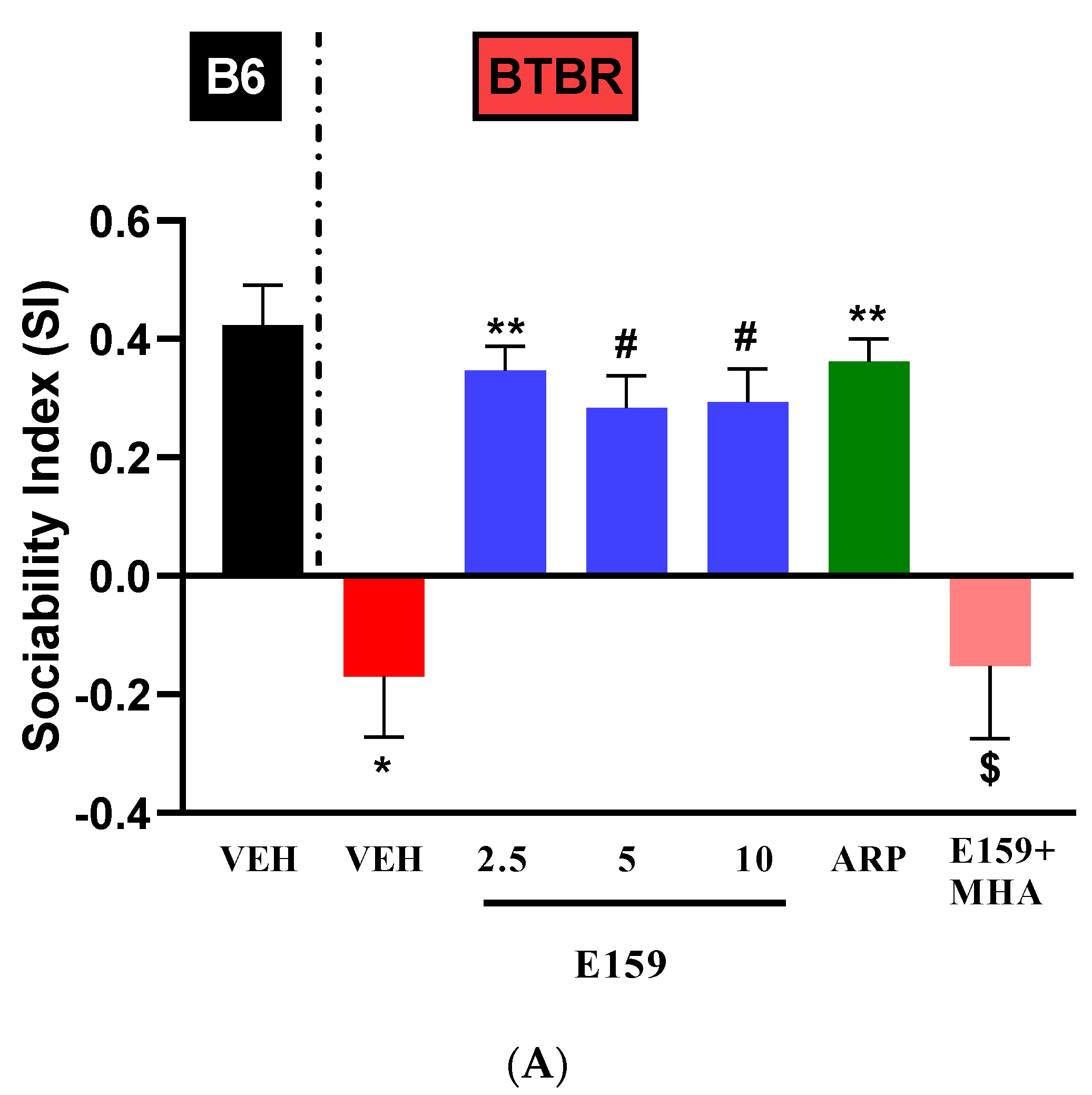
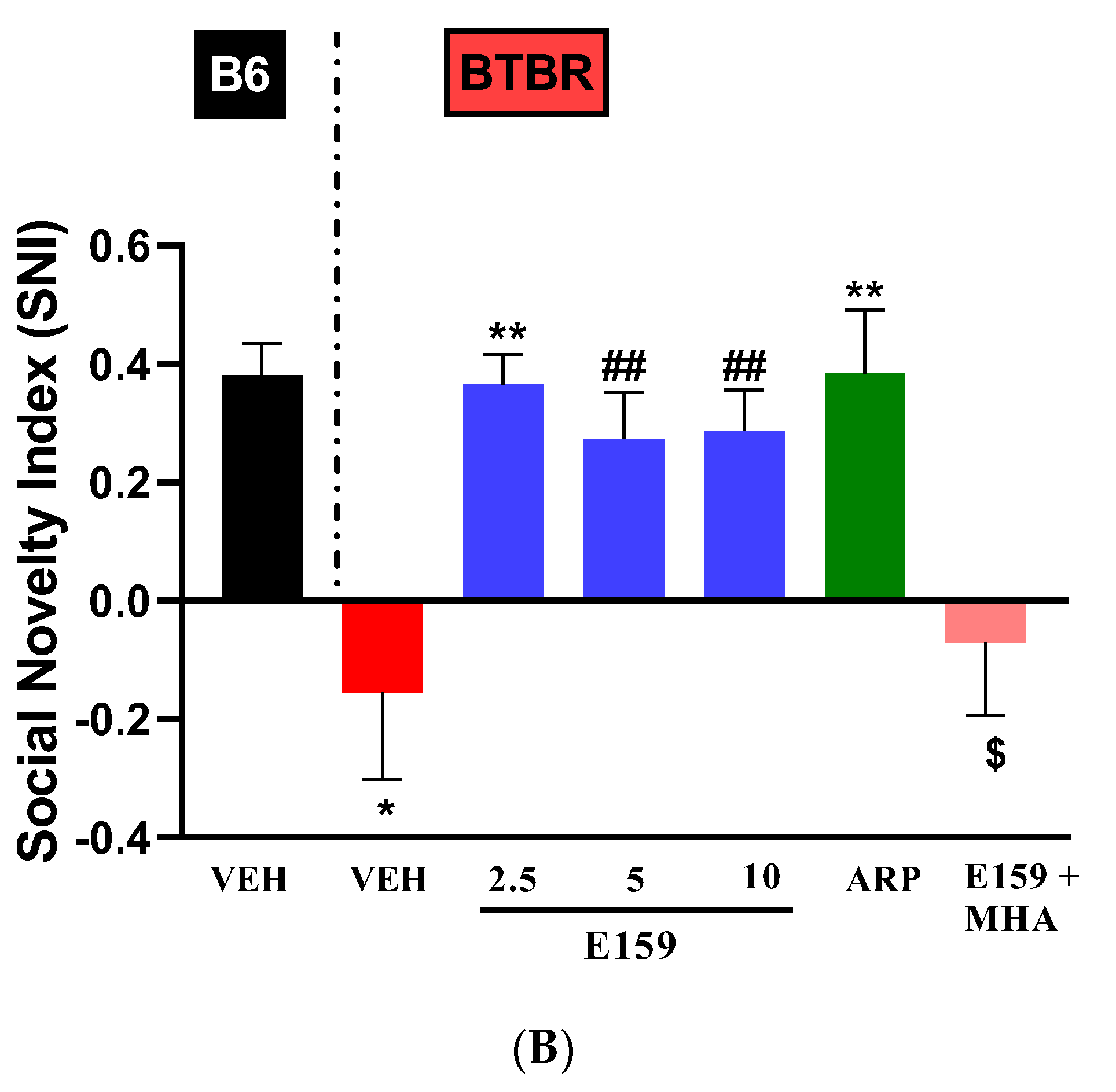
The effects of sub-chronic systemic administration of vehicle, E159 (2.5, 5, and 10 mg/kg, i.p.) and ARP (1 mg/kg, i.p.) on sociability deficits (SI) in BTBR mice in the three-chamber paradigm are depicted in Figure 6A. The data analyses exhibited a substantial impact for strain, treatment, and their interaction (p’s < 0.01). The BTBR mice exhibited a very low percentage of Sociability Index (SI) versus B6 mice (F(1,10) = 23.03, p < 0.001). E159 (2.5, 5, and 10 mg/kg) markedly improved the sociability of our autistic model (p < 0.01). ARP also considerably enhanced the sociability of BTBR mice (p < 0.01). The results also indicated that improvement in SI with E159 (2.5 mg/kg) was statistically comparable to that with ARP (p > 0.99). Additionally, the sociability-enhancing effects of E159 (2.5 mg) were nullified by co-treatment with MHA, H3R agonist (F(1,10) = 5.72, p < 0.05). Furthermore, systemic pretreatment with E159 (2.5, 5, and 10 mg/kg) or ARP (1 mg/kg) did not alter SI in B6 control mice in the three-chamber task (Table 1). Similarly, the effects of sub-chronic systemic administration of E159 (2.5, 5, and 10 mg/kg, i.p.) and ARP (1 mg/kg, i.p.) on social novelty preference were evaluated in BTBR mouse model (Figure 6B). In a two-way ANOVA, both strain and treatment had significant effects, along with a significant strain × treatment interaction (p < 0.05). A considerably lower percentage of Social Novelty Index (SNI) in BTBR animals relative to B6 mice (p < 0.01) was observed. E159 (at all tested doses) appreciably increased SNI versus vehicle administered autistic mice (p < 0.05) (Figure 6B). Similar to SI results, ARP also improved SNI in BTBR animals relative to BTBR animals that received vehicle (p < 0.01). Furthermore, effects of E159 (2.5 mg) on SNI were entirely abolished by co-treatment with MHA [(F(1,10) = 10.57, p < 0.01)]. Notably, systemic pretreatment with E159 (2.5, 5, and 10 mg/kg) or ARP (1 mg/kg) did not alter SNI in B6 mice (Table 1).

2.5. Impact of E159 on Anxiety and Locomotor Activity

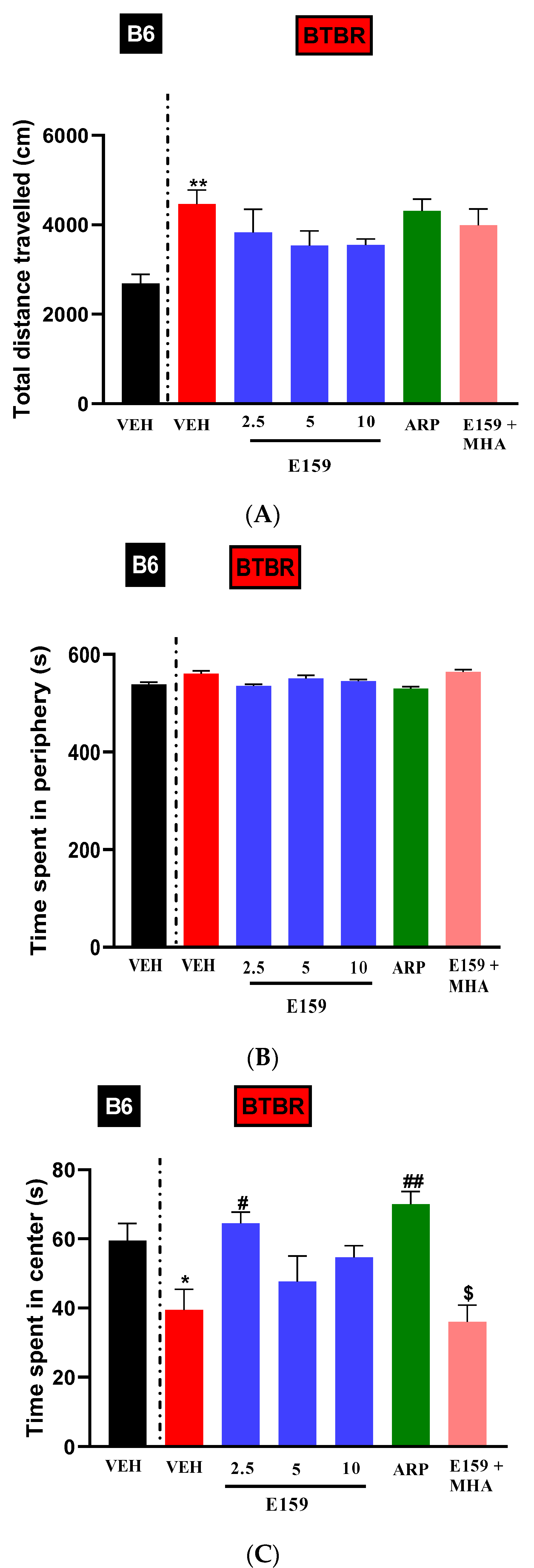
The open field test (OFT) evaluated locomotor ability and anxiety in both assessed strains (Figure 7). Two-way ANOVA results for the travelled distance exhibited substantial effect only for the strain (F(1,60) = 77.40, p < 0.001) (Figure 7A). The BTBR mice that received vehicle treatment travelled significantly more than the B6 mice that received the same treatment. (F(1,10) = 22.27, p < 0.01). Pretreatment with E159 (2.5, 5, and 10 mg/kg, i.p.) or ARP (1 mg/kg, i.p.) did not significantly alter the total distance travelled in either B6 or BTBR mice (Figure 7A, Table 1).
Figure 7B depicts the time spent in the periphery during the OFT following the systemic injections of the vehicle, E159 or ARP. There were no significant effects for strain and treatment (all p’s > 0.05), but significant effect was observed for strain × treatment interaction (F(5,60) = 2.581, p < 0.05). Pretreatment with E159 (2.5, 5, and 10 mg/kg, i.p.) or ARP (1 mg/kg, i.p.) did not significantly alter the time spent in periphery in either B6 or BTBR mice (Figure 7B, Table 1).
Figure 7C shows time spent in the central arena during the OFT after treatment with vehicle, E159, or ARP in B6 and BTBR animals. The two-way ANOVA revealed considerable effect for strain, treatment, and strain × treatment interaction (p < 0.05). Vehicle administered BTBR animals spent significantly reduced duration in the central zone versus B6 animals (F(1,10) = 6.59, p < 0.05). E159 (2.5 mg/kg) and ARP considerably improved the duration in the central arena by BTBR mice (p < 0.05), whereas E159 (5 and 10 mg/kg) did not produce significant effects (all p’s > 0.05). In B6 mice, time in the central arena was not altered by any of the treatments (Table 1). Notably, the E159 (2.5 mg)-induced increase in center time was decreased by simultaneous administration of MHA (F(1,10) = 23.69; p < 0.01).

2.6. Effects of E159 on the Level of Proinflammatory Cytokines in Cerebellum and Hippocampus of BTBR Mice

Statistical analyses showed that there was a considerable elevation in all three proinflammatory cytokines in autistic mice relative to B6 mice (p < 0.001). E159 at a dose of 2.5 mg/kg alleviated rise in proinflammatory cytokines in BTBR animals. The cytokines were measured in hippocampal and cerebellar tissues (Table 2). The cerebellar levels of cytokines showed considerable decline after systemic pretreatment with E159 (all p < 0.05). Similar decline in inflammatory response was also observed in hippocampus (p < 0.01) with reduced TNF-α, IL-6, and IL-1β. In addition, ARP notably reduced proinflammatory cytokines in BTBR animal strain (all p < 0.05). Additionally, the beneficial effects of E159 on cerebellar and hippocampal cytokines were prevented upon co-administration with (R)-α-methylhistamine (MHA) (p < 0.05) (Table 2).

2.7. Effect of E159 on Autophagy

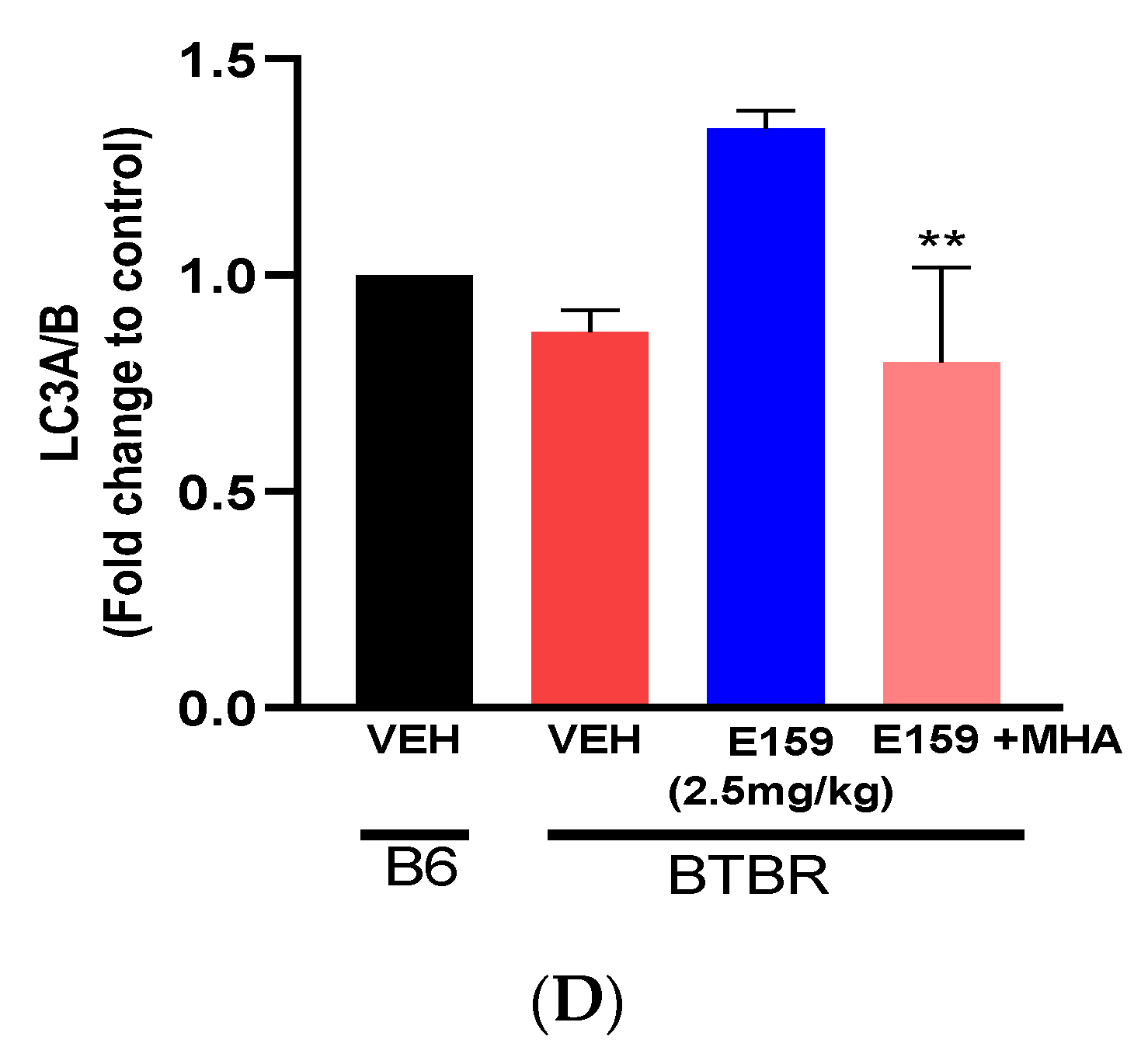
The impact of sub-chronic injections of the E159 (2.5 mg/kg) on the autophagic proteins in autistic BTBR strain is illustrated in Figure 8. The proteins mTOR, p-mTOR, LC3, and Beclin 1 in the cerebellum of BTBR mice were detected by Western blotting (Figure 8A). The mean protein expression in B6 mice was set as a fold change of 1 on graph. Densitometric analysis of cerebellar tissues from vehicle BTBR mice showed significantly elevated level of p-mTOR/ mTOR (Figure 8B) relative to B6 mice treated with vehicle (p < 0.05). Nevertheless, administration of E159 (2.5 mg/kg) significantly decreased p-mTOR in the cerebellum of E159 treated BTBR mice, which further indicates the inhibition of mTOR activity and activation of autophagy. This reduction in protein expression was reversed upon co-injection with H3R agonist MHA (10 mg/kg). Furthermore, a significant decline in levels of Beclin 1 and LC3 (A/B) in cerebellum of autistic strain relative to the B6 strain was observed, indicating impairment of autophagy (p < 0.05) (Figure 8C,D). The systemic sub-chronic treatment with E159 (2.5 mg/kg) provided elevation in the LC3 (A/B) and Beclin 1 levels in BTBR mice. Furthermore, the increase in the levels of LC3 (A/B) and Beclin 1 brought about by E159 (2.5 mg/kg) treatment, was reversed with the administration of MHA, an H3R agonist.

3. Discussion

ASD is characterized by deficits in sociability and repetitive behaviors, with histamine acting through H3 receptors (H3Rs) influencing functions like circadian rhythms and sensory sensitivity, which overlap with ASD symptoms [26,47]. Studies have linked histaminergic signaling via H3Rs to autism-like repetitive behaviors, supported by post-mortem brain analyses, genetic studies, and animal models. Disruptions in histaminergic signaling are also implicated in Tourette syndrome, a disorder associated with stereotypies similar to those in ASD [48,49]. H3R antagonists have shown promise in reducing ASD-like behaviors in several animal models, suggesting their potential therapeutic benefit [23,33].
BTBR mice serve as an idiopathic model of ASD due to their autism-like symptoms, such as repetitive behaviors and reduced sociability [41]. Studies suggest immunological changes in ASD models are linked to abnormal central nervous system development and ASD-like behaviors [50]. Altered immune profiles affect repetitive behaviors and social interactions in BTBR mice [51,52]. BTBR mice were selected for this study because they exhibit behavioral and inflammatory profiles similar to human ASD, including social deficits and abnormal self-grooming, unlike the highly sociable and normally grooming C57BL/6J (B6) mice [35,53]. This study evaluated the impact of the highly selective and potent H3R antagonist E159 on autism-like behaviors in BTBR mice and examined the effects of sub-chronic treatment with E159 on pro-inflammatory cytokines in cerebellum and hippocampus of treated mice. Also, the study aimed to investigate the effect of E159 on the expression of autophagic proteins LC3A/B and Beclin 1 in the cerebellum of treated mice.
In line with several previous studies, BTBR mice showed significant grooming compared to B6 mice [54]. Rapanelli et al. (2017) have reported that repetitive behaviors in mice arise as a result of brain histamine deficiency [49,55]. E159 (2.5, 5, 10 mg/kg, i.p.) treatment dose dependently ameliorated this repetitive stereotypic behavior in BTBR mice effectively, which was comparable to the effect of ARP (1 mg/kg, i.p). Spontaneous alternation or the Y-maze test relies on animals’ normal tendency to explore new environments. In addition to the assessment of repetitive behaviors, it also tests the spatial working memory in mice [56]. Animals with impaired memory repeatedly enter the previously explored arm, showing fewer spontaneous alternations. Similar to previous studies, BTBR mice demonstrated reduced alternation compared to B6 mice, reflecting attentional and cognition deficits [57]. Our results showed that E159 significantly improved spontaneous alternation in BTBR mice, which further confirms that E159 treatment can restore impaired short-term memory. This aligns with a previous study where the Y-maze test shows an augmented spontaneous-alternation rate following treatment with H3R antagonist thioperamide in a model of LPS-induced neuroinflammation [58]. The reference drug ARP also significantly reduced self-grooming as well as enhanced the percentage of alternation in BTBR mice, which is in accordance with several previous experimental studies [55,59,60].
Our findings also showed that E159 effectively improved social deficits in BTBR mice, specifically enhancing sociability and social novelty behaviors. Improvements in sociability and social novelty were most significant with the 2.5 mg/kg dose of E159, while effects at higher doses (5 and 10 mg/kg) were less pronounced. These results align with a previous study where H3R antagonism decreased phencyclidine-induced social behavior impairments in animals [31]. Additionally, the reference drug ARP improved sociability and social novelty in BTBR mice, with effects comparable to those of E159. To account for the potential confounding effects of general locomotor activity and exploratory behavior on the outcomes of other behavioral studies, an open-field test was also carried out in BTBR and B6 mice. BTBR mice consistently showed greater distances traveled compared to control mice. E159 did not impact movement in autistic mice. E159 treatment in control B6 mice showed no adverse effects, indicating it does not alter baseline anxiety or exploratory behaviors. E159 also improved the time BTBR mice spent in the central arena, indicating it can modulate anxiety-associated fear levels, but it did not impact hyperactivity as it had no effect on the total distance traveled.
In BTBR mice, 2.5 mg/kg of E159 caused a significantly greater improvement in sociability, and spontaneous alternation, as well as reduced repetitive grooming behavior compared to higher doses of 5 and 10 mg/kg. This indicates that the optimal behavioral improvements occurred at 2.5 mg/kg, while higher doses may have caused off-target effects. These findings are also consistent with a previous study of E159 where significant improvement in memory in a DIZ-induced amnesia model, was obtained with 2.5 mg/kg of E159 compared to higher doses of 5 and 10 mg/kg [40]. Conversely, all doses of E159 had no impact on the behavior of B6 animals, thereby ruling out any potential interfering effects of the treatment. Additionally, the improvement in behavioral impairment observed with E159 at 2.5 mg/kg was similar to those achieved with standard drug ARP. Therefore, an abrogation study using co-administration of the H3R agonist MHA was conducted with E159 at 2.5 mg/kg, as this was identified as the most optimal dose for minimizing any off-target effects that could occur with 5 or 10 mg/kg of E159. Notably, the co-administration of (R)-α-methylhistamine (MHA), an H3R agonist, nullified all beneficial effects on repetitive behavior, enhancement in alternation and social activity behavior exhibited by E159 (2.5 mg), strongly correlating the role of histaminergic neurotransmission in behavioral and cognitive improvements observed in BTBR mice following treatment with E159 (2.5 mg/kg, i.p.).
ASD patients often show motor coordination deficits and delayed motor skill development linked to cerebellar dysfunction. Temporal lobe abnormalities, particularly in the amygdala and hippocampus, also contribute to autism-like symptoms [3,61,62]. Immune dysregulation plays a critical role in ASD, with microglia activation leading to inflammation and immune dysfunction. Elevated pro-inflammatory cytokines are associated with ASD symptoms in both humans and BTBR mice. Children with ASD frequently exhibit abnormal immune responses, increased cytokine levels, and behavioral issues, such as learning deficits and stereotypical behaviors [11,63,64]. Excessive TNF-α production is a key factor in systemic inflammation and ASD development, suggesting that targeting inflammation could be a potential therapeutic approach for alleviating ASD symptoms [65,66].
Histamine itself can trigger the activation of microglia and inflammation, yet it has anti-inflammatory properties under stress-induced pathological conditions [67,68]. Several studies suggest that histamine interacts with its receptors on microglia and astrocytes, influencing their phenotypes and reducing neuroinflammation [69]. In a study, H3R antagonism was found to increase histamine release, activate H2 receptors, and trigger the PKA/cAMP/CREB pathway, reducing NF-κB/CBP interactions and shifting microglia from a pro-inflammatory M1 state to an anti-inflammatory M2 state, alleviating neuroinflammation [70]. Astrocytes, key regulators of brain homeostasis, can adopt pro-inflammatory (A1) or anti-inflammatory (A2) phenotypes. Thioperamide, an H3R antagonist, was shown to reduce inflammation by shifting astrocytes from A1 to A2 through CREB activation [58,70]. In our study, we observed a significant elevation of TNF-α, IL-6, and IL-1β in the cerebellum and hippocampus of BTBR mice. Consistent with the above-mentioned studies, H3R antagonist E159 (2.5 mg/kg) effectively reduced these proinflammatory markers, thereby inhibiting neuroinflammation in the brain. Moreover, ARP demonstrated a similar significant reduction in proinflammatory cytokines. Conversely, co-administration of E159 (2.5 mg) with the H3R agonist (R)-α-methylhistamine (MHA) increased pro-inflammatory cytokine levels, indicating that E159′s beneficial effects are mediated through interaction with central H3Rs, with brain histamine playing a role in its neuroprotective effects on ASD-like symptoms in BTBR mice (Table 2).
Neuronal autophagy plays a pivotal role in neuronal interaction, signaling, and development, and any disruption in this process can adversely impact memory formation, synaptic plasticity, and structural remodeling [71]. The appropriate axonal and dendritic growth is essential for maintaining neuronal equilibrium, with impaired organelles or proteins typically degraded to facilitate structural plasticity during development [72]. Consistent evidence underscores the significance of autophagy in dendritic, axonal, and synaptic development and maturation. Inhibition of autophagy disrupts the process leading to social behavior deficits and contributing to the development of ASD and other mental illnesses [73,74,75].
The cerebellum shows consistent abnormalities in individuals with ASD. Research suggests that in addition to motor coordination and balance, it plays a key role in cognitive functions such as executive function, working memory, and language—areas commonly impaired in those with ASD. This highlights the importance of cerebellar dysfunction in the condition [76]. Purkinje cells, the primary output cells of the cerebellum, play a key role in neurotransmission to the cortex. Research using animal models of ASD has demonstrated that dysfunction in Purkinje cells is linked to the behavioral abnormalities characteristic of ASD-like behaviors [77]. Early anatomical studies of postmortem ASD brain tissue revealed a significant reduction in Purkinje cell numbers in the lateral hemisphere. In cerebellar samples of individuals with ASD, abnormal activation of microglia and astrocytes, along with significant accumulation of monocytes and macrophages, is observed, particularly in the granular layer and white matter. These inflammatory changes are associated with notable histological abnormalities, including a reduction in Purkinje cells [77,78]. ASD patients also exhibit changes like abnormal cerebellar vermis size and overall cerebellar volume differences. Altered expression of GABA-related enzymes GAD65 and GAD67 in the cerebellum is well-documented in ASD. Reduced GAD65/67 levels in ASD impair inhibitory signaling, synaptic plasticity, and cerebellar computation, disrupting connections between the basket and Purkinje cells and affecting downstream targets like the deep cerebellar nuclei [79]. Functionally, impaired cerebellar development affects both motor and cognitive functions, with abnormalities linked to social interaction deficits [80]. The cerebellum is increasingly recognized as a key brain structure involved in the pathology of ASD; hence, we have focused on the cerebellum to explore the impact of E159 on autophagy within this region. Autophagy also acts as a protective mechanism against oxidative stress by regulating cellular ROS levels and removing damaged proteins and organelles [81,82]. It also suppresses the generation of inflammatory factors triggered by lipopolysaccharide by regulating innate immune signaling pathways and inflammasome activity. Earlier research indicates that activation of ROS and autophagy contribute to microglial activity [83]. Several upstream signals like PI3K-AKT, AMPK, and TSC1/2 regulate mTOR activity. A variety of genetic conditions are linked to ASD, including tuberous sclerosis complex, phosphatase and tensin homolog, hamartoma tumor syndrome, fragile X syndrome, and neurofibromatosis 1. Rodent models of these conditions have exhibited elevated mTORC1 activity in the brain, along with ASD-like behavioral deficits, which were reversed by treatment with the mTORC1 inhibitor rapamycin [84]. The AKT-mTOR pathway is an important signaling cascade associated with long-term plasticity and contributes to cognitive dysfunction. In addition to synaptic plasticity, mTOR has also been widely implicated in the inhibition of autophagy. Excessive activation of mTOR signaling relates to disruption in both glial and neuronal development, which is associated with the pathogenesis of ASD [85,86]. Several autophagy-related genes such as Beclin 1 are crucial in the process of autophagy [87]. Light chain3 (LC3) also serves as a significant indicator of autophagy [88]. A decrease in the levels of LC3 and Beclin1 leads to mitochondrial dysfunction and build-up of ROS, causing NLRP3 to generate IL-1β, escalating inflammation and neurotoxicity. This process ultimately results in neurodegeneration and neuronal death [89]. H3R activation can activate multiple intracellular signaling pathways, such as the PI3K/AKT pathway and mitogen-activated protein kinase (MAPK) pathway [90]. To further explore the impact of H3R antagonist E159, we analyzed the expression of autophagic markers, LC3A/B, and Beclin 1 in the cerebellum of BTBR mice. In this study, BTBR mice exhibited a reduction in the LC3A/B levels as well as decreased levels of Beclin1. These findings suggest impaired autophagy in the cerebellum. Also, cerebellar tissues from BTBR mice showed significantly elevated levels of p-mTOR/ mTOR compared to the B6 control strain. However, treatment with E159 (2.5 mg/kg) significantly decreased p-mTOR levels in the cerebellum of E159-administered BTBR animals, which indicates the inhibition of mTOR activity. This is consistent with a previous preclinical study where H3R antagonism inhibits phosphorylation of mTOR and reinforces autophagy [36]. Wang et al. (2019) explored the effects of LC1405, a novel H3R antagonist, on cognitive deficits caused by Aβ in a mouse model of Alzheimer’s disease (APP/PS1). The study revealed that LC1405 effectively slowed disease progression by improving memory and learning, while also preventing neurodegeneration [91]. These effects were mediated through H3R modulation of cAMP/CREB and PI3K/AKT /GSK3β signaling. H3R blockade by E159 could potentially modulate the PI3K/AKT pathway leading to reduced mTOR activity and increased autophagy. Future studies are required to investigate whether E159 exerts its effects by modulating these upstream pathways, which could provide a more comprehensive understanding of its mechanistic role in both autophagy regulation and the treatment of autism. Notably, the inhibition of mTOR and consequent increase in the level of LC3A/B and Beclin 1 was reduced when the mice were treated with MHA (10 mg/kg) in addition to E159 (2.5 mg/kg), which further advocates the role of brain histamine in the protective actions of H3R antagonist E159. These preliminary findings indicate that H3R antagonist E159, which maintains crucial interactions in docking studies at H3R, could play a role in regulating the mTOR signaling pathway associated with autophagy and improve autism-like symptoms.

4. Materials and Methods

4.1. Molecular Docking Studies

Schrodinger 2022-4 was employed for docking purposes, with ligands prepared in their ionized forms (protonated N4 piperazine nitrogen, +1 charge) [92]. ConfGen was used to generate bioactive conformations (water environment at physiological pH, targeting 20 conformers per ligand) [93]. Only five conformers with the lowest energy were chosen for docking experiments. Docking was performed using the standard protocol on a rigid receptor with a ligand-centered grid (cubical box of A, extra precision) [94]. To validate the docking approach, native ligands were re-docked with high accuracy. Induced-fit docking was conducted with the Glide IGD module. The putative binding energy of ligands (dG) was calculated using Prime MM-GBSA [95].
Molecular dynamics simulations for generated complexes, conducted for 250 ns at 300 K (pressure 1.01325 bar, ensemble class NPγT), were carried out in Desmond [96] with calculated docking pose constituting the starting point for simulation. The protein’s membrane orientation was sourced from the OPM database [97]. The simulation extended for 250 ns, utilizing the TIP3P [98] solvent and POPC membrane model, and generated 1000 frames. Selected frames were analyzed using the Simulation Interaction Diagram tool and visual assessment. The figures shown were performed using the Maestro Schrodinger package.

4.2. In Vivo Studies

4.2.1. Animals

Behavioral experiments were conducted using male BTBR T+Itpr3tf/J (BTBR) and C57BL/6J (B6) mice. At the start of the study, the BTBR mice were 8–10 weeks old and weighed between 27–32 g, and the B6 mice were 8–10 weeks old and weighed between 22–28 g. The animals were housed in the CMHS Animal facility at UAE University with a 12 h light/dark cycle with regulated temperature and humidity and unrestricted access to food and water. All necessary measures were taken to ensure the ethical treatment of animals during our study. All procedures were conducted following approval from the Institutional Animal Ethics Committee of UAE University (Approval No. ERA-2017-5603). The behavioral studies were conducted between 8:00 a.m. and 3:00 p.m. To minimize animal suffering, we used the fewest number of animals to achieve our study objectives. Only male mice were selected for the study to reduce within-group variability caused by hormonal changes during the estrogenic cycle in female mice.

4.2.2. Drug Compounds and Biochemical Materials

The test compound, E159 underwent development and in vitro pharmacological assessment at the Department of Technology and Biotechnology of Drugs (Kraków, Poland) [39]. E159 exhibits high and selective binding affinities to H3R compared to H4R and H1R. Aripiprazole (ARP, 1 mg/kg, i.p.), the reference drug, was procured from Sigma-Aldrich (St. Louis, MO, USA). Additionally, CNS-penetrant H3R agonist (R)-α-methylhistamine (MHA, 10 mg/kg) served for confirmatory studies following its sub-chronic systemic co-administration and was obtained from Sigma-Aldrich. The compounds were administered i.p. 30 min prior to the behavioral tests. Each animal received an injection adjusted to its body weight, with a volume of 10 mL/kg. Vehicle treatment consisted of 0.9% normal saline. The drug doses were selected according to prior studies [40,57]. Commercially available ELISA kits for proinflammatory cytokines (IL-1β, IL-6, and TNF-α) were procured from R&D Systems (Minneapolis, MN, USA). Bovine serum albumin (BSA), primary antibodies, and secondary antibodies were purchased from Cell Signaling, Danvers, MA, USA. PVDF membrane was sourced from Bio-Rad Laboratories (Hercules, CA, USA). The Pierce™ BCA Protein Assay Kit and Chemiluminescence Pico Kit were obtained from Thermo Fisher Scientific (Rockford, IL, USA).

4.2.3. Study Design and Treatment

Prior to the experiment, all mice were acclimated for a week. The treatment lasted for 21 days in a sub-chronic regimen. B6 mice used as the control group (group 1, n = 6) received VEH. BTBR mice injected with VEH (group 2, n = 6) functioned as the control group for ASD-related features. BTBR mice were administered with varying doses of E159 (2.5, 5 and 10 mg/kg, i.p.) (groups 3–5 respectively, n = 6). As the reference compound, Aripiprazole (ARP) was administered at a dosage of 1 mg/kg to BTBR mice (group 6, n = 6). For the abrogation studies, E159 (2.5 mg/kg) was co-administered with (R)-α-methylhistamine (MHA, 10 mg/kg, i.p.) (group 7) in BTBR mice. Besides the previously described groups, four additional groups of B6 mice (n = 6) were treated with E159 (2.5–10 mg/kg) and ARP to control for any potential confounding effects of these treatments on the behaviors of the control B6 mice (Table 1). The sub-chronic treatment (given i.p.) started a week prior to behavior experiments and continued until the sacrifice. On the last day of systemic treatment, after completing all behavioral tests, the animals were sacrificed. Their skulls were opened, and the brains were extracted. The hemispheres were separated, and the cerebellum and hippocampus were isolated and promptly frozen in liquid nitrogen for future biochemical analysis.

4.2.4. Behavioral Assessments

Self-Grooming

The assessment of self-grooming was carried out as described in previous studies [99,100]. Following a 10 min habituation period, the duration of grooming was measured for the second 10 min testing phase.

Y-Maze or Spontaneous Alteration

The test assesses rodents’ exploratory behavior when introduced to a novel environment [57,101]. It also tests the working memory function in mice [56]. The Y-maze test began with mice being placed into one arm of a Y-shaped maze. The test duration was 8 min. Three consecutive, non-repeating arm entries were considered correct spontaneous alternations (SAB). Over an 8 min period, the total movements made by the mice, as well as the percentage of alternations were then analyzed.

Three Chamber Social Test (TCT)

A TCT assessed sociability and social novelty in mice in accordance with previous reports [32,35,102]. The experiment used a three-chambered apparatus with a central chamber and two side chambers accessible through square doors. The test comprised a 30 min duration: a habituation phase, followed by sessions with novel and familiar mice placed in separate chambers. Social behavior was assessed using the Sociability Index (SI) and Social Novelty Index (SNI), based on the duration of exploring novel versus familiar mice.

Open Field Test (OFT)

The OFT was employed to assess the impact of treatments on both anxiety-like behaviors and locomotor activity in tested animals [32,103,104]. Briefly, the test began with a 5 min acclimatization period. The study measured both the total distance traveled throughout the arena and the duration spent in the central versus peripheral zones for ten min. Reduced time spent in the central area suggested anxiety-like behavior, whereas the total distance traveled reflected the animals’ overall locomotor activity [105].

4.3. Biochemical Investigations

4.3.1. Brain Collection and Tissue Processing

The cerebellum and hippocampus were stored at −80 °C for future experiments [35]. RIPA buffer consisting of protease inhibitors and phosphatase inhibitors was used to homogenize the tissues, followed by centrifugation at 12,000 rpm (4 °C, 30 min) for removal of tissue debris. The supernatants were separated and used later for the estimation of proinflammatory cytokines and Western blot analysis.

4.3.2. Pro-Inflammatory Cytokine Assessments

ELISA was used to quantify TNF-α, IL-1β, and IL-6 in the cerebellum and hippocampus, following the manufacturer’s guidelines [106,107].

4.3.3. Western Blot

The cerebellum homogenates were analyzed for protein concentration based on earlier reports [107]. Proteins separated on 12% gel were transferred to a PVDF membrane pre-activated with methanol using 90 V for 1 h 30 min. The membranes after blocking with 5% BSA (1 h, 4 °C) were incubated overnight with monoclonal antibodies against Actin, mTOR, p-mTOR, LC3A/B (all at 1:1000 dilution, Cell Signaling Technologies, Danvers, MA, USA), and Beclin1 (1:1000, Santa Cruz, CA, USA) at 4 °C. The following day after washing with TBST, the membranes were incubated with HRP-conjugated secondary antibodies for 3 h at 4 °C. The Super Signal West Pico PLUS Chemiluminescent Substrate (Thermo Scientific, Rockford, IL, USA) was used to visualize the protein bands and quantified with ImageJ software (Version 1.8.0) (NIH).

4.4. Statistical Analyses

Data were presented as mean ± SEM. A two-way ANOVA combined with Tukey’s post hoc test was employed to examine the effects of drug treatment. For proinflammatory cytokines and protein expressions, one-way ANOVA was used. Statistical analyses were conducted with GraphPad Prism (Version 8.0), and p-values below 0.05 denoted statistical significance (p < 0.05).

5. Conclusions

Brain histamine influences numerous functions, such as memory, long-term social recognition, cognition, learning, and emotions. In the current series of investigations, we observed that H3R antagonist E159 effectively alleviated ASD-related behaviors in BTBR mouse model of autism, specifically enhancing sociability and reducing repetitive behaviors, which are core symptoms of ASD. These improvements can be attributed to E159′s ability to modulate histaminergic neurotransmission as MHA, a centrally acting H3R agonist reversed these E159-provided effects. These behavioral improvements were observed simultaneously with a significant reduction of neuroinflammation and profound mitigation of deficits in autophagy. Our preliminary observations suggest that targeting H3Rs with antagonists like E159 could be a promising therapeutic approach for alleviating ASD-related symptoms.

Limitation

While we demonstrate that E159 ameliorates autism-like behaviors and enhances autophagy through modulation of the mTOR pathway, we did not extensively investigate the upstream signaling factors or related pathways influencing the regulation of autophagy. Secondly, our study focused on the effects of sub-chronic E159 treatment in BTBR mice, and we did not evaluate its long-term impact. This limits our understanding of the compound’s potential chronic effects, which warrants further investigation. Lastly, we did not evaluate the activation or inactivation of specific cerebellar cell types following treatment with test compound E159. Understanding the cell-specific functional responses of H3Rs would offer valuable insights into its modulating role in the cerebellum. Future studies employing techniques such as immunohistochemistry and electrophysiology will be necessary to address these gaps.

Author Contributions

B.S. was responsible for the study concept, design, acquisition, and analysis of animal data; S.D.T., N.Z.H.J.M., E.A.B.A.A. and P.J. conducted behavioral and biochemical experiments. K.K. conducted docking studies to histamine receptors; K.K.-K. and D.Ł. were responsible for the generation, synthesis, and pharmacological in vitro characterization of the H3R antagonist E159; B.S. and S.D.T. drafted the manuscript. All authors have read and agreed to the published version of the manuscript.

Funding

The Office of Graduate Studies and Research of UAE University is thanked for the support provided to B.S. with funds (12M099, 12R207, and 12M182). This study was also supported by statutory funds from the Jagiellonian University Medical College, Kraków, Poland (N42/DBS/000386 (DŁ)) and (N42/DBS/000385 (KK)).

Institutional Review Board Statement

All experiments were approved by the Institutional Animal Ethics Committee in CMHS /United Arab Emirates (Approval No. ERA-2017-5603).

Informed Consent Statement

Not applicable.

Data Availability Statement

Data is contained within the article.

Conflicts of Interest

The authors declare no conflict of interest.

References

  1. Thomas, S.D.; Jha, N.K.; Ojha, S.; Sadek, B. mTOR Signaling Disruption and Its Association with the Development of Autism Spectrum Disorder. Molecules 2023, 28, 1889. [Google Scholar] [CrossRef] [PubMed]
  2. Kumar, S.; Reynolds, K.; Ji, Y.; Gu, R.; Rai, S.; Zhou, C.J. Impaired Neurodevelopmental Pathways in Autism Spectrum Disorder: A Review of Signaling Mechanisms and Crosstalk. J. Neurodev. Disord. 2019, 11, 10. [Google Scholar] [CrossRef]
  3. Dana, H.; Tahtasakal, R.; Sener, E.F. Animal Models of Autism: A Perspective from Autophagy Mechanisms. J. Transl. Genet. Genom. 2020, 4, 251–262. [Google Scholar] [CrossRef]
  4. Jiang, P.; Zhou, L.; Zhao, L.; Fei, X.; Wang, Z.; Liu, T.; Tang, Y.; Li, D.; Gong, H.; Luo, Y.; et al. Puerarin Attenuates Valproate-Induced Features of ASD in Male Mice via Regulating Slc7a11-Dependent Ferroptosis. Neuropsychopharmacology 2024, 49, 497–507. [Google Scholar] [CrossRef] [PubMed]
  5. Bjorklund, G.; Saad, K.; Chirumbolo, S.; Kern, J.K.; Geier, D.A.; Geier, M.R.; Urbina, M.A. Immune Dysfunction and Neuroinflammation in Autism Spectrum Disorder. Acta Neurobiol. Exp. 2016, 76, 257–268. [Google Scholar] [CrossRef]
  6. Siniscalco, D.; Schultz, S.; Brigida, A.L.; Antonucci, N. Inflammation and Neuro-Immune Dysregulations in Autism Spectrum Disorders. Pharmaceuticals 2018, 11, 56. [Google Scholar] [CrossRef] [PubMed]
  7. Pangrazzi, L.; Balasco, L.; Bozzi, Y. Oxidative Stress and Immune System Dysfunction in Autism Spectrum Disorders. Int. J. Mol. Sci. 2020, 21, 3293. [Google Scholar] [CrossRef]
  8. Jiang, C.-C.; Lin, L.-S.; Long, S.; Ke, X.-Y.; Fukunaga, K.; Lu, Y.-M.; Han, F. Signalling Pathways in Autism Spectrum Disorder: Mechanisms and Therapeutic Implications. Signal Transduct. Target. Ther. 2022, 7, 229. [Google Scholar] [CrossRef]
  9. Eftekharian, M.M.; Ghafouri-Fard, S.; Noroozi, R.; Omrani, M.D.; Arsang-jang, S.; Ganji, M.; Gharzi, V.; Noroozi, H.; Komaki, A.; Mazdeh, M.; et al. Cytokine Profile in Autistic Patients. Cytokine 2018, 108, 120–126. [Google Scholar] [CrossRef] [PubMed]
  10. Usui, N.; Kobayashi, H.; Shimada, S. Neuroinflammation and Oxidative Stress in the Pathogenesis of Autism Spectrum Disorder. Int. J. Mol. Sci. 2023, 24, 5487. [Google Scholar] [CrossRef]
  11. Vargas, D.L.; Nascimbene, C.; Krishnan, C.; Zimmerman, A.W.; Pardo, C.A. Neuroglial Activation and Neuroinflammation in the Brain of Patients with Autism. Ann. Neurol. 2005, 57, 67–81. [Google Scholar] [CrossRef] [PubMed]
  12. Lee, G.A.; Lin, Y.-K.; Lai, J.-H.; Lo, Y.-C.; Yang, Y.-C.S.H.; Ye, S.-Y.; Lee, C.-J.; Wang, C.-C.; Chiang, Y.-H.; Tseng, S.-H. Maternal Immune Activation Causes Social Behavior Deficits and Hypomyelination in Male Rat Offspring with an Autism-Like Microbiota Profile. Brain Sci. 2021, 11, 1085. [Google Scholar] [CrossRef] [PubMed]
  13. Onore, C.; Yang, H.; Van de Water, J.; Ashwood, P. Dynamic Akt/mTOR Signaling in Children with Autism Spectrum Disorder. Front. Pediatr. 2017, 5, 43. [Google Scholar] [CrossRef] [PubMed]
  14. Cui, Y.; Yang, M.; Wang, Y.; Ren, J.; Lin, P.; Cui, C.; Song, J.; He, Q.; Hu, H.; Wang, K.; et al. Melatonin Prevents Diabetes-Associated Cognitive Dysfunction from Microglia-Mediated Neuroinflammation by Activating Autophagy via TLR4/Akt/mTOR Pathway. FASEB J. 2021, 35, e21485. [Google Scholar] [CrossRef]
  15. Han, H.-E.; Kim, T.-K.; Son, H.-J.; Park, W.J.; Han, P.-L. Activation of Autophagy Pathway Suppresses the Expression of iNOS, IL6 and Cell Death of LPS-Stimulated Microglia Cells. Biomol. Ther. 2013, 21, 21–28. [Google Scholar] [CrossRef] [PubMed]
  16. Wang, K. Autophagy and Apoptosis in Liver Injury. Cell Cycle 2015, 14, 1631–1642. [Google Scholar] [CrossRef]
  17. Matsuzawa-Ishimoto, Y.; Hwang, S.; Cadwell, K. Autophagy and Inflammation. Annu. Rev. Immunol. 2018, 36, 73–101. [Google Scholar] [CrossRef]
  18. Deng, Z.; Zhou, X.; Lu, J.-H.; Yue, Z. Autophagy Deficiency in Neurodevelopmental Disorders. Cell Biosci. 2021, 11, 214. [Google Scholar] [CrossRef]
  19. Lieberman, O.J.; Cartocci, V.; Pigulevskiy, I.; Molinari, M.; Carbonell, J.; Broseta, M.B.; Post, M.R.; Sulzer, D.; Borgkvist, A.; Santini, E. mTOR Suppresses Macroautophagy During Striatal Postnatal Development and Is Hyperactive in Mouse Models of Autism Spectrum Disorders. Front. Cell. Neurosci. 2020, 14, 70. [Google Scholar] [CrossRef]
  20. Huber, K.M.; Klann, E.; Costa-Mattioli, M.; Zukin, R.S. Dysregulation of Mammalian Target of Rapamycin Signaling in Mouse Models of Autism. J. Neurosci. 2015, 35, 13836–13842. [Google Scholar] [CrossRef]
  21. Agam, G.; Taylor, Z.; Vainer, E.; Golan, H.M. The Influence of Choline Treatment on Behavioral and Neurochemical Autistic-like Phenotype in Mthfr-Deficient Mice. Transl. Psychiatry 2020, 10, 316. [Google Scholar] [CrossRef] [PubMed]
  22. Eissa, N.; Sadeq, A.; Sasse, A.; Sadek, B. Role of Neuroinflammation in Autism Spectrum Disorder and the Emergence of Brain Histaminergic System. Lessons Also for BPSD? Front. Pharmacol. 2020, 11, 886. [Google Scholar] [CrossRef] [PubMed]
  23. Baronio, D.; Castro, K.; Gonchoroski, T.; de Melo, G.M.; Nunes, G.D.F.; Bambini-Junior, V.; Gottfried, C.; Riesgo, R. Effects of an H3R Antagonist on the Animal Model of Autism Induced by Prenatal Exposure to Valproic Acid. PLoS ONE 2015, 10, e0116363. [Google Scholar] [CrossRef] [PubMed]
  24. Wright, C.; Shin, J.H.; Rajpurohit, A.; Deep-Soboslay, A.; Collado-Torres, L.; Brandon, N.J.; Hyde, T.M.; Kleinman, J.E.; Jaffe, A.E.; Cross, A.J.; et al. Altered Expression of Histamine Signaling Genes in Autism Spectrum Disorder. Transl. Psychiatry 2017, 7, e1126. [Google Scholar] [CrossRef] [PubMed]
  25. Eissa, N.; Azimullah, S.; Jayaprakash, P.; Jayaraj, R.L.; Reiner, D.; Ojha, S.K.; Beiram, R.; Stark, H.; Łażewska, D.; Kieć-Kononowicz, K.; et al. The Dual-Active Histamine H3 Receptor Antagonist and Acetylcholine Esterase Inhibitor E100 Ameliorates Stereotyped Repetitive Behavior and Neuroinflammmation in Sodium Valproate Induced Autism in Mice. Chem. Biol. Interact. 2019, 312, 108775. [Google Scholar] [CrossRef] [PubMed]
  26. Sadek, B.; Saad, A.; Sadeq, A.; Jalal, F.; Stark, H. Histamine H3 Receptor as a Potential Target for Cognitive Symptoms in Neuropsychiatric Diseases. Behav. Brain Res. 2016, 312, 415–430. [Google Scholar] [CrossRef]
  27. Guilloux, J.-P.; Samuels, B.A.; Mendez-David, I.; Hu, A.; Levinstein, M.; Faye, C.; Mekiri, M.; Mocaer, E.; Gardier, A.M.; Hen, R.; et al. S 38093, a Histamine H3 Antagonist/Inverse Agonist, Promotes Hippocampal Neurogenesis and Improves Context Discrimination Task in Aged Mice. Sci. Rep. 2017, 7, 42946. [Google Scholar] [CrossRef]
  28. Eissa, N.; Khan, N.; Ojha, S.K.; Łazewska, D.; Kieć-Kononowicz, K.; Sadek, B. The Histamine H3 Receptor Antagonist DL77 Ameliorates MK801-Induced Memory Deficits in Rats. Front. Neurosci. 2018, 12, 42. [Google Scholar] [CrossRef]
  29. Brown, J.W.; Whitehead, C.A.; Basso, A.M.; Rueter, L.E.; Zhang, M. Preclinical Evaluation of Non-Imidazole Histamine H3 Receptor Antagonists in Comparison to Atypical Antipsychotics for the Treatment of Cognitive Deficits Associated with Schizophrenia. Int. J. Neuropsychopharmacol. 2013, 16, 889–904. [Google Scholar] [CrossRef]
  30. Zhang, M.; Jiao, J.; Hu, X.; Yang, P.; Huang, Y.; Situ, M.; Guo, K.; Cai, J.; Huang, Y. Exploring the Spatial Working Memory and Visual Perception in Children with Autism Spectrum Disorder and General Population with High Autism-like Traits. PLoS ONE 2020, 15, e0235552. [Google Scholar] [CrossRef]
  31. Griebel, G.; Pichat, P.; Pruniaux, M.-P.; Beeské, S.; Lopez-Grancha, M.; Genet, E.; Terranova, J.-P.; Castro, A.; Sánchez, J.A.; Black, M.; et al. SAR110894, a Potent Histamine H3-Receptor Antagonist, Displays Procognitive Effects in Rodents. Pharmacol. Biochem. Behav. 2012, 102, 203–214. [Google Scholar] [CrossRef] [PubMed]
  32. Eissa, N.; Jayaprakash, P.; Azimullah, S.; Ojha, S.K.; Al-Houqani, M.; Jalal, F.Y.; Łażewska, D.; Kieć-Kononowicz, K.; Sadek, B. The Histamine H3R Antagonist DL77 Attenuates Autistic Behaviors in a Prenatal Valproic Acid-Induced Mouse Model of Autism. Sci. Rep. 2018, 8, 13077. [Google Scholar] [CrossRef] [PubMed]
  33. Eissa, N.; Azimullah, S.; Jayaprakash, P.; Jayaraj, R.L.; Reiner, D.; Ojha, S.K.; Beiram, R.; Stark, H.; Łażewska, D.; Kieć-Kononowicz, K.; et al. The Dual-Active Histamine H3 Receptor Antagonist and Acetylcholine Esterase Inhibitor E100 Alleviates Autistic-Like Behaviors and Oxidative Stress in Valproic Acid Induced Autism in Mice. Int. J. Mol. Sci. 2020, 21, 3996. [Google Scholar] [CrossRef]
  34. Thomas, S.D.; Abdalla, S.; Eissa, N.; Akour, A.; Jha, N.K.; Ojha, S.; Sadek, B. Targeting Microglia in Neuroinflammation: H3 Receptor Antagonists as a Novel Therapeutic Approach for Alzheimer’s Disease, Parkinson’s Disease, and Autism Spectrum Disorder. Pharmaceuticals 2024, 17, 831. [Google Scholar] [CrossRef]
  35. Venkatachalam, K.; Eissa, N.; Awad, M.A.; Jayaprakash, P.; Zhong, S.; Stölting, F.; Stark, H.; Sadek, B. The Histamine H3R and Dopamine D2R/D3R Antagonist ST-713 Ameliorates Autism-like Behavioral Features in BTBR T+tf/J Mice by Multiple Actions. Biomed. Pharmacother. 2021, 138, 111517. [Google Scholar] [CrossRef]
  36. Yan, H.; Zhang, X.; Hu, W.; Ma, J.; Hou, W.; Zhang, X.; Wang, X.; Gao, J.; Shen, Y.; Lv, J.; et al. Histamine H3 Receptors Aggravate Cerebral Ischaemic Injury by Histamine-Independent Mechanisms. Nat. Commun. 2014, 5, 3334. [Google Scholar] [CrossRef]
  37. Meyza, K.Z.; Blanchard, D.C. The BTBR Mouse Model of Idiopathic Autism—Current View on Mechanisms. Neurosci. Biobehav. Rev. 2017, 76, 99–110. [Google Scholar] [CrossRef] [PubMed]
  38. Wu, H.; Zhao, G.; Liu, S.; Zhang, Q.; Wang, P.; Cao, Y.; Wu, L. Supplementation with Selenium Attenuates Autism-like Behaviors and Improves Oxidative Stress, Inflammation and Related Gene Expression in an Autism Disease Model. J. Nutr. Biochem. 2022, 107, 109034. [Google Scholar] [CrossRef]
  39. Łażewska, D.; Ligneau, X.; Schwartz, J.-C.; Schunack, W.; Stark, H.; Kieć-Kononowicz, K. Ether Derivatives of 3-Piperidinopropan-1-Ol as Non-Imidazole Histamine H3 Receptor Antagonists. Bioorg. Med. Chem. 2006, 14, 3522–3529. [Google Scholar] [CrossRef]
  40. Alachkar, A.; Łażewska, D.; Kieć-Kononowicz, K.; Sadek, B. The Histamine H3 Receptor Antagonist E159 Reverses Memory Deficits Induced by Dizocilpine in Passive Avoidance and Novel Object Recognition Paradigm in Rats. Front. Pharmacol. 2017, 8, 709. [Google Scholar] [CrossRef]
  41. McFarlane, H.G.; Kusek, G.K.; Yang, M.; Phoenix, J.L.; Bolivar, V.J.; Crawley, J.N. Autism-like Behavioral Phenotypes in BTBR T+tf/J Mice. Genes Brain Behav. 2008, 7, 152–163. [Google Scholar] [CrossRef]
  42. Amodeo, D.A.; Jones, J.H.; Sweeney, J.A.; Ragozzino, M.E. Differences in BTBR T+ Tf/J and C57BL/6J Mice on Probabilistic Reversal Learning and Stereotyped Behaviors. Behav. Brain Res. 2012, 227, 64–72. [Google Scholar] [CrossRef]
  43. Shimamura, T.; Shiroishi, M.; Weyand, S.; Tsujimoto, H.; Winter, G.; Katritch, V.; Abagyan, R.; Cherezov, V.; Liu, W.; Han, G.W.; et al. Structure of the Human Histamine H1 Receptor Complex with Doxepin. Nature 2011, 475, 65–70. [Google Scholar] [CrossRef]
  44. Peng, X.; Yang, L.; Liu, Z.; Lou, S.; Mei, S.; Li, M.; Chen, Z.; Zhang, H. Structural Basis for Recognition of Antihistamine Drug by Human Histamine Receptor. Nat. Commun. 2022, 13, 6105. [Google Scholar] [CrossRef]
  45. Im, D.; Kishikawa, J.; Shiimura, Y.; Hisano, H.; Ito, A.; Fujita-Fujiharu, Y.; Sugita, Y.; Noda, T.; Kato, T.; Asada, H.; et al. Structural Insights into the Agonists Binding and Receptor Selectivity of Human Histamine H4 Receptor. Nat. Commun. 2023, 14, 6538. [Google Scholar] [CrossRef] [PubMed]
  46. Jończyk, J.; Malawska, B.; Bajda, M. Hybrid Approach to Structure Modeling of the Histamine H3 Receptor: Multi-Level Assessment as a Tool for Model Verification. PLoS ONE 2017, 12, e0186108. [Google Scholar] [CrossRef]
  47. Kas, M.J.; Glennon, J.C.; Buitelaar, J.; Ey, E.; Biemans, B.; Crawley, J.; Ring, R.H.; Lajonchere, C.; Esclassan, F.; Talpos, J.; et al. Assessing Behavioural and Cognitive Domains of Autism Spectrum Disorders in Rodents: Current Status and Future Perspectives. Psychopharmacology 2014, 231, 1125–1146. [Google Scholar] [CrossRef]
  48. Karagiannidis, I.; Dehning, S.; Sandor, P.; Tarnok, Z.; Rizzo, R.; Wolanczyk, T.; Madruga-Garrido, M.; Hebebrand, J.; Nöthen, M.M.; Lehmkuhl, G.; et al. Support of the Histaminergic Hypothesis in Tourette Syndrome: Association of the Histamine Decarboxylase Gene in a Large Sample of Families. J. Med. Genet. 2013, 50, 760–764. [Google Scholar] [CrossRef]
  49. Rapanelli, M.; Frick, L.; Pogorelov, V.; Ohtsu, H.; Bito, H.; Pittenger, C. Histamine H3R Receptor Activation in the Dorsal Striatum Triggers Stereotypies in a Mouse Model of Tic Disorders. Transl. Psychiatry 2017, 7, e1013. [Google Scholar] [CrossRef]
  50. Estes, M.L.; McAllister, A.K. Immune Mediators in the Brain and Peripheral Tissues in Autism Spectrum Disorder. Nat. Rev. Neurosci. 2015, 16, 469–486. [Google Scholar] [CrossRef]
  51. Onore, C.E.; Careaga, M.; Babineau, B.A.; Schwartzer, J.J.; Berman, R.F.; Ashwood, P. Inflammatory Macrophage Phenotype in BTBR T+tf/J Mice. Front. Neurosci. 2013, 7, 158. [Google Scholar] [CrossRef] [PubMed]
  52. Ahmad, S.F.; Ansari, M.A.; Nadeem, A.; Bakheet, S.A.; Alshammari, M.A.; Khan, M.R.; Alsaad, A.M.S.; Attia, S.M. S3I-201, a Selective Stat3 Inhibitor, Restores Neuroimmune Function through Upregulation of Treg Signaling in Autistic BTBR T+ Itpr3tf/J Mice. Cell. Signal. 2018, 52, 127–136. [Google Scholar] [CrossRef] [PubMed]
  53. McTighe, S.M.; Neal, S.J.; Lin, Q.; Hughes, Z.A.; Smith, D.G. The BTBR Mouse Model of Autism Spectrum Disorders Has Learning and Attentional Impairments and Alterations in Acetylcholine and Kynurenic Acid in Prefrontal Cortex. PLoS ONE 2013, 8, e62189. [Google Scholar] [CrossRef] [PubMed]
  54. Silverman, J.L.; Tolu, S.S.; Barkan, C.L.; Crawley, J.N. Repetitive Self-Grooming Behavior in the BTBR Mouse Model of Autism Is Blocked by the mGluR5 Antagonist MPEP. Neuropsychopharmacology 2010, 35, 976–989. [Google Scholar] [CrossRef]
  55. Choi, H.J.; Im, S.J.; Park, H.R.; Park, S.; Kim, C.-E.; Ryu, S. Long-Term Effects of Aripiprazole Treatment during Adolescence on Cognitive Function and Dopamine D2 Receptor Expression in Neurodevelopmentally Normal Rats. Clin. Psychopharmacol. Neurosci. 2019, 17, 400–408. [Google Scholar] [CrossRef]
  56. Kraeuter, A.-K.; Guest, P.C.; Sarnyai, Z. The Y-Maze for Assessment of Spatial Working and Reference Memory in Mice. Methods Mol. Biol. 2019, 1916, 105–111. [Google Scholar] [CrossRef]
  57. Eissa, N.; Venkatachalam, K.; Jayaprakash, P.; Falkenstein, M.; Dubiel, M.; Frank, A.; Reiner-Link, D.; Stark, H.; Sadek, B. The Multi-Targeting Ligand ST-2223 with Histamine H3 Receptor and Dopamine D2/D3 Receptor Antagonist Properties Mitigates Autism-Like Repetitive Behaviors and Brain Oxidative Stress in Mice. Int. J. Mol. Sci. 2021, 22, 1947. [Google Scholar] [CrossRef]
  58. Wang, J.; Liu, B.; Xu, Y.; Luan, H.; Wang, C.; Yang, M.; Zhao, R.; Song, M.; Liu, J.; Sun, L.; et al. Thioperamide Attenuates Neuroinflammation and Cognitive Impairments in Alzheimer’s Disease via Inhibiting Gliosis. Exp. Neurol. 2022, 347, 113870. [Google Scholar] [CrossRef] [PubMed]
  59. Shin, C.Y.; Kim, H.-S.; Cha, K.-H.; Won, D.H.; Lee, J.-Y.; Jang, S.W.; Sohn, U.D. The Effects of Donepezil, an Acetylcholinesterase Inhibitor, on Impaired Learning and Memory in Rodents. Biomol. Ther. 2018, 26, 274–281. [Google Scholar] [CrossRef] [PubMed]
  60. Lamothe, H.; Schreiweis, C.; Mondragón-González, L.S.; Rebbah, S.; Lavielle, O.; Mallet, L.; Burguière, E. The Sapap3−/− Mouse Reconsidered as a Comorbid Model Expressing a Spectrum of Pathological Repetitive Behaviours. Transl. Psychiatry 2023, 13, 26. [Google Scholar] [CrossRef]
  61. Mostofsky, S.H.; Goldberg, M.C.; Landa, R.J.; Denckla, M.B. Evidence for a Deficit in Procedural Learning in Children and Adolescents with Autism: Implications for Cerebellar Contribution. J. Int. Neuropsychol. Soc. 2000, 6, 752–759. [Google Scholar] [CrossRef] [PubMed]
  62. Marko, M.K.; Crocetti, D.; Hulst, T.; Donchin, O.; Shadmehr, R.; Mostofsky, S.H. Behavioural and Neural Basis of Anomalous Motor Learning in Children with Autism. Brain 2015, 138, 784–797. [Google Scholar] [CrossRef] [PubMed]
  63. Ashwood, P.; Krakowiak, P.; Hertz-Picciotto, I.; Hansen, R.; Pessah, I.; Van de Water, J. Elevated Plasma Cytokines in Autism Spectrum Disorders Provide Evidence of Immune Dysfunction and Are Associated with Impaired Behavioral Outcome. Brain Behav. Immun. 2011, 25, 40–45. [Google Scholar] [CrossRef] [PubMed]
  64. Matta, S.M.; Hill-Yardin, E.L.; Crack, P.J. The Influence of Neuroinflammation in Autism Spectrum Disorder. Brain Behav. Immun. 2019, 79, 75–90. [Google Scholar] [CrossRef]
  65. Li, X.; Chauhan, A.; Sheikh, A.M.; Patil, S.; Chauhan, V.; Li, X.-M.; Ji, L.; Brown, T.; Malik, M. Elevated Immune Response in the Brain of Autistic Patients. J. Neuroimmunol. 2009, 207, 111–116. [Google Scholar] [CrossRef]
  66. Goines, P.E.; Ashwood, P. Cytokine Dysregulation in Autism Spectrum Disorders (ASD): Possible Role of the Environment. Neurotoxicol. Teratol. 2013, 36, 67–81. [Google Scholar] [CrossRef]
  67. Barata-Antunes, S.; Cristóvão, A.C.; Pires, J.; Rocha, S.M.; Bernardino, L. Dual Role of Hista mine on Microglia-Induced Neurodegeneration. Biochim. Biophys. Acta (BBA)-Mol. Basis Dis. 2017, 1863, 764–769. [Google Scholar] [CrossRef]
  68. Saraiva, C.; Barata-Antunes, S.; Santos, T.; Ferreiro, E.; Cristóvão, A.C.; Serra-Almeida, C.; Ferreira, R.; Bernardino, L. Histamine Modulates Hippocampal Inflammation and Neurogenesis in Adult Mice. Sci. Rep. 2019, 9, 8384. [Google Scholar] [CrossRef]
  69. Zhou, Z.; An, Q.; Zhang, W.; Li, Y.; Zhang, Q.; Yan, H. Histamine and Receptors in Neuroinflammation: Their Roles on Neurodegenerative Diseases. Behav. Brain Res. 2024, 465, 114964. [Google Scholar] [CrossRef]
  70. Wang, J.; Liu, B.; Sun, F.; Xu, Y.; Luan, H.; Yang, M.; Wang, C.; Zhang, T.; Zhou, Z.; Yan, H. Histamine H3R Antagonist Counteracts the Impaired Hippocampal Neurogenesis in Lipopolysaccharide-Induced Neuroinflammation. Int. Immunopharmacol. 2022, 110, 109045. [Google Scholar] [CrossRef]
  71. Rubinsztein, D.C.; Codogno, P.; Levine, B. Autophagy Modulation as a Potential Therapeutic Target for Diverse Diseases. Nat. Rev. Drug Discov. 2012, 11, 709–730. [Google Scholar] [CrossRef]
  72. Nixon, R.A.; Cataldo, A.M. Lysosomal System Pathways: Genes to Neurodegeneration in Alzheimer’s Disease. J. Alzheimers Dis. 2006, 9, 277–289. [Google Scholar] [CrossRef] [PubMed]
  73. Bowling, H.; Klann, E. Shaping Dendritic Spines in Autism Spectrum Disorder: mTORC1-Dependent Macroautophagy. Neuron 2014, 83, 994–996. [Google Scholar] [CrossRef] [PubMed]
  74. Tang, G.; Gudsnuk, K.; Kuo, S.-H.; Cotrina, M.L.; Rosoklija, G.; Sosunov, A.; Sonders, M.S.; Kanter, E.; Castagna, C.; Yamamoto, A.; et al. Loss of mTOR-Dependent Macroautophagy Causes Autistic-like Synaptic Pruning Deficits. Neuron 2014, 83, 1131–1143. [Google Scholar] [CrossRef] [PubMed]
  75. Zhang, J.; Zhang, J.-X.; Zhang, Q.-L. PI3K/AKT/mTOR-Mediated Autophagy in the Development of Autism Spectrum Disorder. Brain Res. Bull. 2016, 125, 152–158. [Google Scholar] [CrossRef] [PubMed]
  76. Biswas, M.S.; Roy, S.K.; Hasan, R.; Pk, M.M.U. The Crucial Role of the Cerebellum in Autism Spectrum Disorder: Neuroimaging, Neurobiological, and Anatomical Insights. Health Sci. Rep. 2024, 7, e2233. [Google Scholar] [CrossRef]
  77. Zhou, L.; Jiang, P.; Zhao, L.; Fei, X.; Tang, Y.; Luo, Y.; Gong, H.; Wang, X.; Li, X.; Li, S.; et al. Ligustilide Inhibits Purkinje Cell Ferritinophagy via the ULK1/NCOA4 Pathway to Attenuate Valproic Acid-Induced Autistic Features. Phytomedicine 2024, 126, 155443. [Google Scholar] [CrossRef]
  78. Mapelli, L.; Soda, T.; D’Angelo, E.; Prestori, F. The Cerebellar Involvement in Autism Spectrum Disorders: From the Social Brain to Mouse Models. Int. J. Mol. Sci. 2022, 23, 3894. [Google Scholar] [CrossRef]
  79. Sydnor, L.M.; Aldinger, K.A. Structure, Function, and Genetics of the Cerebellum in Autism. J. Psychiatry Brain Sci. 2022, 7, e220008. [Google Scholar] [CrossRef]
  80. Su, L.-D.; Xu, F.-X.; Wang, X.-T.; Cai, X.-Y.; Shen, Y. Cerebellar Dysfunction, Cerebro-Cerebellar Connectivity and Autism Spectrum Disorders. Neuroscience 2021, 462, 320–327. [Google Scholar] [CrossRef]
  81. Giordano, S.; Darley-Usmar, V.; Zhang, J. Autophagy as an Essential Cellular Antioxidant Pathway in Neurodegenerative Disease. Redox Biol. 2014, 2, 82–90. [Google Scholar] [CrossRef] [PubMed]
  82. Menezo, Y.J.R.; Elder, K.; Dale, B. Link Between Increased Prevalence of Autism Spectrum Disorder Syndromes and Oxidative Stress, DNA Methylation, and Imprinting: The Impact of the Environment. JAMA Pediatr. 2015, 169, 1066–1067. [Google Scholar] [CrossRef]
  83. He, Y.; She, H.; Zhang, T.; Xu, H.; Cheng, L.; Yepes, M.; Zhao, Y.; Mao, Z. P38 MAPK Inhibits Autophagy and Promotes Microglial Inflammatory Responses by Phosphorylating ULK1. J. Cell Biol. 2018, 217, 315–328. [Google Scholar] [CrossRef]
  84. Sato, A.; Ikeda, K. Genetic and Environmental Contributions to Autism Spectrum Disorder Through Mechanistic Target of Rapamycin. Biol. Psychiatry Glob. Open Sci. 2022, 2, 95–105. [Google Scholar] [CrossRef] [PubMed]
  85. Kaeberlein, M. mTOR Inhibition: From Aging to Autism and Beyond. Scientifica 2013, 2013, 849186. [Google Scholar] [CrossRef]
  86. Takei, N.; Nawa, H. mTOR Signaling and Its Roles in Normal and Abnormal Brain Development. Front. Mol. Neurosci. 2014, 7, 28. [Google Scholar] [CrossRef]
  87. Kang, R.; Zeh, H.J.; Lotze, M.T.; Tang, D. The Beclin 1 Network Regulates Autophagy and Apoptosis. Cell Death Differ. 2011, 18, 571–580. [Google Scholar] [CrossRef]
  88. Qin, L.; Dai, X.; Yin, Y. Valproic Acid Exposure Sequentially Activates Wnt and mTOR Pathways in Rats. Mol. Cell. Neurosci. 2016, 75, 27–35. [Google Scholar] [CrossRef] [PubMed]
  89. Chen, X.; Zhou, X.; Cheng, X.; Lin, L.; Wang, Q.; Zhan, R.; Wu, Q.; Liu, S. Protective Effect of Ferulic Acid on Lipopolysaccharide-Induced BV2 Microglia Inflammation via AMPK/mTOR Signaling Pathway. Molecules 2023, 28, 3482. [Google Scholar] [CrossRef]
  90. Bongers, G.; Bakker, R.A.; Leurs, R. Molecular Aspects of the Histamine H3 Receptor. Biochem. Pharmacol. 2007, 73, 1195–1204. [Google Scholar] [CrossRef]
  91. Wang, L.; Fang, J.; Jiang, H.; Wang, Q.; Xue, S.; Li, Z.; Liu, R. 7-Pyrrolidinethoxy-4′-Methoxyisoflavone Prevents Amyloid β–Induced Injury by Regulating Histamine H3 Receptor-Mediated cAMP/CREB and AKT/GSK3β Pathways. Front. Neurosci. 2019, 13, 334. [Google Scholar] [CrossRef]
  92. Schrödinger. Release 2022-4: Schrödinger Suite 2022-4; Schrödinger, LLC: New York, NY, USA, 2017. [Google Scholar]
  93. Watts, K.S.; Dalal, P.; Murphy, R.B.; Sherman, W.; Friesner, R.A.; Shelley, J.C. ConfGen: A Conformational Search Method for Efficient Generation of Bioactive Conformers. J. Chem. Inf. Model. 2010, 50, 534–546. [Google Scholar] [CrossRef] [PubMed]
  94. Friesner, R.A.; Banks, J.L.; Murphy, R.B.; Halgren, T.A.; Klicic, J.J.; Mainz, D.T.; Repasky, M.P.; Knoll, E.H.; Shelley, M.; Perry, J.K.; et al. Glide: A New Approach for Rapid, Accurate Docking and Scoring. 1. Method and Assessment of Docking Accuracy. J. Med. Chem. 2004, 47, 1739–1749. [Google Scholar] [CrossRef]
  95. Jacobson, M.P.; Pincus, D.L.; Rapp, C.S.; Day, T.J.F.; Honig, B.; Shaw, D.E.; Friesner, R.A. A Hierarchical Approach to All-Atom Protein Loop Prediction. Proteins Struct. Funct. Genet. 2004, 55, 351–367. [Google Scholar] [CrossRef]
  96. Bowers, K.J.; Chow, E.; Xu, H.; Dror, R.O.; Eastwood, M.P.; Gregersen, B.A.; Klepeis, J.L.; Kolossvary, I.; Moraes, M.A.; Sacerdoti, F.D.; et al. Scalable Algorithms for Molecular Dynamics Simulations on Commodity Clusters. In Proceedings of the 2006 ACM/IEEE Conference on Supercomputing (SC’06), Tampa, FL, USA, 11–17 November 2006. [Google Scholar] [CrossRef]
  97. Lomize, M.A.; Lomize, A.L.; Pogozheva, I.D.; Mosberg, H.I. OPM: Orientations of Proteins in Membranes Database. Bioinformatics 2006, 22, 623–625. [Google Scholar] [CrossRef]
  98. Jorgensen, W.L.; Chandrasekhar, J.; Madura, J.D.; Impey, R.W.; Klein, M.L. Comparison of Simple Potential Functions for Simulating Liquid Water. J. Chem. Phys. 1983, 79, 926–935. [Google Scholar] [CrossRef]
  99. Cai, Y.; Wang, L.; Xiao, R.; Li, X.; He, X.; Gao, J.; Xu, H.; Fan, X. Autism-like Behavior in the BTBR Mouse Model of Autism Is Improved by Propofol. Neuropharmacology 2017, 118, 175–187. [Google Scholar] [CrossRef]
  100. Sun, J.; Yuan, Y.; Wu, X.; Liu, A.; Wang, J.; Yang, S.; Liu, B.; Kong, Y.; Wang, L.; Zhang, K.; et al. Excitatory SST Neurons in the Medial Paralemniscal Nucleus Control Repetitive Self-Grooming and Encode Reward. Neuron 2022, 110, 3356–3373.e8. [Google Scholar] [CrossRef]
  101. Yeshurun, S.; Rogers, J.; Short, A.K.; Renoir, T.; Pang, T.Y.; Hannan, A.J. Elevated Paternal Glucocorticoid Exposure Modifies Memory Retention in Female Offspring. Psychoneuroendocrinology 2017, 83, 9–18. [Google Scholar] [CrossRef] [PubMed]
  102. Wang, L.; Cai, Y.; Fan, X. Metformin Administration during Early Postnatal Life Rescues Autistic-Like Behaviors in the BTBR T+ Itpr3tf/J Mouse Model of Autism. Front. Behav. Neurosci. 2018, 12, 290. [Google Scholar] [CrossRef]
  103. Seibenhener, M.L.; Wooten, M.C. Use of the Open Field Maze to Measure Locomotor and Anxiety-like Behavior in Mice. J. Vis. Exp. 2015, 96, 52434. [Google Scholar] [CrossRef]
  104. Eissa, N.; Jayaprakash, P.; Stark, H.; Łażewska, D.; Kieć-Kononowicz, K.; Sadek, B. Simultaneous Blockade of Histamine H3 Receptors and Inhibition of Acetylcholine Esterase Alleviate Autistic-Like Behaviors in BTBR T+ Tf/J Mouse Model of Autism. Biomolecules 2020, 10, 1251. [Google Scholar] [CrossRef] [PubMed]
  105. Prut, L.; Belzung, C. The Open Field as a Paradigm to Measure the Effects of Drugs on Anxiety-like Behaviors: A Review. Eur. J. Pharmacol. 2003, 463, 3–33. [Google Scholar] [CrossRef]
  106. Javed, H.; Azimullah, S.; Abul Khair, S.B.; Ojha, S.; Haque, M.E. Neuroprotective Effect of Nerolidol against Neuroinflammation and Oxidative Stress Induced by Rotenone. BMC Neurosci. 2016, 17, 58. [Google Scholar] [CrossRef] [PubMed]
  107. Eissa, N.; Awad, M.A.; Thomas, S.D.; Venkatachalam, K.; Jayaprakash, P.; Zhong, S.; Stark, H.; Sadek, B. Simultaneous Antagonism at H3R/D2R/D3R Reduces Autism-like Self-Grooming and Aggressive Behaviors by Mitigating MAPK Activation in Mice. Int. J. Mol. Sci. 2022, 24, 526. [Google Scholar] [CrossRef]
Figure 1. Chemical structure and pharmacological in vitro binding affinity profile of E159 on selected human histamine receptor subtypes.
Pharmaceuticals 17 01293 g001
Figure 2. Left panel: Predicted binding mode of E159 (left) within histamine H3R receptor binding site. Hydrogen bonds are shown with yellow dashed lines, salt bridges with magenta lines, cation-π interactions with green lines, and π−π interactions with blue lines. Roman numerals indicate the respective TMs; Right panel: Summary of ligand-protein contacts from MD simulation (top; hydrogen bond is shown with a purple dashed line, π−π interactions as green and cation-π as red lines), and contacts histogram (bottom; green for hydrogen bonds, violet for hydrophobic contacts, blue for water bridges; X-axis represents interaction fraction (1.0 = 100% simulation time), Y-axis represents particular interacting amino acids.
Pharmaceuticals 17 01293 g002
Figure 3. (Left panel): Orientation of E159 during the 250 ns MD simulation. Different colors represent distinct frames: 0 ns is shown in blue, transitioning through the violet spectrum (dark to light: 25–100 ns) to grey (125 ns) and vice versa through the orange spectrum (light to dark: 150–225 ns) to red (250 ns). (Right panel): Time evolution of RMSD for ligand (magenta) and protein (grey blue and dark red) for specific frames relative to the reference frame at 0 ns.
Pharmaceuticals 17 01293 g003
Figure 4. E159 mitigated compulsive grooming in BTBR mice. Both B6 and BTBR mice, were administered an intraperitoneal injection of a vehicle, E159 or ARP before the assessment of self-grooming. BTBR mice exhibited a significantly higher grooming duration in comparison to B6 mice. E159 as well as ARP, significantly decreased self-grooming in the autistic model. Additionally, the impact of co-injection of (R)-α-methylhistamine (MHA) on E159 (2.5 mg/kg)-induced reduction in grooming duration in BTBR mice was evaluated. * p < 0.001 relative to B6 mice treated with vehicle, ## p < 0.001 relative to BTBR mice treated with vehicle, $$ p < 0.01 relative to E159 (2.5 mg)-treated autistic mice, (n = 6). (mean ± SEM, n = 6/group).
Pharmaceuticals 17 01293 g004
Figure 5. E159 treatment enhanced the alternation behavior in autistic mice. A significantly lower alternation behavior was seen in BTBR mice compared to B6 mice. However, E159 or ARP considerably improved the alternation in BTBR mice. The impact of MHA (10 mg/kg) co-administration on the enhancement of alternation behavior induced by E159 (2.5 mg) in autistic mice was evaluated. * p < 0.001 relative to B6 mice treated with vehicle. ** p < 0.01, ## p < 0.05 relative to BTBR mice treated with vehicle. $ p < 0.01 relative to E159 (2.5 mg/kg, i.p.)-treated BTBR mice. (mean ± SEM, n = 6/group).
Pharmaceuticals 17 01293 g005
Figure 6. E159 improved impaired sociability in the autistic mice. Mice explored all three chambers for two consecutive 10 min sessions. Results measured included (A) the Sociability Index (SI) and (B) the Social Novelty Index (SNI). BTBR mice were administered E159 or ARP. The impact of co-administering MHA (10 mg/kg) on the enhancement of SI and SNI induced by 2.5 mg of E159 in the autistic model was evaluated. (A) SI: * p < 0.001 relative to B6 mice treated with vehicle. ** p < 0.001, # p < 0.01 relative to BTBR mice treated with vehicle, $ p < 0.05 relative to E159 (2.5 mg)-treated BTBR mice. (B) SNI: * p < 0.01 relative to B6 mice treated with vehicle, ** p < 0.01, ## p < 0.05 relative to BTBR mice treated with vehicle, $ p < 0.01 relative to E159 (2.5 mg)-treated BTBR mice, (n = 6). (mean ± SEM, n = 6/group).
Pharmaceuticals 17 01293 g006aPharmaceuticals 17 01293 g006b
Figure 7. Treatment with E159 had no discernible impact on locomotor ability in autistic mice. (A) BTBR mice showed significantly greater distances travelled relative to B6 mice. (B) Pretreatment with E159 or ARP did not significantly affect the duration spent in periphery in autistic mice. (C) Additionally, BTBR mice spent less duration in the center relative to B6 mice. Data are shown as mean ± SEM. * p < 0.05, ** p < 0.01 relative to B6 mice treated with vehicle. # p < 0.05, ## p < 0.01 relative to BTBR mice treated with vehicle. $ p < 0.01 versus BTBR mice treated with E159 (2.5 mg) (n = 6).
Pharmaceuticals 17 01293 g007
Figure 8. The proteins p-mTOR, mTOR, LC3A/B and Beclin-1 were determined by Western blotting (A). BTBR mice showed decreased expression level of Beclin-1 and LC3A/B in the cerebellum with increase in the levels of p-mTOR/ mTOR which further suggests autophagic deficiency. E159 ameliorates autophagic deficits with reduced levels of p-mTOR/ mTOR (B), and increased Beclin-1 (C) and LC3A/B (D). # p < 0.05 versus B6 mice treated with vehicle. * p < 0.05 versus BTBR mice treated with vehicle. ** p < 0.05,*** p < 0.01 versus E159 (2.5 mg)-treated BTBR mice (n = 3).
Pharmaceuticals 17 01293 g008aPharmaceuticals 17 01293 g008b
Table 1. Behavioral outcomes in control B6 mice following systemic pretreatment with E159 and ARP.
Behavioral TestVEHE159 (mg/kg, i.p.)ARP (1 mg/kg, i.p.)
2.5510
Self-grooming (s)58.5 ± 2.8957.5 ± 1.7157.5 ± 2.1755 ± 3.1559.33 ± 3.93
Spontaneous alteration (%)76.75 ± 3.173.99 ± 3.7872.54 ± 1.9874.58 ± 1.5578.69 ± 0.54
Open FieldTime in center (s)59.5 ± 4.9456.93 ± 4.4257.83 ± 2.8154.5 ± 6.6962.5 ± 2.43
Time in periphery (s)538.8 ± 4.5543 ± 4.36542.2 ± 2.81545.5 ± 6.69537.5 ± 2.43
Total distance travelled (cm)2691 ± 203.62584 ± 219.52545 ± 2132505 ± 215.52730 ± 95.66
Three Chamber TestSociability Index (SI)0.42 ± 0.070.41 ± 0.050.40 ± 0.030.39 ± 0.080.43 ± 0.05
Social Novelty Index (SNI)0.38 ± 0.050.36 ± 0.090.37 ± 0.070.37 ± 0.040.35 ± 0.07
Data are summarized as mean ± SEM (n = 6). No considerable differences were identified among VEH (Vehicle), E159, or ARP administered B6 control mice.
Table 2. E159 mitigated neuroinflammation in cerebellum and hippocampus of BTBR mice.
Treatment GroupsCerebellumHippocampus
Proinflammatory CytokinesProinflammatory Cytokines
TNF-αIL-6IL-1βTNF-αIL-6IL-1β
B6 (Ctrl)
(VEH)
203.8 ± 4.967 ± 6.29161.5 ± 3.13191.4 ± 9.4151.42 ± 1.970.66 ± 12.1
BTBR (Ctrl)
(VEH)
275.7 ± 13.86 *118.1 ± 8.58 *340.9 ± 8.702 *259.9 ± 10.11 *95.08 ± 5.31 *166.2 ± 11.36 *
BTBR
(E159, 2.5 mg/kg)
224.6 ± 5.03 ###72.26 ± 5.86 **198.3 ± 10.5 ##207.0 ± 11.81 ##58.52 ± 2.18 **96.73 ± 7.93 ##
BTBR
(ARP, 1 mg/kg)
229.4 ± 6.5 ###77.54 ± 3.84 ##207.7 ± 10.84 ##199.8 ± 3.45 ***59.69 ± 4.83 ***94.80 ± 15.51 ##
BTBR
(E159, 2.5 mg/kg) + MHA
268.6 ± 15.49 $100.3 ± 8.28 $273.5 ± 15.29 $$244.8 ± 6.99 $90.46 ± 9.29 $$147.2 ± 6.48 $
Tumor Necrosis Factor (TNF-α, pg/mg protein) and interleukin (IL-1β and IL-6, pg/mg protein) were assessed. BTBR mice exhibited considerable elevation in tested cytokine levels in cerebellum and hippocampus compared to control mice. E159 or ARP were administered for 21 days in BTBR mouse model. E159 considerably decreased cytokine levels in cerebellum and hippocampus. The modulation of proinflammatory cytokines by E159 (2.5 mg) was evaluated following a chronic (21-day) co-treatment with MHA. * p < 0.001 vs. B6 mice treated with vehicle. ** p < 0.001, *** p < 0.01, ## p < 0.01, ### p < 0.05 vs. BTBR mice treated with vehicle. $ p < 0.05, $$ p < 0.01 vs. BTBR mice treated with E159 (2.5 mg), (mean ± SEM, n = 6).
Disclaimer/Publisher’s Note: The statements, opinions and data contained in all publications are solely those of the individual author(s) and contributor(s) and not of MDPI and/or the editor(s). MDPI and/or the editor(s) disclaim responsibility for any injury to people or property resulting from any ideas, methods, instructions or products referred to in the content.