Review

Indole-Based and Cyclopentenylindole-Based Analogues Containing Fluorine Group as Potential 18F-Labeled Positron Emission Tomography (PET) G-Protein Coupled Receptor 44 (GPR44) Tracers

1
Department of Translational Research & Cellular Therapeutics, Arthur Riggs Diabetes and Metabolism Research Institute, Beckman Research Institute of the City of Hope, Duarte, CA 91010, USA
2
Department of Immunology & Theranostics, Arthur Riggs Diabetes and Metabolism Research Institute, Beckman Research Institute of the City of Hope, Duarte, CA 91010, USA
*
Authors to whom correspondence should be addressed.
Pharmaceuticals 2023, 16(9), 1203; https://doi.org/10.3390/ph16091203
Submission received: 14 July 2023 / Revised: 7 August 2023 / Accepted: 22 August 2023 / Published: 24 August 2023
(This article belongs to the Section Radiopharmaceutical Sciences)

Abstract

:
Recently, growing evidence of the relationship between G-protein coupled receptor 44 (GPR44) and the inflammation-cancer system has garnered tremendous interest, while the exact role of GPR44 has not been fully elucidated. Currently, there is a strong and urgent need for the development of non-invasive in vivo GPR44 positron emission tomography (PET) radiotracers that can be used to aid the exploration of the relationship between inflammation and tumor biologic behavior. Accordingly, the choosing and radiolabeling of existing GPR44 antagonists containing a fluorine group could serve as a viable method to accelerate PET tracers development for in vivo imaging to this purpose. The present study aims to evaluate published (2000-present) indole-based and cyclopentenyl-indole-based analogues of the GPR44 antagonist to guide the development of fluorine-18 labeled PET tracers that can accurately detect inflammatory processes. The selected analogues contained a crucial fluorine nuclide and were characterized for various properties including binding affinity, selectivity, and pharmacokinetic and metabolic profile. Overall, 26 compounds with favorable to strong binding properties were identified. This review highlights the potential of GPR44 analogues for the development of PET tracers to study inflammation and cancer development and ultimately guide the development of targeted clinical therapies.

1. Introduction

1.1. Molecular Imaging of Inflammation in Cancer

Chronic inflammation is known as one trigger for the development of cancer, and it promotes all stages of tumorigenesis [1]. Several inflammatory molecules, signaling pathways, and cellular infiltrates play important roles in the biological behavior of tumors [2,3,4]. As a double-edged sword, inflammation responses may protect the body against foreign invaders in certain circumstances, but in other conditions, they may predispose the body to the onset of cancer when tumors escape such responses and utilize them to help their own replication [5]. Chronic inflammatory processes relate to all stages of tumor development as well as treatment [6], while oncogenic changes can generate an inflammatory microenvironment that promotes the development of tumors [7]. This vicious cycle is now well understood as including both cancer-related inflammation and inflammation-induced cancer. Direct cell-to-cell interaction as well as communication by inflammatory mediators such as cytokines or prostanoids underlies this interaction [8].
Novel mechanistic insights into the dynamic changes in the cellular physiology of inflammation that occurs in cancer is being pursued, with the goal of leveraging these results to reduce disease burden through the acceleration of tumor-specific therapeutic approaches and improved monitoring of therapeutic responses. Traditionally, clinical evaluations for cancer-associated inflammation rely mainly on immunological assays of biological fluids and tissue samples [9,10]. However, these diagnostic tools do not reveal dynamic tumor immune cellular physiology and inter-and intra-lesion heterogeneity in these interactions. Therefore, imaging technology plays an important role for its capability to monitor these tumor-inflammation interactions in vivo and in real-time in a non-invasive manner [11,12], which can be used not only for detection, but also for determining the inflammatory stage and precise location of premalignant lesions to aid in directing treatments. Furthermore, monitoring inflammation and tumor dynamics using such imaging technologies could provide the insight into cancer basic biology and accelerate drug development to combat inflammation [13].
Currently, there is a strong and urgent need for the development of non-invasive in vivo imaging techniques that can be used for these purposes. A vast range of molecular imaging strategies have been developed over the last decade, such as magnetic resonance imaging (MRI), positron emission tomography (PET), and single-photon emission computerized tomography (SPECT) [14]. Among these, PET uses tracer molecules with radioactively labeled nuclides so that they can be visualized in the body to determine molecular and functional processes, and is regarded as a highly sensitive and qualitative medical imaging technique [15]. Studies using PET alone [16,17,18] or in combination with other techniques, such as computerized tomography (CT) [19] and MRI [20,21], have shown the potential of this technique to be developed as a highly sensitive and qualitative medical in vivo imaging technique [19]. [18F]-fluorodeoxyglucose (FDG) has been established as a useful PET tracer in a number of studies. In 2009, Neumann et al. reported that PET coupled with [18F]-FDG was superior in revealing and thereby diagnosing inflammation in the large-vessel vasculitis, while conventional techniques such as MRI and computerized axial tomography (CAT) scans showed misleading results [22]. In 2015, Hess et al. reported a pilot study that showed PET coupled with [18F]-FDG could be used to evaluate the inflammatory process associated with venous thromboembolic disorders [23]. PET coupled with [18F]-FDG has also been used to study large vessel arterial inflammation [24] and fever of unknown origin in canines [19]. While the potential of PET in revealing inflammation in various diseases has been confirmed, the currently available markers, such as [18F]-FDG, have lacked specificity and resolution, and are insufficient for capturing low-grade inflammation processes [13]. Therefore, there is a need for specific PET biomarkers that can be used to track inflammation and tumor development with acceptable specificity and resolution [19,25].

1.2. G-Protein Coupled Receptor 44 (GPR44) in Cancer-Related Inflammation as Well as Inflammation-Induced Cancer

GPR44, also referred to as prostaglandin D2 receptor 2 (DP2) and chemoattractant receptor-homologous molecule expressed on T helper type 2 cells (CRTH2), was first isolated by Marchese et al. in 1999 [26,27]. It comprises seven transmembrane alpha helices, which has been recently mentioned in the literature [28,29]. As a lipid mediator, GPR44 is expressed in the gastrointestinal tract, brain, endocrine system, gall bladder, and muscle, but is also released in the immune system by immunoglobulin E (IgE)-activated mast cells, T-helper type 2 cells (Th2 cells), eosinophils, and dendritic cells [27,29,30,31,32]. In fact, GPR44 has been discovered to play a significant role in the process of inflammation, giving rise to inflammatory disorders of an array of organs [8]. It was reported that GPR44 underlaid the pathophysiology of asthma [8,30,31,33] such as inducing and amplifying the inflammation cascade [34], upregulating a series of inflammatory cytokines expression in Th2 cells [30], and activating eosinophils [30,31,35]. Currently, GPR44 has drawn increasing attention for the pathogenesis of cancer-related inflammation, inflammation-induced cancer, diabetes, inflammatory bowel disease, chronic kidney diseases, osteoarthritis, central nervous system diseases, and alopecia [8].
As an important mediator in the process of inflammation, GPR44 contributes to canceration in various systems of the human body according to previous research in lung cancer [36], gastric cancer [37], leukemia [38,39], osteosarcoma, colorectal cancer [40,41], breast cancer [42], multiple myeloma [43], and hepatocellular carcinoma [44]. For example, GPR44 activation can promote cardiomyocytes apoptosis through autocrine stimulation of reactive oxygen species and tumor necrosis factor α (TNF-α) production in a mitogen-activated protein kinase (MAPK) pathway-dependent way [45]. In apoptosis of human osteoclasts, GPR44 also plays a key role through extracellular signal-regulated kinase (ERK)1/2 and Akt signaling pathways [46]. Another study showed GPR44 expression and signaling in colorectal cancer tissue triggered proliferation, p-ERK1/2 and vascular endothelial growth factor (VEGF) expression and release, and increased metastatic activity, which could be used as a therapeutic target in colorectal cancer progression [40]. In a human study with a sample size of 80, data indicated that the mRNA expression level of the GPR44 was significantly increased in peripheral blood mononuclear cells (PBMCs) in patients with gastric cancer compared to healthy controls [47]. In a study on lung cancer using a mouse model, GPR44 expression was detected in vascular cells and growing tumor [48].
In 2001, binding assays revealed that the GPR44 agonist was pro-inflammatory mediator prostaglandin D2 (PGD2) [49,50]. PGD2 is a lipid molecule that has been implicated in asthma, allergic rhinitis, and other diseases involving type 2 inflammation. PGD2 binding to GPR44 causes an increase in intracellular levels of Ca2+, which inhibits cyclic adenosine monophosphate (cAMP) production and subsequently induces cell migration and activation [51]. In addition to GPR44 binding, PGD2 is also a ligand of D-prostanoid (DP1) and, when expressed at elevated levels, thromboxane receptor (TP). It was reported that the role of prostaglandin D2 (PGD2) in inflammation was medicated through its interaction with G-protein coupled receptors. When signaling through GPR44 receptor, PGD2 became pro-inflammatory in chronic helminth infections [52] and allergic reactions [53,54].
While the exact role of GPR44 in the inflammation-cancer system has not been fully elucidated, these studies indicated the tumorigenic properties of GPR44. Therefore, GPR44 is a promising target to aid the further exploration of the relationship between inflammation and tumor biologic behavior.

1.3. GPR44 and Fluorine-18 PET Tracers

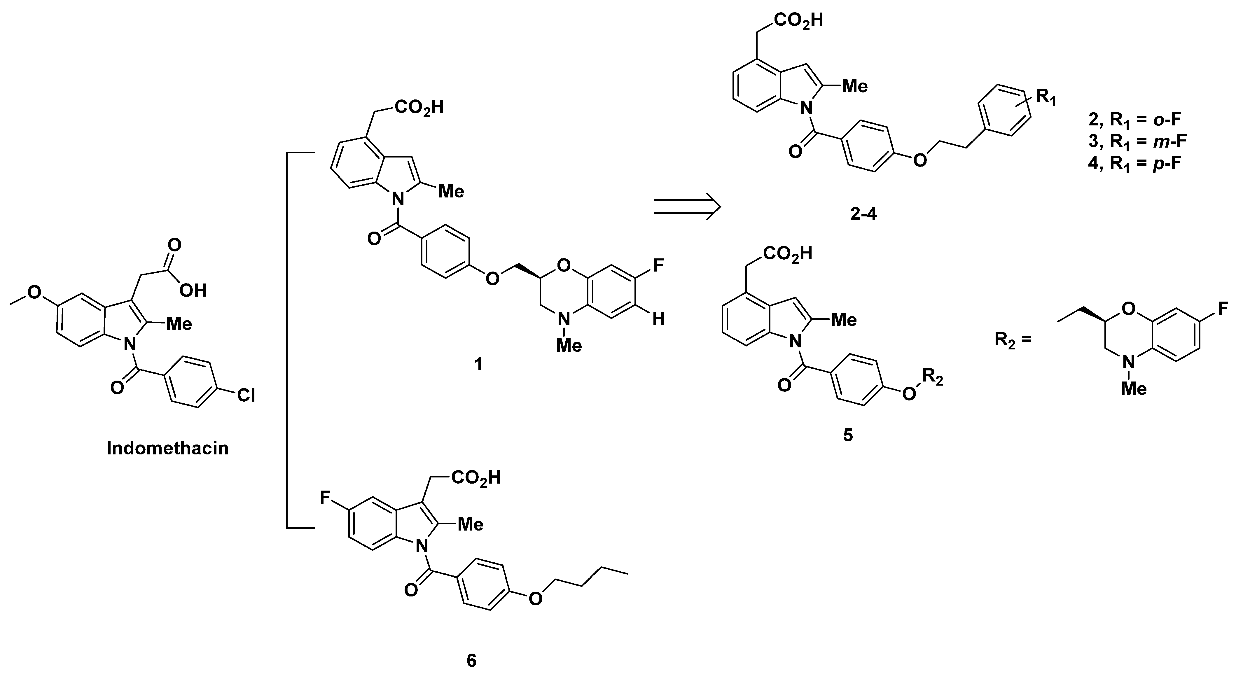
Currently, carbon-11 has been mostly used to radiolabel GPR44 antagonists for PET imaging. For example, AZ12204657 [55] (IC50 = 2.5 nM and Kd = 2.9 µg) and MK-7246 [56] (Figure 1), molecules with high affinities GPR44, are carbon-11 labeled antagonists. Although using 11C-labeling GPR44 PET tracers in animals has been shown to be effective in research, there are many limitations with using 11C-labeling for PET tracers. One of the main drawbacks of using carbon-11 is its half-life of only 20 min in addition to a viability of only 2 h. Because of its short half-life and viability time, mass production and use of a carbon-11 labeled tracer is highly inefficient. Therefore, using carbon-11 radiolabels faces a major limitation in practical use, hindering the extent of their applicability. Additionally, as the clinical use of PET tracers needs to be completed within three half-lives and the requirement that carbon-11 has to be artificially produced by an on-site cyclotron, administrations of these tracers to multiple patients, delivery to remote sites, and use in research are severely hindered.
18F-labeling is preferable over 11C-labeling due to its significantly greater half-life of 110 min, which allows for up to 10 h of PET imaging. The longer half-life and viability thus enable its mass production and distribution [57]. Using fluorine-18 is also beneficial given its positron emission (β+ decay) of 97%, favorable van der Waals radius (1.47 Å), low maximum positron energy (0.635 MeV), and high electronegativity. In addition, its high-energy bond with carbon allows increased tracer stability at higher temperatures and better oxidation resistance. The advantages of 18F-labeling compared to labeling with other radionuclide include better in vivo biological properties and biological behavior [58,59].
In our previous report, we summarized ramatroban-based GPR44 ligands containing a fluorine group and assessed their potential as targets for the development of novel 18F-labeled PET tracers [60]. The present paper aims to continue to summarize current indole-based potential GPR44 analogues and cyclopentenylindole-based potential GPR44 analogues containing a fluorine group and evaluate their potential as treatment targets, to provide a more complete map of current GPR44 analogues containing a fluorine group as options for cancer-related inflammation PET tracer development.

2. Indole-Based Potential GPR44 Analogues

In this section, we evaluate a series of known analogues containing an isolated indole group not fused with other rings, with the exception of those already discussed as ramatroban (Figure 1) and ramatroban-based analogues [60]. These compounds could be considered as analogues of the well-characterized GPR44 antagonist indomethacin (Figure 1), a nonsteroidal anti-inflammatory drug (NSAID) commonly used to reduce fever, pain, stiffness, and swelling from inflammation [61]. Indomethacin is not described here due to its lack of a fluorine atom that may allow for 18F radiolabeling.
For substantially the same reasons, another indole-based GPR44 antagonist developed for treating respiratory diseases including asthma, AZD1981, is not discussed in more detail here due to the lack of a fluorine atom [62].

2.1. Ono Pharmaceutical Co Analogues

2.1.1. Ono Pharmaceutical Co Analogues in 2004–2005

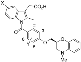
In light of the correlation of PGD2 with allergic diseases, Torisu et al. at Ono Pharmaceutical Co developed a series of human prostaglandin D2 receptor antagonists in 2004 [63]. Among the antagonists, compounds 14 were created with a 4-position acetic acid residue from N-benzoyl-2-methylindole and change in the p-alkoxy moiety, corresponding to the lipophilic moiety of PGD2 receptors (Figure 2).
Table 1 showed the greatest binding potential to DP, with a Ki of 5.3 nM and IC50 of 0.81 nM. The o-fluoro derivative 2 exhibited the next best affinity and selectivity for DP at a moderate Ki of 46 nM and an IC50 of 150 nM. m-Fluoro compound 3 and p-fluoro compound 4 both exhibited a similar subpar Ki and IC50, each at a value greater than 130 nM (hDP, Table 1).
Although compounds 14 all exhibited the selectivity for DP, their bindings to targets DP1 and GPR44 were not discussed in this research. Due to the lack of evidence of selectivity for GPR44, it is uncertain if these compounds can be further developed as PET tracers for GPR44, as the compounds may have the risk of off-target binding to DP1.
In pursuit of an optimal DP1 antagonist, Torisu et al. further formulated a series of N-benzoyl-2-methylindole-4-acetic acids from a p-phenyloxy-ethyloxy derivative in 2004 [64]. The compound with good binding to DP1 was identified as compound 5, evaluated through [3H]PGD2 and cAMP inhibition binding assays. The 4N-methylbenzo-morpholinyl-2-methyloxy analogue 5 demonstrated weak binding to EP, with Ki values for EP1–4 all above 1.7 µM. The binding to DP1, meanwhile, was better with a Ki of 5.3 nM and IC50 of 0.81 nM. In vivo testing of compound 5 for inhibition of PGD2-induced vascular permeability in guinea pig conjunctiva showed that a 0.3 mg/kg oral dose provoked 60% inhibition [64].
In 2005, Torisu et al. continued the search for a selective DP1 antagonist modified from indomethacin analogues and specifically created from 4-substituted phenylhydrazines. The Ki of compound 6 for DP1, measured through an [3H]PGD2 binding assay, was subpar at 110 nM. Correspondingly, the IC50 for DP1 was 1600 nM (mDP) [65]. However, selectivity for DP1 was high in that the Ki values for binding to EP1–4 were all more than 10 µM, with the exception of 2.8 µM for EP2. Of note, all the prostanoid receptors in this study were derived from mice [65].

2.1.2. Recommendations

Although compounds 16 all exhibited the selectivity for DP, there is unfortunately no data on the affinity and selectivity to GPR44. These compounds, therefore, need further characterization for development as GPR44 PET tracers.

2.1.3. Ono Pharmaceutical Co Analogues in 2011

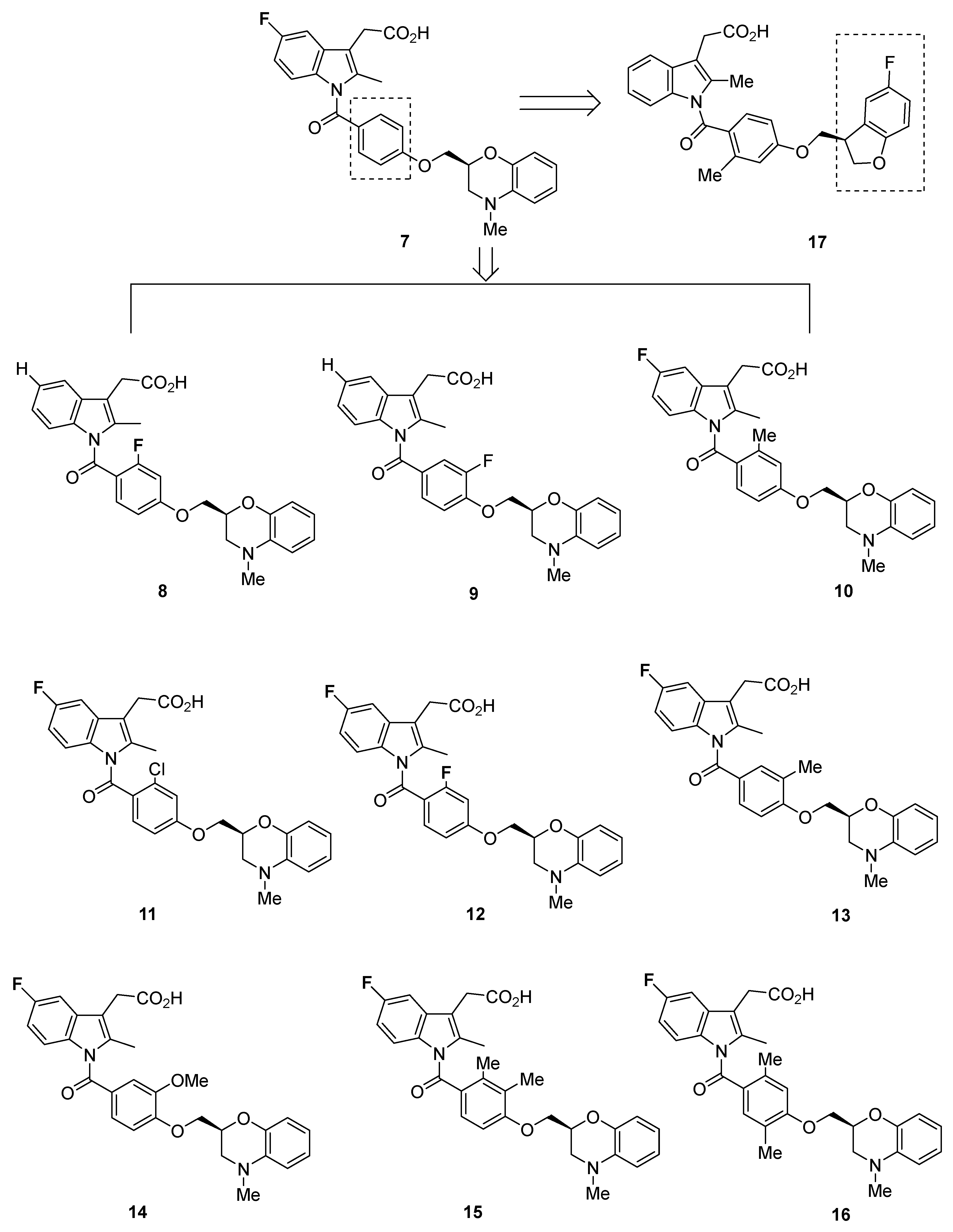
In 2011, Iwahashi et al. from Ono Pharmaceutical Co. synthesized novel DP1 antagonists based on indomethacin [66]. The Ki and IC50 values of the compounds were determined through [3H]PGD2 and cAMP inhibition assays, respectively (Table 2). Of note is that the values were elucidated through binding to a mouse prostanoid receptor.
The 5-fluoro-included 2-methyl indole-3-acetic acid 7 exhibited a particularly high affinity for DP1, with a Ki of 5.7 nM and IC50 of 1.8 nM. Compound 7 was also shown to be specific for DP1, with a Ki above 250 nM for binding to other prostanoids (EP1–4, FP, TP, and IP).
Further modifications of the indole and phenyl groups yielded compounds 816 (Figure 3). With the exception of 9 (Ki = 240 nM), 13 (Ki = 59 nM and IC50 = 42 nM), 14 (IC50 = 68 nM), 15 (IC50 = 32 nM), and 16 (Ki = 14 nM and IC50 = 2.8 nM), all compounds had Ki and IC50 values below 6 nM. Although the binding affinity for DP1 appeared to be high in these compounds, the selectivity was not as pronounced as would be optimal, with the Ki for human IP at values ranging from 93 to 320 nM (with the exception of 14 with its Ki of 1.5 µM).
Substitution of a benzofuran-3-yl moiety led to compound 17 (Figure 3), which had similarly low Ki and IC50 values (5.4 and 1.8 nM, respectively) but suboptimal selectivity (IP Ki = 520 nM) [66].

2.1.4. Recommendations

The compounds developed here were only tested as DP1 antagonists, and were not characterized for binding affinity to GPR44. However, on the basis of the high affinity to DP1, these compounds are not promising for use as GPR44 PET tracers.

2.2. Oxagen Analogues

2.2.1. Oxagen Analogues in 2005 and 2007

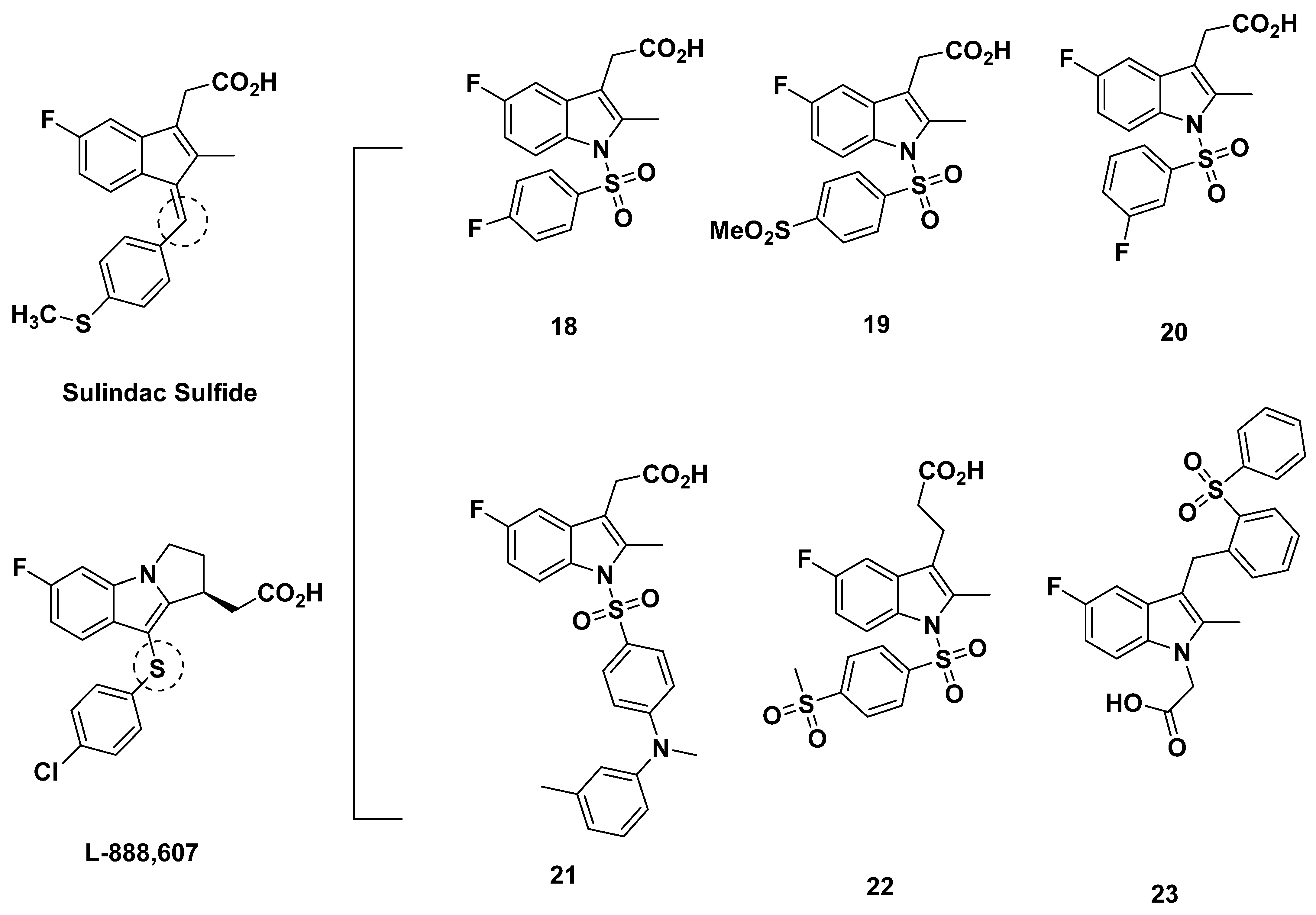
In 2005, Armer et al. from Oxagen aimed to discover a selective antagonist of GPR44 based on a common indole acetic acid structure [67]. The molecules were designed around a (5-fluoro-2-methyl-1H-indolyl3-yl) acetic acid core due to the promising binding affinities of sulindac sulfide and L-888,607 (Figure 4) [68,69], compounds which shared this core, and the optimal structure-activity relationship (SAR) of compound libraries sharing the core. The addition of an N-sulfonyl group was further characterized to increase affinity, and the novel compounds 1820 (Figure 4) were created as such.
Compounds 18 and 20 both exhibited selectivity, with binding to DP1 low at Ki values greater than 6 µM. However, the binding affinity to GPR44, though better, was found to be also low at 71 and 225 nM, respectively. Reflecting the low Ki values, the IC50 for inhibition of PGD2-induced Ca2+ flux was above 150 nM for both compounds [67].
Compound 19 showed better affinity with an improved Ki of 68 nM, a Ca2+ IC50 of 19 nM, an eosinophil shape change assay IC50 of 74 nM, and a Th2 cell chemotaxis IC50 of 67 nM. In rat and human microsomes, compound 19 did not exhibit concerning levels of metabolism, with no cytochrome P450 (CYP450) isoforms inhibited or induced at a dose of 1 µM. Compound 19 additionally did not exhibit cytotoxicity in the HepG2 hepatocarcinoma cell line, nor influence in a human ether-a-go-go-related (hERG) channel assay, which marks potential inducement of the heart condition acquired long QT syndrome (LQTS). In rats, intravenous and oral administration revealed promising pharmacokinetic properties. Intravenous administration of 1 mg/kg yielded an acceptable Cmax of 1801 ng/mL, an area under the curve (AUC) of 619 ng/(mL·h), a clearance of 40 (mL/min)/kg, and a half-life of 2.2 h. The oral dose of 10 mg/kg yielded a five-times lower Cmax at 1.8 h, four-times higher AUC, a half-life of 5.5 h, an acceptable bioavailability of 56%, and unidentified clearance. The discrepancy may be due to the differing ways of administration, as well as the ten-times higher oral dose [67].
Oxagen also disclosed in a patent publication the 1-acetic-acid derivative (compound 21) with a sulphonyl spacer between the indole and aromatic groups. However, this compound exhibited a low affinity and selectivity for GPR44, with its Ki of 222 nM for GPR44 and a Ki of 86 nM for DP1, indicating potential off-target binding [31].

2.2.2. Recommendations

Due to the low affinity for GPR44, shown by the high Ki and IC50 binding values, compounds 1821 may not be optimal compounds to serve as efficacious GPR44 PET ligands.

2.2.3. Oxagen Analogues in 2010

In a 2010 review [70], Norman described an indole-1-sulfonyl-3-acetic acid developed by Oxagen (compound 22) in Figure 4 that was similar to compound 19 created by Armer et al., save for the replacement of the acetic group attached to the aromatic ring by a propanoic group [67]. The IC50 value of the compound was subpar at 68 nM, but the selectivity for GPR44, as opposed to other prostanoid receptors, was adequate.
Norman additionally reviewed compound 23, disclosed by Oxagen in a patent filing, whose properties were not discussed [70,71].

2.2.4. Recommendations

As such, due to the paucity of information regarding compounds 2223, these compounds need further characterization before development for use as PET tracers. However, compound 22 could be reasonably disregarded due to the subpar binding affinity.

2.2.5. OC-459 (OC000459/Timapiprant)

In 2012, Pettipher et al. from Oxagen characterized a 3-quinolinylmethylindol-1-yl-acetic acid, in which the indole scaffold was reversed [72]. This compound, known as OC-459, OC000459, and Timapiprant, is a selective GPR44 antagonist (Figure 1). Of note, the structure included in this review has not been confirmed by Oxagen, but was characterized as OC-459 by Amira. In addition, although Pettipher et al. were the first to publish a non-clinical study of OC-459, it appears that the unpublished development of OC-459 had already been undertaken by other parties [73,74,75,76,77,78].
In a [3H]PGD2 competition binding assay in Chinese hamster ovary (CHO) cells, OC-459 showed a high binding affinity for GPR44 at a Ki of 13 nM. The Ki value on the native receptor in human Th2 lymphocytes was similarly low at 4 nM. The IC50 value of Ca2+ inhibition and Th2 lymphocyte chemotaxis was consistent with the Ki values at 28 nM. OC-459 was also shown to inhibit IL-13 production by Th2 cells at an IC50 of 19 nM and inhibit PGD2-induced Th2 cell survival at an IC50 of 35 nM. For eosinophil shape change, OC-459 inhibited the response at an IC50 of 11 nM.
The [3H]PGD2 displacement in rat GPR44 was comparable at 3 nM, indicating that rats may be used as an in vivo model without major concerns. Selectivity was also promising, as the Ki or IC50 values for OC-459 binding to DP1, EP1–4, FP, IP, and TP were all above 10 µM. A concentration of 10 µM OC-459 also exerted no effects on 69 receptors, ion channels, transporters, and 17 enzymes. These enzymes included the cyclooxygenases cyclooxygenase 1 (COX1) and cyclooxygenase 1 (COX2), in recombinant form, and the native COX1, as expressed in platelets.
OC-459 was also characterized for plasma protein binding and found to bind strongly at 99.1% and 99.8% in human and rat plasma, respectively. The pharmacokinetic profile of OC-459 was elucidated in male Sprague-Dawley rats. At a dose of 2 mg/kg intravenously, OC-459 had a Cmax of 5660.4 ng/mK at 0.08 h, a half-life of 1.3 h, a clearance of 8.4 mL/kg/min, an AUC24h of 4486.5 ng/mL·h, a volume of distribution of 0.5 L/kg, and an undiscerned bioavailability. At the same concentration dosed orally, the Cmax was 1543 ng/mL at 1.3 h, the half-life was 2.9 h, the clearance was 6.4 mL/kg/min, the AUC24h was 5262.5 ng/mL·h, the volume of distribution was not found, and the bioavailability was high at 111%. Of the values elucidated, all seemed characteristic of a well-absorbed and promising compound.
The pharmacologic profile of OC-459 was briefly delved into, and it was characterized as a competitive antagonist. OC-459 also appeared to dissociate at a quick rate from GPR44 with a half-life of 0.41 min [79].
The inhibitory effects of OC-459 were further elucidated in rats orally administered OC-459, which reduced 13,14-dihydro-15-keto-PGD2 (DK-PGD2)-induced eosinophil count. Similarly, eosinophil numbers were reduced following OC-459 oral administration in guinea pigs.
As the first GPR44 antagonist found to act clinically on allergic disease, OC-459 was initially characterized in clinical studies by Barnes et al. in 2012 [80]. This double-blind, parallel group trial included 65 subjects with moderate persistent asthma who were given 200 mg of OC-459 twice daily. The compound ended up gradually increasing its forced expiratory volume in 1 s (FEV1). In addition, OC-459 reduced circulating IgE and night-time symptoms. The plasma concentration of OC-459 was detected as 600 ng/mL, indicating that the GPR44 receptors had been successfully blocked. Importantly, OC-459 was shown to have a tolerability comparable to that of the placebo. In essence, the clinical study confirmed the promising properties of OC-459 from in vivo preclinical models, and established OC-459 for treatment of disease [81,82].
In 2012, a single center, randomized, double-blind, placebo-controlled, two-way crossover study was published [80]. It indicated that OC-459 could reduce symptoms of allergic rhinoconjunctivitis, at least in those allergic to grass pollen. Again, the compound was well-tolerated and acted more quickly than in the previous study, even though the dose was still 200 mg twice daily. An unexpected benefit to OC-459 administration was persistent inhibition, lasting up to 4 weeks. The response may be due to the apoptosis and clearance of Th2 lymphocytes induced by GPR44 inhibition, as OC-459 has a half-life of less than a day [83].
Further clinical studies characterized OC-459 as an inhibitor of eosinophils in corticosteroid-dependent or corticosteroid-refractory eosinophilic esophagitis patients and an inhibitor of the late asthmatic response, sputum eosinophil number and eosinophil shape change from steroid-naive asthmatic patients [84]. In mice, OC-459 also reduced inflammation and its corresponding markers, eosinophil migration, and symptoms of 2,4,6-trinitrobenzenesulfonic acid (TNBS)-induced colitis [85].
However, in 2010, progress on OC-459 decreased following licensing of rights to Eleventa and Atopix, reportedly due to financial concerns. Later, another study published in 2021 found that OC-459 treatment had little impact on the clinicopathological changes induced by rhinovirus RV-A16 infection in patients treated with inhaled corticosteroids, though the same study also concluded that OC-459 was safe and well-tolerated [76].

2.2.6. Recommendations

Although the Ki of OC-459 is not as low as other promising compounds, the extensive characterization of comparable IC50 values and good safety profile make it attractive for further development as a GPR44 PET tracer.

2.3. AstraZeneca Analogues

Birkinshaw et al. from AstraZeneca derived a series of indomethacin-based compounds in 2006 (Figure 5) [86]. In [3H]PGD2 displacement assays, the IC50 of most of the compounds (2426) were suitably low at 5.4, 6.0, and 7.1 nM, respectively; the IC50 of compound 27, meanwhile, was high at 68 nM, indicating the low binding affinity of compound 27. Apart from in vivo potency studies, Birkinshaw et al. calculated the whole blood potency of the compounds, a marker of plasma protein binding. Except for the unmeasured compound 27, the whole blood potency was high at values ranging about 790 nM. This indicates the possibly low activity of the compound in blood. As could be predicted, the plasma protein binding was high at a value greater than 99.1 nM. The lipophilicity of all compounds, however, was acceptable at values from 0.5 to 1.8, measured as log D7.4. In vitro assessment of clearance yielded low values below 2.6 µL/min/106 cells in human hepatocytes (with the exception of compound 27, which again, could not be identified) but extremely divergent values in rat hepatocytes. The values ranged from <1 µL/min/106 cells for compound 26, 6.5 µL/min/106 cells for compound 25, and above 16 µL/min/106 cells for compounds 24 and 27. The discrepancy in values between human and rat cells may point to species-dependent clearance, but the consistently low clearance in human hepatocytes is promising, as it indicates an acceptable compound metabolism.

Recommendations

Due to the high inhibitory potential of compounds 2426, all these compounds may be assessed as potential GPR44 PET tracers. The high whole blood potency and discrepancy in rat and human clearance measurements may be taken into consideration in the development, but should not preclude compounds 2426 from consideration [86].

2.4. AstraZeneca Analogues

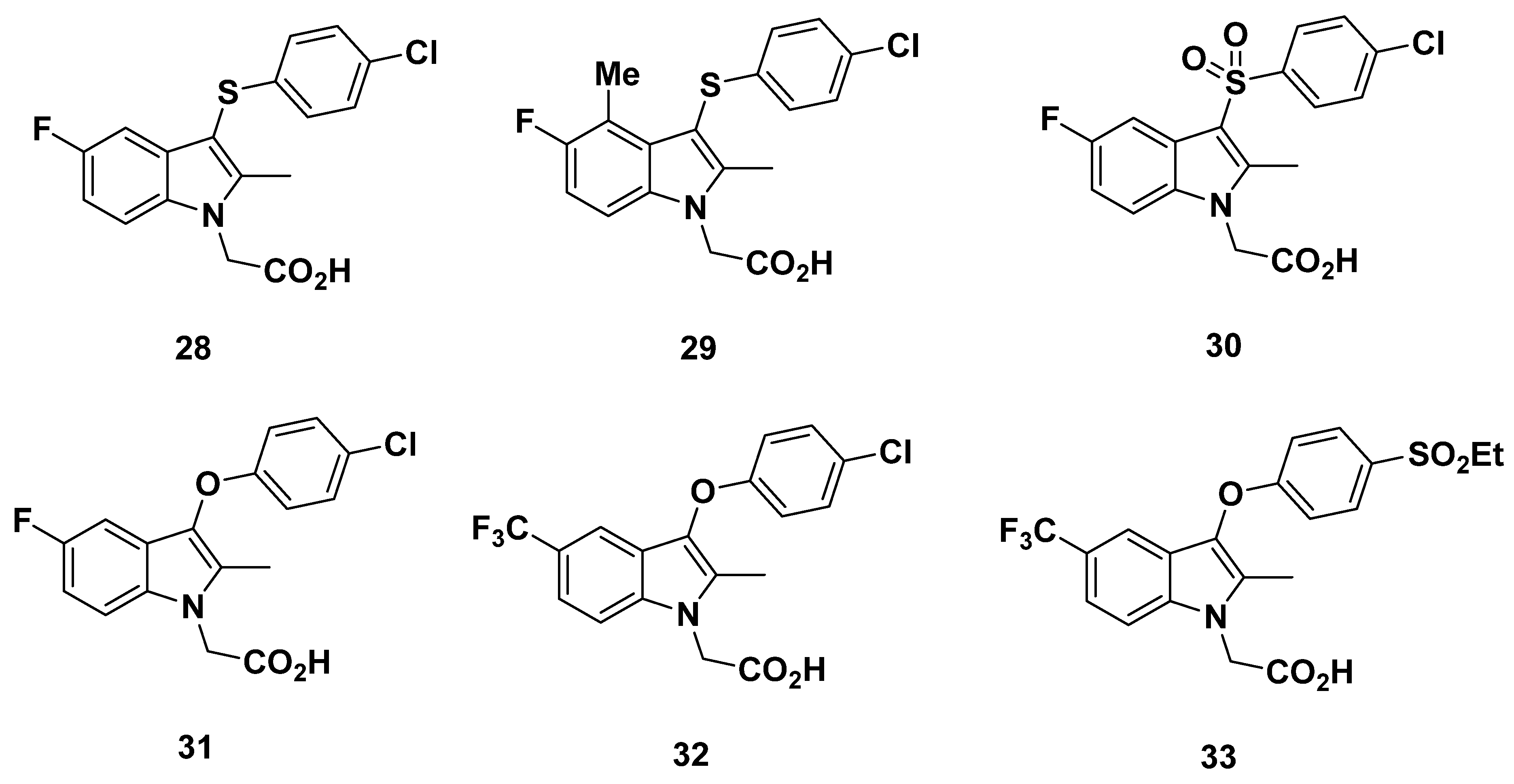
In 2011, Luker et al. from AstraZeneca developed a series of novel GPR44 antagonists based on a 3-thioaryl indole found through screening AstraZeneca compounds similar to indomethacin (Figure 6) [87].
Examination on substitutions led to compounds 2829, both of which exhibited a suitable affinity for GPR44 at IC50 values of 1.6 and 1.3 nM, respectively (as measured through a [3H]PGD2 assay). To determine specificity, binding to human aldose reductase (ALD2), which had been shown to be inhibited by indomethacin, and related aldehyde reductase (ALD1) were evaluated. ALD2 values were adequately large at >7.9 µM and 3.5 µM, respectively. ALD1 values were slightly lower at 3.1 µM and 2.2 µM, respectively. The clearance from rat hepatocytes (19 and 17 µL/min/106 cells, respectively) and human liver microsomes (<3 µL/min/mg for both compounds 2829) was also assessed. These low clearance values are preferable properties.
Incidentally, compound 28 has been disclosed as a compound developed by Pettipher et al. from Oxagen [88]. The Ki of the compound was reportedly 5 nM [89].
In contrast to compounds 2829, sulfone and oxygen-linked compounds 3033 had a less optimal affinity for GPR44, with IC50 values of 7.1, 5.0, 11, and 3.5 nM, respectively. For all compounds save for compound 33, the aldose reductase (ALR2) and aldehyde reductase (ALR1) selectivities were unable to be determined. Compound 33 showed an optimal binding with an IC50 value greater than 10 µM for ALR2 but only at 3.9 µM for ALR2. This selectivity was interesting in that other compounds in the study with the same aryloxy side chains as compound 33 showed an even lower IC50 for ALR1 for reasons undetermined [87].

Recommendations

The high affinity of compounds 2829, 31 and 33, with an IC50 of no more than 5 nM, makes them promising for development into GPR44 PET tracers. Compound 32, with its IC50 of 11 nM, may also be considered. However, off-target binding to ALR2 and ALR1 must be taken into account, and further characterization of binding affinity to other prostanoid receptors, as well as in vivo pharmacokinetics, would be recommended.

2.5. Pfizer Analogues

2.5.1. Development of Analogues

In a patent published in 2002, Pfizer disclosed compound 34 (Figure 7), which was reported to have 40-fold selectivity for GPR44 over DP1 [90]. Similarly, compound 35 (Figure 7) was reported to have a GPR44 pIC50 of 8.15.
In 2012, Kaila et al. from Pfizer developed a series of compounds based on diazine indole acetic acids [91]. Synthesized to inhibit GPR44 to reduce symptoms of asthma, the compounds were each subjected to a cAMP hGPR44 fluorescence resonance energy transfer (FRET) assay in vitro, and those with promising results were further subjected to pharmacokinetic characterization.
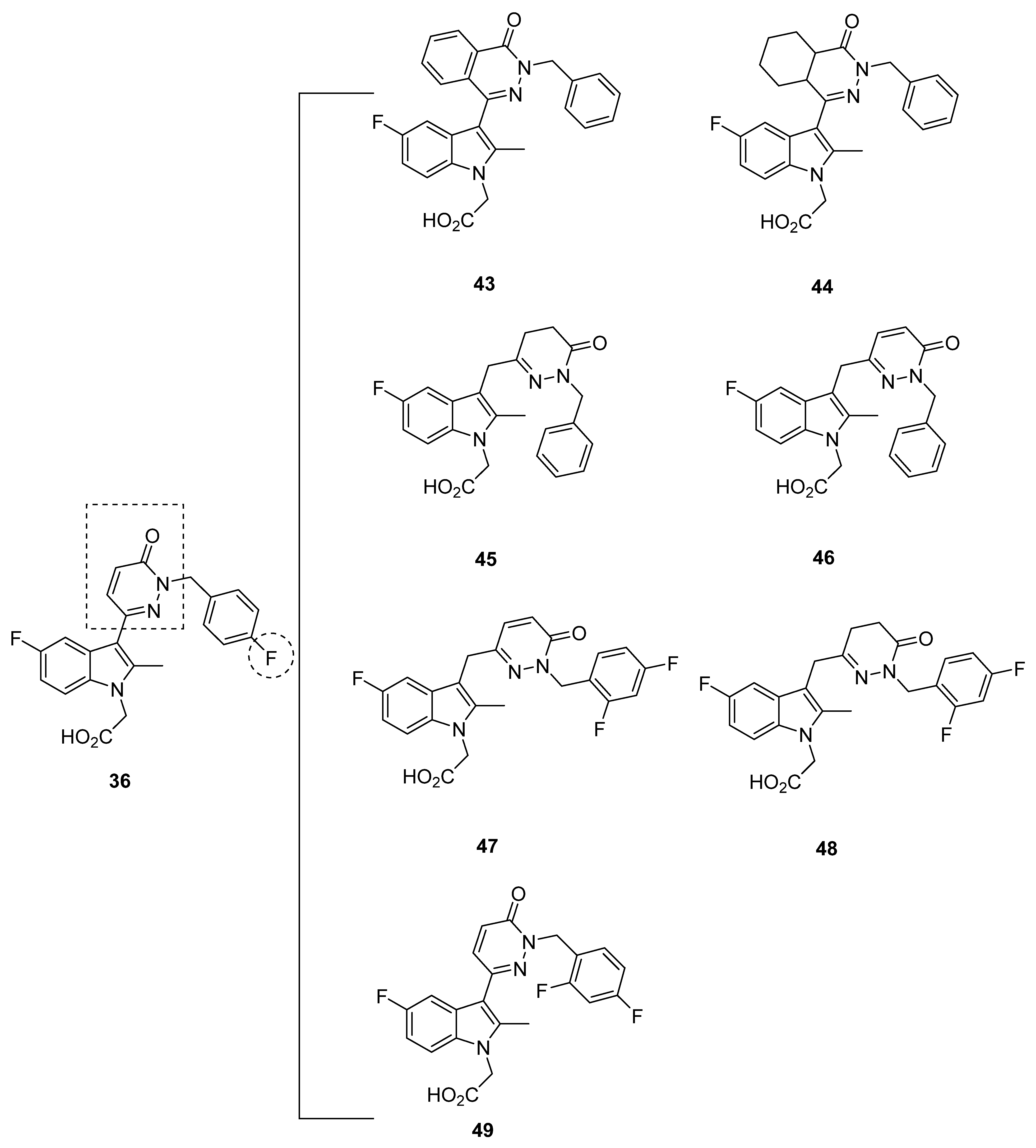
The substitution of the 5-position of the indole and 3-position of the diazine in compound 36 (Figure 7) resulted in a moderate affinity for GPR44 at an IC50 of 20 nM. The lipophilicity at a calculated partition coefficient (clogP) of 3.5 was also promising, as well as an unconcerning microsomal stability, an indicator of metabolism and clearance rate, at greater than 30 min. As might be inferred by the microsomal stability value, the clearance was low at 6.9 mL/min/kg in mice. However, the bioavailability of 11% precluded further studies on compound 36, as well as a 0.3 × 10⁻⁶ cm/s in a Caco-2 cell assay, which measured intestinal absorption. The low value, reflecting a low level of absorption, may be expected from the lipophilicity.
The modification of compound 36 through the addition of hydrophobic groups resulted in the development of compound 37 (Figure 7), which exhibited a moderate affinity for GPR44 at an IC50 of 23 nM. The lipophilicity was again on the higher end of the acceptable level, with a clogP of 3.33. The ionization of the compound, measured through pKa, was 4.02, indicating that it contained an ionizable group. The Caco-2 cell assay found that compound 37 exhibited permeability of 0.5 × 10⁻⁶ cm/s, albeit at a reduced pH of 6.5, the permeability was reportedly reduced due to the carboxylic acid moiety of the compounds.
Substitutions at the 7-position of the A ring of the indole core reduced affinity in compounds 3842 (Figure 7). In these compounds, the IC50 was not ideal, with values above 76 nM and even rising to greater than 1000 nM. Thus, although the lipophilicity stayed relatively constant at a clogP ranging from around 1 to 4 and the pKa was around a value of 3.8, the low affinity of these compounds to GPR44 disqualified them from further characterization.
Reduction of the diazine ring was explored in hopes of increasing membrane permeability, resulting in several compounds in which affinity was compromised (compounds 4344) while others were promising (Figure 8). The IC50 of compounds 4344, as well as compound 45, were all above 11 nM. Unfortunately, a discrepancy in the data for compound 45 was exhibited, in which the 16 nM IC50 of compound 45 was later labeled as 1.5 nM instead. Due to further studies with the eosinophil shape change assay described later, however, we decided to consider compound 45 as promising.
On the other hand, the IC50 values for compounds 4648 were 9, 3.8, and 4 nM, respectively. A compound described briefly, compound 49, also had a moderate IC50 of 10 nM. The solubility of compounds 4748 in aqueous solution was further determined at a pH of 6.6, yielding 0.318 and 0.954 mg/mL; these values may inform development of the compounds, once made into tracers, about their dissolvability in solution.
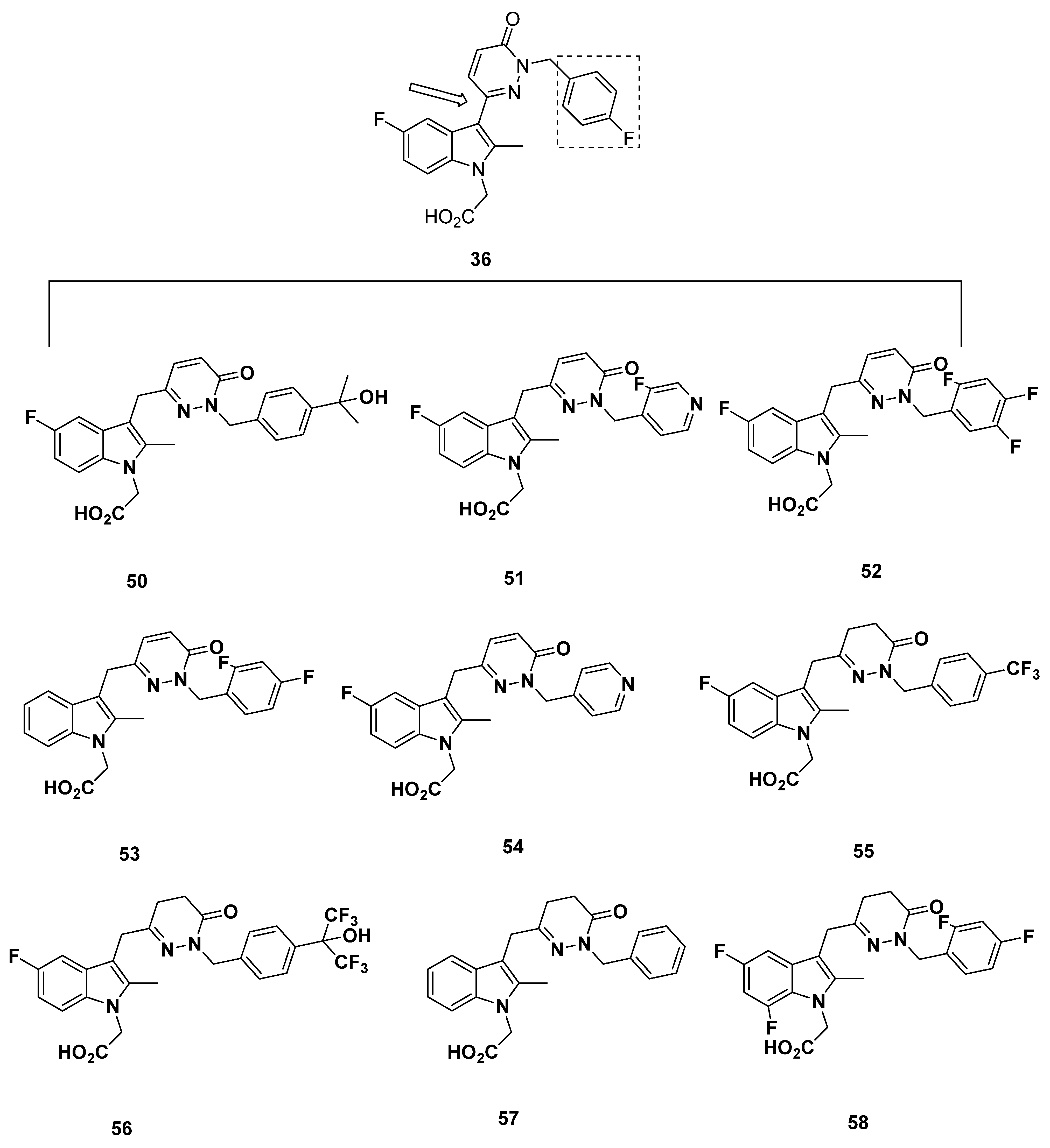
Additional compounds 5058 with a pyridazine linker were developed (Figure 9). The IC50 of compounds 5355 and 5758 in both the FRET assay and the eosinophil shape change assay were all above 10 nM, with the lowest being 12 nM and the highest being 180 nM, and therefore should not be considered on account of low binding affinity. The IC50 values of compounds 5052, meanwhile, were all in a promising range. It proved difficult to compare compounds 5052, however, because of the heterogeneity in FRET assay and eosinophil shape change assay results, in which compound 50 had values of 5 and 13 nM, compound 51 had values of 6 and 6 nM, and compound 52 had values of 0.8 and 10 nM, respectively. An interesting case was presented with compound 56, for which the FRET assay IC50 was an acceptable 7 nM but the eosinophil shape change assay IC50 was 57 nM. None of these compounds have been further characterized.
In a further assessment of compound potency, a basophil chemotaxis assay was run in which the IC50 values were well correlated to those found in the FRET assay. Meanwhile, the similarly strong correlation between the FRET assay and hGPR44 binding assay with [3H]PGD2 confirmed the GPR44 binding potency. Eosinophil shape change assays of the promising compounds 45 and 47 confirmed the IC50 of the FRET assay, with only the slight change in compound 45 IC50 to 4 nM.
To establish the selectivity of the compounds, a cAMP time-resolved FRET (TR-FRET) assay with the lymphocyte NK-92 cells (DP1) and thromboxane B2 (TXB2), an inactive metabolite of thromboxane A2 (TXA2), the major ligand of TP, was carried out. Undisclosed representative compounds from the series were tested and found to have a selectivity of three orders in magnitude.
In the Caco-2 cell assay at pH 6.5, compounds 4647 exhibited adequate permeability at 4.3 and 7.1 × 10⁻⁶ cm/s, whereas compound 49 had a slightly lower permeability at 2.1 × 10⁻⁶ cm/s. The increased permeability of these compounds indicates that adsorption occurs in the jejunum microvilli, due to this region’s slightly acidic pH. Although pharmacokinetic data was not further characterized, the Caco-2 cell assay results suggest that for compounds 4647 and 49, the permeability will not hamper adsorption or oral bioavailability. Interestingly, compound 47 exhibited a high plasma protein binding of 98% in an equilibrium dialysis experiment, but this was determined not to bar membrane permeability.
In addition, the compounds were found to bind at similar values to human and murine GPR44, indicating little concern if mouse models are to be used for in vivo studies. As such, an oxazolone-induced contact hypersensitivity (CHS) mouse model was then used to test compounds 45 and 47, selected due to their permeability, the close correlation between the FRET and eosinophil shape change assays, and structure diversity and potential toxicity.
At an oral dose of 3 mg/kg twice daily, compounds 45 and 47 exhibited a close 34% and 30% reduction in swelling. In rats, compounds 45 and 47 exhibited similar optimal properties, with a clearance of 13 and 12 mL/min/kg, the half-life of 4.3 and 3 h, and steady-state volume of distribution of 1.7 and 1.6 L/kg, respectively. The AUC of compound 47 was higher at 5066 h·kg·ng/mL·mg relative to the 3568 h·kg·ng/mL·mg of compound 45, which corresponded well to the increased Cmax (772 ng/mL as opposed to 484 ng/mL) and bioavailability (37% as opposed to 28%) of compound 47. For compound 47 specifically, the potential for drug-drug interaction was thought to be negligible, as evidenced by the CYP inhibition IC50, which ranged from 16 to 100 µM. In addition, the AMES test for identifying mutagens showed compound 32 had no effect, as well as no inhibition of human ERG at 33 µM compound 47.
The promising compound 48 was also assessed in mice and showed lower clearance (9 mL/min/kg), a comparable half-life (4 h), a lower steady-state volume (1.12 L/kg), a higher AUC (7526 h·kg·ng/mL·mg), a higher Cmax (1889 ng/mL), and a higher bioavailability (41%) as compared to compounds 45 and 47. However, compound 48 was not further profiled.
On account of its more optimal profile, compound 47 was extensively characterized, starting from the determination of a minimum effective dose at 1 mg/kg for house dust mite (HDM)-induced allergic airway disease in mice. At an oral dose of 20 mg/kg daily, compound 47 inhibited airway inflammation and leukocyte counts, save for that of macrophages, in bronchoalveolar lavage. H&E staining confirmed the inhibitory effect of compound 47, showing that it could decrease cell infiltration and epithelial and blood vessel hyperplasia and hypertrophy.
The efficacy of compound 47 was also evaluated in a sheep model of Ascaris-induced airway bronchoconstriction and hyper-responsiveness. Induced responses of both early and late phase allergic airway bronchoconstriction, as well as inflammation and hyper-responsiveness, responded to 3 mg/kg intravenous compound 47 twice daily, and apparently created a comparable effect at even 1 mg/kg intravenously. In addition, 3 mg/kg intravenously applied compound 47 attenuated the length of time of tracheal mucus velocity from 50% to 70% of baseline at 4 h [91]. These compounds have been used by Babu et al. in a machine-learning model to study the structure-activity relationship, which offers more insight on the observed behavior of these compounds. We will provide recommendations after reviewing Babu et al.’s publication in the next section.

2.5.2. Mathematical Modeling

In 2016, Babu et al. at SRM University evaluated GPR44 antagonists using a series of mathematical models to assess the quantitative structure-activity relationship [92]. Specifically, these models included Comparative Molecular Field Analysis (CoMFA) to predict structure-activity relationship, Comparative Molecular Similarity Indices Analysis (CoMSIA) to correlate molecular properties with binding affinity, Topomer CoMFA to assist alignment issues with CoMFA, and Hologram Quantitative Structure Activity Relationship (HQSAR) for specialized molecular signatures. They used compounds disclosed by Kaila et al. to make up a training set to establish the model, and a test set to validate the model.
In addition, the activity structure fragments’ contribution to the molecule was determined, and an IC50 value was predicted to assess the validity of the model for some of Kaila et al. compounds, including compounds 3640, 4243, 4548, 5056, and 58 discussed above (Figure 7, Figure 8 and Figure 9). In all, the predicted IC50 matched well to the actual IC50. Table 3 lists the actual properties and calculated properties of these compounds.
The compound with the highest activity reported by Kaila et al. was compound 52, with an IC50 of 0.8 nm. Thus, this compound was further modeled for its steric and electrostatic contours. Specifically, for compound 52, the 5-position of the indole ring proved conducive to activity, particularly the electronegative atom, but the 7-position did not.
Indicated by the raised IC50, compounds 40 and 42 were structurally different from compound 52, with replacements at the 5 and 7 regions of the indole core. In one of the fragments, the para benzyl ring and ortho diazine lent an increase in binding affinity.
The regional contribution to binding affinity was characterized for compound 52. For instance, the 5-position fluorine and 4, 6, and 7-position hydrogen of the indole core, as well as the diazine ring, again were shown to contribute to compound 52′s inhibitory qualities.
In compounds 3640 and 4244, the meta benzyl ring also contributed to inhibitory qualities; however, when there was no co-expression of the pyridazine linker as in compound 42, the meta benzyl ring lost this quality. In compound 47, meanwhile, the diazine ring had high activity, whereas compound 42 exhibited activity at hydrogen at position 7 in the indole core.
The highly active compound 52 was further characterized for steric and electrostatic contours as part of CoMFA analysis, in which the ortho and meta-attached benzyl on diazine was seen to supplement inhibitory activity. For compound 37, however, the methyl sulfur dioxide at the 5-position of the indole core was instead detrimental to activity. In addition, compounds 40 and 42 exhibited lower activity due to the replacement of the 5-position fluorine in the indole core, indicating the importance of the original fluorine.
Further characterization of compound 52 in CoMSIA contour maps confirmed previous results, as well as indicating that compounds 4748 shared the beneficial para position of the benzyl group; this may have been due to a hydrophobic group at the para position. Likewise, an increase in affinity due to the 5-position of the indole ring may be attributed to a hydrophobic group at the position. In compounds 4748 and 52, the hydrophobic group was fluorine at the para position of the benzyl group. However, hydrophobicity at the 7-position of the indole core and meta position of the diazine ring resulted in decreased affinity of compounds 40 and 42. The effects of an H-bond donor group were also important to consider, as the H-bond donor in the hydroxyl group of the carboxylic acid attached to the indole core facilitated improved binding. In contrast, an H-bond donor group at the meta and para position of the benzyl ring was detrimental to binding potency [92].
As Babu et al. reported, the modeling overall identified the presence of “(a) sterically bulky, electronegative, hydrophobic atom at para position of benzyl ring, (b) electronegative and hydrophobic atom at 5th position of indole ring could improve the activity of these compounds. The pyridazine linker and carboxylic acid are very important to make the compound more potent with higher activity” [92]. Based on these findings, the authors designed molecules based on our findings and found that the predicted activities of newly designed molecules were in the range of highly active molecules used in this study.

2.5.3. Recommendations

The low IC50 values of compounds 4549, as well as compounds 5052, mark their potential for use as GPR44 PET tracers. As compound 47 has been extensively characterized and found to be efficacious, this compound may be particularly promising. The lack of extensive testing, though, does not preclude compounds 4549 for further development.
In addition, several structural features, including electronegativity and hydrophobicity at the para position of the benzyl ring as well as the 5-position of the indole core were found to lead to improved inhibition. The presence of pyridazine linkers and carboxylic acids were also identified as lending significant qualities to the inhibitors. These findings of this modeling may well-inform the development of future GPR44 antagonists.

2.6. Almirall Analogues

2.6.1. Almirall Analogues in 2013

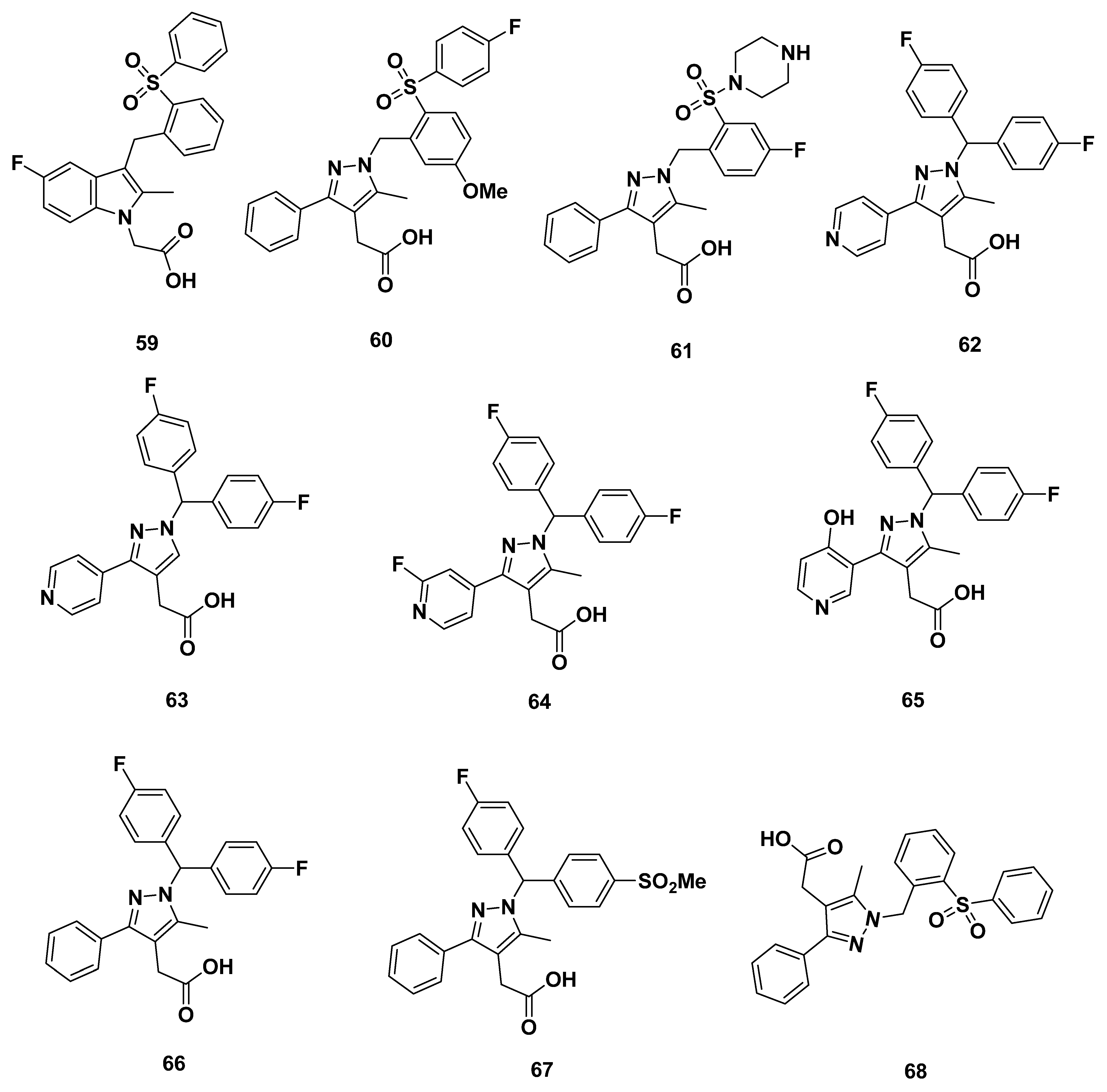
Andrés et al. from Almirall synthesized a series of GPR44 antagonists on the basis of expanding the ring of the indole core in 2013 [93]. High throughput screening of 100 compounds by homogeneous cAMP assay yielded the initial pyrazole-4-acetic acid precursor substructure. The ortho-sulphonyl benzyl tail containing compound 59 (Figure 10) exhibited a low IC50 in both the eosinophil shape change assay from a reference that could not be located but which was referred to by Andrés et al., and GTPγS binding assay, which measured GPR44 signal transduction; the values were 5 and 16 nM, respectively.
Further changes, including separating the indole core to benzene ring connected with a pyrazole ring, resulted in compounds 6061. Compound 60 had a high GTPγS IC50 of 36 nM, indicating subpar binding affinity. The addition of a strong basic terminus group in compound 61 resulted in a much worse binding affinity where GTPγS IC50 = 5000 nM.
Based on the optimization of the substituents of the pyrazole core and dibenzyl derivative tail groups, compounds 6267 were created. Unfortunately, the binding affinity of all the compounds was poor at GTPγS IC50 greater than 46 nM, with a median IC50 of 705 nM.
Other compounds in the series were profiled, including compound 62 with its IC50 of 46 nM, in which all showed <40% inhibition at 10 µM for DP1 and the TP. Dissociation kinetics were elucidated from membranes using the GTPγS assay, in which most compounds had rapid dissociation. The slowest dissociating compound was compound 62 with its half-life of 30 min. Although none of these compounds (save for 62) contained the necessary fluorine group, the results may be representative of general compounds with an expanded indole core.
In all, compound 68 in the series was further profiled, but that compound did not contain the necessary fluorine group. The profile showed qualities characteristic of a lipophilic carboxylic acid, with adequate absorption, low clearance, and a long terminal half-life. However, overly high amounts of plasma protein binding were concerning. The compounds 6061 may be presumed to have similar properties, but the later compounds (6267), based off a polar pyridyl substitution, should not have concerns with decreased plasma protein binding and receptor ligand interactions [93].

2.6.2. Recommendations

As such, compounds 5960 may be considered on the basis of their IC50 values. Caution should be taken for compounds 60 on account of the plasma protein binding and subsequent detriments to receptor ligand interactions.

2.6.3. Almirall Analogues in 2016

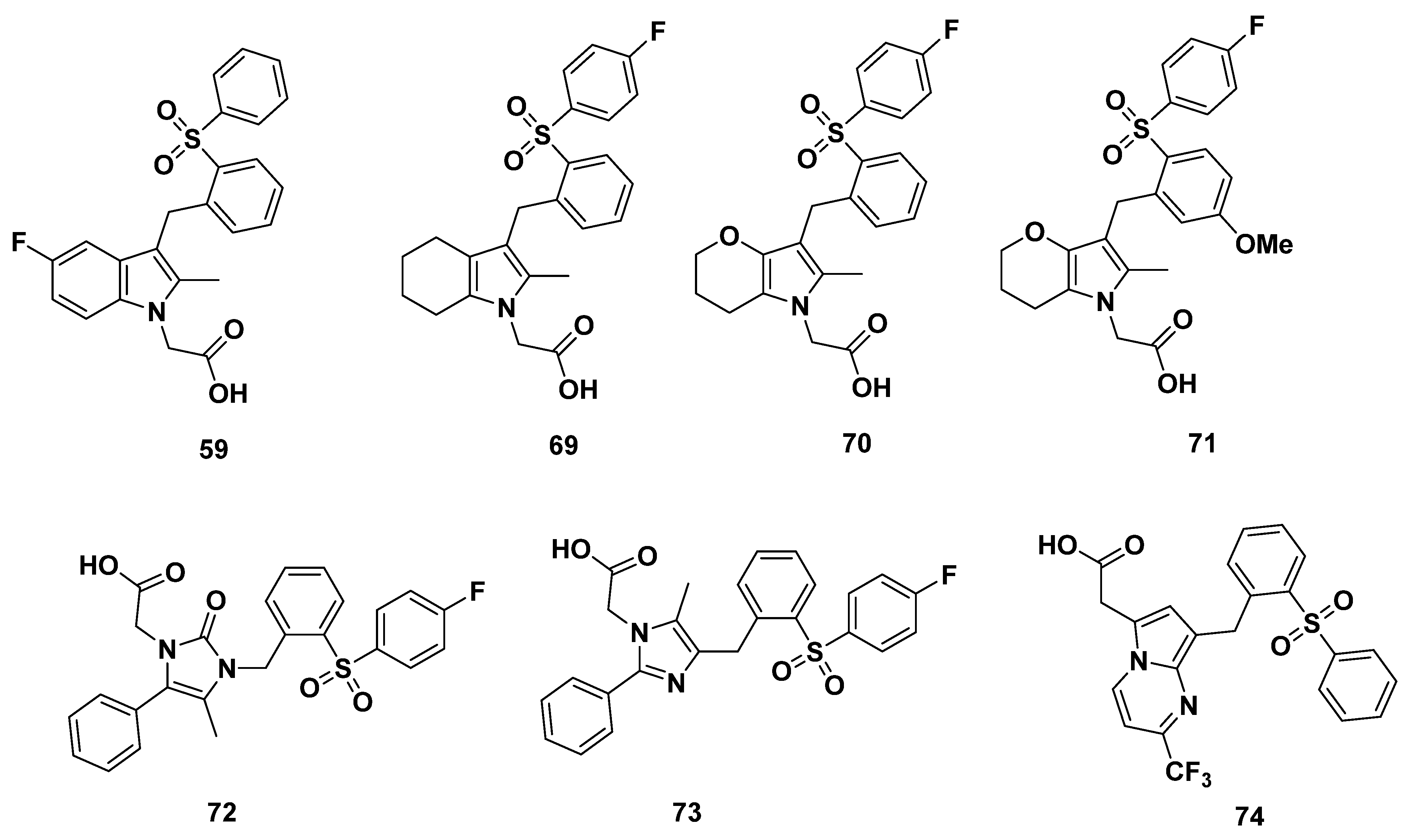
Buil et al. of Almirall synthesized a series of GPR44 antagonists with differing cores (Figure 11) [94]. In 2016, they further described compound 59, an indole GPR44 antagonist, with a moderate GTPγS IC50 of 14 nM and adequate dissociation half-life of 1.3 h.
Another compound, 69, showed a high GTPγS IC50 of 104 nM. On the other hand, the pyrrole analogues 7071 exhibited exactly the same GTPγS IC50 of 2 nM and dissociation half-life of 21 h. The low IC50 indicates high binding affinity, and the lack of differences between the methoxy-fluoro tail substitution of 71 and mono-fluoro substitution of 70 indicates a lack of their relevance to receptor binding. The long half-life indicates that the compounds may reside for a long time in vivo, perhaps due to the stability imparted by the extra-carbonyl group. In addition, the para-fluoro substituent in the tail may also impart a structure less prone to decomposition [94].
Changing the core to have the structure of imidazolone (compound 72), imidazole (compound 73) or pyrrolopyrimidine (compound 74), compound 73 with an imidazole core appeared to be superior than the other two, with a GTPγS IC50 of 8 nM, while compound 74 had an elevated GTPγS IC50 of 47 nM, and that of compound 72 was as high as 10,000 nM.

2.6.4. Recommendation

As the compounds 59, 7071, and 73 all have moderate to good binding affinity to GPR44, they may all be considered. However, additional characterization would prove useful.

2.7. Actelion Analogues

In 2013, Valdenaire et al. from Actelion screened around 80,000 chemical compounds for their GPR44 binding affinity using high-throughput screening [95]. An assessment of properties led Valdenaire et al. to develop a series of compounds based on 1H-indole-3-carbaldehyde, all of which possessed a fluorine atom. Among them, compounds 7578 (Figure 12) were assessed for inhibitory qualities through a Ca2+ assay in human embryonic kidney cells (HEK-293). While compounds 7778 had IC50 values of 45 and 100 nM, respectively, compounds 7576 were decidedly more promising with IC50 values of 10 nM [96].
Another series of compounds were designed to have a thiobenzimidazole core, including compounds 7982 (Figure 12). Among these compounds, 7980 and 82 each exhibited a desirably high GPR44 affinity with an IC50 of 5, 2 and 3 nM, respectively, while compound 81 showed a relatively higher value of 45 nM. Compound 80 was characterized more extensively, where it showed consistently high GPR44 affinity under different assay conditions, high selectivity over DP1 (with IC50 over 10,000 nM), good chemical and human plasma stability (e.g., 100% of the parent was recovered after incubation in human plasma for 4 h, and 95% was recovered after incubation in simulated gastric fluid (SGF) for 1 h), as well as good solubility in buffer solutions (399 ng/mL or higher). The intrinsic clearance (Clint) in rat liver microsomes (RLM) was 60 µL/min/mg protein and was 20 in suspended hepatocytes (RHepa) µL/min/106 cells and 13 µL/min/mg protein in human liver microsomes (HLM).
Compound 80 was a weak inhibitor of the cytochrome CYP450 isoform CYP2D6 and CYP2C9, with IC50 values higher than 50 µM in each case. Low inhibition of CYP3A4 was observed with an IC50 of 10 and 15 µM in mid and test conditions each. Compound 80 was given to Wistar rats at an oral dosage of 10 mg/kg and showed an acceptable 19% oral bioavailability [95].

Recommendations

Although the IC50 values of compounds 7576 appear to be a reasonable basis for recommending their development into GPR44 PET tracers, further characterization of pharmacokinetic properties or in vivo assessments, for instance, would be of much use. On the other hand, represented by the thorough characterization of 80, the series including 7980 and 82 appear to have good properties that make them candidates to be developed as PET tracers.

3. Cyclopentenylindole-Based Potential GPR44 Analogues

Here, we describe a series of analogues sharing a common cyclopentenylindole group.

3.1. Actimis Analogues

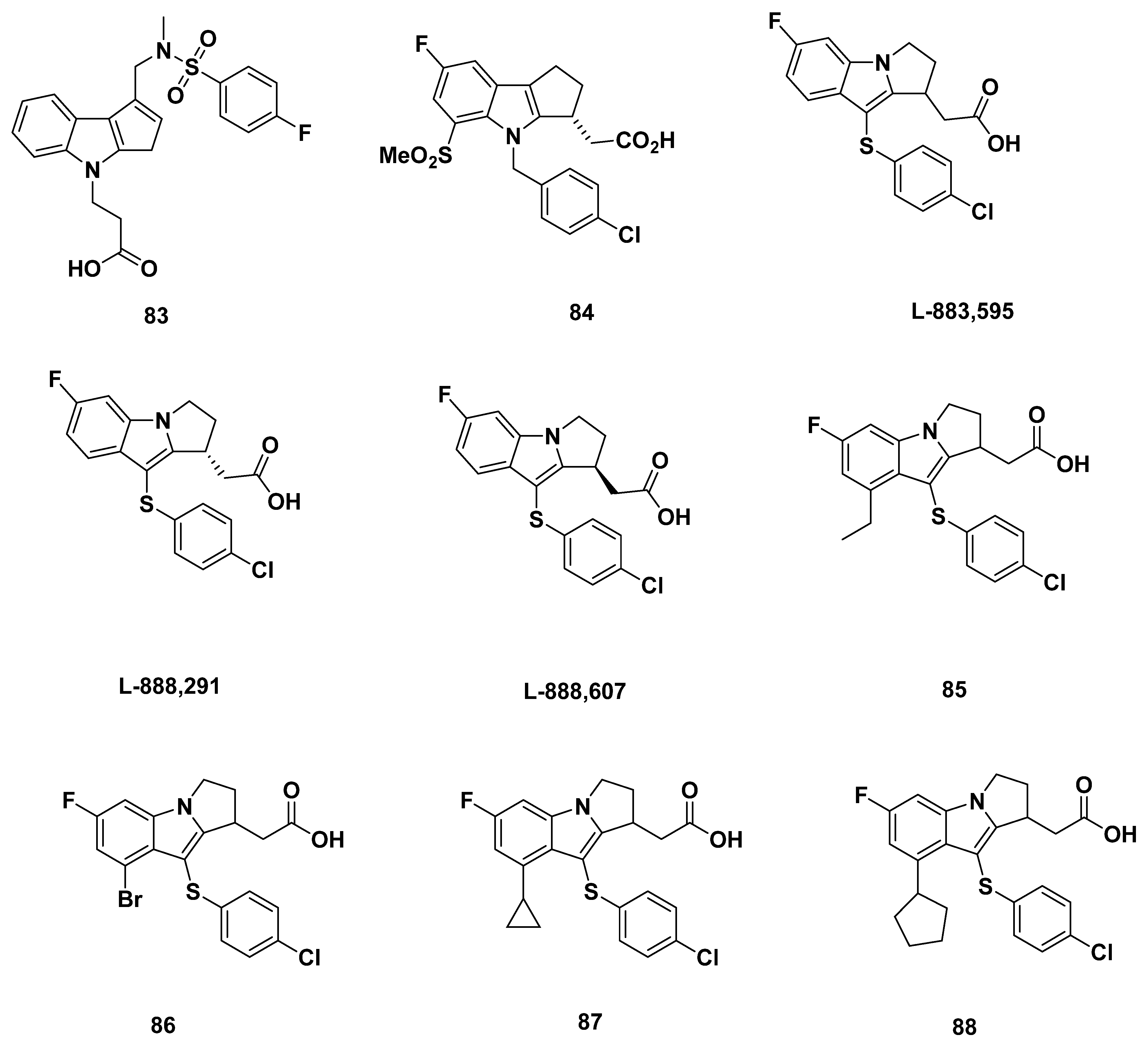
In a 2005 review paper by Ly et al. from Actimis [97], compound 83 (Figure 13) was characterized. The compound description was based off a 2003 patent application WO 03/09742 from Shionogi in Japan. In place of the conventional acetic acid attached to the nitrogen atom of the indole group, compound 83 had an ethanoic acid. It also contained a p-fluorophenyl sulfonamide attached to the cyclopentenyl ring. The compound was reported to inhibit GPR44 at an IC50 value of 37 nM.

Recommendations

Compound 83 does not have a particularly promising IC50 value, so we recommend further characterization of its properties.

3.2. Merck Analogue

3.2.1. Merck Analogue in 2005

In 2005, Campos et al. from Merck characterized the asymmetric synthesis of a reported PGD2 receptor antagonist 84 (Figure 13), but no further details about binding affinity were provided [98].

3.2.2. Recommendations

Due to the paucity of information at this time, we are unable to make a recommendation about this compound.

3.2.3. L-888,607, L-883,595, and L-888,291

In 2005, Gervais et al. from Merck developed L-888,607 (Figure 13), reportedly the first synthetic, potent, and selective GPR44 agonist [69].
At first, the racemic compound L-883,595 (Figure 13) was identified as a base for modification through screening. Although the affinity to GPR44 was optimal at a Ki of 4 nM, L-883,595 was shown to have potential off-target binding to DP1 at a Ki of 211 nM, with both values measured through binding assays.
Separating the enantiomers of L-883,595 resulted in the L-888,291 (Figure 13) and L-888,607, which were the R and S enantiomers, respectively. L-888,291 was not promising, as it had an approximately equal binding affinity for GPR44 and DP1 (48 and 40 nM, respectively).
L-888,607, meanwhile, was promising with a high binding affinity for GPR44 at a Ki of 0.8 nM, and a less concerning binding affinity for DP1 (2331 nM) that was over 1000 times of that for GPR44.
A series of L-883,595 analogues were also characterized, in which substitution at the 6-position of the indole ring was explored. However, all did not exhibit as promising an affinity to GPR44 as L-888,607, although compound 85, produced through substitution by an alkyl group, showed the lowest Ki at 12 nM. Other compounds included compound 86, with a Ki of 21 nM, compound 87, with a Ki of 20 nM, and compound 88, with a Ki of 94 nM.
Further equilibrium binding assays characterized promising properties of L-888,607, with a binding affinity (Ki) 360-fold lower for the TP receptor and more than 1000-fold lower for six other prostanoid receptors (EP1–EP4, FP, and IP). Additionally, potential off-target binding to chemokine receptors (CCR1, CCR2, CCR4, CCR5, CXCR1, CXCR2, and CXCR3), anaphylatoxin receptors (C2aR and C5aR), and the cyclooxygenases-1 and -2 was shown to be minimal, as no significant binding was found at 10 µM L-888,607.
L-888,607 was additionally confirmed as a strong inhibitor through measurements of cAMP inhibition (EC50 = 0.5 nM), eosinophil shape change induction, and eosinophil chemotaxis (at an optimal concentration of 100 nM).
The pharmacokinetic profile was elucidated following intravenous or oral administration to mice. The results showed no causes for concern, with a maximum concentration of 36.1 M, half-life of 2.9 h, trough level at 8 h of 3.5 M, and AUC0–8h of 87.1 M after intravenous administration of 5 mg/kg L-888,607. The oral administration of 20 mg/kg L-888,607 showed similar values, with a maximum concentration at 31.6 M, half-life of 4 h, trough level at 8 h of 15.4 M, and AUC0–8h of 166 M. The bioavailability was sufficient at 48%.

3.2.4. Recommendations

As a result of the high binding affinity and specificity to GPR44, as well as the optimal in vivo pharmacokinetic properties, L-888,607 can be considered for the development of a GPR44 PET tracer.

3.2.5. MK-0524

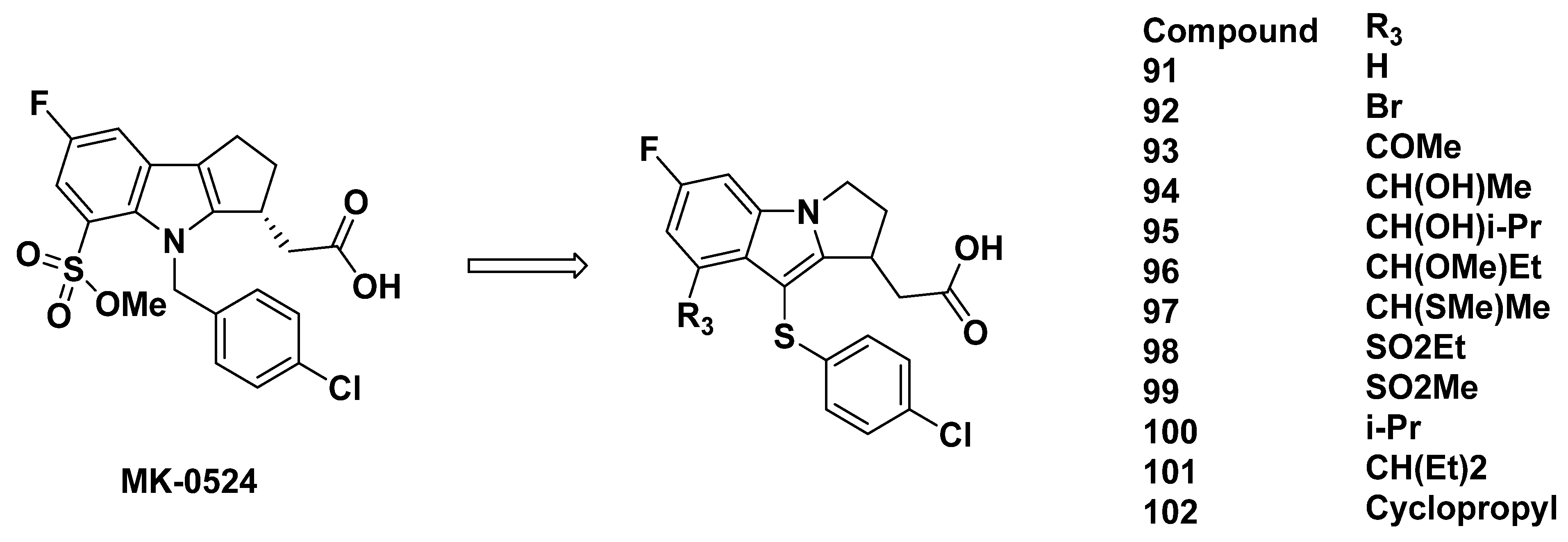
MK-0524, [(3R)-4-(4-chlorobenzyl)-7-fluoro-5-(methylsulfonyl)-1,2,3,4-tetrahydrocyclopenta[b]indol-3-yl] acetic acid, also referred to as laropiprant commercially, was developed by Sturino et al. from Merck in 2007 (Figure 14) [82]. It was initially targeted for the treatment of niacin-induced flushing and was also considered a promising compound to alleviate allergic disorders. MK-0524 was a selective DP antagonist, demonstrating potent inhibition of PGD2-induced cAMP production with a Ki of 0.57 nM, an IC50 of 0.09 nM in washed platelets, and an IC50 of 4.0 nM in platelet-rich plasma [82]. In contrast, MK-0524 exhibited much lower selectivity for TP, as shown by its Ki of 2.95 nM and IC50 of 770 nM in platelet-rich plasma [82].
Due to its desirable selectivity for DP1 and therapeutic potential, MK-0524 has been characterized extensively. A study intravenously administered [14C]MK-0524 in rats, dogs, and monkeys and evaluated its metabolism, pharmacokinetics, and excretion [99]. MK-0524 was metabolized primarily through acyl glucuronidation into the major metabolite, acyl glucuronide, and subsequently excreted via the bile ducts in both rats and dogs. The minor metabolites, monohydroxylated epimers and the keto-metabolite, occurred in trace amounts [100]. Following intravenous dosing at 1 and 5 mg/kg, rats exhibited a low plasma clearance of 2 mL/min/kg, a small volume of distribution of 0.8 L/kg, and a moderately long half-life of 7.5 [99]. The plasma concentrations of acyl glucuronide remained low compared to the parent compound. In dogs receiving 1 and 5 mg/kg intravenous doses, the mean values reflected a similar trend at 6 mL/min/kg, 5 L/kg, and 13 h. Unlike rats, the plasma concentrations of acyl glucuronide in dogs were substantial with AUC (0–∞) and Cmax values ranging from 8 to 18% for MK-0524. The values varied in monkeys after 3 mg/kg intravenous administration, in which the plasma clearance was 8 mL/min/kg, the volume of distribution was low at 1 L/kg, and the half-life was 3 h. Acyl glucuronide plasma concentrations were higher in monkeys than in rats and dogs, with an AUC (0–∞) value of 27%. Thus, the ratio of acyl glucuronidation to the parent compound was found to be species-specific.
Another study showed that MK-0524 underwent qualitatively similar metabolism in humans [100]. Extensive glucuronidation in hepatocytes and liver microsomes was found using human liver microsomes, human intestinal microsomes, and recombinant human uridine diphosphate glucuronosyltransferases. In a clinical study, the metabolism and excretion of [14C]MK-0524 were evaluated in six healthy human participants using 40 mg of oral administration [101]. The results confirmed that the main metabolite was acyl glucuronic acid conjugate, which was by excretion into bile and then feces.

3.2.6. Recommendations

A significant disadvantage of MK-0524 is its off-target DP1 agonist activity. Therefore, MK-0524 is not a suitable molecule for the development of a GPR44 tracer.

3.2.7. Merck Analogues in 2007

In 2007, Sturino et al. from Merck reported two additional analogues that exhibited improved DP1 selectivity [82]. The bromide compound 89 (Figure 14) was measured for DP1 binding affinity, and showed a Ki 1.5 nM. The methylketone compound 90 (Figure 14), a transformed product of compound 89, had a similarly strong binding affinity at a Ki of 1.1 nM. The IC50 values for DP in washed platelets and platelet-rich plasma were similar for both compounds 89 (0.26 nM and 12 nM, respectively) and 90 (0.29 nM and 15 nM, respectively), although the platelet-rich plasma Ki values were interestingly much higher than the other assays.
The ligand-receptor interactions of compound 89′s binding to DP1 were elucidated by Mu et al. of Sanofi Aventis in 2011 [83].
Similarly, the IC50 values for binding to TP in platelet-rich plasma were higher at 80 and 1400 nM, respectively. The concern of the selectivity of both compounds was further confirmed by Ki values, as compounds 8990 exhibited a binding affinity for TP of 14 and 0.84 nM, respectively.
As measured in rat hepatocytes, compounds 8990 were shown to be largely metabolized, with only 42% and 35% remaining, respectively, following 2 h incubation. In pharmacodynamic characterizations of the compounds in rats, compounds 8990 continued to show optimal properties, including adequate half-lives at 12 and 4 h, respectively, moderate bioavailability at 41% and 52%, respectively, and low plasma clearance of 1.4 and 1.9 mL/min, respectively. The Cmax (10 and 21 10 mg/kg, respectively) concentration 6 h post-dose (3.7 and 7.5 10 mg/kg, respectively), and volume of distribution (0.4 and 0.5, respectively) values also were of little cause for concern.
In the metabolic route, intravenous cannulation surveying found that compounds 8990 did not accumulate in the bile fluid, as Cmax was 29 and 91 µM, respectively. The tmax of the compounds reflected the maximum concentration at values of 1–1.5 h and 2.5–3 h, respectively. Hydroxylation and glucuronidation were cited as the main routes of metabolism.
In further investigation of the profile of compounds 8990, the compounds were radiolabeled with [14C] and studied in rat and human microsomes and hepatocytes. The covalent binding of compound 89 was equivalent at 33 pmol equiv/mg of protein per h for both human microsomes and hepatocytes, but varied from 460 to 74 pmol equiv/mg·h, respectively, in rat microsomes and hepatocytes. The variation for compound 90 was more severe with rat microsomes (290 pmol equiv/mg·h), rat hepatocytes (45 pmol equiv/mg·h), human microsomes (46 pmol equiv/mg·h), and human hepatocytes (16 pmol equiv/mg·h) all differing. The microsomal system often afforded values higher than the hepatocyte system, which may be due to the action of reactive intermediates. The high binding in at least the rat system was promising, but the covalent binding was low in the human system. These disparate results may indicate species-specific antagonism, potentially limiting the in vivo testing of compounds 8990.

3.2.8. Recommendations

Due to the uncertainty of the binding affinity to GPR44, strong off-target binding to DP1, and potential off-target binding to TP, compounds 8990 are not recommended.

3.2.9. Merck Analogues in 2008

Another series of compounds based on MK-0524 was developed by Beaulieu et al. from Merck in 2008 [102]. In these compounds, the nitrogen atom and 3-position carbon on the indole of MK-0524 were exchanged (Figure 15).
The compounds 91102 (Figure 15) resulted from substitutions at the 4-position carbon on the indole group. Among the various substituents, compounds 96 and 99 showed good binding affinity to DP1 with Ki values of less than 2 nM and IC50 values in platelet-rich plasma of 6.7 and 10.4 nM, respectively. The selectivity was also strong, as the compounds were 170-fold more selective for DP1 than TP. The remaining compounds did not exhibit either high enough binding affinity or selectivity for DP1.
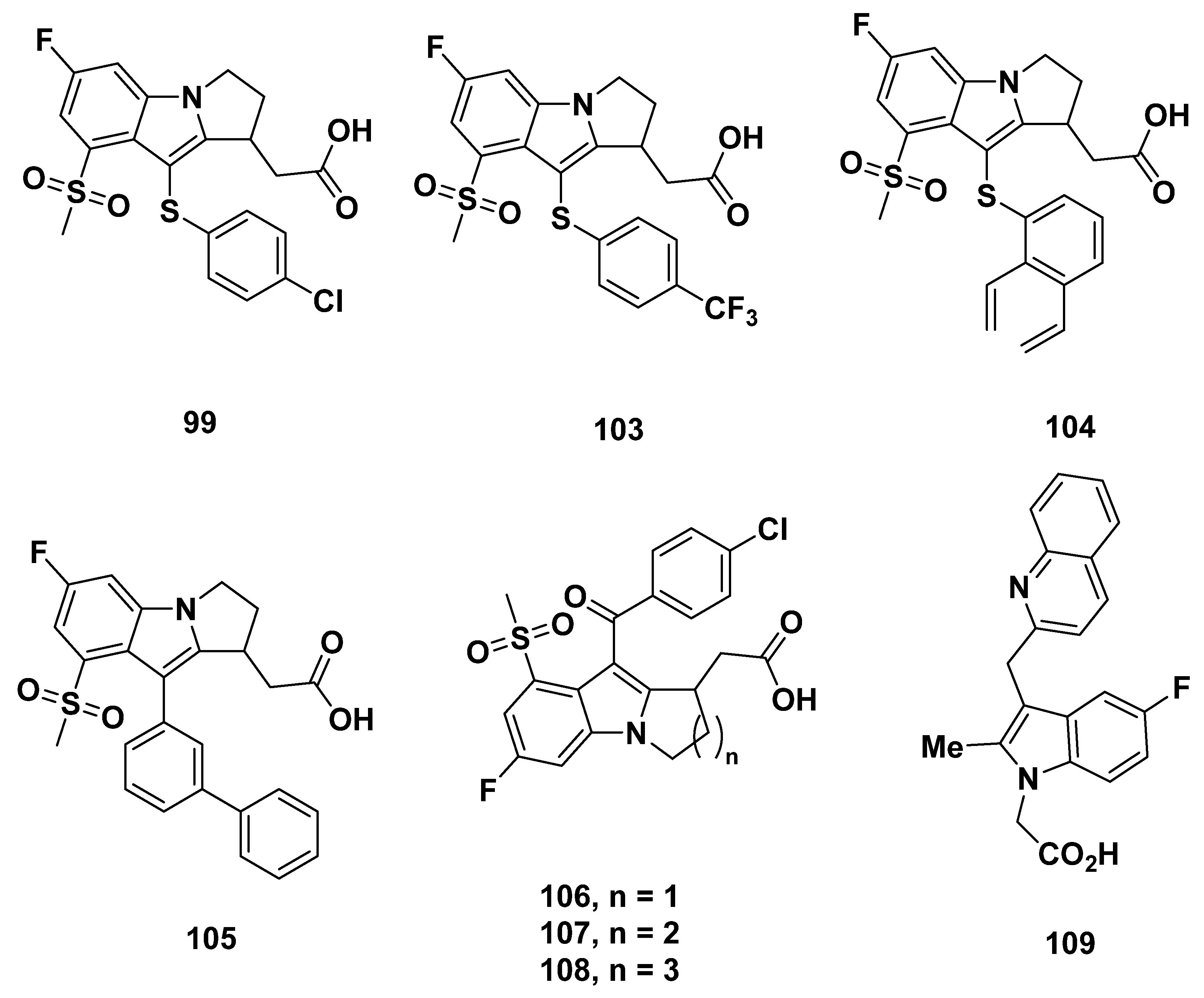
Through substitution of the 3-position carbon of the indole group, compounds 103105 were developed (Figure 16). All of the compounds were strong DP1 antagonists, with a Ki of less than 2 nM and IC50 at most 40 nM. The selectivity for DP1 over TP was also over 150-fold.
Further development led to compounds 106108, in which the number of carbons on the indole-fused ring differs. The binding affinity, represented by Ki and IC50 values, appeared to decrease as ring size increased. The Ki value was the lowest for compound 108 at 1.4 nM and IC50 of 5.2 nM, although the selectivity may be a cause for concern at a TP Ki of 675 nM. Whereas compound 107 exhibited similar properties to compound 108, compound 106 had a much greater selectivity with a TP Ki of 6972 nM, but lowered binding affinity at a Ki for DP1 of 6.9 nM.

3.2.10. Recommendations

As a consequence of the unidentified GPR44 binding affinity and potential off-target binding to DP1, these compounds may not serve as optimal GPR44 PET tracers.

3.3. Astellas Analogues in 2008

Compound 109 (Figure 16) was briefly described as an indole acetic acid in a 2012 paper by Ito et al. from Astellas, and cited in a 2005 patent by Middlemiss et al. [103].

Recommendations

We are unable to make a recommendation due to the lack of information about this compound.

4. Potential Fluorination Methods

According to the strategies discussed above, Eriksson, O. et al. have developed 11C-MK-7246 [104,105] and 18F-MK-7246 [106] from MK-7246, and our team has developed 18F-TM30089 from TM30089 and two other 18F-labeled GPR44 PET tracers [107]. These potential 18F-labeled PET tracers displayed high specific binding to GPR44 in murine pig and monkey. It validated the upside of such a strategy, i.e., the prominent GPR44 ligands have been thoroughly characterized (affinity, lipophilicity, protein binding, etc.), which makes it a highly efficient process to screen the most promising GPR44 PET tracers.
For the radiolabeling strategies for developing 18F-labeled GPR44 ligands from “cold” GPR44 candidates (Figure 17), there are many publications about methodologies for 18F-labeling [108,109,110]. Direct and indirect radiolabeling methods may be utilized to incorporate fluorine-18.

4.1. Concerted Nucleophilic Aromatic Substitution (CSNAr) with 18F-

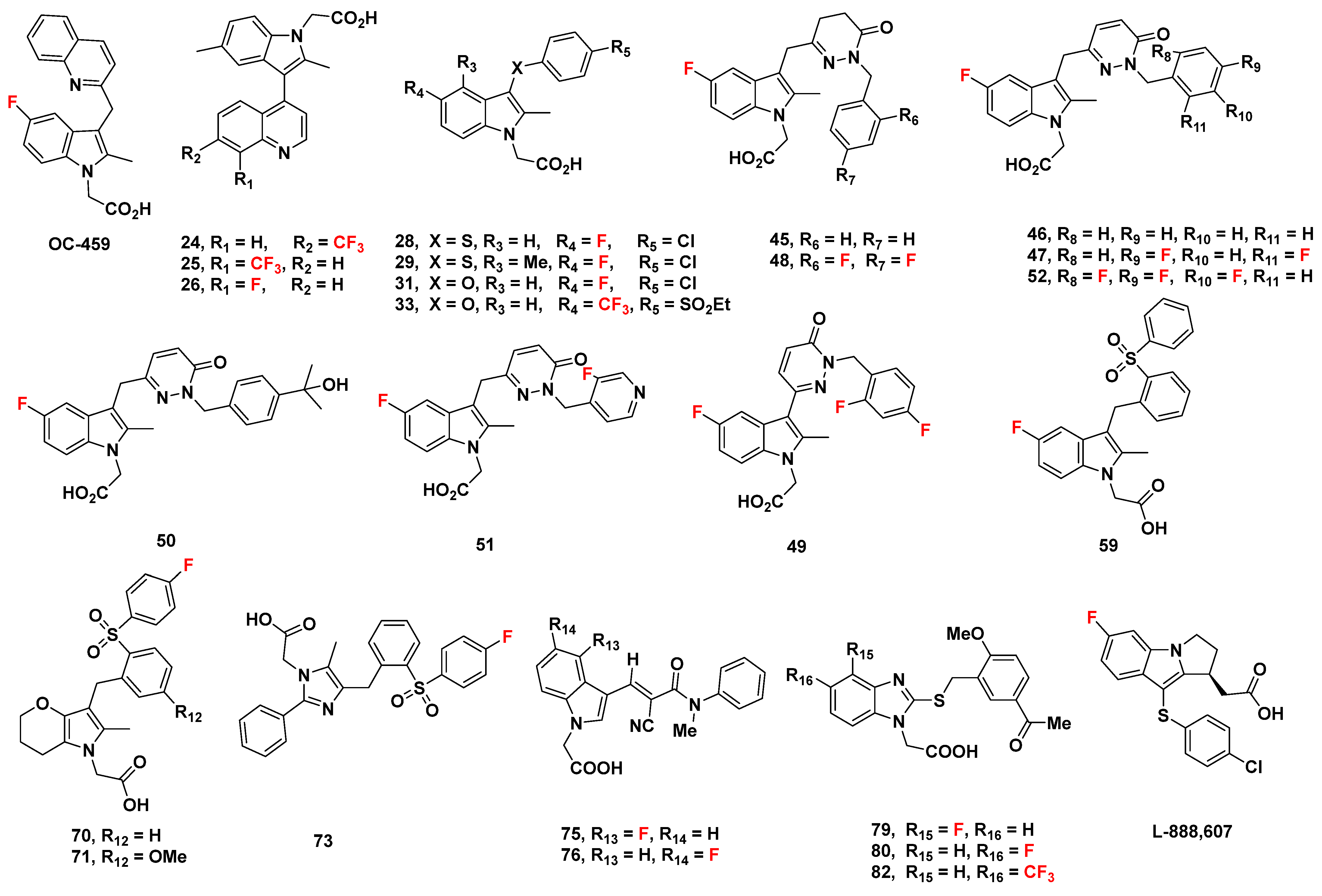
Orit Jacobson (2015) stated that “18F nucleophilic aromatic substitution (SNAr) requires sufficient activation of the phenyl ring, which can be achieved by electron withdrawing group(s) (such as −NO2, −CN, −CF3, carbonyl, or sulfur dioxide groups) in the ortho or para position to the leaving group” [110]. The most common and efficient leaving groups for no-carrier-added nucleophilic aromatic substitutions are trimethylammonium salt and the nitro group [109]. This 18F-labeling method is potentially suitable for compounds such as compounds 70, 71, and 73 (Figure 18). Lower temperatures of fluorination correspond to the precursors with a trimethylammonium group compared with nitro group-containing precursors.
Traditional two-step SNAr (nucleophilic aromatic substitution) requires the presence of an electron-withdrawing group located ortho or para to the leaving group, a constraint that severely limits its applicability. In order to enable the rate-determining step of SNAr (addition of the 18F nuclide to form the Meisenheimer complex), the ring must be pi-deficient, as only pi-deficient arenes can adequately stabilize the anionic Meisenheimer complex [111].
However, a concerted nucleophilic aromatic substitution reaction (CSNAr) has been presented by Constanze N. Neumann et al., in which substitution is not restricted to electron-deficient arenes, as it does not proceed via the formation of a Meisenheimer intermediate (Figure 19) [112]. They show that the deoxyfluorination of phenols with PhenoFluor favors CSNAr over conventional stepwise displacement, demonstrating that it is a viable alternative to two-step SNAr. By combining the addition (nucleophilic attack) and elimination steps into a single, concerted step in CSNAr, there is significantly less build-up of negative charge, lowering the activation energy barrier and removing the need for electron-withdrawing groups to stabilize the anionic intermediate.
One potential limitation may be present in the elution procedure outlined by Constanze N. Neumann et al., which, despite eliminating the need for the azeotropic drying of 18F-fluoride, cannot be used with substrates containing carboxylic acids, as carboxylic acid prevents the formation of key uronium intermediate [112]. There are certainly different ways to modify the procedure to accommodate substrates with carboxylic acid, but further experimental verification would be necessary to ensure viability. Pre-treating the carboxylate group of precursors with a cation [K+c2.2.2]2C2O4 to make a cationic chelate [K+c2.2.2]2CO3 could potentially improve this drawback.
CSNAr is potentially suitable for labeling all 26 promising GPR44 candidates. However, while it can be a powerful method for the synthesis of radiolabeled compounds, potentially offering improved yields compared to traditional methods, each specific reaction still requires substantial experimental validation to precisely understand its efficacy.

4.2. Other Traditional Approaches

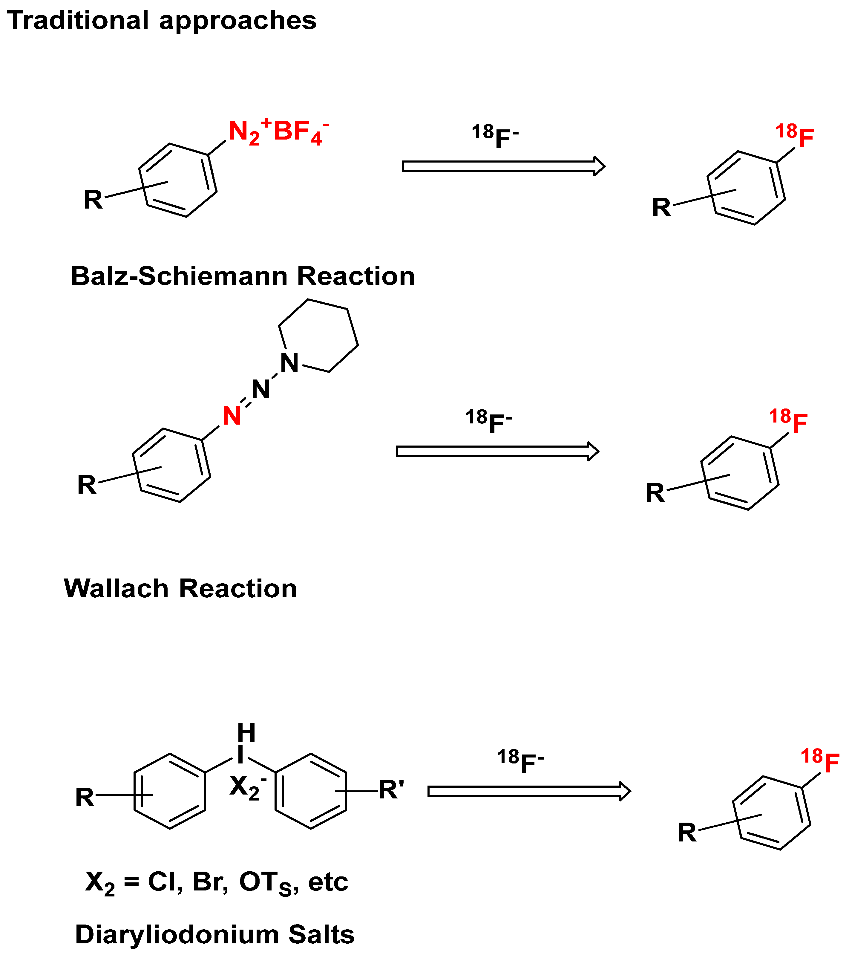
Labeling aromatic rings with 18F in the absence of SNAr-activating groups and in a regiospecific manner could be accomplished (Figure 20) using the Balz−Schiemann reaction [113], Wallach Reaction [114], and Diaryliodonium Salts method [115,116].
The Balz-Schiemann reaction, used to convert aromatic amines into aromatic fluorides, involves the synthesis of a dry aryldiazonium tetrafluoroborate from an aromatic amine, followed by thermal decomposition into the desired aromatic fluoride, boron trifluoride, and nitrogen. The significance of this method arises from its wide applicability in fluorinating both electron-excessive and electron-deficient aryl amines.
However, it is limited by several factors: (1) substrate scope, restricted to primary aromatic amines, (2) low specific activity caused by competing incorporation of non-radioactive fluoride from monolabeled BF4, (3) a maximum theoretical RCY of 25% due to isotopic exchange, (4) byproducts, as a significant amount of potentially toxic waste may be produced, (5) reaction conditions, as stringent anhydrous conditions and high temperatures are required [117].
These limitations have been at least partially addressed by modifications (e.g., photoredox catalysis, hypervalent iodine (III) catalysis, continuous flow, specialized low-polarity solvents, in situ diazotization with tert-butyl nitrite or [NO] [BF4], etc.) [118].
Because the Balz-Schiemann reaction is applicable only to primary aromatic amines, it may not be suitable for compounds 24, 25, 33, and 82. Additionally, because diazotiza-tion typically requires acidic conditions, it may not be suitable for compounds containing functional groups susceptible to hydrolysis (e.g., esters and amides). Strongly deactivating groups on the aromatic ring (e.g., –NO2, –CF3, –C(=O) R, –C(=O) H, –SO3H, etc.) may prevent formation of the diazonium ion. This reaction may be suitable for compounds OC-459, L-888,607, 26, 28, 29, 31, 59, 79, and 80.
Another potential fluorination method, the Wallach reaction, involves the thermal decomposition of an aryl triazine to an aryldiazonium salt under acidic conditions. The greater stability of triazine compared to diazonium salts gives the Wallach reaction a significant advantage over the Balz-Schiemann reaction [118]. Additionally, triazene-masked diazonium ions can be utilized not only in solution-based reaction processes but also on solid substrates, making them compatible with different combinatorial techniques. However, the two-step elimination-addition process (through a cationic intermediate, which can react with any existing nucleophile) of the Wallach reaction could result in the production of multiple byproducts and subsequently a reduced RCY. More detailed information (for example, via further testing) regarding its functional group compatibility is necessary to determine whether or not it is suitable for fluorinating each GPR44 ligand [110].
Diaryliodonium salts, a category of hypervalent iodine (III) reagents possessing some covalent properties (demonstrated by X-ray structures), are used as versatile reagents in the synthesis of aryl fluorides. Their relative electrophilicity (compared to halide anion salts) and good solubility in most organic solvents makes them promising for use in nucleophilic aromatic fluorination [119].
A Cu-catalyzed approach using KF as a source of fluorine (explored by Naoko Ichiishi et. al.) possesses wide substrate applicability, high selectivity, and high functional group tolerance. This addresses several limitations of fluorination using diaryliodonium salts: high temperatures, aryl exchange limiting RCY, and production of unwanted regioisomers. (tBuCN)2Cu(OTf), CuI(OTf)·benzene, CuII(OTf)2 were all shown to be effective catalysts for the fluorination of compounds with the general structure [Mes–I–Ar]BF4 (with Ar representing an electron-rich aromatic ring), demonstrating considerable potential for further exploration with other traditionally challenging precursors. Cu catalysis also significantly enhanced selectivity and yield in substrates bearing electron-withdrawing groups as well [120].
The regioselectivity of electrophilic fluorination using 18F-F2 can be improved using aryltrimethyltin as a precursor, producing fewer byproducts and a higher RCY [110].

4.3. Novel 18F-Labeling Strategies

Wei Chen et al. has developed a method for the fluorination of aromatic C-H bonds using a [18F] F- salt under blue light illumination, a strategy demonstrated to be effective in tracer studies conducted on mice. This method uses single-electron photooxidants to catalytically produce cationic arene radicals from the original aromatic substrates, serving as reactive intermediates that can more readily undergo substitution reactions via nucleophilic attack (thus enabling C-18F bond conversion) [121].
A later report demonstrates that polarity-reversed photoredox-catalyzed deoxyfluorination, proceeding via cation radical accelerated SNAr, allows for the fluorination of electron-rich aromatic compounds at mild temperatures (mitigating the need for electron-withdrawing groups). Their approach targets the C-O bond in phenol derivatives, establishing a method for unsymmetrical diaryl ethers that relies on the preferential oxidation of the more electron-rich aromatic ring. Utilizing an acridinium-based photooxidant, [18F] TBAF, and excess phase-transfer reagent TBAHCO3 (minimizing Hofmann elimination of [18F] TBAF) under 450 nm LEDs, they obtained excellent RCYs while minimizing byproduct formation [122].
Ultimately, the feasibility of such reactions would highly depend on the specific substrates and reaction conditions. Thus, while theoretically possible, whether a specific photoredox-catalyzed SNAr reaction could occur would need to be confirmed experimentally.

4.4. [18F] Trifluoromethylation (Compounds 24, 25, 33, and 82)

The earliest 18F-trifluoromethylation methods rely on halex exchange, a type of SNAr involving the replacement of an aromatic halogen with a different nucleophilic halide. However, such techniques are limited in substrate scope (applicable only to electron-deficient arenes), radiochemical yield, and specific activity [123]. Furthermore, their usage of aryl difluoromethylene precursors with halide-based leaving groups necessitates stringent conditions to enable the rate-determining elimination step.
An alternative method, [18F] fluorodecarboxylation proceeding via the Ag(I)-catalyzed electrophilic addition of [18F] F- from [18F] Selectfluor bis(triflate), has been previously used to synthesize 18F-trifluoromethylarenes. While it is applicable to a broader range of substrates and possesses wider functional group tolerance than nucleophilic halex exchange, its efficacy is limited by its operational complexity [123,124].
A more recent fluorination method developed by Gouverneur et al. in 2013 involves the Cu(I)-catalyzed single-operation installation of [18F]CF3 into a (hetero)aryl iodide precursor, directly forming [18F]CF3 (hetero)arenes and negating the requirement for an arylCF2 precursor possessing a distinct leaving group favorable for [18F] F- substitution. In this approach, the (hetero)aryl-CF2-18F linkage is directly constructed through two main steps: (1) halide-induced decarboxylation of methyl chlorodifluoroacetate in the presence of [18F] F-, producing a trifluoromethide intermediate that can be captured by copper iodide to form [18F] CuCF3, (2) cross-coupling of [18F] CuCF3 with the (hetero)aryl iodide precursor, producing the desired [18F] trifluoromethyl (hetero)arene [124]. Riss et al. presents an alternative method for forming [18F] CuCF3 via a similar stepwise process that uses CHF2I in place of methyl chlorodifluoroacetate [125]. This approach offers several significant advantages, namely wide functional group tolerance, high RCY and regioselectivity, and amenability to a variety of bioactive molecules, which is especially relevant when developing potential GPR44 PET tracers. However, a limitation of this method is its incompatibility with unprotected amines, alcohols, and carboxylic acids. This could partially be resolved by protecting amine and carboxylate groups as carbamates and esters, respectively, then removing these protecting groups once the reaction is complete [126].
Cu(I)-catalyzed trifluoromethylation of heteroaryl iodides appears to be promising for the labeling of compounds 24, 25, 44, and 82, though experimental validation is necessary to ensure that this method is applicable to each specific target molecule.

5. Conclusions

In addition to ramatroban analogues evaluated previously, there are a number of indole-based and cyclopentenylindole-based compounds with fluorine atoms that demonstrated high affinity and selectivity to GPR44. These compounds have the potential to be developed into GPR44 18F-labeled PET tracers for in vivo imaging of inflammation-related research, which can provide important insights into inflammation-cancer dynamics.

Author Contributions

Writing—original draft preparation, R.Y., K.X.H., L.A.H., M.J., H.Z., K.L., A.G., T.L. and J.L.; writing—review and editing, R.Y., K.X.H., L.A.H., M.J., H.Z., K.L., A.G., J.C., Z.L., T.L., J.E.S., F.K. and J.L. All authors have read and agreed to the published version of the manuscript.

Funding

This work was supported by the Oxnard Foundation (Grant No. 50214-2012049).

Institutional Review Board Statement

Not applicable.

Informed Consent Statement

Not applicable.

Data Availability Statement

Data sharing is not applicable.

Acknowledgments

We acknowledge financial support to this work from the Oxnard Foundation Project for Non-Invasive Imaging.

Conflicts of Interest

The authors declare no conflict of interest.

References

  1. Greten, F.R.; Grivennikov, S.I. Inflammation and Cancer: Triggers, Mechanisms, and Consequences. Immunity 2019, 51, 27–41. [Google Scholar] [CrossRef] [PubMed]
  2. Sever, R.; Brugge, J.S. Signal transduction in cancer. Cold Spring Harb. Perspect. Med. 2015, 5, a006098. [Google Scholar] [CrossRef] [PubMed]
  3. Zhao, H.; Wu, L.; Yan, G.; Chen, Y.; Zhou, M.; Wu, Y.; Li, Y. Inflammation and tumor progression: Signaling pathways and targeted intervention. Signal Transduct. Target. Ther. 2021, 6, 263. [Google Scholar] [CrossRef]
  4. Harjunpaa, H.; Asens, M.L.; Guenther, C.; Fagerholm, S.C. Cell Adhesion Molecules and Their Roles and Regulation in the Immune and Tumor Microenvironment. Front. Immunol. 2019, 10, 1078. [Google Scholar] [CrossRef] [PubMed]
  5. Murata, T.; Maehara, T. Discovery of anti-inflammatory role of prostaglandin D2. J. Vet. Med. Sci. 2016, 78, 1643–1647. [Google Scholar] [CrossRef]
  6. Elinav, E.; Nowarski, R.; Thaiss, C.A.; Hu, B.; Jin, C.C.; Flavell, R.A. Inflammation-induced cancer: Crosstalk between tumours, immune cells and microorganisms. Nat. Rev. Cancer 2013, 13, 759–771. [Google Scholar] [CrossRef]
  7. Mantovani, A.; Allavena, P.; Sica, A.; Balkwill, F. Cancer-related inflammation. Nature 2008, 454, 436–444. [Google Scholar] [CrossRef] [PubMed]
  8. Jandl, K.; Heinemann, A. The therapeutic potential of CRTH2/DP2 beyond allergy and asthma. Prostaglandins Other Lipid Mediat. 2017, 133, 42–48. [Google Scholar] [CrossRef]
  9. Shioga, T.; Kondo, R.; Ogasawara, S.; Akiba, J.; Mizuochi, S.; Kusano, H.; Mihara, Y.; Tanigawa, M.; Kinjyo, Y.; Naito, Y.; et al. Usefulness of Tumor Tissue Biopsy for Predicting the Biological Behavior of Hepatocellular Carcinoma. Anticancer Res. 2020, 40, 4105–4113. [Google Scholar] [CrossRef]
  10. Wang, S.M.; Zhang, K.; Tan, S.Y.; Xin, J.Y.; Yuan, Q.Y.; Xu, H.H.; Xu, X.; Liang, Q.; Christiani, D.C.; Wang, M.L.; et al. Circular RNAs in body fluids as cancer biomarkers: The new frontier of liquid biopsies. Mol. Cancer 2021, 20, 13. [Google Scholar] [CrossRef]
  11. Unterrainer, M.; Eze, C.; Ilhan, H.; Marschner, S.; Roengvoraphoj, O.; Schmidt-Hegemann, N.S.; Walter, F.; Kunz, W.G.; Rosenschold, P.M.A.; Jeraj, R.; et al. Recent advances of PET imaging in clinical radiation oncology. Radiat. Oncol. 2020, 15, 88. [Google Scholar] [CrossRef] [PubMed]
  12. Ming, Y.; Wu, N.; Qian, T.; Li, X.; Wan, D.Q.; Li, C.; Li, Y.; Wu, Z.; Wang, X.; Liu, J.; et al. Progress and Future Trends in PET/CT and PET/MRI Molecular Imaging Approaches for Breast Cancer. Front. Oncol. 2020, 10, 1301. [Google Scholar] [CrossRef]
  13. Liu, C.H.; Abrams, N.D.; Carrick, D.M.; Chander, P.; Dwyer, J.; Hamlet, M.R.J.; Kindzelski, A.L.; PrabhuDas, M.; Tsai, S.A.; Vedamony, M.M.; et al. Imaging inflammation and its resolution in health and disease: Current status, clinical needs, challenges, and opportunities. FASEB J. 2019, 33, 13085–13097. [Google Scholar] [CrossRef] [PubMed]
  14. Gangadaran, P.; Ahn, B.C. Molecular Imaging: A Useful Tool for the Development of Natural Killer Cell-Based Immunotherapies. Front. Immunol. 2017, 8, 1090. [Google Scholar] [CrossRef]
  15. Kang, N.Y.; Soetedjo, A.A.P.; Amirruddin, N.S.; Chang, Y.T.; Eriksson, O.; Teo, A.K.K. Tools for Bioimaging Pancreatic beta Cells in Diabetes. Trends Mol. Med. 2019, 25, 708–722. [Google Scholar] [CrossRef]
  16. Tarkin, J.M.; Joshi, F.R.; Evans, N.R.; Chowdhury, M.M.; Figg, N.L.; Shah, A.V.; Starks, L.T.; Martin-Garrido, A.; Manavaki, R.; Yu, E.; et al. Detection of Atherosclerotic Inflammation by 68Ga-DOTATATE PET Compared to [18F]FDG PET Imaging. J. Am. Coll. Cardiol. 2017, 69, 1774–1791. [Google Scholar] [CrossRef] [PubMed]
  17. Emami, H.; Singh, P.; MacNabb, M.; Vucic, E.; Lavender, Z.; Rudd, J.H.; Fayad, Z.A.; Lehrer-Graiwer, J.; Korsgren, M.; Figueroa, A.L.; et al. Splenic metabolic activity predicts risk of future cardiovascular events: Demonstration of a cardiosplenic axis in humans. JACC Cardiovasc. Imaging 2015, 8, 121–130. [Google Scholar] [CrossRef]
  18. Kwiecinski, J.; Dey, D.; Cadet, S.; Lee, S.E.; Otaki, Y.; Huynh, P.T.; Doris, M.K.; Eisenberg, E.; Yun, M.; Jansen, M.A.; et al. Peri-Coronary Adipose Tissue Density Is Associated With 18F-Sodium Fluoride Coronary Uptake in Stable Patients With High-Risk Plaques. JACC Cardiovasc. Imaging 2019, 12, 2000–2010. [Google Scholar] [CrossRef]
  19. Grobman, M.; Cohn, L.; Knapp, S.; Bryan, J.N.; Reinero, C. F-18-FDG-PET/CT as adjunctive diagnostic modalities in canine fever of unknown origin. Vet. Radiol. Ultrasoun. 2018, 59, 107–115. [Google Scholar] [CrossRef]
  20. Coughlin, J.M.; Wang, Y.; Minn, I.; Bienko, N.; Ambinder, E.B.; Xu, X.; Peters, M.E.; Dougherty, J.W.; Vranesic, M.; Koo, S.M.; et al. Imaging of Glial Cell Activation and White Matter Integrity in Brains of Active and Recently Retired National Football League Players. JAMA Neurol. 2017, 74, 67–74. [Google Scholar] [CrossRef]
  21. Cipriano, P.W.; Yoon, D.; Gandhi, H.; Holley, D.; Thakur, D.; Hargreaves, B.A.; Kennedy, D.J.; Smuck, M.W.; Cheng, I.; Biswal, S. F-18-FDG PET/MRI in Chronic Sciatica: Early Results Revealing Spinal and Nonspinal Abnormalities. J. Nucl. Med. 2018, 59, 967–972. [Google Scholar] [CrossRef] [PubMed]
  22. Neumann, T.; Oelzner, P.; Freesmeyer, M.; Hansch, A.; Opfermann, T.; Hein, G.; Wolf, G. Images in cardiovascular medicine. Diagnosis of large-vessel vasculitis by [18F] fluorodeoxyglucose-positron emission tomography. Circulation 2009, 119, 338–339. [Google Scholar] [CrossRef] [PubMed]
  23. Hess, S.; Madsen, P.H.; Iversen, E.D.; Frifelt, J.J.; Hoilund-Carlsen, P.F.; Alavi, A. Efficacy of FDG PET/CT imaging for venous thromboembolic disorders: Preliminary results from a prospective, observational pilot study. Clin. Nucl. Med. 2015, 40, e23–e26. [Google Scholar] [CrossRef]
  24. Tawakol, A.; Ishai, A.; Takx, R.A.P.; Figueroa, A.L.; Ali, A.; Kaiser, Y.; Truong, Q.A.; Solomon, C.J.E.; Calcagno, C.; Mani, V.; et al. Relation between resting amygdalar activity and cardiovascular events: A longitudinal and cohort study. Lancet 2017, 389, 834–845. [Google Scholar] [CrossRef]
  25. Rondina, M.T.; Lam, U.T.; Pendleton, R.C.; Kraiss, L.W.; Wanner, N.; Zimmerman, G.A.; Hoffman, J.M.; Hanrahan, C.; Boucher, K.; Christian, P.E.; et al. (18)F-FDG PET in the evaluation of acuity of deep vein thrombosis. Clin. Nucl. Med. 2012, 37, 1139–1145. [Google Scholar] [CrossRef]
  26. Marchese, A.; George, S.R.; Kolakowski, L.F., Jr.; Lynch, K.R.; O’Dowd, B.F. Novel GPCRs and their endogenous ligands: Expanding the boundaries of physiology and pharmacology. Trends Pharmacol. Sci. 1999, 20, 370–375. [Google Scholar] [CrossRef]
  27. Haba, R.; Shintani, N.; Onaka, Y.; Kanoh, T.; Wang, H.; Takenaga, R.; Hayata, A.; Hirai, H.; Nagata, K.Y.; Nakamura, M.; et al. Central CRTH2, a second prostaglandin D2 receptor, mediates emotional impairment in the lipopolysaccharide and tumor-induced sickness behavior model. J. Neurosci. 2014, 34, 2514–2523. [Google Scholar] [CrossRef]
  28. Wang, L.; Yao, D.; Deepak, R.; Liu, H.; Xiao, Q.; Fan, H.; Gong, W.; Wei, Z.; Zhang, C. Structures of the Human PGD2 Receptor CRTH2 Reveal Novel Mechanisms for Ligand Recognition. Mol. Cell. 2018, 72, 48–59.e4. [Google Scholar] [CrossRef]
  29. Eriksson, O. GPR44 as a Target for Imaging Pancreatic Beta-Cell Mass. Curr. Diabetes Rep. 2019, 19, 49. [Google Scholar] [CrossRef]
  30. Domingo, C.; Palomares, O.; Sandham, D.A.; Erpenbeck, V.J.; Altman, P. The prostaglandin D2 receptor 2 pathway in asthma: A key player in airway inflammation. Respir. Res. 2018, 19, 189. [Google Scholar] [CrossRef]
  31. Pettipher, R.; Hansel, T.T.; Armer, R. Antagonism of the prostaglandin D2 receptors DP1 and CRTH2 as an approach to treat allergic diseases. Nat. Rev. Drug Discov. 2007, 6, 313–325. [Google Scholar] [CrossRef] [PubMed]
  32. Uhlen, M.; Fagerberg, L.; Hallstrom, B.M.; Lindskog, C.; Oksvold, P.; Mardinoglu, A.; Sivertsson, A.; Kampf, C.; Sjostedt, E.; Asplund, A.; et al. Proteomics. Tissue-based map of the human proteome. Science 2015, 347, 1260419. [Google Scholar] [CrossRef] [PubMed]
  33. Eriksson, O.; Johnstrom, P.; Cselenyi, Z.; Jahan, M.; Selvaraju, R.K.; Jensen-Waern, M.; Takano, A.; Sorhede Winzell, M.; Halldin, C.; Skrtic, S.; et al. In Vivo Visualization of beta-Cells by Targeting of GPR44. Diabetes 2018, 67, 182–192. [Google Scholar] [CrossRef] [PubMed]
  34. Xue, L.; Salimi, M.; Panse, I.; Mjosberg, J.M.; McKenzie, A.N.; Spits, H.; Klenerman, P.; Ogg, G. Prostaglandin D2 activates group 2 innate lymphoid cells through chemoattractant receptor-homologous molecule expressed on TH2 cells. J. Allergy Clin. Immunol. 2014, 133, 1184–1194. [Google Scholar] [CrossRef]
  35. Moraes-Ferreira, R.; Brandao-Rangel, M.A.R.; Gibson-Alves, T.G.; Silva-Reis, A.; Souza-Palmeira, V.H.; Aquino-Santos, H.C.; Frison, C.R.; Oliveira, L.V.F.; Albertini, R.; Vieira, R.P. Physical Training Reduces Chronic Airway Inflammation and Mediators of Remodeling in Asthma. Oxidative Med. Cell. Longev. 2022, 2022, 5037553. [Google Scholar] [CrossRef]
  36. Wang, J.J.; Mak, O.T. Induction of apoptosis in non-small cell lung carcinoma A549 cells by PGD2 metabolite, 15d-PGJ2. Cell Biol. Int. 2011, 35, 1089–1096. [Google Scholar] [CrossRef]
  37. Zhang, B.; Bie, Q.; Wu, P.; Zhang, J.; You, B.; Shi, H.; Qian, H.; Xu, W. PGD2/PTGDR2 Signaling Restricts the Self-Renewal and Tumorigenesis of Gastric Cancer. Stem. Cells 2018, 36, 990–1003. [Google Scholar] [CrossRef]
  38. Qian, F.; Arner, B.E.; Kelly, K.M.; Annageldiyev, C.; Sharma, A.; Claxton, D.F.; Paulson, R.F.; Prabhu, K.S. Interleukin-4 treatment reduces leukemia burden in acute myeloid leukemia. FASEB J. 2022, 36, e22328. [Google Scholar] [CrossRef] [PubMed]
  39. Caligiuri, M.; Li, Z.; Ma, R.; Tang, H.; Zhang, J.; Marcucci, G.; Yu, J. Human ILC1s target leukemia stem cells and control development of AML. Res. Sq. 2023. [Google Scholar] [CrossRef]
  40. Dash, P.; Ghatak, S.; Topi, G.; Satapathy, S.R.; Ek, F.; Hellman, K.; Olsson, R.; Mehdawi, L.M.; Sjolander, A. High PGD2 receptor 2 levels are associated with poor prognosis in colorectal cancer patients and induce VEGF expression in colon cancer cells and migration in a zebrafish xenograft model. Br. J. Cancer 2022, 126, 586–597. [Google Scholar] [CrossRef]
  41. Ma, X.B.; Xu, Y.Y.; Zhu, M.X.; Wang, L. Prognostic Signatures Based on Thirteen Immune-Related Genes in Colorectal Cancer. Front. Oncol. 2020, 10, 591739. [Google Scholar] [CrossRef]
  42. Wang, P.P.; Fu, Y.; Chen, Y.Y.; Li, Q.; Hong, Y.; Liu, T.; Ding, Z.Y. Nomogram Personalizes and Visualizes the Overall Survival of Patients with Triple-Negative Breast Cancer Based on the Immune Genome. BioMed Res. Int. 2020, 2020, 4029062. [Google Scholar] [CrossRef]
  43. Di Lullo, G.; Marcatti, M.; Heltai, S.; Brunetto, E.; Tresoldi, C.; Bondanza, A.; Bonini, C.; Ponzoni, M.; Tonon, G.; Ciceri, F.; et al. Th22 cells increase in poor prognosis multiple myeloma and promote tumor cell growth and survival. Oncoimmunology 2015, 4, e1005460. [Google Scholar] [CrossRef] [PubMed]
  44. Gao, Q.; Wang, X.Y.; Qiu, S.J.; Zhou, J.; Shi, Y.H.; Zhang, B.H.; Fan, J. Tumor stroma reaction-related gene signature predicts clinical outcome in human hepatocellular carcinoma. Cancer Sci. 2011, 102, 1522–1531. [Google Scholar] [CrossRef]
  45. Koyani, C.N.; Windischhofer, W.; Rossmann, C.; Jin, G.; Kickmaier, S.; Heinzel, F.R.; Groschner, K.; Alavian-Ghavanini, A.; Sattler, W.; Malle, E. 15-deoxy-Δ12,14-PGJ2 promotes inflammation and apoptosis in cardiomyocytes via the DP2/MAPK/TNFalpha axis. Int. J. Cardiol. 2014, 173, 472–480. [Google Scholar] [CrossRef] [PubMed]
  46. Yue, L.; Haroun, S.; Parent, J.L.; de Brum-Fernandes, A.J. Prostaglandin D2 induces apoptosis of human osteoclasts through ERK1/2 and Akt signaling pathways. Bone 2014, 60, 112–121. [Google Scholar] [CrossRef] [PubMed]
  47. Bie, Q.L.; Zhang, P.; Su, Z.L.; Zheng, D.; Ying, X.Y.; Wu, Y.M.; Yang, H.J.; Chen, D.Y.; Wang, S.J.; Xu, H.X. Polarization of ILC2s in Peripheral Blood Might Contribute to Immunosuppressive Microenvironment in Patients with Gastric Cancer. J. Immunol. Res. 2014, 2014, 923135. [Google Scholar] [CrossRef]
  48. Murata, T.; Lin, M.I.; Aritake, K.; Matsumoto, S.; Narumiya, S.; Ozaki, H.; Urade, Y.; Hori, M.; Sessa, W.C. Role of prostaglandin D2 receptor DP as a suppressor of tumor hyperpermeability and angiogenesis in vivo. Proc. Natl. Acad. Sci. USA 2008, 105, 20009–20014. [Google Scholar] [CrossRef]
  49. Hirai, H.; Tanaka, K.; Yoshie, O.; Ogawa, K.; Kenmotsu, K.; Takamori, Y.; Ichimasa, M.; Sugamura, K.; Nakamura, M.; Takano, S.; et al. Prostaglandin D2 selectively induces chemotaxis in T helper type 2 cells, eosinophils, and basophils via seven-transmembrane receptor CRTH2. J. Exp. Med. 2001, 193, 255–262. [Google Scholar] [CrossRef]
  50. Monneret, G.; Gravel, S.; Diamond, M.; Rokach, J.; Powell, W.S. Prostaglandin D2 is a potent chemoattractant for human eosinophils that acts via a novel DP receptor. Blood 2001, 98, 1942–1948. [Google Scholar] [CrossRef]
  51. Kupczyk, M.; Kuna, P. Targeting the PGD2/CRTH2/DP1 Signaling Pathway in Asthma and Allergic Disease: Current Status and Future Perspectives. Drugs 2017, 77, 1281–1294. [Google Scholar] [CrossRef]
  52. Oyesola, O.O.; Shanahan, M.T.; Kanke, M.; Mooney, B.M.; Webb, L.M.; Smita, S.; Matheson, M.K.; Campioli, P.; Pham, D.; Fruh, S.P.; et al. PGD2 and CRTH2 counteract Type 2 cytokine-elicited intestinal epithelial responses during helminth infection. J. Exp. Med. 2021, 218, e20202178. [Google Scholar] [CrossRef]
  53. Luna-Gomes, T.; Magalhaes, K.G.; Mesquita-Santos, F.P.; Bakker-Abreu, I.; Samico, R.F.; Molinaro, R.; Calheiros, A.S.; Diaz, B.L.; Bozza, P.T.; Weller, P.F.; et al. Eosinophils as a Novel Cell Source of Prostaglandin D2: Autocrine Role in Allergic Inflammation. J. Immunol. 2011, 187, 6518–6526. [Google Scholar] [CrossRef]
  54. Rittchen, S.; Heinemann, A. Therapeutic Potential of Hematopoietic Prostaglandin D2 Synthase in Allergic Inflammation. Cells 2019, 8, 619. [Google Scholar] [CrossRef]
  55. Jahan, M.; Johnström, P.; Selvaraju, R.K.; Svedberg, M.; Winzell, M.S.; Bernström, J.; Kingston, L.; Schou, M.; Jia, Z.; Skrtic, S.; et al. The development of a GPR44 targeting radioligand [(11)C]AZ12204657 for in vivo assessment of beta cell mass. EJNMMI Res. 2018, 8, 113. [Google Scholar] [CrossRef]
  56. Gallant, M.; Beaulieu, C.; Berthelette, C.; Colucci, J.; Crackower, M.A.; Dalton, C.; Denis, D.; Ducharme, Y.; Friesen, R.W.; Guay, D.; et al. Discovery of MK-7246, a selective CRTH2 antagonist for the treatment of respiratory diseases. Bioorg. Med. Chem. Lett. 2011, 21, 288–293. [Google Scholar] [CrossRef] [PubMed]
  57. Park, C.; Lee, B.S.; Chi, D.Y. High efficiency synthesis of F-18 fluoromethyl ethers: An attractive alternative for C-11 methyl groups in positron emission tomography radiopharmaceuticals. Org. Lett. 2013, 15, 4346–4349. [Google Scholar] [CrossRef] [PubMed]
  58. Debnath, S.; Hao, G.; Guan, B.; Thapa, P.; Hao, J.; Hammers, H.; Sun, X. Theranostic Small-Molecule Prodrug Conjugates for Targeted Delivery and Controlled Release of Toll-like Receptor 7 Agonists. Int. J. Mol. Sci. 2022, 23, 7160. [Google Scholar] [CrossRef]
  59. Debnath, S.; Zhou, N.; McLaughlin, M.; Rice, S.; Pillai, A.K.; Hao, G.; Sun, X. PSMA-Targeting Imaging and Theranostic Agents-Current Status and Future Perspective. Int. J. Mol. Sci. 2022, 23, 1158. [Google Scholar] [CrossRef]
  60. Huang, L.A.; Huang, K.X.; Tu, J.; Kandeel, F.; Li, J. Ramatroban-Based Analogues Containing Fluorine Group as Potential 18F-Labeled Positron Emission Tomography (PET) G-Protein Coupled Receptor 44 (GPR44) Tracers. Molecules 2021, 26, 1433. [Google Scholar] [CrossRef]
  61. Hirai, H.; Tanaka, K.; Takano, S.; Ichimasa, M.; Nakamura, M.; Nagata, K. Cutting edge: Agonistic effect of indomethacin on a prostaglandin D2 receptor, CRTH2. J. Immunol. 2002, 168, 981–985. [Google Scholar] [CrossRef] [PubMed]
  62. Schmidt, J.A.; Bell, F.M.; Akam, E.; Marshall, C.; Dainty, I.A.; Heinemann, A.; Dougall, I.G.; Bonnert, R.V.; Sargent, C.A. Biochemical and pharmacological characterization of AZD1981, an orally available selective DP2 antagonist in clinical development for asthma. Br. J. Pharmacol. 2013, 168, 1626–1638. [Google Scholar] [CrossRef] [PubMed]
  63. Torisu, K.; Kobayashi, K.; Iwahashi, M.; Nakai, Y.; Onoda, T.; Nagase, T.; Sugimoto, I.; Okada, Y.; Matsumoto, R.; Nanbu, F.; et al. Discovery of a new class of potent, selective, and orally active prostaglandin D2 receptor antagonists. Bioorg. Med. Chem. 2004, 12, 5361–5378. [Google Scholar] [CrossRef] [PubMed]
  64. Torisu, K.; Kobayashi, K.; Iwahashi, M.; Nakai, Y.; Onoda, T.; Nagase, T.; Sugimoto, I.; Okada, Y.; Matsumoto, R.; Nanbu, F.; et al. Discovery of orally active prostaglandin D2 receptor antagonists. Bioorg. Med. Chem. Lett. 2004, 14, 4891–4895. [Google Scholar] [CrossRef] [PubMed]
  65. Torisu, K.; Kobayashi, K.; Iwahashi, M.; Egashira, H.; Nakai, Y.; Okada, Y.; Nanbu, F.; Ohuchida, S.; Nakai, H.; Toda, M. Development of a prostaglandin D2 receptor antagonist: Discovery of a new chemical lead. Eur. J. Med. Chem. 2005, 40, 505–519. [Google Scholar] [CrossRef]
  66. Iwahashi, M.; Shimabukuro, A.; Onoda, T.; Matsunaga, Y.; Okada, Y.; Matsumoto, R.; Nambu, F.; Nakai, H.; Toda, M. Discovery of selective indole-based prostaglandin D₂ receptor antagonist. Bioorg. Med. Chem. 2011, 19, 4574–4588. [Google Scholar] [CrossRef]
  67. Armer, R.E.; Ashton, M.R.; Boyd, E.A.; Brennan, C.J.; Brookfield, F.A.; Gazi, L.; Gyles, S.L.; Hay, P.A.; Hunter, M.G.; Middlemiss, D.; et al. Indole-3-acetic acid antagonists of the prostaglandin D2 receptor CRTH2. J. Med. Chem. 2005, 48, 6174–6177. [Google Scholar] [CrossRef]
  68. Shen, T.Y.; Winter, C.A. Chemical and biological studies on indomethacin, sulindac and their analogs. Adv. Drug Res. 1977, 12, 90–245. [Google Scholar]
  69. Gervais, F.G.; Morello, J.P.; Beaulieu, C.; Sawyer, N.; Denis, D.; Greig, G.; Malebranche, A.D.; O’Neill, G.P. Identification of a potent and selective synthetic agonist at the CRTH2 receptor. Mol. Pharmacol. 2005, 67, 1834–1839. [Google Scholar] [CrossRef]
  70. Norman, P. DP2 receptor antagonists in development. Expert Opin. Investig. Drugs 2010, 19, 947–961. [Google Scholar] [CrossRef]
  71. Armer, R.E.; Pettipher, E.R.; Whittaker, M.; Wynne, G.M.; Vile, J.; Schroer, F. Compounds Having Crth2 Antagonist Activity. European Patent EP2250161B1, 19 January 2009. [Google Scholar]
  72. Pettipher, R.; Vinall, S.L.; Xue, L.; Speight, G.; Townsend, E.R.; Gazi, L.; Whelan, C.J.; Armer, R.E.; Payton, M.A.; Hunter, M.G. Pharmacologic profile of OC000459, a potent, selective, and orally active D prostanoid receptor 2 antagonist that inhibits mast cell-dependent activation of T helper 2 lymphocytes and eosinophils. J. Pharmacol. Exp. Ther. 2012, 340, 473–482. [Google Scholar] [CrossRef]
  73. Sokolowska, M.; Rovati, G.E.; Diamant, Z.; Untersmayr, E.; Schwarze, J.; Lukasik, Z.; Sava, F.; Angelina, A.; Palomares, O.; Akdis, C.A.; et al. Effects of non-steroidal anti-inflammatory drugs and other eicosanoid pathway modifiers on antiviral and allergic responses: EAACI task force on eicosanoids consensus report in times of COVID-19. Allergy 2022, 77, 2337–2354. [Google Scholar] [CrossRef] [PubMed]
  74. Currie, C.; Framroze, B.; Singh, D.; Lea, S.; Bjerknes, C.; Hermansen, E. Assessing the Anti-Inflammatory Effects of an Orally Dosed Enzymatically Liberated Fish Oil in a House Dust Model of Allergic Asthma. Biomedicines 2022, 10, 2574. [Google Scholar] [CrossRef]
  75. Vatrella, A.; Maglio, A.; Pelaia, C.; Ciampo, L.; Pelaia, G.; Vitale, C. Eosinophilic inflammation: An Appealing Target for Pharmacologic Treatments in Severe Asthma. Biomedicines 2022, 10, 2181. [Google Scholar] [CrossRef] [PubMed]
  76. Farne, H.; Glanville, N.; Johnson, N.; Kebadze, T.; Aniscenko, J.; Regis, E.; Zhu, J.; Trujillo-Torralbo, M.B.; Kon, O.M.; Mallia, P.; et al. Effect of CRTH2 antagonism on the response to experimental rhinovirus infection in asthma: A pilot randomised controlled trial. Thorax 2021, 77, 950–959. [Google Scholar] [CrossRef]
  77. Tang, M.; Zhang, Y.; Chen, Y. The impact of CRTH2 antagonist OC 000459 on pulmonary function of asthma patients: A meta-analysis of randomized controlled trials. Postepy Dermatol. Alergol. 2021, 38, 566–571. [Google Scholar] [CrossRef]
  78. Bonafoux, D.; Abibi, A.; Bettencourt, B.; Burchat, A.; Ericsson, A.; Harris, C.M.; Kebede, T.; Morytko, M.; McPherson, M.; Wallace, G.; et al. Thienopyrrole acetic acids as antagonists of the CRTH2 receptor. Bioorganic Med. Chem. Lett. 2011, 21, 1861–1864. [Google Scholar] [CrossRef] [PubMed]
  79. Sykes, D.A.; Bradley, M.E.; Riddy, D.M.; Willard, E.; Reilly, J.; Miah, A.; Bauer, C.; Watson, S.J.; Sandham, D.A.; Dubois, G.; et al. Fevipiprant (QAW039), a Slowly Dissociating CRTh2 Antagonist with the Potential for Improved Clinical Efficacy. Mol. Pharmacol. 2016, 89, 593–605. [Google Scholar] [CrossRef]
  80. Barnes, N.; Pavord, I.; Chuchalin, A.; Bell, J.; Hunter, M.; Lewis, T.; Parker, D.; Payton, M.; Collins, L.P.; Pettipher, R.; et al. A randomized, double-blind, placebo-controlled study of the CRTH2 antagonist OC000459 in moderate persistent asthma. Clin. Exp. Allergy 2012, 42, 38–48. [Google Scholar] [CrossRef]
  81. Mangiavacchi, F.; Crociani, L.; Sancineto, L.; Marini, F.; Santi, C. Continuous Bioinspired Oxidation of Sulfides. Molecules 2020, 25, 2711. [Google Scholar] [CrossRef]
  82. Sturino, C.F.; O’Neill, G.; Lachance, N.; Boyd, M.; Berthelette, C.; Labelle, M.; Li, L.; Roy, B.; Scheigetz, J.; Tsou, N.; et al. Discovery of a potent and selective prostaglandin D2 receptor antagonist, [(3R)-4-(4-chloro-benzyl)-7-fluoro-5-(methylsulfonyl)-1,2,3,4-tetrahydrocyclopenta[b]indol-3-yl]-acetic acid (MK-0524). J. Med. Chem. 2007, 50, 794–806. [Google Scholar] [CrossRef] [PubMed]
  83. Mu, L.; Aguiar, J.; Ardati, A.; Cao, B.; Gardner, C.J.; Gillespy, T.; Harris, K.; Lim, S.; Marcus, R.; Morize, I.; et al. Understanding DP receptor antagonism using a CoMSIA approach. Bioorg. Med. Chem. Lett. 2011, 21, 66–75. [Google Scholar] [CrossRef] [PubMed]
  84. Singh, D.; Cadden, P.; Hunter, M.; Pearce Collins, L.; Perkins, M.; Pettipher, R.; Townsend, E.; Vinall, S.; O’Connor, B. Inhibition of the asthmatic allergen challenge response by the CRTH2 antagonist OC000459. Eur. Respir. J. 2013, 41, 46–52. [Google Scholar] [CrossRef] [PubMed]
  85. Radnai, B.; Sturm, E.M.; Stancic, A.; Jandl, K.; Labocha, S.; Ferreiros, N.; Grill, M.; Hasenoehrl, C.; Gorkiewicz, G.; Marsche, G.; et al. Eosinophils Contribute to Intestinal Inflammation via Chemoattractant Receptor-homologous Molecule Expressed on Th2 Cells, CRTH2, in Experimental Crohn’s Disease. J. Crohn’s Colitis 2016, 10, 1087–1095. [Google Scholar] [CrossRef] [PubMed]
  86. Birkinshaw, T.N.; Teague, S.J.; Beech, C.; Bonnert, R.V.; Hill, S.; Patel, A.; Reakes, S.; Sanganee, H.; Dougall, I.G.; Phillips, T.T.; et al. Discovery of potent CRTh2 (DP2) receptor antagonists. Bioorg. Med. Chem. Lett. 2006, 16, 4287–4290. [Google Scholar] [CrossRef]
  87. Luker, T.; Bonnert, R.; Brough, S.; Cook, A.R.; Dickinson, M.R.; Dougall, I.; Logan, C.; Mohammed, R.T.; Paine, S.; Sanganee, H.J.; et al. Substituted indole-1-acetic acids as potent and selective CRTh2 antagonists-discovery of AZD1981. Bioorg. Med. Chem. Lett. 2011, 21, 6288–6292. [Google Scholar] [CrossRef]
  88. Pettipher, R.; Whittaker, M. Update on the development of antagonists of chemoattractant receptor-homologous molecule expressed on Th2 cells (CRTH2). From lead optimization to clinical proof-of-concept in asthma and allergic rhinitis. J. Med. Chem. 2012, 55, 2915–2931. [Google Scholar] [CrossRef]
  89. Andrés, M.; Bravo, M.; Buil, M.A.; Calbet, M.; Castillo, M.; Castro, J.; Eichhorn, P.; Ferrer, M.; Lehner, M.D.; Moreno, I.; et al. 2-(1H-Pyrazol-1-yl)acetic acids as chemoattractant receptor-homologous molecule expressed on Th2 lymphocytes (CRTh2) antagonists. Eur. J. Med. Chem. 2014, 71, 168–184. [Google Scholar] [CrossRef]
  90. Bauer, P.H.; Ceng, J.B.; Gladue, R.P.; Li, B.; Neote, K.S.; Zhang, J. Methods for the Identification of Compounds Useful for the Treatment of Disease States Mediated by Prostaglandin D2. European Patent EP1170594, 9 January 2002. [Google Scholar]
  91. Kaila, N.; Huang, A.; Moretto, A.; Follows, B.; Janz, K.; Lowe, M.; Thomason, J.; Mansour, T.S.; Hubeau, C.; Page, K.; et al. Diazine indole acetic acids as potent, selective, and orally bioavailable antagonists of chemoattractant receptor homologous molecule expressed on Th2 cells (CRTH2) for the treatment of allergic inflammatory diseases. J. Med. Chem. 2012, 55, 5088–5109. [Google Scholar] [CrossRef]
  92. Babu, S.; Rupa, M.; Nagarajan, S.K.; Sohn, H.; Madhavan, T. Molecular Modeling Study on Diazine Indole Acetic Acid Derivatives for CRTH2 Inhibitory Activity. Comb. Chem. High Throughput Screen. 2016, 19, 444–460. [Google Scholar] [CrossRef]
  93. Andrés, M.; Bravo, M.; Buil, M.A.; Calbet, M.; Castro, J.; Domènech, T.; Eichhorn, P.; Ferrer, M.; Gómez, E.; Lehner, M.D.; et al. 2-(1H-Pyrazol-4-yl)acetic acids as CRTh2 antagonists. Bioorg. Med. Chem. Lett. 2013, 23, 3349–3353. [Google Scholar] [CrossRef]
  94. Buil, M.A.; Calbet, M.; Castillo, M.; Castro, J.; Esteve, C.; Ferrer, M.; Forns, P.; González, J.; López, S.; Roberts, R.S.; et al. Structure-activity relationships (SAR) and structure-kinetic relationships (SKR) of sulphone-based CRTh2 antagonists. Eur. J. Med. Chem. 2016, 113, 102–133. [Google Scholar] [CrossRef]
  95. Pothier, J.; Riederer, M.A.; Peter, O.; Leroy, X.; Valdenaire, A.; Gnerre, C.; Fretz, H. Novel 2-(2-(benzylthio)-1H-benzo[d]imidazol-1-yl)acetic acids: Discovery and hit-to-lead evolution of a selective CRTh2 receptor antagonist chemotype. Bioorg. Med. Chem. Lett. 2012, 22, 4660–4664. [Google Scholar] [CrossRef] [PubMed]
  96. Valdenaire, A.; Pothier, J.; Renneberg, D.; Riederer, M.A.; Peter, O.; Leroy, X.; Gnerre, C.; Fretz, H. Evolution of novel tricyclic CRTh2 receptor antagonists from a (E)-2-cyano-3-(1H-indol-3-yl)acrylamide scaffold. Bioorg. Med. Chem. Lett. 2013, 23, 944–948. [Google Scholar] [CrossRef] [PubMed]
  97. Ly, T.W.; Bacon, K.B. Small-molecule CRTH2 antagonists for the treatment of allergic inflammation: An overview. Expert Opin. Investig. Drugs 2005, 14, 769–773. [Google Scholar] [CrossRef]
  98. Campos, K.R.; Journet, M.; Lee, S.; Grabowski, E.J.; Tillyer, R.D. Asymmetric synthesis of a prostaglandin D2 receptor antagonist. J. Org. Chem. 2005, 70, 268–274. [Google Scholar] [CrossRef]
  99. Chang, S.W.; Reddy, V.; Pereira, T.; Dean, B.J.; Xia, Y.Q.; Seto, C.; Franklin, R.B.; Karanam, B.V. The pharmacokinetics and disposition of MK-0524, a Prosglandin D2 Receptor 1 antagonist, in rats, dogs and monkeys. Xenobiotica 2007, 37, 514–533. [Google Scholar] [CrossRef]
  100. Dean, B.J.; Chang, S.; Silva Elipe, M.V.; Xia, Y.Q.; Braun, M.; Soli, E.; Zhao, Y.; Franklin, R.B.; Karanam, B. Metabolism of MK-0524, a prostaglandin D2 receptor 1 antagonist, in microsomes and hepatocytes from preclinical species and humans. Drug Metab. Dispos. 2007, 35, 283–292. [Google Scholar] [CrossRef]
  101. Karanam, B.; Madeira, M.; Bradley, S.; Wenning, L.; Desai, R.; Soli, E.; Schenk, D.; Jones, A.; Dean, B.; Doss, G.; et al. Absorption, metabolism, and excretion of [14C]MK-0524, a prostaglandin D2 receptor antagonist, in humans. Drug Metab. Dispos. 2007, 35, 1196–1202. [Google Scholar] [CrossRef] [PubMed]
  102. Beaulieu, C.; Guay, D.; Wang, Z.; Leblanc, Y.; Roy, P.; Dufresne, C.; Zamboni, R.; Berthelette, C.; Day, S.; Tsou, N.; et al. Identification of prostaglandin D2 receptor antagonists based on a tetrahydropyridoindole scaffold. Bioorg. Med. Chem. Lett. 2008, 18, 2696–2700. [Google Scholar] [CrossRef]
  103. Ito, S.; Terasaka, T.; Zenkoh, T.; Matsuda, H.; Hayashida, H.; Nagata, H.; Imamura, Y.; Kobayashi, M.; Takeuchi, M.; Ohta, M. Discovery of novel and potent CRTH2 antagonists. Bioorg. Med. Chem. Lett. 2012, 22, 1194–1197. [Google Scholar] [CrossRef]
  104. Cheung, P.; Zhang, B.; Puuvuori, E.; Estrada, S.; Amin, M.A.; Ye, S.; Korsgren, O.; Odell, L.R.; Eriksson, J.; Eriksson, O. PET Imaging of GPR44 by Antagonist [11C]MK-7246 in Pigs. Biomedicines 2021, 9, 434. [Google Scholar] [CrossRef]
  105. Eriksson, J.; Roy, T.; Sawadjoon, S.; Bachmann, K.; Skold, C.; Larhed, M.; Weis, J.; Selvaraju, R.K.; Korsgren, O.; Eriksson, O.; et al. Synthesis and preclinical evaluation of the CRTH2 antagonist [11C]MK-7246 as a novel PET tracer and potential surrogate marker for pancreatic beta-cell mass. Nucl. Med. Biol. 2019, 71, 1–10. [Google Scholar] [CrossRef] [PubMed]
  106. Cheung, P.; Amin, M.A.; Zhang, B.; Lechi, F.; Korsgren, O.; Eriksson, J.; Odell, L.R.; Eriksson, O. [(18)F]MK-7246 for Positron Emission Tomography Imaging of the Beta-Cell Surface Marker GPR44. Pharmaceutics 2023, 15, 499. [Google Scholar] [CrossRef]
  107. Kandeel, F.; Li, J.; Peng, J. Compositions and Methods for G-Protein-Coupled Receptor 44 Detection. U.S. Patent Application No. PCT/US2021/047091, 23 August 2021. [Google Scholar]
  108. van der Born, D.; Pees, A.; Poot, A.J.; Orru, R.V.A.; Windhorst, A.D.; Vugts, D.J. Fluorine-18 labelled building blocks for PET tracer synthesis. Chem. Soc. Rev. 2017, 46, 4709–4773. [Google Scholar] [CrossRef] [PubMed]
  109. Tredwell, M.; Gouverneur, V. 18F labeling of arenes. Angew. Chem. Int. Ed. Engl. 2012, 51, 11426–11437. [Google Scholar] [CrossRef]
  110. Jacobson, O.; Kiesewetter, D.O.; Chen, X. Fluorine-18 radiochemistry, labeling strategies and synthetic routes. Bioconjug. Chem. 2015, 26, 1–18. [Google Scholar] [CrossRef]
  111. Ajenjo, J.; Destro, G.; Cornelissen, B.; Gouverneur, V. Closing the gap between 19F and 18F chemistry. EJNMMI Radiopharm. Chem. 2021, 6, 33. [Google Scholar] [CrossRef]
  112. Neumann, C.N.; Hooker, J.M.; Ritter, T. Concerted nucleophilic aromatic substitution with 19F and 18F. Nature 2016, 534, 369–373. [Google Scholar] [CrossRef] [PubMed]
  113. Coenen, H.H. Fluorine-18 labeling methods: Features and possibilities of basic reactions. In Ernst Schering Research Foundation Workshop; Springer: Berlin/Heidelberg, Germany, 2007. [Google Scholar] [CrossRef]
  114. Tewson, T.J.; Raichle, M.E.; Welch, M.J. Preliminary studies with [18F]haloperidol: A radioligand for in vivo studies of the dopamine receptors. Brain Res. 1980, 192, 291–295. [Google Scholar] [CrossRef]
  115. Pike, V.W.; Aigbirhio, F.I. Reactions of Cyclotron-Produced [18F]Fluoride with Diaryliodonium-Salts—A Novel Single-Step Route to No-Carrier-Added [18]Fluoroarenes. J. Chem. Soc. Chem. Comm. 1995, 21, 2215–2216. [Google Scholar] [CrossRef]
  116. Basuli, F.; Wu, H.T.; Griffiths, G.L. Syntheses of meta-[18F]fluorobenzaldehyde and meta-[18F]fluorobenzylbromide from phenyl(3-Formylphenyl) iodonium salt precursors. J. Label. Compd. Rad. 2011, 54, 224–228. [Google Scholar] [CrossRef]
  117. Yang, L.; Zhang, C.P. Revisiting the Balz-Schiemann Reaction of Aryldiazonium Tetrafluoroborate in Different Solvents under Catalyst- and Additive-Free Conditions. ACS Omega 2021, 6, 21595–21603. [Google Scholar] [CrossRef]
  118. Preshlock, S.; Tredwell, M.; Gouverneur, V. 18F-Labeling of Arenes and Heteroarenes for Applications in Positron Emission Tomography. Chem. Rev. 2016, 116, 719–766. [Google Scholar] [CrossRef] [PubMed]
  119. Wang, B.; Cerny, R.L.; Uppaluri, S.; Kempinger, J.J.; Dimagno, S.G. Fluoride-Promoted Ligand Exchange in Diaryliodonium Salts. J. Fluor. Chem. 2010, 131, 1113–1121. [Google Scholar] [CrossRef]
  120. Ichiishi, N.; Canty, A.J.; Yates, B.F.; Sanford, M.S. Cu-catalyzed fluorination of diaryliodonium salts with KF. Org. Lett. 2013, 15, 5134–5137. [Google Scholar] [CrossRef]
  121. Chen, W.; Huang, Z.; Tay, N.E.S.; Giglio, B.; Wang, M.; Wang, H.; Wu, Z.; Nicewicz, D.A.; Li, Z. Direct arene C-H fluorination with 18F via organic photoredox catalysis. Science 2019, 364, 1170–1174. [Google Scholar] [CrossRef]
  122. Tay, N.E.S.; Chen, W.; Levens, A.; Pistritto, V.A.; Huang, Z.; Wu, Z.H.; Li, Z.B.; Nicewicz, D.A. 19F- and 18F-arene deoxyfluorination via organic photoredox-catalysed polarity-reversed nucleophilic aromatic substitution. Nat. Catal. 2020, 3, 734–742. [Google Scholar] [CrossRef] [PubMed]
  123. Mizuta, S.; Stenhagen, I.S.R.; O’Duill, M.; Wolstenhulme, J.; Kirjavainen, A.K.; Forsback, S.J.; Tredwell, M.; Sandford, G.; Moore, P.R.; Huiban, M.; et al. Catalytic Decarboxylative Fluorination for the Synthesis of Tri- and Difluoromethyl Arenes. Org. Lett. 2013, 15, 2648–2651. [Google Scholar] [CrossRef]
  124. Huiban, M.; Tredwell, M.; Mizuta, S.; Wan, Z.; Zhang, X.; Collier, T.L.; Gouverneur, V.; Passchier, J. A broadly applicable [18F]trifluoromethylation of aryl and heteroaryl iodides for PET imaging. Nat. Chem. 2013, 5, 941–944. [Google Scholar] [CrossRef] [PubMed]
  125. Ruhl, T.; Rafique, W.; Lien, V.T.; Riss, P.J. Cu(I)-mediated 18F-trifluoromethylation of arenes: Rapid synthesis of 18F-labeled trifluoromethyl arenes. Chem. Commun. 2014, 50, 6056–6059. [Google Scholar] [CrossRef] [PubMed]
  126. Ghosh, A.K.; Brindisi, M. Organic Carbamates in Drug Design and Medicinal Chemistry. J. Med. Chem. 2015, 58, 2895–2940. [Google Scholar] [CrossRef] [PubMed]
Figure 1. Molecular structures of compounds MK-7246, Ramatroban, Indomethacin, AZD1981, and OC-459.
Pharmaceuticals 16 01203 g001
Figure 2. Molecular structures of compounds 16.
Pharmaceuticals 16 01203 g002
Figure 3. Molecular structures of compounds 717. The dashed box (compound 7) highlights the differences between compound 7 and compounds 8–16. The dashed box (compound 17) highlights the substitution of a benzofuran-3-yl moiety.
Pharmaceuticals 16 01203 g003
Figure 4. Molecular structures of compounds sulindac sulfide, L-888,607, and 1823. The dashed circles highlight the differences in the linker.
Pharmaceuticals 16 01203 g004
Figure 5. Molecular structures of compounds 24–27.
Pharmaceuticals 16 01203 g005
Figure 6. Molecular structures of compounds 2833.
Pharmaceuticals 16 01203 g006
Figure 7. Molecular structures of compounds 3442. The dashed box and dashed circle highlight the differences between compound 36 and compounds 37–42.
Pharmaceuticals 16 01203 g007
Figure 8. Molecular structures of compounds 36, 4349. The dashed box and dashed circle highlight the differences between compound 36 and compounds 43–49.
Pharmaceuticals 16 01203 g008
Figure 9. Molecular structures of compounds 36, 5058. The dashed box highlights the differences between compound 36 and compounds 5058. The arrow indicates the change in the linker.
Pharmaceuticals 16 01203 g009
Figure 10. Molecular structures of compounds 5968.
Pharmaceuticals 16 01203 g010
Figure 11. Molecular structures of compounds 59, 6974.
Pharmaceuticals 16 01203 g011
Figure 12. Molecular structures of compounds 7582.
Pharmaceuticals 16 01203 g012
Figure 13. Molecular structures of compounds 83–88, L-883,595, L-888,291, L-888,607.
Pharmaceuticals 16 01203 g013
Figure 14. Molecular structures of compounds MK-0524, 8990.
Pharmaceuticals 16 01203 g014
Figure 15. Molecular structures of compounds 91102.
Pharmaceuticals 16 01203 g015
Figure 16. Molecular structures of compounds 99, and 103109.
Pharmaceuticals 16 01203 g016
Figure 17. All potential “cold” 18F-labelled GPR44 candidates. F is shown in red color.
Pharmaceuticals 16 01203 g017
Figure 18. Potential labeling compounds 70, 71, and 73 by SNAr. The labeling position is shown in red color.
Pharmaceuticals 16 01203 g018
Figure 19. Potential labeling method by CSNAr for almost all promising GPR44 candidates except compounds 24, 25, 33, and 82. The labeling position is shown in red color.
Pharmaceuticals 16 01203 g019
Figure 20. Potential labeling methods for labeling almost all promising GPR44 candidates, excluding compounds 24, 25, 33, and 82. The labeling position is shown in red color.
Pharmaceuticals 16 01203 g020
Table 1. Activity profiles of compounds 16 [63,64].
Compd.Binding Ki (µM)IC50 (µM) b
mEP1mEP2mEP3mEP4hIPmDPhDPhDP
1>102.11.7>100.260.0230.00530.00081
20.270.241.41.90.0260.00240.0460.15
31.20.732.01.6NT a0.0190.200.37
40.200.712.83.50.290.00930.130.47
5>102.11.7>10NT a0.0230.00530.00081
6>102.8>10>10NT a0.11NT aNT a
a NT: not tested.; b IC50 (µM): hDP receptor antagonist activity.; m/h: mouse/human.
Table 2. Effect of substitution of the N-benzoyl moiety on the activity profiles of compounds 816 [64].
Comp. XYBinding Ki (nM)IC50 a
(nM)
Binding Ki (nM)
mDPhIP
8Pharmaceuticals 16 01203 i001H2-F3.82.5110
9H3-F240NT b210
10F2-Me2.73.2130
11F2-Cl5.21.6200
12F2-F3.80.7320
13F3-Me5942160
14F3-OMeNT b681500
15F2,3-MeNT b32230
16F2,5-Me142.893
a IC50 (nM): mDP receptor antagonist activity.; b NT: not tested.
Table 3. Actual and predicted pIC50 of compounds 36, 3740, 4243, 4548, 5056, and 58 [90].
CompoundActual pIC50Predicted pIC50Residual
36 *7.6997.892−0.193
37 *7.6387.776−0.138
387.0466.9020.144
397.1196.9900.129
40 *6.6116.1900.421
426.0306.149−0.119
43 *7.9598.317−0.358
458.8248.971−0.147
468.0468.083−0.037
478.5238.4530.070
48 *8.3988.2660.132
508.3019.168−0.867
518.2228.1770.045
529.0979.120−0.023
537.6997.807−0.108
547.9217.8890.032
55 *7.7968.134−0.338
568.1558.1090.046
587.5537.663−0.110
* Test set compounds for Topomer CoMFA.
Disclaimer/Publisher’s Note: The statements, opinions and data contained in all publications are solely those of the individual author(s) and contributor(s) and not of MDPI and/or the editor(s). MDPI and/or the editor(s) disclaim responsibility for any injury to people or property resulting from any ideas, methods, instructions or products referred to in the content.