Review

Quinoxaline 1,4-Dioxides: Advances in Chemistry and Chemotherapeutic Drug Development

by
Galina I. Buravchenko
and
Andrey E. Shchekotikhin
*
Gause Institute of New Antibiotics, 119021 Moscow, Russia
*
Author to whom correspondence should be addressed.
Pharmaceuticals 2023, 16(8), 1174; https://doi.org/10.3390/ph16081174
Submission received: 16 July 2023 / Revised: 8 August 2023 / Accepted: 14 August 2023 / Published: 17 August 2023
(This article belongs to the Special Issue Novel Heterocyclic Compounds for Drug Discovery)

Abstract

:
N-Oxides of heterocyclic compounds are the focus of medical chemistry due to their diverse biological properties. The high reactivity and tendency to undergo various rearrangements have piqued the interest of synthetic chemists in heterocycles with N-oxide fragments. Quinoxaline 1,4-dioxides are an example of an important class of heterocyclic N-oxides, whose wide range of biological activity determines the prospects of their practical use in the development of drugs of various pharmaceutical groups. Derivatives from this series have found application in the clinic as antibacterial drugs and are used in agriculture. Quinoxaline 1,4-dioxides present a promising class for the development of new drugs targeting bacterial infections, oncological diseases, malaria, trypanosomiasis, leishmaniasis, and amoebiasis. The review considers the most important methods for the synthesis and key directions in the chemical modification of quinoxaline 1,4-dioxide derivatives, analyzes their biological properties, and evaluates the prospects for the practical application of the most interesting compounds.

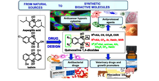
Graphical Abstract

1. Introduction

It is well known that nitrogen-containing heterocycles are privileged structures in medicinal chemistry because their derivatives have various biological properties [1]. The importance of nitrogen heterocycles as key pharmacophoric moieties lies in the synthesis of novel anticancer compounds [2,3,4,5,6]. Indeed, the presence of different nitrogen electron-donor atoms in the structure improves the interaction with target proteins, enzymes, and receptors through the formation of several types of interactions, such as hydrogen bonds, dipole-dipole, hydrophobic interactions, van der Waals forces, and π-stacking interactions. The chemistry of N-oxides of heterocyclic compounds is one of the dynamically developing areas of organic synthesis. Aliphatic and aromatic N-oxides are known as prodrugs due to their ability to be reduced by various oxidoreductases expressed in bacterial and tumor cells [7,8]. Quinoxaline 1,4-dioxides attract close attention due to their wide spectrum of biological activity. The presence of two N-oxide groups determines the pharmaceutical properties of quinoxaline-1,4-dioxides, such as antibacterial, antitumor, antifungal, insecticidal, herbicidal, and antiparasitic. The synthetic availability and biological potential of quinoxaline 1,4-dioxides define the prospects for their use in the targeted design of compounds with practical and valuable properties.
Moreover, the interest in heterocyclic N-oxides is explained by the presence of such structures in certain biologically active compounds found in natural sources. An example of a natural compound based on quinoxaline 1,4-dioxide is 6-chloro-2-quinoxalinecarboxylic acid 1,4-dioxide (1, Figure 1) produced by Streptomyces ambofaciens, which can inhibit the growth of gram-positive bacteria. It has been noted that esters and amides of this acid also have antibacterial activity [9]. Iodinin (2, Figure 1) is another example of a derivative of natural origin with a related structure to quinoxaline 1,4-dioxide [10]. Iodinin (2) was first identified and isolated from the bacterial culture Chromobacterium iodinum [11], and its antimicrobial properties were described in 1943 by H. McIlwain [12]. The results of recent studies have shown that iodinin (2) exhibits high cytotoxicity and selectivity for acute myeloid leukemia and promyelocytic leukemia cells (EC50 values for apoptotic cell death were 40 times lower than for non-tumor cells). Notably, iodinin (2) induced apoptosis in tumor cell isolates from patients with a poor survival prognosis [13,14].
Since the second half of the twentieth century, many researchers have observed the pronounced antibacterial activity of synthetic quinoxaline 1,4-dioxides. Some derivatives have been patented as antimicrobial agents and have found application in husbandry as promoters that increase animal weight gain [15].
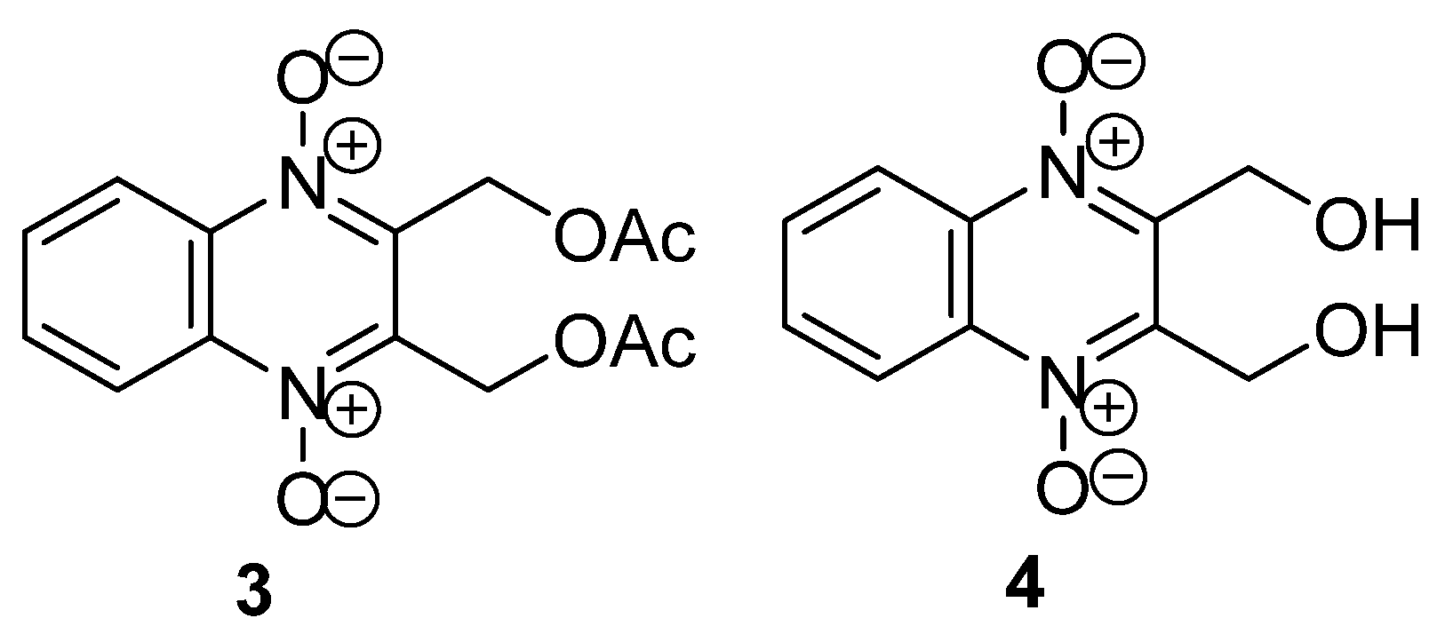
Certain quinoxaline derivatives have also been patented and used in clinical practice as antibacterial drugs. For example, quinoxidine (3; 2,3-bis(acetoxymethyl)quinoxaline 1,4-dioxide, Figure 2) and dioxidine (4; 2,3-bis(hydroxymethyl)quinoxaline 1,4-dioxide, Figure 2) have been used in the clinic since the 1970s as broad-spectrum antibacterial agents [16].
Further studies have shown that quinoxaline 1,4-dioxides are a promising scaffold for the development of new drugs for the treatment of tuberculosis and parasitic infections such as malaria, trypanosomiasis, leishmaniasis, amoebiasis, and trichomoniasis [17]. For instance, derivative 5 (Figure 3) [18] has demonstrated promising antituberculosis activity, while compounds 6 and 7 exhibited high fungicidal [19] and antiviral [20] effects, respectively (Figure 3). Derivatives of quinoxaline-2-carbonitrile 1,4-dioxide, such as compound 8 (Figure 3), exhibit selective cytotoxicity against solid tumor cells under hypoxic conditions [21].
However, despite the high biological activity of quinoxaline 1,4-dioxides, the derivatives described in the literature have several disadvantages associated with low solubility, mutagenicity [22], photoallergic reactions [23,24], and the development of bacterial resistance [25].
Methods for the synthesis and biological activity of quinoxaline 1,4-dioxides have been previously discussed in several papers [26,27,28,29,30,31,32,33,34,35,36]. However, the reviews published in the last twenty years have primarily focused on the biological aspects of quinoxaline 1,4-dioxides, providing limited consideration of their chemical properties and structure-activity relationships. In addition, the current reviews on the chemistry and biology of quinoxaline 1,4-dioxides do not contain up-to-date information published since 2016. Moreover, the information regarding the synthesis methods and chemical modifications of this class of compounds has not been systematized yet. Therefore, there is an interest in the general analysis and summarization of the data in the field of synthesis, modification, and biological properties of quinoxaline 1,4-dioxides, as well as the coverage of emerging trends in their study in medicinal chemistry and pharmaceuticals.

2. Methods of the Synthesis of Quinoxaline 1,4-Dioxides

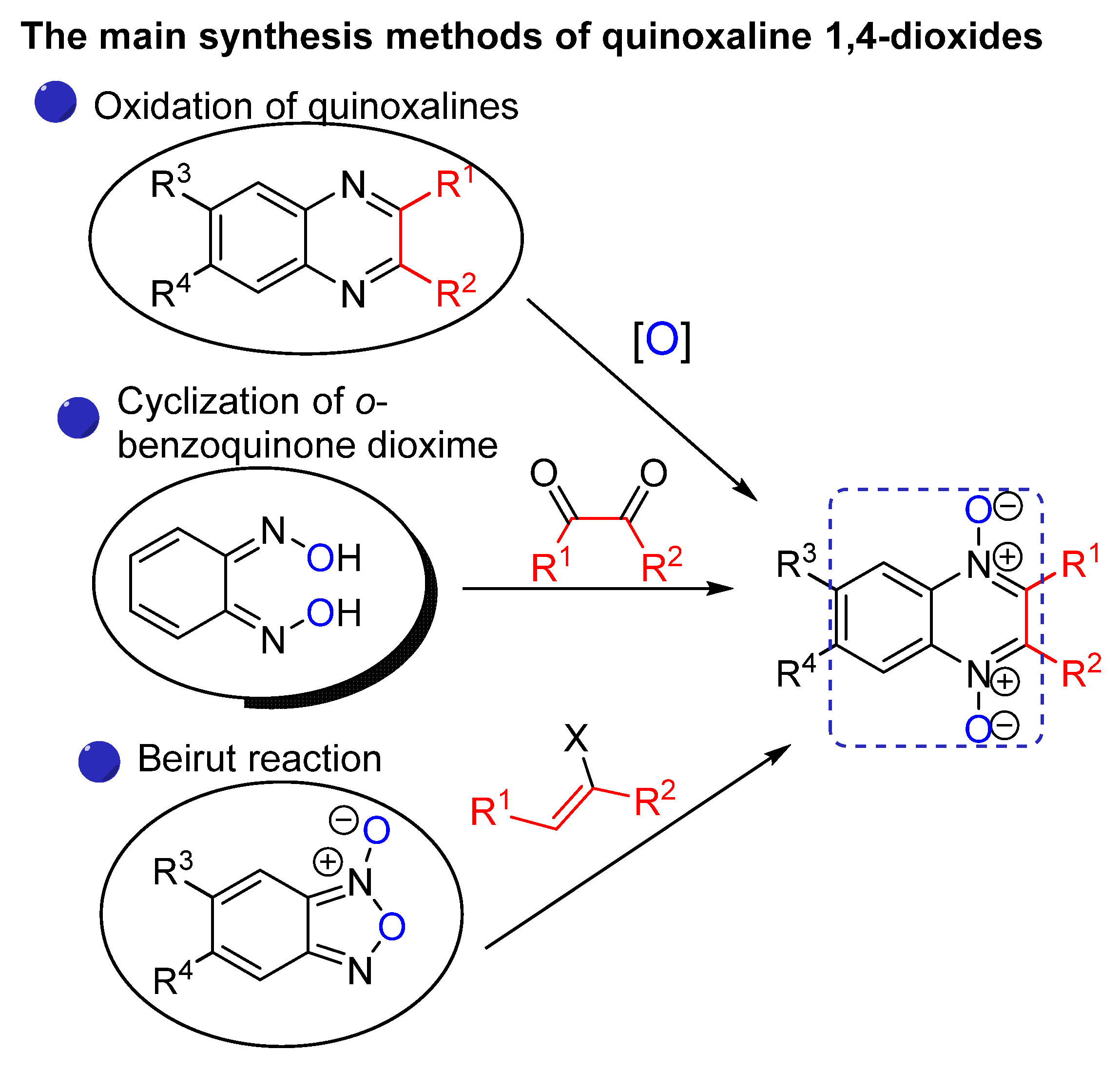
Synthetic approaches to the formation of quinoxaline 1,4-dioxides are significantly limited (Figure 4). Prior to 1965, the primary method for their synthesis was the direct oxidation of quinoxaline derivatives. Additionally, benzene-unsubstituted quinoxaline 1,4-dioxides can be obtained by the condensation of o-benzoquinone dioxime with 1,2-dicarbonyl derivatives. However, currently, a more efficient method for obtaining quinoxaline 1,4-dioxide derivatives has become the heterocyclization of benzofuroxans with enols or enamines by the Beirut reaction.
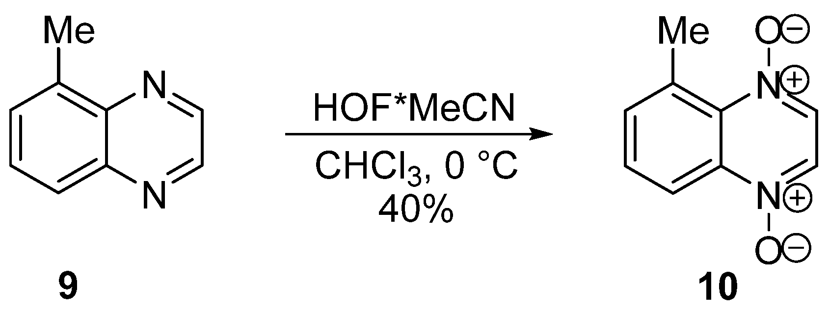
The oxidation of quinoxalines with peroxy acids or hydrogen peroxide does not result in a preparative yield of the desired quinoxaline 1,4-dioxides. This limitation arises from the deactivation of the heterocyclic core, preventing further electrophilic attack due to the formation of an N-oxide fragment following the oxidation of one of the nitrogen atoms [37]. In 2006, M. Carmeli and S. Rozen developed a method based on the application of a complex of hypofluorous acid with acetonitrile. This approach allows oxidizing of quinoxalines to quinoxaline 1,4-dioxides with quantitative yields, even for derivatives with electron-withdrawing substituents and sterically hindered 5-substituted derivatives. For instance, quinoxaline 9 can be oxidized to derivative 10 with a substituent at position 5, which is otherwise challenging to access using alternative methods (Scheme 1) [37,38].
Several examples of the formation of a fragment of quinoxaline 1,4-dioxide via cyclization of o-benzoquinone dioxime with 1,2-dicarbonyl compounds are known. In 1970, E. Abushanab [39] described the interaction of 1,2-diketones with o-benzoquinone dioxime (11), leading to 2,3-disubstituted derivatives (compound 12, Scheme 2) in low yields. However, the formation of 2-hydroxyquinoxaline 1,4-dioxides as the main products, such as compound 13, was observed in good yields when α-ketoaldehydes were used for this condensation (Scheme 2) [39].
The most significant preparative method for the synthesis of quinoxaline 1,4-dioxides is the cyclization of benzofuroxans by the Beirut reaction, developed by M.J. Haddadin and C.H. Issidorides in 1965 [40].
Initially, quinoxaline 1,4-dioxides were obtained by the condensation of benzofuroxans with enamines. Thus, the interaction of benzofuroxan (14a) with morpholinylcyclohexene gives 2,3-tetramethylenequinoxaline 1,4-dioxide (15) in moderate yield (Scheme 3) [41]. Later, the synthetic possibilities of the Beirut reaction were expanded by using a large number of starting substrates. In 1972, M.J. Haddadin and C.H. Issidorides used this method to synthesize phenazine 5,10-dioxides, analogs of the antibiotic Iodinin. The authors found that cyclization of benzofuroxan (14a) with hydroquinone in the presence of a base gives 2-hydroxyphenazine 5,10-dioxide (16) (Scheme 3) [42]. In the same year, A. Tanaka et al. showed the possibility of using alkynes in the Beirut reaction for the synthesis of 2,3-disubstituted quinoxaline 1,4-dioxides. Thus, the derivative 17 was obtained from benzofuroxan (14a) and 2-(furan-2-ylethynyl)-5-nitrofuran in the presence of methylamine, although in low yield (Scheme 3) [43]. Cyclization of benzofuroxan (14a) with aryl- and heteroaryl ethylenes (Scheme 3) was proposed for the synthesis of 2-substituted quinoxaline 1,4-dioxides (for example, derivative 18) by J. Li et al. [44].
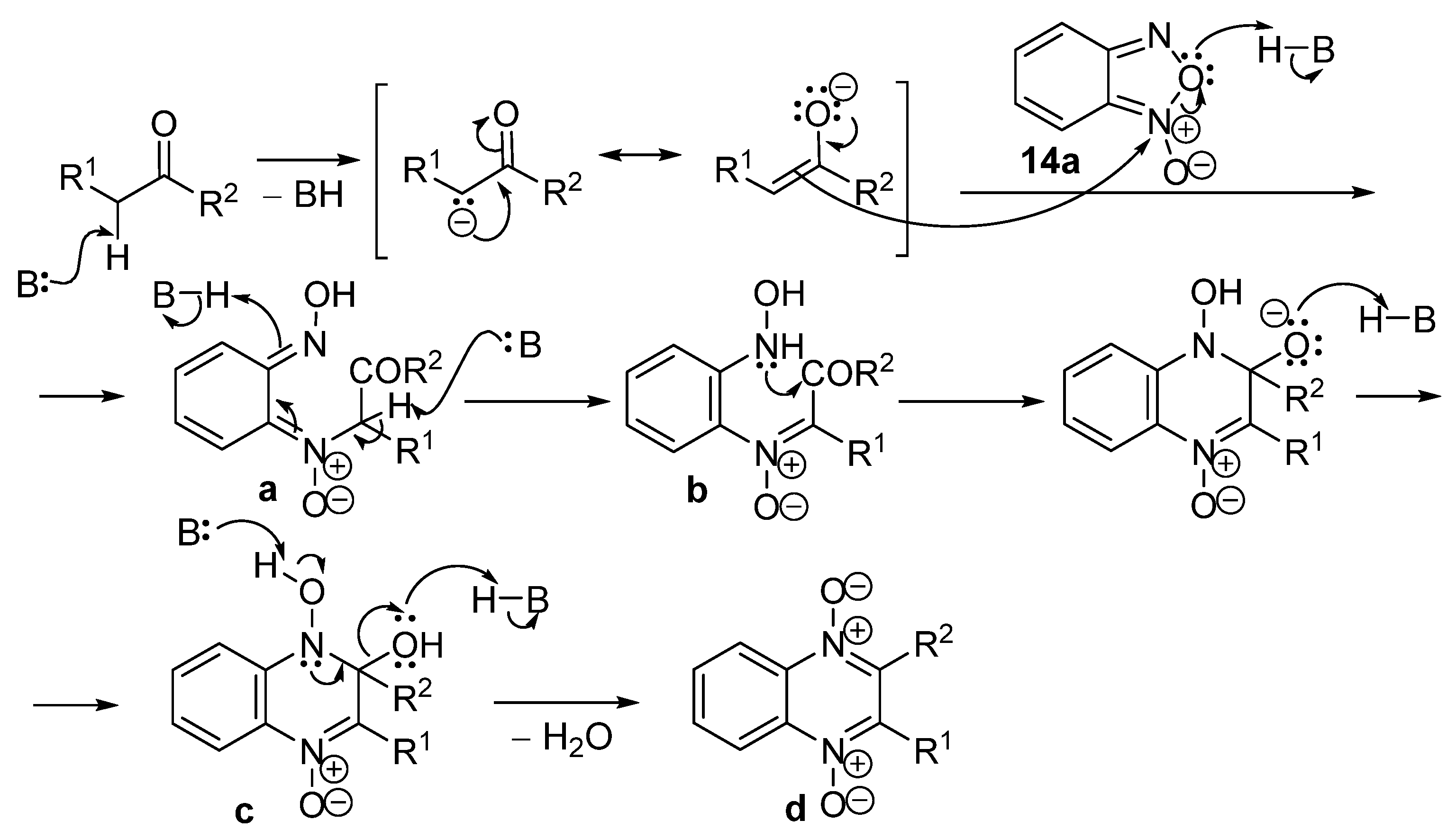
Several possible variants of the Beirut reaction mechanism have been described [27,41,45,46]. The first stage of the proposed mechanism includes the nucleophilic attack of the formed enolate ion on the electrophilic nitrogen atom of benzofuroxan 14a, leading to intermediate a [27]. Based on the mechanism of the Beirut reaction (Figure 5), the opening of benzofuroxan 14a in one of the transition states is accompanied by an attack of the oxime nitrogen atom on the electrophilic carbonyl group of intermediate b, which causes the formation of dihydroxyquinoxaline c. Further elimination of water from intermediate c leads to the formation of the quinoxaline 1,4-dioxide (d).
In 1995, A. Monge et al. described the synthesis of 3-aminoquinoxaline-2-carbonitrile 1,4-dioxide 19a by the Beirut reaction from malononitrile and benzofuroxan 14a (Scheme 4) when developing antiparasitic agents [47]. A similar cyclization with malonic ester in the presence of sodium hydride leading to 2-hydroxyquinoxaline 1,4-dioxide 20 in high yield was reported by Y. Xu et al. (Scheme 4) [48].
Cyclization of benzofuroxans with 1,3-diketones, β-ketoesters, or β-ketoamides has been proposed to obtain 2-acyl derivatives of quinoxaline 1,4-dioxide [49]. Thus, the interaction of 5,6-difluorobenzofuroxan (14b) with acetylacetone, acetoacetic ester, or N-phenylacetoacetamide in triethylamine at 0–10 °C gives 6,7-difluoroquinoxaline 1,4-dioxides 2122a, 23, respectively, with yields of 65–83% (Scheme 5) [50].
T. Lima et al. evaluated the effect of acid-base catalysts in the Beirut reaction on the yield of 2-carboethoxyquinoxaline 1,4-dioxide during the study of the condensation of benzofuroxan (14a) with benzoylacetic esters [51]. Thus, the target products were obtained in the presence of organic bases in less than 7% yields. In the case of acid catalysis, the key derivatives formed slightly higher yields (~13%). Catalysis with potassium fluoride on alumina in the absence of an organic solvent leads to the formation of desired products in acceptable yields (31–40%). A higher yield (50–64%) of derivatives of 3-phenylquinoxaline-2-carboxylic acid 1,4-dioxide was achieved using K2CO3 as a catalyst in acetone or DMF.
T. Sun et al. carried out the heterocyclization of benzofuroxan 14a and carbonyl compounds to substituted quinoxaline 1,4-dioxides via supramolecular catalysis by β-cyclodextrin in water [52]. A similar procedure was used to obtain conjugates of 6(7)-haloquinoxaline 1,4-dioxides with primaquine [53] or with N-aryl-substituted 1,2,3-triazoles [54], which have antimalarial and antimycobacterial activity.
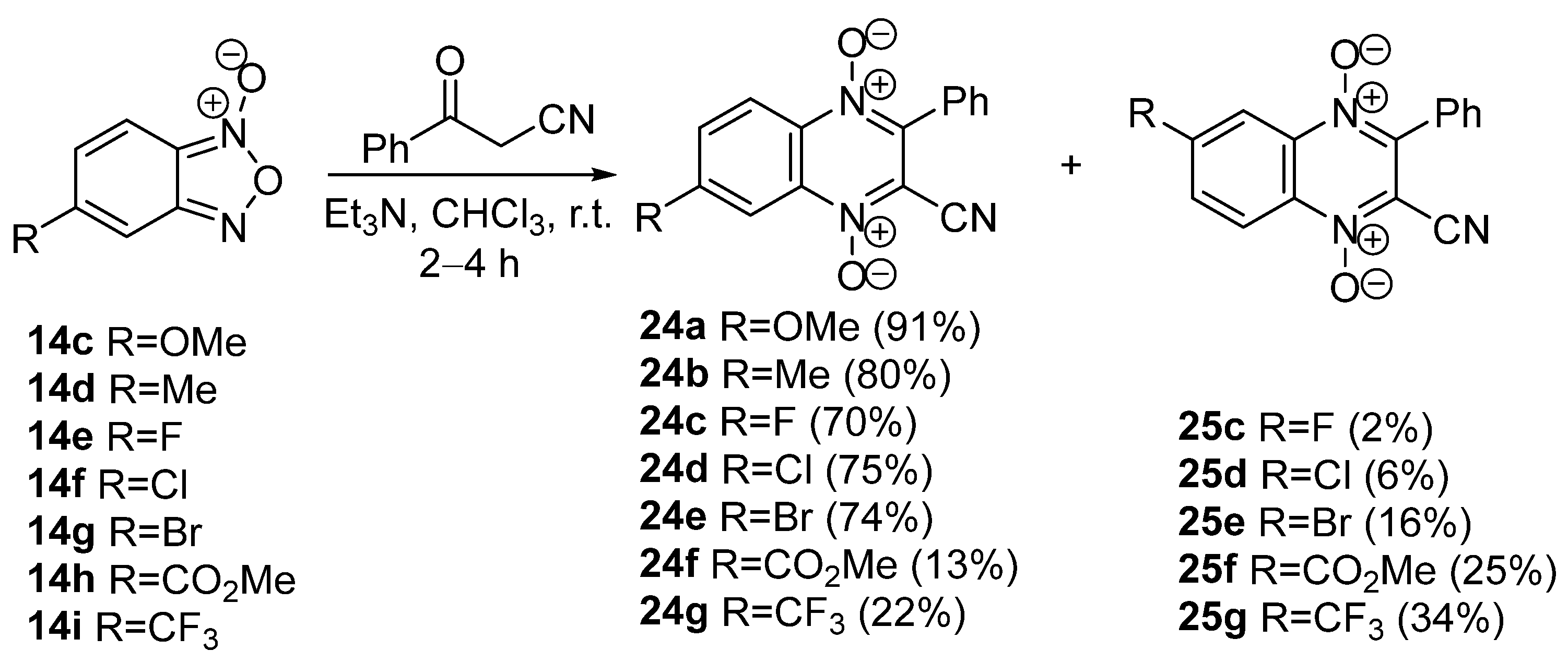
In some cases, the Beirut reaction between monosubstituted benzofuroxans and CH-acids leads to a mixture of 7- and 6-substituted quinoxaline 1,4-dioxides [55,56,57], which is explained by the presence of tautomeric equilibrium in benzofuroxans [58,59]. A study of the regioselectivity of the Beirut reaction between monosubstituted benzofuroxans 14ci and benzoylacetonitrile revealed that, along with the 7-isomers 24ag, their 6-substituted analogs 25cg [60] are formed in the reaction (Scheme 6). It is shown that the yield of the 6-isomer increases with the growth of electron-withdrawing properties of the substituent in the starting benzofuroxan and correlates well with the Hammett constant. Thus, the 6-isomer is not formed in the case of derivatives with electron-donating groups, while the 6-substituted compounds 25f,g with CO2Me- and CF3-groups are the main products of the reaction. The structure of the isomers was accurately confirmed by NMR spectroscopy and X-ray analysis. Compounds 24ag and 25f,g showed high antiproliferative activity against breast adenocarcinoma cells (MCF-7, MDA-MB-231) under hypoxic conditions. Moreover, the structural isomers 24cg and 25cg had considerable differences in properties.
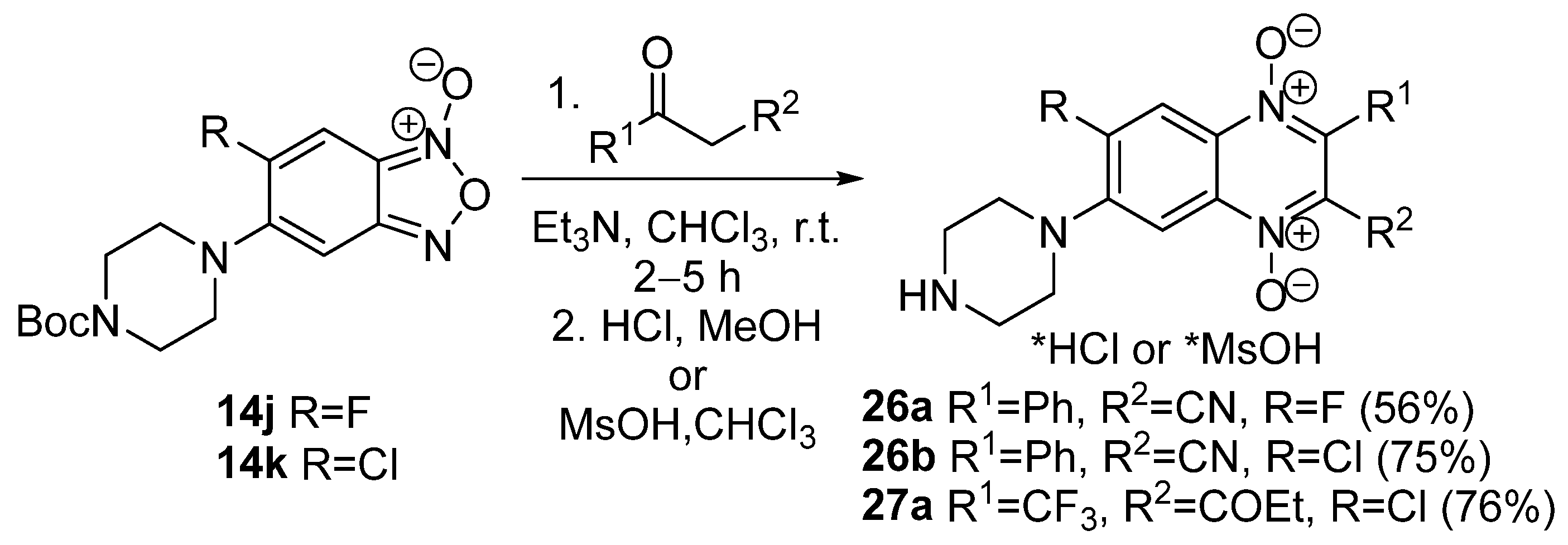
The reaction of aminobenzofuroxans 14j,k with benzoylacetonitrile in chloroform in the presence of Et3N also yields 7-amino-substituted quinoxaline-2-carbonitrile 1,4-dioxides 26a,b (Scheme 7) [61]. This method of heterocyclization was later used to synthesize 7-(piperazin-1-yl)-3-trifluoromethylquinoxaline 1,4-dioxide 27a (Scheme 7) [62]. The 7-amino derivatives of 26a,b have high aqueous solubility and downregulate the expression of HIF1α, BCL2, and ERα and induce apoptosis in MCF-7 cells at submicromolar concentrations.
In summary, the Beirut cyclization is currently the main preparative method for the synthesis of quinoxaline 1,4-dioxides with various substituents at positions 2, 3, 6, and 7. Ongoing research is focused on studying regioselectivity and exploring the possibilities of varying starting compounds, catalysts, and conditions to increase the yield of target quinoxaline 1,4-dioxides.

3. Chemical Properties of Quinoxaline 1,4-Dioxides

The reactivity of quinoxaline 1,4-dioxides is generally similar to the properties of heteroaromatic N-oxides. As a result, most of the reactions of quinoxaline 1,4-dioxides described in the literature involve the modification of the N-oxide fragment or are related to the transformation of the functional groups of the side chains and the benzene ring of quinoxaline.
Since the N-oxide fragment in the quinoxaline 1,4-dioxide facilitates many reactions, its reduction holds important synthetic value for the preparation of quinoxaline derivatives. Deoxygenation of heteroaromatic N-oxides can proceed in the presence of various reducing agents, such as catalytic hydrogenation, complex metal hydrides, sodium hypophosphite, titanium (III) chloride, dissolving metals, ascorbic acid, trivalent phosphorus compounds, and a number of sulfur-containing compounds [63,64,65,66]. However, not all reagents are suitable for the preparative reduction of quinoxaline 1,4-dioxides to quinoxalines due to the tendency of these substrates to undergo deoxygenation to mono-N-oxides or the formation of dihydro- and tetrahydroquinoxalines [67].
Quinoxaline 1,4-dioxides are sensitive to UV irradiation and, as a result of photoinduced rearrangement, can yield various products that depend on the structure of the starting compound. In particular, 2-substituted quinoxaline 1,4-dioxides 28a,b isomerize to 3-oxoquinoxaline 1-N-oxides 30a and 30b in high yield through the intermediate oxaziridines 29 under photolysis of their aqueous solutions (Scheme 8) [68].
Reduction and rearrangements occurring in the 1,4-dioxide core are often observed under basic conditions [69,70,71,72,73], by treatment of nucleophiles [74,75,76] or acid halides [77,78,79], which frequently reduce the yield of the target products and limit the possibility of transforming the functional groups of quinoxaline 1,4-dioxides. At the same time, the electron-withdrawing nature of the N-oxide fragments in the quinoxaline nucleus makes it possible to carry out a nucleophilic substitution of the leaving groups in positions 2, 3 and 6, 7 of the heterocyclic core under relatively mild conditions.

3.1. Transformations of Alkyl and Acyl Groups

Transformations of alkyl groups in the quinoxaline core are one of the most important approaches for obtaining a wide range of derivatives. For example, the synthesis of the original antibacterial drugs quinoxidine and dioxidine (compounds 3 and 4, Figure 2) is based on the modification of the methyl groups of the heterocycle. Bromination of 2,3-dimethylquinoxaline 1,4-dioxide (12) yields 2,3-bis(bromomethyl)quinoxaline 1,4-dioxide (31), which, upon treatment with acetic acid in the presence of triethylamine, gives quinoxidine (3). Hydrolysis of the di-O-acetyl derivative 3 in aqueous methanol leads to dioxidine (4) in good yield (Scheme 9) [80].
The CH-groups in the methyl attached to the pyrazine ring of quinoxaline 1,4-dioxides have high acidity, which explains the facilitation of the condensation of 2-benzoyl-3-methylquinoxaline 1,4-dioxides 32a,b with dimethylacetal of N,N-dimethylformamide (DMADMF) in o-xylene, which gives high yields of enamines 33a,b, respectively (Scheme 10) [81].
Similarly, enamine 34 was obtained from 2-carboethoxy-7-chloro-3-methylquinoxaline 1,4-dioxide and DMADMF [82]. The transamination of this derivative with morpholine gives enamine 35, while the reaction with anilines is accompanied by cyclization to pyridin-2-ones, leading to the formation of derivatives 36ac in good yields (Scheme 11).
The synthesis of aldehyde-derived quinoxaline 1,4-dioxides by the Beirut reaction is limited by the accessibility of the starting reagents. M. Quiliano et al. described the preparation of 2-formylquinoxaline 1,4-dioxides, developed for obtaining analogs of isoniazid [83,84]. Oxidation of 2,3-dimethylquinoxaline 12 by treatment with SeO2 under microwave irradiation gave 2-formylquinoxaline 37a, which upon condensation with a derivative of hydrazine, resulted in hydrazones 38a,b (Scheme 12). Compounds 38a,b showed high antitubercular activity against M. tuberculosis H37Rv with low cytotoxicity for non-tumor cells (Vero cells).
Another example of the use of 2-formylquinoxaline 1,4-dioxide for the synthesis of antimycobacterial agents was presented by Zhang H. et al. [85]. The condensation of carbaldehydes 39a,b with amines formed the corresponding N-substituted 2-thiazolidinonequinoxaline 1,4-dioxides, which further underwent intramolecular cyclization with thioglycolic acid to give 2-thiazolidinone-substituted quinoxalines 40a,b in good yield (Scheme 13). Derivative 40b demonstrated high antimycobacterial activity (MIC = 1.6 μg/mL, M. tuberculosis H37Rv), as well as significant antifungal activity (MIC = 2–4 μg/mL) against a panel of pathogenic fungi (Candida spp., Aspergillus spp., Cryptococcus spp.) comparable to the reference drug amphotericin B (MIC = 0.5–2 μg/mL).
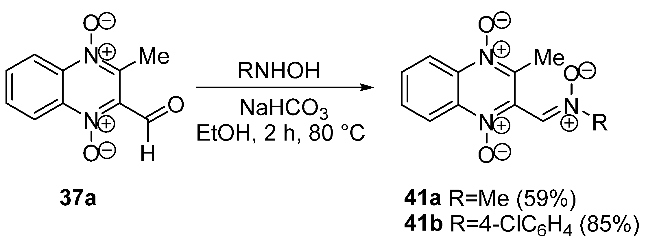
In addition, 2-formylquinoxaline 1,4-dioxide 37a was applied for the synthesis of nitrones 41a,b via its condensation with N-substituted hydroxylamines (Scheme 14) [86]. Derivatives 41a,b exhibited high antibacterial activity against Gram-positive and Gram-negative bacteria, including resistant strains, both in vitro and in vivo. It has been shown that the introduction of an additional N-oxide fragment into position 3 leads to an increase in the selectivity of the antibacterial effect of these derivatives against Gram-negative bacteria.
An alternative synthesis of quinoxalinylnitrones based on the condensation of 2-methylquinoxaline 1,4-dioxide (28b) with p-nitrosodimethylaniline, resulted in a high yield of nitrone 42 (Scheme 15) [87].
B.B. Michniak et al. described the conversion of 2-formylquinoxaline 1,4-dioxide (39a) to quinoxaline-2-carbonitrile 1,4-dioxide (44) via O-acyloxime 43 (Scheme 16) [88].
Y. Pan et al. used the modification of the acetyl residue for the diversification of quinoxaline 1,4-dioxides and increasing the solubility of compounds in aqueous solutions [89]. Bromination of ketone 21b and subsequent substitution of the halogen in α-bromoketone 45 with amines gave α-aminoketones 46a,b in high yield (Scheme 17). Treatment of the bromo derivative 45 with thiols yielded β-mercaptoketones 47a,b. The obtained compounds demonstrated high antimycobacterial activity against the M. tuberculosis H37Rv strain without cytotoxic effects on non-tumor cells (Vero cells).
The condensation of 2-acetyl-3-methylquinoxaline 1,4-dioxide 21c with p-chlorobenzaldehyde in NaOH solution leading to chalcone 48 with high yield (Scheme 18) is described in several papers [81,90]. An example of the modification of chalcones based on quinoxaline 1,4-dioxide is the heterocyclization of compound 48 with thiourea and hydrazine, resulting in derivatives of thiopyrimidine 49 and pyrazole 50, respectively (Scheme 18) [81]. The obtained compounds 49, 50 exhibit high antitumor activity against hepatocellular carcinoma HepG2 and are two orders of magnitude more active than the reference agent tirapazamine under hypoxic conditions.
An alternative method for the synthesis of chalcones based on quinoxaline 1,4-dioxide was developed by Burguete A. et al. [91]. The Wittig reaction of 2-acetyl-3-methylquinoxaline 1,4-dioxide 37b under microwave irradiation yielded enone 51, which has high antioxidant and anti-inflammatory activity (Scheme 19) [92].
A. Monge’s group carried out cyclopropanation of the conjugated double bonds of chalcones based on quinoxaline 1,4-dioxide using the Corey-Tchaikovsky reaction [90]. Thus, the reaction of enone 51 with sulfoxonium methylide generated from trimethyloxosulfonium iodide (TMSOI) gave the cyclopropyl derivative 52 in low yield (Scheme 20).
Compound 52 demonstrated good antimalarial activity against the P. falciparum FCR-3 strain.

3.2. Reactions of Amino Derivatives of Quinoxaline 1,4-Dioxides

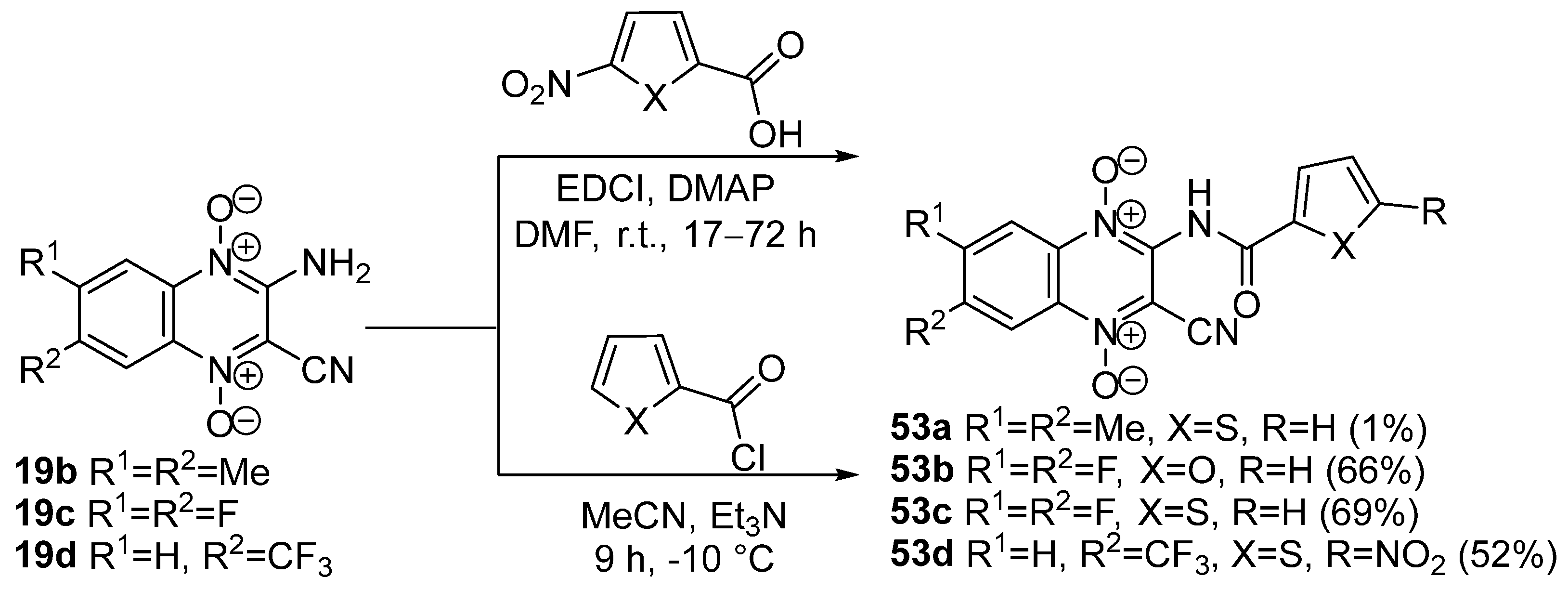
A. Monge et al. demonstrated that 3-aminoquinoxaline-2-carbonitrile 1,4-dioxides 19bd can be acylated using acid chlorides, resulting in the formation of the corresponding amides 53ad (Scheme 21) [93]. Condensation of 5-nitrofuran- and 5-nitrothiophene-2-carboxylic acids 53ad in the presence of carbodiimide (EDCI) and DMAP was used for the synthesis of the corresponding amides (Scheme 21). Derivative 53a exhibits MIC values close to the reference rifampicin against M. tuberculosis H37Rv. Compounds 53b and 53c have comparable activity with the anti-Chagas drug nifurtimox for the T. cruzi Tulahuen 2 strain.
The reaction of 2-aminoquinoxaline 1,4-dioxides 19a,b with isocyanates leads to cyclic carbamates 54a,b. The ring opening of these carbamates with alcohols results in the formation of alkylcarbamates of quinoxaline 1,4-dioxides 55a,b (Scheme 22) [94].
Soliman D.H. et al. described an interesting modification of the amino group in the pyrazine ring of quinoxaline, allowing the obtainment of tricyclic diazaphosphorins 57a,b as a result of cyclization of 3-aminoquinoxaline 1,4-dioxides 19a and 56 by treatment with phosphorus(V) sulfide in pyridine. Further methylation of diazaphosphorins 57a,b yielded N,S,S-trimethyl derivatives 58a,b (Scheme 23). These compounds demonstrated high activity against the prostate cancer cells (PC3), comparable to doxorubicin [95]. In addition, compound 58b was able to inhibit VEGFR-2 and SRC kinases (IC50 = 18 μM).

3.3. Metal-Containing Chelates of Quinoxaline 1,4-Dioxides

One of the promising strategies in the search for new antitumor agents is the development of metal-containing chelates [96]. For example, copper(II) complexes with amino acids [97], casiopeins [98], oligopeptides [99,100], and mono- and bis-thiosemicarbazones [101,102] have demonstrated significant antitumor activity in human tumor models in vivo. The study of complexes with transition metals showed that quinoxaline-2-carboxilic acid 1,4-dioxide could act as a mono- or bidentate ligand for different coordination ions (Cr(III), Mn(II), Fe(III), Co(II), Ni(II), Cu(II), Zn(II), Ce(III), Nd(III), V(IV)) [103].
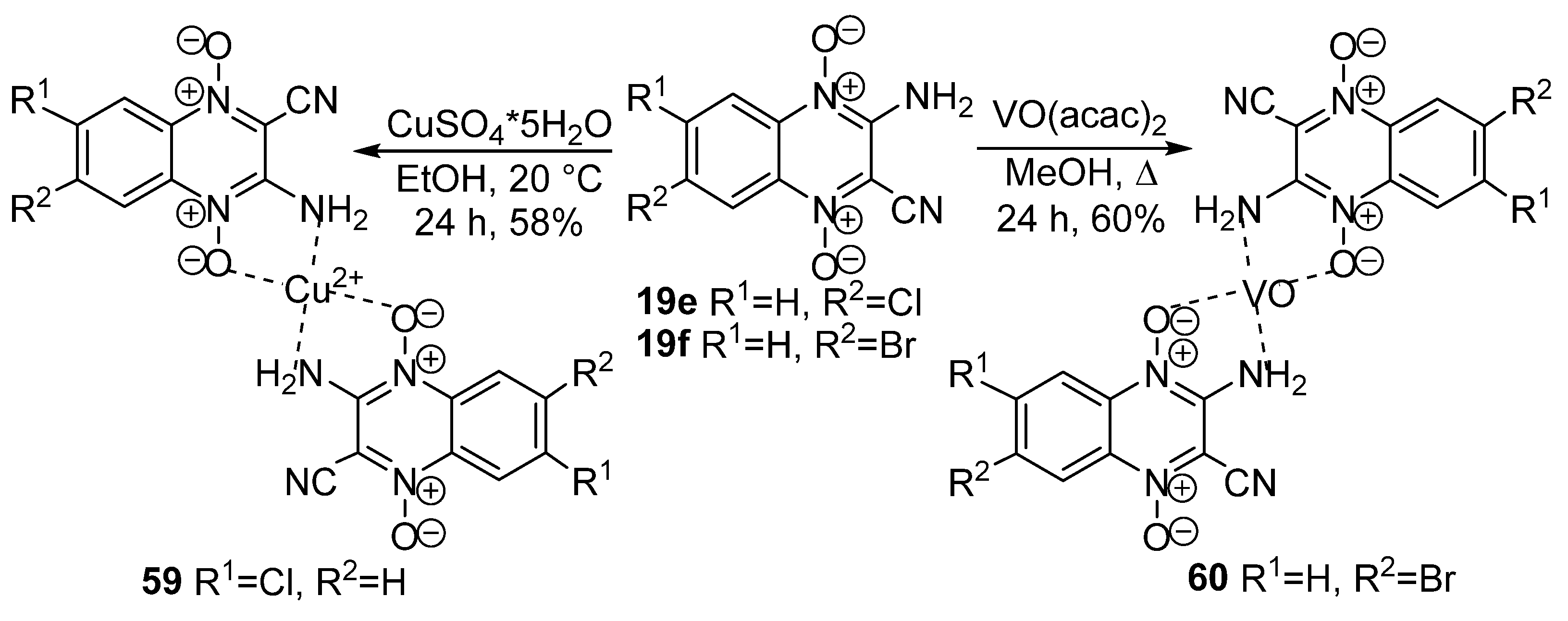
Another example of a ligand for chelates is 3-aminoquinoxaline-2-carbonitrile 1,4-dioxides 19e,f, which forms complexes with Cu(II) (59) [104] or V(IV) (60), respectively (Scheme 24) [105].
Derivative 60 has caused hypoglycemic and antihyperlipidemic effects using in vitro models, while the copper complex 59 demonstrated promising antiproliferative potency and selectivity under hypoxic conditions.

3.4. Nucleophilic Substitution of Halogen Atoms

Despite the high biological activity of some quinoxaline 1,4-dioxide derivatives, their study is complicated by low solubility in pharmacologically acceptable aqueous media [106]. One way to increase the bioavailability of organic compounds is by introducing substituents containing salt-forming groups, such as amino groups. The presence of a di-N,N′-oxide fragment in the quinoxaline core increases the reactivity of quinoxalines in nucleophilic substitution reactions. Therefore, the most important methods of diversification of quinoxaline 1,4-dioxides are based on the modification of halogen atoms, alkoxy- or alkylthio-groups in the activated positions of the heterocycle.
M.A. Ortega et al. demonstrated that 3-aminoquinoxaline-2-carbonitrile 1,4-dioxides 19b and 19e do not undergo diazotization reactions under classical conditions due to the low basicity of the amino group and the low solubility of such compounds. However, it was possible to produce a diazonium salt and replace the diazo group with a halogen by the Sandmeyer reaction. Thus, treatment of amines 19b and 19e with tert-butyl nitrite, heating in acetonitrile in the presence of CuCl2 in an inert atmosphere gave 3-chloro derivatives 61a,b, although in low yield (Scheme 25) [107]. Further nucleophilic substitution of the chlorine atom in compounds 61a,b by diamines in chloroform in the presence of K2CO3 led to 3-amino derivatives, such as piperazines 62a,b, in low yields (Scheme 25). An alternative method for introducing amine residues into position 3 of quinoxaline 1,4-dioxides is based on the substitution of alkylthio-, aryloxy-, and sulfonyl-groups, which gives 3-amino derivatives in high yields [108,109,110,111,112].
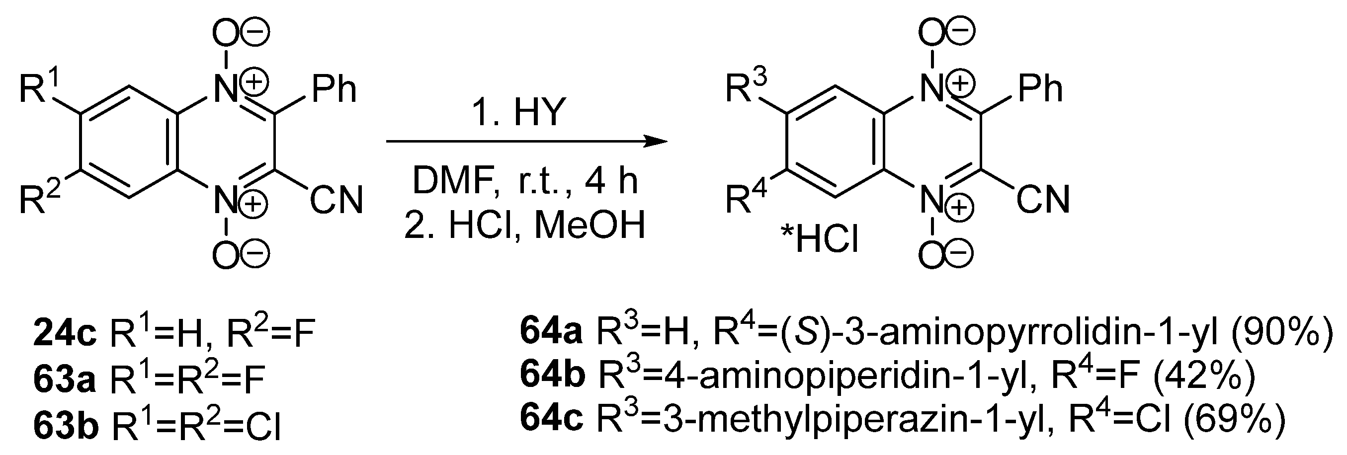
The substitution of halogen atoms in the benzene ring activated by electron-withdrawing groups in the pyrazine nucleus of quinoxalines proceeds more efficiently. Thus, a series of amino derivatives 64ac was synthesized by treating an excess of diamines with halogen derivatives of 3-phenylquinoxaline-2-carbonitrile 1,4-dioxides 24c and 63a,b in DMF, yielding moderate-to-good yields (40–85%) (Scheme 26). It should be noted that for 6,7-dihalogen derivatives, the reaction proceeds regioselectively, leading to 6-amino-substituted 64b,c, which is explained by the electron-withdrawing effect of the CN group in position 2 of the heterocycle [61,113]. The structure of these compounds was confirmed by the 13C NMR spectra, as well as X-ray analysis. Amino derivatives 64ac at low micromolar concentrations inhibited the proliferation of a panel of tumor cell lines of various tissue origins and also showed outstanding selectivity for malignant cells under hypoxia.
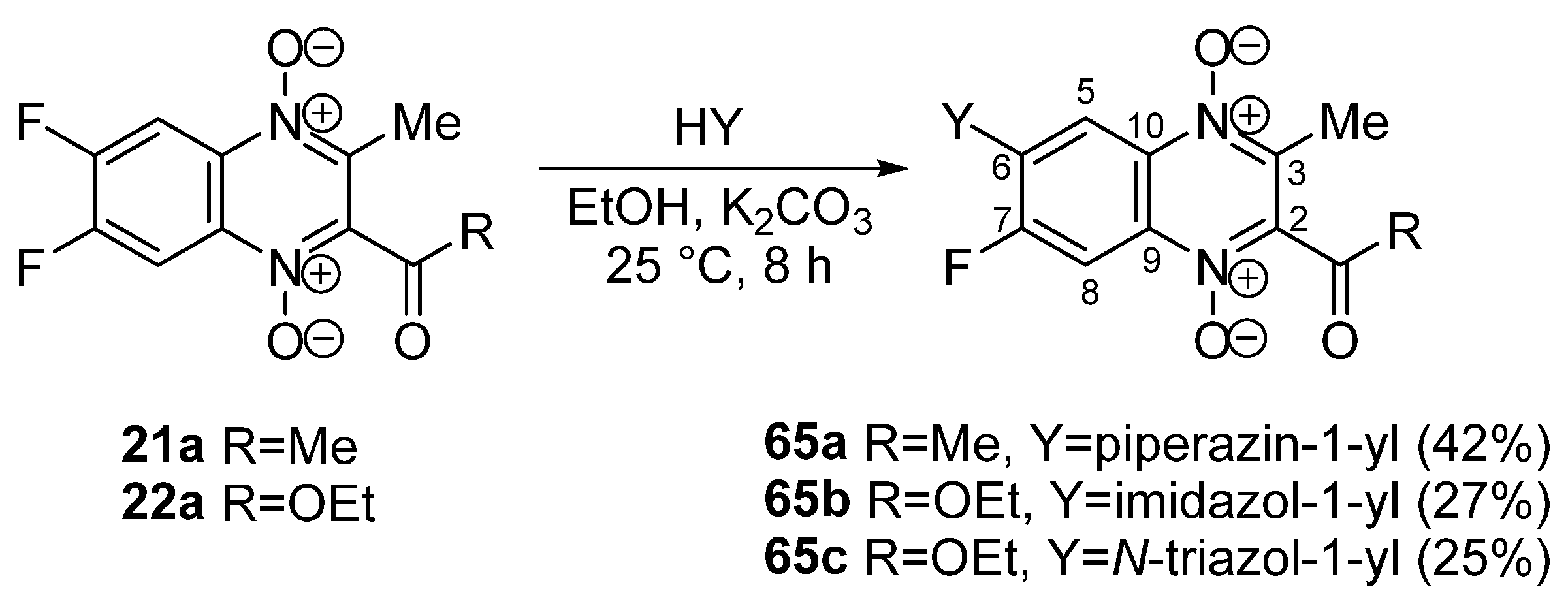
In 2021, a similar approach was used by N. Zhang et al. to modify 2-acyl-6,7-difluoro-3-methylquinoxaline 1,4-dioxides 2122a. The treatment of derivatives 2122a with piperazine or azoles in ethanol in the presence of K2CO3 yielded 6-amino derivatives 65ac (Scheme 27), which exhibit promising antituberculosis potency [114].
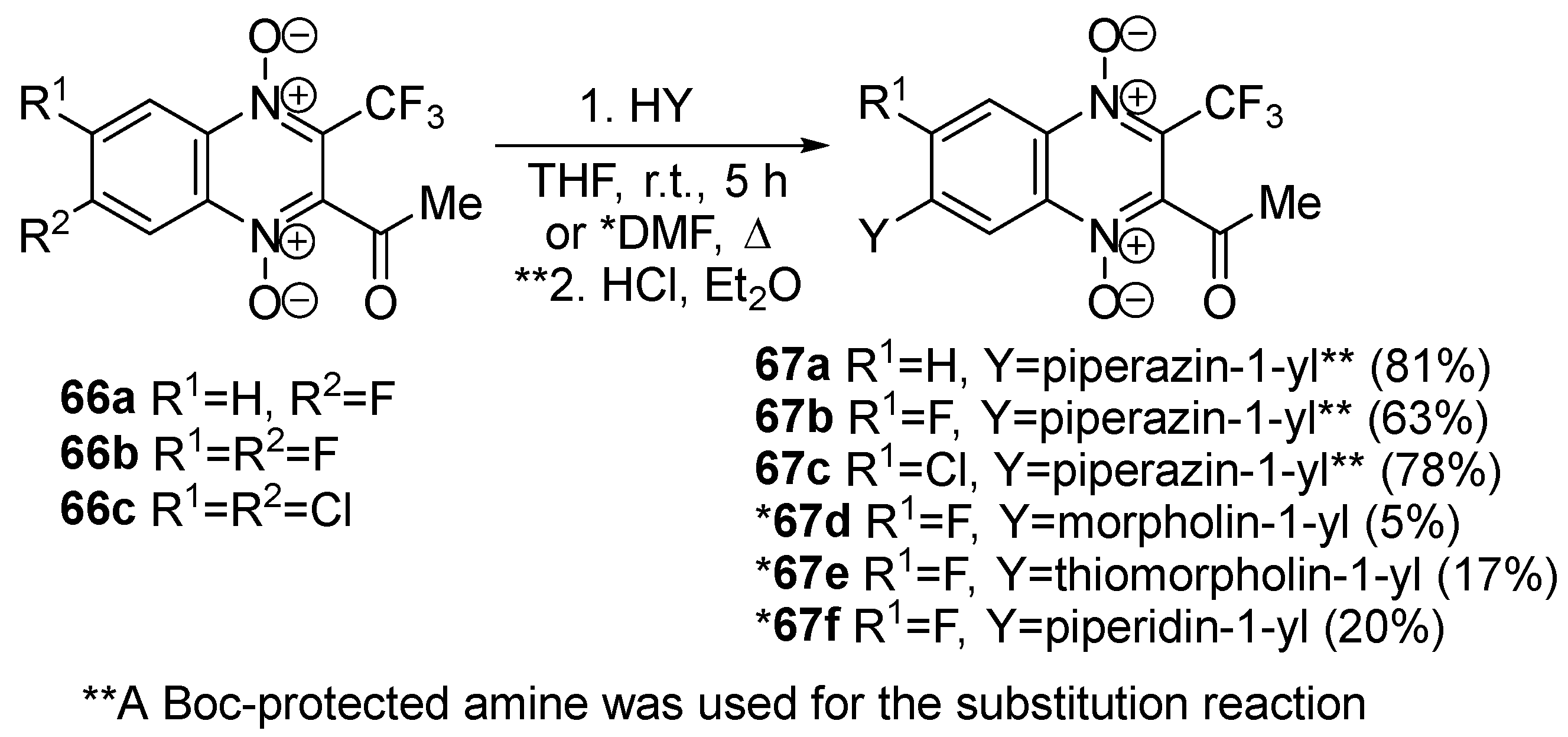
A similar method for modifying 2-acyl-3-trifluoromethylquinoxaline 1,4-dioxides was used for the development of new antibacterial agents [62,115,116]. However, derivatives with two electron-withdrawing groups in positions 2 and 3 have a high tendency for deoxygenation of the 1,4-dioxide moiety [116,117]. The substitution of the halogen atom in derivatives 66ac proceeds more efficiently when using Boc-piperazine to give amino derivatives 67af (Scheme 28).
In the case of 6,7-dihalogen derivatives, the reaction also proceeds retrospectively but leads to 7-amino derivatives 67bf, the structure of which was confirmed by analysis of the 13C NMR spectra [62].

3.5. Transformations of Carboxylic Acids and Their Derivatives

1,4-Dioxides of quinoxaline-2-carboxylic acids and their derivatives have high biological activity and a wide range of pharmacological properties. Therefore, it is important to analyze their chemical properties and the most valuable methods of their transformations. The 1,4-dioxides of quinoxaline-2-carboxylic acids are unstable and easily decarboxylated. Thus, quinoxaline 1,4-dioxide 28a is formed from acid 68 when heated in propanol (Scheme 29) [56].
The hydrolysis of ester 22b to acid 70 was successfully carried out by heating in aqueous ethanol in the presence of triethylamine and catalytic amounts of calcium chloride (Scheme 30). It has been shown that calcium ions accelerate the hydrolysis of esters of 1,4-dioxides of quinoxaline-2-carboxylic acids due to the formation of complex 69 [118].
The direct alkaline hydrolysis of 2-carboethoxy-7-fluoroquinoxaline 1,4-dioxide 65b proceeds in low yield and gives acid 71, as described in [114]. This is apparently explained by the instability of quinoxaline 1,4-dioxides in the presence of bases (Scheme 31).
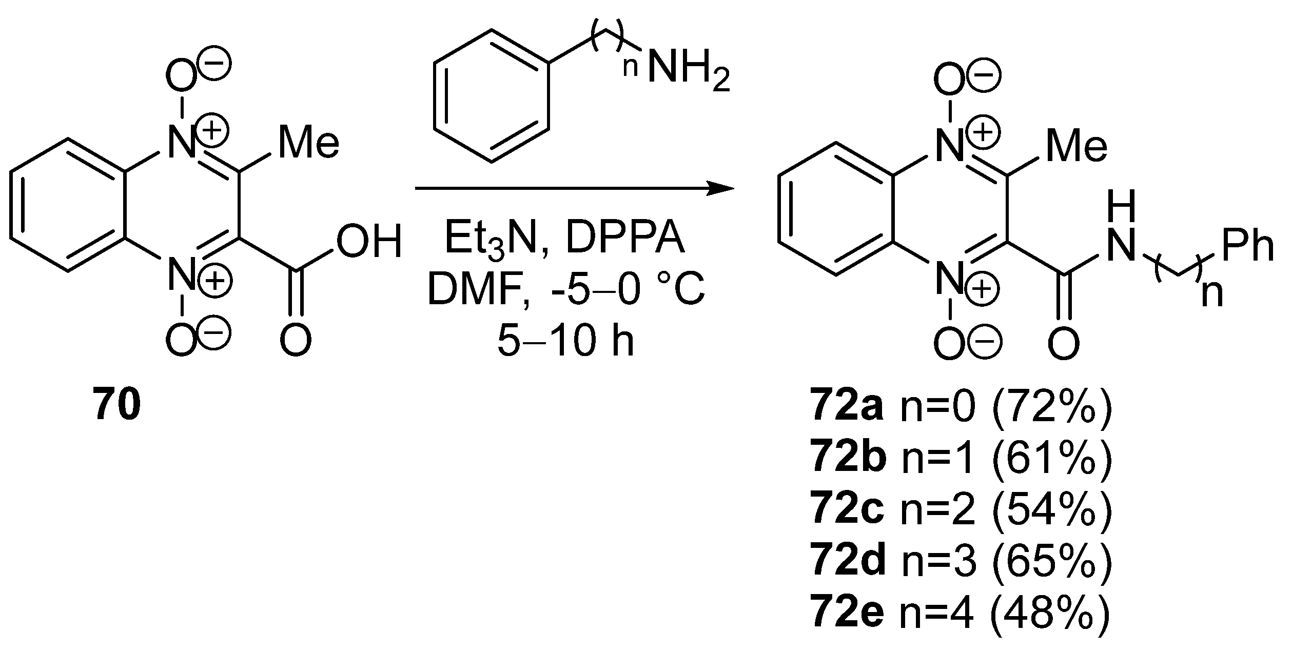
S.S. Sabri et al. synthesized amides from quinoxaline-2-carboxylic acid 70 using diphenylphospharylazide (DPPA) as an activating agent. Carboxamides 72ae were obtained in good yields from acid 70 and primary amines in the presence of DPPA and triethylamine in DMF (Scheme 32) [119].
The Curtius rearrangement can be used for the synthesis of some 2-amino derivatives of quinoxaline 1,4-dioxide from azides of 1,4-dioxides of quinoxaline-2-carboxylic acids. Hydrazinolysis and subsequent nitrosation of ester 22c gave the azide of 3-methylquinoxaline-2-carboxylic acid 73. Azide 73 rearranges into 2-alkyl carbamates 74ac or substituted ureas 75ac when boiled in an alcohol or amine solution, respectively (Scheme 33) [120].
The transformations of the hydrazide of 3-methylquinoxaline-2-carboxylic acid 76, obtained from the corresponding ethyl ester and hydrazine, are applied to the functionalization of the quinoxaline 1,4-dioxide scaffold at position 2 [82]. Condensation of hydrazide 76 with benzaldehydes in the presence of acetic acid in ethanol gave hydrazones 77a and 77b (Scheme 34).
Treatment of hydrazide 76 with carbon disulfide leads to the cyclization of 2-(1,3,4-oxadiazol-2-thione) 78 (Scheme 34). Boiling hydrazide 76 with p-nitrobenzoic acid in the presence of phosphoryl chloride gave 2-(1,3,4-oxadiazolyl)quinoxaline 1,4-dioxide 79.

4. Chemotherapeutic Properties of Quinoxaline 1,4-Dioxides

The unique biological properties of quinoxaline 1,4-dioxides have attracted significant interest from scientists for their use as an important scaffold for drug development. As mentioned earlier, aliphatic and aromatic N-oxides are known as prodrugs due to their ability to be reduced by various oxidoreductases expressed in bacterial and tumor cells. Moreover, the attention given to heterocyclic N-oxides, particularly quinoxaline derivatives, can be explained by the presence of this scaffold in natural bioactive compounds, such as iodinin, its analog myxin, echinomycin, and aspergilic acid [121,122].

4.1. Antibacterial and Antifungal Activity of Quinoxaline 1,4-Dioxides

In 1938, the investigation of the biological properties of quinoxaline 1,4-dioxides started with the discovery of the antibacterial activity of its closest analog, iodinin [123,124]. Subsequent studies have revealed that the mechanism of the antibacterial action of quinoxaline 1,4-dioxides is associated with the ability of heterocyclic N-oxides to reduction by oxidoreductases, leading to the generation of ROS, DNA damage, inhibition of DNA and RNA synthesis, morphological changes in the bacterial cell wall, as well as suppression of extracellular bacterial nuclease and α-toxin action [125,126,127,128]. Thus, the single-electron reduction of NADPH-dependent monooxygenases (P450) of quinoxaline 1,4-dioxide (28a, Scheme 8) leads to the formation of an oxygen-sensitive intermediate radical 80, generating reactive hydroxyl radical and metabolite 81 (Scheme 35) [129].
The medicinal drugs in this class of heterocycles include quinoxidine and dioxidine (compounds 3 and 4, Figure 2), which were discovered in the 1970s and are still used in parenteral forms and in various topical drug formulations. Dioxidine (4, Figure 2) is a reserve drug for the treatment of purulent infections. Washing of burning and purulent-necrotic wounds with dioxidine promotes faster wound surface cleansing, stimulates wound regeneration, and enhances epithelization [130]. Quinoxidine (3, Figure 2) acts on both Gram-positive organisms (including Staphylococcus spp., resistant to other antimicrobial drugs) and Gram-negative bacteria (K. pneumoniae, P. aeruginos, Proteus spp.). Dioxidine (4, Figure 2) causes loosening of the cell wall and agglomeration of DNA fibrils in Staphylococcus aureus cells [113]. In anaerobic conditions, the antibacterial activity of this drug increases by 8–128 times, depending on the type of pathogen [113]. Dioxidine (4, Figure 2) does not exhibit cross-resistance with other antimicrobial drugs. Recent studies have confirmed the low toxicity of dioxidine, including the absence of cardiotoxicity, immunotoxicity, and toxic effects on the adrenal glands and other organs, which makes it suitable for use as a new dosage form for the treatment of pharyngitis and tonsillitis [131,132].
In the pursuit of quinoxaline 1,4-dioxides with antibacterial and antifungal activity, it has been shown that 2-phenylsulfonyl-substituted quinoxaline 1,4-dioxides hold promise for the development of new antimicrobial agents. Among the synthesized series, derivatives 82a,b (Figure 6) demonstrated the highest activity against Enterococcus faecalis and Enterococcus faecium strains, with MIC values ranging from 0.4 to 1.9 μg/mL [133].
A. Carta et al. have discovered promising derivatives with high anti-candida activity against Candida spp. The authors have demonstrated that the 2-chlorinated derivatives 83a,b exhibit the highest antifungal activity (MIC = 0.39–0.78 μg/mL, Figure 6) [134]. Furthermore, the water-soluble 3-trifluoromethyl derivatives 27a and 67c (Figure 6) have shown antimicrobial activity against Gram-positive and Gram-negative bacteria and mycobacteria, comparable to reference drugs ciprofloxacin and rifampicin, with MIC values ranging from 0.25 to 10 μg/mL [62]. Additionally, compounds 27a and 67c demonstrated high antifungal activity comparable to amphotericin B against fungal cultures of C. albicans ATCC 10,231 and M. canis B-200. An analysis of the structure-activity relationship of the 3-trifluoromethylquinoxaline 1,4-dioxide series (for example, derivatives 27a and 67c, Figure 6) revealed that the introduction of an additional piperazine residue negatively affects the antimicrobial properties of these compounds. Furthermore, substituents in position 2 of the quinoxaline core do not significantly influence the antimicrobial activity of such derivatives. In particular, 7-(piperazin-1-yl)-3-trifluoromethylquinoxaline 1,4-dioxides demonstrated high activity against Gram-positive bacteria, while their effect on Gram-negative bacteria and fungi was noticeably lower.
The study of the mechanism of antibacterial action on the dual reporter test system pDualrep2 revealed that the 3-trifluoromethyl derivatives 27a, 67c (Figure 6), as well as the reference dioxidine (4, Figure 2), induced a bacterial SOS-response in the strain E. coli BW25513 [62].

4.2. Antimycobacterial Activity of Quinoxaline 1,4-Dioxides

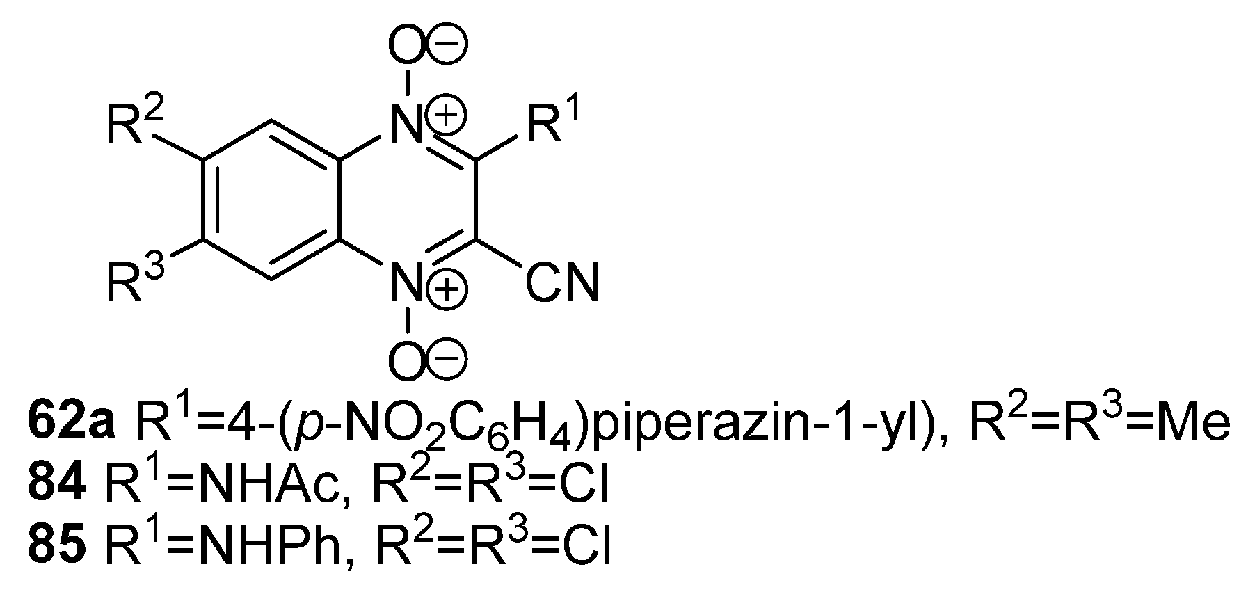
Previously, based on the structural similarity of quinoxaline 1,4-dioxides with vitamin K, which plays an important role in the metabolism of M. tuberculosis [135], A. Monge et al. tested a series of 3-aminoquinoxaline-2-carbonitrile 1,4-dioxides (62a, 84, 85, Figure 7, Scheme 25) against clinical isolates of M. tuberculosis with MDR [136]. The screening results for antimycobacterial properties demonstrated high activity of some derivatives of this type (for example, compounds 62a, 84, 85, Figure 7) against M. tuberculosis H37Rv strain, highlighting the significant role of N-oxide fragments in their ability to inhibit the growth of the mycobacterium [137,138,139]. Moreover, compound 62a exhibited a high selectivity index (SI) (SI = IC50,Vero/MIC > 124).
The replacement of the nitrile group at position 2 of quinoxaline with a carboxamide moiety, acyl, or carboxyl groups increased the inhibitory potency and, more importantly, reduced the cytotoxicity of such derivatives by an order of magnitude (21be, 22bd, 8687ac, Figure 8) [140,141]. Among the 2-carboxamide derivatives, compounds 62a and 84, 85 were the most active (Figure 8), with MIC values of 0.89, 0.42, and 0.14 μg/mL, respectively. In the series of 2-acyl derivatives, 2-acetylquinoxaline 1,4-dioxides 21be showed the highest activity (Figure 8), although these derivatives had slightly higher MIC values (0.87, 3.13, 0.8, and 4.3 μg/mL, respectively) compared to their analogs with carboxamide (compounds 62a, 84, 85) and ester (compounds 22bd, 87ac) groups. The quinoxaline-2-carboxylates 22bd and 87ac exhibited the highest activity against M. tuberculosis H37Rv among all tested derivatives (MIC = 0.56, 2.30, 1.50, and 0.01 μg/mL, respectively), surpassing the reference drug rifampicin in activity. Notably, compounds 87b and 87c showed activity against the majority of tested strains of M. tuberculosis with MDR.
Some studies have demonstrated that 3-alkylquinoxaline 1,4-dioxides with carboxamide (compounds 86ac), acyl (compounds 21be), alkoxycarbonyl (compounds 22bd, 87ac), or thioaryl (compound 88a) substituents at position 2 also possess high antituberculosis activity (Figure 8) [142]. Compound 88a exhibited moderate MIC values (30–60 μg/mL); however, in bacteriostatic concentrations, it acted synergistically with isoniazid, reducing its MIC by more than two orders of magnitude [143].
Compounds 65bd (Figure 8) demonstrated high antimycobacterial activity against M. tuberculosis H37Rv, as well as low cytotoxicity for non-tumor cells (MIC ≤ 0.25 μg/mL, SI(Vero) = 169–412) [114]. The 6-(1,3,4-triazol-1-yl) substituted derivative 65d showed the highest antitubercular activity in a model of macrophages infected with M. tuberculosis. Additionally, compound 65d disrupted the integrity of membranes and energy homeostasis of M. tuberculosis and significantly increased the level of ROS in eukaryotic cells. Consequently, it induced autophagy in infected macrophages, suggesting an indirect blockage of intracellular replication of M. tuberculosis. Pharmacological studies in vivo demonstrated that compound 65d was reasonably safe and possessed favorable pharmacokinetic properties.
The analysis of the obtained data for 2-carbonyl-containing quinoxaline 1,4-dioxides (for example, compounds 21ae; 22bd; 87ac and 65bd, Figure 8) revealed that the acetyl group at position 2 of the quinoxaline led to moderate to good antimycobacterial activity. Replacement of this moiety with a butyryl or isovaleryl group dramatically reduced the activity. However, the ethoxycarbonyl group at the C2 carbon atom of the quinoxaline ring generally enhanced antimycobacterial activity. In contrast, a benzyl analog did not alter the activity of such derivatives. Nevertheless, the introduction of an isopropyl ester group at the 2 position leads to a significant decrease in antimycobacterial activity. Moreover, the presence of a piperidine, morpholine, or pyrrolidine group in the structure of derivatives containing a 2-carbonyl fragment negatively affected the activity against the M. tuberculosis strain. Interestingly, the introduction of an imidazole or a 1,2,4-triazole group at position 6 of the heterocyclic ring resulted in a significant increase in the anti-TB potency of the corresponding compounds (derivatives 65bd, Figure 8). This indicates that an aromatic nitrogen heterocyclic group attached to the C6 carbon atom may be beneficial for activity.
Compound 40b (Figure 8) exhibited high antituberculosis activity (IC50 = 1.6 μg/mL) against M. tuberculosis H37Rv and showed high selectivity with weak cytotoxic effect (SI = 60.6) on non-tumor cells (Vero cells) [85].

4.3. Antiparasitic Activity of Quinoxaline 1,4-Dioxides

The resistance of parasites belonging to the Plasmodium genus, including Plasmodium falciparum, to antimalarial drugs such as chloroquine, mefloquine, and artemisin-based combination therapy, poses a significant challenge in the treatment and control of malaria [144]. A. Monge et al. have developed a series of quinoxaline 1,4-dioxides that exhibit activity against P. falciparum, both in vitro and in vivo [145]. Based on the SAR analysis, the authors have several derivatives with high activity against chloroquine-resistant P. falciparum FcB1 strain, including compounds 32c, 62cd, 88bd, and 89a,b (Figure 9) [146]. Among these, the 2-carbonitrile derivatives 89a and 89b (Figure 9) showed the highest activity, with an IC50 of 5–6 μM against P. falciparum. In an in vivo experiment using Rj:SWISS (IOPs Orl) mice infected with a resistant strain of Plasmodium berghei NK65 Vincke and Lips 1948 was shown that at four times administration of 20 mg/kg of derivatives 89a and 89b led to a suppression of parasitemia by 70% and 88% on the fifth day, respectively. However, increasing the dosage of the tested substances resulted in the toxic death of animals.
Mechanistic studies have shown that quinoxaline 1,4-dioxide derivatives act as irreversible inhibitors of peroxiredoxin-2 and exhibit strong lytic activity against Plasmodium spp. [147].
A. Monge et al. have recently disclosed the antileishmaniasis activity of 3-acylaminoquinoxaline-2-carbonitrile 1,4-dioxides (for example, derivatives 90, 91) [148,149]. Among tested compounds, 90, 91 (Figure 10) exhibited the most activity against both amastigote and promastigote forms of Leishmania amazonensis while showing relatively low cytotoxicity for mammalian cells. In vitro and in silico studies revealed some structure-activity relationship correlations. First of all, the presence of a halogen atom at position 7 of quinoxaline was found to be critical for anti-leishmaniasis activity. Additionally, electron-donating substituents at position 7 were shown to reduce the potency of such derivatives by an order of magnitude. K.F. Chacón-Vargas et al. described a series of 7-isopropoxycarbonyl-substituted derivatives that inhibit the growth of amastigotes of Leishmania mexicana, with compounds 92, 93 demonstrating the highest activity and selectivity against this parasite [150].
Further studies have revealed high activity against the causative agent of amoebiasis, Entamoeba histolytica, for certain quinoxaline 1,4-dioxides [151,152]. The A. Monge group designed and synthesized a library of 2-acyl-3-methylquinoxaline 1,4-dioxides. Notably, compounds containing the thienyl fragment at position 2, as well as electron-withdrawing groups in the benzene ring of quinoxaline, exhibited the highest antiamoebic potency [151]. The most active compound was 3-trifluoromethyl quinoxaline 1,4-dioxide 94 (Figure 11) with an IC50 value of 0.35 μM, although it showed relatively low selectivity for mammalian cells (SI [IC50(Vero cells)/IC50(E. histolytica)] = 16.7). The 3-methyl analog (compound 95, Figure 11) was four times less active than the 3-trifluoromethyl derivative 94 (IC50 = 1.4 μM, E. histolytica), but it displayed a higher selectivity index (SI > 60).
A. Carta et al. also observed the antitrichomonas activity of 6,7-difluoro-3-methylquinoxaline 1,4-dioxides, including compounds 90b and 96 (Figure 12) [142]. It was found that 2-phenylthio derivatives of 3-methylquinoxaline 1,4-dioxide exhibited 20–30 times higher activity than the reference drug metronidazole against the protist parasite Trichomonas vaginalis SS22. For instance, the derivative 90b suppressed the growth of the protozoan at submicromolar concentrations (IC50 = 0.4 μM). Interestingly, sulfone 96 (Figure 12) [153] displayed four times lower activity (IC50 = 1.6 μM) than the non-oxidized analog 90b, but it more effectively blocked the growth of T. vaginalis compared to the reference drug metronidazole (IC50 = 12.5 μM).
E. Torres and co-authors developed a series of quinoxaline 1,4-dioxides with activity against Trypanosoma cruzi, the causative agent of trypanosomiasis (Chagas disease), contained at position 2, the carbonitrile fragment, carboxyl or ester groups [154]. The evaluation of the biological properties of the synthesized compounds revealed promising derivatives (for example, compounds 53c, 97, Figure 13, [155]) with high activity against T. cruzi. It is shown that derivatives 53c, 97 (Figure 13) exhibited IC50 values similar to the reference drug nifurtimox with a selectivity index SI > 10. The authors conducted a SAR analysis, revealing that electron-withdrawing substituents at position 3 and, more critically, at position 6 and/or 7 of quinoxaline play a key role in the inhibitory ability of these derivatives. Conversely, the introduction of electron-donating groups into the heterocyclic nucleus led to a decrease in the trypanosomicidal activity of quinoxaline 1,4-dioxides. The proposed mechanism of antitrypanosomal activity of the lead compounds 53c and 97 (Figure 13) is based on the inhibition of mitochondrial dehydrogenases in T. cruzi cells.
Derivatives 67df (Figure 13) also exhibited high antiparasitic activity against epimastigotes of T. cruzi, with IC50 < 1.5 μM, surpassing the activity of reference drugs nifurtimox and benznidazole [116,117].
The study of the relationship between structure and antiparasitic activity revealed the key role of the nitrile group at position 2 of quinoxaline 1,4-dioxide (for example, derivatives 53c; 89a,b; 62c,d; 90; 91, Figure 9, Figure 10 and Figure 13). It should be noted that similar to the antitumor activity of such derivatives, replacing the carbonitrile group with carboxyl and carboxamide groups, which have close electronic influences, leads to a partial or complete loss of the activity of the compounds. It was found that the substituents at position 3 of the heterocycle also play an important role in the antiparasitic properties of quinoxaline 1,4-dioxides. In general, the presence of an aromatic fragment at the C3 carbon atom of quinoxaline 1,4-dioxides significantly increases the activity of such derivatives. Furthermore, electron-donating substituents at position 3 of the heterocycle positively affect the ability of the compounds to block the growth of protozoa and enhance the selectivity indexes of such derivatives against parasites. Additionally, the introduction of a halogen atom into the benzene ring of quinoxaline leads to an increased ability of the compounds to suppress the growth of parasites.

4.4. Anticancer Properties of Quinoxaline 1,4-Dioxides

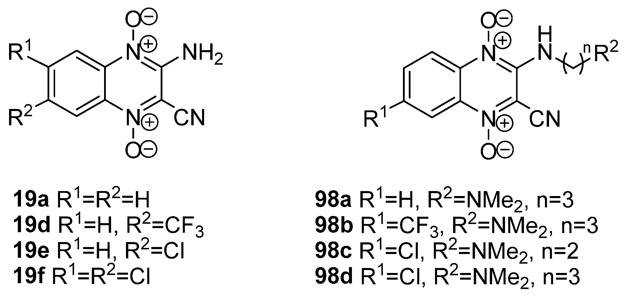
It is known that quinoxaline-2-carbonitrile 1,4-dioxides have high antiproliferative activity against tumor cells of various histogenesis, and their activity increases in hypoxia. A voltammetric study of the electrochemical properties of series derivatives has shown that the introduction of electron-withdrawing groups into the benzene ring of quinoxaline is accompanied by a significant shift of the redox potential to the positive region. This shift correlates with an increase in the cytotoxicity of such compounds under hypoxia [156]. Taking into account the results of electrochemical studies, U. Das et al. synthesized 3-aminoquinoxaline-2-carbonitrile 1,4-dioxides 19af (Figure 14), which selectively induced tumor cells death under hypoxic conditions with low cytotoxicity against non-malignant cells [157]. Further optimization of the structure of quinoxalines 19af by introducing aminoalkylamino-groups into position 3 of quinoxaline (compounds 98ad, Figure 13) leads to an increase in the water solubility of such derivatives while retaining the high antitumor activity of the compounds [158]. It should also be noted that the coordination complexes of 3-aminoquinoxaline-2-carbonitrile 1,4-dioxides 59 and 60 with metals (Scheme 24) exhibit high antiproliferative activity against solid tumor cells, with improved bioavailability and toxicity properties [159].
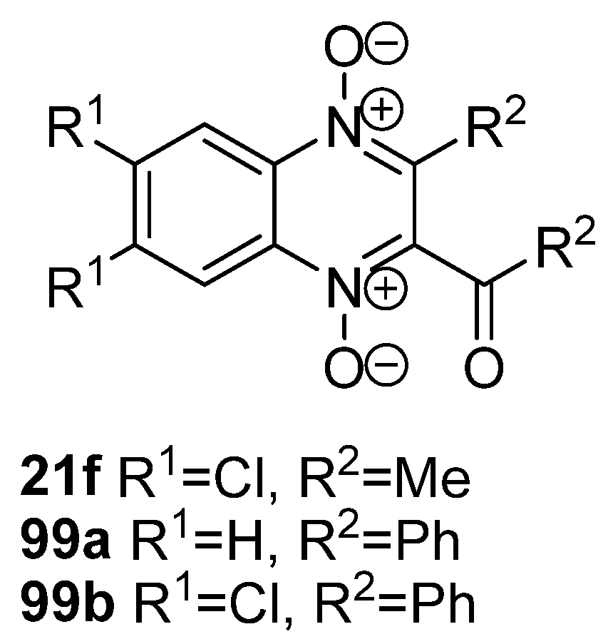
H.U. Gali-Muhtasib et al. [160] have shown that compounds 21f, 99a,b (Figure 15) induce apoptosis and inhibit the growth of colon carcinoma cells T-84 at micromolar concentrations, as well as inhibit the cell cycle in both normoxia and hypoxia. Compounds 21f, 99a,b were found to increase transcript levels of apoptotic transforming growth factor β1 (TGF-β1) and decrease the level of TGF-α mRNA. In addition, derivatives 21f, 99a,b suppress the expression of the antiapoptotic protein gene Bcl-2a and increase the expression level of proapoptotic proteins p53 and p21, which are key mediators that cause apoptosis and cell cycle blocking.
H.U. Gali-Muhtasib et al. observed the effect of quinoxaline 1,4-dioxides on the expression of mRNA transcription factor HIF-1α in T-cell leukemia [161]. It is known that the 1α subunit of the heterodimeric transcription factor HIF regulates a metabolic adaptation to oxygen deficiency and plays an important role in the transcriptional activation of genes involved in angiogenesis. The screening results showed that compounds 99a and 99b (Figure 15) effectively reduce the expression of HIF-1a mRNA, while their non-chlorinated analog 21f does not affect the expression of the HIF-1α gene [162]. It was also found that compound 99a induces the arrest of the cells in G2/M-phases and inhibits the expression of cyclin B, causing apoptosis of tumor cells. The derivative 99a reduces the level of expression of the Bcl-2α gene encoding the antiapoptotic Bcl-2α protein and increases the expression of the proapoptotic BAX protein [163].
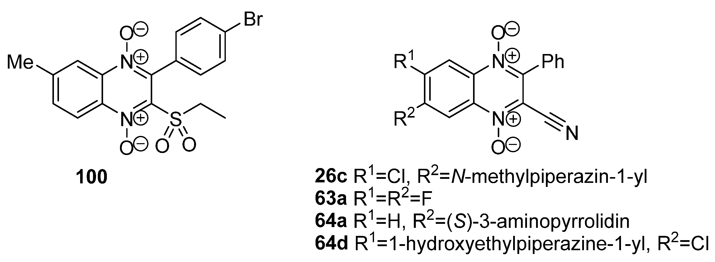
In 2008, Weng Q. et al. [164] found a highly active derivative 100 (Figure 16) capable of inhibiting the growth of a wide panel of tumor cell lines at low concentrations (IC50 = 0.2–8.9 µM) under hypoxic conditions. Recent studies, including virtual screening, the synthesis of polysubstituted derivatives, and the evaluation of their biological activity, have led to the discovery of a series of 3-phenylquinoxaline-2-carbonitrile 1,4-dioxides (compounds 26c, 63a, 64a and 64d, Figure 16) [61,114,165], which exhibit remarkable cytotoxicity. It was shown that compound 63a (Figure 16) induces apoptosis in human tumor cells of various histogenesis. Moreover, lead compounds 26c and 64a,d (Figure 16) have in one order highest activity and selectivity under hypoxic conditions than the reference antitumor hypoxic cytotoxin tirapazamine [61,114].
Analysis of the structure-activity relationship in a series of antitumor agents based on quinoxaline 1,4-dioxide reveals an essential role for the carbonitrile group at position 2 of the heterocyclic ring (for example, derivatives 19af; 26c; 63a; 64a,d; 98ad, Figure 14 and Figure 16). Additionally, substituents at position 3 of the heterocycle are equally important for the antiproliferative properties of quinoxaline 1,4-dioxides. Among these compounds, congeners with a benzene ring at position 3 of the quinoxaline (such as compounds 21f; 26c; 63a; 64a,d; 99a,b; 100, Figure 15 and Figure 16) exhibit the highest activity, whereas compounds with different substituents in this position (for example, compounds 19af; 98ad, Figure 14) show lower levels of activity. Another crucial factor for the ability of compounds to inhibit tumor cell growth under both normoxic and hypoxic conditions is the nature and location of substituents at positions 6 and 7 of quinoxaline 1,4-dioxide. Introduction of linear diamines into the benzene ring of quinoxaline results in a complete loss of antiproliferative activity, whereas the presence of cyclic diamines significantly increases activity, particularly under hypoxic conditions. Notably, derivatives containing substituents at position 7 of quinoxaline exhibit noticeably higher activity compared to their corresponding regioisomeric analogs with substituents at position 6. Furthermore, the introduction of a halogen atom into the quinoxaline structure positively affects the ability of compounds to inhibit tumor cell growth. Remarkably, the addition of a chlorine atom proves to be the most crucial modification for enhancing the hypoxic cytotoxicity of quinoxaline 1,4-dioxide derivatives.
Compounds 26c and 64a,d are also able to induce apoptosis in the myelogenous leukemia cell line and its doxorubicin-resistant subline K562/4 cell with the expression of P-glycoprotein (P-gp) [61,114].

5. Targeting Signaling Pathways in Cells by Quinoxaline 1,4-Dioxide Derivatives

The search for ways to selectively induce tumor cell death is one of the key goals in the development of new anticancer drugs. Therefore, the discovery of the hypoxia-selective cytotoxic mode of action of quinoxaline 1,4-dioxides stimulated further study of the mechanisms of their antitumor activity. It has been shown that, similarly to the antibacterial action, the antitumor potency of quinoxaline 1,4-dioxides is associated with their bioreduction in tumor cells by reductases expressed under hypoxic conditions, leading to the generation of ROS and the damage of DNA in malignant cells [166,167,168].
As noted above, quinoxaline 1,4-dioxide derivatives are able to trigger a cascade of various proapoptotic processes and suppress the mechanisms of cell resistance to xenobiotics [61,114,165]. It has been shown that quinoxaline derivatives induce tumor cell death via apoptosis [169,170,171]. Additionally, due to the outstanding hypoxic selectivity of several quinoxaline 1,4-dioxides, this class has a high potential for the development of drugs for the targeted treatment of solid tumors. Thus, previous works [61,114,165] have shown that the hypoxic selectivity of compounds 26c, 64a,d, and 99b (Scheme 12, Figure 15 and Figure 16) is associated with the inhibition of expression and activity of the hypoxia-induced factor HIF-1α in tumor cells. At the same time, amino derivatives 26c and 64a,d have a significant effect on the signaling pathways of the estrogen receptor ERα, which supports the growth of hormone-dependent tumor cells MCF-7 and can be considered as double blockers of HIF-1α/ERα, modulating the activity of the HIF-1α and ERK1/2 signaling pathways. In addition, the induction of apoptosis in HCT-116p53KO tumor cells with a deletion of the tumor suppressor p53, observed for derivative 63a [60,61], is evidence that tumor cell death induced by quinoxaline 1,4-dioxides can proceed via a p53-independent mechanism [165,171].
The compounds 100 (Figure 16) and 99b (Figure 15) could reduce the expression of vascular endothelial growth factor (VEGF) under hypoxic conditions. Quincetone (104, Figure 17) and derivative 100 (Figure 16) also down-regulated the expression of mitogen-activated protein kinases (MAPKs) involved in the mitochondrial apoptosis pathway. When studying the molecular mechanisms of action of olaquindox, D. Li and co-workers found that it can induce DNA damage and S-phase arrest, contributing to an increase in the expression of GADD45a, cyclin A, Cdk2, p21, and p53, a decrease in cyclin D1, as well as activation of phosphorylation of c-Jun N-terminal kinases (p-JNK), p38 (p-p38), and extracellular signal-regulated kinases (p-ERK) [172].
The A. Monge group found that the leishmanicidal activity of compound 92 (Figure 10) is associated with the induction of necrosis of L. mexicana caused by ROS production, leading to damage to the cell membrane, phosphatidylserine flip-flop, disruption of the mitochondrial membrane potential, and subsequent fragmentation of the parasite DNA [173]. The study of the mechanism of antiparasitic action showed that thioredoxin reductase (EhTrxR) is the main biological target of quinoxaline 1,4-dioxides in the cells of E. histolytica [174]. Compound 95 (Figure 11) acts as a substrate that disrupts the electron transfer in the active site of the EhTrxR enzyme, which leads to an increase in ROS levels and induces oxidative stress, causing changes in chromatin, cell granularity, and the redistribution of vacuoles.

6. Agricultural Use of Quinoxaline 1,4-Dioxide Derivatives

Quinoxaline 1,4-dioxide derivatives have been used for over 50 years as animal growth promoters, significantly improving feed efficiency in animal husbandry [175]. The most well-known compounds (101104) are shown in Figure 17 [176,177].
Carbadox (CARB, 101, Figure 17) acts on Gram-positive bacteria but is inactive against Gram-negative bacteria. It was patented by Pfizer in 1968. As a feed additive, it stimulates the growth of pigs and is also used to control swine dysentery (caused by Serpulina hyodysenteriae), bacterial enteritis (caused by Salmonella spp.), and nasal infections (caused by Bordetella bronchiseptica) [178]. Carbadox improves the retention of dietary proteins [23,24] and increases the ratio of RNA to DNA and protein to DNA in the lean muscles of young pigs [179]. Additionally, it reduces the energy requirement of the gastrointestinal tract, spleen, and pancreas in pigs [180]. However, the use of carbadox as a feed additive for livestock has been banned in Canada and the EU since 2004 due to the experimental evidence showing its carcinogenic and genotoxic properties [181]. Nonetheless, it is still used as a therapeutic and preventive agent in the treatment of bacterial enteritis of pigs and porcine dysentery caused by B. hyodysenteriae in the United States, Canada, and some other countries.
Olaquindox (OLAQ, 102, Figure 17), developed by Bayer in 1967, is used to stimulate the growth of pigs. The drug improves the functioning of the gastrointestinal tract in animals, suppressing pathogenic Gram-positive and Gram-negative microflora. It is also used to prevent swine dysentery caused by B. hyodysenteriae [182]. Carbadox (101) and olaquindox (102) can irritate mucous membranes and cause superficial dermatitis and photoallergic reactions in animals [183].
Studies have shown that quinoxaline 1,4-dioxide derivatives are not present in meat after 10 days of keeping pigs before slaughter. Furthermore, carbadox and olaquindox exhibit low acute toxicity [183]. The LD50 for administration of carbadox peros in rats is 850 mg/kg, and in mice, it is 2810 mg/kg. Olaquindox (102) is somewhat less toxic, with an oral LD50 for rabbits ranging from 1000 to 2000 mg/kg and about 1600 mg/kg for rats. The subcutaneous LD50 of olaquindox in rats is 1275 mg/kg. However, both carbadox and olaquindox have shown genotoxicity using in vitro and in vivo studies. Carbadox (101) induced chromosome damage in human lymphocytes, and olaquindox damaged DNA in Chinese hamster cell culture. Additionally, carbadox (101) and olaquindox (102) caused an increase in the number of micronuclear polychromatic erythrocytes in the bone marrow of rats, indicating their genotoxicity in animals. The metabolites of carbadox (101) and olaquindox (102) could potentially be carcinogenic, thus requiring careful monitoring of their content in the meat and animal fat.
Mequindox (MEQ, 21b, Figure 17), created by Pfizer, exhibits high antibacterial activity against Gram-negative bacteria, particularly Salmonella spp., and is widely used in China as a feed additive for animals and as a veterinary drug for dysentery and white diarrhea in pigs [184].
Quincetone (QUIN, 103, Figure 17), developed by the Lanzhou Institute of Animal Husbandry and Veterinary Drugs (Chinese Academy of Agricultural Sciences, Lanzhou, China), is active against B. hyodysenteriae, Salmonella spp., E. coli, and other Gram-negative bacilli. Quincetone (103) is approved as an animal growth promoter in China and has been used in swine, poultry, and aquaculture since 2003 [185].
Cyadox (CYAD, 104, Figure 17), described by Chemapol Benelux, exhibits antibacterial activity against Staphylococcus hyicus, Pasteurella multocida, E. coli and also shows a beneficial effect as a growth promoter in broiler chickens and pigs. Cyadox (104) has low toxicity and high safety, making it a potential substituent for olaquindox (102) and carbadox (101) in animal husbandry [186].
Several quinoxaline 1,4-dioxide derivatives, such as compounds 105 and 106 (Figure 18), have shown herbicidal activity against dicotyledonous and cereal plants by disrupting the functioning of photosystem I [187]. Due to the presence of two N-oxide groups, compounds 105 and 106 compete with NADP+ in the active site of ferredoxin-NADP+ reductase, initiating numerous oxidative transformations in plant cells, leading to their death.
In conclusion, the synthetic availability and the ability to modulate desired pharmacological properties make quinoxaline 1,4-dioxide derivatives as a promising class of heterocyclic compounds for the development of new agrochemical agents.

7. Toxicological Properties of Quinoxaline 1,4-Dioxides

The ability of quinoxaline 1,4-dioxides to generate ROS in cells, thereby inducing DNA single- or double-stranded breaks, also underlies their mutagenic action. For example, T. Negishi and co-workers proved the mutagenicity of compounds 101104 (Figure 17) and 3 (Figure 2), as well as some other quinoxaline 1,4-dioxides, on Salmonella typhimurium TA 98 and TA 100 strains [188]. It was shown that the mutagenicity of quinoxaline 1,4-dioxides is associated with the presence of two N-oxide groups since quinoxaline and quinoxaline 1-N-oxide did not exhibit mutagenic activity under the same conditions. In addition, using in vivo experiments (a micronuclear test in mice) additionally confirmed the clastogenicity of quinoxaline 1,4-dioxides [22]. Thus, olaquindox (102, Figure 17) induces chromosome aberrations when administered orally at a dose of 100 mg/kg, while carbadox (101, Figure 17) exhibits the same effect at a dose of 800 mg/kg [189]. Nevertheless, in contrast to the derivatives 101 and 102, the agricultural agent cyadox (104, Figure 17) did not show a damaging effect on chromosomes, even at a dose of 1200 mg/kg. It was found that derivative 101 has a teratogenic effect and leads to intrauterine malformation of the fetus and death of the embryo using in vivo experiments on rats [190]. Teratogenic, embryotoxic, and mutagenic effects have been shown for the clinically used dioxidine, which limits its use [191].
The phototoxicity of some derivatives of quinoxaline 1,4-dioxide was observed in [192], indicating the ability of quinoxaline 1,4-dioxides used in agriculture and veterinary practice to cause photoallergic reactions in animals. It was found that the reactive oxaziridine intermediate 29 and its 2,3-disubstituted derivatives, as well as its dioxaziridine analogs (Scheme 8) generated under UV irradiation, engage in the chemical interaction with proteins. In particular, irradiation of 3-phenylquinoxaline-2-carbonitrile 1,4-dioxide (89b, Figure 9) by the long-wavelength part of the visible spectrum leads to selective cleavage of the bond at the 2’-position of deoxyguanosine [193]. An enhancement of photoinduced bond breaking in pBR322 plasmid DNA caused by compound 89b (Figure 9) occurs with the further participation of NADPH-dependent enzymes under anaerobic conditions. Notably, in contrast to derivative 89b (Figure 9), DNA damage induced by quinoxaline 1,4-dioxide (28a, Scheme 8) was independent of NADPH-dependent enzymes.

8. Conclusions

The search for new possibilities to functionalize quinoxaline 1,4-dioxides has attracted special attention from chemists due to the unique biological properties and high pharmacological activity of this class of compounds. Over the past decade, several effective methods for their synthesis have been developed, and classical methods for the preparation and modification of quinoxaline 1,4-dioxides have been improved. Progress in the chemistry of this class has allowed the evaluation of bioactivities for a wide range of functionalized derivatives. Recent experimental works demonstrate the continued scientific and practical interest in quinoxaline 1,4-dioxides. Therefore, a systematization of preparation methods of quinoxaline 1,4-dioxides are in demand to develop new schemes for the synthesis of these compounds and contribute to further evaluations of this class of heterocycles.
In recent years, there has been a high priority placed on modifying the structure of quinoxaline 1,4-dioxide to develop drugs with greater selectivity or improved therapeutic properties. However, analyzing the relationship between biological activity and the structure of quinoxaline 1,4-dioxides, considering multiple types of activity simultaneously, and evaluating specific structural features, is a challenging task. General observations include the following:
-
the introduction of electron-withdrawing substituents at positions 3 and 6/7 of the quinoxaline moiety promotes more readily reduction by bacterial or eukaryotic reductases, resulting in increased cytotoxicity and antimicrobial activity of such derivatives;
-
the presence of a carbonitrile moiety at position 2 of the pyrazine ring enhances antiproliferative activity, while ester or carboxamide fragments at this position improve the antibacterial properties of quinoxaline 1,4-dioxides;
-
overall, the presence of a halogen atom in the quinoxaline core enhances the biological activity of the compounds, with chlorine being the most significant;
-
the introduction of electron-donating groups at C3 or C7 carbon atoms of the heterocycle increases the hypoxic cytotoxicity of quinoxaline-2-carbonitrile 1,4-dioxide derivatives.
However, the activity of such compounds strongly depends on the structure of the substituents. The presence of linear alkylamino groups at these positions had a negative effect, resulting in a complete loss of antiproliferative properties and selectivity under hypoxic conditions. [61,108]. On the other hand, the presence of cyclic amino groups at positions 3 or 7 leads to a significant increase in the hypoxic selectivity of quinoxaline-2-carbonitrile 1,4-dioxides. Additionally:
-
lipophilic substituents at positions 2, 6, or 7 improve the antituberculous potential of the derivatives;
-
acylamino groups at position 3 of the quinoxaline ring have a positive effect on suppressing the growth of protozoa (Leishmania spp. and Plasmodium spp);
-
the introduction of salt-forming fragments leads to water-soluble derivatives while retaining their biological potency.
However, due to the limited available data on target proteins and the necessity for further searches for intracellular reductases involved in the activation of quinoxaline 1,4-dioxides, a comprehensive understanding of the mechanism of action remains essential. H. Zhang et al. conducted QSAR analysis for antimycobacterial activity using CoMFA and CoMSIA models [89]. The results of 3D-QSAR modeling identified several critical requirements for the structure of quinoxaline 1,4-dioxides for their antituberculosis properties. This indicates the specificity of the binding of this class of compounds to intracellular targets important for inhibiting pathogen growth. Electron-withdrawing and sterically bulky substituents, particularly halogen atoms, at position 7 of the quinoxaline are preferred for increasing antimycobacterial activity. Furthermore, the introduction of hydrogen bond donors at the C2 carbon atom of the pyrazine ring enhances activity against Mycobacterium spp.
Despite the observed progress, the further development of methods for preparing new quinoxaline 1,4-dioxides and the study of their mode of action remains an urgent task. Addressing these tasks could broaden the practical applications for this unique class of heterocyclic compounds.

Author Contributions

Conceptualization and formal analysis A.E.S. and G.I.B.; writing—original draft preparation, G.I.B.; writing—review and editing, A.E.S. and G.I.B.; supervision, A.E.S. All authors have read and agreed to the published version of the manuscript.

Funding

The study was partially financially supported by the Russian Science Foundation (RSF grant number 21-45-00018).

Institutional Review Board Statement

Not applicable.

Informed Consent Statement

Not applicable.

Data Availability Statement

Data sharing not applicable.

Acknowledgments

The authors thank A.A. Vatlin and D.A. Maslov for supervision of the project RSF 21-45-00018.

Conflicts of Interest

The authors declare no conflict of interest.

References

  1. Vitaku, E.; Smith, D.T.; Njardarson, J.T. Analysis of the structural diversity, substitution patterns, and frequency of nitrogen heterocycles among U.S. FDA Approved Pharmaceuticals. J. Med. Chem. 2014, 57, 10257–10274. [Google Scholar] [CrossRef] [PubMed]
  2. Mohan, C.D.; Anilkumar, N.C.; Rangappa, S.; Shanmugam, M.K.; Mishra, S.; Chinnathambi, A.; Alharbi, S.A.; Bhattacharjee, A.; Sethi, G.; Kumar, A.P.; et al. Novel 1,3,4-oxadiazole induces anticancer activity by targeting NF-κB in hepatocellular carcinoma cells. Front. Oncol. 2018, 8, 42. [Google Scholar] [CrossRef] [PubMed]
  3. Pecoraro, C.; De Franco, M.; Carbone, D.; Bassani, D.; Pavan, M.; Cascioferro, S.; Parrino, B.; Cirrincione, G.; Dall’Acqua, S.; Moro, S.; et al. 1,2,4-Amino-triazine derivatives as pyruvate dehydrogenase kinase inhibitors: Synthesis and pharmacological evaluation. Eur. J. Med. Chem. 2023, 249, 115134. [Google Scholar] [CrossRef]
  4. Moniot, S.; Forgione, M.; Lucidi, A.; Hailu, G.S.; Nebbioso, A.; Carafa, V.; Baratta, F.; Altucci, L.; Giacché, N.; Passeri, D.; et al. Development of 1,2,4-oxadiazoles as potent and selective inhibitors of the human deacetylase sirtuin 2: Structure-activity relationship, X-ray crystal ctructure, and anticancer activity. Eur. J. Med. Chem. 2017, 60, 2344–2360. [Google Scholar] [CrossRef] [PubMed]
  5. Carbone, D.; De Franco, M.; Pecoraro, C.; Bassani, D.; Pavan, M.; Cascioferro, S.; Parrino, B.; Cirrincione, G.; Dall’Acqua, S.; Moro, S.; et al. Discovery of the 3-amino-1,2,4-triazine-based library as selective PDK1 inhibitors with therapeutic potential in highly aggressive pancreatic ductal adenocarcinoma. Int. J. Mol. Sci. 2023, 24, 3679. [Google Scholar] [CrossRef]
  6. Abdel-Aziz, A.K.; Dokla, E.M.E.; Abouzid, K.A.M.; Minucci, S. Discovery of EMD37, a 1,2,4-oxadiazole derivative, as a novel endoplasmic reticulum stress inducer with potent anticancer activity. Biochem. Pharmacol. 2022, 206, 115316. [Google Scholar] [CrossRef]
  7. Patterson, L.H. Rationale for the use of aliphatic N-oxides of cytotoxic anthraquinones as prodrug DNA binding agents: A new class of bioreductive agent. Cancer Metastasis Rev. 1993, 12, 119–134. [Google Scholar] [CrossRef]
  8. Shen, X.; Gates, K.S. Enzyme-activated generation of reactive oxygen species from heterocyclic N-oxides under aerobic and anaerobic conditions and its relevance to hypoxia-selective prodrugs. Chem. Res. Toxicol. 2019, 32, 348–361. [Google Scholar] [CrossRef]
  9. Nicola, O.; O’Neill, A.J. Revisiting unexploited antibiotics in search of new antibacterial drug candidates: The case of MSD-819 (6-chloro-2-quinoxalinecarboxylic acid 1,4-dioxide). J. Antibiot. 2017, 70, 317–319. [Google Scholar] [CrossRef]
  10. Turner, J.M.; Messenger, A.J. Occurrence, biochemistry and physiology of phenazine pigment production. Adv. Microb. Physiol. 1986, 27, 211–275. [Google Scholar] [CrossRef]
  11. Cimmino, A.; Evidente, A.; Mathieu, V.; Andolfi, A.; Lefranc, F.; Kornienko, A.; Kiss, R. Phenazines and cancer. Nat. Prod. Rep. 2012, 29, 487–501. [Google Scholar] [CrossRef]
  12. McIlwain, H. Bacterial inhibition by metabolite analogues. Part V. Reactions and antibacterial properties of p-diazine di-N-oxides. J. Chem. Soc. 1943, 88, 322–325. [Google Scholar] [CrossRef]
  13. Viktorsson, E.Ö.; Melling Grøthe, B.; Aesoy, R.; Sabir, M.; Snellingen, S.; Prandina, A.; Åstrand, O.A.H.; Bonge-Hansen, T.; Døskeland, S.O.; Herfindal, L.; et al. Total synthesis and antileukemic evaluations of the phenazine 5,10-dioxide natural products iodinin, myxin and their derivatives. Bioorg. Med. Chem. 2017, 25, 2285–2293. [Google Scholar] [CrossRef]
  14. Myhren, L.; Nygaard, G.; Gausdal, G.; Sletta, H.; Teigen, K.; Degnes, K.; Zahlsen, K.; Brunsvik, A.; Bruserud, Ø.; Døskeland, S.O.; et al. Iodinin (1,6-Dihydroxyphenazine 5,10-dioxide) from Streptosporangium sp. induces apoptosis selectively in myeloid leukemia cell lines and patient cells. Mar. Drugs 2013, 11, 332–349. [Google Scholar] [CrossRef] [PubMed]
  15. Schwartz, B.S.; Pollak, J.; Bailey-Davis, L.; Hirsch, A.G.; Cosgrove, S.E.; Nau, C.; Kress, A.M.; Glass, T.A.; Bandeen-Roche, K. Antibiotic use and childhood body mass index trajectory. Int. J. Obes. 2016, 40, 615–621. [Google Scholar] [CrossRef] [PubMed]
  16. Dvoryantseva, G.G.; Lindeman, S.V.; Aleksanyan, M.S.; Struehkov, Y.T.; Teten’ehuk, K.P.; Khabarova, L.S.; Elina, A.S. Connection between the structure and the antibacterial activity of the N-oxides of quinoxalines. Molecular structure of dioxidine and quinoxidine. Pharm. Chem. J. 1990, 24, 672–677. [Google Scholar] [CrossRef]
  17. Vicente, E.; Lima, L.M.; Bongard, E.; Charnaud, S.; Villar, R.; Solano, B.; Burguete, A.; Perez-Silanes, S.; Aldana, I.; Vivas, L.; et al. Synthesis and structure-activity relationship of 3-phenylquinoxaline 1,4-di-N-oxide derivatives as antimalarial agents. Eur. J. Med. Chem. 2008, 43, 1903–1910. [Google Scholar] [CrossRef]
  18. Zarranz, B.; Jaso, A.; Aldana, I.; Monge, A. Synthesis and antimycobacterial activity of new quinoxaline-2-carboxamide 1,4-di-N-oxide derivatives. J. Med. Chem. 2003, 11, 2149–2156. [Google Scholar] [CrossRef]
  19. Edwards, M.L.; Bambury, R.E.; Ritter, H.W. 3-Substituted 2-formylquinoxaline 1,4-dioxides. J. Med. Chem. 1975, 18, 637–639. [Google Scholar] [CrossRef] [PubMed]
  20. Kleim, J.P.; Bender, R.; Billhardt, U.M.; Meichsner, C.; Riess, G.; Rösner, M.; Winkler, I.; Paessens, A. Activity of a novel quinoxaline derivative against human immunodeficiency virus type 1 reverse transcriptase and viral replication. Antimicrob. Agents Chemother. 1993, 37, 1659–1664. [Google Scholar] [CrossRef]
  21. Monge, A.; Palop, J.A.; Cerain, A.L.; Senador, V.; Martinez-Crespo, F.J.; Sainz, Y.; Narro, S.; Garcia, E.; de Miguel, C.; Gonzalez, M.; et al. Greenhow hypoxia-selective agents derived from quinoxaline 1,4-di-N-oxides. J. Med. Chem. 1995, 38, 1786–1792. [Google Scholar] [CrossRef]
  22. Tucker, M.J. Carcinogenic action of quinoxaline 1,4-dioxide in rats. J. Natl. Cancer Inst. 1975, 55, 137–146. [Google Scholar] [CrossRef]
  23. Zaynoun, S.; Johnson, B.; Frain-Bell, W. The investigation of Quindoxin photosensitivity. Contact Derm. 1976, 2, 343–352. [Google Scholar] [CrossRef] [PubMed]
  24. De Vries, H.; Bojarski, J.; Donker, A.A.; Bakri, A.; Beyersbergen van Henegouwen, G.M. Photochemical reactions of Quindoxin, Olaquindox, Carbadox and Cyadox with protein, indicating photoallergic properties. Toxicology 1990, 63, 85–95. [Google Scholar] [CrossRef] [PubMed]
  25. Vieira, M.; Pinheiro, C.; Fernandes, R.; Noronha, J.P.; Prudencio, C. Antimicrobial activity of quinoxaline 1,4-dioxide with 2-and 3-substituted derivatives. Microbiol. Res. 2014, 169, 287–293. [Google Scholar] [CrossRef]
  26. Carta, A.; Corona, P.; Loriga, M. Quinoxaline 1,4-dioxide: A versatile scaffold endowed with manifold activities. Curr. Med. Chem. 2005, 12, 2259–2272. [Google Scholar] [CrossRef] [PubMed]
  27. Lima, L.M.; do Amaral, D.N. Beirut reaction and its application in the synthesis of quinoxaline-N,N′-dioxides bioactive compounds. Rev. Virtual Quim. 2013, 5, 1075–1100. [Google Scholar] [CrossRef]
  28. Agrawal, N.; Bhardwaj, A. An appraisal on synthetic and pharmaceutical perspectives of quinoxaline 1,4-di-N-oxide scaffold. Chem. Biol. Drug Des. 2022, 100, 346–363. [Google Scholar] [CrossRef]
  29. Rivera, G. Quinoxaline 1,4-di-N-oxide derivatives: Are they unselective or selective inhibitors? Mini Rev. Med. Chem. 2022, 22, 15–25. [Google Scholar] [CrossRef]
  30. Dos Santos Fernandes, G.F.; Pavan, A.R.; dos Santos, J.L. Heterocyclic N-oxides—A promising class of agents against tuberculosis, malaria and neglected tropical diseases. Curr. Pharm. Des. 2018, 24, 1325–1340. [Google Scholar] [CrossRef]
  31. Hamama, W.S.; Waly, S.M.; Said, S.B.; Zoorob, H.H. Highlights on the chemistry of 2-amino-3-cyano-quinoxaline 1,4-dioxides and their derivatives. Synth. Commun. 2020, 50, 1737–1757. [Google Scholar] [CrossRef]
  32. Cheng, G.; Sa, W.; Cao, C.; Guo, L.; Hao, H.; Liu, Z.; Wang, X.; Yuan, Z. Quinoxaline 1,4-di-N-Oxides: Biological activities and mechanisms of actions. Front. Pharmacol. 2016, 7, 64. [Google Scholar] [CrossRef] [PubMed]
  33. González, M.; Cerecetto, H.; Monge, A. Quinoxaline 1,4-dioxide and phenazine 5,10-dioxide. Bioact. Heterocycl. V 2007, 11, 179–211. [Google Scholar] [CrossRef]
  34. Vicente, E.; Villar, R.; Pérez-Silanes, S.; Aldana, I.; Goldman, R.C.; Monge, A. Quinoxaline 1,4-di-N-oxide and the potential for treating tuberculosis infectious disorders. Curr. Drug Targets 2011, 11, 196–204. [Google Scholar] [CrossRef]
  35. Keri, R.S.; Pandule, S.S.; Budagumpi, S.; Nagaraja, B.M. Quinoxaline and quinoxaline-1,4-di-N-oxides: An emerging class of antimycobacterials. Arch. Pharm. 2018, 351, e1700325. [Google Scholar] [CrossRef]
  36. Montana, M.; Montero, V.; Khoumeri, O.; Vanelle, P. Quinoxaline moiety: A potential scaffold against Mycobacterium tuberculosis. Molecules 2021, 26, 4742. [Google Scholar] [CrossRef]
  37. Carmeli, M.; Rozen, S. A new efficient route for the formation of quinoxaline N-oxides and N,N′-dioxides using HOF-CH3CN. J. Org. Chem. 2006, 71, 5761–5765. [Google Scholar] [CrossRef]
  38. Wang, R.; Zhang, M.; Wang, W.; Wang, X.; Yuan, Y.; Li, J. Synthesis, crystal structure and calculation of oxides of 2-methylamino-3-methyl quinoxaline. J. Mol. Struct. 2020, 1222, 128826. [Google Scholar] [CrossRef]
  39. Abushanab, E. One-step synthesis of quinoxaline 1,4-dioxides and related compounds. J. Org. Chem. 1970, 35, 4279–4280. [Google Scholar] [CrossRef]
  40. Haddadin, M.J.; Issidorides, C.H. Enamines with isobenzofuroxan: A novel synthesis of quinoxaline-di-N-oxides. Tetrahedron Lett. 1965, 6, 3253–3256. [Google Scholar] [CrossRef]
  41. Jie, J.L. Name Reactions: A Collection of Detailed Reaction Mechanisms, 3rd ed.; Springer: New York, NY, USA, 2006; Volume 1, p. 504. [Google Scholar]
  42. El-Haj, M.J.A.; Dominy, B.W.; Johnston, J.D.; Haddadin, M.J.; Issidorides, C.H. New route to phenazine 5,10-dioxides and related compounds. J. Org. Chem. 1972, 37, 589–593. [Google Scholar] [CrossRef]
  43. Tanaka, A.A.; Usui, T. Studies on furan derivatives. Preparation of 2-substituted 3-(5-nitro-2-furyl) quinoxaline 1,4-dioxides and determination of their antibacterial activity. Chem. Pharm. Bull. 1981, 29, 110–115. [Google Scholar] [CrossRef]
  44. Li, J.; Ji, M.; Hua, W.; Hu, H. Synthesis of 2-substituted-quinoxaline 1,4-dioxides. Ind. J. Chem. B 2001, 40, 1230–1231. [Google Scholar] [CrossRef]
  45. McFarland, J.W. 2,3-Dihydroquinoxaline 1,4-dioxides as intermediates in the reaction between benzofurazan 1-oxide and enamines. J. Org. Chem. 1971, 36, 1842–1843. [Google Scholar] [CrossRef]
  46. Atfah, A.; Hill, J. Reaction of 2,1,3-benzoxadiazole 1-oxide with ethyl 2,4-dioxo-4-phenylbutyrate. A route to 2-benzoylquinoxaline, its 1,4-dioxide, and related compounds. J. Chem. Soc. Perkin Trans. 1 1989, 2, 221–224. [Google Scholar] [CrossRef]
  47. Monge, A.; Palop, J.A.; Urbasos, I.; Fernandez-Alvarez, E. New quinoxaline and pyrimido [4,5-b]quinoxaline derivatives. Potential antihypertensive and blood platelet antiaggregating agents J. Heterocycl. Chem. 1989, 26, 1623–1626. [Google Scholar] [CrossRef]
  48. Xu, Y.; Wu, F.; Yao, Z.; Zhang, M.; Jiang, S. Synthesis of quinoxaline 1,4-di-N-oxide analogues and crystal structure of 2-carbomethoxy-3-hydroxyquinoxaline-di-N-oxide. Molecules 2011, 16, 6894–6901. [Google Scholar] [CrossRef]
  49. Anderson, R.F.; Yadav, P.; Shinde, S.S.; Hong, C.R.; Pullen, S.M.; Reynisson, J.; Wilso, W.R.; Hay, M.P. Radical chemistry and cytotoxicity of bioreductive 3-substituted quinoxaline di-N-oxides. Chem. Res. Toxicol. 2016, 29, 1310–1324. [Google Scholar] [CrossRef] [PubMed]
  50. Chupakhin, O.N.; Kotovskaya, S.K.; Perova, N.M.; Baskakova, Z.M.; Charushin, V.N. Synthesis of new fluorine-containing derivatives of quinoxaline 1,4-dioxides and condensed systems derived from them. Chem. Heterocycl. Compd. 1999, 35, 459–469. [Google Scholar] [CrossRef]
  51. Lima, L.M.; Zarranz, B.; Marin, A.; Solano, B.; Vicente, E.; Perez Silanes, S.; Aldana, I.; Monge, A. Comparative use of solvent-free KF-Al2O3 and K2CO3 in acetone in the synthesis of quinoxaline 1,4-dioxide derivatives designed as antimalarial drug candidates. J. Heterocycl. Chem. 2005, 42, 1381–1385. [Google Scholar] [CrossRef]
  52. Sun, T.; Zhao, W.-J.; Hao, A.-Y.; Sun, L.-Z. Effective promotion of Beirut reaction by β-cyclodextrin in water. Synth. Commun. 2011, 41, 3097–3105. [Google Scholar] [CrossRef]
  53. Bonilla-Ramírez, L.; Rios, A.; Quiliano, M.; Ramírez-Calderon, G.; Beltrán-Hortelano, I.; Franetich, J.F.; Corcuera, L.; Bordessoulles, M.; Vettorazzi, A.; López de Cerain, A.; et al. Novel antimalarial chloroquine- and primaquine-quinoxaline 1,4-di-N-oxide hybrids: Design, synthesis, Plasmodium life cycle stage profile, and preliminary toxicity studies. Eur. J. Med. Chem. 2018, 158, 68–81. [Google Scholar] [CrossRef] [PubMed]
  54. Srinivasarao, S.; Nandikolla, A.; Suresh, A.; Ewa, A.-K.; Głogowska, A.; Ghosh, B.; Kumar, B.K.; Murugesan, S.; Pulya, S.; Aggrawal, H.; et al. Discovery of 1,2,3-triazole based quinoxaline-1,4-di-N-oxide derivatives as potential anti-tubercular agents. Bioorg. Chem. 2020, 100, 103955. [Google Scholar] [CrossRef]
  55. Gasco, A.M.; Ermondi, G.; Fruttero, R.; Gasco, A. Benzofurazanyl- and benzofuroxanyl-1,4-dihydropyridines: Synthesis, structure and calcium entry blocker activity. Eur. J. Med. Chem. 1996, 31, 3–10. [Google Scholar] [CrossRef]
  56. Haddadin, M.J.; Taha, M.U.; Jarrar, A.A.; Issidorides, C.H. Reaction of benzofurazan oxide with unsymmetrical 1,3-diketones; steric and polar effects. Tetrahedron 1976, 32, 719–724. [Google Scholar] [CrossRef]
  57. Haddadin, M.J.; Issidorides, C.H. The Beirut reaction. Heterocycles 1993, 3, 1503–1525. [Google Scholar] [CrossRef]
  58. Cerecetto, H.; Gonzalez, M.; Lavaggi, M.L.; Porcal, W. Preparation of phenazine N5,N10-dioxides. Effects of benzofuroxan substituents in the outcome of their expansion reaction with phenolates. J. Braz. Chem. Soc. 2005, 16, 1290–1296. [Google Scholar] [CrossRef]
  59. Ermondi, G.; Visentin, S.; Boschi, D.; Fruttero, R.; Gasco, A. Structural investigation of Ca2+ antagonists benzofurazanyl and benzofuraxanyl-1,4-dihydropiridines. J. Mol. Struct. 2000, 523, 149–154. [Google Scholar] [CrossRef]
  60. Buravchenko, G.I.; Scherbakov, A.M.; Korlukov, A.D.; Dorovatovskii, P.V.; Shchekotikhin, A.E. Revision of the regioselectivity of the Beirut reaction of monosubstituted benzofuroxans with benzoylacetonitrile. 6-Substituted quinoxaline-2-carbonitrile 1,4-dioxides: Structural characterization and estimation of anticancer activity and hypoxia selectivity. Curr. Org. Chem. 2020, 17, 29–39. [Google Scholar] [CrossRef]
  61. Buravchenko, G.I.; Scherbakov, A.M.; Dezhenkova, L.G.; Monzote, L.; Shchekotikhin, A.E. Synthesis of 7-amino-6-halogeno-3-phenylquinoxaline-2-carbonitrile 1,4-dioxides: A way forward for targeting hypoxia and drug resistance of cancer cells. RSC Adv. 2021, 11, 38782–38795. [Google Scholar] [CrossRef]
  62. Buravchenko, G.I.; Maslov, D.A.; Alam, M.S.; Grammatikova, N.E.; Frolova, S.G.; Vatlin, A.A.; Tian, X.; Ivanov, I.V.; Bekker, O.B.; Kryakvin, M.A.; et al. Synthesis and characterization of novel 2-acyl-3-trifluoromethylquinoxaline 1,4-dioxides as potential antimicrobial agents. Pharmaceuticals 2022, 15, 155. [Google Scholar] [CrossRef]
  63. Dirlam, J.P.; McFarland, J.W. Selective monodeoxygenation of certain quinoxaline 1,4-dioxides with trimethyl phosphite. J. Org. Chem. 1977, 42, 1360–1364. [Google Scholar] [CrossRef]
  64. Kluge, A.F.; Maddox, M.L.; Lewis, G.S. Formation of quinoxaline monoxides from reaction of benzofurazan oxide with enones and carbon-13 NMR correlations of quinoxaline N-oxides. J. Org. Chem. 1980, 45, 1909–1914. [Google Scholar] [CrossRef]
  65. Demirdji, S.H.; Haddadin, M.J.; Issidorides, C.H. Deoxygenation of quinoxaline and phenazine N-oxides by catalytic transfer reduction and by iodide in the presence of pyridine/sulfur trioxide complex. J. Heterocycl. Chem. 1983, 20, 1735–1737. [Google Scholar] [CrossRef]
  66. Novacek, L.; Nechvatal, M. Partial reduction of quinoxaline 1,4-dioxide derivatives with L-ascorbic acid. Collect. Czech. Chem. Commun. 1988, 53, 1302–1306. [Google Scholar] [CrossRef]
  67. Homaidan, F.R.; Issidorides, C.H. Deoxygenation of 2,3-disubstituted quinoxaline 1,4-dioxides. Heterocycles 1981, 16, 411–415. [Google Scholar] [CrossRef]
  68. Jarrar, A.A.; Fataftah, Z.A. Photolysis of some quinoxaline-1,4-di-oxides. Tetrahedron 1977, 33, 2127–2129. [Google Scholar] [CrossRef]
  69. Hirota, T.; Namba, T.; Sasaki, K. Polycyclic N-hetero compounds. XXII Reaction of pyridine N-oxides and pyrazine di-N-oxides with formamide. J. Heterocycl. Chem. 1987, 24, 949–953. [Google Scholar] [CrossRef]
  70. Koyama, T.; Nanba, T.; Hirota, T.; Ohmori, S.; Yamato, M. Polycyclic N-hetero compounds. XIII. Reactions of pyridine N-oxides with formamide. Chem. Pharm. Bull. 1977, 25, 964–967. [Google Scholar] [CrossRef]
  71. Elina, A.S.; Musatova, I.S.; Titkova, R.M.; Dubinskii, R.A.; Goizman, M.S. Redox transformations of 2-formylquinoxaline hydrate and diethylacetal N,N′-dioxides. Chem. Heterocycl. Compd. 1980, 16, 861–863. [Google Scholar] [CrossRef]
  72. Epstein, W.W.; Sweat, F.W. Dimethyl sulfoxide oxidations. Chem. Rev. 1967, 67, 247–260. [Google Scholar] [CrossRef]
  73. Elina, A.S.; Tsyrul’nikova, L.G.; Syrova, G.P. N-oxides of the quinoxaline series. XVI. Redox reactions in the N-oxides of α-hydroxymethyl derivatives of quinoxaline. Chem. Heterocycl. Compd. 1969, 5, 115–117. [Google Scholar] [CrossRef]
  74. Jarrar, A.A. Photochemistry of some quinoxaline 1,4-dioxides. J. Heterocycl. Chem. 1978, 15, 177–179. [Google Scholar] [CrossRef]
  75. Vega, A.O.; Gil, M.J.; Fernandez-Alvarez, E. Synthesis of 1-hydrazinopyridazino [4,5-b]quinoxaline and related compounds. J. Heterocycl. Chem. 1984, 21, 1271–1276. [Google Scholar] [CrossRef]
  76. Lima, L.M.; Vicente, E.; Solano, B.; Perez-Silanes, S.; Aldana, I.; Monge, A. Unexpected reduction of ethyl 3-phenylquinoxaline-2-carboxylate 1,4-di-N-oxide derivatives by amines. Molecules 2008, 13, 78–85. [Google Scholar] [CrossRef]
  77. Ahmed, Y.; Qureshi, M.I.; Habib, M.S.; Farooqi, M.A. Quinoxaline derivatives. XII. The reactions of quinoxaline 1,4-dioxides with acetic anhydride. Bull. Chem. Soc. Jpn. 1987, 60, 1145–1148. [Google Scholar] [CrossRef]
  78. Ahmed, Y.; Habib, M.S.; Qureshi, M.I.; Faroogi, M.A. Quinoxaline derivatives. XI. Reaction of quinoxaline 1,4-dioxide and some of its derivatives with acetyl chloride. J. Org. Chem. 1973, 38, 2176–2179. [Google Scholar] [CrossRef]
  79. Elina, A.S.; Tsyrul’nikova, L.G.; Musatova, I.S. N-oxides of the quinoxaline series XVIII. Reactions of di-N-oxides of α-methyl derivatives of quinoxaline and pyrazine with p-nitrosodimethylaniline. Pharm. Chem. J. 1967, 1, 241–244. [Google Scholar] [CrossRef]
  80. Ley, K.; Seng, F.; Eholzer, U.; Nast, R.; Schubart, R. New syntheses of quinoxaline and phenazine di-N-oxides. Angew. Chem. 1969, 8, 596–597. [Google Scholar] [CrossRef]
  81. Amin, K.M.; Ismail, M.M.F.; Noaman, E.; Soliman, D.H.; Ammar, Y.A. New quinoxaline 1,4-di-N-oxides. Part 1: Hypoxia-selective cytotoxins and anticancer agents derived from quinoxaline 1,4-di-N-oxides. Bioorg. Med. Chem. 2006, 14, 6917–6923. [Google Scholar] [CrossRef]
  82. Said, S.; El-Ablack, F.; Elbeheiry, H.M. Synthesis and characterization of newly fused 1,2-dihydropyrido [3,4-b], bridged oxadiazol-2-yl, 4-substituted-benzylidene hydrazide and arylidene 6-chloroquinoxaline 1,4-dioxides. J. Braz. Chem. Soc. 2018, 29, 2060–2071. [Google Scholar] [CrossRef]
  83. Quiliano, M.; Pabón, A.; Ramirez-Calderon, G.; Barea, C.; Deharo, E.; Galiano, S.; Aldana, I. New hydrazine and hydrazide quinoxaline 1,4-di-N-oxide derivatives: In silico ADMET, antiplasmodial and antileishmanial activity. Bioorg. Med. Chem. Lett. 2017, 27, 1820–1825. [Google Scholar] [CrossRef] [PubMed]
  84. Torres, E.; Moreno, E.; Ancizu, S.; Barea, C.; Galiano, S.; Aldana, I.; Monge, A.; Pérez-Silanes, S. New 1,4-di-N-oxide-quinoxaline-2-ylmethylene isonicotinic acid hydrazide derivatives as anti-Mycobacterium tuberculosis agents. Bioorg. Med. Chem. Lett. 2011, 21, 3699–3703. [Google Scholar] [CrossRef]
  85. Zhang, H.; Zhang, J.; Qu, W.; Xie, S.; Huang, L.; Chen, D.; Tao, Y.; Liu, Z.; Pan, Y.; Yuan, Z. Design, synthesis, and biological evaluation of novel thiazolidinone-containing quinoxaline-1,4-di-N-oxides as antimycobacterial and antifungal agents. Front. Chem. 2020, 8, 598. [Google Scholar] [CrossRef] [PubMed]
  86. Kim, H.K.; Miller, L.F.; Bambury, R.E.; Ritter, H.W. Nitrones. 7. alpha-Quinoxalinyl-N-substituted nitrone 1,4-dioxides. J. Med. Chem. 1977, 20, 557–560. [Google Scholar] [CrossRef]
  87. Elina, A.S. 1,4-Dioxide nitrone derivatives quinoxalines and their biological action. Khim. Farm. Zh. 1967, 1, 69–73. [Google Scholar]
  88. Michniak, B.B. Amines and amides as penetration enhancers. In Percutaneous Penetration Enhancers; Smith, E.W., Maibach, H.I., Eds.; CRC Press: Boca Raton, FL, USA, 1995; pp. 79–82. ISBN 0-8493-2605-2. [Google Scholar]
  89. Pan, Y.; Li, P.; Xie, S.; Tao, Y.; Chen, D.; Dai, M.; Hao, H.; Huang, L.; Wang, Y.; Wang, L.; et al. Synthesis, 3D-QSAR analysis and biological evaluation of quinoxaline 1,4-di-N-oxide derivatives as antituberculosis agents. Bioorg. Med. Chem. Let. 2016, 26, 4146–4153. [Google Scholar] [CrossRef] [PubMed]
  90. Gil, A.; Pabón, A.; Galiano, S.; Burguete, A.; Pérez-Silanes, S.; Deharo, E.; Monge, A.; Aldana, I. Synthesis, biological evaluation and structure-activity relationships of new quinoxaline derivatives as anti-Plasmodium falciparum agents. Molecules 2014, 19, 2166–2180. [Google Scholar] [CrossRef] [PubMed]
  91. Burguete, A.; Pontiki, E.; Hadjipavlou-Litina, D.; Ancizu, S.; Villar, R.; Solano, B.; Moreno, E.; Torres, E.; Pe´rez, S.; Aldana, I.; et al. Synthesis and biological evaluation of new quinoxaline derivatives as antioxidant and anti-inflammatory agents. Chem. Biol. Drug Des. 2011, 77, 255–267. [Google Scholar] [CrossRef]
  92. Burguete, A.; Pontiki, E.; Hadjipavlou-Litina, D.; Villar, R.; Vicente, E.; Solano, B.; Ancizu, S.; Perez-Silanes, S.; Aldana, I.; Monge, A. Synthesis and anti-inflammatory/antioxidant activities of some new ring substituted 3-phenyl-1-(1,4-di-N-oxidequinoxalin-2-yl)-2-propen-1-one derivatives and of their 4,5-dihydro-(1H)-pyrazole an alogues. Bioorg. Med. Chem. Lett. 2007, 17, 6439–6443. [Google Scholar] [CrossRef]
  93. Ancizu, S.; Moreno, E.; Torres, E.; Burguete, A.; Pérez-Silanes, S.; Benítez, D.; Villar, R.; Solano, B.; Marín, A.; Aldana, I.; et al. Heterocyclic-2-carboxylic acid (3-cyano-1,4-di-N-oxidequinoxalin-2-yl)amide derivatives as hits for the development of neglected disease drugs. Molecules 2009, 14, 2256–2272. [Google Scholar] [CrossRef] [PubMed]
  94. Martínez Crespo, F.J.; Palop, J.A.; Sainz, Y.; Narro, S.; Senador, V.; Gonzalez, M.; Lopez De Cerain, A.; Monge, A.; Hamilton, E.; Barker, A.J. 4-Cyano-2-oxo-1,2,4-oxadiazolo [2,3-a]quinoxaline 5-N-oxides. New synthetic method and reaction with alcohols. Potential cytotoxic activity. J. Heterocycl. Chem. 1996, 33, 1671–1677. [Google Scholar] [CrossRef]
  95. Abdel-Sattar, S.; Elgazwy, H.; Soliman, D.H. Design, synthesis and evaluation of 1,3,2-diazaphosphorin [4,5-b]quinoxaline-5,10-di-N-oxide derivatives as novel VEGFR-2 and SRC Kinase inhibitors in the treatment of prostate cancer. JPS Conf. Proc. 2013, 4, 77–86. [Google Scholar] [CrossRef]
  96. Urquiola, C.; Gambino, D.; Cabrera, M.; Lavaggi, M.L.; Cerecetto, H.; Gonza´lez, M.; Lo´pez de Cerain, A.; Monge, A.; Costa-Filho, A.J.; Torre, M.H. New copper-based complexes with quinoxaline N1,N4-dioxide derivatives, potential antitumoral agents. J. Inorg. Biochem. 2008, 102, 119–126. [Google Scholar] [CrossRef] [PubMed]
  97. Stanila, A.; Marcu, A.; Rusu, D.; Rusu, M.; David, L. Spectroscopic studies of some copper(II) complexes with amino acids. J. Mol. Struct. 2007, 834–836, 364–368. [Google Scholar] [CrossRef]
  98. Marın-Hernandez, A.; Gracia-Morb, I.; Ruiz-Ramırez, L.; Moreno-Sanchez, R. Toxic effects of copper-based antineoplastic drugs (Casiopeinas 1) on mitochondrial functions. Biochem. Pharmacol. 2003, 65, 1979–1989. [Google Scholar] [CrossRef] [PubMed]
  99. Kállay, C.; Várnagy, K.; Micera, G.; Sanna, D.; Sóvágó, I. Copper(II) complexes of oligopeptides containing aspartyl and glutamyl residues. Potentiometric and spectroscopic studies. J. Inorg. Biochem. 2005, 99, 1514–1525. [Google Scholar] [CrossRef]
  100. Matera-Witkiewicz, A.; Brasuń, J.; Świątek-Kozłowska, J.; Pratesi, A.; Ginanneschi, M.; Messori, L. Short-chain oligopeptides with copper(II) binding properties: The impact of specific structural modifications on the copper(II) coordination abilities. J. Inorg. Biochem. 2009, 103, 678–688. [Google Scholar] [CrossRef]
  101. Pahontu, E.; Fala, V.; Gulea, A.; Poirier, D.; Tapcov, V.; Rosu, T. Synthesis and characterization of some new Cu(II), Ni(II) and Zn(II) complexes with salicylidene thiosemicarbazones: Antibacterial, antifungal and in vitro antileukemia activity. Molecules 2013, 18, 8812–8836. [Google Scholar] [CrossRef]
  102. Lim, S.; Price, K.A.; Chong, S.-F.; Paterson, B.M.; Caragounis, A.; Barnham, K.J.; Crouch, P.J.; Peach, J.M.; Dilworth, J.R.; White, A.R.; et al. Copper and zinc bis(thiosemicarbazonato) complexes with a fluorescent tag: Synthesis, radiolabelling with copper-64, cell uptake and fluorescence studies. J. Biol. Inorg. Chem. 2009, 15, 225–235. [Google Scholar] [CrossRef]
  103. Karayannis, N.M.; Speca, A.N.; Chasan, D.E.; Pytlewski, L.L. Coordination complexes of the N-oxides of aromatic diimines and diazines. Coord. Chem. Rev. 1976, 20, 37–80. [Google Scholar] [CrossRef]
  104. Torre, M.H.; Gambino, D.; Araujo, J.; Cerecetto, H.; González, M.; Lavaggi, M.L.; Azqueta, A.; López de Cerain, A.; Monge, A.; Abra, U.; et al. Novel Cu(II) quinoxaline N1,N4-dioxide complexes as selective hypoxic cytotoxins. Eur. J. Med. Chem. 2005, 40, 473–480. [Google Scholar] [CrossRef] [PubMed]
  105. Noblia, P.; Vieites, M.; Torre, M.H.; Costa-Filho, A.J.; Cerecetto, H.; Gonzalez, M.; Lavaggi, M.L.; Adachi, Y.; Sakurai, H.; Gambino, D. Novel vanadyl complexes with quinoxaline 1,4-dioxide derivatives as potent in vitro insulin-mimetic compounds. J. Inorg. Biochem. 2006, 100, 281–287. [Google Scholar] [CrossRef] [PubMed]
  106. Hu, Y.; Xia, Q.; Shangguan, S.; Liu, X.; Hu, Y.; Sheng, R. Synthesis and biological evaluation of 3-aryl-quinoxaline-2-carbonitrile 1,4-di-N-oxide derivatives as hypoxic selective anti-tumor agents. Molecules 2012, 17, 9683–9696. [Google Scholar] [CrossRef] [PubMed]
  107. Ortega, M.A.; Morancho, M.J.; Martinez-Crespo, F.J.; Sainz, Y.; Montoya, M.E.; Lopez de Cerain, A.; Monge, A. New quinoxalinecarbonitrile 1,4-di-N-oxide derivatives as hypoxic-cytotoxic agents. Eur. J. Med. Chem. 2000, 35, 21–30. [Google Scholar] [CrossRef]
  108. Aguirre, G.; Cerecetto, H.; Di Maio, R.; González, M.; Alfaro, M.E.M.; Jaso, A.; Zarranz, B.; Ortega, M.A.; Aldana, I.; Monge-Vega, A. Quinoxaline N,N′-dioxide derivatives and related compounds as growth inhibitors of Trypanosoma cruzi. Structure–activity relationships. Bioorg. Med. Chem. Lett. 2004, 14, 3835–3839. [Google Scholar] [CrossRef]
  109. Usta, J.A.; Haddadin, M.J.; Issidorides, C.H.; Jarrar, A.A. Preparation of some functionalized quinoxaline 1,4-dioxides. J. Heterocycl. Chem. 1981, 18, 655–658. [Google Scholar] [CrossRef]
  110. Vicente, E.; Villar, R.; Burguete, A.; Solano, B.; Ancizu, S.; Perez-Silanes, S.; Aldana, I.; Monge, A. Substitutions of fluorine atoms and phenoxy groups in the synthesis of quinoxaline 1,4-di-N-oxide derivatives. Molecules 2008, 13, 86–95. [Google Scholar] [CrossRef] [PubMed]
  111. Monge, A.; Martinez-Crespo, F.J.; Lopez de Cerain, A.; Palop, J.A.; Narro, S.; Senador, V.; Marin, A.; Sainz, Y.; Gonzalez, M.; Hamilton, E.; et al. Hypoxia-selective agents derived from 2-quinoxalinecarbonitrile 1,4-di-N-oxides. J. Med. Chem. 1995, 38, 4488–4494. [Google Scholar] [CrossRef]
  112. Aldana, I.; Ortega, M.A.; Jaso, A.; Zarranz, B.; Oporto, P.; Gimenez, A.; Monge, A.; Deharo, E. Anti-malarial activity of some 7-chloro-2-quinoxalinecarbonitrile-1,4-di-N-oxide derivatives. Pharmazie 2003, 58, 68–69. [Google Scholar]
  113. Buravchenko, G.I.; Scherbakov, A.M.; Dezhenkova, L.G.; Bykov, E.E.; Solovieva, S.E.; Korlukov, A.A.; Sorokin, D.N.; Fidalgo, L.M.; Shchekotikhin, A.E. Discovery of derivatives of 6(7)-amino-3-phenylquinoxaline-2-carbonitrile 1,4-dioxides: Novel, hypoxia-selective HIF-1α inhibitors with strong antiestrogenic potency. Bioorg. Chem. 2020, 104, 104324. [Google Scholar] [CrossRef] [PubMed]
  114. Zhang, H.; Lu, Q.; Zhang, J.; Qu, W.; Xie, S.; Huang, L.; Yuan, Z.; Pan, Y. Discovery of novel nitrogenous heterocyclic-containing quinoxaline-1,4-di-N-oxides as potent activator of autophagy in M. tb-infected macrophages. Eur. J. Med. Chem. 2021, 223, 113657. [Google Scholar] [CrossRef] [PubMed]
  115. Santivañez-Veliz, M.; Pérez-Silanes, S.; Torres, E.; Moreno-Viguri, E. Design and synthesis of novel quinoxaline derivatives as potential candidates for treatment of multidrug-resistant and latent tuberculosis. Bioorg. Med. Chem. Lett. 2016, 26, 2188–2193. [Google Scholar] [CrossRef] [PubMed]
  116. Pérez-Silanes, S.; Torres, E.; Arbillaga, L.; Varela, J.; Cerecetto, H.; González, M.; Azqueta, A.; Moreno-Viguri, E. Synthesis and biological evaluation of quinoxaline di-N-oxide derivatives with in vitro trypanocidal activity. Bioorg. Med. Chem. Lett. 2016, 26, 903–906. [Google Scholar] [CrossRef] [PubMed]
  117. Pérez-Silanes, S.; Devarapally, G.; Torres, E.; Moreno-Viguri, E.; Aldana, I.; Monge, A.; Crawford, P.W. Cyclic voltammetric study of some anti-chagas-active 1,4-dioxidoquinoxalin-2-ylketone derivatives. Helv. Chim. Acta 2013, 96, 217–227. [Google Scholar] [CrossRef]
  118. Stumm, G.; Niclas, H.-J. An improved and efficient synthesis of quinoxalinecarboxamide 1,4-dioxides from benzofuroxan and acetoacetamides in the presence of calcium salts. J. Prakt. Chem. 1989, 331, 736–744. [Google Scholar] [CrossRef]
  119. Sabri, S.S.; El-Abadelah, M.M.; Al-Bitar, B.A. Synthesis and spectroscopic studies on some new substituted 2-quinoxalinecarboxamides and their N-oxides. Heterocycles 1987, 26, 699–711. [Google Scholar] [CrossRef]
  120. Ismail, M.M.F.; Amin, K.M.; Noaman, E.; Soliman, D.H.; Ammar, Y.A. New quinoxaline 1,4-di-N-oxides: Anticancer and hypoxia-selective therapeautic agents. Eur. J. Med. Chem. 2010, 45, 2733–2738. [Google Scholar] [CrossRef]
  121. Liu, J.; Zhao, Y.; Fu, Z.Q.; Liu, F. Monooxygenase LaPhzX is involved in self-resistance mechanisms during the biosynthesis of N-oxide phenazine Myxin. J. Agric. Food Chem. 2021, 69, 13524–13532. [Google Scholar] [CrossRef]
  122. Dutcher, J.D.; Wintersteiner, O. The Structure of aspergillic acid. J. Biol. Chem. 1944, 155, 359–360. [Google Scholar] [CrossRef]
  123. Watanabe, K.; Hotta, K.; Praseuth, A.; Koketsu, K.; Migita, A.; Boddy, C.N.; Wang, C.C.C.; Oguri, H.; Oikawa, H. Total biosynthesis of antitumor nonribosomal peptides in Escherichia coli. Nat. Chem. Biol. 2006, 2, 423–428. [Google Scholar] [CrossRef] [PubMed]
  124. Clemo, G.R.; McIlwain, H. The Phenazine Series. Part VII. The Pigment of Chromobacterium iodinum; The phenazine di-N-oxides. J. Chem. Soc. 1938, 100, 479–483. [Google Scholar] [CrossRef]
  125. Suter, W.; Rosselet, A.; Knusel, F. Mode of action of quindoxin and substituted quinoxaline-di-N-oxides on Escherichia coli. Antimicrob. Agents Chemother. 1978, 13, 770–783. [Google Scholar] [CrossRef]
  126. Fadeeva, N.I.; Padeĭskaia, E.N.; Degtiareva, I.N.; Pershin, G.N. Effect of quinolaline di-N-oxide derivatives on the DNAse and plasmocoagulase of Staphylococcus aureus. Farmakol. Toksikol. 1978, 41, 613–617. [Google Scholar] [PubMed]
  127. Huang, X.-J.; Zhang, H.-H.; Wang, X.; Huang, L.-L.; Zhang, L.-Y.; Yan, C.-X.; Liu, Y.; Yuan, Z.-H. ROS mediated cytotoxicity of porcine adrenocortical cells induced by QdNOs derivatives in vitro. Chem. Biol. Interact. 2010, 185, 227–234. [Google Scholar] [CrossRef]
  128. Xu, F.; Cheng, G.; Hao, H.; Wang, Y.; Wang, X.; Chen, D.; Peng, D.; Liu, Z.; Yuan, Z.; Dai, M. Mechanisms of antibacterial action of quinoxaline 1,4-di-N-oxides against Clostridium perfringens and Brachyspira hyodysenteriae. Front. Microbiol. 2016, 7, 1948–1959. [Google Scholar] [CrossRef] [PubMed]
  129. Azqueta, A.; Pachon, G.; Cascante, M.; Creppy, E.E.; de Cerain, A.L. DNA damage induced by a quinoxaline 1, 4-di-N-oxide derivative (hypoxic selective agent) in Caco-2 cells evaluated by the comet assay. Mutagenesis 2005, 20, 165–171. [Google Scholar] [CrossRef]
  130. Popov, D.A.; Anuchina, N.M.; Terentyev, A.A.; Kostyuk, G.V.; Blatun, L.A.; Rusanova, E.V.; Aleksandrova, I.A.; Pkhakadze, T.Y.; Bogomolova, N.S.; Terekhova, L.P. Dioxidin: Antimicrobial activity and prospects of its clinical use at present. Antibiot. Chemother. 2013, 58, 37–42. (In Russian) [Google Scholar]
  131. Shabatina, T.I.; Morosov, Y.N.; Soloviev, A.V.; Shabatin, A.V.; Vernaya, O.I.; Melnikov, M.Y. Cryochemical production of drug nanoforms: Particle size and crystal phase control of the antibacterial medication 2,3-quinoxalinedimethanol-1,4-dioxide (Dioxidine). Nanomaterials 2021, 11, 1588. [Google Scholar] [CrossRef] [PubMed]
  132. Zhuravleva, M.; Roshchina, E.; Loseva, C.; Gurov, A. Assessment of the toxicological profile of the new dioxidine dosage form (solution for topical and external use, 0.025%). Antibiot. Chemother. 2022, 67, 24–32. [Google Scholar] [CrossRef]
  133. Usai, D.; Sanna, P.; Sechi, L.A.; Carta, A.; Paglietti, G.; Zanettu, S. In vitro activity of quinoxaline 1, 4-dioxides derivatives against enterococci clinical strains. Ig. Mod. 2004, 121, 289–300. [Google Scholar]
  134. Carta, A.; Paglietti, G.; Nikookar, M.E.R.; Sanna, P.; Sechi, L.; Zanetti, S. Novel substituted quinoxaline 1,4-dioxides with in vitro antimycobacterial and anticandida activity. Eur. J. Med. Chem. 2002, 37, 355–366. [Google Scholar] [CrossRef]
  135. Iland, C.N. Effect of antibacterial analogues of vitamin K on M. tuberculosis. Nature 1948, 161, 1010. [Google Scholar] [CrossRef] [PubMed]
  136. Moreno, E.; Ancizu, S.; Pérez-Silanes, S.; Torres, E.; Aldana, I.; Monge, A. Synthesis and antimycobacterial activity of new quinoxaline-2-carboxamide 1,4-di-N-oxide derivatives. Eur. J. Med. Chem. 2010, 45, 4418–4426. [Google Scholar] [CrossRef]
  137. Sainz, Y.; Montoya, M.E.; Martinez-Crespo, F.J.; Ortega, M.A.; Lopez de Cerain, A.; Monge, A. New quinoxaline 1,4-di-N-oxides for treatment of tuberculosis. Arzneim. Forsch. Drug Res. 1999, 49, 55–59. [Google Scholar] [CrossRef]
  138. Ortega, M.A.; Sainz, Y.; Montoya, M.E.; Lopez De Cerain, A.; Monge, A. Synthesis and antituberculosis activity of new 2-quinoxalinecarbonitrile 1,4-di-N-oxides. Pharmazie 1999, 54, 24–25. [Google Scholar] [PubMed]
  139. Montoya, M.E.; Sainz, Y.; Ortega, M.A.; Lopez De Cerain, A.; Monge, A. Synthesis and antituberculosis activity of some new 2-quinoxalinecarbonitriles. Farmaco 1998, 53, 570–573. [Google Scholar] [CrossRef]
  140. Jaso, A.; Zarranz, B.; Aldana, I.; Monge, A. Synthesis of new quinoxaline-2-carboxylate 1,4-dioxide derivatives as anti-Mycobacterium tuberculosis agents. J. Med. Chem. 2005, 48, 2019–2025. [Google Scholar] [CrossRef]
  141. Jaso, A.; Zarranz, B.; Aldana, I.; Monge, A. Synthesis of new 2-acetyl and 2-benzoyl quinoxaline 1,4-di-N-oxide derivatives as anti-Mycobacterium tuberculosis agents. Eur. J. Med. Chem. 2003, 38, 791–800. [Google Scholar] [CrossRef]
  142. Carta, A.; Loriga, M.; Paglietti, G.; Mattana, A.; Fiori, P.L.; Mollicotti, P.; Sechi, L.; Zanettic, S. Synthesis, antimycobacterial, antitrichomonas and anticandida in vitro activities of 2-substituted-6,7-difluoro-3-methylquinoxaline 1,4-dioxides. Eur. J. Med. Chem. 2004, 39, 195–203. [Google Scholar] [CrossRef]
  143. Zanetti, S.; Sechi, L.A.; Molicotti, P.; Cannas, S.; Bua, A.; Deriu, A.; Carta, A.; Paglietti, G. In vitro activity of new quinoxaline 1,4-dioxide derivatives against strains of Mycobacterium tuberculosis and other mycobacteria. Int. J. Antimicrob. Agents 2005, 25, 179–181. [Google Scholar] [CrossRef] [PubMed]
  144. Noreen, N.; Ullah, A.; Salman, S.M.; Mabkhot, Y.; Alsayari, A.; Badshah, S.L. New insights into the spread of resistance to artemisinin and its analogues. J. Glob. Antimicrob. Resist. 2021, 27, 142–149. [Google Scholar] [CrossRef] [PubMed]
  145. Zarranz, B.; Jaso, A.; Aldana, I.; Monge, A.; Maurel, S.; Deharo, E.; Jullian, V.; Sauvain, M. Synthesis and antimalarial activity of new 3-arylquinoxaline-2-carbonitrile derivatives. Arzneim. Forsch./Drug Res. 2004, 55, 754–761. [Google Scholar] [CrossRef]
  146. Zarranz, B.; Jaso, A.; Lima, L.M.; Aldana, I.; Monge, A.; Maurel, S.; Sauvain, M. Antiplasmodial activity of 3-trifluoromethyl-2-carbonylquinoxaline di-N-oxide derivatives. Rev. Bras. Cienc. Farm. 2006, 42, 357–361. [Google Scholar] [CrossRef]
  147. Brizuela, M.; Huang, H.M.; Smith, C.; Burgio, G.; Foote, S.J.; McMorran, B.J. Treatment of erythrocytes with 2-cys peroxiredoxin inhibitor, Conoidin A, prevents the growth of Plasmodium falciparum and enhances parasite sensitivity to chloroquine oxides. PLoS ONE 2014, 9, e92411. [Google Scholar] [CrossRef]
  148. Barea, C.; Pabon, A.; Galiano, S.; Perez-Silanes, S.; Gonzalez, G.; Deyssard, C.; Monge, A.; Deharo, E.; Aldana, I. Antiplasmodial and leishmanicidal activities of 2-cyano-3-(4-phenylpiperazine-1-carboxamido)quinoxaline 1,4-dioxide derivatives. Molecules 2012, 17, 9451–9461. [Google Scholar] [CrossRef] [PubMed]
  149. Villalobos-Rocha, J.C.; Sanchez-Torres, L.; Nogueda-Torres, B.; Segura-Cabrera, A.; Garcia-Perez, C.A.; Bocanegra-Garcia, V.; Palos, I.; Monge, A.; Rivera, G. Anti-Trypanosoma cruzi and anti-leishmanial activity by quinoxaline-7-carboxylate 1,4-di-N-oxide derivatives. Parasitol. Res. 2014, 113, 2027–2035. [Google Scholar] [CrossRef]
  150. Chacón-Vargas, K.F.; Andrade-Ochoa, S.; Nogueda-Torres, B.; Juárez-Ramírez, D.C.; Lara-Ramírez, E.E.; Mondragón-Flores, R.; Monge, A.; Rivera, G.; Sánchez-Torres, L.E. Isopropyl quinoxaline-7-carboxylate 1,4-di-N-oxide derivatives induce regulated necrosis like cell death on Leishmania (Leishmania) mexicana. Parasitol. Res. 2017, 117, 45–58. [Google Scholar] [CrossRef]
  151. Duque-Montano, B.E.; Gomez-Caro, L.C.; Sanchez-Sanchez, M.; Monge, A.; Hernandez-Baltazar, E.; Rivera, G.; Torres-Angeles, O. Synthesis and in vitro evaluation of new ethyl and methyl quinoxaline-7-carboxylate 1,4-di-N-oxide against Entamoeba histolytica. Bioorg. Med. Chem. 2013, 21, 4550–4558. [Google Scholar] [CrossRef]
  152. Jones, W.R.; Landquist, J.K.; Stewart, G.T. Synthetic amoebicides. II. The anti-amoebic action of quinoxaline-1,4-dioxide and some derivatives. Br. J. Pharmacol. Chemother. 1953, 8, 286–289. [Google Scholar] [CrossRef]
  153. Glazer, E.A.; Chappel, L.R. Pyridoquinoxaline N-oxides. A new class of antitrichomonal agents. J. Med. Chem. 1982, 25, 868–870. [Google Scholar] [CrossRef] [PubMed]
  154. Torres, E.; Moreno-Viguri, E.; Galiano, S.; Devarapally, G.; Crawford, P.W.; Azqueta, A.; Arbillaga, L.; Varela, J.; Birriel, E.; Di Maio, R.; et al. Novel quinoxaline 1,4-di-N-oxide derivatives as new potential antichagasic agents. Eur. J. Med. Chem. 2013, 66, 324–334. [Google Scholar] [CrossRef]
  155. Gerpe, A.; Boiani, L.; Hernandez, P.; Sortino, M.; Zacchino, S.; Gonzalez, M.; Cerecetto, H. Naftifine-analogues as anti-Trypanosoma cruzi agents. Eur. J. Med. Chem. 2010, 45, 2154–2164. [Google Scholar] [CrossRef] [PubMed]
  156. Miller, E.; Xia, Q.; Cella, M.; Nenninger, A.; Mruzik, M.; Brillos-Monia, K.; Hu, Y.Z.; Sheng, R.; Ragain, C.M.; Crawford, P. Voltammetric study of some 3-aryl-quinoxaline-2-carbonitrile 1,4-di-N-oxide derivatives with anti-tumor activities. Molecules 2017, 22, 1442. [Google Scholar] [CrossRef] [PubMed]
  157. Das, U.; Pati, H.N.; Panda, A.K.; De Clercq, E.; Balzarini, J.; Molnar, J.; Barath, Z.; Ocsovszki, I.; Kawase, M.; Zhou, L.; et al. 2-(3-Aryl-2-propenoyl)-3-methylquinoxaline-1,4-dioxides: A novel cluster of tumor-specific cytotoxins which reverse multidrug resistance. Bioorg. Med. Chem. 2009, 17, 3909–3915. [Google Scholar] [CrossRef]
  158. Ganley, B.; Chowdhury, G.; Bhansali, J.; Daniels, J.S.; Gates, K.S. Redox-activated, hypoxia-selective DNA cleavage by quinoxaline 1,4-di-N-oxide. Bioorg. Med. Chem. 2001, 9, 2395–2401. [Google Scholar] [CrossRef]
  159. Urquiola, C.; Vieites, M.; Torre, M.H.; Cabrera, M.; Lavaggi, M.L.; Cerecetto, H.; González, M.; López de Cerain, A.; Monge, A.; Smircich, P.; et al. Cytotoxic palladium complexes of bioreductive quinoxaline N1,N4-dioxide prodrugs. Bioorg. Med. Chem. 2009, 17, 1623–1629. [Google Scholar] [CrossRef]
  160. Gali-Muhtasib, H.U.; Haddadin, M.J.; Rahhal, D.N.; Younes, I.H. Quinoxaline 1,4-dioxides as anticancer and hypoxia-selective drugs. Oncol. Rep. 2001, 8, 679–684. [Google Scholar] [CrossRef]
  161. Diab-Assef, M.; Haddadin, M.J.; Yared, P.; Assaad, C.; Gali-Muhtasib, H.U. Quinoxaline 1,4-dioxides: Hypoxia-selective therapeutic agents. Mol. Carcinog. 2002, 33, 198–205. [Google Scholar] [CrossRef]
  162. Gali-Muhtasib, H.; Sidani, M.; Geara, F.; Mona, A.-D.; Al-Hmaira, J.; Haddadin, M.J.; Zaatari, G. Quinoxaline 1,4-dioxides are novel angiogenesis inhibitors that potentiate antitumor effects of ionizing radiation. Int. J. Oncol. 2004, 2, 1121–1131. [Google Scholar] [CrossRef]
  163. Gali-Muhtasib, H.U.; Diab-Assaf, M.K.; Haddadin, M.J. Quinoxaline 1,4-dioxides induce G2/M cell cycle arrest and apoptosis in human colon cancer cells. Cancer Chemother. Pharmacol. 2005, 55, 369–378. [Google Scholar] [CrossRef]
  164. Weng, Q.; Wang, D.; Guo, P.; Fang, L.; Hu, Y.; He, Q.; Yang, B. Q39, a novel synthetic quinoxaline 1,4-di-N-oxide compound with anti-cancer activity in hypoxia. Eur. J. Pharmacol. 2008, 581, 262–269. [Google Scholar] [CrossRef] [PubMed]
  165. Scherbakov, A.M.; Borunov, A.M.; Buravchenko, G.I.; Andreeva, O.E.; Kudryavtsev, I.A.; Dezhenkova, L.G.; Shchekotikhin, A.E. Novel quinoxaline-2-carbonitrile-1,4-dioxide derivatives suppress HIF1α activity and circumvent MDR in cancer cells. Cancer Investig. 2018, 36, 199–209. [Google Scholar] [CrossRef] [PubMed]
  166. Yin, J.; Glaser, R.; Gates, K.S. On the reaction mechanism of tirapazamine reduction chemitry: Unimolecular N-OH homolysis, stepwise dehydration, or triazenering-opening. Chem. Res. Toxicol. 2012, 25, 634–645. [Google Scholar] [CrossRef] [PubMed]
  167. Wang, X.; Zhang, H.; Huang, L.; Pan, Y.; Li, J.; Chen, D.; Cheng, G.; Hao, H.; Tao, Y.; Liu, Z.; et al. Deoxidation rates play a critical role in DNA damage mediated by important synthetic drugs, quinoxaline 1,4-dioxides. Chem. Res. Toxicol. 2015, 28, 470–481. [Google Scholar] [CrossRef]
  168. Zeng, Y.; Ma, J.; Zhan, Y.; Xu, X.; Zeng, Q.; Liang, J.; Chen, X. Hypoxia-activated prodrugs and redox-responsive nanocarriers. Int. J. Nanomed. 2018, 13, 6551–6574. [Google Scholar] [CrossRef]
  169. Sibiya, M.A.; Raphoko, L.; Mangokoana, D.; Makola, R.; Nxumalo, W.; Matsebatlela, T.M. Induction of cell death in human A549 cells using 3-(quinoxaline-3-yl)prop-2-ynyl methanosulphonate and 3-(quinoxaline-3-yl)prop-2-yn-1-ol. Molecules 2019, 24, 407–423. [Google Scholar] [CrossRef]
  170. Zamudio-Vazquez, R.; Ivanova, S.; Moreno, M.; Hernandez-Alvarez, M.I.; Giralt, E.; Bidon-Chanal, A.; Zorzano, A.; Albericio, F.; Tulla-Puche, J. A new quinoxaline-containing peptide induces apoptosis in cancer cells by autophagy modulation. Chem. Sci. 2015, 6, 4537–4549. [Google Scholar] [CrossRef]
  171. El-Khatib, M.; Geara, F.; Haddadin, M.J.; Gali-Muhtasib, H. Cell death by the quinoxaline dioxide DCQ in human colon cancer cells is enhanced under hypoxia and is independent of p53 and p21. Radiat. Oncol. 2010, 5, 107. [Google Scholar] [CrossRef]
  172. Li, D.; Dai, C.; Yang, X.; Li, B.; Xiao, X.; Tang, S. GADD45a regulates olaquindox-induced DNA damage and S-phase arrest in human hepatoma G2 cells via JNK/p38 pathways. Molecules 2017, 22, 124. [Google Scholar] [CrossRef]
  173. Chacón-Vargas, K.; Nogueda-Torres, B.; Sánchez-Torres, L.; Suarez-Contreras, E.; Villalobos-Rocha, J.; Torres-Martinez, Y.; Lara-Ramirez, E.E.; Fiorani, G.; Krauth-Siegel, R.L.; Bolognesi, M.L.; et al. Trypanocidal activity of quinoxaline 1,4-di-N-oxide derivatives as Trypanothione reductase inhibitors. Molecules 2017, 22, 220. [Google Scholar] [CrossRef] [PubMed]
  174. Soto-Sánchez, J.; Caro-Gómez, L.A.; Paz-González, A.D.; Marchat, L.A.; Rivera, G.; Moo-Puc, R.; Arias, D.G.; Ramírez-Moreno, E. Biological activity of esters of quinoxaline-7-carboxylate 1,4-di-N-oxide against E. histolytica and their analysis as potential thioredoxin reductase inhibitors. Parasitol. Res. 2020, 119, 695–711. [Google Scholar] [CrossRef] [PubMed]
  175. Norris, L.C. The significant advances of the past fifty years in poultry nutrition. Poult. Sci. 1958, 37, 256–274. [Google Scholar] [CrossRef]
  176. Cheng, G.; Li, B.; Wang, C.; Zhang, H.; Liang, G.; Weng, Z.; Hao, H.; Wang, X.; Liu, Z.; Dai, M.; et al. Systematic and molecular basis of the antibacterial action of quinoxaline 1,4-di-N-oxides against Escherichia coli. PLoS ONE 2015, 10, e0136450. [Google Scholar] [CrossRef] [PubMed]
  177. Zou, D.-J.; Zheng, Q.-F.; Huang, X.-J.; Wang, X.; Ihsan, A. Potential benefits of quinoxaline 1,4-dioxides in aldosterone dysmetabolism disease—A medical hypothesis. J. Anim. Sci. 2011, 1, 121–127. [Google Scholar] [CrossRef]
  178. Butaye, P.; Devriese, L.A.; Haesebrouck, F. Antimicrobial growth promoters used in animal feed: Effects of less well known antibiotics on Gram-positive bacteria. Clin. Microbiol. Rev. 2003, 16, 175–188. [Google Scholar] [CrossRef]
  179. Yen, J.T.; Nienaber, J.A.; Pond, W.G.; Varel, V.H. Effect of carbadox on growth, fasting metabolism, thyroid function and gastrointestinal tract in young pigs. J. Nutr. 1985, 115, 970–979. [Google Scholar] [CrossRef]
  180. Yen, J.T.; Pond, W.G. Effects of carbadox, copper, or Yucca shidigera extract on growth performance and visceral weight of young pigs. J. Anim. Sci. 1993, 71, 2140–2146. [Google Scholar] [CrossRef]
  181. Constable, P.; Hinchcliff, K.W.; Done, S.; Gruenberg, W. Practical Antimicrobial Therapeutics, 11th ed.; Saunders Ltd.: Nottingham, UK, 2017; pp. 153–174. [Google Scholar] [CrossRef]
  182. Yang, B.; Huang, L.; Wang, Y.; Liu, Y.; Tao, Y.; Chen, D.; Liu, Z.; Fang, K.; Chen, Y.; Yuan, Z. Residue depletion and tissue-plasma correlation of methyl-3-quinoxaline-2-carboxylic acid after dietary administration of Olaquindox in pigs. J. Agric. Food Chem. 2010, 58, 937–942. [Google Scholar] [CrossRef]
  183. Čihák, R.; Vontorková, M. Cytogenetic effects of quinoxaline-1,4-dioxide-type growth-promoting agents. Mutat. Res. Genet. Toxicol. Environ. Mutagen. 1983, 117, 311–316. [Google Scholar] [CrossRef]
  184. Huang, L.; Yin, F.; Pan, Y.; Chen, D.; Li, J.; Wan, D.; Liu, Z.; Yuan, Z. Metabolism, distribution, and elimination of mequindox in pigs, chickens, and rats. J. Agric. Food Chem. 2015, 63, 9839–9849. [Google Scholar] [CrossRef]
  185. Li, J.S.; Zhao, R.C.; Lu, R.H. Quincetone growth promoter in fat chicklings. Ind. Vet. J. 2005, 82, 1149–1151. [Google Scholar]
  186. Hao, H.; Guo, W.; Iqbal, Z.; Cheng, G.; Wang, X.; Dai, M.; Huang, L.; Wang, Y.; Peng, D.; Liu, Z.; et al. Impact of cyadox on human colonic microflora in chemostat models. Regul. Toxicol. Pharmacol. 2013, 67, 335–343. [Google Scholar] [CrossRef] [PubMed]
  187. Cerecetto, H.; Dias, E.; Di Maio, R.; Gonzalez, M.; Pacce, S.; Saenz, P.; Seoane, G.; Suescun, L.; Mombru, A.; Fernandez, G.; et al. Synthesis and herbicidal activity of N-oxide derivatives. J. Agric. Food Chem. 2000, 48, 2995–3002. [Google Scholar] [CrossRef] [PubMed]
  188. Negishi, T.; Tanaka, K.; Hayatsu, H. Mutagenicity of carbadox and several quinoxaline 1,4-dioxide derivatives. Chem. Pharm. Bull. 1980, 28, 1347–1349. [Google Scholar] [CrossRef] [PubMed]
  189. Cihak, R.; Srb, V. Cytogenetic effects of quinoxaline-1,4-dioxide-type growth-promoting agents: I. Micronucleus test in rats. Mutat. Res. Genet. Toxicol. 1983, 116, 129–135. [Google Scholar] [CrossRef]
  190. Yoshimura, H. Teratogenic assessment of carbadox in rats. Toxicol. Lett. 2002, 129, 115–118. [Google Scholar] [CrossRef] [PubMed]
  191. Fonshteĭn, L.M.; Revazova, I.A.; Zolotareva, G.N.; Abilev, S.K.; Akin’shina, L.N. Mutagenic activity of dioxydine. Genetika 1978, 14, 900–908. [Google Scholar]
  192. Elina, A.S.; Musatova, I.S.; Padeiskaya, E.N.; Shvarts, Y.G. Esters of 2,3-bis(hydroxymethyl)quinoxaline 1,4-di-N-oxide and their biological action. Pharm. Chem. J. 1977, 11, 781–785. [Google Scholar] [CrossRef]
  193. Fuchs, T.; Gates, K.S.; Hwang, J.T.; Greenberg, M.M. Photosensitization of guanine-specific DNA damage by a cyano-substituted quinoxaline di-N-oxide. Chem. Res. Toxicol. 1999, 12, 1190–1194. [Google Scholar] [CrossRef]
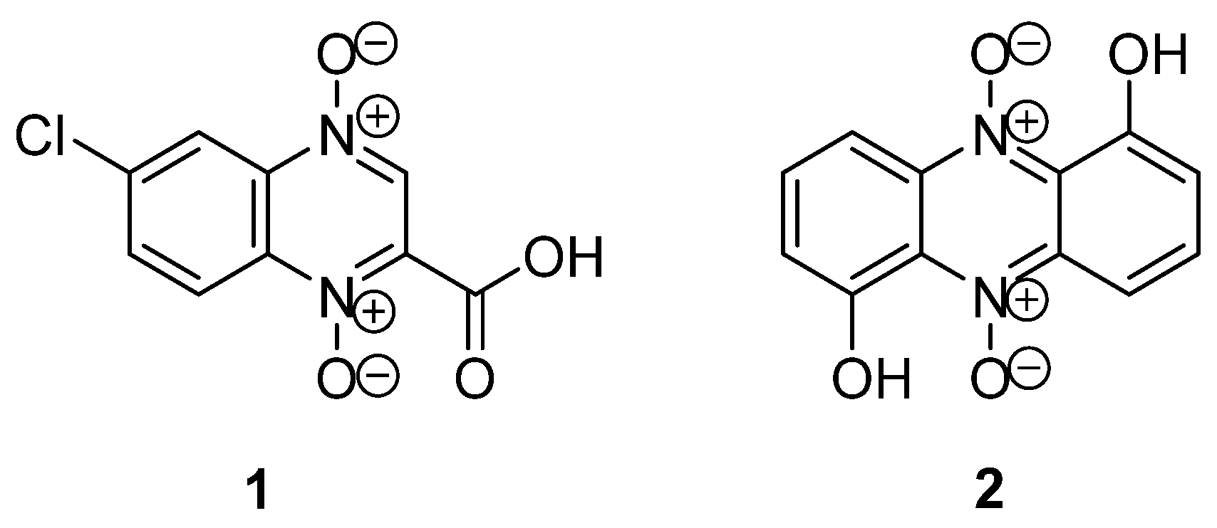
Figure 1. Examples of heterocyclic N-oxides of natural origin.
Pharmaceuticals 16 01174 g001
Figure 2. Antibacterial drugs based on quinoxaline 1,4-dioxide.
Pharmaceuticals 16 01174 g002
Figure 3. Derivatives of quinoxaline 1,4-dioxide with miscellaneous biological activity.
Pharmaceuticals 16 01174 g003
Figure 4. The main approaches for the synthesis of quinoxaline 1,4-dioxides.
Pharmaceuticals 16 01174 g004
Scheme 1. Synthesis of quinoxaline 1,4-dioxides via oxidation reaction.
Pharmaceuticals 16 01174 sch001
Scheme 2. Synthesis of 2,3-substituted quinoxaline 1,4-dioxides from o-benzoquinone.
Pharmaceuticals 16 01174 sch002
Scheme 3. Examples of quinoxaline 1,4-dioxides obtained from benzofuroxan by the Beirut reaction.
Pharmaceuticals 16 01174 sch003
Figure 5. Proposed mechanism of the Beirut reaction.
Pharmaceuticals 16 01174 g005
Scheme 4. Synthesis of 3-amino- and 3-hydroxyquinoxaline 1,4-dioxides.
Pharmaceuticals 16 01174 sch004
Scheme 5. Method of preparation of 2-acylquinoxaline 1,4-dioxides.
Pharmaceuticals 16 01174 sch005
Scheme 6. Example of the synthesis of 6- and 7-regioisomeric quinoxaline 1,4-dioxides from monosubstituted benzofuroxans.
Pharmaceuticals 16 01174 sch006
Scheme 7. Synthesis of derivatives of 7-aminoquinoxaline 1,4-dioxides by the Beirut reaction.
Pharmaceuticals 16 01174 sch007
Scheme 8. Photoinduced rearrangement of 2-substituted quinoxaline 1,4-dioxides.
Pharmaceuticals 16 01174 sch008
Scheme 9. Synthetic route for the preparation of quinoxidine (3) and dioxidine (4).
Pharmaceuticals 16 01174 sch009
Scheme 10. Transformation of the 2-methyl group of quinoxaline 1,4-dioxides 32a,b into enamines.
Pharmaceuticals 16 01174 sch010
Scheme 11. Transamination reactions of the enamine-derived quinoxaline 1,4-dioxide 34.
Pharmaceuticals 16 01174 sch011
Scheme 12. Synthesis of antimycobacterial derivatives based on molecular hybridization of arylhydrazides with a quinoxaline scaffold.
Pharmaceuticals 16 01174 sch012
Scheme 13. Synthesis of antimycobacterial derivatives based on conjugates of quinoxaline and thiazolidinone.
Pharmaceuticals 16 01174 sch013
Scheme 14. Synthesis of nitrones by the condensation of 2-formylquinoxaline 1,4-dioxide 37a with hydroxylamines.
Pharmaceuticals 16 01174 sch014
Scheme 15. Example of the synthesis of nitrone from 2-methylquinoxaline 1,4-dioxides.
Pharmaceuticals 16 01174 sch015
Scheme 16. Synthesis of quinoxaline-2-carbonitrile 1,4-dioxide by transformation of an aldehyde group.
Pharmaceuticals 16 01174 sch016
Scheme 17. Synthesis of some quinoxaline 1,4-dioxides by transformation of acetyl group.
Pharmaceuticals 16 01174 sch017
Scheme 18. Modification of quinoxaline-derived chalcones into thiopyrimidino- and pyrazoloquinoxaline heterocyclic hybrids.
Pharmaceuticals 16 01174 sch018
Scheme 19. Synthesis of quinoxaline-derived chalcones by the Wittig reaction.
Pharmaceuticals 16 01174 sch019
Scheme 20. Cyclopropanation of chalcone based on quinoxaline 1,4-dioxide by the Corey-Tchaikovsky reaction.
Pharmaceuticals 16 01174 sch020
Scheme 21. Acylation reactions of 3-aminoquinoxaline-2-carbonitrile 1,4-dioxides.
Pharmaceuticals 16 01174 sch021
Scheme 22. Transformation of 3-amino group of quinoxaline-2-caronitrile 1,4-dioxides into alkylcarbamates.
Pharmaceuticals 16 01174 sch022
Scheme 23. Modification of 3-aminoquinoxaline-2-carboxamide (2-carbonirile) 1,4-dioxides using P2S5.
Pharmaceuticals 16 01174 sch023
Scheme 24. Preparation of metal complexes of 3-aminoquinoxaline-2-carbonitrile 1,4-dioxides.
Pharmaceuticals 16 01174 sch024
Scheme 25. Synthetic route for the synthesis of 3-aminoquinoxaline 1,4-dioxides via 2-chloro derivatives.
Pharmaceuticals 16 01174 sch025
Scheme 26. Synthesis of 6(7)-aminoquinoxaline-2-carbonitrile 1,4-dioxides by nucleophilic substitution of halogens.
Pharmaceuticals 16 01174 sch026
Scheme 27. Synthesis of 3-acyl-6-aminoquinoxaline 1,4-dioxides.
Pharmaceuticals 16 01174 sch027
Scheme 28. Variations of the synthesis of 7-amino-3-trifluoromethylquinoxaline 1,4-dioxides via substitution reaction.
Pharmaceuticals 16 01174 sch028
Scheme 29. Decarboxylation of quinoxaline-2-carboxylic acid 1,4-dioxide.
Pharmaceuticals 16 01174 sch029
Scheme 30. CaCl2-Catalyzed hydrolysis of 2-ethoxycarbonyl derivative of quinoxaline 1,4-dioxide.
Pharmaceuticals 16 01174 sch030
Scheme 31. Example of alkaline hydrolysis of 2-ethoxycarbonyl derivative of quinoxaline 1,4-dioxide.
Pharmaceuticals 16 01174 sch031
Scheme 32. Transformation of quinoxaline-2-carboxilic acid 1,4-dioxide into amides using DPPA.
Pharmaceuticals 16 01174 sch032
Scheme 33. Synthesis of substituted ureas on quinoxaline scaffold via Curtius rearrangement.
Pharmaceuticals 16 01174 sch033
Scheme 34. Example of transformations of hydrazide of quinoxaline-2-carboxilic acid 1,4-dioxide.
Pharmaceuticals 16 01174 sch034
Scheme 35. One-electron intracellular reduction of quinoxaline 1,4-dioxide by reductases.
Pharmaceuticals 16 01174 sch035
Figure 6. Examples of the structures of quinoxaline 1,4-dioxides with antibacterial and antifungal activity.
Pharmaceuticals 16 01174 g006
Figure 7. Derivatives of quinoxaline-2-carbonitrile 1,4-dioxide with antimycobacterial activity.
Pharmaceuticals 16 01174 g007
Figure 8. Examples of derivatives with antimycobacterial activity.
Pharmaceuticals 16 01174 g008
Figure 9. Examples of quinoxaline 1,4-dioxides active against P. falciparum.
Pharmaceuticals 16 01174 g009
Figure 10. Quinoxaline 1,4-dioxides inhibit the growth of Leishmania spp.
Pharmaceuticals 16 01174 g010
Figure 11. Derivatives of quinoxaline 1,4-dioxide with promising activity against E. histolytica.
Pharmaceuticals 16 01174 g011
Figure 12. Quinoxaline 1,4-dioxides inhibit the growth of T. vaginalis.
Pharmaceuticals 16 01174 g012
Figure 13. Quinoxaline 1,4-dioxides with antitrypanosomal activity.
Pharmaceuticals 16 01174 g013
Figure 14. Hypoxia-selective anticancer cytotoxins based on quinoxaline 1,4-dioxide scaffold.
Pharmaceuticals 16 01174 g014
Figure 15. Quinoxaline 1,4-dioxide derivatives with promising antiproliferative activity.
Pharmaceuticals 16 01174 g015
Figure 16. Cytotoxic derivatives of quinoxaline 1,4-dioxide.
Pharmaceuticals 16 01174 g016
Figure 17. Animal growth promoters based on the quinoxaline 1,4-dioxide scaffold: carbadox (101), olaquindox (102), mequindox (21b), quincetone (103), and cyadox (104).
Pharmaceuticals 16 01174 g017
Figure 18. Quinoxaline 1,4-dioxides with herbicidal activity.
Pharmaceuticals 16 01174 g018
Disclaimer/Publisher’s Note: The statements, opinions and data contained in all publications are solely those of the individual author(s) and contributor(s) and not of MDPI and/or the editor(s). MDPI and/or the editor(s) disclaim responsibility for any injury to people or property resulting from any ideas, methods, instructions or products referred to in the content.