Next Article in Journal
Pinostrobin and Tectochrysin Conquer Multidrug-Resistant Cancer Cells via Inhibiting P-Glycoprotein ATPase
Next Article in Special Issue
Virtual Screening of Benzimidazole Derivatives as Potential Triose Phosphate Isomerase Inhibitors with Biological Activity against Leishmania mexicana
Previous Article in Journal
Direct-Acting Antivirals and Host-Targeting Approaches against Enterovirus B Infections: Recent Advances
 
 
pharmaceuticals-logo
Article Menu
Article

ACW-02 an Acridine Triazolidine Derivative Presents Antileishmanial Activity Mediated by DNA Interaction and Immunomodulation

by
Sonaly Lima Albino
1,2,3,
Willian Charles da Silva Moura
2,3,
Malu Maria Lucas dos Reis
2,
Gleyton Leonel Silva Sousa
2,4,
Pablo Rayff da Silva
5,
Mayara Gabriele Carvalho de Oliveira
6,
Tatiana Karla dos Santos Borges
7,
Lucas Fraga Friaça Albuquerque
7,
Sinara Mônica Vitalino de Almeida
8,
Maria do Carmo Alves de Lima
9,
Selma Aparecida Souza Kuckelhaus
6,
Igor José dos Santos Nascimento
2,3,
Francisco Jaime Bezerra Mendonca Junior
10,
Teresinha Gonçalves da Silva
1 and
Ricardo Olímpio de Moura
2,3,*
1
Programa de Pós Graduação em Inovação Terapêutica, Universidade Federal de Pernambuco, Recife 50670-901, Brazil
2
Laboratório de Desenvolvimento e Síntese de Fármacos, Departamento de Farmácia, Universidade Estadual da Paraíba, Campina Grande 58429-500, Brazil
3
Programa de Pós Graduação em Ciências Farmacêuticas, Universidade Estadual da Paraíba, Campina Grande 58429-500, Brazil
4
Programa de Pós Graduação em Química, Universidade Federal Rural do Rio de Janeiro, Seropédica 23890-000, Brazil
5
Programa de Pós Graduação em Produtos Naturais, Sintéticos e Bioativos, Universidade Federal da Paraiba, João Pessoa 58051-900, Brazil
6
Área de Morfologia, Faculdade de Medicina—UnB, Universidade de Brasília, Campus Darcy Ribeiro/Asa Norte, Brasília 70910-900, Brazil
7
Laboratório de Imunologia Celular, Área de Patologia, Faculdade de Medicina, Campus Darcy Ribeiro, Brasília 70910-900, Brazil
8
Faculdade de Ciências, Educação e Tecnologia de Garanhuns (FACETEG), Universidade de Pernambuco, Garanhuns 55290-000, Brazil
9
Laboratório de Química e Inovação Terapêutica, Departamento de Antibióticos, Universidade Federal de Pernambuco, Recife 50670-901, Brazil
10
Laboratório de Síntese e Vetorização de Moléculas, Universidade Estadual da Paraíba, João Pessoa 58071-160, Brazil
*
Author to whom correspondence should be addressed.
Pharmaceuticals 2023, 16(2), 204; https://doi.org/10.3390/ph16020204
Submission received: 27 December 2022 / Revised: 26 January 2023 / Accepted: 27 January 2023 / Published: 29 January 2023
(This article belongs to the Special Issue Drug Discovery of Antiprotozoal Agents)

Abstract

:
The present study proposed the synthesis of a novel acridine derivative not yet described in the literature, chemical characterization by NMR, MS, and IR, followed by investigations of its antileishmanial potential. In vitro assays were performed to assess its antileishmanial activity against L. amazonensis strains and cytotoxicity against macrophages through MTT assay and annexin V-FITC/PI, and the ability to perform an immunomodulatory action using CBA. To investigate possible molecular targets, its interaction with DNA in vitro and in silico targets were evaluated. As results, the compound showed good antileishmanial activity, with IC50 of 6.57 (amastigotes) and 94.97 (promastigotes) µg mL−1, associated with non-cytotoxicity to macrophages (CC50 > 256.00 µg mL−1). When assessed by flow cytometry, 99.8% of macrophages remained viable. The compound induced an antileishmanial effect in infected macrophages and altered TNF-α, IL-10 and IL-6 expression, suggesting a slight immunomodulatory activity. DNA assay showed an interaction with the minor grooves due to the hyperchromic effect of 47.53% and Kb 1.17 × 10⁶ M−1, and was sustained by docking studies. Molecular dynamics simulations and MM-PBSA calculations propose cysteine protease B as a possible target. Therefore, this study demonstrates that the new compound is a promising molecule and contributes as a model for future works.

1. Introduction

Leishmaniasis is a parasitosis caused by macrophage infection of protozoa from the genus Leishmania, transmitted to mammal host through the bite of blood-feeding females of phlebotomine sand flies [1]. It has been recognized as one of the main neglected tropical diseases (NTDs) of global health concern, estimating that over one billion people are living in endemic areas at risk of infection. Accordingly, each year more than two million new cases occur, associated with high levels of morbidity and mortality [2,3].
Leishmania parasites exist in two forms during their life cycle (amastigotes and promastigotes) and are capable of manifesting three main clinical forms, generally classified as cutaneous leishmaniasis (CL), mucocutaneous leishmaniasis (MCL) and visceral leishmaniasis (VL, also known as kala-azar). Depending on the parasite species and the cellular immune mechanisms of the host, different clinical, histopathological and immunopathological manifestations are developed [4,5].
The immune mechanism response of the host to the infection relies on myeloid cells such as dendritic cells and macrophages, which develops a complex relation [6]. Leishmania protozoa use macrophages as important host cells for establishing the infection. In order to adapt, the parasite regulates phagosome maturation, promoting the alteration of macrophage defenses such as oxidative damage, antigen presentation, immune activation and apoptosis, while there is an increase in nutrient availability, causing the macrophage to become a favorable environment for its growth and avoiding destruction [7].
Current chemotherapy includes drugs such as sodium stibogluconate, meglumine antimoniate, amphotericin B, paromomycin, and pentamidine. These, however, have limitations regarding the emergence of resistant strains, high costs, prolonged administration, and the manifestation of side-effects such as nephrotoxicity, ototoxicity, and hepatotoxicity [8,9]. In this sense, the development of new candidates for leishmanicidal drugs that are more effective against parasites, associated with less toxicity to the human host, stands out as a priority.
In this sense, the acridine nucleus has demonstrated to be an eligible scaffold due to its potential as a chemotherapeutic precursor [10]. As an antiparasitic acridine compound of clinical application, mepacrine stands out due to its wide spectrum of biological action, with its main application in the treatment of malaria in chloroquine-resistant strains. Therefore, several studies aimed at obtaining leishmanicidal compounds are based on the chemical structure of this compound, maintaining the 9-amino-2-methoxy-6-chloroacridine core while altering moieties linked to the 9-amino groups [11], as observed in the study developed by Serafim et al. [12], in which the authors have synthesized and evaluated the antipromastigote activity of thiophene-acridine derivatives against L. (L.) amazonensis strains, highlighting compounds ACS01 and ACS02 as potent and selective, with IC50 values of 9.60 and 10.95 µM against antimony-sensitive strains, and 14.83 and 16.36 µM against resistant strains, respectively. Furthermore, both molecules interacted with DNA with binding constants of 104 M−1, in which the activity is attributed to the 6-chloro-2-methoxy-acridine moiety, as also previously reported by Zhang et al. [13].
Based on these previous data, our group proposed the synthesis of a novel acridine derivative containing the 6-chloro-2-methoxy-acridine moiety, through a rational strategy of molecular design based on a privileged structure, linked to a triazolidine-dithione nucleus obtained through an intramolecular spontaneous cyclization, followed by in silico and in vitro evaluation of its antileishmanial activity, mechanism of action through DNA interaction and immunomodulatory properties.

2. Results

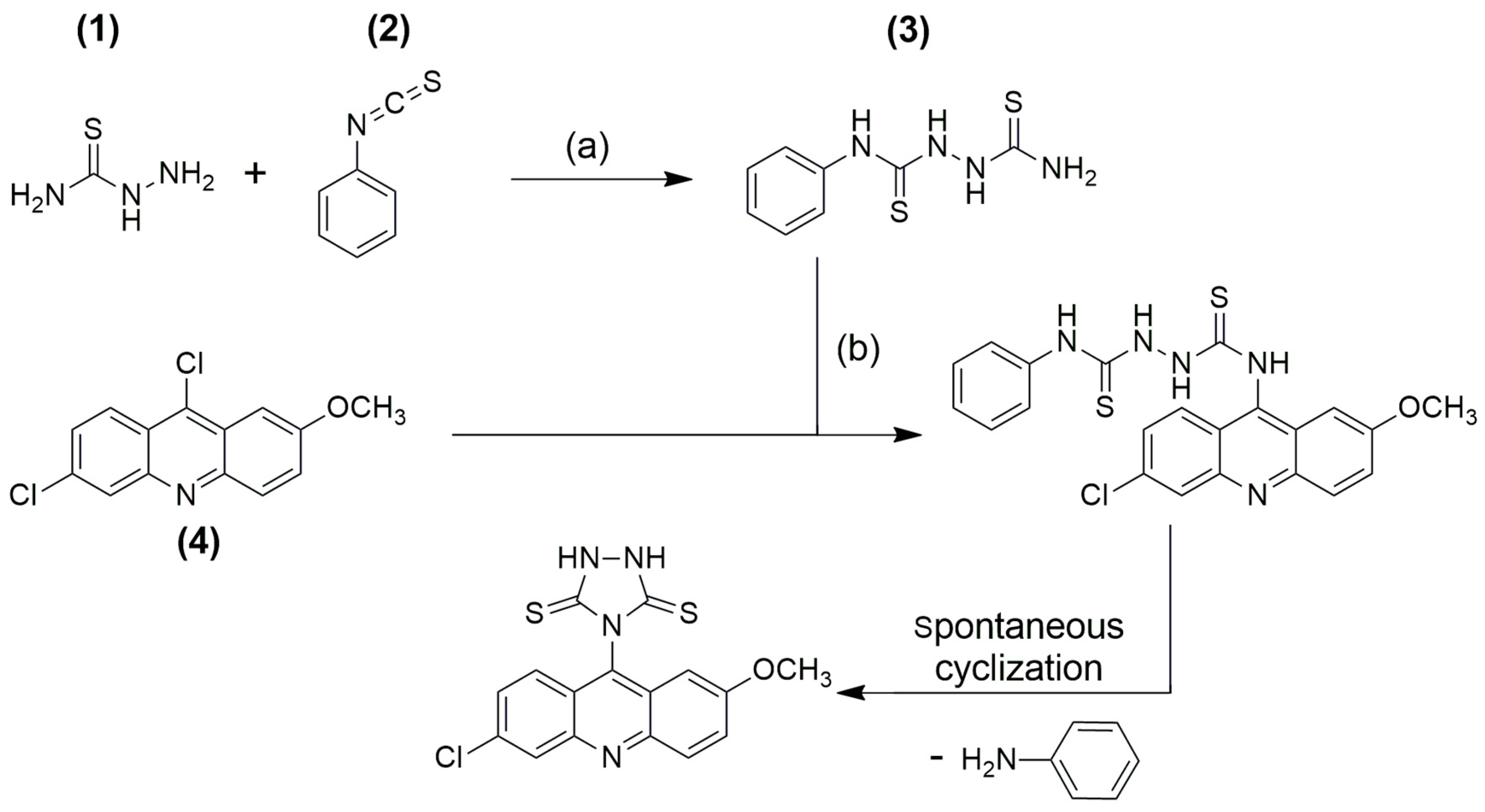
2.1. Synthesis and Characterization

In order to develop a new compound for the treatment of leishmania, a novel triazolidine acridine derivative was synthesized, obtained accidentally by spontaneous cyclization from a coupling reaction between two scaffolds: thiosemicarbazide and disubstituted 9-chloro-acridine. Scheme 1 outlines the synthesis with the sequence of the reactions to obtain compound ACW-02. The target compound was obtained in two steps, as described in the experimental section. The synthesized compound is being reported for the first time and its structure was confirmed by 1H NMR and 13C NMR spectroscopy, infrared spectroscopy and mass spectrometry, where the results were in agreement with the described structure. These data are present in the Supplementary Material (Figures S1–S4).
Through 1H NMR analysis, it was possible to identify a singlet for two symmetrical hydrogens with displacement at δ 12.79 ppm, correspondent to the triazolidine NH, corroborating with the findings of Tiwari et al. [14], which observed a peak in δ 12.48 ppm referring to triazolidine NH. Peaks characteristic of singlet (s), doublet (d), double doublet (dd), double triplet (dt) and multiplet (m) with displacements ranging between δ 7.36–8.83 ppm have been observed, indicative of the acridine ring. Furthermore, a singlet with an integral for three hydrogens in δ 3.88 ppm was recognized, correspondent to the OCH3 attached to the acridine ring. The 13C NMR spectrum also confirmed the structure by the presence of signals correspondent to the acridine nucleus, but mainly by the presence of thiocarbonyl groups, which showed symmetric displacement at δ 194.64 ppm, just as it was observed by Tiwari et al. [14]. Additionally, a peak in δ 55.86 ppm relative to OCH3 attached to the acridine ring was observed.
Infrared results also assisted in the structural characterization of the novel triazolidine acridine derivative. The following bands were observed: 3242–3147 cm−1, suggestive of axial deformation of secondary N-H; 3085–3056 cm−1, suggestive of aromatic C-H axial deformation; 1621 and 1482 cm−1, characteristic of C=CAr vibration; 1208 cm−1, suggesting the axial deformation vibration of C=S bound to nitrogen; 1197 cm−1, suggestive of asymmetric axial deformation of C=C-O attributed to the methoxyl group attached to the acridine ring. Ultimately, mass spectrometry (MS) was useful to confirm the structure of the novel synthesized compound, exhibiting the result of m/z = 375.81.

2.2. Antileishmanial and Cytotoxic Activity Evaluation

The antileishmanial potential of compound ACW-02 was explored in vitro against amastigotes and promastigotes forms of Leishmania amazonensis, responsible for causing the clinical cutaneous form of the pathology. For this purpose, Amphotericin B, a well-known commercial drug widely used in the treatment, was used as a positive control. It was observed that the synthesized compound showed good activity against the amastigote strain, with an IC50AMA of 6.57 µg mL−1. Nonetheless, against the promastigote strain, a satisfactory result was not obtained (IC50PRO = 94.97 µg mL−1). These results are presented in Table 1.
As for the cytotoxic effect, the synthesized compounds did not show cytotoxicity against JJ74 macrophage strains up to the highest concentration evaluated (256.00 μg mL−1). Thus, the compounds ACW-02 showed high rates of selectivity index for the amastigote forms of the parasite, with a value of >38.94. This contrasts with the results obtained for amphotericin, which, although it is a potent drug, presents a high level of toxicity, as already described in the literature [15] and can be observed in the low selectivity index (1.05) obtained.
Additionally, in the evaluation of the cytotoxic effect of the synthesized compound on macrophages, indications of immunomodulatory activity were observed through the identification of its stimulatory effects on the proliferation of such cells. The is quantified in EC50 value in Table 1 (EC50 = 9.46 μg mL−1). Therefore, in order to clarify the effects of the synthesized compound on macrophages and the possibility of an immunomodulatory effect, further studies were carried out.

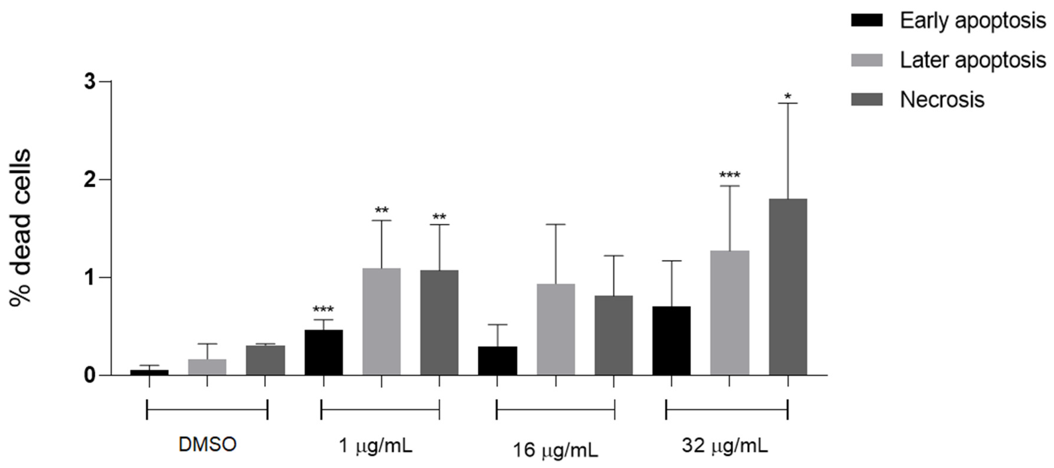
2.3. Cytotoxicity Evaluation in Macrophages by Annexin V-FITC/PI Double Staining Assay

In order to understand in more detail the effect of the synthesized compound on the J774 macrophages, a cell cycle evaluation assay using the annexin V-FITC/PI method was performed. The results reaffirm the compound’s non-cytotoxicity to macrophages, with the percentage of living cells corresponding to approximately 98%, up to the highest concentration tested. The results are shown in the graph in Figure 1.
As demonstrated, in the absence of the compound, all cells were viable as the percentage of initial, late apoptosis and necrosis was 0%. In contrast, in a concentration of 1 µg mL−1, the percentage of cells in initial, late apoptosis and necrosis was 0.46, 1.09 and 1.07, respectively. When increased to the concentration of 16 µg mL−1, there is a decrease in the percentages, these being 0.29, 0.94 and 0.81, respectively. Finally, at a concentration of 32 µg mL−1, the percentages increase to 0.70, 1.27 and 1.8, respectively. In comparison to negative control, which has shown values of 0.06, 0.17 and 0.31%, ACW-02 exhibits approximate values, highlighting its viability as non-cytotoxic to macrophages.

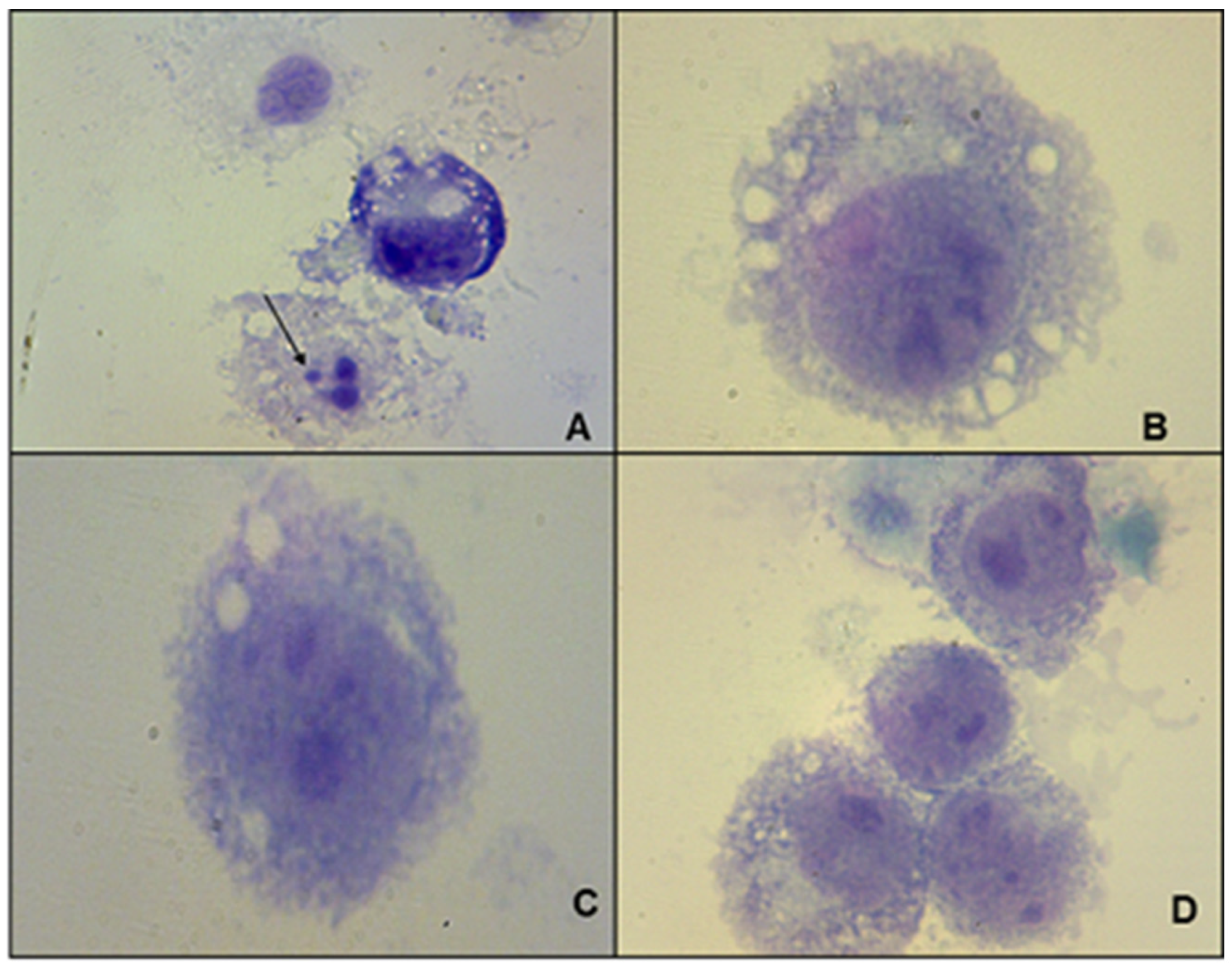
2.4. Determination of the Microbicidal Effect in Cultures of J774 Macrophages Infected with L. amazonensis

Previous studies provided information about the antileishmanial activity of the evaluated compound associated with a possible immunomodulatory activity on macrophages. Given the importance of this cell for the progression of the pathogenesis in question, this possibility represents a pertinent association of mechanisms. Therefore, the effect of ACW-02 on macrophages infected with L. amazonensis was evaluated, in order to verify its immunomodulatory potential. The results are shown in Table 2.
Our results evaluated by the J774 macrophage infection index showed the microbicidal potential of ACW-02. In comparison to infected and untreated cells (0 µg mL−1), it was observed that the addition of ACW-02 reduced the infection rate in all concentrations (1, 16 or 32 µg mL−1), due to the decrease in the percentage of infected macrophages and the number of Leishmania ingested by macrophages (Table 2).
The microbicidal effect of ACW-02 is shown in Figure 2. The low infection rate, especially at the lowest concentrations of ACW-02 (1 or 16 µg mL−1), possibly interfered with the functionality of macrophages, thus favoring the elimination of parasites.

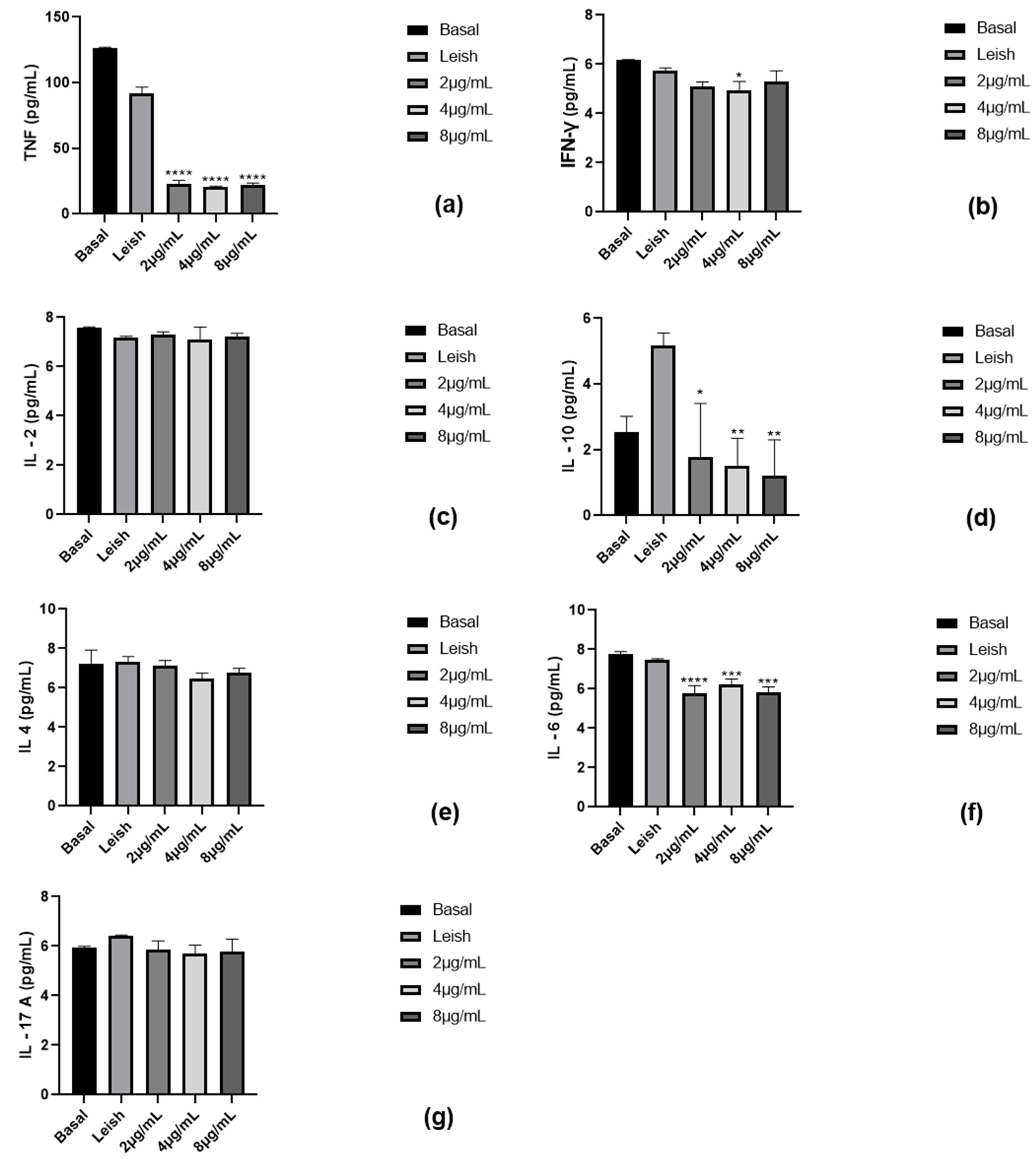
2.5. Assessment of Cytokine Production by Macrophages

Evaluations were made concerning the effect of the compound ACW-02, at concentrations 2, 4, and 8 µg mL−1, on the modulation of the Th1 (T helper 1), Th2 (T helper 2), and Th17 (T helper 17) response through the expression of the respective cytokines: TNF-α (Tumor Necrosis Factor-α), IFN-γ (Interferon-γ), IL-2 (Interleukin-2), IL-10 (Interleukin-10), IL-4 (Interleukin-4), IL -6 (Interleukin-6) and IL-17A (Interleukin-17A). The response expressed by the basal macrophage (untreated and uninfected) and macrophage infected with L. amazonensis (untreated) were used as negative and positive controls, respectively (Figure 3).
The progression of the infection caused by Leishmania protozoa is directly related to the immune response developed by the host, especially the T-cell mediated immunity and the expression of pro-inflammatory and/or anti-inflammatory cytokines, that can result in an asymptomatic, self-healing, or chronic leishmaniasis. The cellular immunity generated by the Th1 is considered to be the key mediator of resistance to Leishmania, as they are associated with the secretion of pro-inflammatory cytokines (e.g. TNF-α, IFN-γ, IL-2) that activate the MΦ killing machinery, leading to parasite inactivation [16,17,18]. Nevertheless, according to the results obtained in our experiments, there is a significant decrease in the expression of TNF-α (Figure 3a) with the addition of the acridine compound ACW-02, at the lowest up until the highest concentration evaluated, thus favoring an anti-inflammatory action. Further, the effect of the compound on IFN-γ and IL-2 cytokines was not significant.
On the other hand, cytokines expressed by Th2 cells (e.g. IL-4, IL-6, IL-10) are related to an anti-inflammatory action, as they are capable of suppressing the host’s immune system, causing macrophage depletion and, consequently, promoting development of the intracellular forms of the parasite and disease progression [17]. According to our findings, a decrease in IL-10 expression is observed (Figure 3d), approaching basal values at the lowest concentration tested (2 µg mL−1). Moreover, there is a significant decrease in IL-6 expression at all concentrations evaluated. Opposed to the previous results, the decrease in the expression of an anti-inflammatory interleukin will favor a pro-inflammatory effect, that is, a leishmanicidal action. Results found for the other evaluated cytokine, IL-4, were not significant.
Lastly, Th17 is involved in the production of IL-17, a highly pro-inflammatory cytokine with significant effects on stromal cells, resulting in the recruitment of leukocytes and forming an interaction between innate and adaptive immunity [16,19]. However, according to the results obtained, the compound ACW-02 does not show significant activity on the cytokine IL-17A (Figure 3g).

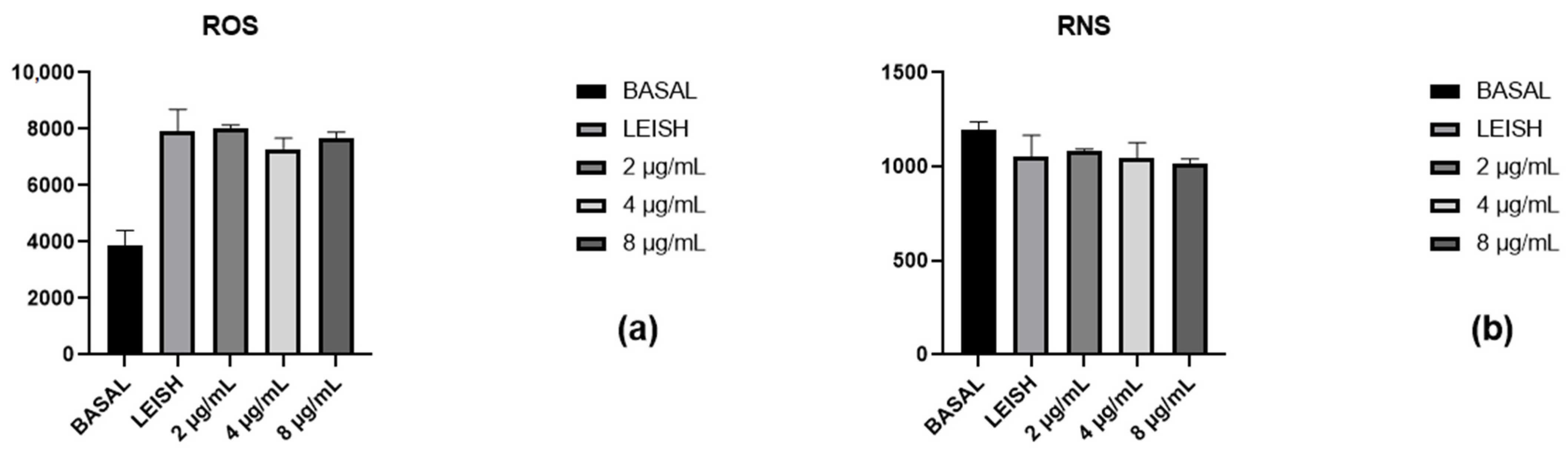
2.6. Reactive Oxygen and Nitrogen Species Evaluation

The production of reactive oxygen (ROS) and nitrogen (RNS) species is of significant relevance to disease progression. The activation of macrophages as a response to an infectious process induces the overproduction of reactive oxygen species (ROS), including superoxide, hydrogen peroxide, hydroxyl radicals, and reactive nitrogen species (RNS), such as nitric oxide (NO). ROS and RNS exhibit high microbicidal capacity, as these species can generate oxidative damage to parasite’s biomolecules, such as lipids, proteins, and DNA, leading to loss of membrane integrity, defective replication, and ultimately cell death [20,21].
In order to evaluate the ability of the synthesized compound to induce the formation of reactive species, evaluations were carried out that showed that the compound under evaluation at concentrations 2, 4, and 8 µg mL−1 did not significantly affect the production of ROS, when compared to the infected and untreated macrophages, as shown in Figure 4a.
Similarly, at the same concentrations (2, 4, and 8 µg mL−1), ACW-02 presented no significant alteration on RNS production in comparison to infected and untreated macrophages, as shown in Figure 4b.

2.7. Interaction with DNA

In order to propose a potential target that justifies the antileishmanial action of the acridine compound ACW-02, interaction studies with the DNA molecule were performed.
The UV–vis absorption spectra of ACW-02 showed significant absorption in the region between 400–500 nm. This derivative exhibited, in the absence of ctDNA, maximum peak at 460 nm, while in the presence of ctDNA, it presented a maximum peak of 461 nm. The maximum absorption wavelengths of the compound in the absence and presence of ctDNA can be observed in Table 3.
In these studies, the model of McGhee and Von Hippel [22] was used to estimate the binding constant (Kb) from the spectrophotometric data. The interaction by intercalation can be characterized by the hypochromic effect and by the wavelength alteration for the red region (bathochromic effect) [23,24,25,26], whereas the occurrence of hyperchromism with little or no redshift is associated with electrostatic attachment or interaction with DNA groove (major or minor) [27,28]. According to the absorption results, ACW-02 presented hyperchromism of 47.23% and Kb (M−1) = 1.17 × 10⁶ in the presence of ctDNA (Figure 5).
According to the data, ACW-02 presented characteristics suggestive of interaction either by intercalation or by interaction with DNA grooves due to its Kb value. Kb values for intercalation complexes with DNA ranges from 1 × 10⁴ to 1 × 10⁶ M−1, while Kb values of groove binders range from 1 × 10⁵ to 1 × 10⁹ M−1 [29]. To confirm this effect, molecular docking studies were performed.

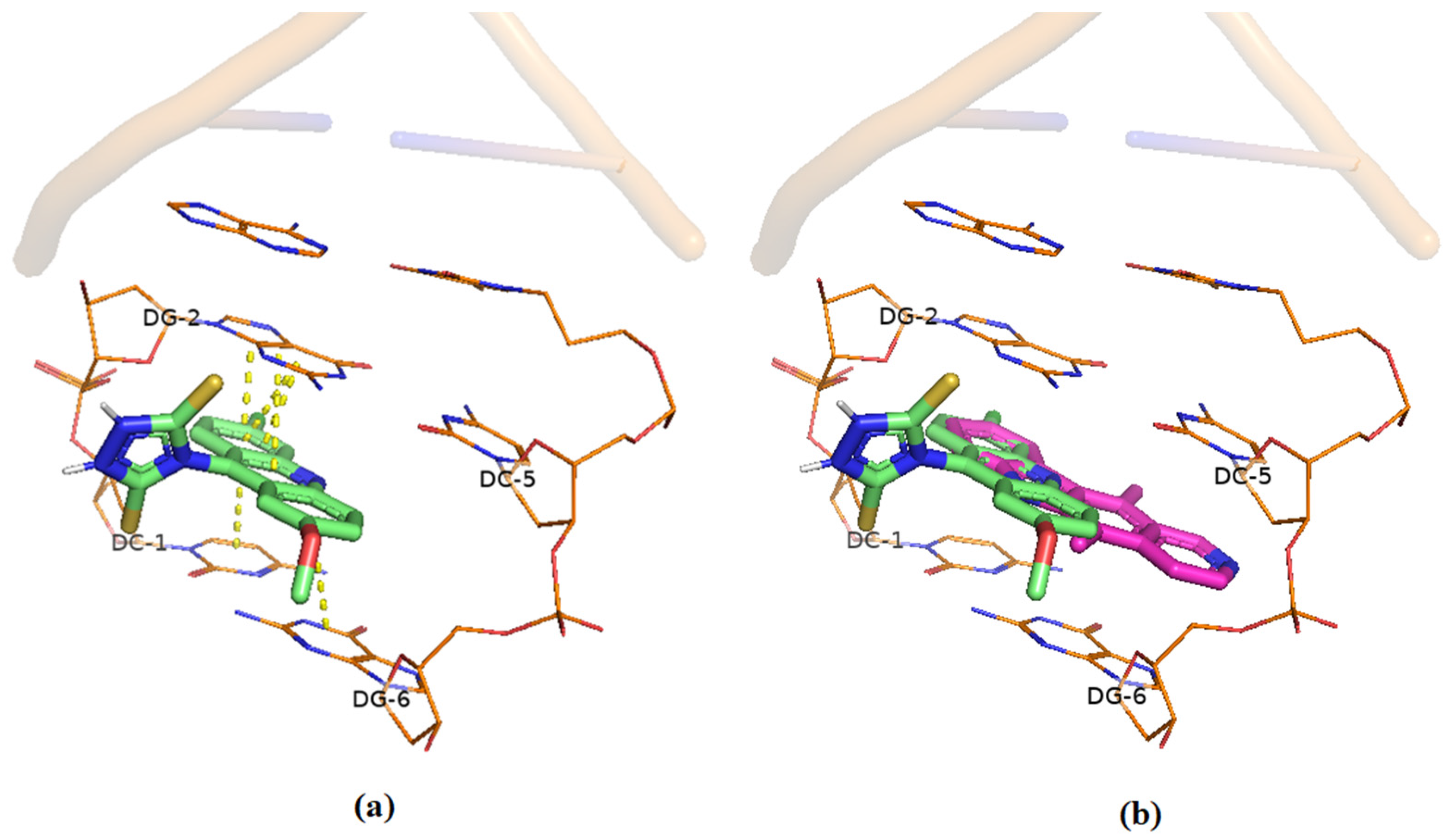
Docking Studies of Interaction with DNA

The DNA intercalation capacity of ACW-02 in silico was evaluated using the crystallographic fitting model available in the PDB for Ellipticin (PDB ID: 1Z3F). Figure 6 shows the highest-scoring ACW-02 conformer (71.77) after the molecular fit study. The acridine portion of the compound indicated more affinity to intercalate between the nitrogenous bases of DNA through π-π electron stacking. This stacking limit between the bases was delimited by the steric hindrance of the cyclic triazolidine-derived portion in the structure. As proposed in spectroscopic studies, molecular anchoring also indicated the ability of double interaction with DNA, with the triazolidine fragment capable of interacting with the side chains of the macromolecule. Although ACW-02 had a lower score than ellipticin (75.68), they showed great similarity in terms of pose and intermolecular interactions performed with DNA (Figure 6), highlighting the ability of the acridine derivative for new interactions in the receptor.
Although these results indicate the ability of the compound to interact with DNA, this is probably not the only mechanism of action performed by the compound under evaluation, since there is no structural difference between the DNA molecules of the parasite and the host that provides selectivity to the compound, and, consequently, justifies the entirety of the leishmanicidal action. For this reason, the possibility of new targets was evaluated in silico to investigate possible complementary mechanisms of action.

2.8. Theoretical Study of Possible Mechanisms of Action

2.8.1. Molecular Docking

The high selectivity presented in the in vitro tests of ACW-02 against Leishmania amazonensis supported the in silico study of possible mechanisms of action involved in the survival of the parasite. Some metabolic pathways explored in the disease, such as: redox balance (Trypanothione reductase, TryR); regulation of DNA supercoiling by topoisomerases I (Topo I); and the enzymes involved in ergosterol biosynthesis (14-alpha-ester demethylase, CYP51) were explored by molecular docking [12,30,31,32]. Moreover, cysteine proteases, such as cysteine protease B from Leishmania amazonensis (CPBLa) are promising drug targets against parasitic diseases, essential for the biochemical processes and survival of parasites [33]. The crystallographic models used for the theoretical studies were obtained from the PDB and the CPBLa was built using homology modeling. Validations by redocking, when possible, were performed and presented acceptable results for the study [34]. The ACW-02 presented scores lower than the standards complexed to the targets studied in the docking (Table 4). However, information about the main residues and polar interactions intrinsic to the formation of the ligand-receptor complex were reproduced and indicated good affinities for the docking of ACW-02 in the studied targets; characteristics such as planarity and points of polar interactions in the cyclic triazolidine-derived portion were essential for the performance of the scaffold in this study. For example, in TryR (PDB ID: 4APN), polar interactions with residues such as Lys61 and hydrophobic interactions with residues Val58 and Leu62 are reported to be essential for docking in the enzyme. In this study, ACW-02 demonstrated the ability to reproduce these interactions and, additionally, pointed out other interactions with nearby residues in the aforementioned cavity [10,35]. Finally, interaction analysis CPBLa reveled H-bond with the key residues Cys153 and van der Waals with His289 and Gly151, proposing that the acridine derivative can perform on the cysteine protease as a drug target.
Planar condensed fragments are commonly associated with mechanisms that directly or indirectly involve DNA, due to their validated intercalation capability, as visualized in aforementioned in silico and in vitro studies with DNA performed in this paper [36,37]. The docking results found suggest that Topo IB (PDB ID: 2B9S) is a possible mechanism for ACW-02. Due to the absence of a co-crystallized ligand to the topoisomerase I target, Camptothecin (CPT) was used as a standard compound, given its elucidated mechanism of action as a topoisomerase I inhibitor [10]. The intrinsic polar interactions of Camptothecin in docking with LDTopoI (63.98) were reproduced with residues Arg190 and Lys211 [38]. As in the docking with DNA, the cyclic portion linked to acridine induced a steric restriction for an improved stacking between DNA topoisomerase I bases (Figure 7A), however, this same fragment was responsible for additional interactions with the enzymatic portion of topo I, which justifies the similarity between the scores found for ACW-02 and Camptothecin.
On the other hand, the docking analysis of the ACW-02 in CPBLa (Figure 7B) has several interactions that can propose this drug target. First, acridine nitrogen interacts with the Cys153 by H-bond, and the aromatic ring is positioned to perform van der Waals interactions with the catalytic residue of His289. In addition, interactions amide-π Stacked are formed with the residue Asn288 and π-alkyl with Leu287 and Ala288. Finally, repulsive interactions are observed with hydrophobic residues such as Gly151, Gly193, and Gly194. Similar interactions were found in docking analyses of the standard compound. Finally, the most effective fit score of ACW-02 compared with the standard (50.97 and 47.77, respectively) and most effective interactions propose the CPBLa as the possible drug target. Thus, to confirm our findings, MD simulations and MM-PBSA calculations were performed and discussed in the following topics. Two-dimensional figures of these dockings are present in the Supplementary Material (Figures S7–S10).
Table 4. Molecular docking results of ACW-02 against potential antileishmanial targets.
4APN (TryR)3L4D (CYP51)2B9S (Topo I)CPBLa
ACW-0254.2258.7460.3450.97
Standard *67.5878.7763.9847.77
RMSD0.761.02--
* Ligand co-crystalized in 4APN for TryR; ligand co-crystalized in 3L4D for CYP51; Camptothecin for Topo I; and compound 1c [39] for CPBLa.

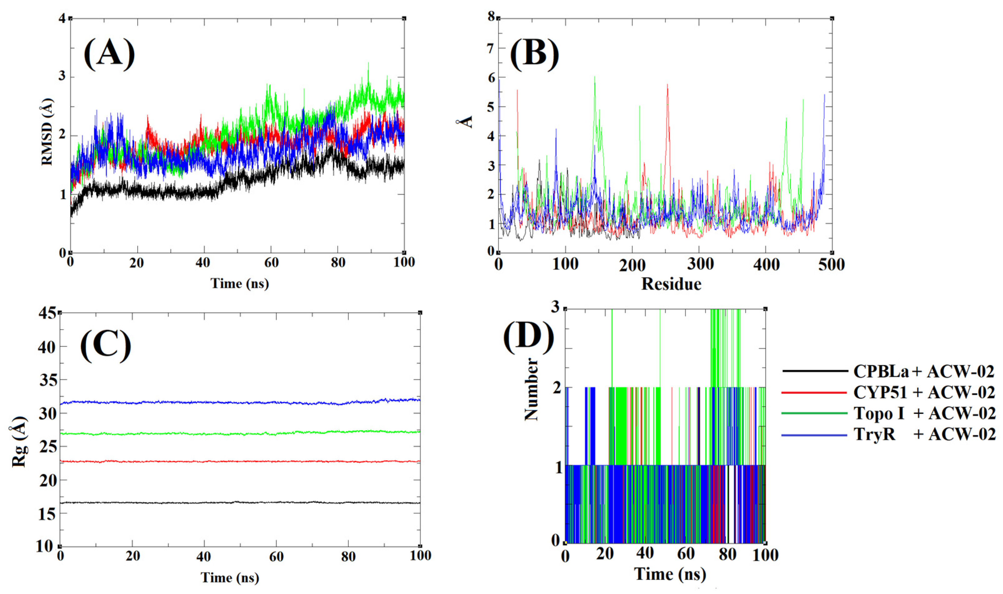
2.8.2. Molecular Dynamics (MD) Simulations

Molecular dynamics simulations were performed in trajectory of 100 ns (Figure 8) to confirm the drug target of ACW-02. The RMSD plot (Figure 8A) reveals the best stability of ACW-02 in complex with CPBLa, showing values lower 2 Å. On the other hand, the compound in complex with the other targets shows up to 3 Å. In addition, the RMSF plots (Figure 8B) reveal lower fluctuations of the residues for the complex with CPBLa, suggesting the best stability of the complex. The stiffness and protein compaction is shown in the Rg plot (Figure 8C), in which better protein compaction for the complex with CPBLa (between 15 and 16 Å), different of the other complexes that presented values higher than 15. Finally, the compounds showed similar h-bonds interactions around the trajectory (Figure 8D), up to 2 for the complexes with CPBLa, CYP51, TryR, and up to 3 for Topo I. The MD results are according to other reports and generate useful information that can be used to propose the complex stability and interaction with the targets [40,41,42]. Thus, these findings suggest the best stability of the ACW-02 with the CPBLa and can be a possible drug target.

2.8.3. MM-PBSA Calculations

MM-PBSA calculations using the MD simulations trajectories were used to re-score the affinities energies and determine the attractive forces involved in the formation of the ACW-02 complexes against the proposed targets (Table 5). As observed in molecular docking studies, the affinity energy of ACW-02 against CPBLa by MM-PBSA (ΔGbind = −103.13 KJ/mol) was superior to the complex with other proposed targets. Furthermore, it is clear that van der Waals interactions are essential for complex formation (DGvdw = −151.75 KJ/mol) when compared to electrostatic forces (0.68 KJ/mol). The polar solvation and SASA energy show low solvation (61.09 and −13.15 KJ/mol, respectively), indicating more excellent permanence of the ligand in a hydrophobic environment free of water molecules when compared to its interaction with other targets. Finally, the results suggest CPBLa as one of the main targets of ACW-02.

3. Discussion

The compound under study is an unprecedented triazolidine-acridine derivative. The initial design of the compound contemplated the coupling reaction between the di-substituted acridine nucleus and the intermediate N-phenylhydrazine-carbothioamide, a thiosemicarbazide derivative, selected during the design stage due to the inherent potential of the fragment that gives it a broad spectrum of biological activity [43,44], including anti-protozoal action [45,46,47]. However, during the synthesis stage, the triazolidine-dithione nucleus was obtained through a nucleophilic substitution reaction between the carbothioamide intermediate and the acridine nucleus, followed by an intramolecular attack favored by the energy provided by the heating of the reaction and by the free protons in the reaction medium, inducing an autocyclization and a fragmentation of the aniline portion from the carbothioamide subunit, forming a new triazolidine-thione nucleus coupled to the substituted acridine. Thus, due to the novelty of the compound, it was decided to continue the studies by observing the contributions of this new ring to the acridine scaffold.
The reaction to obtain the final product occurred in an ethanolic and acidic medium, at reflux temperature, and was finalized by cooling the reaction to room temperature, followed by the addition of distilled water and precipitation of the product, subsequently filtrated. Ultimately, ACW-02 was elucidated by 1H and 13C NMR, infrared, and mass spectrometry techniques, showing signs that confirmed the proposed structure.
Therefore, given the potential of acridine compounds as chemotherapeutic agents, with several studies exploring and reporting their anticancer anticancer [13,48,49,50,51], antibacterial [52,53,54] and antiparasitic [12,55,56] properties, this study aimed to evaluate the potential antileishmanial activity of the new synthetically obtained compound, given the urgent need for efficient drugs for this purpose.
Leishmaniasis comprises the pathology caused by protozoa of the genus Leishmania. In its digenetic life cycle, the parasite assumes metacyclic promastigote and amastigote forms in the vertebrate host. The promastigote forms are initially inoculated into the host through the bite of the sandfly mosquito and, in the bloodstream, the parasites penetrate phagocytic cells through interactions between their surface molecules and macrophage receptors. In the phagocytic vacuole, these differentiate into the amastigote form and multiply intensely until the infected macrophage ruptures. These free amastigotes, in turn, infect other macrophages and continue the cycle [4,57]. Therefore, depending on the drug’s affinity for each parasitic form, different stages of the infection progression will be achieved. According to the studies carried out, the compound ACW-02 presented an IC50 value of 6.57 µg mL−1 against amastigote forms of L. amazonensis, and 94.97 µg mL−1 against promastigote forms of the same species.
Depending on the infecting species, different clinical manifestations may develop. The species under study, Leishmania amazonensis, is an important etiological agent that causes American cutaneous leishmaniasis (ACL) and is capable of establishing a broad spectrum of clinical manifestations, varying from: cutaneous, localized or diffuse, which are the most common; mucocutaneous; and even visceral leishmaniasis [58]. Thus, it is observed that the amastigote forms are more sensitive to the pharmacological action of ACW-02 than the promastigote forms, indicating the possible potentiality of the compound against these forms of L. amazonensis in the treatment of ACL.
Moreover, based on the report by Serafim et al. [12], the hybridization of the thiophene portion with the 6,9-dichloro-2-methoxy-acridine ring was beneficial to the evaluated antipromastigote activity against L. amazonensis, obtaining IC50 values between 9.62 and 69.11 µM. However, the hybridization of the non-substituted acridine ring to the thiophene portion caused a loss in activity, highlighting then the relevance of the substitution in the acridine ring by groups with positive mesomeric effect (–OCH3 and –Cl) for antileishmanial activity. As this study involves obtaining an unprecedented molecule that possesses the substituted acridine nucleus, the same as that used by Serafim et al. [12], it is supposed that this portion of the molecular structure of ACW-02 is associated with the antileishmanial activity presented.
Considering the importance of macrophages during the installation and progression of the Leishmania infectious process [59], studies involving the effects of ACW-02 on J774 macrophages were carried out. The determination of CC50 demonstrates that the compound did not show cytotoxicity up to the highest concentration tested (256.00 µg mL−1), and this result is confirmed in more detail through the evaluation of cytotoxicity in macrophages by Annexin V-FITC/PI double staining assay, which showed low percentages of cells (<2%) in initial, late apoptosis and necrosis after treatment with ACW-02 at concentrations of 1, 16, and 32 µg mL−1. Thus, evaluating the first result mentioned, the calculated selectivity index of ACW-02 to L. amazonensis amastigote cells was above 38.94, providing a safety margin.
Additionally, a stimulatory effect on macrophages was observed at a concentration of 9.46 µg mL−1 in preliminary studies and the possibility of the compound’s ability to play an immunomodulatory effect on macrophages was evaluated in more comprehensive studies. The determination of the microbicidal effect contributed to the supposed possibility of altering the functionality of the macrophage in the face of parasitic infection with the addition of the ACW-02 compound, due to the alteration in the parameters that evaluate the percentage of infected cells, the average of Leishmanias and percentage of infection, especially in lower concentrations (1 and 16 µg mL−1) and, accordingly, closer to the EC50 value obtained. Moreover, it is supposed that the highest concentration (32 µg mL−1) may have caused a supersaturation on the culture medium during the test, thus causing a modification in the solubility of the compound and consequent decrease in biological activity, due to the lower interaction of the compound with the infected macrophages.
To determine the influence of the compound directly on the components of the immune system, targeted studies were carried out. The control of the infection and disease progression caused by Leishmania sp. is associated with the generation of pro-inflammatory and anti-inflammatory immune responses. Stimulation of a Th1 response and the downmodulation of a Th2 response promotes macrophage activation and, consequently, assists in the host control of Leishmania parasite burden and clinical cure [18,60]. The results obtained in response to Th2 demonstrated a significant decrease in the expression of IL-10 and IL-6 cytokines, thus contributing to an antileishmanial effect. These results corroborate with the aforementioned studies and may justify the macrophage’s ability to perform a microbicidal effect against Leishmania. However, these results are not sufficiently conclusive, since there is also a significant decrease in TNF-α expression, which, on the other hand, exerts a pro-leishmania effect. Thus, the effect accomplished by reducing TNF-α is antagonistic to the one obtained by decreasing IL-10 and IL-6.
Similarly, macrophage activation promotes the production of reactive oxygen and nitrogen species to induce oxidative damage to the parasite [20]. However, the results did not demonstrate an alteration in these components that would justify the performance of an immunomodulatory activity by ACW-02 at the concentrations evaluated (2–8 µg mL−1). Thus, due to the lack of results that establish the immunomodulatory effect as justification for the entirety of the antileishmanial activity, other pharmacological targets were evaluated in silico and in vitro.
The acridine nucleus, present in the structure of the compound ACW-02, stands out as a potential scaffold. Its biological activity can be attributed to the planarity of its aromatic structures, which can interact by intercalation or interactions with the DNA grooves alone, interfering in cellular properties [61,62,63]. In view of this potential mechanism of action of acridine compounds, the interaction of the compound ACW-02 with DNA was evaluated.
The presence of aromatic groups without substituents may favor the interaction of the complex through strong hydrophobic or non-covalent interactions between the electron states of the chromophore and DNA base pairs, leading to a decrease in the transition energy π → π*. However, despite the acridine ring possible interactions, the hyperchromic effect observed in this compound may be due to the additional electrostatic interactions of the methoxyl (–OCH3), of the acridine ring, which has σ+ and π- effects, and of the chlorine atom (–Cl), which has σ+ and π+ effects, resulting in a reduction of the stacking of bases of this biomolecule [64,65,66].
In general, the non-substituted acridine nucleus presents intercalating activity [67,68] through π-π interactions with DNA base pairs. The insertion of the triazolidine nucleus, presenting electron acceptor groups (C=S) and electron donors/acceptors (–NH), may favor the stabilization of the supramolecular DNA-ligand complex, through electrostatic or non-covalent interactions [69,70,71]. Thus, favoring a possible intercalation and interaction with DNA grooves.
In silico molecular docking studies were used as a strategy to reaffirm the interaction with DNA through studies on the ellipticine-complexed DNA target (PDB ID: 1Z3F), demonstrating an ACW-02 score proximate to that of the ellipticine standard (71.77 and 75.68, respectively).
Aiming to elucidate possible molecular mechanisms that could contribute to the entirety of the antileishmanial action of the compound ACW-02 and justify its selectivity for the parasite, theoretical studies of molecular docking were performed in pharmacological targets exclusive and essential to the Leishmania (TryR, Topo I, CPBLa, and CYP51). The results demonstrated the in silico affinity of the compound to the targets and interactions with essential amino acid residues capable of propagating a tissue response. In view of these findings, important interactions of the compound ACW-02 are observed, especially against the topoisomerase I target, in which the compound presented a score value comparable to the control compound Camptothecin (60.34 and 63.98, respectively). This enzyme is responsible for the relaxation of supercoiled DNA that occurs during DNA replication and transcription processes, and its blockage can lead to cell death [72]. Therefore, topoisomerase I is a promising target for antileishmanial compounds since the enzyme is overpressed during Leishmania’s rapid life cycle and is structurally distinct from the human topoisomerase I [37].
On the other hand, ACW-02 reveled the best interaction against the CPBLa by molecular docking (fit score of 50.97 and 47.77 for the standard) and interactions with key residues (Cys153, His289, and Gly151). Similar to other cysteine protease inhibitors [73], the interactions with key residues are related to inhibiting the enzyme’s catalytic functions and preventing the parasite’s life cycle [74,75]. In addition, the best stability in MD simulations and great ΔGbind through MM-PBSA calculations (−103.13 KJ/mol) propose the CPBLa as a possible drug target. Others works highlight these methods as essential to predict drug targets of leishmaniasis [76,77]. Furthermore, these findings are in agreement with other works that underline the acridine derivatives as promising cysteine protease inhibitors [78].
Moreover, these findings corroborate with in silico and in vitro results of interaction with DNA, suggesting the possibility of the compound performing a dual pharmacological activity, that is, being able to intercalate with DNA bases and inhibit the activity of the Leishmania topoisomerase I enzyme and CPBLa.
Therefore, further studies that support in the mechanistic elucidation that can justify the leishmanicidal effect of the compound, whether in silico, in vitro, or in vivo, are envisioned by our research group, especially aiming to clarify its activity against topoisomerase I and CPBLa from Leishmania, as well as the expansion of the chemical series to obtain new derivatives that may present more favorable results.

4. Materials and Methods

4.1. General Procedure for the Synthesis of Triazolidine Acridine Derivative

The intermediate N-phenylhydrazine-carbothioamide (3) was obtained through a nucleophilic addition reaction between thiosemicarbazide (2) and isothiocyanate (1), in ethanol at 50 °C. Then, 6,9-dichloro-2-methoxy acridine (Sigma Aldrich, Saint Louis, MO, USA) (4) was coupled to the intermediate N-phenylhydrazine-carbothioamide by an aromatic nucleophilic substitution reaction at 78 °C in ethanolic medium and acid, followed by spontaneous cyclization according to Scheme 1. The reaction was monitored by Analytical Thin Chromatography (CCDA), determining the end of the reaction. The reaction was filtered and the obtained crystals were washed with ice-cold distilled water, and then recrystallized in ethanol. The product was then analyzed by 1H and 13C NMR (Agilent NMR spectrometer, mod. Mercury Plus 500 MHz, OXFORD 300 magneto-NMR, Santa Clara, CA, USA), infrared spectroscopy (IRPrestige-21 Spectrophotometer, Shimadzu, Kyoto, Japan) and mass spectrometry (Shimadzu® AXIMA series MALDI-TOF/MS, Kyoto, Japan).

4-(6-Chloro-2-methoxyacridin-9-yl)-1,2,4-triazolidine-3,5-dithione (ACW-02)

Orange crystals. Formula: C16H11ClN4OS2; M.W.: 374.8610 g mol−1; Yield: 50%; Melting point: 282–284 °C; Rƒ: 0.50 (n-hexane/EtOAc 7:3). 1H NMR (500 MHz, DMSO-d6): δ 3.88 (3H, s, OCH3); 7.36 (1H, dd, J = 2.05 Hz; J = 9.05 Hz, H-12); 7.52 (1H, dd, J = 2.9 Hz; J = 9.05 Hz, H-01); 7.62 (1H, d, J = 9.1 Hz, H-06); 7.65 (1H, d, J = 2 Hz, H-14); 8.25 (1H, d, J = 2.9 Hz, H-03); 8.83 (1H, d, J = 2.9 Hz, H-11); 12.79 (2H, s, NH). 13C NMR (100 MHz, DMSO-d6): δ 194.64 (C-19, C-16); 156.34 (C-13); 138.50 (C-4); 136.27 (C-8); 132.48 (C-3); 131.33 (C-2); 130.46 (C-5); 127.57 (C-9); 126.32 (C-6); 126.96 (C-12); 120.90 (C-1); 117.82 (C-11); 108.43 (C-14); 55.86 (OCH3). IR (KBr, cm−1): 3242–3147 (N-H); 3085–3056 (C-HAr); 1621–1482 (C=CAr); 1482 (N-C=S); 1208 (C=S); 1197 (C=C-O). MALDI-TOF MS m/z [M+H]+: calculated = 374.0063; found = 375.0096.

4.2. Antileishmanial and Cytotoxic Activity

4.2.1. Cultures of L. amazonensis

The amastigote cultures of L. amazonesis MHOM/BR/pH8 strain were kept under cryopreservation until transferred to NNN medium (Novy–MacNeal–Nicolle) and cultured at 26 °C until the parasites reached the log growth phase. Then, the suspension was transferred to Schneider culture medium (Sigma-Aldrich, St. Louis, MO, USA), supplemented with 10% inactivated fetal bovine serum and 0.2% gentamicin sulfate, at 26 °C, so that the parasites returned to the log phase of growth. After that stage, the promastigote forms were incubated at 37 °C, producing a growth curve and the formation of the axenic amastigote strains [79].
The promastigote forms of L. amazonensis MHOM/BR/pH8 strain were preserved in Schneider medium (Sigma-Aldrich, St Louis, MO, USA) supplemented with 20% fetal bovine serum (FBS) and 1% streptomycin/penicillin, at 26 °C, in a B.O.D incubator—J. Prolab, São José dos Pinhais, Brasil, model JP. 100 (LBCM/CPAM). The promastigote forms were used in the exponential growth phase in all stages of the experiment [80].

4.2.2. Macrophage Cultive

The macrophages J774A.1 strain (ATCC TIB-67) was kept under cryopreservation. The thawing process started with the transfer of the cryopreserved content to a Falcon tube containing 5 mL of DMEM culture medium (Gibco®, Billings, MT, USA), supplemented with 10% inactivated fetal bovine serum, in addition to 1% non-amino acids essential oils and 1% gentamicin sulfate. The tube was centrifuged, and the cells were resuspended and transferred to a culture bottle kept in an incubator at 37 °C with 5% CO2 [81].

4.2.3. Determination of the Inhibitory and Cytotoxic Effects in L. amazonensis and JJ74 Macrophage Cultures

To assess inhibitory or cytotoxic concentrations of the synthesized compound capable to eliminate 50% of the cells in the cultures of L. amazonensis amastigotes (IC50) and cytotoxic concentrations in JJ74 macrophage cultures (CC50), MTT [3-(4,5-dimethylthiazol-2-yl)-2,5-diphenyltetrazolium bromide] (Sigma-Aldrich) method was used [80]. In a 96-well-plate, the compound was diluted in the following concentrations: 0, 0.5, 1, 2, 4, 8, 16, 32, 64, 128, 256 μg mL−1. In parallel, 2.0 × 10⁵ axenic amastigotes and 1.0 × 10⁵ JJ74 macrophages were distributed per well, according to the assay in question. The plates were incubated for 24 h and 2 h, respectively, at 37 °C with 5% CO2. Then, 10 μL of MTT was added per well and the cultures were re-incubated for a period of 4 h. Subsequently, after the incubation time, 50 μL of DMSO was added to each well for the solubilization of the formazan crystals. The absorbances were read on a SpectraMAX GeminiXS ® plate spectrophotometer (Molecular Devices LLC, San Jose, CA, USA) at a wavelength of 570 nm [82].
On the other hand, the methodology for evaluating the antipromastigote activity in L. amazonensis strains was based on the Neubauer chamber method, as described by Serafim et al. [12]. Promastigotes in the logarithmic growth phase were collected, counted, and diluted in Schneider medium (Sigma-Aldrich, St. Louis, MO, USA) supplemented with 20% FBS in a concentration of 1 × 10⁶ cells mL−1. Subsequently, the cells were incubated with different concentrations of the compound (10; 5; 2.5, 1.25; and 0.625 μg/mL). The negative control consisted of cells incubated only in Schneider medium, while the positive control comprehended the use of the commercial drug Amphotericin B® (Cristália, Itapira, SP, Brazil). Culture growth was observed after 72 h of incubation at 25 °C by counting viable cells using a Neubauer chamber, and calculated using the following Equation (1):
No. Leishmania/mL = No. of cells counted × No. of Neubauer chamber quadrants × the dilution used × 104 (Neubauer chamber correction factor)
The concentration that inhibits 50% of parasite growth (IC50) was the parameter used to estimate growth inhibition. Therefore, IC50 was determined after 72 h of cultivation by linear regression analysis with SPSS 8.0 software for Windows. Each test was performed in 2 independent experiments, with different cultures, and in technical triplicate.

4.2.4. Cytotoxicity Evaluation in Macrophages by Annexin V-FITC/PI Double Staining Assay

The cytotoxicity assay in JJ74 macrophages was performed the molecule through the method of Annexin V-FITC/PI double staining assay by flow cytometer BD LSRFortessaTM (BDBiosciences, San Jose, CA, USA). This assay is based on the ability of annexin V to bind to phosphatidylserine, which is externalized in apoptotic events, and propidium iodide to bind to the DNA of cells that have lost membrane integrity. Thus, this study allows the detection of viable cells (Annexin–/PI–), early apoptotic cells (Annexin+/PI–), late apoptotic cells (Annexin+/PI+) and necrotic cells (Annexin–/PI+) [83,84].
The analyses took place in 96-well plates, and 4 × 10⁴ macrophages were distributed per well, with the addition of the molecule in concentrations of 1, 16 and 32 μg mL−1. These were incubated in DMEM (Gibco®, Billings, MT, USA) medium for two hours at 37 °C with 5% CO2. Subsequently, 2 μL of annexin + 2 μL PI was added to each well, followed by ten minutes of incubation. Lastly, the plates were centrifuged, with the removal of 100 μL of the supernatant and the addition of 200 μL of saline. Fluorescence-activated cells were analyzed in a flow cytometer. The analyses were performed in sextuplicate [85].

4.2.5. Microbicidal Assay

The J774 macrophage cells were quantified and distributed (1.5 × 10⁵ cells/excavation) in 24 excavation-plates in DMEM medium, adding coverslips previously washed in each well to promote cell adhesion. The cells were incubated for 2 h at 37 °C with 5% CO2. Subsequent to the cell adhesion process, the excavations were washed with STF pH 7.2; 37 °C, followed by the addition of the amastigote forms of L. amazonensis (1.5 × 10⁶/excavation). To promote macrophage infection, these were incubated for 12 h at 37 °C with 5% CO2, in DMEM medium added with 10% Bovine Fetal Serum (Sigma Aldrich, St. Louis, MO, USA).
To remove the non-phagocyted Leishmanias, the excavations were washed three times with sterile FTS at 37 °C. The molecule was then incubated at the before-mentioned concentrations during a 2 h treatment period. Finally, the surplus medium was removed, the excavations were exposed to drying and fixed in methanol, being later stained with Giemsa 10% solution and evaluated by optical microscopy (1000×).
The microbicidal effect was evaluated by determining infection in 200 macrophages, being expressed as a product of the mean of phagocyted Leishmanias by the percentage of infected macrophages. To determine the estimated percentage of macrophages adhered to the coverslips, the cells present in 10% of the coverslips were quantified with the assistance of optical microscopy (400×) [86].

4.2.6. Assessment of Cytokine Production by Macrophages

The quantification of cytokines IL-2, IL-4, IL-6, IL-10, IL-17A, INF-γ, and TNF-α was performed in the supernatant of cell cultures of J774 macrophages infected with L. amazonensis amastigotes, by cytometry bead array (CBA), according to the manufacturer’s instructions (BD Bioscience, Franklin Lakes, NJ, USA). Macrophages were adhered to a plate infected with L. amazonensis and treated with the compound under analysis at concentrations of 2, 4, and 8 μg mL−1. Uninfected and untreated macrophages were used as the negative control, while the untreated macrophages infected with L. amazonensis were used as the positive control. After the 24-h treatment period, 50 μL of the supernatant from each well was transferred to a conical tube, followed by the addition of 50 μL of the capture bead mixture and 50 μL of the detection reagent. A calibration curve containing the following concentrations of each cytokine was added to the experiment: 0 pg mL−1, 20 pg mL−1, 40 pg mL−1, 80 pg mL−1, 156 pg mL−1, 312.5 pg mL−1, 625 pg mL−1, 1250 pg mL−1, 2500 pg mL−1 and 5000 pg mL−1. The fluorescence intensity in each sample was captured on the BD LRS II FORTESSA flow cytometer using the DIVA program version 7 (BD Bioscience), and the fluorescence data were processed using the FCAP array program version 3 (BD Bioscience). The experiments were performed in triplicate [86].

4.2.7. Evaluation of the Production of Reactive Oxygen and Nitrogen Species

The quantification of ROS and RNS was performed using the probe 2′,7′-dichlorodihydrofluorescein diacetate (DCF-DA) and 4-amino-5-methylamino-2′,7′-difluorofluorescein diacetate (DAF-FM diacetate), which produce, in the presence of reactive species, 2-7-dichlorofluorescein (DCF) and 4-amino-5-methylamino-2′,7′-difluorofluorescein (DAF-FM), respectively, which fluoresce and remain inside the cell. Macrophages were adhered to a plate infected with L. amazonensis and treated with the compound under analysis at concentrations of 2, 4, and 8 μg mL−1. Uninfected and untreated macrophages were used as the negative control, while the untreated macrophages infected with L. amazonensis were used as the positive control. After the 24-h treatment period, the plates were incubated for 30 min with the DCF-DA probe diluted in saline pH 7.2 and 1 h with the DAF-FM diacetate probe diluted in saline pH 7.2, respectively, in a humid chamber at 37 °C with 5% CO2, protected from light. Then, the cells were washed with saline, pH 7.2, at room temperature and resuspended with 200 µL of saline under the same conditions. The fluorescence intensity in each sample was captured on the BD LRS II FORTESSA flow cytometer using the DIVA program version 7 (BD Bioscience) and the mean fluorescence intensity (MFI) was obtained after processing the data in the FlowJoTM version 10.6.1. The experiment was performed in triplicate and the results were calculated as the difference in MFI in each group [86].

4.3. ctDNA Interaction Evaluation by UV–vis Absorption

The interaction in vitro of ACW-02 and calf thymus DNA (ctDNA) (Sigma Aldrich, Saint Louis, MO, USA) was conducted in 10 mM Tris-HCl buffer (pH 7.6), in a rectangular quartz cuvette with 1 cm path length at 25 °C. CtDNA concentration in Tris-HCl was defined as micromolar equivalents of the base pairs [87]. The derivative stock solution was prepared in DMSO (1 mM), and subsequently, diluted in Tris-HCl buffer, on concentrations ranging from 10 to 100 µM. Afterward, the absorption spectral titration experiment was performed by keeping the compound in a constant concentration (50 µM) and modifying DNA concentration (0–100 µM bp). The intrinsic binding constant (Kb) of the compound with ctDNA was calculated by fitting the data to Equation (2), in which coefficients of apparent (εa), bound (εb) and free extinction (εf) were used [22]:
[DNA]/(εa − εf) = (DNA)/(εb − εf) + 1/Kb(εb − εf)
The data were fitted by using the software SigmaPlot 10.0. Plot fitting of [DNA]/(εa − εf) vs. [DNA] utilized the Kb acquired from the percentage of the slope to the Y intercept [88].

4.4. Statistical Analysis

The normality of the variables was analyzed employing the Kolmogorov-Smirnov test and the homogeneity of the variances, using the Bartlett test. Paired t-test or Wilcoxon test were used to compare two normal or non-normal samples, respectively. For multiple comparisons, the ANOVA test was employed, for parametric or non-parametric data, respectively. Analyzes and graphical representations were performed using the Prism® Software Package program (GraphPad ®, Boston, MA, USA, 1997). Differences were considered significant at p value < 0.05 [89].

4.5. Homology Modeling

Because there is no crystallographic structure of the cysteine protease B from Leishmania amazonensis (CPBla), it was decided to carry out a homology modeling and thus build the model similar to other works [90,91]. Initially, the amino acid sequence for the required cysteine protease was obtained from the National Center for Biotechnology Information Search database (NCBI, (https://www.ncbi.nlm.nih.gov/, accessed on 10 September 2022) [92], under the code AAP21894, consisting of 353 amino acids. Then, a search for proteins with structural identity was performed using the Basic local alignment search tool server (BLAST, https://blast.ncbi.nlm.nih.gov/Blast.cgi, accessed on 10 September 2022) [93]. Thus, the search revealed 100 homologous sequences, so the one with the best alignment was with the cruzain from T. cruzi (PDB: 1aim) [94], used as a template. Finally, the model was built using the Swiss-Model web software (https://swissmodel.expasy.org/, accessed on 10 September 2022) [95] and validated through the Ramachandran graph generated by the SAVES web software (https://saves.mbi.ucla.edu/, accessed on 12 September 2022) [96]. Finally, the developed model was aligned with the original PDB (1aim) using the PyMol software, and the Root Mean Square deviation (RMSD) value was calculated for the final validation of the model [97].

4.6. Molecular Docking Studies

The studied structures were first treated based on the semi-empirical theory at the PM6 level using the Spartan 14 software. After being optimized, they were submitted to the study of molecular docking through the Gold 5.8.1 program. The structure of the dodecamer (PDB ID: 1Z3F) was used as a DNA model, trypanothione reductase from L. infantum (PDB ID: 4APN), topoisomerase I from L. donovani (PDB ID: 2B9S), and 14-alpha-ester demethylase from L. infantum (PDB ID: 3L4D) were selected and obtained from the Protein Data Bank, in view of the homology between L. amazonensis, L. donovani and L. infantum species genetic sequence [10,98,99,100,101,102]. To validate the study, redocking for targets were performed with co-crystallized ligands. Through the docking results provided by GOLD software, the intermolecular interactions were determined and used as a starting point to measure the affinity of the ligand for the interaction site. The conformation with the best score was analyzed using the Pymol 2.3.2 software, evaluating the distance and connection with the receptor for the outcomes of molecular modeling studies [103].

4.7. Molecular Dynamics Simulations

The complexes of ACW-02 with TryR, Topo I, CYP51, and CPBLa obtained in molecular docking were used in molecular dynamic (MD) simulations using the GROMACS® software. Initially, charge and hydrogens were added, and water molecules were removed from the complexes using the DockPrep available in Chimera® software. Next, the CHARMM36 force field and TIP3P solvation method were selected. At the same time, Ligand topologies were generated using the web software SwissParam (http://www.swissparam.ch/, accessed on 29 September 2022) [104]. Then, a 1.0 nm triclinic box was created, and ions and water were added in a physiological concentration (0.15 M). The system equilibrations were started, with 10,000 steps by the conjugate gradient method, and subsequently, a total minimization at 20,000 steps. With the minimization of system energy, the NvT (constant Number of particles, Volume, and Temperature) and NpT (constant Number of particles, Pressure, and Temperature) balances were employed in 300 K at 10 ns. Finally, the simulation was performed at 100 ns with the system equilibrated. After obtaining the trajectory files, the Root-mean-square deviation (RMSD), root-mean-square fluctuation (RMSF), radius of gyration (Rg), and H-bond plots were generated using the Xmgrace® software. This protocol is according to other works performed by our research team [105,106,107].

4.8. MM-PBSA Calculations

After the molecular dynamic simulations, the trajectory files were used to calculate the free binding energy of the ligand in complex with the macromolecule through the Molecular Mechanics/Poisson–Boltzmann Surface Area (MM-PBSA) method. This method is used frequently in high-throughput virtual screenings to reduce the incidence of false positives [108]. In this way, it’s possible to calculate the Gibbs free-biding energy (ΔGbinding) during the simulation, using van der Waals and electrostatic (unbound) interactions [109]. The difference between the free energy of complex protein-ligand (Gcpx) and unbound protein and ligand (Grec) are used to calculate the ΔGbinding. Finally, this value is calculated based on the sum of the changes in the solvation entropy (−TΔSsol), binding energy (ΔEbind), and conformational entropy (−TΔSconf) (Equation (3)) [110]. Thus, ΔGbinding and interactions parameters calculated by MM-PBSA were performed using the g_mmpbsa tool [108] accoupled in the GROMACS® software with the trajectory files of the molecular dynamics simulation. The ΔGbinding values were obtained as the average free-interaction and solvation energies [109].
ΔGbinding = EbindingTΔSsolTΔSconf

5. Conclusions

In conclusion, a novel active antileishmanial compound has been synthesized, with high selectivity for amastigote forms of L. amazonensis and non-toxicity for macrophages up to the highest concentration tested. Additionally, the compound appeared to perform immunomodulatory activity toward macrophages’ infection management due to the down expression of the pro-Leishmania cytokines IL-10 and IL-6. Furthermore, evaluated in silico and in vitro, the compound ACW-02 presented the ability to bind with DNA, suggesting this as one of the potential targets responsible for the activity performed. Additional theoretical studies of molecular docking, dynamics and MM-PBSA calculations in Leishmania targets contributed to a possible mechanistic elucidation and justification of the selectivity of the compound for the parasite cell by topoisomerase IB and CPBLa inhibition. Therefore, this preliminary study supports the potential antiparasitic action of this acridine compound, encouraging further studies involving this proposal.

Supplementary Materials

The following supporting information can be downloaded at: https://www.mdpi.com/article/10.3390/ph16020204/s1, Figure S1: 1H NMR spectrum of ACW-02 (500 MHz, DMSO-d6); Figure S2: 13C NMR spectrum of ACW-02 (125 MHz, DMSO-d6); Figure S3: Extended Mass spectrum of ACW-02 by MALDI-TOF; Figure S4: Infrared (IR) of ACW-02; Figure S5: Annexin V-FITC/PI assay with control group; Figure S6: Effect of compound ACW-02 on macrophages. B7–B12—1 µG mL−1. C1–C6—16 µG mL−1. C7–C12—32 µG mL−1; Table S1: Descriptive statistics of ACW-02 in vitro experiments; Figure S7: Molecular docking of ACW-02 (A) on trypanothione reductase from L. infantum (PDB ID: 4APN); Figure S8: Molecular docking of ACW-02 (A) on sterol 14-alpha demethylase (CYP51) from L. infantum (PDB ID: 3L4D); Figure S9: Molecular docking of ACW-02 on topoisomerase 1 (TOP I) from L. donovani (PDB ID: 2B9S); Figure S10: Molecular docking of ACW-02 on cysteine protease B from L. amazonensis (CPBLa).

Author Contributions

Participated in the research design: R.O.d.M., T.G.d.S., S.A.S.K., F.J.B.M.J. and M.d.C.A.d.L.; performed the experiments and the data analysis: S.L.A., W.C.d.S.M., M.M.L.d.R., G.L.S.S., M.G.C.d.O., T.K.d.S.B., L.F.F.A., S.M.V.d.A., I.J.d.S.N., F.J.B.M.J. and R.O.d.M.; wrote or contributed to the writing of the manuscript: S.L.A., W.C.d.S.M., M.M.L.d.R., G.L.S.S., P.R.d.S., I.J.d.S.N. and R.O.d.M. All authors have read and agreed to the published version of the manuscript.

Funding

This study was financed in part by Coordenação de Aperfeiçoamento Pessoal de Nível Superior, Brasil (CAPES, Finance Code 001), Fundação de Amparo a Ciência e Tecnologia do Estado de Pernambuco (FACEPE, Finance Code APQ-0537-2.10/19), Produtividade em Pesquisa (PROPESQ/PRPGP/UEPB/2017), and Fundação de Apoio à Pesquisa do Estado da Paraíba (FAPESQ-PB, Finance Code 013/2018).

Institutional Review Board Statement

Not applicable.

Informed Consent Statement

Not applicable.

Data Availability Statement

Data is contained within the article and Supplementary Materials.

Acknowledgments

This work was supported by grants from Coordenação de Aperfeiçoamento de Pessoal de Nível Superior (CAPES), Conselho Nacional de Desenvolvimento Científico e Tecnológico (CNPq), Fundação de Amparo à Ciência e Tecnologia do Estado de Pernambuco (FACEPE) and Fundação de Apoio à Pesquisa do Estado da Paraíba (FAPESQ-PB). Authors also thank to Universidade Estadual da Paraíba (UEPB), Universidade Federal de Pernambuco (UFPE), Universidade de Brasília (UnB), Universidade Federal Rural do Rio de Janeiro (UFRRJ) and Universidade Estadual de Pernambuco (UPE). In addition, the authors would like to thank the “Centro Nacional de Processamento de Alto Desempenho em São Paulo (CENAPAD-SP)” for using their resources.

Conflicts of Interest

The authors declare no conflict of interest. The funders had no role in the design of the study; in the collection, analyses, or interpretation of data; in the writing of the manuscript, or in the decision to publish the results.

References

  1. Ready, P.D. Biology of Phlebotomine Sand Flies as Vectors of Disease Agents. Annu. Rev. Entomol. 2013, 58, 227–250. [Google Scholar] [CrossRef] [PubMed]
  2. Valdivieso, E.; Mejías, F.; Torrealba, C.; Benaim, G.; Kouznetsov, V.V.; Sojo, F.; Rojas-Ruiz, F.A.; Arvelo, F.; Dagger, F. In Vitro 4-Aryloxy-7-Chloroquinoline Derivatives Are Effective in Mono- and Combined Therapy against Leishmania Donovani and Induce Mitocondrial Membrane Potential Disruption. Acta Trop. 2018, 183, 36–42. [Google Scholar] [CrossRef]
  3. World Health Organization. Leishmaniasis. Available online: https://www.who.int/health-topics/leishmaniasis (accessed on 29 July 2022).
  4. Anversa, L.s.; Tiburcio, M.G.S.; Richini-Pereira, V.n.B.; Ramirez, L.E. Human Leishmaniasis in Brazil: A General Review. Rev. Assoc. Med. Bras. 2018, 64, 281–289. [Google Scholar] [CrossRef] [Green Version]
  5. Upadhyay, A.; Kushwaha, P.; Gupta, S.; Dodda, R.P.; Ramalingam, K.; Kant, R.; Goyal, N.; Sashidhara, K.V. Synthesis and Evaluation of Novel Triazolyl Quinoline Derivatives as Potential Antileishmanial Agents. Eur. J. Med. Chem. 2018, 154, 172–181. [Google Scholar] [CrossRef]
  6. Soulat, D.; Bogdan, C. Function of Macrophage and Parasite Phosphatases in Leishmaniasis. Front. Immunol. 2017, 8, 1838. [Google Scholar] [CrossRef] [PubMed] [Green Version]
  7. Podinovskaia, M.; Descoteaux, A. Leishmania and the Macrophage: A Multifaceted Interaction. Future Microbiol. 2015, 10, 111–129. [Google Scholar] [CrossRef]
  8. Mcgwire, B.S.; Satoskar, A.R. Leishmaniasis: Clinical Syndromes and Treatment. QJM Int. J. Med. 2014, 107, 7–14. [Google Scholar] [CrossRef] [PubMed] [Green Version]
  9. Upadhyay, A.; Chandrakar, P.; Gupta, S.; Parmar, N.; Singh, S.K.; Rashid, M.; Kushwaha, P.; Wahajuddin, M.; Sashidhara, K.V.; Kar, S. Synthesis, Biological Evaluation, Structure-Activity Relationship, and Mechanism of Action Studies of Quinoline-Metronidazole Derivatives Against Experimental Visceral Leishmaniasis. J. Med. Chem. 2019, 62, 5655–5671. [Google Scholar] [CrossRef] [PubMed]
  10. Almeida, F.S.; Sousa, G.L.S.; Rocha, J.C.; Ribeiro, F.F.; de Oliveira, M.R.; de Lima Grisi, T.C.S.; Araújo, D.A.M.; Michelangela, M.S.; Castro, R.N.; Amaral, I.P.G.; et al. In Vitro Anti-Leishmania Activity and Molecular Docking of Spiro-Acridine Compounds as Potential Multitarget Agents against Leishmania Infantum. Bioorg. Med. Chem. Lett. 2021, 49, 128289. [Google Scholar] [CrossRef]
  11. Albino, S.L.; da Silva, J.M.; de Caldas Nobre, M.S.; de Medeiros E Silva, Y.M.S.; Santos, M.B.; de Araújo, R.S.A.; do Carmo Alves de Lima, M.; Schmitt, M.; de Moura, R.O. Bioprospecting of Nitrogenous Heterocyclic Scaffolds with Potential Action for Neglected Parasitosis: A Review. Curr. Pharm. Des. 2020, 26, 4112–4150. [Google Scholar] [CrossRef]
  12. De Lima Serafim, V.; Félix, M.B.; Frade Silva, D.K.; Rodrigues, K.A.d.F.; Andrade, P.N.; de Almeida, S.M.V.; de Albuquerque dos Santos, S.; de Oliveira, J.F.; de Lima, M.d.C.A.; Mendonça-Junior, F.J.B.; et al. New Thiophene–Acridine Compounds: Synthesis, Antileishmanial Activity, DNA Binding, Chemometric, and Molecular Docking Studies. Chem. Biol. Drug Des. 2018, 91, 1141–1155. [Google Scholar] [CrossRef] [PubMed]
  13. Zhang, W.; Zhang, B.; Zhang, W.; Yang, T.; Wang, N.; Gao, C.; Tan, C.; Liu, H.; Jiang, Y. Synthesis and Antiproliferative Activity of 9-Benzylamino-6-Chloro-2-Methoxy-Acridine Derivatives as Potent DNA-Binding Ligands and Topoisomerase II Inhibitors. Eur. J. Med. Chem. 2016, 116, 59–70. [Google Scholar] [CrossRef] [PubMed]
  14. Tiwari, J.; Saquib, M.; Singh, S.; Tufail, F.; Singh, J.; Singh, J. Catalyst-Free Glycerol-Mediated Green Synthesis of 5′-Thioxospiro[Indoline-3,3′-[1,2,4]Triazolidin]-2-Ones/Spiro[Indoline-3,3′-[1,2,4]Triazolidine]-2,5′-Diones. Synth. Commun. 2017, 47, 1999–2006. [Google Scholar] [CrossRef]
  15. Tiwari, N.; Gedda, M.R.; Tiwari, V.K.; Singh, S.P.; Singh, R.K. Limitations of Current Therapeutic Options, Possible Drug Targets and Scope of Natural Products in Control of Leishmaniasis. Mini-Rev. Med. Chem. 2017, 18, 26–41. [Google Scholar] [CrossRef]
  16. Gonçalves-de-Albuquerque, S.d.C.; Pessoa-e-Silva, R.; Trajano-Silva, L.A.M.; de Goes, T.C.; de Morais, R.C.S.; Oliveira, C.N.d.C.; de Lorena, V.M.B.; de Paiva-Cavalcanti, M. The Equivocal Role of Th17 Cells and Neutrophils on Immunopathogenesis of Leishmaniasis. Front. Immunol. 2017, 8, 1437. [Google Scholar] [CrossRef] [Green Version]
  17. Mirzaei, A.; Maleki, M.; Masoumi, E.; Maspi, N. A Historical Review of the Role of Cytokines Involved in Leishmaniasis. Cytokine 2021, 145, 155297. [Google Scholar] [CrossRef] [PubMed]
  18. Gabriel, Á.; Valério-Bolas, A.; Palma-Marques, J.; Mourata-Gonçalves, P.; Ruas, P.; Dias-Guerreiro, T.; Santos-Gomes, G. Cutaneous Leishmaniasis: The Complexity of Host’s Effective Immune Response against a Polymorphic Parasitic Disease. J. Immunol. Res. 2019, 2019, 2603730. [Google Scholar] [CrossRef] [PubMed] [Green Version]
  19. Tesmer, L.A.; Lundy, S.K.; Sarkar, S.; Fox, D.A. Th17 Cells in Human Disease. Immunol. Rev. 2008, 223, 87–113. [Google Scholar] [CrossRef]
  20. Tomiotto-Pellissier, F.; Bortoleti, B.T.d.S.; Assolini, J.P.; Gonçalves, M.D.; Carloto, A.C.M.; Miranda-Sapla, M.M.; Conchon-Costa, I.; Bordignon, J.; Pavanelli, W.R. Macrophage Polarization in Leishmaniasis: Broadening Horizons. Front. Immunol. 2018, 9, 2529. [Google Scholar] [CrossRef] [Green Version]
  21. Kima, P.E. Leishmania Molecules That Mediate Intracellular Pathogenesis. Microbes Infect. 2014, 16, 721–726. [Google Scholar] [CrossRef]
  22. McGhee, J.D.; von Hippel, P.H. Theoretical Aspects of DNA-Protein Interactions: Co-Operative and Non-Co-Operative Binding of Large Ligands to a One-Dimensional Homogeneous Lattice. J. Mol. Biol. 1974, 86, 469–489. [Google Scholar] [CrossRef] [PubMed]
  23. Pawar, S.; Joshi, R.; Ottoor, D. Spectroscopic and Molecular Docking Study to Understand the Binding Interaction of Rosiglitazone with Bovine Serum Albumin in Presence of Valsartan. J. Lumin. 2018, 197, 200–210. [Google Scholar] [CrossRef]
  24. Tokala, R.; Thatikonda, S.; Sana, S.; Regur, P.; Godugu, C.; Shankaraiah, N. Synthesis and in Vitro Cytotoxicity Evaluation of β-Carboline-Linked 2,4-Thiazolidinedione Hybrids: Potential DNA Intercalation and Apoptosis-Inducing Studies. New J. Chem. 2018, 42, 16226–16236. [Google Scholar] [CrossRef]
  25. Wang, H.; Cai, Z.; Zheng, S.; Ma, H.; Lin, H.; Zheng, X. Design, Synthesis and Biological Evaluation of Some Novel Thiazole-2- Carboxamide Derivatives as Antitumor Agents. Lett. Drug Des. Discov. 2017, 15, 388–397. [Google Scholar] [CrossRef]
  26. Zhou, Y.; Song, T.; Cao, Y.; Gong, G.; Zhang, Y.; Zhao, H.; Zhao, G. Synthesis and Characterization of Planar Chiral Cyclopalladated Ferrocenylimines: DNA/HSA Interactions and in Vitro Cytotoxic Activity. J. Organomet. Chem. 2018, 871, 1–9. [Google Scholar] [CrossRef]
  27. Aneja, B.; Arif, R.; Perwez, A.; Napoleon, J.V.; Hasan, P.; Rizvi, M.M.A.; Azam, A.; Rahisuddin; Abid, M. N-Substituted 1,2,3-Triazolyl-Appended Indole-Chalcone Hybrids as Potential DNA Intercalators Endowed with Antioxidant and Anticancer Properties. ChemistrySelect 2018, 3, 2638–2645. [Google Scholar] [CrossRef]
  28. Sama, F.; Raizada, M.; Ashafaq, M.; Ahamad, M.N.; Mantasha, I.; Iman, K.; Shahid, M.; Rahisuddin; Arif, R.; Shah, N.A.; et al. Synthesis, Structure and DNA Binding Properties of a Homodinuclear Cu(II) Complex: An Experimental and Theoretical Approach. J. Mol. Struct. 2019, 1176, 283–289. [Google Scholar] [CrossRef]
  29. Ihmels, H.; Otto, D. Intercalation of Organic Dye Molecules into Double-Stranded DNA—General Principles and Recent Developments. Top. Curr. Chem. 2005, 258, 161–204. [Google Scholar] [CrossRef]
  30. Emami, S.; Tavangar, P.; Keighobadi, M. An Overview of Azoles Targeting Sterol 14α-Demethylase for Antileishmanial Therapy. Eur. J. Med. Chem. 2017, 135, 241–259. [Google Scholar] [CrossRef]
  31. Menezes, T.M.; de Almeida, S.M.V.; de Moura, R.O.; Seabra, G.; de Lima, M.d.C.A.; Neves, J.L. Spiro-Acridine Inhibiting Tyrosinase Enzyme: Kinetic, Protein-Ligand Interaction and Molecular Docking Studies. Int. J. Biol. Macromol. 2019, 122, 289–297. [Google Scholar] [CrossRef]
  32. Turcano, L.; Torrente, E.; Missineo, A.; Andreini, M.; Gramiccia, M.; Di Muccio, T.; Genovese, I.; Fiorillo, A.; Harper, S.; Bresciani, A.; et al. Identification and Binding Mode of a Novel Leishmania Trypanothione Reductase Inhibitor from High Throughput Screening. PLoS Negl. Trop. Dis. 2018, 12, e0006969. [Google Scholar] [CrossRef] [Green Version]
  33. Dos Santos Nascimento, I.J.; de Aquino, T.M.; da Silva-Júnior, E.F. Cruzain and Rhodesain Inhibitors: Last Decade of Advances in Seeking for New Compounds Against American and African Trypanosomiases. Curr. Top. Med. Chem. 2021, 21, 1871–1899. [Google Scholar] [CrossRef] [PubMed]
  34. Bell, E.W.; Zhang, Y. DockRMSD: An Open-Source Tool for Atom Mapping and RMSD Calculation of Symmetric Molecules through Graph Isomorphism. J. Cheminform. 2019, 11, 40. [Google Scholar] [CrossRef] [Green Version]
  35. Verma, R.K.; Prajapati, V.K.; Verma, G.K.; Chakraborty, D.; Sundar, S.; Rai, M.; Dubey, V.K.; Singh, M.S. Molecular Docking and In Vitro Antileishmanial Evaluation of Chromene-2-Thione Analogues. ACS Med. Chem. Lett. 2012, 3, 243–247. [Google Scholar] [CrossRef] [Green Version]
  36. Prada, C.F.; Álvarez-Velilla, R.; Balaña-Fouce, R.; Prieto, C.; Calvo-Álvarez, E.; Escudero-Martínez, J.M.; Requena, J.M.; Ordóñez, C.; Desideri, A.; Pérez-Pertejo, Y.; et al. Gimatecan and Other Camptothecin Derivatives Poison Leishmania DNA-Topoisomerase IB Leading to a Strong Leishmanicidal Effect. Biochem. Pharmacol. 2013, 85, 1433–1440. [Google Scholar] [CrossRef] [Green Version]
  37. Tejería, A.; Pérez-Pertejo, Y.; Reguera, R.M.; Balaña-Fouce, R.; Alonso, C.; Fuertes, M.; González, M.; Rubiales, G.; Palacios, F. Antileishmanial Effect of New Indeno-1,5-Naphthyridines, Selective Inhibitors of Leishmania Infantum Type IB DNA Topoisomerase. Eur. J. Med. Chem. 2016, 124, 740–749. [Google Scholar] [CrossRef]
  38. Mamidala, R.; Majumdar, P.; Jha, K.K.; Bathula, C.; Agarwal, R.; Chary, M.T.; Mazumdar, H.K.; Munshi, P.; Sen, S. Identification of Leishmania Donovani Topoisomerase 1 Inhibitors via Intuitive Scaffold Hopping and Bioisosteric Modification of Known Top 1 Inhibitors. Sci. Rep. 2016, 6, 26603. [Google Scholar] [CrossRef] [Green Version]
  39. De Almeida, L.; Alves, K.F.; Maciel-Rezende, C.M.; Jesus, L.d.O.P.; Pires, F.R.; Junior, C.V.; Izidoro, M.A.; Júdice, W.A.d.S.; dos Santos, M.H.; Marques, M.J. Benzophenone Derivatives as Cysteine Protease Inhibitors and Biological Activity against Leishmania (L.) Amazonensis Amastigotes. Biomed. Pharmacother. 2015, 75, 93–99. [Google Scholar] [CrossRef]
  40. Dos Santos Nascimento, I.J.; da Silva-Júnior, E.F. TNF-α Inhibitors from Natural Compounds: An Overview, CADD Approaches, and Their Exploration for Anti-Inflammatory Agents. Comb. Chem. High Throughput Screen. 2022, 25, 2317–2340. [Google Scholar] [CrossRef]
  41. Dos Santos Nascimento, I.J.; de Aquino, T.M.; da Silva-Júnior, E.F. Molecular Docking and Dynamics Simulations Studies of a Dataset of NLRP3 Inflammasome Inhibitors. Recent Adv. Inflamm. Allergy Drug Discov. 2022, 15, 80–86. [Google Scholar] [CrossRef]
  42. Dos Santos Nascimento, I.J.; de Aquino, T.M.; da Silva-Júnior, E.F. Repurposing FDA-Approved Drugs Targeting SARS-CoV2 3CLpro: A Study by Applying Virtual Screening, Molecular Dynamics, MM-PBSA Calculations and Covalent Docking. Lett. Drug Des. Discov. 2022, 19, 637–653. [Google Scholar] [CrossRef]
  43. Acharya, P.T.; Bhavsar, Z.A.; Jethava, D.J.; Patel, D.B.; Patel, H.D. A Review on Development of Bio-Active Thiosemicarbazide Derivatives: Recent Advances. J. Mol. Struct. 2021, 1226, 129268. [Google Scholar] [CrossRef]
  44. Gazieva, G.A.; Kravchenko, A.N. Thiosemicarbazides in the Synthesis of Five- and Six-Membered Heterocyclic Compounds. Russ. Chem. Rev. 2012, 81, 494–523. [Google Scholar] [CrossRef]
  45. Taha, M.; Ismail, N.H.; Ali, M.; Rashid, U.; Imran, S.; Uddin, N.; Khan, K.M. Molecular Hybridization Conceded Exceptionally Potent Quinolinyl-Oxadiazole Hybrids through Phenyl Linked Thiosemicarbazide Antileishmanial Scaffolds: In Silico Validation and SAR Studies. Bioorg. Chem. 2017, 71, 192–200. [Google Scholar] [CrossRef] [PubMed]
  46. Borges, A.P.; Carneiro, Z.A.; Prado, F.S.; Souza, J.R.; Furlan e Silva, L.H.; Oliveira, C.G.; Deflon, V.M.; de Albuquerque, S.; Leite, N.B.; Machado, A.E.H.; et al. Cu(I) Complexes with Thiosemicarbazides Derived from p-Toluenesulfohydrazide: Structural, Luminescence and Biological Studies. Polyhedron 2018, 155, 170–179. [Google Scholar] [CrossRef]
  47. Leite, A.C.L.; de Lima, R.S.; Moreira, D.R.d.M.; Cardoso, M.V.d.O.; Gouveia de Brito, A.C.; Farias dos Santos, L.M.; Hernandes, M.Z.; Kiperstok, A.C.; de Lima, R.S.; Soares, M.B.P. Synthesis, Docking, and In Vitro Activity of Thiosemicarbazones, Aminoacyl-Thiosemicarbazides and Acyl-Thiazolidones against Trypanosoma Cruzi. Bioorg. Med. Chem. 2006, 14, 3749–3757. [Google Scholar] [CrossRef] [PubMed]
  48. De Almeida, S.M.V.; Lafayette, E.A.; Silva, W.L.; de Lima Serafim, V.; Menezes, T.M.; Neves, J.L.; Ruiz, A.L.T.G.; de Carvalho, J.E.; de Moura, R.O.; Beltrão, E.I.C.; et al. New Spiro-Acridines: DNA Interaction, Antiproliferative Activity and Inhibition of Human DNA Topoisomerases. Int. J. Biol. Macromol. 2016, 92, 467–475. [Google Scholar] [CrossRef] [PubMed]
  49. Janočková, J.; Plšíková, J.; Kašpárková, J.; Brabec, V.; Jendželovský, R.; Mikeš, J.; Kova̘, J.; Hamu̘aková, S.; Fedoročko, P.; Kuča, K.; et al. Inhibition of DNA Topoisomerases i and II and Growth Inhibition of HL-60 Cells by Novel Acridine-Based Compounds. Eur. J. Pharm. Sci. 2015, 76, 192–202. [Google Scholar] [CrossRef]
  50. Kava, H.W.; Murray, V. CpG Methylation Increases the DNA Binding of 9-Aminoacridine Carboxamide Pt Analogues. Bioorg. Med. Chem. 2016, 24, 4701–4710. [Google Scholar] [CrossRef]
  51. Singh, P.; Kumar, A.; Sharma, A.; Kaur, G. Identification of Amino Acid Appended Acridines as Potential Leads to Anti-Cancer Drugs. Bioorg. Med. Chem. Lett. 2015, 25, 3854–3858. [Google Scholar] [CrossRef]
  52. Mitra, P.; Chakraborty, P.K.; Saha, P.; Ray, P.; Basu, S. Antibacterial Efficacy of Acridine Derivatives Conjugated with Gold Nanoparticles. Int. J. Pharm. 2014, 473, 636–643. [Google Scholar] [CrossRef] [PubMed]
  53. Palaniraja, J.; Kumar, S.S.; Ramki, S.; Arunachalam, P.; Roopan, S.M. Conventional Spectroscopic Identification of Biologically Active Imidazo-Pyrimido Fused Acridines: In Vitro Anti-Bacterial and Anti-Feedant Activity. J. Mol. Liq. 2017, 230, 634–640. [Google Scholar] [CrossRef]
  54. De Silva, R.T.; Perera, R.P.; Nanayakkara, C.M. Design, Synthesis and Antibacterial Activity Evaluation of 9-Phenyl-10-(2-Phenylalkyl) Acridinium Bromide: A Novel Acridine Based Antibacterial Agent. Curr. Trends Biotechnol. Pharm. 2014, 8, 114–123. [Google Scholar]
  55. Chtita, S.; Ghamali, M.; Hmamouchi, R.; Elidrissi, B.; Bourass, M.; Larif, M.; Bouachrine, M.; Lakhlifi, T. Investigation of Antileishmanial Activities of Acridines Derivatives against Promastigotes and Amastigotes Form of Parasites Using Quantitative Structure Activity Relationship Analysis. Adv. Phys. Chem. 2016, 2016, 5137289. [Google Scholar] [CrossRef] [Green Version]
  56. Fernández-Calienes Valdés, A. Acridine and Acridinones: Old and New Structures with Antimalarial Activity. Open Med. Chem. J. 2011, 5, 11–20. [Google Scholar] [CrossRef]
  57. Dostálová, A.; Volf, P. Leishmania Development in Sand Flies: Parasite-Vector Interactions Overview. Parasites Vectors 2012, 5, 276. [Google Scholar] [CrossRef] [PubMed] [Green Version]
  58. Silva, T.F.; Tomiotto-Pellissier, F.; Pasquali, A.K.S.; Pinto-Ferreira, F.; Pavanelli, W.R.; Conchon-Costa, I.; Navarro, I.T.; Caldart, E.T. Phenotypical and Genotypical Differences among Leishmania (Leishmania) Amazonensis Isolates That Caused Different Clinical Frames in Humans and Dogs: A Systematic Review. Acta Trop. 2021, 221, 106018. [Google Scholar] [CrossRef] [PubMed]
  59. De Menezes, J.P.; Saraiva, E.M.; Da Rocha-Azevedo, B. The Site of the Bite: Leishmania Interaction with Macrophages, Neutrophils and the Extracellular Matrix in the Dermis. Parasites Vectors 2016, 9, 264. [Google Scholar] [CrossRef] [Green Version]
  60. Santos-Mateus, D.; Passero, F.; Rodrigues, A.; Pereira, M.D.A. The Battle between Leishmania and the Host Immune System at a Glance. Int. Trends Immun. 2016, 4, 28–34. [Google Scholar] [CrossRef]
  61. Gao, C.; Liu, F.; Luan, X.; Tan, C.; Liu, H.; Xie, Y.; Jin, Y.; Jiang, Y. Novel Synthetic 2-Amino-10-(3,5-Dimethoxy)Benzyl-9(10H)-Acridinone Derivatives as Potent DNA-Binding Antiproliferative Agents. Bioorg. Med. Chem. 2010, 18, 7507–7514. [Google Scholar] [CrossRef]
  62. Liu, Q.; Zhang, J.; Wang, M.Q.; Zhang, D.W.; Lu, Q.-S.; Huang, Y.; Lin, H.H.; Yu, X.Q. Synthesis, DNA Binding and Cleavage Activity of Macrocyclic Polyamines Bearing Mono- or Bis-Acridine Moieties. Eur. J. Med. Chem. 2010, 45, 5302–5308. [Google Scholar] [CrossRef] [PubMed]
  63. Janovec, L.; Kožurková, M.; Sabolová, D.; Ungvarský, J.; Paulíková, H.; Plšíková, J.; Vantová, Z.; Imrich, J. Cytotoxic 3,6-Bis((Imidazolidinone)Imino)Acridines: Synthesis, DNA Binding and Molecular Modeling. Bioorg. Med. Chem. 2011, 19, 1790–1801. [Google Scholar] [CrossRef] [PubMed]
  64. Aleksić, M.M.; Kapetanović, V. An Overview of the Optical and Electrochemical Methods for Detection of DNA—Drug Interactions. Acta Chim. Slov. 2014, 61, 555–573. [Google Scholar]
  65. Rescifina, A.; Zagni, C.; Varrica, M.G.; Pistarà, V.; Corsaro, A. Recent Advances in Small Organic Molecules as DNA Intercalating Agents: Synthesis, Activity, and Modeling. Eur. J. Med. Chem. 2014, 74, 95–115. [Google Scholar] [CrossRef] [PubMed]
  66. Sirajuddin, M.; Ali, S.; Badshah, A. Drug-DNA Interactions and Their Study by UV-Visible, Fluorescence Spectroscopies and Cyclic Voltametry. J. Photochem. Photobiol. B Biol. 2013, 124, 1–19. [Google Scholar] [CrossRef]
  67. Lang, X.; Li, L.; Chen, Y.; Sun, Q.; Wu, Q.; Liu, F.; Tan, C.; Liu, H.; Gao, C.; Jiang, Y. Novel Synthetic Acridine Derivatives as Potent DNA-Binding and Apoptosis-Inducing Antitumor Agents. Bioorg. Med. Chem. 2013, 21, 4170–4177. [Google Scholar] [CrossRef] [PubMed]
  68. Zhao, J.; Li, W.; Ma, R.; Chen, S.; Ren, S.; Jiang, T. Design, Synthesis and DNA Interaction Study of New Potential DNA Bis-Intercalators Based on Glucuronic Acid. Int. J. Mol. Sci. 2013, 14, 16851–16865. [Google Scholar] [CrossRef] [PubMed]
  69. Nepali, K.; Sharma, S.; Sharma, M.; Bedi, P.M.S.; Dhar, K.L. Rational Approaches, Design Strategies, Structure Activity Relationship and Mechanistic Insights for Anticancer Hybrids. Eur. J. Med. Chem. 2014, 77, 422–487. [Google Scholar] [CrossRef]
  70. Shi, J.H.; Liu, T.T.; Jiang, M.; Chen, J.; Wang, Q. Characterization of Interaction of Calf Thymus DNA with Gefitinib: Spectroscopic Methods and Molecular Docking. J. Photochem. Photobiol. B Biol. 2015, 147, 47–55. [Google Scholar] [CrossRef]
  71. Zhu, J.; Chen, L.; Dong, Y.; Li, J.; Liu, X. Spectroscopic and Molecular Modeling Methods to Investigate the Interaction between 5-Hydroxymethyl-2-Furfural and Calf Thymus DNA Using Ethidium Bromide as a Probe. Spectrochim. Acta Part A Mol. Biomol. Spectrosc. 2014, 124, 78–83. [Google Scholar] [CrossRef]
  72. Li, M.; Liu, Y. Topoisomerase I in Human Disease Pathogenesis and Treatments. Genom. Proteom. Bioinform. 2016, 14, 166–171. [Google Scholar] [CrossRef] [PubMed] [Green Version]
  73. Silva, L.R.; Guimarães, A.S.; do Nascimento, J.; do Santos Nascimento, I.J.; da Silva, E.B.; McKerrow, J.H.; Cardoso, S.H.; da Silva-Júnior, E.F. Computer-Aided Design of 1,4-Naphthoquinone-Based Inhibitors Targeting Cruzain and Rhodesain Cysteine Proteases. Bioorg. Med. Chem. 2021, 41, 116213. [Google Scholar] [CrossRef] [PubMed]
  74. Mottram, J.C.; Brooks, D.R.; Coombs, G.H. Roles of Cysteine Proteinases of Trypanosomes and Leishmania in Host-Parasite Interactions. Curr. Opin. Microbiol. 1998, 1, 455–460. [Google Scholar] [CrossRef]
  75. McKerrow, J. Cysteine Protease Inhibitors as Chemotherapy for Parasitic Infections. Bioorg. Med. Chem. 1999, 7, 639–644. [Google Scholar] [CrossRef] [PubMed]
  76. Federico, L.B.; Silva, G.M.; Gomes, S.Q.; Francischini, I.A.G.; Barcelos, M.P.; dos Santos, C.B.R.; Costa, L.T.; Campos Rosa, J.M.; de Paula da Silva, C.H.T. Potential Colchicine Binding Site Inhibitors Unraveled by Virtual Screening, Molecular Dynamics and MM/PBSA. Comput. Biol. Med. 2021, 137, 104817. [Google Scholar] [CrossRef]
  77. Singh, S.; Prajapati, V.K. Exploring Actinomycetes Natural Products to Identify Potential Multi-Target Inhibitors against Leishmania Donovani. 3 Biotech 2022, 12, 235. [Google Scholar] [CrossRef]
  78. Eberle, R.J.; Olivier, D.S.; Amaral, M.S.; Gering, I.; Willbold, D.; Arni, R.K.; Coronado, M.A. The Repurposed Drugs Suramin and Quinacrine Cooperatively Inhibit SARS-CoV-2 3CLpro In Vitro. Viruses 2021, 13, 873. [Google Scholar] [CrossRef]
  79. Kückelhaus, C.S.; Kückelhaus, S.A.S.; Tosta, C.E.; Muniz-Junqueira, M.I. Pravastatin Modulates Macrophage Functions of Leishmania (L.) Amazonensis-Infected BALB/c Mice. Exp. Parasitol. 2013, 134, 18–25. [Google Scholar] [CrossRef] [PubMed]
  80. De Araújo-Vilges, K.M.; de Oliveira, S.V.; Couto, S.C.P.; Fokoue, H.H.; Romero, G.A.S.; Kato, M.J.; Romeiro, L.A.S.; Leite, J.R.S.A.; Kuckelhaus, S.A.S. Effect of Piplartine and Cinnamides on Leishmania Amazonensis, Plasmodium Falciparum and on Peritoneal Cells of Swiss Mice. Pharm. Biol. 2017, 55, 1601–1607. [Google Scholar] [CrossRef] [Green Version]
  81. Liu, Y.; Deng, G.; Wang, X.; Luo, J.; Qian, X.; Ling, W. Cyanidin-3-O-β-Glucoside Polarizes LPS-Induced M1 into M2 Macrophage in J774 Cells via PPARγ-Mediated NF-ΚB and STAT6 Signaling Pathway. J. Funct. Foods 2021, 77, 104314. [Google Scholar] [CrossRef]
  82. Rodrigues, K.A.D.F.; Dias, C.N.D.S.; Neris, P.L.D.N.; Rocha, J.D.C.; Scotti, M.T.; Scotti, L.; Mascarenhas, S.R.; Veras, R.C.; De Medeiros, I.A.; Keesen, T.D.S.L.; et al. 2-Amino-Thiophene Derivatives Present Antileishmanial Activity Mediated by Apoptosis and Immunomodulation in Vitro. Eur. J. Med. Chem. 2015, 106, 1–14. [Google Scholar] [CrossRef] [PubMed]
  83. Da Cruz, A.C.N.; Brondani, D.J.; de Santana, T.I.; da Silva, L.O.; Borba, E.F.d.O.; de Faria, A.R.; de Albuquerque, J.F.C.; Piessard, S.; Ximenes, R.M.; Baratte, B.; et al. Biological Evaluation of Arylsemicarbazone Derivatives as Potential Anticancer Agents. Pharmaceuticals 2019, 12, 169. [Google Scholar] [CrossRef] [PubMed] [Green Version]
  84. Zakharova, O.; Nevinsky, G.; Politanskaya, L.; Baev, D.; Ovchinnikova, L.; Tretyakov, E. Evaluation of Antioxidant Activity and Cytotoxicity of Polyfluorinated Diarylacetylenes and Indoles toward Human Cancer Cells. J. Fluor. Chem. 2019, 226, 109353. [Google Scholar] [CrossRef]
  85. Hawash, M.; Qneibi, M.; Jaradat, N.; Abualhasan, M.; Amer, J.; Amer, E.H.; Ibraheem, T.; Hindieh, S.; Tarazi, S.; Sobuh, S. The Impact of Filtered Water-Pipe Smoke on Healthy versus Cancer Cells and Their Neurodegenerative Role on AMPA Receptor. Drug Chem. Toxicol. 2022, 45, 2292–2300. [Google Scholar] [CrossRef] [PubMed]
  86. Kückelhaus, S.A.S.; de Aquino, D.S.; Borges, T.K.; Moreira, D.C.; Leite, L.d.M.; Muniz-Junqueira, M.I.; Kückelhaus, C.S.; Sierra Romero, G.A.; Prates, M.V.; Bloch, C.; et al. Phylloseptin-1 Is Leishmanicidal for Amastigotes of Leishmania Amazonensis inside Infected Macrophages. Int. J. Environ. Res. Public Health 2020, 17, 4856. [Google Scholar] [CrossRef]
  87. Wolfe, A.; Shimer, G.H.; Meehan, T. Polycyclic Aromatic Hydrocarbons Physically Intercalate into Duplex Regions of Denatured DNA. Biochemistry 1987, 26, 6392–6396. [Google Scholar] [CrossRef] [PubMed]
  88. Gouveia, R.G.; Ribeiro, A.G.; Segundo, M.Â.S.P.; de Oliveira, J.F.; de Lima, M.d.C.A.; de Lima Souza, T.R.C.; de Almeida, S.M.V.; de Moura, R.O. Synthesis, DNA and Protein Interactions and Human Topoisomerase Inhibition of Novel Spiroacridine Derivatives. Bioorg. Med. Chem. 2018, 26, 5911–5921. [Google Scholar] [CrossRef] [PubMed]
  89. Kückelhaus, S.A.S.; Leite, J.R.S.A.; Muniz-Junqueira, M.I.; Sampaio, R.N.; Bloch, C.; Tosta, C.E. Antiplasmodial and Antileishmanial Activities of Phylloseptin-1, an Antimicrobial Peptide from the Skin Secretion of Phyllomedusa Azurea (Amphibia). Exp. Parasitol. 2009, 123, 11–16. [Google Scholar] [CrossRef] [PubMed]
  90. Schröder, J.; Noack, S.; Marhöfer, R.J.; Mottram, J.C.; Coombs, G.H.; Selzer, P.M. Identification of Semicarbazones, Thiosemicarbazones and Triazine Nitriles as Inhibitors of Leishmania Mexicana Cysteine Protease CPB. PLoS ONE 2013, 8, e77460. [Google Scholar] [CrossRef] [Green Version]
  91. Freitas, P.; Castilho, T.; de Almeida, L.; Maciel-Rezende, C.; Costa, L.; Viegas Junior, C.; Marques, M.; dos Santos, M.; da Silveira, N. An in Silico Study of Benzophenone Derivatives as Potential Non-Competitive Inhibitors of Trypanosoma Cruzi and Leishmania Amazonensis Cysteine Proteinases. J. Braz. Chem. Soc. 2017, 29, 515–527. [Google Scholar] [CrossRef]
  92. Pruitt, K.D.; Tatusova, T.; Maglott, D.R. NCBI Reference Sequences (RefSeq): A Curated Non-Redundant Sequence Database of Genomes, Transcripts and Proteins. Nucleic Acids Res. 2007, 35, D61–D65. [Google Scholar] [CrossRef] [PubMed] [Green Version]
  93. Camacho, C.; Coulouris, G.; Avagyan, V.; Ma, N.; Papadopoulos, J.; Bealer, K.; Madden, T.L. BLAST+: Architecture and Applications. BMC Bioinform. 2009, 10, 421. [Google Scholar] [CrossRef] [PubMed] [Green Version]
  94. Gillmor, S.A.; Craik, C.S.; Fletterick, R.J. Structural Determinants of Specificity in the Cysteine Protease Cruzain. Protein Sci. 1997, 6, 1603–1611. [Google Scholar] [CrossRef] [PubMed] [Green Version]
  95. Schwede, T. SWISS-MODEL: An Automated Protein Homology-Modeling Server. Nucleic Acids Res. 2003, 31, 3381–3385. [Google Scholar] [CrossRef] [Green Version]
  96. Laskowski, R.A.; MacArthur, M.W.; Moss, D.S.; Thornton, J.M. PROCHECK: A Program to Check the Stereochemical Quality of Protein Structures. J. Appl. Crystallogr. 1993, 26, 283–291. [Google Scholar] [CrossRef]
  97. Hawash, M.; Qaoud, M.T.; Jaradat, N.; Abdallah, S.; Issa, S.; Adnan, N.; Hoshya, M.; Sobuh, S.; Hawash, Z. Anticancer Activity of Thiophene Carboxamide Derivatives as CA-4 Biomimetics: Synthesis, Biological Potency, 3D Spheroid Model, and Molecular Dynamics Simulation. Biomimetics 2022, 7, 247. [Google Scholar] [CrossRef]
  98. Canals, A.; Purciolas, M.; Aymamí, J.; Coll, M. The Anticancer Agent Ellipticine Unwinds DNA by Intercalative Binding in an Orientation Parallel to Base Pairs. Acta Crystallogr. Sect. D Biol. Crystallogr. 2005, 61, 1009–1012. [Google Scholar] [CrossRef] [Green Version]
  99. Baiocco, P.; Poce, G.; Alfonso, S.; Cocozza, M.; Porretta, G.C.; Colotti, G.; Biava, M.; Moraca, F.; Botta, M.; Yardley, V.; et al. Inhibition of Leishmania Infantum Trypanothione Reductase by Azole-Based Compounds: A Comparative Analysis with Its Physiological Substrate by X-ray Crystallography. ChemMedChem 2013, 8, 1175–1183. [Google Scholar] [CrossRef] [Green Version]
  100. Hargrove, T.Y.; Wawrzak, Z.; Liu, J.; Nes, W.D.; Waterman, M.R.; Lepesheva, G.I. Substrate Preferences and Catalytic Parameters Determined by Structural Characteristics of Sterol 14α-Demethylase (CYP51) from Leishmania Infantum. J. Biol. Chem. 2011, 286, 26838–26848. [Google Scholar] [CrossRef] [Green Version]
  101. Davies, D.R.; Mushtaq, A.; Interthal, H.; Champoux, J.J.; Hol, W.G.J. The Structure of the Transition State of the Heterodimeric Topoisomerase I of Leishmania Donovani as a Vanadate Complex with Nicked DNA. J. Mol. Biol. 2006, 357, 1202–1210. [Google Scholar] [CrossRef]
  102. Tschoeke, D.A.; Nunes, G.L.; Jardim, R.; Lima, J.; Dumaresq, A.S.R.; Gomes, M.R.; Pereira, L.d.M.; Loureiro, D.R.; Stoco, P.H.; de Matos Guedes, H.L.; et al. The Comparative Genomics and Phylogenomics of Leishmania Amazonensis Parasite. Evol. Bioinform. 2014, 10, 131–153. [Google Scholar] [CrossRef]
  103. De Almeida, P.S.V.B.; de Arruda, H.J.; Sousa, G.L.S.; Ribeiro, F.V.; de Azevedo-França, J.A.; Ferreira, L.A.; Guedes, G.P.; Silva, H.; Kummerle, A.E.; Neves, A.P. Cytotoxicity Evaluation and DNA Interaction of RuII-Bipy Complexes Containing Coumarin-Based Ligands. Dalt. Trans. 2021, 50, 14908–14919. [Google Scholar] [CrossRef] [PubMed]
  104. Zoete, V.; Cuendet, M.A.; Grosdidier, A.; Michielin, O. SwissParam: A Fast Force Field Generation Tool for Small Organic Molecules. J. Comput. Chem. 2011, 32, 2359–2368. [Google Scholar] [CrossRef]
  105. Roque Marques, K.M.; do Desterro, M.R.; de Arruda, S.M.; de Araújo Neto, L.N.; do Carmo Alves de Lima, M.; de Almeida, S.M.V.; da Silva, E.C.D.; de Aquino, T.M.; da Silva-Júnior, E.F.; de Araújo-Júnior, J.X.; et al. 5-Nitro-Thiophene-Thiosemicarbazone Derivatives Present Antitumor Activity Mediated by Apoptosis and DNA Intercalation. Curr. Top. Med. Chem. 2019, 19, 1075–1091. [Google Scholar] [CrossRef] [PubMed]
  106. Silva-Junior, E.F.; Barcellos Franca, P.H.; Quintans-Junior, L.J.; Mendonca-Junior, F.J.B.; Scotti, L.; Scotti, M.T.; de Aquino, T.M.; de Araujo-Junior, J.X. Dynamic Simulation, Docking and DFT Studies Applied to a Set of Anti-Acetylcholinesterase Inhibitors in the Enzyme β-Secretase (BACE-1): An Important Therapeutic Target in Alzheimer’s Disease. Curr. Comput. Aided Drug Des. 2017, 13, 266–274. [Google Scholar] [CrossRef] [PubMed]
  107. Braga, T.C.; Silva, T.F.; Maciel, T.M.S.; da Silva, E.C.D.; da Silva-Júnior, E.F.; Modolo, L.V.; Figueiredo, I.M.; Santos, J.C.C.; de Aquino, T.M.; de Fátima, Â. Ionic Liquid-Assisted Synthesis of Dihydropyrimidin(Thi)One Biginelli Adducts and Investigation of Their Mechanism of Urease Inhibition. New J. Chem. 2019, 43, 15187–15200. [Google Scholar] [CrossRef]
  108. Kumari, R.; Kumar, R.; Lynn, A. G_mmpbsa—A GROMACS Tool for High-Throughput MM-PBSA Calculations. J. Chem. Inf. Model. 2014, 54, 1951–1962. [Google Scholar] [CrossRef] [PubMed]
  109. Sarma, H.; Mattaparthi, V.S.K. Structure-Based Virtual Screening of High-Affinity ATP-Competitive Inhibitors Against Human Lemur Tyrosine Kinase-3 (LMTK3) Domain: A Novel Therapeutic Target for Breast Cancer. Interdiscip. Sci. Comput. Life Sci. 2019, 11, 527–541. [Google Scholar] [CrossRef]
  110. Wang, F.; Wu, F.-X.; Li, C.-Z.; Jia, C.-Y.; Su, S.-W.; Hao, G.-F.; Yang, G.-F. ACID: A Free Tool for Drug Repurposing Using Consensus Inverse Docking Strategy. J. Cheminform. 2019, 11, 73. [Google Scholar] [CrossRef] [PubMed]
Scheme 1. Synthesis of triazolidine acridines derivative. Reagents and conditions: (1) Thiosemicarbazide; (2) Isothiocyanate; (3) N-phenylhydrazine-carbothioamide; (4) 6,9-Dichloro-2-methoxyacridine; (a) EtOH, 50 °C; (b) EtOH, HCl, 78 °C.
Pharmaceuticals 16 00204 sch001
Figure 1. Effect of ACW-02 on J774 macrophages through the annexin V-FITC/PI method after 2 h. Asterisks represent statistically differences between treated and untreated macrophages; * p < 0.05; ** p < 0.01; *** p < 0.001. ANOVA followed by Tukey’s multiple comparisons test.
Pharmaceuticals 16 00204 g001
Figure 2. Macrophages J774 experimentally infected by Leishmania amazonensis. (A) negative control (0 µg mL−1), (B) ACW-02 treated in concentration of 1 µg mL−1, (C) 16 µg mL−1 and (D) 32 µg mL−1. The black arrow indicates the presence of the macrophage-internalized L. amazonensis amastigote. Stain: Giemsa 10%. Bars = 10 µm.
Pharmaceuticals 16 00204 g002
Figure 3. Assessment of Th1 immune response ((a) TNF-α, (b) IFN-γ, (c) IL-2), Th2 ((d) IL-10, (e) IL-4, (f) IL-6) and Th17 ((g) IL-17A). Basal: Macrophages uninfected and untreated. Leish: Macrophages infected with L. amazonensis and untreated. Asterisks represent statistically differences between treated and infected macrophages untreated; * p < 0.05; ** p < 0.01; *** p < 0.001; **** p < 0.0001. ANOVA followed by Tukey’s multiple comparisons test.
Pharmaceuticals 16 00204 g003
Figure 4. Evaluation of the effect of ACW-02 on the production of reactive (a) oxygen and (b) nitrogen species. Basal: Macrophages uninfected and untreated. Leish: Macrophages infected with L. amazonensis and untreated. ANOVA followed by Tukey’s multiple comparisons test.
Pharmaceuticals 16 00204 g004
Figure 5. Absorption spectrum (10 μM) in the absence and presence of increasing amount of ACW-02.
Pharmaceuticals 16 00204 g005
Figure 6. Proposed fit for ACW-02 in DNA (PDB ID: 1Z3F). (a) on the left, the proposed molecular fit of ACW-02 in DNA; hydrophobic interactions with DNA bases are shown in yellow dashed lines. (b) overlap of ellipticin fitting (magenta) with the ACW-02 (green).
Pharmaceuticals 16 00204 g006
Figure 7. Docking analysis of Camptothecin (green) and ACW-02 (yellow) in LdTopo IB (PDB ID: 2B9S) (A), and ACW-02 with CPBLa (B). The polar interactions are highlighted as dashed lines in the respective colors of the derivatives studied.
Pharmaceuticals 16 00204 g007
Figure 8. MD simulations plots of (A) RMSD; (B) RMSF, (C) Rg; and (D) h-bonds of the CPBLa; CYP51; Topo I and TryR in complex with ACW-02.
Pharmaceuticals 16 00204 g008
Table 1. Data on 50% growth inhibition of L. amazonensis, cytotoxic concentration on macrophages (CC50), concentration that induces stimulatory effect on macrophages (EC50), and selectivity index (SI), of ACW-02 and positive control Amphotericin B.
COMPOUNDIC50AMA
(µg mL−1)
IC50PRO
(µg mL−1)
CC50
(µg mL−1)
EC50
(µg mL−1)
SI (CC50/IC50AMA)
ACW-026.57 ± 0.0494.97 ± 0.06>256.009.46 ± 0.06>38.94
AMPHOTERICIN B2.94 ± 0.071.41 ± 0.0033.09 ± 0.012.80 ± 0.00931.05
Table 2. Data on ACW-02 microbicidal effect assay.
ACW-2
(µg mL−1)
Infected Cells (%)Mean of LeishmaniasInfection Rate (%)
022.002.4053.00
112.001.2014.50
1611.501.2013.50
3211.501.8021.00
Table 3. UV–vis absorption data of triazolidine acridine in the absence and presence of ctDNA.
Compoundλmax Free (nm)λmax Bound (nm)∆λ (nm)Hyperchr a (%)Hypochr b (%)Kb (M−1)
ACW-02 + DNA460461147.53-1.17 × 10⁶
a Hyperchromicity for complexes formed by compounds and 100 μM of ctDNA in comparison to free ligands. b Hypochromicity for complexes formed by compounds and 100 μM of ctDNA in comparison to free ligands. Kb: affinity constant.
Table 5. Binding energy and interaction parameters calculated by MM-PBSA.
2B9S (Topo I)CPBLa
ΔGbing (KJ/mol)−42.17 ± 21.36−103.13 ± 8.11
SASA Energy (KJ/mol)−10.66 ± 1.69−13.15 ± 0.78
Polar solvation energy (KJ/mol)65.34 ± 23.6361.09 ± 5.20
Electrostatic energy (KJ/mol)−1.87 ± 19.590.68 ± 3.60
Van der Waals energy (KJ/mol)−94.98 ± 25.52−151.75 ± 7.79
Disclaimer/Publisher’s Note: The statements, opinions and data contained in all publications are solely those of the individual author(s) and contributor(s) and not of MDPI and/or the editor(s). MDPI and/or the editor(s) disclaim responsibility for any injury to people or property resulting from any ideas, methods, instructions or products referred to in the content.

