Review

Synergistic Effect of Plant Compounds in Combination with Conventional Antimicrobials against Biofilm of Staphylococcus aureus, Pseudomonas aeruginosa, and Candida spp.

by
Graziana Bonincontro
1,†,
Sarah Adriana Scuderi
2,†,
Andreana Marino
2,* and
Giovanna Simonetti
1,*
1
Department of Environmental Biology, Sapienza University of Rome, P.le Aldo Moro, 5, 00185 Roma, Italy
2
Department of Chemical, Biological, Pharmaceutical and Environmental Sciences, University of Messina, Viale Ferdinando Stagno D’Alcontres, 98100 Messina, Italy
*
Authors to whom correspondence should be addressed.
These authors contributed equally to this work.
Pharmaceuticals 2023, 16(11), 1531; https://doi.org/10.3390/ph16111531
Submission received: 16 September 2023 / Revised: 20 October 2023 / Accepted: 26 October 2023 / Published: 30 October 2023
(This article belongs to the Special Issue Synergistic Effects of Plant Derivatives with Other Drugs)

Abstract

:
Bacterial and fungal biofilm has increased antibiotic resistance and plays an essential role in many persistent diseases. Biofilm-associated chronic infections are difficult to treat and reduce the efficacy of medical devices. This global problem has prompted extensive research to find alternative strategies to fight microbial chronic infections. Plant bioactive metabolites with antibiofilm activity are known to be potential resources to alleviate this problem. The phytochemical screening of some medicinal plants showed different active groups, such as stilbenes, tannins, alkaloids, terpenes, polyphenolics, flavonoids, lignans, quinones, and coumarins. Synergistic effects can be observed in the interaction between plant compounds and conventional drugs. This review analyses and summarises the current knowledge on the synergistic effects of plant metabolites in combination with conventional antimicrobials against biofilms of Staphylococcus aureus, Pseudomonas aeruginosa, and Candida albicans. The synergism of conventional antimicrobials with plant compounds can modify and inhibit the mechanisms of acquired resistance, reduce undesirable effects, and obtain an appropriate therapeutic effect at lower doses. A deeper knowledge of these combinations and of their possible antibiofilm targets is needed to develop next-generation novel antimicrobials and/or improve current antimicrobials to fight drug-resistant infections attributed to biofilm.

1. Introduction

Bacteria and fungi exist predominantly within biofilms in natural and clinical settings. Bacterial and fungal biofilms are a structured community of microbial cells adherent to a surface and enclosed in a self-produced polymeric matrix that plays an essential role in many human diseases. These are sessile microbial communities constructed by surface attachment of planktonic bacteria, followed by cell–cell interactions with extracellular matrix that develop into growing colonies having a complex three-dimensional structure, thereby contributing to chronic infections [1].
According to the National Institute of Health (NIH, USA), about 80% of all microbial infections are caused by biofilms [2]. Infections and diseases associated with biofilms include skin infections, chronic wounds, otitis media, vaginal infections, keratitis, and respiratory and urinary tract infections. Staphylococcus aureus, Pseudomonas aeruginosa, and Candida spp. are responsible for many types of chronic biofilm-associated infections [3].
S. aureus is an opportunistic Gram-positive bacterium which causes several human infections, including tissue infection (skin, respiratory tract, bone articulations, endocarditis) and invasive infection, which can lead to sepsis and toxic shock syndrome; S. aureus biofilm-associated infections are also involved [4].
P. aeruginosa is a Gram-negative opportunistic pathogen commonly found in soil and water [5]. P. aeruginosa infections are mostly related to the presence of bacterial biofilm and include keratitis and respiratory mucosal and urinary tract infections [2].
Candida spp. colonise the oral cavity and the gastrointestinal and genitourinary tracts. Candida spp. are fungal pathogens responsible for superficial mucosal infections and for the majority of the reported global deaths by fungi in both immunocompromised and immunocompetent individuals [6]. Candida is implicated in biofilm-associated infections such as skin, oral, and vaginal infections and infections involving implanted medical devices.
Nowadays, antibiotics and antiseptics appear to be less active against bacterial and fungal infections characterised by the presence of biofilm, for reasons mostly attributable to poor penetration of the extracellular matrix [7].
Therefore, the research of new compounds or alternative therapeutic approaches to combat biofilm represents a critical concern. Studies have demonstrated the antibiofilm effects of several natural compounds such as phenols, essential oils, terpenoids, lectins, and alkaloids, revealing their abilities to not only inhibit biofilm formation but destroy mature biofilm structures too [8,9].
The purpose of this article is to review the current knowledge concerning the antibiofilm property of phytochemicals with antimicrobial properties in combination with antibiotics and synthetic antimicrobials against S. aureus, P. aeruginosa, and Candida spp., with a view to their future application for the control of these microorganisms.
Synergism activity studies published between 2011 and 2023 were selected as a result of internet database searches, such as Google Scholar, PubMed, and Sciencedirect, using the following keywords: “Staphylococcus aureus biofilm”, “Pseudomonas aeruginosa biofilm”, “Candida biofilm”, “chronic infections”, “resistance”, “synergism”, “natural compounds”, “antibacterial drugs”, “antifungal drugs”, “antibiofilm”, and “polymicrobial biofilm”.

2. Biofilm of Bacteria and Yeasts

Biofilms are communities of microorganisms in nature that are attached to a biological or abiotic surface and are surrounded by a self-generated extracellular matrix mainly composed of polysaccharides, secreted proteins, and extracellular DNA [10,11]. A biofilm can consist of a single microbial species or a combination of different species in the microbial composition of their community and microenvironment. Biofilm growth allows bacteria and fungi a protected lifestyle that withstands hostile environmental conditions [12]. Furthermore, biofilms protect microorganisms from the host’s immune system and also increase their resistance to conventional antimicrobials by approximately 1000-fold [13,14,15].
Chronic S. aureus, P. aeruginosa, and Candida species infections associated with biofilms have become increasingly difficult to treat with current antimicrobials [16,17,18,19].

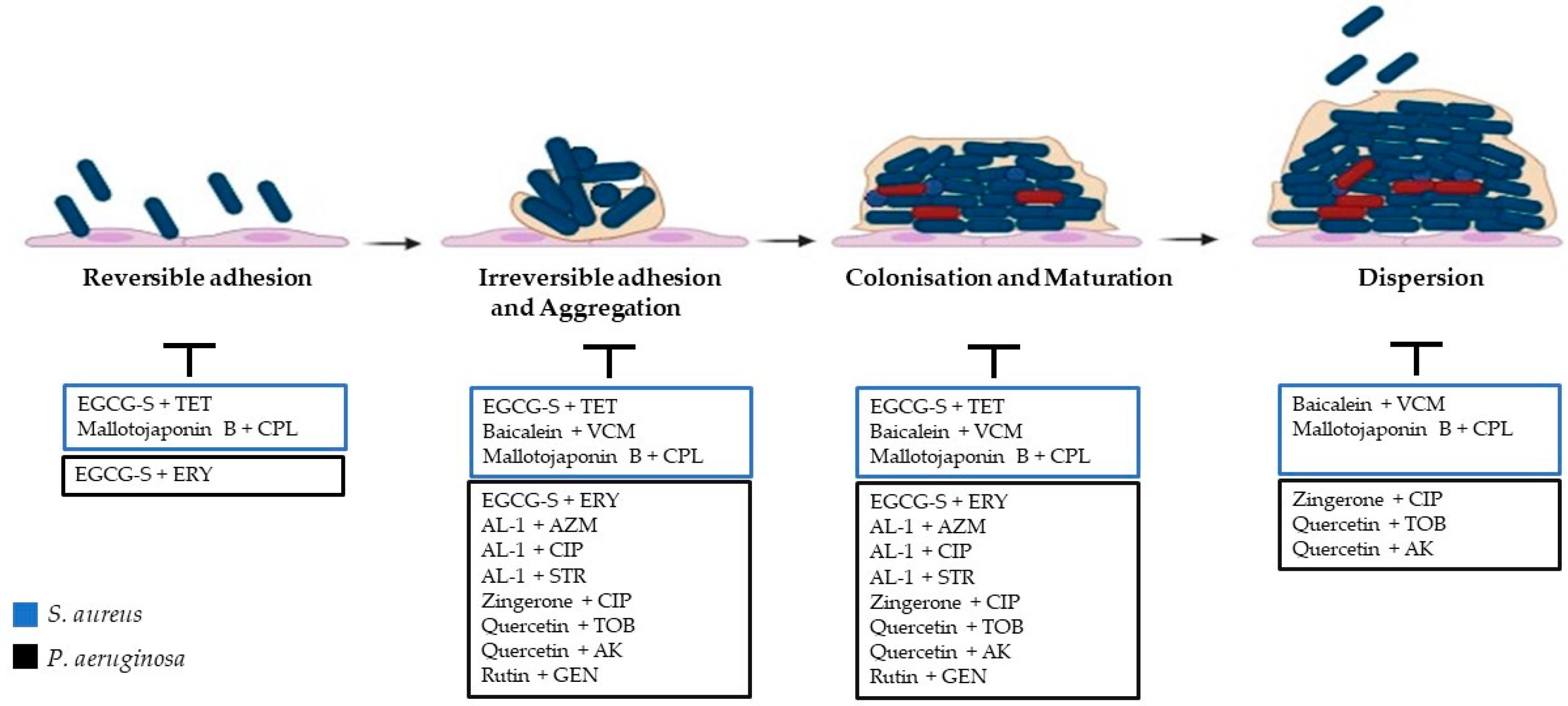
2.1. Steps of Biofilm Formation

The steps leading to microbial biofilm formation are dynamic and complex [12,20,21,22]. The biofilm development process includes the initial adhesion of microorganisms onto a surface, reversible attachment to the substratum, irreversible attachment and colonisation, aggregation and expansion, biofilm maturation, and finally biofilm dispersal and controlled detachment [15].

2.1.1. Adhesion

The initial reversible adhesion constitutes the serendipitous meeting between a surface and a planktonic microorganism [23]. Microorganisms begin biofilm formation due to specific outer pressure, such as nutrition, extreme pH, extreme temperature, UV radiation, high salt concentrations, desiccation, high pressure, or antibiotic treatment. It initiates with the favourable interaction between planktonic cells transported to the surfaces by sedimentation, convection, Brownian motion, or hydrodynamic forces [11]. This primary adhesion is achieved through the effects of electrostatic forces, Lifshitz–van der Waals interactions, and hydrophobic interactions [23,24].
The irreversible attachment is attained with the involvement of bacterial and fungal structural adhesins [25]. Bacterial adherence structures, including pili/fimbriae, non-fimbrial adhesions, and flagella, were identified to be involved in the development of biofilms [26].
The biofilms of S. aureus can be classified into ica-independent and ica-dependent biofilms. The ica-independent biofilms are more frequently found in methicillin-resistant S. aureus (MRSA) isolates. These biofilms involve proteins belonging to the family of microbial surface component recognizing adhesive matrix molecules (MSCRAMMs), such as fibrinogen and fibronectin-binding proteins FnBPA and FnBPB, and Staphylococcus protein A (Spa), S. aureus surface proteins C and G (SasC and SasG), clumping factor B (ClfB), extracellular adherence protein (Eap), and biofilm-associated protein (Bap). The ica-dependent biofilms are associated with exopolysaccharide intercellular adhesin/poly-N-acetylglucosamine (PIA/PNAG), encoded by the icaADBC operon, involved in intercellular adhesion [27,28].
P. aeruginosa cells begin irreversible attachment by making strong adhesive forces and aligning their long axes parallel to the surface. During infection, the type IV pili, potentially, can initiate the adhesion between P. aeruginosa and the host surface [29]. The type IV pili bind the glycolipids asialo-GM1 and asialo-GM2 on epithelial cells of the surfaces [30]. Functional amyloid fibrils (Fap), which provide mechanical robustness to the biofilm, increase adhesion and cell aggregation [31].
Candida adhesins, which are cell wall proteins, play a pivotal role in this process by facilitating attachment to various cells, including epithelial cells and other microorganisms, as well as abiotic surfaces, through the binding to specific amino acids or sugar residues. In the case of C. albicans, numerous adhesins are part of the Als (agglutinin-like sequence) family. Similar ALS-like proteins have also been identified in C. parapsilosis, C. tropicalis, C. dubliniensis, C. lusitaniae, and C. guilliermondii [32]. C. albicans Eap1 participates in adhesion to polystyrene substrates; Hwp2, Pga1, and members of the Sap family play direct or indirect roles in adhesion to human cell lines or in cellular aggregation [33]. Recently, Thierry Mourer and colleagues have demonstrated that amyloid protein Pga59 is used by the fungal cell to mediate cell–substrate interactions and biofilm formation. The Pga59 cell wall protein is an amyloid-forming protein involved in adhesion and biofilm establishment in the pathogenic yeast C. albicans [34].

2.1.2. Irreversible Adhesion and Aggregation

A cell-to-cell signalling mechanism referred to as quorum sensing (QS) also coordinates the initial formation of microbial biofilms [35].
Both Gram-negative and Gram-positive bacteria employ QS mechanisms to regulate biofilm formation by releasing signalling molecules (autoinducers, AIs) [36]. Gram-negative bacteria primarily produce acyl homoserine lactones (AHLs), whereas Gram-positive bacteria use oligopeptides [37,38,39]. In C. albicans QS, the best-known quorum-sensing molecules are farnesol, farnesoic acid, and tyrosol [40].
The irreversible adhesion of bacteria is progressed through the production of exopolysaccharides (EPSs) regulated by QS. The main constituents of EPSs, including polysaccharides, proteinaceous pili/fimbriae, other protein components, lipids, DNAs, and other compounds, depend on the species and the environmental factors [41,42,43,44,45]. Secondary messenger c-di-GMP is considered as one of the incentives for the transition from reversible to irreversible adhesion through EPS production and the structures of the cell’s surface [46]. EPS constituents are most often stabilised by intermolecular networks which involve specific binding proteins or networking between ECM constituents. Overall, EPS plays serious roles in adherence to surfaces, signalling, cell–cell recognition, biofilm formation, formed biofilm, protection of cells, retention of water, and genetic exchange [43,45,47,48]. EPS secretion continues till the third step of biofilm production to ensure the protected adhesion of bacteria and fungi to the surfaces.
The S. aureus auxiliary gene regulator (arg) QS system has a role in controlling the development of biofilms. agrB, a gene associated with the secretion of virulence factors, stimulates biofilm dispersion through increased production of the SplABCDEF serine proteases and the Aur metalloproteinase. PSMs have surfactant-like characteristics that are involved in the dispersion of biofilm. Other systems such as TCSs ArlRS, SaeRS, and SrrAB in S. aureus regulate virulence genes and biofilm formation. In S. aureus, a subpopulation of cells undergoes lysis to release extracellular DNA that glues together the ECM. The holin-like CidABC and anti-holin-like LrgAB systems control this process [27]. CidA facilitates the release of genomic DNA and the production of biofilms via controlling the actions of murein hydrolases. The LrgAB, regulated from protein LytSR TCS, prevents CidA-mediated lysis [27].
P. aeruginosa cells produce EPS and form clusters following the early attachment steps. EPS is made up of lipids, proteins, nucleic acids, and polysaccharides [49]. The exopolysaccharides’ composition includes alginate, polysaccharide synthesis locus (Psl), and pellicle polysaccharide (Pel). Alginate is mainly produced by P. aeruginosa clinical isolates originating from the lungs of cystic fibrosis (CF) patients. It is a linear polymer consisting of β-D-mannuronic acid and α-L-guluronic acid, controlled by the algACD operon, and has an important role in the protection and structural stability of biofilm [19]. Psl is a neutral polysaccharide composed of a repeating pentasaccharide, consisting of D-mannose, D-glucose, and L-rhamnose, mainly involved in the initial attachment of cells and early biofilm formation, whereas Pel is a cellulose-sensitive exopolysaccharide composed of 1→4 linked partially acetylated galactosamine and glucosamine sugars, essential for late-stage biofilm formation and maturation [49,50,51,52,53]. Moreover, CdrA accessory proteins also exist in biofilm matrices [54]. P. aeruginosa also produces two small soluble lectins, LecA and LecB, which bind to the repeating sugar unit in the exopolysaccharide and help P. aeruginosa adhere to targets in the host organism [55,56,57]. Extracellular DNA (eDNA)-dependent matrix is a critical component for several functions that maintain the stability of biofilm and the survival of bacteria in this environment. eDNA is released by cell death and lysis, which mainly occur in the interior of biofilms (e.g., the stalk of the mushrooms). eDNA promotes cell–cell adhesion and biofilm self-organisation and enables the formation of a stable biofilm structure [58]. In P. aeruginosa, eDNA overcomes the electrostatic repulsion between negatively charged polysaccharide strands by binding to Ca2+ to produce “cationic bridging” [25,59,60,61]. The cationic bridge promotes cell aggregation, enhances P. aeruginosa biofilms, and aids in initial attachment to foreign surfaces. Additionally, eDNA can interact with Pel and Psl to create fibre-like networks, which is likely to increase the biofilm’s resilience [62,63,64].
Once yeast cells have started to adhere to each other and to a surface, the transitional stage or morphological modification, where yeast cells change into hyphae via the transcriptional regulators Bcr1, Brg1, Efg1, Tec1, Ndt80, and Rob1, occurs. Hwp1 is required for hyphal development, mating, and the maintenance of biofilm integrity.
In the context of polymicrobial biofilms, the hyphae contribute to the architectural stability of the biofilm and act as a support for yeast cells, pseudohyphae, and other microorganisms [65]. During cell proliferation, yeasts accumulate an extracellular matrix (ECM) [66]. There are four main macromolecular ECM constituents: proteins (55%), carbohydrates (25%), lipids (15%), and nucleic acid. There are more proteins identified in the biofilm matrix, including glycolysis enzymes that may degrade extracellular biopolymers acting as a source of energy. In C. albicans biofilm, the dominant polysaccharide in the ECM is the mannans glucan complex, namely, α-1,2 branched α-1,6 mannan, which is associated with linear β-1,6 glucan and a small amount of β-1,3 glucan. The profile lipid is composed of glycerolipids (99.5%), with fewer sphingolipids (0.5%). In addition, the only sterol detected was ergosterol, though at low concentrations. EPS gives a three-dimensional structure to the formed biofilm in which the cells may stay immobilised. In C. tropicalis, glucose and hexosamine are the major carbohydrates present. Small amounts of proteins, uronic acid, and phosphorus may also be present [32]. Another key contributor to the stability and growth of C. albicans biofilms in the ECM is eDNA with predominantly random non-coding sequences. eDNA is likely be released via autolysis [55]. The ECM is partially self-produced and secreted by C. albicans cells within the biofilm but may also contain aggregates of C. albicans cells and lysed host cells, as well as epithelial cells and neutrophils. Two transcription factors involved in regulating ECM production for C. albicans biofilms are Zap1 and Rlm1 [67,68,69].

2.1.3. Colonisation and Maturation

Bacteria have minimal metabolic activity at deeper levels of mature biofilms. However, specific channels permit the exchange of nutrients and products between microorganisms enclosed within the biofilm and the outer environment, which favours the colonisation and maturation of the microorganisms [48]. Once microorganisms have irreversibly attached to a surface, the process of biofilm maturation begins. Bacteria continue to reproduce using the AIs signals within the embedded EPS matrix, and this leads to the formation of microcolonies and the maturation of biofilms [22,46]. During the process of maturation, the gene expression pattern of the sessile cells differs significantly from that of the planktonic cells; motility within the microcolonies, such as the production of bacterial surface structures, is restricted. Bacteria within deeper areas of mature biofilms possess lower metabolic activity. The minimal availability of nutrients and hypoxic conditions of the environment have been recognised as factors that stimulate the transition of bacteria in persister cells, a subpopulation of dormant phenotypic variants, detected in biofilm chronic infections. In staphylococcal biofilms, persister cells comprise a subpopulation of cells highly tolerant to antibiotics without being genetically resistant [70].
Yeasts produce microcolonies, and the coalescence of microcolonies yields the basal level of the biofilm. In the final maturation step, there is a wide accumulation of extracellular matrix material [71]. Persister cells also form, and although they make up only a small part of the biofilm population, these cells are associated with cases of tolerance to antimicrobial treatments [65]. C. albicans mature biofilms exhibit a dense network composed of blastophores and hyphae surrounded by ECM [71].

2.1.4. Dispersion

After the maturation of biofilm, the microorganisms detach and disperse to start a new biofilm community [12]. The biofilm detachment process, also known as the dispersion stage, represents the final process of biofilm development.
It is regarded as a strategy of bacterial cells to detach from the biofilms and begin another biofilm life cycle [72]. The biofilm dispersal process is regulated by environmental signals (oxygen, nutrients, temperature, and signalling molecules), the intracellular reduction in the concentration of c-di-GMP, and the upregulation of motility or QS genes, though many bacterial dispersal signals remain cryptic. The dispersion mechanisms differ among bacteria; however, the process can be divided into three common steps: escape of cells from the microcolonies, movement of freed cells, and attachment of the cells to another substrate [73,74].
During the biofilm dispersal phase, yeast cells disperse non-adherent budding daughter cells to make new communities [32,75]. The detachment of cells from biofilms involves mainly yeast cells, not filaments. The released cells display higher levels of adhesion to plastic or endothelial cells, probably due to their increased propensity to produce hyphae. In addition, the released cells were phenotypically distinct and more virulent than planktonic cells in a disseminated infection model. Thus, biofilm dispersion yields a unique class of yeast cell with increased ability to create new biofilms and cause infection [71,75].
The dissemination and colonisation of new target substrates explain recalcitrant microbial chronic infections. Moreover, biofilms protect the invading microorganisms against the immune system of the host via the impaired activation of phagocytes and the complement system [76].
Consequently, biofilm-associated diseases are persistent infections that are slowly developing, rarely resolved by the immune system, and refractory to antimicrobial treatments.

3. Bacterial and Fungal Resistance

3.1. Bacterial Biofilm Resistance

It is believed that biofilm-related organisms account for more than 65% of all microbial infections and exhibit high resistance to antimicrobial agents and components of the host defence system (both innate and adaptive) [77]. Within the biofilm, the bacteria adapt to environmental anoxia and nutrient limitations, showing an altered metabolism and altered gene expression profile, concomitant with lower metabolic activity, reduced cell proliferation, and increased nutrient sequestration. In addition, biofilm growth is associated with an increased level of mutations, and the proximity of the biofilm-embedded cells facilitates the horizontal transfer of resistance genes between the bacteria [27]. The principal concern with biofilm-related infections involves the difficulty in fully eradicating the infection, despite aggressive antimicrobial therapy. There are two primary mechanisms that explain how bacterial pathogens can evade antimicrobial therapy: resistance and tolerance. Resistance, which is measured by the minimum inhibitory concentration (MIC), refers to the ability of bacteria to multiply despite treatment with an antimicrobial compound. Resistance is typically inherited from mother bacteria or acquired from horizontal gene transfer, which is facilitated by the close proximity of bacterial cells within a biofilm. Resistance can be mediated by several processes, including drug inactivation or modifications, alteration of the binding site, reduced drug accumulation through increased efflux or decreased entry, and use of alternative metabolic pathways. Tolerance, by comparison, is defined as the capability of a microbe to survive despite treatment with a bactericidal antibiotic to which it is susceptible based on the minimum inhibitory concentration value [7].
In S. aureus infections, a significant problem is the rapid development of antibiotic resistance associated with biofilms [18]. Cell heterogeneity can exist in staphylococcal biofilms due to the development of persister cells through the acquisition of antibiotic resistance genes through horizontal gene transfer [78,79]. Among the various mechanisms by which this complex phenomenon may occur, those involving antibiotic efflux, enzyme activity, and reduced permeability are noteworthy [18]. The treatment of biofilm infection requires sensitive and well-penetrating antibiotics to ensure a sufficient concentration of effective antibiotics at the site of the biofilm infection. Bacterial cells within a biofilm are likely to encounter subinhibitory concentrations of antibiotics, which were shown to potentially stimulate biofilm production and alter the composition of the biofilm matrix [70]. Hence, tetracyclines, macrolides, rifamycins, lincosamides, quinolones, fusidic acid, oxazolidinones, sulfonamides, and nitroimidazole are preferred to glycopeptides, aminoglycosides, polymyxins, and β-lactamases because they can penetrate deeper [18]. The secretion of β-lactamase from a biofilm-embedded bacteria into the biofilm matrix can prevent the β-lactam antibiotics from acting on a neighbouring cell even if the latter does not produce the enzyme [27].
In chronic P. aeruginosa infections, the typical biofilm lifestyle significantly diminishes the effectiveness of antimicrobial treatments, as it results in inherent tolerance. This tolerance encompasses physical and physiological factors, along with biofilm-specific genes that can temporarily shield against antibiotics, thereby fostering the emergence of resistance [80]. The negatively charged polysaccharides (especially the Pel polysaccharide and alginate of P. aeruginosa) can effectively sequestrate the positively charged aminoglycoside class of antibiotics, such as tobramycin, thus preventing them from penetrating the deeper layers of the biofilm [81]. The eDNA through chelating cations such as magnesium ions forms a cation-limited environment that results in the induction of the PhoPQ and PmrAB TCSs in P. aeruginosa. These TCSs regulate cationic antimicrobial peptide resistance by upregulating the PA3552-PA3559 operon. The DNA-induced expression of PA3552-PA3559 results in up to a 2560-fold increase in the resistance to cationic antimicrobial peptides and a 640-fold increase in the resistance to aminoglycosides [82]. Biofilm-associated P. aeruginosa expresses the gene ndvB, which encodes a glycosyltransferase that catalyses the synthesis of periplasmic β-(1→3)-cyclic glucans. The glucans are thought to promote aminoglycoside resistance by sequestering the antibiotics (e.g., tobramycin) in the periplasm away from their cellular target. When P. aeruginosa biofilms are exposed to β-lactam antibiotics or colistin, various resistance mechanisms are induced, such as the increased expression of β-lactamase and the production of modified lipopolysaccharides, which make the bacteria resistant to colistin and other polymyxin antibiotics. tssC1, which is implicated in type VI secretion (T6S), is upregulated in P. aeruginosa biofilms. The upregulation of tssC1 is important for the induction of biofilm-associated antibiotic resistance to tobramycin, gentamicin, and ciprofloxacin. The expression of RND efflux pumps and genes involved in type III secretion were upregulated in antibiotic-resistant biofilms of P. aeruginosa that have developed in the presence of azithromycin [27].

3.2. Candida Biofilm Resistance

Biofilm, a self-protected life mode of the Candida species [16,17], is the main cause of antibiotic-resistant Candida infections. Biofilm-associated infections are hard to treat and constitute a challenge for clinicians because of their persistence and resistance to the majority of known antifungal drugs. Candida is able to form biofilms both on biotic and abiotic surfaces, for example on mucosae and medical implants.
Biofilm resistance is multifactorial and mechanistically complex and uses both methods akin to traditional, planktonic antifungal resistance as well as mechanisms specific to the biofilm lifestyle [17]. However, it can be mainly attributed to three features: production of an extracellular matrix; drug efflux pump overexpression, and persister cell presence [83].
In total, 55% of the extracellular matrix is made up of proteins, followed by 25% of carbohydrates, 15% of lipids, and 5% of extracellular DNA, and it plays a major role in Candida biofilm resistance [83]. Its two components, extracellular DNA and β-glucan, increase biofilm resistance to different antifungals in an unclear way; this may also entail reduced drug penetration and a matrix–antifungal sequestration mechanism [84]. It is also believed that the extracellular matrix plays a role in the preservation of nutrients, water, and enzymes [85,86]. The formation of biofilm matrix β-1,3 glucan, biofilm adhesion, and resistance to azole treatments are all hampered when this matrix delivery pathway is disrupted.
The efflux pumps are mainly involved in resistance mechanisms during the early biofilm developmental phase [87]. With transcriptional analysis, it has been possible to show the greater expression of efflux pump genes following 12 h of biofilm formation in contrast to 48 h of full biofilm formation [88]. These findings further support the theory of time-specific efflux pump functionality [88]. C. glabrata and C. tropicalis biofilms are also characterised by the upregulation of efflux pumps [89,90].
Furthermore, at the later stages of biofilm growth, the content of ergosterol in the cell membranes of biofilm cells is much lower than that of planktonic cells [17]. This reduced presence of ergosterol may impair the effectiveness of medications that target ergosterol, such amphotericin B and azoles.
Namely, alterations in the transcriptional profile of sterol pathway genes have been shown, examining the various phases of biofilm formation [91,92]. Through microarray analysis, Nett and colleagues have demonstrated that an increased transcription of ERG11, which codes for the azole drug target, and ERG25, which codes for a putative C4 methyl sterol oxidase, may be involved in the C4-demethylation of intermediates in ergosterol biosynthesis [92].
Candida biofilm is also characterised by the presence of a subpopulation of cells, called persister cells, metabolically dormant variants of microbial deep-seated biofilm-forming microbes that are resistant to a variety of medications, such as amphotericin B, azoles, and chlorhexidine [17]. They form only upon adherence to a surface [83].
Persister cells are present within both Candida and bacterial biofilms [93]. C. albicans and C. krusei biofilms frequently possess persister cells, whereas C. glabrata spp. do not [93,94].
General stress response upregulation, cell density, and quorum sensing are the main features which allow biofilm-specific resistance mechanisms [83].
The greater number of fungal cells in a biofilm as opposed to planktonic development seems to be responsible for drug resistance [95,96]. Perumal and Chaffin [95] demonstrated that both planktonic and biofilm cells revealed higher azole drug resistance at higher than at lower cell densities, indicating that antifungal resistance is influenced by high biofilm cell density.
Candida biofilm cell density is strictly linked with quorum sensing. C. albicans produces two main quorum-sensing molecules, tyrosol and farnesol, which have conflicting effects [97,98]. It has been demonstrated that farnesol prevents the development of biofilms and the yeast-to-hyphae transition [85,98], and it seems to negatively regulate drug resistance. This effect has been observed for C. albicans and C. dubliniensis biofilms [99,100].
In addition, greater stress responses to biofilm presence can influence drug susceptibility and promote drug resistance [83]. Uppuluri and colleagues identified calcineurin, a Ca2+ calmodulin-activated serine/threonine-specific protein phosphatase, crucial for morphogenesis, virulence, and homeostasis, as being involved in biofilm resistance [101]. Hsp90, one of the heat shock proteins that stabilises several host proteins, constitutes another stress response pathway contributing to Candida biofilm resistance [102].

4. Antimicrobial Activity of Plant Compounds

For 350 million years, plants have survived various environmental stresses, both of abiotic and biotic origin [103]. Environmental (abiotic) factors can be considered as the lack of nutrients and oxygen, drought, salt, unfavourable temperature swings, intense sunshine, and conditions brought on by human activity, including pollution, pesticide use, and elevated UV radiation [104,105]. Herbivore pests, nematodes, bacteria, fungus, viruses, and insects are examples of biotic influences. [106]. To withstand such assaults, plants detect pathogens and initiate immune responses, such as fortifying the cell wall, synthesising lytic enzymes, and producing pathogenesis-related proteins [107].
Moreover, plants produce a broad range of compounds that constitute a defence mechanism against antagonistic biotic and abiotic stresses [103]. They are called ‘secondary metabolites’ because they are products of secondary plant metabolism and are biologically dynamic.
These compounds may exist in a constitutively stored, inactive state within plant cells or on the plant’s surface before encountering microorganisms, or they can be synthesised after infection from existent forerunners. Alternatively, their biological synthesis can be induced in reaction to an infection [108].
Phytoalexins are the inducible compounds and include terpenoids, glycosteroids, flavonoids, and polyphenols [103].
Secondary metabolites not only improve the defence against biotic and abiotic stressors but also provide a substantial supply of antiviral, antifungal, antimalarial, antibacterial, antioxidant, anti-inflammatory, anticancer, antidiabetic, and immunosuppressive compounds [109,110]. Secondary metabolites are also nutritional constituents that can improve the physiological and cellular activities in animals and humans that consume them [111].
The use of plants as therapies in traditional medicine is nowadays well established in several diseases. The most important advantages linked with the use of herbal plants are their safety, including reduced cost-effectiveness in comparison to better patient tolerance and fewer unwanted side effects, sustainability, improved biodegradability, and easy availability [112].
Plants enriched in natural secondary metabolites may be a promising approach to the discovery of potential alternative therapeutics to combat antimicrobial resistance. Specifically, plants have been widely documented as capable of producing drug resistance inhibitors that facilitate the delivery of antimicrobial compounds [112].
Different antimicrobial mechanisms have been very well recognised: the degradation of the proton driving force with ion leakage, natural action of the cell components, disruption of the outer membrane of Gram-negative microorganisms through the release of lipopolysaccharides, interaction with membrane proteins, such as ATPases and other enzymes, and inhibition of enzyme synthesis [113].
Regarding antibiofilm activity, the possibility of using natural compounds that can inhibit quorum sensing has been proposed to treat biofilm-associated infections [27].
Numerous secondary plant metabolites have been demonstrated to possess anti-quorum and antibiofilm properties, despite typically lacking direct antibacterial activity [103,114]. Examples are halogenated thiophenones, which can manage the growth of biofilms [115]; baicalin hydrate and cinnamaldehyde target P. aeruginosa’s acyl-homoserine lactone-based quorum sensing system [116]; hamamelitannin targets S. aureus’s peptide-based system; and baicalin improves P. aeruginosa’s susceptibility to tobramycin [116].
Sesamin and Sesamolin, two lignans separated from the plant Sesamum indicum (L.), have been shown to stop previously infected worms from becoming infected in a P. aeruginosa infection model in Caenorhabditis elegans. The mechanism seems to be their capacity to attenuate the quorum sensing-regulated virulence factors of the bacteria [117].
However, there are no plant-derived antibiotics, although plants fight infections successfully, probably thanks to a successful defence mechanism they developed, based on “synergy” between the several secondary metabolites they synthesise [103].
Combination therapy is one method that can be used to increase the efficacy of antimicrobial therapy for difficult-to-treat infections, such as biofilm-related infections, given the dearth of new antimicrobial drugs with novel mechanisms of action and the rise in infections brought on by resistant microorganisms [118].
This strategy focuses on using natural enhancers to boost the antimicrobial medicine’s antibiofilm activity at concentrations where the antibiotic previously exhibited no antibiofilm activity. Thus, researchers have also been interested in how phytocompounds interact with antibiotics or chemotherapeutic antimicrobial agents. Numerous studies have shown that these phytocompounds exhibit promising synergistic effects when combined with these medications, offering benefits for treatment such as a wider range of efficacy, enhanced safety and tolerability, decreased toxicity, and decreased resistance to antimicrobial agents [119].
Therapeutic interactions are described as synergistic, indifferent, and antagonistic. This interaction is defined by the fractional inhibitory concentration index (FICI) [120]. The FICI is a non-parametric model defined by the following equation:
Σ FIC = FIC A + FIC B
where FIC A = MIC of drug A in combination/MIC of drug A alone; FICB = MIC of drug B in combination/MIC of drug B alone [121].
The following values obtained from the FICI equation establish the interaction relations: values ≤0.5 indicate synergistic interactions, values of 4.0 indicate antagonistic effects, and values between these two indicate no interaction [120].

5. Synergy between Antibacterial Chemotherapeutics and Natural Compounds against S. aureus

Biofilm formation adds to the resistance phenotype of the microbial pathogens towards host defence and conventional drug therapy, often resulting in serious and persistent infections [122]. Thus, a multifaceted approach is needed to combat this problem by discovering novel antimicrobial agents and/or new methodological strategies [123,124]. In the last decade, the interest in medicinal and aromatic plants and their components has remarkably increased.
Accordingly, Shinde and colleagues [125] focused on the effect of epigallocatechin gallate (EGCG), a polyphenol/catechin of green tea. The chemical structure features of EGCG include multiple hydroxyl groups and a gallate moiety. EGCG is prominently found in green tea (Camellia sinensis) leaves, accounting for a significant portion of their polyphenol content. EGCG exerts antioxidant, anti-inflammatory, anti-tumour, and antimicrobial effects on various Gram-positive and Gram-negative bacteria [125]. The antimicrobial effects of EGCG are exerted through several mechanisms, including cell membrane injury, enzyme inhibition, and the impairment of fatty acid biosynthesis. EGCG compromises cell wall integrity by interfering with the polysaccharides that make up the glycocalyx and bind peptidoglycan, affecting the biofilm attachment to the surface. Recently, reports hypothesised that the synergism between EGCG and several antibacterial substances can counteract bacterial biofilm by deteriorating the extracellular matrix elements and favouring the antibiotics’ action [126]. In this context, Shinde and colleagues demonstrated that a modified lipid-soluble EGCG, epigallocatechin-3-gallate-stearate (EGCG-S), in combination with tetracycline, is able to inhibit 94% of S. aureus ATCC 14990 biofilm formation [125]. Several studies demonstrated that EGCG impairs the assembly of phenol-soluble modulins (PSMs), Bap formation, and the integrity of the cell wall, hypothesising that this polyphenol might influence the cell adhesion to the surface. The potential of EGCG as a synergistic agent with antibiotics could be due to breaks in the extracellular matrix components, which allow antibiotic permeation, favouring their effect against biofilm [125,126].
Another natural compound drawing great interest is baicalein, which is an extract of Scutellaria baicalensis with antibacterial activity. A study by Chen and colleagues revealed baicalein’s antibiofilm properties using clinically isolated strains of S. aureus 17546 (t037). The molecular mechanism of its antibiofilm activity was studied also in combination with antibiotics such as vancomycin (VCM), showing its synergic effect [127]. It has been demonstrated that baicalein was able to prevent S. aureus biofilm development also at subinhibitory concentrations. Although baicalein (32 µg/mL) and VCM (4 µg/mL) alone did not exert any effect on bacterial counts in established biofilms, their combination was able to trigger a dramatic effect on the dispersal step. The authors demonstrated that baicalein could inhibit S. aureus biofilm formation by downregulating the quorum-sensing system regulators, like agrA, RNAIII, and sarA. The synergism between baicalein and VCM caused biofilm formation inhibition, the destruction of formed biofilm, and increased permeability to VCM [127].
Mallotojaponin B, a phenolic compound obtained from Mallotus oppositifolius, possesses several antibacterial effects. Nguena-Dongue and colleagues investigated the combinatory effect of mallotojaponin B with chloramphenicol against methicillin-resistant S. aureus ATCC 33591. Mallotojaponin B, belonging to the phenol family, exerts its effects through the alteration of membrane structural components or the inactivation of cell essential functions. Moreover, mallotojaponin B is able to alter membrane permeability and integrity and modify several intracellular functions induced by the hydrogen bonding of phenolic compounds to enzymes. MRSA ATCC 33591, which is chloramphenicol-resistant, was susceptible to mallotojaponin B’s activity. Nguena-Dongue and colleagues investigated the effects of their combinations against MRSA. Interestingly, a synergistic effect (FICI 0.393) with MIC reductions in a range from 12.5 μg/mL (MIC) to 0.781 μg/mL (1/16 MIC) for mallotojaponin B and from 250 μg/mL (MIC) to 1.95 μg/mL (1/128 MIC) for chloramphenicol was demonstrated [128]. These results demonstrated that the combination of mallotojaponin B and chloramphenicol exerts bactericidal synergistic effects on MRSA through the alteration and destruction of the cell membrane and biofilms formed by MRSA, suggesting that this combination could be a potential strategy against methicillin-resistant S. aureus (Table 1; Figure 1).

6. Synergy between Antibacterial Chemotherapeutics and Natural Compounds against P. aeruginosa

In P. aeruginosa-associated infections, antibiotic resistance is mediated in part by surface-attached biofilms [133]. Their framework becomes a physical barrier to antibiotic penetration and provides an altered microenvironment for pathogen survival inside the host [133]. Thus, there is an urgent need for revamping the treatment measures to counter bacterial biofilm infections like P. aeruginosa infection as well as to control the emergence of highly virulent strains. Previous studies have focused on the antibiofilm potential of various plant natural compounds including phenols, essential oils, terpenoids, lectins, alkaloids, polypeptides, and polyacetylenes, demonstrating that they not only inhibit biofilm formation but also eliminate mature biofilm structures [129,133]. The combination of plant extracts and antibiotics represents a template for developing antibiofilm drugs against P. aeruginosa biofilm-associated infections [134]. Several active substances present in ginger (Z. officinale) such as gingerols, shogaols, paradols, gingerdiols, and zingerone have been widely used in traditional herbal medicine [129]. Some of these chemical compounds are found to be clinically effective for the treatment of various diseases, including P. aeruginosa infections [129]. According to this, it has been demonstrated that zingerone could be a potential antibiofilm agent by making the biofilm prone to antibiotic treatment and by altering its formation process, demonstrating a synergistic effect with antibiotics. When the biofilm was grown in the presence of zingerone (10,000 µg/mL), ciprofloxacin (0.06 µg/mL) showed a significantly high eradication efficiency because zingerone modulated the biofilm formation, making it thin [129]. Altered biofilms were more susceptible to ciprofloxacin; so, biofilm grown in the presence of zingerone was more easily eradicated by ciprofloxacin as compared to the normally grown biofilms [129]. Therefore, ciprofloxacin can better penetrate thinner biofilms in combination with zingerone, becoming more efficient at eradicating P. aeruginosa biofilms. The results of the study suggested that zingerone has potent antibiofilm inhibition and eradication activities which increase the antibiotic effect [129].
Another report demonstrated the synergistic effect of 14-alpha-lipoyl andrographolide (AL-1), obtained from Andrographis paniculate, on P. aeruginosa-associated biofilm when combined with azithromycin, ciprofloxacin, and gentamicin [130]. The report revealed the antibiofilm properties of AL-1 and its ability to sensitise the bacterium to several antibiotics through synergistic effects. The combinatory treatment between AL-1 and antibiotics was demonstrated to decrease the production of EPS and pyocyanin compared to antibiotics alone, suggesting that the synergism effect is due to the inhibition of biofilm synthesis. The results have proven that AL-1 could be an effective compound to counteract P. aeruginosa infection when used in association with the most common antibiotics [130].
Rutin, a flavonoid isolated from the peels of Citrus sinensis with antimicrobial properties, has been studied for its antibiofilm potential against P. aeruginosa, in combination with the conventional antibiotic gentamicin [131]. The results showed that rutin exhibited an MIC at 800 μg/mL against P. aeruginosa. When the bacterium was treated with rutin (200 μg/mL) and gentamicin (2.5 μg/mL), the prevention of biofilm formation was improved in a synergistic manner [131]. Adherence and biofilm formation were attributed to EPS generation, which was similarly markedly inhibited in the tested strain, indicating a potential synergistic action in combination with gentamicin. The findings of the study confirm that rutin could be potentially used as an adhesion inhibitor to treat P. aeruginosa infections, displaying an enhanced antibiofilm property when combined with gentamicin. Even though the mechanism of inhibition of biofilm formation by rutin is unclear, the study indicates the potential use of rutin as a promising antibiofilm agent against P. aeruginosa as it induces ROS generation in bacteria, leading to oxidative stress and the death of bacteria [131]. Moreover, quercetin demonstrated interesting antibacterial effects too. The development of drug resistance in opportunistic pathogens represents one of the major healthcare challenges correlated with infection management [132]. Combination therapy has numerous advantages due to the simultaneous effect of two drugs on two separate cellular targets [132]. Nevertheless, the selection of the drugs should offer safety and a synergistic interaction against most of the strains. The efficacy of antibiotics in combination with quercetin, a natural flavonoid, against biofilm-forming P. aeruginosa strains previously isolated from catheter-associated urinary tract infections has been studied. Based on the antibiotic susceptibility pattern, the synergistic effect of quercetin (10 mg/mL) with selected antibiotics amikacin and tobramycin was tested by evaluating the MIC and FICI values. Quercetin was demonstrated to inhibit biofilm formation with an MIC value of 500 μg/mL; however, the combinations with the antibiotics amikacin and tobramycin were demonstrated to further inhibit biofilm formation, showing FICI values of 0.25 and 0.5, respectively. The effects of the synergistic combinations were further assessed with time–kill and biofilm cell viability assays. Quercetin and selected antibiotics affected biofilm formation and biofilm cell viability, showing 80% inhibition. These results demonstrated that synergistic combinations penetrate the matrix of the biofilm and cause the death of cells. In vitro infection studies showed that the drug combinations decreased the infection rate significantly by reducing the HEK-293T cell killing effect caused by P. aeruginosa [132]. A modified lipid-soluble EGCG, epigallocatechin-3-gallate-stearate (EGCG-S) (100 μg/mL), in combination with erytromycin (15 μg/mL), is able to inhibit 95% of P. aeruginosa ATCC-CRM-9027 biofilm formation, suggesting the potential of EGCG-S as a synergistic agent with antibiotics and as an antibiofilm agent [125]. It was demonstrated that EGCG influences the ability of Fap to form fibrils. Combinations with antibiotics favour the two agents’ penetration and their action on bacterial cells [135,136] (Table 1; Figure 1).

7. Synergy between Antifungal Chemotherapeutics and Natural Compounds against Candida spp.

Plants, in their ongoing battle for survival, have developed a wide range of defensive molecules to protect themselves from attacks by plant pathogenic fungi. These molecules, often present in plants as natural compounds, have demonstrated remarkable antifungal properties. In this context, numerous studies have explored the potential applications of these molecules in the fight against fungal infections, particularly those caused by Candida species. These investigations have focused on their synergistic interactions with synthetic antifungals, particularly in the context of biofilm-associated infections, offering an intriguing perspective for the development of effective antifungal therapies.
Acorus calamus Linn. is widely spread across Central Asia, North America, and Eastern Europe. Indian medicine uses this plant’s rhizome to treat several diseases like epilepsy, mental ailments, chronic diarrhoea, and dysentery and for the following symptoms and conditions: helminthiasis, amenorrhea, dysmenorrhea, nephropathy, calculi, strangury, hoarseness, flatulence, and dyspepsia [137]. α- and β-asarones, the primary bioactive substances, showed several bioactive features like antibacterial and antifungal properties [118,138,139,140,141]. The in vitro synergistic activity of α-asarone and β-asarone purified from Acorus calamus rhizomes and amphotericin B, fluconazole, and clotrimazole against Candida species has been reported by Kumar and colleagues.
α-asarone (8 μg/mL) combined with 0.03 μg/mL of amphotericin B reduces C. albicans biofilm by 36%. C. tropicalis biofilm is reduced by 39% using a combination of 0.25 μg/mL of amphotericin B and 4 μg/mL of α-asarone [118]. β-asarone showed a lower antibiofilm activity against both C. albicans and C. tropicalis when combined with amphotericin B. β-asarone (1 μg/mL) and amphotericin B (0.03 μg/mL) combined inhibited C. tropicalis biofilm by 21%. C. albicans biofilm is inhibited at the same rate by the combination of 8 μg/mL β-asarone and 0.06 μg/mL amphotericin B. It is evident from the results obtained by Kumar and colleagues that α-asarone and β-asarone had synergistic interactions also with fluconazole and clotrimazole. The MIC values decreased by eight times, and the inhibition of biofilms was significantly higher than with the individual compounds. α-asarone (8 μg/mL) and fluconazole (0.06 μg/mL) resulted in the most active combination against C. albicans with an inhibition rate of 40% of the biofilm formation [118]. β-asarone has been observed to suppress the morphogenesis and biofilm growth of C. albicans at subinhibitory doses [142].
The antifungal properties of plant-derived polyphenols are nowadays well known.
EGCG has garnered attention for its multifaceted biological activities, including its antifungal properties. In scientific research, EGCG has demonstrated inhibitory effects against various fungal species. It has been shown to inhibit the growth and yeast-to-hypha transition of C. albicans [143,144,145,146,147]. Several studies demonstrated that EGCG may synergise with fluconazole, amphotericin B, and ketoconazole against C. albicans [144,148]. Synergistic effects have been observed against all Candida spp. biofilm when EGCG was combined with amphotericin B by Ning and colleagues. C. albicans ATCC 10231 biofilm was reduced by 90% with 750 μg/mL of EGCG and 0.13 μg/mL of amphotericin B with a FICI of 0.19 [149]. The combination of 375 μg/mL of EGCG with 1.56 μg/mL of amphotericin B showed an inhibition of C. parapsilosis biofilm of 90% with a FICI of 0.27. In addition, 3000 μg/mL of EGCG with 0.19 μg/mL of amphotericin B showed a 90% inhibition of C. tropicalis biofilm with a FICI of 0.19. The EGCG–amphotericin B combination resulted in a synergistic effect also against C. krusei (FICI = 0.31), C. glabrata (FICI = 0.31), and C. kefyr (FICI = 0.5). C. krusei is the most sensitive to this combination, with a sessile minimum inhibitory concentration 90 (SMIC90) of 187.5 μg/mL for EGCG and 0.16 μg/mL for amphotericin B [149]. In a study conducted by Behbehani and colleagues, the synergistic effects of EGCG in combination with fluconazole or ketoconazole against mature biofilms of Candida species strains were investigated [150]. Notably, no interaction was observed when EGCG was combined with fluconazole against two C. albicans strains (ATCC 24433 and a clinical isolate), as indicated by FICI values exceeding 0.5. However, in all other cases, the FICI values were below 0.5, signifying a synergistic interaction between EGCG and fluconazole or ketoconazole [150]. Similarly, Ning and colleagues investigated synergism between EGCG and fluconazole or miconazole. They performed an antibiofilm assay using C. albicans SC 5314, C. albicans ATCC 10231, C. parapsilosis ATCC 22019, C. tropicalis ATCC 13803, C. glabrata ATCC 66032, C. kefyr ATCC 46764, and C. krusei ATCC 14243. All the strains were found to be more sensitive to the EGCG in combination with fluconazole or miconazole. The concentrations needed to obtain biofilm inhibition were lower when used together. The combination of EGCG with fluconazole appeared to be additive rather than synergistic against C. parapsilosis, C. krusei, and C. kefyr, as indicated by the FICI value (FICI values of 0.56, 0.75, and 0.63, respectively), whereas the combination with miconazole had no synergistic effect only against C. kefyr (FICI = 0.63) [149]. This polyphenolic compound’s potential as an antifungal agent is attributed to its ability to target specific fungal components and pathways. Some authors have reported that the mechanism of action of EGCG on C. albicans involves its inhibitory activity on folic acid metabolism [148]. This phenomenon may elucidate the molecular mechanism behind the synergy observed between EGCG and azole antifungals. Evensen and Braun reported that the inhibition of biofilm formation and maintenance of C. albicans is expedited through the impairment of proteasomal activity caused by EGCG, leading to cellular metabolic and structural disruptions [151].
Tyrosol is a dietary phenolic compound present in virgin olive oil and wine and also a quorum-sensing molecule in C. albicans which stimulates germ-tube formation and yeast-to-hypha transition during biofilm formation [97]. This compound may have a significant impact on preventing diseases like cancer and metabolic, neurological, and cardiovascular disorders [152]. Its use against fungal infections has also been proposed. Cordeiro and colleagues stated that exogenous tyrosol significantly reduces the biofilm formation of clinical isolated C. albicans and C. tropicalis and showed that, when combined with amphotericin B (52.5 and 90 μg/mL, respectively), tyrosol at a concentration of 187.5 μM reduced C. albicans biofilm formation by 69% and C. tropicalis biofilm formation by 63%. The capacity to inhibit the mature biofilms of both C. albicans and C. tropicalis was found to be markedly lower (36.5% and 36.4%, respectively, at the same concentrations used to investigate antibiofilm formation properties) [153]. Fluconazole (3490 μg/mL) and itraconazole (1020 μg/mL) combined with 187.5 μM of tyrosol inhibited C. albicans biofilm formation by 55% and 70%, respectively. However, at the same concentration, these combinations had no activity against mature biofilm. Similarly, fluconazole (4720 μg/mL) and tyrosol (225 μM) were not able to inhibit C. tropicalis mature biofilm, but they have been shown to be active against the biofilm formation at the same concentrations, with inhibition rates of 58 and 61% [153]. Monteiro and colleagues reported that tyrosol significantly decreased the number of adhered cells to the acrylic surface in both single and mixed cultures of C. albicans and C. glabrata [154].
The presence of Pseudolaric Acid A (PAA), a diterpenoid found in the pine bark of Pseudolarix kaempferi, has been reported to exert inhibitory effects on C. albicans. The pine bark of Pseudolarix kaempferi is used in traditional Chinese medicine to treat dermatomycosis. Zhu and colleagues have studied the possible use of PAA in association with fluconazole against C. albicans ATCC 90028. They showed that both PAA and fluconazole lead to a dose-dependent decrease in C. albicans biofilm formation after 1.5 h of incubation. They found that the inhibition rate was about 20% using 8 μg/mL PAA and 23% using 0.25 μg/mL fluconazole. PAA (4 μg/mL) in association with fluconazole (0.5 μg/mL) exhibited a 42% inhibition of biofilm formation in the early stage (1.5 h). PAA (4 μg/mL) in combination with fluconazole (0.5 μg/mL) reduced biofilm formation by 49%, after 6 h of formation [155]. During the mature stage of biofilm formation (24 h), neither PAA nor fluconazole individually exhibited substantial antibiofilm activity, even at the highest drug concentration of 512 μg/mL. However, when PAA (4 μg/mL) and fluconazole (0.5 μg/mL) were combined, there was a 58% reduction in biofilm formation at 24 h. Also, cell adhesion was inhibited by 44% with 4 μg/mL of PAA and 0.5 μg/mL of fluconazole. Zhu and colleagues demonstrated that when fluconazole (0.5 μg/mL) was combined with PAA (4 μg/mL), the numbers of both planktonic cells and hyphae were lower than those in the drug-free control. This suggests that the combination of PAA with fluconazole inhibited the yeast-to-hypha transition and hypha formation, consequently hindering biofilm formation. The yeast-to-hypha transition is an important process in fungi like Candida, where yeast is the predominant growth form, but the transition to a filamentous form is crucial for biofilm formation [155].
The pine bark of Pseudolarix kaempferi also contains another diterpene, Pseudolaric Acid B (PAB), also known for its inhibitory activity against fungi. Li and colleagues showed that the combination of PAB and fluconazole displayed inhibitory effects on both early and mature biofilms. After 6 h of incubation, the combination of 16 µg/mL of fluconazole and 2 µg/mL of PAB on C. tropicalis inhibits 80.36% of biofilm formation. In addition, 64 µg/mL of PAB and 2 µg/mL of fluconazole inhibits 50% of mature biofilm. Among the mechanisms of action of PAB, the authors reported that PAB inhibited spore germination and mycelium formation and caused damage to cell integrity, resulting in cell deformation, swelling, collapse, and perforation of the outer membrane [156].
Coumarins are a group of natural chemical compounds that include a wide range of molecules with a similar basic structure. These compounds are widely distributed in nature and are found in various plants, such as coumarin, which is present in some herbs and spices, including cinnamon and vanilla. Some coumarins have pharmacological and biological properties, such as antioxidant, anti-inflammatory, and other biological properties.
The combination of fluconazole with osthole, a natural coumarin derived from the Cnidium monnieri plant, has been investigated by Li and colleagues against C. albicans SC5314 biofilm formation. The authors demonstrated the synergism of fluconazole and osthole against the planktonic growth and against C. albicans biofilm formation. Osthole or fluconazole alone did not exhibit an antibiofilm effect. However, the combination of fluconazole and osthole had a significant synergistic effect by reducing the biofilm formation by 90% at a concentration of 8 μg/mL for both. When it comes to the mechanism of action, the authors’ experiments, as indicated by the results of an expression profile microarray, revealed significant changes in the expression of genes associated with oxidation–reduction processes, energy metabolism, or transportation following the combined treatment with fluconazole and osthole. From the microarray data, the authors observed that the expression of multidrug transporter genes exhibited an opposite trend following the combined treatment with fluconazole and osthole [157,158].
Essential oils are concentrated blends of terpenes and other aromatic compounds found in some plants. Many essential oils, such as tea tree oil, oregano oil, and cinnamon oil, are known for their antimicrobial properties [159,160,161,162]. These essential oils can be extracted from various parts of the plants, such as the leaves, flowers, roots, or fruits.
The genus Oreganum produces essential oils enriched in carvacrol, eugenol, and thymol. It has been shown that the addition of terpenoids in combination with fluconazole prevented C. albicans biofilm growth in the early phase of formation. Carvacrol at a concentration of 62 μg/mL did not have an inhibitory activity, but it allowed the concentration of fluconazole needed for biofilm formation inhibition to be decreased to 32 μg/mL. At these concentrations, the synergism between carvacrol and fluconazole was confirmed with a FICI value of 0.311. The combination of 125 μg/mL of eugenol with 2 μg/mL of fluconazole also had a synergistic effect, with a FICI of 0.25 against biofilm formation, whereas no interaction was detected against mature biofilm (FICI > 1). The combination of thymol and fluconazole resulted in no synergistic interaction against both biofilm formation and mature biofilm. Furthermore, 500 μg/mL of carvacrol decreases the fluconazole dosage needed to inhibit mature biofilms by 32-fold, although no synergistic effects have been registered (FICI > 0.5). In the manuscript, the authors report that a study on Saccharomyces cerevisiae revealed that eugenol and carvacrol induce membrane disintegration, loss of ions, and interference in the TOR signalling pathway, ultimately leading to a loss in viability. Additionally, terpenoid-induced alterations in permeability and membrane fluidity result in the degradation of the cell wall, which impacts the adherence of C. albicans to solid surfaces, and the increased influx of fluconazole results in the inhibition of biofilm formation [121].
Farnesol is a chemical compound belonging to the sesquiterpene class, which are terpenes composed of three isoprene units. Farnesol is found in various plants and is responsible for their characteristic aroma. It is also used in perfumery and the cosmetics industry for its pleasant scent. Additionally, it has been studied for its potential biological properties, including possible antibacterial and anti-inflammatory effects. Many plants, including citronella, lemon grass, tuberose, cyclamen, rose, neroli, balsam, and musk, contain farnesol in their essential oils, and it is also an autoregulatory molecule produced by C. albicans, which inhibits biofilm formation and the yeast-to-hyphae transition. Farnesol is used to treat hyperlipidemia, atherosclerosis, diabetes, allergic asthma, and obesity [163]. Synergy between farnesol and amphotericin B has been investigated by Katragkou and colleagues [164]. When 14 μM of farnesol was combined with 1 μg/mL of amphotericin B, C. albicans SC5314, a well-characterised strain that forms biofilms, reduced its biofilm formation. The MIC value of farnesol decreased from 600 μM when used alone to 14 μM when combined with amphotericin B, and for amphotericin B, the MIC values decreased from 1.5 μg/mL (alone) to 1 μg/mL (in combination) with a FICI of 0.79. Katrgkou and his group calculated the FIC index of the combination farnesol–fluconazole against C. albicans SC5314. They obtained a synergistic effect (FICI = 0.5) at a concentration of 150 μM of farnesol and 64 μg/mL of fluconazole. The authors observed, concerning the mechanism of action of farnesol, structural distortion, with the biofilm showing a more sparse appearance compared to untreated (control) biofilms. This includes a loss of elongated hyphal elements and a predominance of yeast-like forms. Farnesol has been tested in combination with fluconazole, voriconazole, itraconazole, posaconazole, and isoconazole against C. albicans biofilm formation [164]. The combination posaconazole–farnesol was the most active against C. albicans biofilm formation, with SMIC50 at a concentration of 0.25 μg/mL and 4.69 μg/mL, respectively. At the same concentration, farnesol inhibited 50% of biofilm formation in combination with 0.5 μg/mL of itraconazole or isoconazole. In addition, 1 μg/mL of voriconazole was needed to reduce the biofilm by 50%, together with 4.69 μg/mL of farnesol. The combination of farnesol and fluconazole was less active, and a higher concentration of these compounds was necessary to inhibit 50% of biofilm inhibition (75 μg/mL and 64 μg/mL, respectively). The combination of farnesol with posaconazole resulted in the most active combination, with an SMIC50 of 2.34 μg/mL of farnesol and 0.25 μg/mL of posaconazole against C. auris biofilm formation. The highest concentrations needed were 75 μg/mL and 64 μg/mL for the combination of farnesol/fluconazole. The authors showed that exposure to farnesol significantly increased phospholipase activity in C. albicans. Farnesol induced the production of reactive species in a dose-dependent manner and enhanced resistance to oxidative stress in C. albicans. Additionally, due to its amphiphilic properties, farnesol could be incorporated into cell membranes, affecting their fluidity and integrity. Farnesol was found to influence cellular polarisation and membrane permeability in both C. parapsilosis and C. dubliniensis [165].
Gypenosides (Gyp) are a group of triterpenoid saponins that were isolated from the perennial creeping plant Gynostemma pentaphyllum Makino, which is mostly located in southwest China [166] and is used in traditional Chinese medicine. Gynostemma pentaphyllum extracts possess several pharmacological properties, such as anti-inflammatory, antioxidative, and anticancer properties [167,168]. The combination of fluconazole and Gyp, tested against two fluconazole-resistant clinical C. albicans strains, showed synergistic activity against biofilm in the early stage of formation (4 h). At 8 h and 12 h of incubation, the FICIs were > 0.5, indicating no synergistic activity. The authors showed that the co-administration of fluconazole and Gyp inhibited yeast-to-hyphal conversion. When Gyp and fluconazole were used individually, they each had a slight effect on reducing the density and length of the hyphae in C. albicans cells. Furthermore, Gyp demonstrated antifungal activity against fluconazole-resistant C. albicans by inhibiting efflux pump activity [166].
Asiatic Acid (2α,3β,23-trihydroxyursan-12-en-28-oic acid, AA) is an aglycone of pentacyclic triterpenoids of the ursane class. Mainly Centella asiatica contains this phytocompound, but it is present in a wide variety of different species, including Prunella vulgaris, Nepeta hindostana, and Combretum nelsonii. The effect of AA alone and the interactions of fluconazole and AA against C. albicans biofilms were tested by Wang and colleagues [169]. They found no synergistic activity between AA and fluconazole against both 4 h and 8 h biofilm, with a FICI > 0.5. However, the concentration of AA used in combination was 2-fold decreased, whereas the fluconazole concentration was decreased from 64 μg/mL to 0.125 μg/mL and 0.25 μg/mL for 4 h and 8 h biofilm, respectively. The authors stated that the synergistic effect between AA and fluconazole could be linked to AA’s impact on the efflux pump in resistant C. albicans. Both AA alone and in combination with fluconazole exhibited antifungal activity, possibly through the induction of intracellular reactive oxygen species accumulation. Additionally, the combination of AA and fluconazole inhibited the hyphal growth of C. albicans, which could represent one of the mechanisms underlying the synergistic effect against C. albicans [169].
Cabbage, broccoli, horseradish, Brussels sprouts, and mustard belong to the family Cruciferae and contain isothiocyanates (ITCs). Their presence is considered responsible for excellent chemotherapeutic properties [170]. ITCs, including allyl isothiocyanate (AITC), have potential anticancer and antibacterial effects [171]. Several studies have shown the activities of AITC against foodborne bacterial pathogens and their biofilm and pathogenic fungi [172,173,174,175]. In a study conducted by Raut and colleagues, the anti-Candida biofilm activity of AITC was investigated, both alone and in combination with fluconazole. AITC alone showed a dose-dependent capacity to inhibit early biofilm development. AITC alone at 500 μg/mL inhibited more than 70% of biofilm formation of C. albicans ATCC 90028, and 512 μg/mL of fluconazole was needed to obtain C. albicans biofilm reduction. Due to the combination, the concentrations needed for both AITC and fluconazole decreased. The combination of 125 μg/mL of AITC and 4 μg/mL of fluconazole inhibited the biofilm formation by C. albicans [176]. The fluconazole concentration decreased by 128-fold when used in combination with AITC against biofilm formation with a FICI of 0.257. The AITC–fluconazole combination was synergistic also against the preformed biofilms, with an inhibition rate of 50% at the combination of 250 μg/mL of AITC and 32 μg/mL of fluconazole. The FICI values against the preformed biofilms of C. albicans ATCC 90028 and C. albicans GMC 03 were 0.265 and 0.312, respectively. In terms of the mechanism of action, the authors demonstrated that AITC interfered with serum-induced yeast-to-hypha morphogenesis in a concentration-dependent manner [176].
N-butylphthalide (NBP) is a natural compound derived from celery seeds (Apium graveolens) that has garnered significant interest in the field of medicine for its diverse range of potential therapeutic properties, including neuroprotection, cardiovascular benefits, antioxidant capabilities, and anti-inflammatory effects. In a study evaluating the use of N-butylphthalide and fluconazole in combination against clinical strains of C. albicans, the results showed that N-butylphthalide exhibited antibiofilm activity against biofilms preformed in less than 12 h. The combination of N-butylphthalide and fluconazole demonstrated synergistic interactions against some clinical isolates with preformed biofilms at various time points (4 h, 8 h, and 12 h). However, one clinical isolate displayed resistance, as its 24 h biofilm was not sensitive to the highest concentrations of both N-butylphthalide and fluconazole, whether used individually or in combination. Overall, the authors assert that N-butylphthalide exhibited potential in addressing biofilms; however, its efficacy fluctuated based on the isolate and biofilm maturity, with limited instances of synergistic effects being observed. In terms of mechanism of action, the authors observed that at a concentration of 64 μg/mL, N-butylphthalide induced the formation of loose and patchy hyphae. However, as the concentration increased to 128 μg/mL, the cells mainly remained in the yeast form, with only a few filaments visible in the field of vision. The results also showed that N-butylphthalide caused a significant dose-dependent accumulation of intracellular ROS in C. albicans. Furthermore, N-butylphthalide significantly enhanced drug absorption after 20 min, with cells in the N-butylphthalide-treated group absorbing higher concentrations of Rh6G compared to the control group [177].
Palmatine is an isoquinoline alkaloid present in Rhizoma Coptidis and Mahonia aquifolium. Several studies have elucidated its potential medicinal applications attributable to its pharmacological and biochemical characteristics. Palmatine is effective against bacterial, viral, and fungal infections as well as jaundice, diarrhoea, hypertension, and inflammation [178,179,180]. Wang and colleagues investigated the antibiofilm activity of palmatine, alone and in combination with fluconazole or itraconazole, against C. albicans, C. parapsilosis, C. tropicalis, C. glabrata, C. krusei, and C. guilliermondii [181]. C. parapsilosis was the only sensitive strain to palmatine, fluconazole, and itraconazole used alone. The concentrations needed to obtain 80% biofilm inhibition dramatically decreased when used in combination. For the different strains, the SMIC80 of the combination palmatine–fluconazole ranged from 8 μg/mL to 256 μg/mL for palmatine and from 64 μg/mL to 256 μg/mL for fluconazole. A clear decrease in the concentrations was shown when the compounds were used in combination, and synergistic effects were observed against all the tested strains, with FICI values ranging from 0.125 to 0.375, with C. guilliermondii ATCC 6260 and C. tropicalis GDM2.147 being two of the most sensitive strains [169]. The palmatine–itraconazole combination resulted in an indifferent interaction only when tested against C. glabrata ATCC 2340. The authors demonstrated that, following treatment with palmatine and fluconazole/itraconazole, there was a significant reduction in hyphal and blast conidia cells. Additionally, the concurrent administration of palmatine and fluconazole altered the normal function of efflux pumps in the Candida species tested [181].
Harmine hydrochloride (HMH) is a compound derived from the Chinese traditional herb harmel (Peganum harmala). It is traditionally used in Chinese herbal medicine for its antitussive properties, primarily as a remedy for cough relief [182]. HMH is an alkaloid compound with potential pharmacological effects. While it is traditionally known for its cough-relief properties, it has also garnered interest for its neuropharmacological effects, potential antioxidant activity, and applications beyond respiratory issues. The antibiofilm synergism of HMH combined with three azoles on resistant C. albicans clinical strains has been evaluated by Li and colleagues [183]. When used alone, HMH, fluconazole, itraconazole, and voriconazole have been shown to be ineffective against C. albicans adhesion and biofilm formation. HMH in combination with fluconazole, itraconazole, and voriconazole inhibited C. albicans adhesion with FICI values < 0.5. Regarding the mechanism of action, the authors demonstrated that the drug combination significantly elevates the intracellular concentration of [Ca2+] and the accumulation of intracellular ROS. Furthermore, the activity of metacaspases was substantially enhanced by the drug combination, thereby triggering the apoptotic process [183].
Medicinal plants such as Hydrastis canadensis, Coptis chinensis, Berberis aquifolium, Berberis vulgaris (barberry), and Berberis aristata contain the alkaloid berberine [184]. Plants produce berberine as a defence molecule to protect them against microorganisms. Several studies have investigated its antimicrobial activity against bacteria, fungi, protozoans, viruses, and helminths [185]. Wei and his group investigated the combination of berberine and miconazole using a dual-flow cell model. In this model, C. albicans SC5314 cells were allowed to attach to glass disk surfaces, preconditioned with artificial saliva. Glass discs supplied the right background for dark-field microscopy to visualise the unstained fungal biofilms. When tested individually, miconazole at 16 μg/mL and berberine at 0.8 μg/mL did not inhibit the formation of C. albicans biofilm. However, a significant 91% inhibition of biofilm formation was observed when the combination of berberine and miconazole was used after 24 h [186].
Berberine was discovered to inhibit key enzymes (sterol 24-methyl transferase and chitin synthase) in the ergosterol and chitin biosynthesis pathways, resulting in the enhanced permeability of cell membranes and cell walls in C. albicans [186] (Table 2; Figure 2).

8. Synergy between Antimicrobial Chemotherapeutics and Natural Compounds against Polymicrobial Biofilms

Polymicrobial biofilms, composed of diverse microorganisms embedded within an extracellular polymeric substance matrix, present a significant challenge in various settings. These communities are characterised by complex interactions among microorganisms. Among polymicrobial biofilms, those involving S.aureus, Candida species, and P. aeruginosa have garnered attention due to their clinical relevance.
Polymicrobial biofilms formed by S. aureus and Candida spp. are frequently encountered in healthcare settings, often on medical devices. This interplay between bacteria and fungi can exacerbate infections and hinder treatments.
The combination of S. aureus and P. aeruginosa in biofilms is clinically significant, especially in chronic wounds, respiratory infections, and cystic fibrosis lung infections. These biofilms exhibit enhanced resistance to antibiotics and host immune responses.
Polymicrobial biofilms involving P. aeruginosa and Candida spp. have been observed in various contexts. They can lead to challenging clinical scenarios by combining Pseudomonas’ virulence factors with Candida’s adaptability.
The differences between monomicrobial and polymicrobial biofilms lie in their complexity and the interactions among multiple species. Understanding these intricate microbial communities is essential for improving clinical outcomes and addressing the challenges posed by biofilm-related infections.
Treating polymicrobial biofilms poses a formidable challenge due to the intricate interactions among multiple microbial species. These biofilms often exhibit heightened resistance to conventional antimicrobial agents and immune responses. To combat this issue, some researchers have explored the potential of combining antimicrobial drugs with plant-derived compounds. However, it is worth nothing that only a limited number of studies have delved into this area, investigating the activity of such combinations.
Jafri and Ahmad documented the antibiofilm properties of thymol and eugenol from essential oils, both by themselves and in conjunction with vancomycin and fluconazole, against mixed biofilms of C. albicans and S. aureus. The highest synergy was found between thymol and vancomycin (FICI = 0.125), highlighting the synergy between phytocompounds and antimicrobial medications in varied combinations. Additional FICI values were also recorded for various combinations [187].
Gao et al. investigated the combination of amphotericin B and berberine for its activity against the formation of dual-species biofilms formed by C. albicans and S. aureus. In this study, C. albicans SC5314 and two different S. aureus strains (ATCC 25923 and the clinical S. aureus strain HNS0029) were utilised. The combination of amphotericin B and berberine resulted in a reduction of over 60% in the biomass of the dual-species biofilm formed by C. albicans and S. aureus HNS0029. Similarly, in dual-species biofilms formed by C. albicans and S. aureus ATCC 25923, the combination of the two agents at the same concentrations led to a reduction of 69% [188].
The study of Tan and colleagues assessed the combined effects of 2-aminobenzimidazole and curcumin on C. albicans and S. aureus mixed species biofilm formation and preformed biofilms. The combination exhibited enhanced efficacy against mixed biofilms compared to monotherapy [189] (Table 3).

9. Conclusions

Microbial pathogens living in biofilms are much more resistant to antibiotics and to elimination by the immune system, which has promoted the search for new strategies to control biofilm infections. Many researchers have combined plant compounds with antimicrobial activity with antibiotics or chemotherapeutics. Encouraging results were observed with EGCG in combination with different antimicrobials towards P. aeruginosa, S. aureus, and all species belonging to the genus Candida. These combinations are interesting and raise great expectations, but it must also be borne in mind that for many compounds the mechanism of action is still unknown and that these drugs influence each other and complicate pharmacokinetics. It is also necessary for activities to be standardised to make it possible to compare results, which is often difficult to do. It is certainly necessary for these studies to continue in order to ensure the correct use of combinations and to also make systemic administration possible. To date, one might suggest the topical use of the most active combinations to resolve muco-cutaneous infections, such as chronic wounds.

Author Contributions

Conceptualisation, G.S. and A.M.; methodology, G.S., A.M., G.B. and S.A.S.; investigation, G.S., A.M., G.B. and S.A.S.; data curation, G.S., A.M., G.B. and S.A.S.; writing—original draft preparation, G.B., S.A.S., G.S. and A.M.; writing—review and editing G.B., S.A.S., G.S. and A.M.; supervision, G.S. and A.M. All authors have read and agreed to the published version of the manuscript.

Funding

This research received no external funding.

Institutional Review Board Statement

Not applicable.

Informed Consent Statement

Not applicable.

Data Availability Statement

Data sharing is not applicable.

Conflicts of Interest

The authors declare no conflict of interest.

References

  1. Berlanga, M.; Gomez-Perez, L.; Guerrero, R. Biofilm formation and antibiotic susceptibility in dispersed cells versus planktonic cells from clinical, industry and environmental origins. Antonie Van Leeuwenhoek 2017, 110, 1691–1704. [Google Scholar] [CrossRef]
  2. Qin, S.; Xiao, W.; Zhou, C.; Pu, Q.; Deng, X.; Lan, L.; Liang, H.; Song, X.; Wu, M. Pseudomonas aeruginosa: Pathogenesis, virulence factors, antibiotic resistance, interaction with host, technology advances and emerging therapeutics. Signal Transduct. Target. Ther. 2022, 7, 199. [Google Scholar] [CrossRef] [PubMed]
  3. Serra, R.; Grande, R.; Butrico, L.; Rossi, A.; Settimio, U.F.; Caroleo, B.; Amato, B.; Gallelli, L.; de Franciscis, S. Chronic wound infections: The role of Pseudomonas aeruginosa and Staphylococcus aureus. Expert. Rev. Anti Infect. Ther. 2015, 13, 605–613. [Google Scholar] [CrossRef] [PubMed]
  4. Tong, S.Y.; Davis, J.S.; Eichenberger, E.; Holland, T.L.; Fowler, V.G., Jr. Staphylococcus aureus infections: Epidemiology, pathophysiology, clinical manifestations, and management. Clin. Microbiol. Rev. 2015, 28, 603–661. [Google Scholar] [CrossRef] [PubMed]
  5. Moradali, M.F.; Ghods, S.; Rehm, B.H. Pseudomonas aeruginosa Lifestyle: A Paradigm for Adaptation, Survival, and Persistence. Front. Cell Infect. Microbiol. 2017, 7, 39. [Google Scholar] [CrossRef]
  6. Rokas, A. Evolution of the human pathogenic lifestyle in fungi. Nat. Microbiol. 2022, 7, 607–619. [Google Scholar] [CrossRef]
  7. Maurice, N.M.; Bedi, B.; Sadikot, R.T. Pseudomonas aeruginosa Biofilms: Host Response and Clinical Implications in Lung Infections. Am. J. Respir. Cell Mol. Biol. 2018, 58, 428–439. [Google Scholar] [CrossRef]
  8. Sharifi-Rad, M.; Epifano, F.; Fiorito, S.; Alvarez-Suarez, J.M. Phytochemical Analysis and Biological Investigation of Nepeta juncea Benth. Different Extracts. Plants 2020, 9, 646. [Google Scholar] [CrossRef]
  9. Sharifi-Rad, M.; Mohanta, Y.K.; Pohl, P.; Jaradat, N.; Aboul-Soud, M.A.M.; Zengin, G. Variation of phytochemical constituents, antioxidant, antibacterial, antifungal, and anti-inflammatory properties of Grantia aucheri (Boiss.) at different growth stages. Microb. Pathog. 2022, 172, 105805. [Google Scholar] [CrossRef]
  10. Zhang, J.; Miao, Y.; Zhang, Q.; Sun, Y.; Wu, L.; Peng, Y. Mechanism of stable sewage nitrogen removal in a partial nitrification-anammox biofilm system at low temperatures: Microbial community and EPS analysis. Bioresour. Technol. 2020, 297, 122459. [Google Scholar] [CrossRef]
  11. Muhammad, M.H.; Idris, A.L.; Fan, X.; Guo, Y.; Yu, Y.; Jin, X.; Qiu, J.; Guan, X.; Huang, T. Beyond Risk: Bacterial Biofilms and Their Regulating Approaches. Front. Microbiol. 2020, 11, 928. [Google Scholar] [CrossRef] [PubMed]
  12. Hall-Stoodley, L.; Costerton, J.W.; Stoodley, P. Bacterial biofilms: From the natural environment to infectious diseases. Nat. Rev. Microbiol. 2004, 2, 95–108. [Google Scholar] [CrossRef]
  13. Magana, M.; Sereti, C.; Ioannidis, A.; Mitchell, C.A.; Ball, A.R.; Magiorkinis, E.; Chatzipanagiotou, S.; Hamblin, M.R.; Hadjifrangiskou, M.; Tegos, G.P. Options and Limitations in Clinical Investigation of Bacterial Biofilms. Clin. Microbiol. Rev. 2018, 31, e00084-16. [Google Scholar] [CrossRef] [PubMed]
  14. Roy, R.; Tiwari, M.; Donelli, G.; Tiwari, V. Strategies for combating bacterial biofilms: A focus on anti-biofilm agents and their mechanisms of action. Virulence 2018, 9, 522–554. [Google Scholar] [CrossRef] [PubMed]
  15. Hu, X.; Huang, Y.Y.; Wang, Y.; Wang, X.; Hamblin, M.R. Antimicrobial Photodynamic Therapy to Control Clinically Relevant Biofilm Infections. Front. Microbiol. 2018, 9, 1299. [Google Scholar] [CrossRef]
  16. Tobudic, S.; Kratzer, C.; Lassnigg, A.; Presterl, E. Antifungal susceptibility of Candida albicans in biofilms. Mycoses 2012, 55, 199–204. [Google Scholar] [CrossRef]
  17. Taff, H.T.; Mitchell, K.F.; Edward, J.A.; Andes, D.R. Mechanisms of Candida biofilm drug resistance. Future Microbiol. 2013, 8, 1325–1337. [Google Scholar] [CrossRef]
  18. Tuon, F.F.; Dantas, L.R.; Suss, P.H.; Tasca Ribeiro, V.S. Pathogenesis of the Pseudomonas aeruginosa Biofilm: A Review. Pathogens 2022, 11, 300. [Google Scholar] [CrossRef]
  19. Kamali, E.; Jamali, A.; Ardebili, A.; Ezadi, F.; Mohebbi, A. Evaluation of antimicrobial resistance, biofilm forming potential, and the presence of biofilm-related genes among clinical isolates of Pseudomonas aeruginosa. BMC Res. Notes 2020, 13, 27. [Google Scholar] [CrossRef]
  20. O’Toole, G.; Kaplan, H.B.; Kolter, R. Biofilm formation as microbial development. Annu. Rev. Microbiol. 2000, 54, 49–79. [Google Scholar] [CrossRef]
  21. Galie, S.; Garcia-Gutierrez, C.; Miguelez, E.M.; Villar, C.J.; Lombo, F. Biofilms in the Food Industry: Health Aspects and Control Methods. Front. Microbiol. 2018, 9, 898. [Google Scholar] [CrossRef] [PubMed]
  22. Lopez, D.; Vlamakis, H.; Kolter, R. Biofilms. Cold Spring Harb. Perspect. Biol. 2010, 2, a000398. [Google Scholar] [CrossRef] [PubMed]
  23. Dunne, W.M., Jr. Bacterial adhesion: Seen any good biofilms lately? Clin. Microbiol. Rev. 2002, 15, 155–166. [Google Scholar] [CrossRef] [PubMed]
  24. Carniello, V.; Peterson, B.W.; van der Mei, H.C.; Busscher, H.J. Physico-chemistry from initial bacterial adhesion to surface-programmed biofilm growth. Adv. Colloid. Interface Sci. 2018, 261, 1–14. [Google Scholar] [CrossRef] [PubMed]
  25. Bos, R.; van der Mei, H.C.; Busscher, H.J. Physico-chemistry of initial microbial adhesive interactions--its mechanisms and methods for study. FEMS Microbiol. Rev. 1999, 23, 179–230. [Google Scholar] [CrossRef]
  26. Berne, C.; Ducret, A.; Hardy, G.G.; Brun, Y.V. Adhesins Involved in Attachment to Abiotic Surfaces by Gram-Negative Bacteria. Microbiol. Spectr. 2015, 3, 4. [Google Scholar] [CrossRef]
  27. Sionov, R.V.; Steinberg, D. Targeting the Holy Triangle of Quorum Sensing, Biofilm Formation, and Antibiotic Resistance in Pathogenic Bacteria. Microorganisms 2022, 10, 1239. [Google Scholar] [CrossRef]
  28. Jiang, Q.; Jin, Z.; Sun, B. MgrA Negatively Regulates Biofilm Formation and Detachment by Repressing the Expression of psm Operons in Staphylococcus aureus. Appl. Environ. Microbiol. 2018, 84, e01008-18. [Google Scholar] [CrossRef]
  29. Laventie, B.J.; Sangermani, M.; Estermann, F.; Manfredi, P.; Planes, R.; Hug, I.; Jaeger, T.; Meunier, E.; Broz, P.; Jenal, U. A Surface-Induced Asymmetric Program Promotes Tissue Colonization by Pseudomonas aeruginosa. Cell Host Microbe 2019, 25, 140–152.e146. [Google Scholar] [CrossRef]
  30. Comolli, J.C.; Waite, L.L.; Mostov, K.E.; Engel, J.N. Pili binding to asialo-GM1 on epithelial cells can mediate cytotoxicity or bacterial internalization by Pseudomonas aeruginosa. Infect. Immun. 1999, 67, 3207–3214. [Google Scholar] [CrossRef]
  31. Dueholm, M.S.; Sondergaard, M.T.; Nilsson, M.; Christiansen, G.; Stensballe, A.; Overgaard, M.T.; Givskov, M.; Tolker-Nielsen, T.; Otzen, D.E.; Nielsen, P.H. Expression of Fap amyloids in Pseudomonas aeruginosa, P. fluorescens, and P. putida results in aggregation and increased biofilm formation. Microbiologyopen 2013, 2, 365–382. [Google Scholar] [CrossRef] [PubMed]
  32. Cavalheiro, M.; Teixeira, M.C. Candida Biofilms: Threats, Challenges, and Promising Strategies. Front. Med. 2018, 5, 28. [Google Scholar] [CrossRef] [PubMed]
  33. McCall, A.D.; Pathirana, R.U.; Prabhakar, A.; Cullen, P.J.; Edgerton, M. Candida albicans biofilm development is governed by cooperative attachment and adhesion maintenance proteins. NPJ Biofilms Microbiomes 2019, 5, 21. [Google Scholar] [CrossRef] [PubMed]
  34. Mourer, T.; El Ghalid, M.; Pehau-Arnaudet, G.; Kauffmann, B.; Loquet, A.; Brule, S.; Cabral, V.; d’Enfert, C.; Bachellier-Bassi, S. The Pga59 cell wall protein is an amyloid forming protein involved in adhesion and biofilm establishment in the pathogenic yeast Candida albicans. NPJ Biofilms Microbiomes 2023, 9, 6. [Google Scholar] [CrossRef]
  35. Abraham, W.R. Going beyond the Control of Quorum-Sensing to Combat Biofilm Infections. Antibiotics 2016, 5, 3. [Google Scholar] [CrossRef] [PubMed]
  36. Li, Y.H.; Tian, X. Quorum sensing and bacterial social interactions in biofilms. Sensors 2012, 12, 2519–2538. [Google Scholar] [CrossRef] [PubMed]
  37. Miller, M.B.; Bassler, B.L. Quorum sensing in bacteria. Annu. Rev. Microbiol. 2001, 55, 165–199. [Google Scholar] [CrossRef]
  38. Sperandio, V.; Torres, A.G.; Giron, J.A.; Kaper, J.B. Quorum sensing is a global regulatory mechanism in enterohemorrhagic Escherichia coli O157:H7. J. Bacteriol. 2001, 183, 5187–5197. [Google Scholar] [CrossRef]
  39. Sun, J.; Daniel, R.; Wagner-Dobler, I.; Zeng, A.P. Is autoinducer-2 a universal signal for interspecies communication: A comparative genomic and phylogenetic analysis of the synthesis and signal transduction pathways. BMC Evol. Biol. 2004, 4, 36. [Google Scholar] [CrossRef]
  40. Zawrotniak, M.; Wojtalik, K.; Rapala-Kozik, M. Farnesol, a Quorum-Sensing Molecule of Candida albicans Triggers the Release of Neutrophil Extracellular Traps. Cells 2019, 8, 1611. [Google Scholar] [CrossRef]
  41. Myszka, K.; Czaczyk, K. Characterization of adhesive exopolysaccharide (EPS) produced by Pseudomonas aeruginosa under starvation conditions. Curr. Microbiol. 2009, 58, 541–546. [Google Scholar] [CrossRef]
  42. Kostakioti, M.; Hadjifrangiskou, M.; Hultgren, S.J. Bacterial biofilms: Development, dispersal, and therapeutic strategies in the dawn of the postantibiotic era. Cold Spring Harb. Perspect. Med. 2013, 3, a010306. [Google Scholar] [CrossRef]
  43. Limoli, D.H.; Jones, C.J.; Wozniak, D.J. Bacterial Extracellular Polysaccharides in Biofilm Formation and Function. Microbiol. Spectr. 2015, 3, 3. [Google Scholar] [CrossRef]
  44. Jayathilake, P.G.; Jana, S.; Rushton, S.; Swailes, D.; Bridgens, B.; Curtis, T.; Chen, J. Extracellular Polymeric Substance Production and Aggregated Bacteria Colonization Influence the Competition of Microbes in Biofilms. Front. Microbiol. 2017, 8, 1865. [Google Scholar] [CrossRef]
  45. Costa, O.Y.A.; Raaijmakers, J.M.; Kuramae, E.E. Microbial Extracellular Polymeric Substances: Ecological Function and Impact on Soil Aggregation. Front. Microbiol. 2018, 9, 1636. [Google Scholar] [CrossRef]
  46. Toyofuku, M.; Inaba, T.; Kiyokawa, T.; Obana, N.; Yawata, Y.; Nomura, N. Environmental factors that shape biofilm formation. Biosci. Biotechnol. Biochem. 2016, 80, 7–12. [Google Scholar] [CrossRef]
  47. Dogsa, I.; Kriechbaum, M.; Stopar, D.; Laggner, P. Structure of bacterial extracellular polymeric substances at different pH values as determined by SAXS. Biophys. J. 2005, 89, 2711–2720. [Google Scholar] [CrossRef]
  48. Flemming, H.C.; Wingender, J.; Szewzyk, U.; Steinberg, P.; Rice, S.A.; Kjelleberg, S. Biofilms: An emergent form of bacterial life. Nat. Rev. Microbiol. 2016, 14, 563–575. [Google Scholar] [CrossRef]
  49. Ghafoor, A.; Hay, I.D.; Rehm, B.H. Role of exopolysaccharides in Pseudomonas aeruginosa biofilm formation and architecture. Appl. Environ. Microbiol. 2011, 77, 5238–5246. [Google Scholar] [CrossRef]
  50. Friedman, L.; Kolter, R. Two genetic loci produce distinct carbohydrate-rich structural components of the Pseudomonas aeruginosa biofilm matrix. J. Bacteriol. 2004, 186, 4457–4465. [Google Scholar] [CrossRef]
  51. Jackson, K.D.; Starkey, M.; Kremer, S.; Parsek, M.R.; Wozniak, D.J. Identification of psl, a locus encoding a potential exopolysaccharide that is essential for Pseudomonas aeruginosa PAO1 biofilm formation. J. Bacteriol. 2004, 186, 4466–4475. [Google Scholar] [CrossRef] [PubMed]
  52. Overhage, J.; Schemionek, M.; Webb, J.S.; Rehm, B.H. Expression of the psl operon in Pseudomonas aeruginosa PAO1 biofilms: PslA performs an essential function in biofilm formation. Appl. Environ. Microbiol. 2005, 71, 4407–4413. [Google Scholar] [CrossRef]
  53. Campisano, A.; Schroeder, C.; Schemionek, M.; Overhage, J.; Rehm, B.H. PslD is a secreted protein required for biofilm formation by Pseudomonas aeruginosa. Appl. Environ. Microbiol. 2006, 72, 3066–3068. [Google Scholar] [CrossRef] [PubMed]
  54. Reichhardt, C.; Jacobs, H.M.; Matwichuk, M.; Wong, C.; Wozniak, D.J.; Parsek, M.R. The Versatile Pseudomonas aeruginosa Biofilm Matrix Protein CdrA Promotes Aggregation through Different Extracellular Exopolysaccharide Interactions. J. Bacteriol. 2020, 202, e00216-20. [Google Scholar] [CrossRef] [PubMed]
  55. Mitchell, K.F.; Zarnowski, R.; Andes, D.R. Fungal Super Glue: The Biofilm Matrix and Its Composition, Assembly, and Functions. PLoS Pathog. 2016, 12, e1005828. [Google Scholar] [CrossRef] [PubMed]
  56. Visini, R.; Jin, X.; Bergmann, M.; Michaud, G.; Pertici, F.; Fu, O.; Pukin, A.; Branson, T.R.; Thies-Weesie, D.M.; Kemmink, J.; et al. Structural Insight into Multivalent Galactoside Binding to Pseudomonas aeruginosa Lectin LecA. ACS Chem. Biol. 2015, 10, 2455–2462. [Google Scholar] [CrossRef]
  57. Passos da Silva, D.; Matwichuk, M.L.; Townsend, D.O.; Reichhardt, C.; Lamba, D.; Wozniak, D.J.; Parsek, M.R. The Pseudomonas aeruginosa lectin LecB binds to the exopolysaccharide Psl and stabilizes the biofilm matrix. Nat. Commun. 2019, 10, 2183. [Google Scholar] [CrossRef]
  58. Secchi, E.; Savorana, G.; Vitale, A.; Eberl, L.; Stocker, R.; Rusconi, R. The structural role of bacterial eDNA in the formation of biofilm streamers. Proc. Natl. Acad. Sci. USA 2022, 119, e2113723119. [Google Scholar] [CrossRef]
  59. Whitchurch, C.B.; Tolker-Nielsen, T.; Ragas, P.C.; Mattick, J.S. Extracellular DNA required for bacterial biofilm formation. Science 2002, 295, 1487. [Google Scholar] [CrossRef]
  60. Flemming, H.C.; Neu, T.R.; Wozniak, D.J. The EPS matrix: The “house of biofilm cells”. J. Bacteriol. 2007, 189, 7945–7947. [Google Scholar] [CrossRef]
  61. Powell, L.C.; Pritchard, M.F.; Ferguson, E.L.; Powell, K.A.; Patel, S.U.; Rye, P.D.; Sakellakou, S.M.; Buurma, N.J.; Brilliant, C.D.; Copping, J.M.; et al. Targeted disruption of the extracellular polymeric network of Pseudomonas aeruginosa biofilms by alginate oligosaccharides. NPJ Biofilms Microbiomes 2018, 4, 13. [Google Scholar] [CrossRef] [PubMed]
  62. Jennings, L.K.; Dreifus, J.E.; Reichhardt, C.; Storek, K.M.; Secor, P.R.; Wozniak, D.J.; Hisert, K.B.; Parsek, M.R. Pseudomonas aeruginosa aggregates in cystic fibrosis sputum produce exopolysaccharides that likely impede current therapies. Cell Rep. 2021, 34, 108782. [Google Scholar] [CrossRef] [PubMed]
  63. Jennings, L.K.; Storek, K.M.; Ledvina, H.E.; Coulon, C.; Marmont, L.S.; Sadovskaya, I.; Secor, P.R.; Tseng, B.S.; Scian, M.; Filloux, A.; et al. Pel is a cationic exopolysaccharide that cross-links extracellular DNA in the Pseudomonas aeruginosa biofilm matrix. Proc. Natl. Acad. Sci. USA 2015, 112, 11353–11358. [Google Scholar] [CrossRef]
  64. Wang, S.; Liu, X.; Liu, H.; Zhang, L.; Guo, Y.; Yu, S.; Wozniak, D.J.; Ma, L.Z. The exopolysaccharide Psl-eDNA interaction enables the formation of a biofilm skeleton in Pseudomonas aeruginosa. Environ. Microbiol. Rep. 2015, 7, 330–340. [Google Scholar] [CrossRef] [PubMed]
  65. Wijaya, J.; Oh, S. Machine learning reveals the complex ecological interplay of microbiome in a full-scale membrane bioreactor wastewater treatment plant. Environ. Res. 2023, 222, 115366. [Google Scholar] [CrossRef] [PubMed]
  66. Vopalenska, I.; St’ovicek, V.; Janderova, B.; Vachova, L.; Palkova, Z. Role of distinct dimorphic transitions in territory colonizing and formation of yeast colony architecture. Environ. Microbiol. 2010, 12, 264–277. [Google Scholar] [CrossRef] [PubMed]
  67. Martins, M.; Uppuluri, P.; Thomas, D.P.; Cleary, I.A.; Henriques, M.; Lopez-Ribot, J.L.; Oliveira, R. Presence of extracellular DNA in the Candida albicans biofilm matrix and its contribution to biofilms. Mycopathologia 2010, 169, 323–331. [Google Scholar] [CrossRef]
  68. Kean, R.; Delaney, C.; Sherry, L.; Borman, A.; Johnson, E.M.; Richardson, M.D.; Rautemaa-Richardson, R.; Williams, C.; Ramage, G. Transcriptome Assembly and Profiling of Candida auris Reveals Novel Insights into Biofilm-Mediated Resistance. mSphere 2018, 3, e00334-18. [Google Scholar] [CrossRef]
  69. Gulati, M.; Nobile, C.J. Candida albicans biofilms: Development, regulation, and molecular mechanisms. Microbes Infect. 2016, 18, 310–321. [Google Scholar] [CrossRef]
  70. Schilcher, K.; Horswill, A.R. Staphylococcal Biofilm Development: Structure, Regulation, and Treatment Strategies. Microbiol. Mol. Biol. Rev. 2020, 84, e00026-19. [Google Scholar] [CrossRef]
  71. Desai, J.V.; Mitchell, A.P. Candida albicans Biofilm Development and Its Genetic Control. Microbiol. Spectr. 2015, 3, 3. [Google Scholar] [CrossRef] [PubMed]
  72. Singh, S.; Singh, S.K.; Chowdhury, I.; Singh, R. Understanding the Mechanism of Bacterial Biofilms Resistance to Antimicrobial Agents. Open Microbiol. J. 2017, 11, 53–62. [Google Scholar] [CrossRef] [PubMed]
  73. Kaplan, J.B. Biofilm dispersal: Mechanisms, clinical implications, and potential therapeutic uses. J. Dent. Res. 2010, 89, 205–218. [Google Scholar] [CrossRef] [PubMed]
  74. Shen, D.; Langenheder, S.; Jurgens, K. Dispersal Modifies the Diversity and Composition of Active Bacterial Communities in Response to a Salinity Disturbance. Front. Microbiol. 2018, 9, 2188. [Google Scholar] [CrossRef] [PubMed]
  75. Uppuluri, P.; Chaturvedi, A.K.; Srinivasan, A.; Banerjee, M.; Ramasubramaniam, A.K.; Kohler, J.R.; Kadosh, D.; Lopez-Ribot, J.L. Dispersion as an important step in the Candida albicans biofilm developmental cycle. PLoS Pathog. 2010, 6, e1000828. [Google Scholar] [CrossRef]
  76. Pokhrel, S.; Boonmee, N.; Tulyaprawat, O.; Pharkjaksu, S.; Thaipisutikul, I.; Chairatana, P.; Ngamskulrungroj, P.; Mitrpant, C. Assessment of Biofilm Formation by Candida albicans Strains Isolated from Hemocultures and Their Role in Pathogenesis in the Zebrafish Model. J. Fungi 2022, 8, 1014. [Google Scholar] [CrossRef]
  77. Jamal, M.; Ahmad, W.; Andleeb, S.; Jalil, F.; Imran, M.; Nawaz, M.A.; Hussain, T.; Ali, M.; Rafiq, M.; Kamil, M.A. Bacterial biofilm and associated infections. J. Chin. Med. Assoc. 2018, 81, 7–11. [Google Scholar] [CrossRef]
  78. Garcia-Betancur, J.C.; Lopez, D. Cell Heterogeneity in Staphylococcal Communities. J. Mol. Biol. 2019, 431, 4699–4711. [Google Scholar] [CrossRef]
  79. Aguila-Arcos, S.; Alvarez-Rodriguez, I.; Garaiyurrebaso, O.; Garbisu, C.; Grohmann, E.; Alkorta, I. Biofilm-Forming Clinical Staphylococcus Isolates Harbor Horizontal Transfer and Antibiotic Resistance Genes. Front. Microbiol. 2017, 8, 2018. [Google Scholar] [CrossRef]
  80. Fernandez-Billon, M.; Llambias-Cabot, A.E.; Jordana-Lluch, E.; Oliver, A.; Macia, M.D. Mechanisms of antibiotic resistance in Pseudomonas aeruginosa biofilms. Biofilm 2023, 5, 100129. [Google Scholar] [CrossRef]
  81. Tseng, B.S.; Zhang, W.; Harrison, J.J.; Quach, T.P.; Song, J.L.; Penterman, J.; Singh, P.K.; Chopp, D.L.; Packman, A.I.; Parsek, M.R. The extracellular matrix protects Pseudomonas aeruginosa biofilms by limiting the penetration of tobramycin. Environ. Microbiol. 2013, 15, 2865–2878. [Google Scholar] [CrossRef] [PubMed]
  82. Mulcahy, H.; Charron-Mazenod, L.; Lewenza, S. Extracellular DNA chelates cations and induces antibiotic resistance in Pseudomonas aeruginosa biofilms. PLoS Pathog. 2008, 4, e1000213. [Google Scholar] [CrossRef] [PubMed]
  83. Kaur, J.; Nobile, C.J. Antifungal drug-resistance mechanisms in Candida biofilms. Curr. Opin. Microbiol. 2023, 71, 102237. [Google Scholar] [CrossRef] [PubMed]
  84. Mitchell, K.F.; Zarnowski, R.; Andes, D.R. The Extracellular Matrix of Fungal Biofilms. Adv. Exp. Med. Biol. 2016, 931, 21–35. [Google Scholar] [CrossRef] [PubMed]
  85. Ramage, G.; Saville, S.P.; Wickes, B.L.; Lopez-Ribot, J.L. Inhibition of Candida albicans biofilm formation by farnesol, a quorum-sensing molecule. Appl. Environ. Microbiol. 2002, 68, 5459–5463. [Google Scholar] [CrossRef]
  86. Flemming, H.C.; Wingender, J. The biofilm matrix. Nat. Rev. Microbiol. 2010, 8, 623–633. [Google Scholar] [CrossRef]
  87. Soto, S.M. Role of efflux pumps in the antibiotic resistance of bacteria embedded in a biofilm. Virulence 2013, 4, 223–229. [Google Scholar] [CrossRef]
  88. Mukherjee, P.K.; Chandra, J.; Kuhn, D.M.; Ghannoum, M.A. Mechanism of fluconazole resistance in Candida albicans biofilms: Phase-specific role of efflux pumps and membrane sterols. Infect. Immun. 2003, 71, 4333–4340. [Google Scholar] [CrossRef]
  89. Bizerra, F.C.; Nakamura, C.V.; de Poersch, C.; Estivalet Svidzinski, T.I.; Borsato Quesada, R.M.; Goldenberg, S.; Krieger, M.A.; Yamada-Ogatta, S.F. Characteristics of biofilm formation by Candida tropicalis and antifungal resistance. FEMS Yeast Res. 2008, 8, 442–450. [Google Scholar] [CrossRef]
  90. Ramage, G.; Rajendran, R.; Sherry, L.; Williams, C. Fungal biofilm resistance. Int. J. Microbiol. 2012, 2012, 528521. [Google Scholar] [CrossRef]
  91. Finkel, J.S.; Mitchell, A.P. Genetic control of Candida albicans biofilm development. Nat. Rev. Microbiol. 2011, 9, 109–118. [Google Scholar] [CrossRef] [PubMed]
  92. Nett, J.E.; Lepak, A.J.; Marchillo, K.; Andes, D.R. Time course global gene expression analysis of an in vivo Candida biofilm. J. Infect. Dis. 2009, 200, 307–313. [Google Scholar] [CrossRef] [PubMed]
  93. LaFleur, M.D.; Kumamoto, C.A.; Lewis, K. Candida albicans biofilms produce antifungal-tolerant persister cells. Antimicrob. Agents Chemother. 2006, 50, 3839–3846. [Google Scholar] [CrossRef] [PubMed]
  94. Al-Dhaheri, R.S.; Douglas, L.J. Absence of amphotericin B-tolerant persister cells in biofilms of some Candida species. Antimicrob. Agents Chemother. 2008, 52, 1884–1887. [Google Scholar] [CrossRef]
  95. Perumal, P.; Mekala, S.; Chaffin, W.L. Role for cell density in antifungal drug resistance in Candida albicans biofilms. Antimicrob. Agents Chemother. 2007, 51, 2454–2463. [Google Scholar] [CrossRef]
  96. Seneviratne, C.J.; Jin, L.J.; Samaranayake, Y.H.; Samaranayake, L.P. Cell density and cell aging as factors modulating antifungal resistance of Candida albicans biofilms. Antimicrob. Agents Chemother. 2008, 52, 3259–3266. [Google Scholar] [CrossRef]
  97. Alem, M.A.; Oteef, M.D.; Flowers, T.H.; Douglas, L.J. Production of tyrosol by Candida albicans biofilms and its role in quorum sensing and biofilm development. Eukaryot. Cell 2006, 5, 1770–1779. [Google Scholar] [CrossRef]
  98. Hornby, J.M.; Jensen, E.C.; Lisec, A.D.; Tasto, J.J.; Jahnke, B.; Shoemaker, R.; Dussault, P.; Nickerson, K.W. Quorum sensing in the dimorphic fungus Candida albicans is mediated by farnesol. Appl. Environ. Microbiol. 2001, 67, 2982–2992. [Google Scholar] [CrossRef]
  99. Jabra-Rizk, M.A.; Shirtliff, M.; James, C.; Meiller, T. Effect of farnesol on Candida dubliniensis biofilm formation and fluconazole resistance. FEMS Yeast Res. 2006, 6, 1063–1073. [Google Scholar] [CrossRef]
  100. Weber, K.; Schulz, B.; Ruhnke, M. The quorum-sensing molecule E,E-farnesol—Its variable secretion and its impact on the growth and metabolism of Candida species. Yeast 2010, 27, 727–739. [Google Scholar] [CrossRef]
  101. Uppuluri, P.; Nett, J.; Heitman, J.; Andes, D. Synergistic effect of calcineurin inhibitors and fluconazole against Candida albicans biofilms. Antimicrob. Agents Chemother. 2008, 52, 1127–1132. [Google Scholar] [CrossRef]
  102. Robbins, N.; Uppuluri, P.; Nett, J.; Rajendran, R.; Ramage, G.; Lopez-Ribot, J.L.; Andes, D.; Cowen, L.E. Hsp90 governs dispersion and drug resistance of fungal biofilms. PLoS Pathog. 2011, 7, e1002257. [Google Scholar] [CrossRef]
  103. Borges, A.; Abreu, A.C.; Dias, C.; Saavedra, M.J.; Borges, F.; Simoes, M. New Perspectives on the Use of Phytochemicals as an Emergent Strategy to Control Bacterial Infections Including Biofilms. Molecules 2016, 21, 877. [Google Scholar] [CrossRef]
  104. Iriti, M.; Faoro, F. Chemical diversity and defence metabolism: How plants cope with pathogens and ozone pollution. Int. J. Mol. Sci. 2009, 10, 3371–3399. [Google Scholar] [CrossRef] [PubMed]
  105. Suzuki, N.; Rivero, R.M.; Shulaev, V.; Blumwald, E.; Mittler, R. Abiotic and biotic stress combinations. New Phytol. 2014, 203, 32–43. [Google Scholar] [CrossRef] [PubMed]
  106. Rausher, M.D. Co-evolution and plant resistance to natural enemies. Nature 2001, 411, 857–864. [Google Scholar] [CrossRef] [PubMed]
  107. War, A.R.; Paulraj, M.G.; Ahmad, T.; Buhroo, A.A.; Hussain, B.; Ignacimuthu, S.; Sharma, H.C. Mechanisms of plant defense against insect herbivores. Plant Signal Behav. 2012, 7, 1306–1320. [Google Scholar] [CrossRef]
  108. Furstenberg-Hagg, J.; Zagrobelny, M.; Bak, S. Plant defense against insect herbivores. Int. J. Mol. Sci. 2013, 14, 10242–10297. [Google Scholar] [CrossRef]
  109. Gonzalez-Lamothe, R.; Mitchell, G.; Gattuso, M.; Diarra, M.S.; Malouin, F.; Bouarab, K. Plant antimicrobial agents and their effects on plant and human pathogens. Int. J. Mol. Sci. 2009, 10, 3400–3419. [Google Scholar] [CrossRef]
  110. Dehghan, H.; Sarrafi, Y.; Salehi, P. Antioxidant and antidiabetic activities of 11 herbal plants from Hyrcania region, Iran. J. Food Drug Anal. 2016, 24, 179–188. [Google Scholar] [CrossRef]
  111. Lahiri, D.; Dash, S.; Dutta, R.; Nag, M. Elucidating the effect of anti-biofilm activity of bioactive compounds extracted from plants. J. Biosci. 2019, 44, 52. [Google Scholar] [CrossRef]
  112. Mahizan, N.A.; Yang, S.K.; Moo, C.L.; Song, A.A.; Chong, C.M.; Chong, C.W.; Abushelaibi, A.; Lim, S.E.; Lai, K.S. Terpene Derivatives as a Potential Agent against Antimicrobial Resistance (AMR) Pathogens. Molecules 2019, 24, 2631. [Google Scholar] [CrossRef] [PubMed]
  113. Alam, M.; Bano, N.; Ahmad, T.; Sharangi, A.B.; Upadhyay, T.K.; Alraey, Y.; Alabdallah, N.M.; Rauf, M.A.; Saeed, M. Synergistic Role of Plant Extracts and Essential Oils against Multidrug Resistance and Gram-Negative Bacterial Strains Producing Extended-Spectrum beta-Lactamases. Antibiotics 2022, 11, 855. [Google Scholar] [CrossRef] [PubMed]
  114. Bouyahya, A.; Chamkhi, I.; Balahbib, A.; Rebezov, M.; Shariati, M.A.; Wilairatana, P.; Mubarak, M.S.; Benali, T.; El Omari, N. Mechanisms, Anti-Quorum-Sensing Actions, and Clinical Trials of Medicinal Plant Bioactive Compounds against Bacteria: A Comprehensive Review. Molecules 2022, 27, 1484. [Google Scholar] [CrossRef] [PubMed]
  115. Lillicrap, A.; Macken, A.; Wennberg, A.C.; Grung, M.; Rundberget, J.T.; Fredriksen, L.; Scheie, A.A.; Benneche, T.; d’Auriac, M.A. Environmental fate and effects of novel quorum sensing inhibitors that can control biofilm formation. Chemosphere 2016, 164, 52–58. [Google Scholar] [CrossRef]
  116. Brackman, G.; Cos, P.; Maes, L.; Nelis, H.J.; Coenye, T. Quorum sensing inhibitors increase the susceptibility of bacterial biofilms to antibiotics in vitro and in vivo. Antimicrob. Agents Chemother. 2011, 55, 2655–2661. [Google Scholar] [CrossRef]
  117. Anju, V.T.; Busi, S.; Ranganathan, S.; Ampasala, D.R.; Kumar, S.; Suchiang, K.; Kumavath, R.; Dyavaiah, M. Sesamin and sesamolin rescues Caenorhabditis elegans from Pseudomonas aeruginosa infection through the attenuation of quorum sensing regulated virulence factors. Microb. Pathog. 2021, 155, 104912. [Google Scholar] [CrossRef]
  118. Kumar, S.N.; Aravind, S.R.; Sreelekha, T.T.; Jacob, J.; Kumar, B.S. Asarones from Acorus calamus in combination with azoles and amphotericin B: A novel synergistic combination to compete against human pathogenic Candida species in vitro. Appl. Biochem. Biotechnol. 2015, 175, 3683–3695. [Google Scholar] [CrossRef]
  119. Lu, M.; Li, T.; Wan, J.; Li, X.; Yuan, L.; Sun, S. Antifungal effects of phytocompounds on Candida species alone and in combination with fluconazole. Int. J. Antimicrob. Agents 2017, 49, 125–136. [Google Scholar] [CrossRef]
  120. Odds, F.C. Synergy, antagonism, and what the chequerboard puts between them. J. Antimicrob. Chemother. 2003, 52, 1. [Google Scholar] [CrossRef]
  121. Doke, S.K.; Raut, J.S.; Dhawale, S.; Karuppayil, S.M. Sensitization of Candida albicans biofilms to fluconazole by terpenoids of plant origin. J. Gen. Appl. Microbiol. 2014, 60, 163–168. [Google Scholar] [CrossRef] [PubMed]
  122. Merghni, A.; Noumi, E.; Hadded, O.; Dridi, N.; Panwar, H.; Ceylan, O.; Mastouri, M.; Snoussi, M. Assessment of the antibiofilm and antiquorum sensing activities of Eucalyptus globulus essential oil and its main component 1,8-cineole against methicillin-resistant Staphylococcus aureus strains. Microb. Pathog. 2018, 118, 74–80. [Google Scholar] [CrossRef] [PubMed]
  123. Lakhundi, S.; Zhang, K. Methicillin-Resistant Staphylococcus aureus: Molecular Characterization, Evolution, and Epidemiology. Clin. Microbiol. Rev. 2018, 31, e00020-18. [Google Scholar] [CrossRef] [PubMed]
  124. Velazquez-Meza, M.E.; Galarde-Lopez, M.; Carrillo-Quiroz, B.; Alpuche-Aranda, C.M. Antimicrobial resistance: One Health approach. Vet. World 2022, 15, 743–749. [Google Scholar] [CrossRef]
  125. Shinde, S.; Lee, L.H.; Chu, T. Inhibition of Biofilm Formation by the Synergistic Action of EGCG-S and Antibiotics. Antibiotics 2021, 10, 102. [Google Scholar] [CrossRef]
  126. Hengge, R. Targeting Bacterial Biofilms by the Green Tea Polyphenol EGCG. Molecules 2019, 24, 2403. [Google Scholar] [CrossRef]
  127. Mao, Y.; Liu, P.; Chen, H.; Wang, Y.; Li, C.; Wang, Q. Baicalein Inhibits the Staphylococcus aureus Biofilm and the LuxS/AI-2 System in vitro. Infect. Drug Resist. 2023, 16, 2861–2882. [Google Scholar] [CrossRef]
  128. Nguena-Dongue, B.N.; Tchamgoue, J.; Ngandjui Tchangoue, Y.A.; Lunga, P.K.; Toghueo, K.R.M.; Zeuko, O.M.; Melogmo, Y.K.D.; Tchouankeu, J.C.; Kouam, S.F.; Fekam, B.F. Potentiation effect of mallotojaponin B on chloramphenicol and mode of action of combinations against Methicillin-resistant Staphylococcus aureus. PLoS ONE 2023, 18, e0282008. [Google Scholar] [CrossRef]
  129. Kumar, L.; Chhibber, S.; Harjai, K. Zingerone inhibit biofilm formation and improve antibiofilm efficacy of ciprofloxacin against Pseudomonas aeruginosa PAO1. Fitoterapia 2013, 90, 73–78. [Google Scholar] [CrossRef]
  130. Zeng, X.; Liu, X.; Bian, J.; Pei, G.; Dai, H.; Polyak, S.W.; Song, F.; Ma, L.; Wang, Y.; Zhang, L. Synergistic effect of 14-alpha-lipoyl andrographolide and various antibiotics on the formation of biofilms and production of exopolysaccharide and pyocyanin by Pseudomonas aeruginosa. Antimicrob. Agents Chemother. 2011, 55, 3015–3017. [Google Scholar] [CrossRef]
  131. Deepika, M.S.; Thangam, R.; Sakthidhasan, P.; Arun, S.; Sivasubramanian, S.; Thirumurugan, R. Combined effect of a natural flavonoid rutin from Citrus sinensis and conventional antibiotic gentamicin on Pseudomonas aeruginosa biofilm formation. Food Control 2018, 90, 282–294. [Google Scholar] [CrossRef]
  132. Vipin, C.; Saptami, K.; Fida, F.; Mujeeburahiman, M.; Rao, S.S.; Athmika; Arun, A.B.; Rekha, P.D. Potential synergistic activity of quercetin with antibiotics against multidrug-resistant clinical strains of Pseudomonas aeruginosa. PLoS ONE 2020, 15, e0241304. [Google Scholar] [CrossRef] [PubMed]
  133. Khan, J.; Tarar, S.M.; Gul, I.; Nawaz, U.; Arshad, M. Challenges of antibiotic resistance biofilms and potential combating strategies: A review. 3 Biotech. 2021, 11, 169. [Google Scholar] [CrossRef]
  134. Diaz-Puertas, R.; Alvarez-Martinez, F.J.; Falco, A.; Barrajon-Catalan, E.; Mallavia, R. Phytochemical-Based Nanomaterials against Antibiotic-Resistant Bacteria: An Updated Review. Polymers 2023, 15, 1392. [Google Scholar] [CrossRef] [PubMed]
  135. Najarzadeh, Z.; Mohammad-Beigi, H.; Nedergaard Pedersen, J.; Christiansen, G.; Sonderby, T.V.; Shojaosadati, S.A.; Morshedi, D.; Stromgaard, K.; Meisl, G.; Sutherland, D.; et al. Plant Polyphenols Inhibit Functional Amyloid and Biofilm Formation in Pseudomonas Strains by Directing Monomers to Off-Pathway Oligomers. Biomolecules 2019, 9, 659. [Google Scholar] [CrossRef]
  136. Sudano Roccaro, A.; Blanco, A.R.; Giuliano, F.; Rusciano, D.; Enea, V. Epigallocatechin-gallate enhances the activity of tetracycline in staphylococci by inhibiting its efflux from bacterial cells. Antimicrob. Agents Chemother. 2004, 48, 1968–1973. [Google Scholar] [CrossRef]
  137. Bhat, S.D.; Ashok, B.K.; Acharya, R.N.; Ravishankar, B. Anticonvulsant activity of raw and classically processed Vacha (Acorus calamus Linn.) rhizomes. Ayu 2012, 33, 119–122. [Google Scholar] [CrossRef]
  138. Aqil, F.; Zahin, M.; Ahmad, I. Antimutagenic activity of methanolic extracts of four ayurvedic medicinal plants. Indian. J. Exp. Biol. 2008, 46, 668–672. [Google Scholar]
  139. Su, L.Y.; Ni, G.H.; Liao, Y.C.; Su, L.Q.; Li, J.; Li, J.S.; Rao, G.X.; Wang, R.R. Antifungal Activity and Potential Mechanism of 6,7, 4’-O-Triacetylscutellarein Combined With Fluconazole Against Drug-Resistant C. albicans. Front. Microbiol. 2021, 12, 692693. [Google Scholar] [CrossRef]
  140. Lee, S.H.; Kim, K.Y.; Ryu, S.Y.; Yoon, Y.; Hahm, D.H.; Kang, S.A.; Cho, S.H.; Lim, J.S.; Moon, E.Y.; Yoon, S.R.; et al. Asarone inhibits adipogenesis and stimulates lipolysis in 3T3-L1 adipocytes. Cell Mol. Biol. 2010, 56 (Suppl. S1), OL1215-1222. [Google Scholar]
  141. Geng, Y.; Li, C.; Liu, J.; Xing, G.; Zhou, L.; Dong, M.; Li, X.; Niu, Y. Beta-asarone improves cognitive function by suppressing neuronal apoptosis in the beta-amyloid hippocampus injection rats. Biol. Pharm. Bull. 2010, 33, 836–843. [Google Scholar] [CrossRef] [PubMed]
  142. Rajput, S.B.; Karuppayil, S.M. beta-Asarone, an active principle of Acorus calamus rhizome, inhibits morphogenesis, biofilm formation and ergosterol biosynthesis in Candida albicans. Phytomedicine 2013, 20, 139–142. [Google Scholar] [CrossRef] [PubMed]
  143. Han, Y. Synergic anticandidal effect of epigallocatechin-O-gallate combined with amphotericin B in a murine model of disseminated candidiasis and its anticandidal mechanism. Biol. Pharm. Bull. 2007, 30, 1693–1696. [Google Scholar] [CrossRef]
  144. Hirasawa, M.; Takada, K. Multiple effects of green tea catechin on the antifungal activity of antimycotics against Candida albicans. J. Antimicrob. Chemother. 2004, 53, 225–229. [Google Scholar] [CrossRef] [PubMed]
  145. Daglia, M. Polyphenols as antimicrobial agents. Curr. Opin. Biotechnol. 2012, 23, 174–181. [Google Scholar] [CrossRef] [PubMed]
  146. Chen, M.; Zhai, L.; Arendrup, M.C. In vitro activity of 23 tea extractions and epigallocatechin gallate against Candida species. Med. Mycol. 2015, 53, 194–198. [Google Scholar] [CrossRef]
  147. Sitheeque, M.A.; Panagoda, G.J.; Yau, J.; Amarakoon, A.M.; Udagama, U.R.; Samaranayake, L.P. Antifungal activity of black tea polyphenols (catechins and theaflavins) against Candida species. Chemotherapy 2009, 55, 189–196. [Google Scholar] [CrossRef]
  148. Navarro-Martinez, M.D.; Garcia-Canovas, F.; Rodriguez-Lopez, J.N. Tea polyphenol epigallocatechin-3-gallate inhibits ergosterol synthesis by disturbing folic acid metabolism in Candida albicans. J. Antimicrob. Chemother. 2006, 57, 1083–1092. [Google Scholar] [CrossRef]
  149. Ning, Y.; Ling, J.; Wu, C.D. Synergistic effects of tea catechin epigallocatechin gallate and antimycotics against oral Candida species. Arch. Oral. Biol. 2015, 60, 1565–1570. [Google Scholar] [CrossRef]
  150. Behbehani, J.M.; Irshad, M.; Shreaz, S.; Karched, M. Synergistic effects of tea polyphenol epigallocatechin 3-O-gallate and azole drugs against oral Candida isolates. J. Mycol. Med. 2019, 29, 158–167. [Google Scholar] [CrossRef]
  151. Evensen, N.A.; Braun, P.C. The effects of tea polyphenols on Candida albicans: Inhibition of biofilm formation and proteasome inactivation. Can. J. Microbiol. 2009, 55, 1033–1039. [Google Scholar] [CrossRef] [PubMed]
  152. Rodriguez-Morato, J.; Boronat, A.; Kotronoulas, A.; Pujadas, M.; Pastor, A.; Olesti, E.; Perez-Mana, C.; Khymenets, O.; Fito, M.; Farre, M.; et al. Metabolic disposition and biological significance of simple phenols of dietary origin: Hydroxytyrosol and tyrosol. Drug Metab. Rev. 2016, 48, 218–236. [Google Scholar] [CrossRef] [PubMed]
  153. Cordeiro Rde, A.; Teixeira, C.E.; Brilhante, R.S.; Castelo-Branco, D.S.; Alencar, L.P.; de Oliveira, J.S.; Monteiro, A.J.; Bandeira, T.J.; Sidrim, J.J.; Moreira, J.L.; et al. Exogenous tyrosol inhibits planktonic cells and biofilms of Candida species and enhances their susceptibility to antifungals. FEMS Yeast Res. 2015, 15, fov012. [Google Scholar] [CrossRef] [PubMed]
  154. Monteiro, D.R.; Feresin, L.P.; Arias, L.S.; Barao, V.A.; Barbosa, D.B.; Delbem, A.C. Effect of tyrosol on adhesion of Candida albicans and Candida glabrata to acrylic surfaces. Med. Mycol. 2015, 53, 656–665. [Google Scholar] [CrossRef] [PubMed]
  155. Zhu, B.; Li, Z.; Yin, H.; Hu, J.; Xue, Y.; Zhang, G.; Zheng, X.; Chen, W.; Hu, X. Synergistic Antibiofilm Effects of Pseudolaric Acid A Combined with Fluconazole against Candida albicans via Inhibition of Adhesion and Yeast-To-Hypha Transition. Microbiol. Spectr. 2022, 10, e0147821. [Google Scholar] [CrossRef]
  156. Li, Z.; Yin, H.; Chen, W.; Jiang, C.; Hu, J.; Xue, Y.; Yao, D.; Peng, Y.; Hu, X. Synergistic Effect of Pseudolaric Acid B with Fluconazole Against Resistant Isolates and Biofilm of Candida tropicalis. Infect. Drug Resist. 2020, 13, 2733–2743. [Google Scholar] [CrossRef] [PubMed]
  157. Li, Z.; Zhu, B.; Chen, W.; Hu, J.; Xue, Y.; Yin, H.; Hu, X.; Liu, W. Pseudolaric Acid A: A Promising Antifungal Agent Against Prevalent Non-albicans Candida Species. Infect. Drug Resist. 2023, 16, 5953–5964. [Google Scholar] [CrossRef]
  158. Li, D.D.; Chai, D.; Huang, X.W.; Guan, S.X.; Du, J.; Zhang, H.Y.; Sun, Y.; Jiang, Y.Y. Potent In Vitro Synergism of Fluconazole and Osthole against Fluconazole-Resistant Candida albicans. Antimicrob. Agents Chemother. 2017, 61, e00436-17. [Google Scholar] [CrossRef]
  159. de Almeida, I.; Alviano, D.S.; Vieira, D.P.; Alves, P.B.; Blank, A.F.; Lopes, A.H.; Alviano, C.S.; Rosa Mdo, S. Antigiardial activity of Ocimum basilicum essential oil. Parasitol. Res. 2007, 101, 443–452. [Google Scholar] [CrossRef]
  160. Zore, G.B.; Thakre, A.D.; Jadhav, S.; Karuppayil, S.M. Terpenoids inhibit Candida albicans growth by affecting membrane integrity and arrest of cell cycle. Phytomedicine 2011, 18, 1181–1190. [Google Scholar] [CrossRef]
  161. Kotan, R.; Kordali, S.; Cakir, A. Screening of antibacterial activities of twenty-one oxygenated monoterpenes. Z. Naturforsch C J. Biosci. 2007, 62, 507–513. [Google Scholar] [CrossRef] [PubMed]
  162. Zore, G.B.; Thakre, A.D.; Rathod, V.; Karuppayil, S.M. Evaluation of anti-Candida potential of geranium oil constituents against clinical isolates of Candida albicans differentially sensitive to fluconazole: Inhibition of growth, dimorphism and sensitization. Mycoses 2011, 54, e99–e109. [Google Scholar] [CrossRef] [PubMed]
  163. Jung, Y.Y.; Hwang, S.T.; Sethi, G.; Fan, L.; Arfuso, F.; Ahn, K.S. Potential Anti-Inflammatory and Anti-Cancer Properties of Farnesol. Molecules 2018, 23, 2827. [Google Scholar] [CrossRef] [PubMed]
  164. Katragkou, A.; McCarthy, M.; Alexander, E.L.; Antachopoulos, C.; Meletiadis, J.; Jabra-Rizk, M.A.; Petraitis, V.; Roilides, E.; Walsh, T.J. In vitro interactions between farnesol and fluconazole, amphotericin B or micafungin against Candida albicans biofilms. J. Antimicrob. Chemother. 2015, 70, 470–478. [Google Scholar] [CrossRef] [PubMed]
  165. Nagy, F.; Vitalis, E.; Jakab, A.; Borman, A.M.; Forgacs, L.; Toth, Z.; Majoros, L.; Kovacs, R. In vitro and in vivo Effect of Exogenous Farnesol Exposure Against Candida auris. Front. Microbiol. 2020, 11, 957. [Google Scholar] [CrossRef] [PubMed]
  166. Liu, Y.; Ren, H.; Wang, D.; Zhang, M.; Sun, S.; Zhao, Y. The synergistic antifungal effects of gypenosides combined with fluconazole against resistant Candida albicans via inhibiting the drug efflux and biofilm formation. Biomed. Pharmacother. 2020, 130, 110580. [Google Scholar] [CrossRef]
  167. Zhao, J.; Ming, Y.; Wan, Q.; Ye, S.; Xie, S.; Zhu, Y.; Wang, Y.; Zhong, Z.; Li, L.; Ye, Q. Gypenoside attenuates hepatic ischemia/reperfusion injury in mice via anti-oxidative and anti-apoptotic bioactivities. Exp. Ther. Med. 2014, 7, 1388–1392. [Google Scholar] [CrossRef]
  168. Yu, H.; Guan, Q.; Guo, L.; Zhang, H.; Pang, X.; Cheng, Y.; Zhang, X.; Sun, Y. Gypenosides alleviate myocardial ischemia-reperfusion injury via attenuation of oxidative stress and preservation of mitochondrial function in rat heart. Cell Stress. Chaperones 2016, 21, 429–437. [Google Scholar] [CrossRef]
  169. Wang, Y.; Lu, C.; Zhao, X.; Wang, D.; Liu, Y.; Sun, S. Antifungal activity and potential mechanism of Asiatic acid alone and in combination with fluconazole against Candida albicans. Biomed. Pharmacother. 2021, 139, 111568. [Google Scholar] [CrossRef]
  170. Agagunduz, D.; Sahin, T.O.; Yilmaz, B.; Ekenci, K.D.; Duyar Ozer, S.; Capasso, R. Cruciferous Vegetables and Their Bioactive Metabolites: From Prevention to Novel Therapies of Colorectal Cancer. Evid. Based Complement. Alternat Med. 2022, 2022, 1534083. [Google Scholar] [CrossRef]
  171. Davaatseren, M.; Hwang, J.T.; Park, J.H.; Kim, M.S.; Wang, S.; Sung, M.J. Allyl isothiocyanate ameliorates angiogenesis and inflammation in dextran sulfate sodium-induced acute colitis. PLoS ONE 2014, 9, e102975. [Google Scholar] [CrossRef] [PubMed]
  172. Rhee, M.S.; Lee, S.Y.; Dougherty, R.H.; Kang, D.H. Antimicrobial effects of mustard flour and acetic acid against Escherichia coli O157:H7, Listeria monocytogenes, and Salmonella enterica serovar Typhimurium. Appl. Environ. Microbiol. 2003, 69, 2959–2963. [Google Scholar] [CrossRef]
  173. Nowicki, D.; Rodzik, O.; Herman-Antosiewicz, A.; Szalewska-Palasz, A. Isothiocyanates as effective agents against enterohemorrhagic Escherichia coli: Insight to the mode of action. Sci. Rep. 2016, 6, 22263. [Google Scholar] [CrossRef] [PubMed]
  174. Borges, A.; Abreu, A.C.; Ferreira, C.; Saavedra, M.J.; Simoes, L.C.; Simoes, M. Antibacterial activity and mode of action of selected glucosinolate hydrolysis products against bacterial pathogens. J. Food Sci. Technol. 2015, 52, 4737–4748. [Google Scholar] [CrossRef] [PubMed]
  175. Abreu, A.C.; Borges, A.; Simoes, L.C.; Saavedra, M.J.; Simoes, M. Antibacterial activity of phenyl isothiocyanate on Escherichia coli and Staphylococcus aureus. Med. Chem. 2013, 9, 756–761. [Google Scholar] [CrossRef] [PubMed]
  176. Raut, J.S.; Bansode, B.S.; Jadhav, A.K.; Karuppayil, S.M. Activity of Allyl Isothiocyanate and Its Synergy with Fluconazole against Candida albicans Biofilms. J. Microbiol. Biotechnol. 2017, 27, 685–693. [Google Scholar] [CrossRef]
  177. Gong, Y.; Liu, W.; Huang, X.; Hao, L.; Li, Y.; Sun, S. Antifungal Activity and Potential Mechanism of N-Butylphthalide Alone and in Combination with Fluconazole against Candida albicans. Front. Microbiol. 2019, 10, 1461. [Google Scholar] [CrossRef]
  178. Vollekova, A.; Kost’alova, D.; Kettmann, V.; Toth, J. Antifungal activity of Mahonia aquifolium extract and its major protoberberine alkaloids. Phytother. Res. 2003, 17, 834–837. [Google Scholar] [CrossRef]
  179. Ishikawa, S.; Tamaki, M.; Ogawa, Y.; Kaneki, K.; Zhang, M.; Sunagawa, M.; Hisamitsu, T. Inductive Effect of Palmatine on Apoptosis in RAW 264.7 Cells. Evid. Based Complement. Alternat Med. 2016, 2016, 7262054. [Google Scholar] [CrossRef]
  180. Wu, J.; Xiao, Q.; Zhang, N.; Xue, C.; Leung, A.W.; Zhang, H.; Xu, C.; Tang, Q.J. Photodynamic action of palmatine hydrochloride on colon adenocarcinoma HT-29 cells. Photodiagnosis Photodyn. Ther. 2016, 15, 53–58. [Google Scholar] [CrossRef]
  181. Wang, T.; Shao, J.; Da, W.; Li, Q.; Shi, G.; Wu, D.; Wang, C. Strong Synergism of Palmatine and Fluconazole/Itraconazole against Planktonic and Biofilm Cells of Candida Species and Efflux-Associated Antifungal Mechanism. Front. Microbiol. 2018, 9, 2892. [Google Scholar] [CrossRef] [PubMed]
  182. Siddiqui, H.; Tasneem, S.; Farooq, S.; Sami, A.; Rahman, A.U.; Choudhary, M.I. Harmaline and its Derivatives Against the Infectious Multi-Drug Resistant Escherichia coli. Med. Chem. 2017, 13, 465–476. [Google Scholar] [CrossRef] [PubMed]
  183. Li, X.; Wu, X.; Gao, Y.; Hao, L. Synergistic Effects and Mechanisms of Combined Treatment With Harmine Hydrochloride and Azoles for Resistant Candida albicans. Front. Microbiol. 2019, 10, 2295. [Google Scholar] [CrossRef] [PubMed]
  184. Schmeller, T.; Latz-Bruning, B.; Wink, M. Biochemical activities of berberine, palmatine and sanguinarine mediating chemical defence against microorganisms and herbivores. Phytochemistry 1997, 44, 257–266. [Google Scholar] [CrossRef]
  185. Quan, H.; Cao, Y.Y.; Xu, Z.; Zhao, J.X.; Gao, P.H.; Qin, X.F.; Jiang, Y.Y. Potent in vitro synergism of fluconazole and berberine chloride against clinical isolates of Candida albicans resistant to fluconazole. Antimicrob. Agents Chemother. 2006, 50, 1096–1099. [Google Scholar] [CrossRef]
  186. Wei, G.X.; Xu, X.; Wu, C.D. In vitro synergism between berberine and miconazole against planktonic and biofilm Candida cultures. Arch. Oral. Biol. 2011, 56, 565–572. [Google Scholar] [CrossRef]
  187. Jafri, H.; Ahmad, I. Thymus vulgaris essential oil and thymol inhibit biofilms and interact synergistically with antifungal drugs against drug resistant strains of Candida albicans and Candida tropicalis. J. Mycol. Med. 2020, 30, 100911. [Google Scholar] [CrossRef]
  188. Gao, S.; Zhang, S.; Zhang, S. Enhanced in vitro antimicrobial activity of amphotericin B with berberine against dual-species biofilms of Candida albicans and Staphylococcus aureus. J. Appl. Microbiol. 2021, 130, 1154–1172. [Google Scholar] [CrossRef]
  189. Tan, Y.; Leonhard, M.; Moser, D.; Ma, S.; Schneider-Stickler, B. Antibiofilm efficacy of curcumin in combination with 2-aminobenzimidazole against single- and mixed-species biofilms of Candida albicans and Staphylococcus aureus. Colloids Surf. B Biointerfaces 2019, 174, 28–34. [Google Scholar] [CrossRef]
Figure 1. Activity of plant compounds in combination with antimicrobial drugs on different steps of bacterial biofilm formation. Regular cells are shown in blue; persister cells are shown in red. Epigallocatechin-3-gallate-stearate (EGCG-S), 14-alpha-lipoyl andrographolide (AL-1), erythromycin (ERY), tetracycline (TET), vancomycin (VCM), chloramphenicol (CPL), azithromycin (AZM), ciprofloxacin (CIP), streptomycin (STR), tobramycin (TOB), amikacin (AK), gentamicin (GEN).
Pharmaceuticals 16 01531 g001
Figure 2. Activity of plant compounds in combination with antimicrobial drugs on different steps of Candida biofilm formation. Harmine hydrochloride (HMH), tyrosol (TYR), Pseudolaric Acid A (PAA), Pseudolaric Acid B (PAB), Asiatic Acid (AA), gypenosides (Gyp), epigallocatechin gallate (EGCG), palmatine (PAL), berberine (BBR), allyl isothiocyanate (AITC), osthole (OST), carvacrol (CARV), eugenol (EUG), thymol (THY), farnesol (FAR), N-butylphthalide (NBP), amphotericin B (AmpB), fluconazole (FLC), itraconazole (ITR), voriconazole (VCZ), posaconazole (POS), isoconazole (ISO), miconazole (MCZ), ketoconazole (KCZ).
Pharmaceuticals 16 01531 g002
Table 1. Synergism between plant compounds and antibacterial agents against Staphylococcus aureus and Pseudomonas aeruginosa.
Plant Compounds
(1)
Drugs
(2)
StrainsPreformed BiofilmBiofilm FormationReference
S. aureus1
(µg/mL)
2
(µg/mL)
Inhibition
(%)
FICI 11
(µg/mL)
2
(µg/mL)
Inhibition
(%)
1 FICI
Epigallocatechin-3-
gallate-stearate
Tetracycline ATCC 14990
CRM-6538
200 1594 [125]
Baicalein Vancomycin17546 (t037)32 4 [127]
Mallotojaponin BChloramphe-
nicol
ATCC 335911.563.973.97 1.56 3.9500.393[128]
P. aeruginosa1
(µg/mL)
2
(µg/mL)
Inhibition
(%)
FICI 11
(µg/mL)
2
(µg/mL)
Inhibition
(%)
FICI
ZingeroneCiprofloxacinPAO-110,0000.06 10,000 0.06 [129]
14-alpha-lipoyl andrographolideAzithromycinPAO-1 269.48 [130]
14-alpha-lipoyl andrographolideGentamicinPAO-1 269.41 [130]
14-alpha-lipoyl andrographolideCiprofloxacinPAO-1 269.40.75 [130]
14-alpha-lipoyl andrographolideStreptomycinPAO-1 269.48 [130]
Rutin GentamicinMTCC 2488 2002.5850.50[131]
QuercetinAmikacinYU-V10, YU-V11, YU-V15, YU-V28,
and PAO-1
1250.5 to 128>90 1250.5 to 128>900.25[132]
QuercetinTobramycinYU-V10, YU-V11, YU-V15, YU-V28 PAO-11250.5 to 128>90 1250.5 to 128>900.50[132]
Epigallocatechin-3-gallate-stearateErythromycinATCC
CRM-9027
1001595 [125]
1 FICI: Fractional Inhibitory Concentration Index.
Table 2. Synergism between plant compounds and antifungal agents against Candida spp.
Plant Compounds
(1)
Drugs
(2)
StrainsPreformed BiofilmBiofilm FormationReference
C. albicans1
(µg/mL)
2
(µg/mL)
inhibition
(%)
FICI 21
(µg/mL)
2
(µg/mL)
inhibition
(%)
FICI 2
α-Asarones from Acorus calamusin rhizomes Amphotericin B MTCC 277 8 0.0336 [118]
α-Asarones from Acorus calamusin rhizomes Fluconazole MTCC 277 8 0.0640 [118]
α-Asarones from Acorus calamusin rhizomes Clotrimazole MTCC 277 160.1239 [118]
β-Asarones from Acorus calamusin rhizomes Amphotericin B MTCC 277 80.0621 [118]
β-Asarones from Acorus calamusin rhizomes Fluconazole MTCC 277 20.0316 [118]
β-Asarones from Acorus calamusin rhizomes Clotrimazole MTCC 277 40.0623 [118]
Pseudolaric Acid A FluconazoleATCC 9002840.558 40.549 [155]
Allyl isothiocyanate FluconazoleATCC 9002825016500.2651254500.257[176]
CarvacrolFluconazoleATCC 9002850032 0.5166232 0.311[121]
EugenolFluconazoleATCC 9002810002 11252 0.25[121]
ThymolFluconazoleATCC 9002820002 1.00110002 1.003[121]
osthole, coumarin derivative Fluconazole SC5314 8 8 90 [158]
Berberine Miconazole SC5314 250,0007800900.25 16,000 800 91 [186]
Palmatine Fluconazole SC5314 64 256 800.2813[181]
Palmatine Itraconazole SC5314 64 128 800.127[181]
Farnesol Fluconazole SC5314 75 64 50 [165]
Farnesol Voriconazole SC5314 4.69 1 50 [165]
Farnesol Itraconazole SC5314 4.69 0.5 50 [165]
Farnesol Posaconazole SC5314 4.69 0.25 50 [165]
Farnesol Isoconazole SC5314 4.69 0.5 50 [165]
Epigallocatechin gallate Miconazole SC5314 1500 400 900.31[149]
Epigallocatechin gallate Fluconazole SC5314 3000 400 900.19[149]
Epigallocatechin gallate Amphotericin B SC5314 3000 1.56 900.37[149]
FarnesolFluconazole SC5314 33.35664 0.5 [164]
FarnesolAmphotericin B SC5314 3.3131 0.79 [164]
FarnesolMicafungin SC5314 66.7110.25 0.49 [164]
Tyrosol Amphotericin B Clinical isolate 25.952.536.5 25.952.569 [153]
Tyrosol Fluconazole Clinical isolate 25.934900 25.9349055 [153]
Tyrosol Itraconazole Clinical isolate 25.910200 25.9102070 [153]
Allyl isothiocyanate Fluconazole GMC 03 25032500.312624500.132[176]
N-butylphthalide Fluconazole Clinical isolate 04 640.580>0.5 [177]
N-butylphthalide Fluconazole Clinical isolate 04 64480>0.5 [177]
Asiatic Acid Fluconazole Clinical isolate 04 320.25800.504 [169]
Asiatic Acid Fluconazole Clinical isolate 08 320.25800.504 [169]
N-butylphthalide Fluconazole Clinical isolate 10 320.5800.25 [177]
N-butylphthalide Fluconazole Clinical isolate 10 324800.26 [177]
N-butylphthalide Fluconazole Clinical isolate 10 648800.27 [177]
Harmine hydrochloride Fluconazole Clinical isolate 10 >1024>128502160.25500.018[183]
Harmine hydrochloride Itraconazole Clinical isolate 10 >1024>64502640.125500.064[183]
Harmine hydrochloride Voriconazole Clinical isolate 10 >1024>64502640.125500.064[183]
Gypenosides Fluconazole Clinical isolate 10 32 0.5800.254 [166]
Asiatic Acid Fluconazole Clinical isolate 10 320.25800.504 [169]
N-butylphthalide Fluconazole Clinical isolate 16 320.5800.25 [177]
N-butylphthalide Fluconazole Clinical isolate 16 322800.25 [177]
N-butylphthalide Fluconazole Clinical isolate 16 644800.26 [177]
Harmine hydrochloride Fluconazole Clinical isolate 16 >1024>128502160.25500.018[183]
Harmine hydrochloride Itraconazole Clinical isolate 16 >1024>64502640.25500.066[183]
Harmine hydrochloride Voriconazole Clinical isolate 16 >1024>64502640.125500.064[183]
Gypenosides Fluconazole Clinical isolate 16 32 180 [166]
Asiatic Acid Fluconazole Clinical isolate 16 320.25800.504 [169]
Palmatine Fluconazole Clinical isolate 73044 64256800.375[181]
Palmatine Itraconazole Clinical isolate 73044 16256801[181]
Palmatine Fluconazole Clinical isolate Z2003 32256800.281[181]
Palmatine Itraconazole Clinical isolate Z2003 32128800.094[181]
Palmatine Fluconazole Clinical isolate Z1402 32256800.156[181]
Palmatine Itraconazole Clinical isolate Z1402 64256800.129[181]
Palmatine Fluconazole Clinical isolate Z1407 6464800.156[181]
Palmatine Itraconazole Clinical isolate Z1407 3232800.281[181]
Palmatine Fluconazole Clinical isolates Z826 64256800.312[181]
Palmatine Itraconazole Clinical isolates Z826 64128800.187[181]
Palmatine Fluconazole Clinical isolate Z1309 32128800.312[181]
Palmatine Itraconazole Clinical isolate Z1309 3264800.266[181]
Epigallocatechin 3-O-gallate Fluconazole CDC 27974 62.532800.563[150]
Epigallocatechin 3-O-gallate Ketoconazole CDC 27974 31.2532800.313[150]
Epigallocatechin 3-O-gallate Fluconazole CDC 28304 31.2532800.375[150]
Epigallocatechin 3-O-gallate Ketoconazole CDC 28304 31.2516800.313[150]
Epigallocatechin 3-O-gallate Fluconazole ATCC 24433 31.2516800.531[150]
Epigallocatechin 3-O-gallate Ketoconazole ATCC 24433 31.258800.281[150]
Epigallocatechin gallate Miconazole ATCC 10231 375 50900.16[149]
Epigallocatechin gallate Fluconazole ATCC 10231 375 800900.28[149]
Epigallocatechin gallate Amphotericin B ATCC 10231 750 0.13900.19[149]
Plant compoundsDrugsC. parapsilosis1
(µg/mL)
2
(µg/mL)
inhibition
(%)
FICI 21
(µg/mL)
2
(µg/mL)
inhibition
(%)
FICI 2
Palmatine FluconazoleATCC 22019 320.0625800.281[181]
Palmatine ItraconazoleATCC 22019 8 128 800.156[181]
Epigallocatechin gallate MiconazoleATCC 22019 6000 800900.5[149]
Epigallocatechin gallate FluconazoleATCC 22019 1500 1600900.56[149]
Epigallocatechin gallate Amphotericin BATCC 22019 375 1.56900.27[149]
C. tropicalis1
(µg/mL)
2
(µg/mL)
inhibition
(%)
FICI 21
(µg/mL)
2
(µg/mL)
inhibition
(%)
FICI 2
α-Asarones from Acorus calamusin rhizomes Amphotericin B MTCC 184
4 0.2539 [118]
α-Asarones from Acorus calamusin rhizomes Fluconazole MTCC 184 16 0.1250 [118]
α-Asarones from Acorus calamusin rhizomes Clotrimazole MTCC 184 8 0.2541 [118]
β-Asarones from Acorus calamusin rhizomes Amphotericin B MTCC 184 1 0.0321 [118]
β-Asarones from Acorus calamusin rhizomes Fluconazole MTCC 184 2 0.0316 [118]
β-Asarones from Acorus calamusin rhizomes Clotrimazole MTCC 184 2 0.0623 [118]
Tyrosol Amphotericin BClinical isolate3119036,4 3119063 [153]
Tyrosol FluconazoleClinical isolate31147200 311472058 [153]
Tyrosol ItraconazoleClinical isolate31110450 311104561 [153]
Palmatine FluconazoleClinical isolate GDM 2.147 8 256 800.125[181]
Palmatine ItraconazoleClinical isolate GDM 2.147 16 32 800.062[181]
Epigallocatechin gallate Miconazole ATCC 13803 1500 800900.31[149]
Epigallocatechin gallate Fluconazole ATCC 13803 1500 400900.19[149]
Epigallocatechin gallate Amphotericin B ATCC 13803 3000 0.19900.19[149]
Pseudolaric Acid B FluconazoleATCC 75064250 2 1680.36 [156]
C. krusei1
(µg/mL)
2
(µg/mL)
inhibition
(%)
FICI 21
(µg/mL)
2
(µg/mL)
inhibition
(%)
FICI 2
Palmatine FluconazoleATCC 1182 8 128 800.281[181]
Palmatine ItraconazoleATCC 1182 128 4 800.312[181]
Epigallocatechin gallate MiconazoleATCC 14243 375 0.20900.14[149]
Epigallocatechin gallate FluconazoleATCC 14243 1500 39.06900.75[149]
Epigallocatechin gallate Amphotericin BATCC 14243 187.5 0.16900.31[149]
C. glabrata1
(µg/mL)
2
(µg/mL)
inhibition
(%)
FICI 21
(µg/mL)
2
(µg/mL)
inhibition
(%)
FICI 2
Palmatine FluconazoleATCC 2340 256 128 800.312[181]
Palmatine ItraconazoleATCC 2340 512 512 800.187[181]
Epigallocatechin 3-O-gallate Fluconazole CDC 27845 15.63 16800.375[150]
Epigallocatechin 3-O-gallate Ketoconazole CDC 27845 15.63 8800.375[150]
Epigallocatechin 3-O-gallate Fluconazole CDC 28398 15.63 16800.313[150]
Epigallocatechin 3-O-gallate Ketoconazole CDC 28398 15.63 16800.313[150]
Epigallocatechin 3-O-gallate Fluconazole ATCC 15126 15.63 8800.281[150]
Epigallocatechin 3-O-gallate Ketoconazole ATCC 15126 15.63 8800.281[150]
Epigallocatechin gallate Miconazole ATCC 66032 1500 200900.31[149]
Epigallocatechin gallate Fluconazole ATCC 66032 6000 400900.5[149]
Epigallocatechin gallate Amphotericin B ATCC 66032 1500 0.63900.31[149]
C. auris1
(µg/mL)
2
(µg/mL)
inhibition
(%)
FICI 21
(µg/mL)
2
(µg/mL)
inhibition
(%)
FICI 2
Farnesol Fluconazole Clinical isolate 10 75 6450 [165]
Farnesol Itraconazole Clinical isolate 10 4.69 0.550 [165]
Farnesol Posaconazole Clinical isolate 10 2.34 0.2550 [165]
Farnesol Isoconazole Clinical isolate 10 9.375 0.12550 [165]
Farnesol Fluconazole Clinical isolate 12 75 6450 [165]
Farnesol Voriconazole Clinical isolate 12 4.69 0.550 [165]
Farnesol Itraconazole Clinical isolate 12 9.375 0.550 [165]
Farnesol Posaconazole Clinical isolate 12 2.34 0.2550 [165]
Farnesol Isoconazole Clinical isolate 12 18.75 0.12550 [165]
Farnesol Fluconazole Clinical isolate 27 75 6450 [165]
Farnesol Voriconazole Clinical isolate 27 9.375 0.550 [165]
Farnesol Itraconazole Clinical isolate 27 9.375 0.550 [165]
Farnesol Posaconazole Clinical isolate 27 2.34 0.2550 [165]
Farnesol Isoconazole Clinical isolate 27 9.38 0.12550 [165]
C. guilliermondi1
(µg/mL)
2
(µg/mL)
inhibition
(%)
FICI 21
(µg/mL)
2
(µg/mL)
inhibition
(%)
FICI 2
Palmatine Fluconazole ATCC 6260 32 256 800.156[181]
Palmatine Itraconazole ATCC 6260 2 64 800.094[181]
C. dubliniensis1
(µg/mL)
2
(µg/mL)
inhibition
(%)
FICI 21
(µg/mL)
2
(µg/mL)
inhibition
(%)
FICI 2
Epigallocatechin 3-O-gallate Fluconazole CDC 27963 15.63 16800.313[150]
Epigallocatechin 3-O-gallate Ketoconazole CDC 27963 15.63 16800.313[150]
Epigallocatechin 3-O-gallate Fluconazole CDC 28551 15.63 16800.375[150]
Epigallocatechin 3-O-gallate Ketoconazole CDC 28551 15.63 8800.375[150]
C. kefir1
(µg/mL)
2
(µg/mL)
inhibition
(%)
FICI 21
(µg/mL)
2
(µg/mL)
inhibition
(%)
FICI 2
Epigallocatechin gallate Miconazole ATCC 46764 750 100900.63[149]
Epigallocatechin gallate Fluconazole ATCC 46764 750 200900.63[149]
2 FICI: Fractional Inhibitory Concentration Index.
Table 3. Synergism between plant compounds and antimicrobial agents against polymicrobial biofilm.
Plant Compounds
(1)
Drugs
(2)
StrainsPreformed BiofilmBiofilm FormationReference
1
(µg/mL)
2
(µg/mL)
Inhibition
(%)
FICI1
(µg/mL)
2
(µg/mL)
Inhibition
(%)
FICI 3
Thymol FluconazoleC. albicans J-01 3.2532800.250[187]
S. aureus
MTCC3160
Eugenol FluconazoleC. albicans J-01 12.5512800.531[187]
S. aureus
MTCC3160
Thymol Vancomycin C. albicans J-01 3.25128800.125[187]
S. aureus
MTCC3160
EugenolVancomycinC. albicans J-01 12.5512800.281[187]
S. aureus
MTCC3160
Thymol FluconazoleC. albicans J-12 3.12256800.187[187]
S. aureus
MTCC3160
Eugenol FluconazoleC. albicans J-12 12.51024800.562[187]
S. aureus
MTCC3160
Thymol VancomycinC. albicans J-12 3.12128800.125[187]
S. aureus
MTCC3160
Eugenol VancomycinC. albicans J-12 50256800.375[187]
S. aureus
MTCC3160
Berberine Amphotericin B C. albicans
SC5314
128 460 [188]
S. aureus
HNS0029
Berberine Amphotericin B C. albicans
SC5314
128 469 [188]
S. aureus
ATCC 25923
Curcumin2-aminobenzimidazoleC. albicans DAY18520020073.3 10010097.6 [189]
S. aureus
ATCC 6538
3 FICI: Fractional Inhibitory Concentration Index.
Disclaimer/Publisher’s Note: The statements, opinions and data contained in all publications are solely those of the individual author(s) and contributor(s) and not of MDPI and/or the editor(s). MDPI and/or the editor(s) disclaim responsibility for any injury to people or property resulting from any ideas, methods, instructions or products referred to in the content.

