Article

Achillea fragrantissima (Forssk.) Sch.Bip Flower Dichloromethane Extract Exerts Anti-Proliferative and Pro-Apoptotic Properties in Human Triple-Negative Breast Cancer (MDA-MB-231) Cells: In Vitro and In Silico Studies

1
Biochemistry Department, College of Science, King Saud University, Riyadh 11495, Saudi Arabia
2
Medical Research Core Facility and Platforms, King Abdullah International Medical Research Center (KAIMRC), King Saud bin Abdulaziz University for Health Sciences (KSAU-HS), Ministry of National Guard—Health Affairs (MNGHA), Riyadh 11481, Saudi Arabia
3
Pharmaceutical Sciences Department, College of Pharmacy, King Saud bin Abdulaziz University for Health Sciences (KSAU-HS), Riyadh 11481, Saudi Arabia
4
Cell and Gene Therapy Group, Medical Genomics Research Department, King Abdullah International Medical Research Center (KAIMRC), King Saud bin Abdulaziz University for Health Sciences (KSAU-HS), Ministry of National Guard—Health Affairs (MNGHA), Riyadh 11481, Saudi Arabia
5
Developmental Medicine Department, King Abdullah International Medical Research Center (KAIMRC), King Saud bin Abdulaziz University for Health Sciences (KSAU-HS), Ministry of National Guard—Health Affairs (MNGHA), Riyadh 11481, Saudi Arabia
6
Botany and Microbiology Department, College of Science, King Saud University, Riyadh 11495, Saudi Arabia
7
Cellular Therapy and Cancer Research Department, King Abdullah International Medical Research Center (KAIMRC), King Saud bin Abdulaziz University for Health Sciences (KSAU-HS), Ministry of National Guard—Health Affairs (MNGHA), Riyadh 11481, Saudi Arabia
*
Author to whom correspondence should be addressed.
These authors contributed equally to this work.
Pharmaceuticals 2022, 15(9), 1060; https://doi.org/10.3390/ph15091060
Submission received: 17 May 2022 / Revised: 15 June 2022 / Accepted: 16 June 2022 / Published: 26 August 2022
(This article belongs to the Special Issue Drug Candidates for the Treatment of Breast Cancer)

Abstract

:
The aggressive triple-negative breast cancer (TNBC) is a challenging disease due to the absence of tailored therapy. The search for new therapies involves intensive research focusing on natural sources. Achillea fragrantissima (A. fragrantissima) is a traditional medicine from the Middle East region. Various solvent extracts from different A. fragrantissima plant parts, including flowers, leaves, and roots, were tested on TNBC MDA-MB-231 cells. Using liquid chromatography, the fingerprinting revealed rich and diverse compositions for A. fragrantissima plant parts using polar to non-polar solvent extracts indicating possible differences in bioactivities. Using the CellTiter-Glo™ viability assay, the half-maximal inhibitory concentration (IC50) values were determined for each extract and ranged from 32.4 to 161.7 µg/mL. The A. fragrantissima flower dichloromethane extract had the lowest mean IC50 value and was chosen for further investigation. Upon treatment with increasing A. fragrantissima flower dichloromethane extract concentrations, the MDA-MB-231 cells displayed, in a dose-dependent manner, enhanced morphological and biochemical hallmarks of apoptosis, including cell shrinkage, phosphatidylserine exposure, caspase activity, and mitochondrial outer membrane permeabilization, assessed using phase-contrast microscopy, fluorescence-activated single-cell sorting analysis, Image-iT™ live caspase, and mitochondrial transition pore opening activity, respectively. Anticancer target prediction and molecular docking studies revealed the inhibitory activity of a few A. fragrantissima flower dichloromethane extract-derived metabolites against carbonic anhydrase IX, an enzyme reported for its anti-apoptotic properties. In conclusion, these findings suggest promising therapeutic values of the A. fragrantissima flower dichloromethane extract against TNBC development.

Graphical Abstract

1. Introduction

Breast cancer is the first-rank diagnosed cancer worldwide and the second leading cause of cancer mortality in the female population, responsible for about 685,000 deaths in 2020 [1]. Due to its heterogeneity in morphological, molecular, and biochemical features, breast cancer is one of the most challenging diseases among all types of cancer [2]. A highly aggressive subtype of breast cancer is triple-negative breast cancer (TNBC), which is mainly characterized by the lack of the expression of the estrogen receptor, progesterone receptor, and human epidermal growth factor receptor 2 [3]. This TNBC subtype, accounting for 10–20% of all breast cancer subtypes, is mostly diagnosed at the advanced stage of the cancer, and contributes to the metastatic disease, which is the most critical condition reducing patient survival [4,5]. The treatment of this heterogeneous TNBC presents a clinical challenge due to the lack of conventional targeted therapies, requiring the development of tailoring treatment, including immune-checkpoint inhibitors and antibody–drug conjugates [5,6]. Although both chemotherapy and surgery can be beneficial, many studies demonstrate that surgical removal of the cancer generates tumor relapse and metastatic recurrence [7]. Thus, chemotherapy remains the first-line standard therapeutic option, paving the way for different systemic therapies, including the development of novel anticancer natural product-based drugs [8,9,10,11].
Traditional herbal medicine and derivative natural compounds have been widely studied for their anticancer properties via different mechanisms leading to cancer cell death, including programmed cell death (i.e., apoptosis) [12,13]. Apoptosis is a complex series of proteolytic network reactions distinguished into two molecular pathways, death receptor-dependent (extrinsic) and mitochondria-dependent (intrinsic) apoptosis involving initiators caspase-8 and caspase-9, respectively [14]. Mitochondria play a regulatory role in the intrinsic apoptosis pathway through the expression of mitochondrial anti-apoptotic proteins, such as Bcl-2, and pro-apoptotic protein members of the Bcl-2 family such as pro-apoptotic proteins Bak (Bcl-2 antagonist or killer) and Bax (Bcl-2-associated X protein). Bax proteins generate oligomerization or heteromerization between Bax/Bak proteins to form large pores in the outer membrane of the mitochondria leading to the increase in outer-membrane permeabilization, which subsequently release cytochrome c, a main component of caspase-9 activation in the apoptosome [14]. The cleavage of these initiator caspases results in the cleavage of executioner caspases (caspase-7, caspase-3), which leads to membrane lipid profile reversion characterized by phosphatidylserine exposure, cytoskeletal and nuclear protein degradation, and deoxyribonucleic acid (DNA) degradation, causing the apoptotic body formation [15,16].
The Achillea genus belongs to the family Asteraceae (i.e., Compositae), which contains over 130 perennial species found in temperate climates in dry or semi-dry habitats, including the species of Achillea fragrantissima (Forssk.) Sch.Bip (A. fragrantissima) (Synonym, Santolina fragrantissima Forssk.), which are known as traditional herbal medicine consumed by Middle Eastern populations [17,18]. It is commonly referred to as Qaisoom (in Arabic, قيصوم) in the Middle East region [19]. This traditional herbal medicine is used to treat numerous illnesses including respiratory diseases, eye infections, smallpox, fever, gastrointestinal disturbances, dysmenorrhea, headache, fatigue, and diabetes [18,20]. In this study, we hypothesize that this understudied local traditional herbal medicine might contain phytochemical(s) that could be potential therapeutic agent(s) against TNBC.

2. Results

2.1. Screening of the Anti-Proliferative Potential of A. fragrantissima Extracts Derived from Flowers, Leaves, and Roots against TNBC Cells

In the search for the discovery of new anticancer natural compound-based drugs for the treatment of TNBC, different parts (i.e., flowers, leaves, and roots) of the plant A. fragrantissima were extracted using various solvents (i.e., methanol, ethanol, dichloromethane, and chloroform). Each aqueous A. fragrantissima extract was then tested for endotoxin contaminants, which was confirmed by the low endotoxin unit (EU) levels reaching 0.0032 EU/µg herb weight. Concerning the chemical analysis, the fingerprinting for each extract was analyzed using high-performance liquid chromatography and ultra-violet detector (HPLC-UVD), which displayed different chromatograms, as seen in Figure 1.
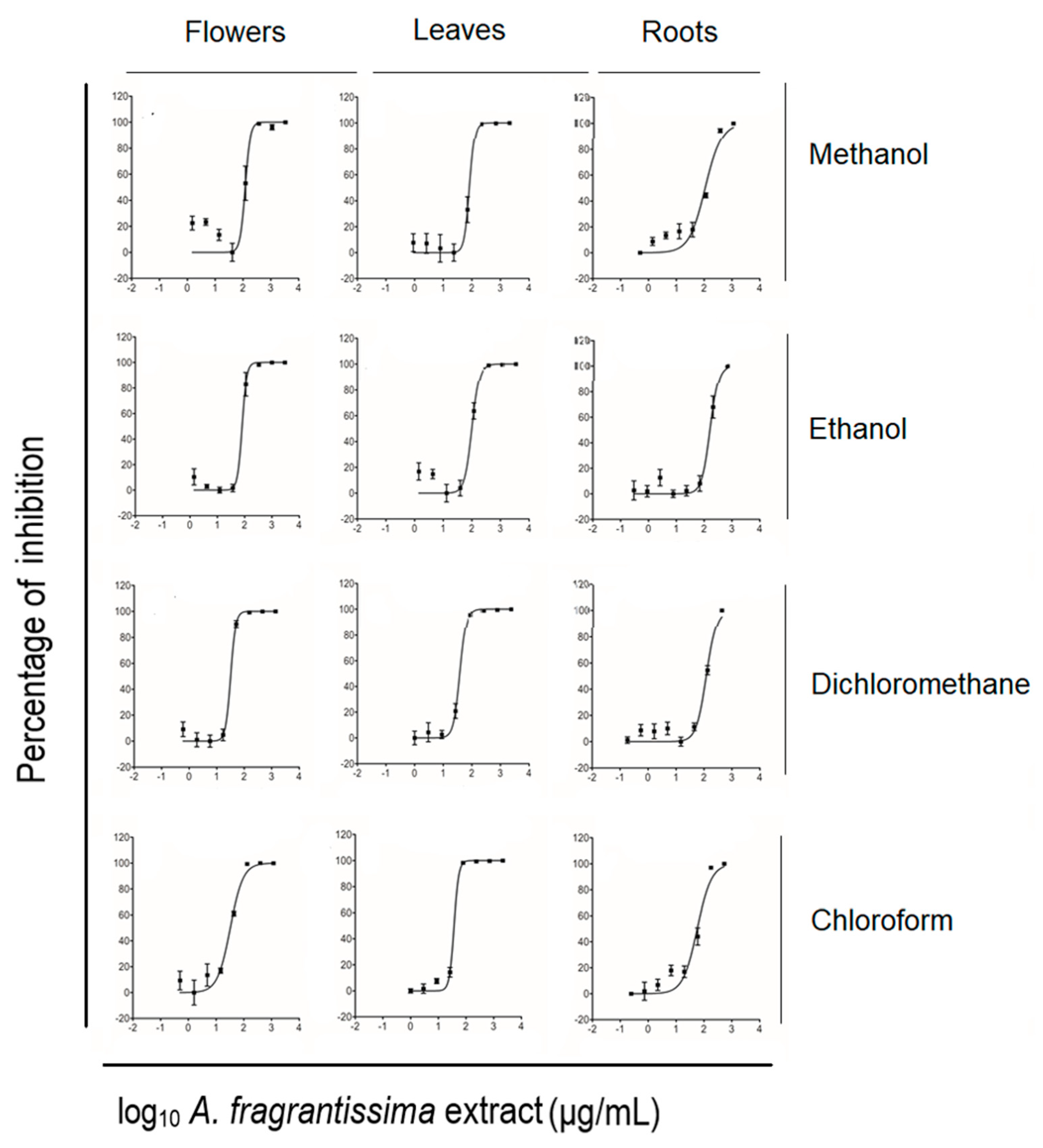
The measurement of half-maximal inhibitory concentrations (IC50) was performed using an ATP-based viability assay. The dichloromethane extract of the A. fragrantissima flowers displayed the lowest IC50 value followed by the chloroform extract. This dichloromethane extract was used for the remaining in vitro investigations for anticancer properties. Based on the ATP generated by living untreated and treated TNBC MDA-MB-231 cells, the IC50 value reflecting the anti-proliferative potential of each extract was determined. All the A. fragrantissima extracts significantly inhibited MDA-MB-231 cell viability in a dose-dependent manner (Figure 2). The mean IC50 values ranged from 32.4 to 161.7 µg/mL, including the dichloromethane and chloroform extracts derived from the flowers showing the lowest mean IC50 values, followed by the leaf-derived extracts, as seen in Table 1. The dichloromethane extract of the A. fragrantissima flower was shown to be the most potent leading to MDA-MB-231 cell growth inhibition and, thus, was selected for further experiments in this study.

2.2. Induction of Apoptosis in MDA-MB-231 Cells by A. fragrantissima Flower Dichloromethane Extract

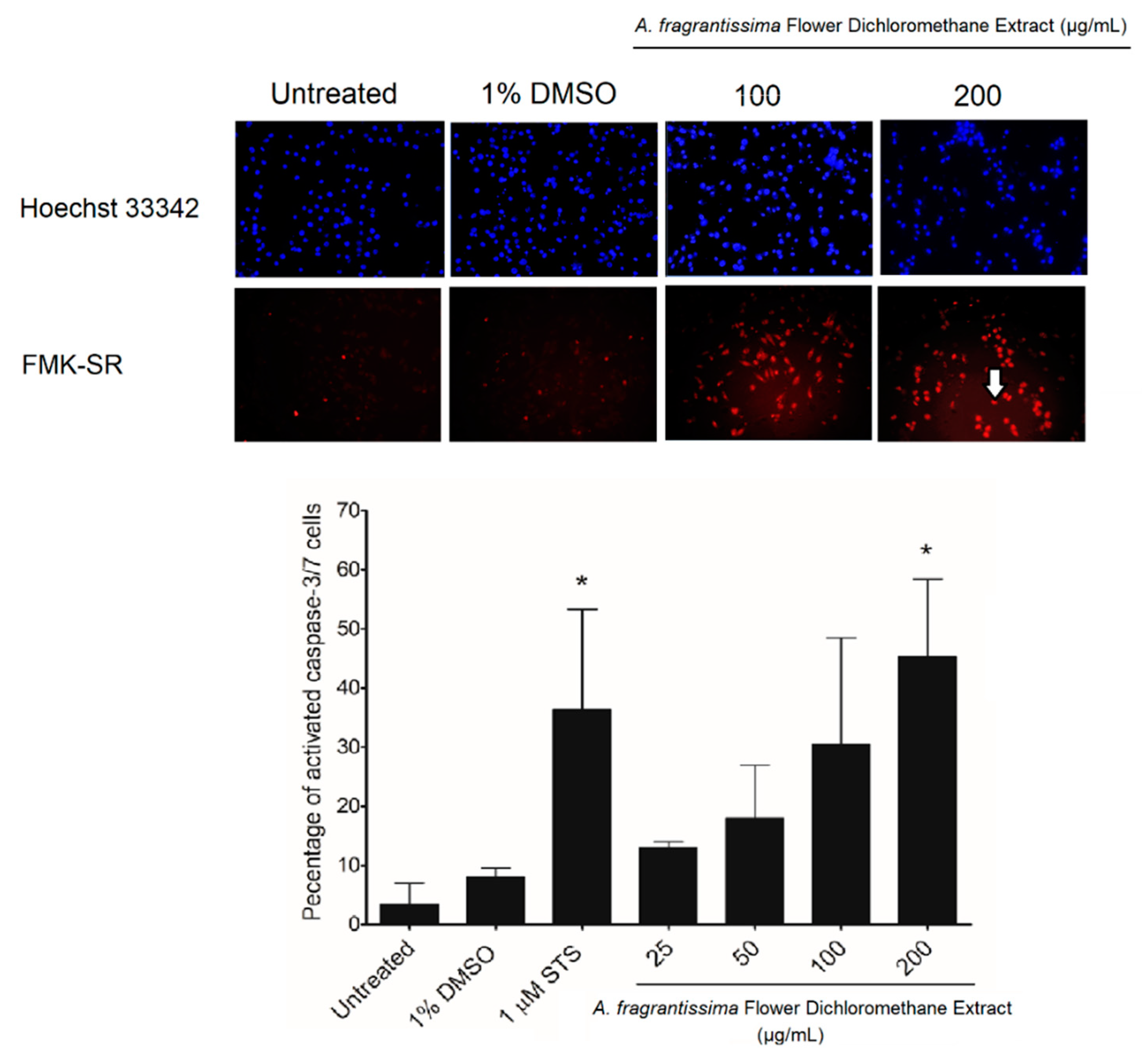
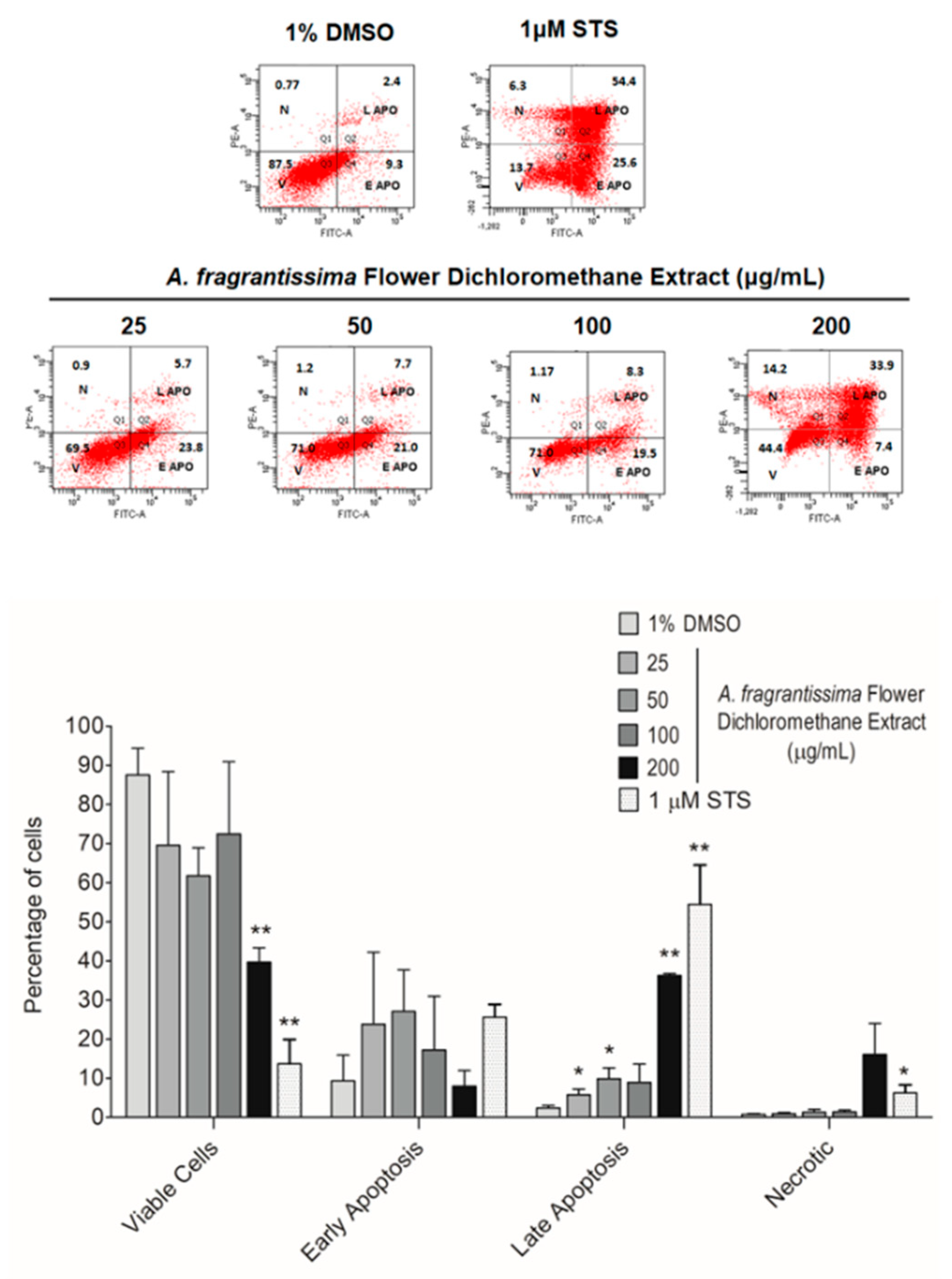
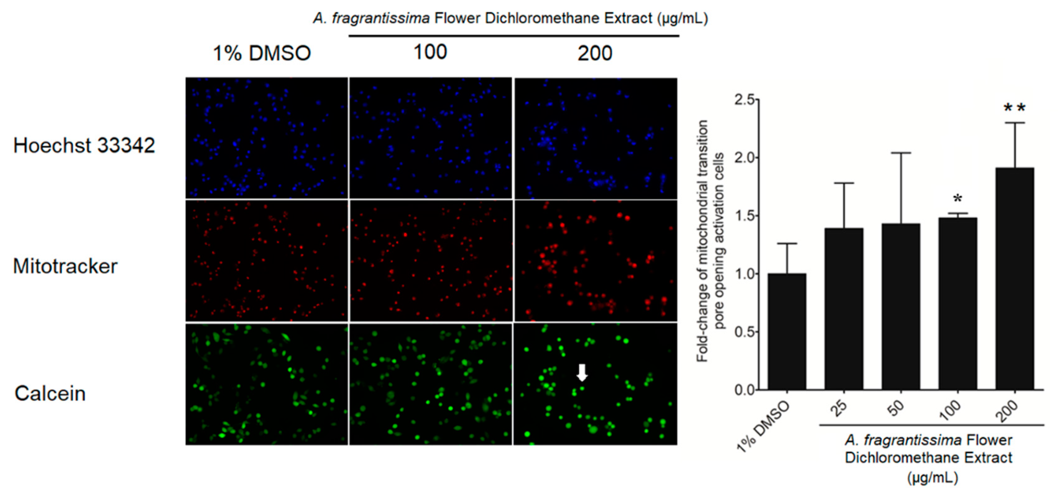
In vitro investigations were performed to determine if the inhibition of MDA-MB-231 cell growth was mediated via the caspase-dependent extrinsic apoptotic pathway. The treatment of MDA-MB-231 cells by the A. fragrantissima flower dichloromethane extract increased observed cell shrinkage in a dose-dependent manner, suggesting apoptosis as the mechanism of cell death (Figure 3). The caspase-dependent apoptosis was confirmed in A. fragrantissima flower dichloromethane extract-treated MDA-MB-231 cells presenting cleaved caspase-3/7; the active form indicated in red fluorescence and was determined under confocal fluorescence microscopy (Figure 4). The confocal images clearly display an increase in caspase-3/7 activity in the cells treated with the protein kinase inhibitor staurosporine (STS, a potent apoptosis inducer) and A. fragrantissima flower dichloromethane extract (25 to 200 µg/mL), in comparison with the untreated and DMSO-treated cells, the negative controls. The status of apoptosis was also determined using the FACS analysis based on the Annexin V/propidium iodide (PI) stain, which clearly showed early and late apoptotic status of A. fragrantissima flower dichloromethane extract-treated cells, in a dose-dependent manner, compared to DMSO-treated cells (Figure 5). The mitochondria potential membrane assay detected an increase in the number of apoptotic whole-cell green fluorescence indicating the involvement of the intrinsic apoptotic pathway in STS- and A. fragrantissima flower dichloromethane extract-treated cells, compared to the untreated and DMSO-treated cells (Figure 6).

2.3. Metabolite Identification Using LC-QTOF

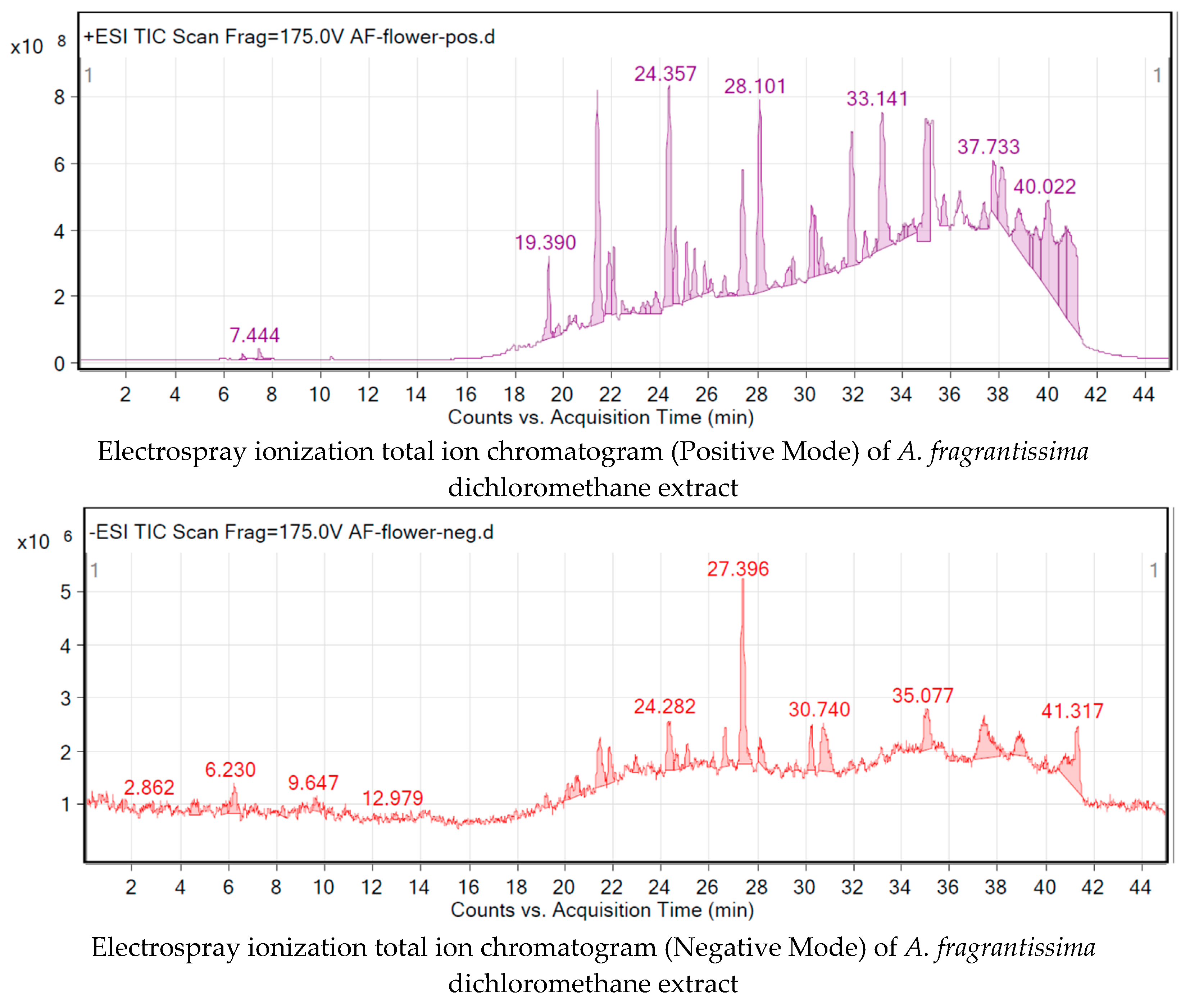
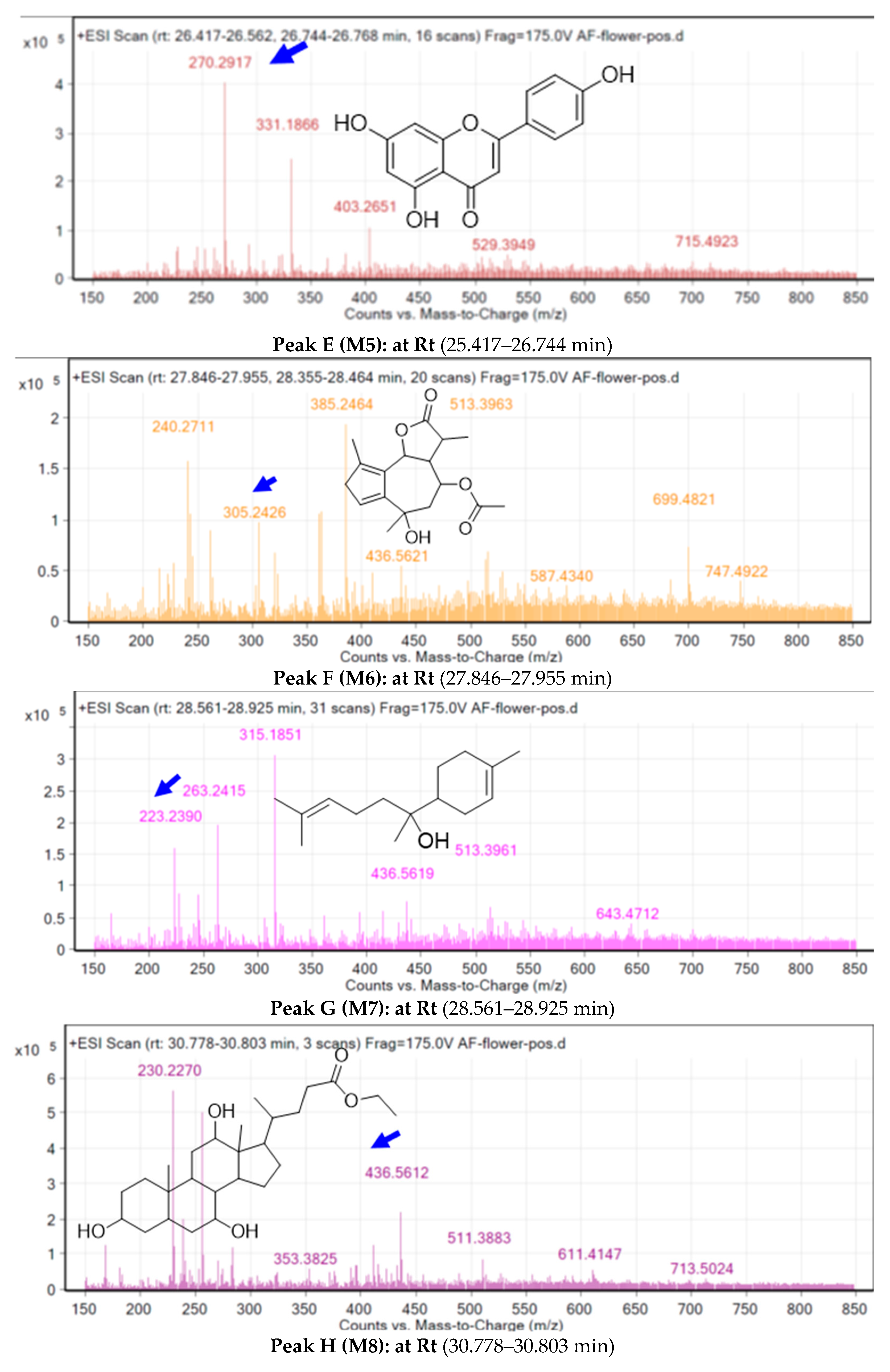
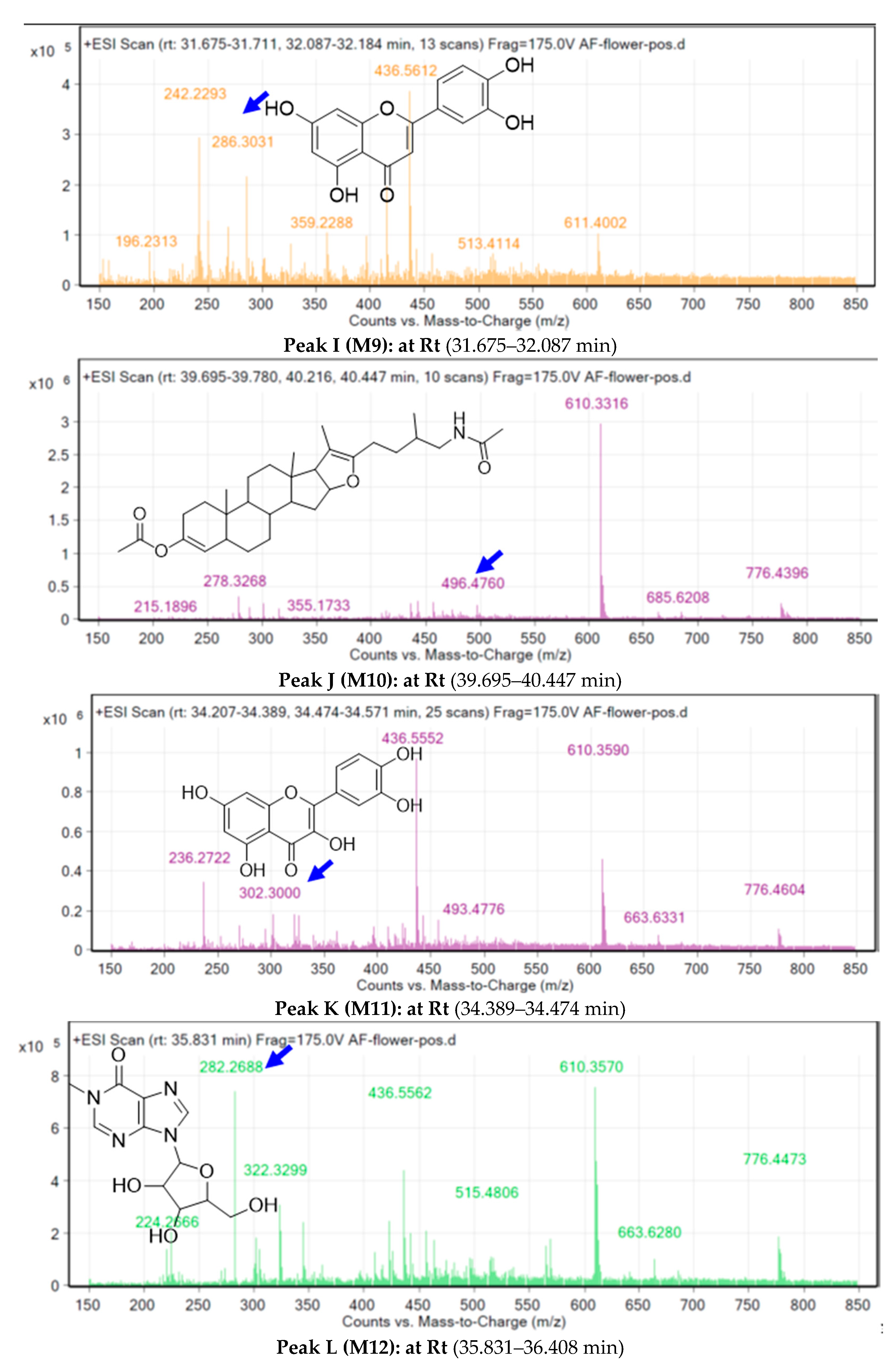
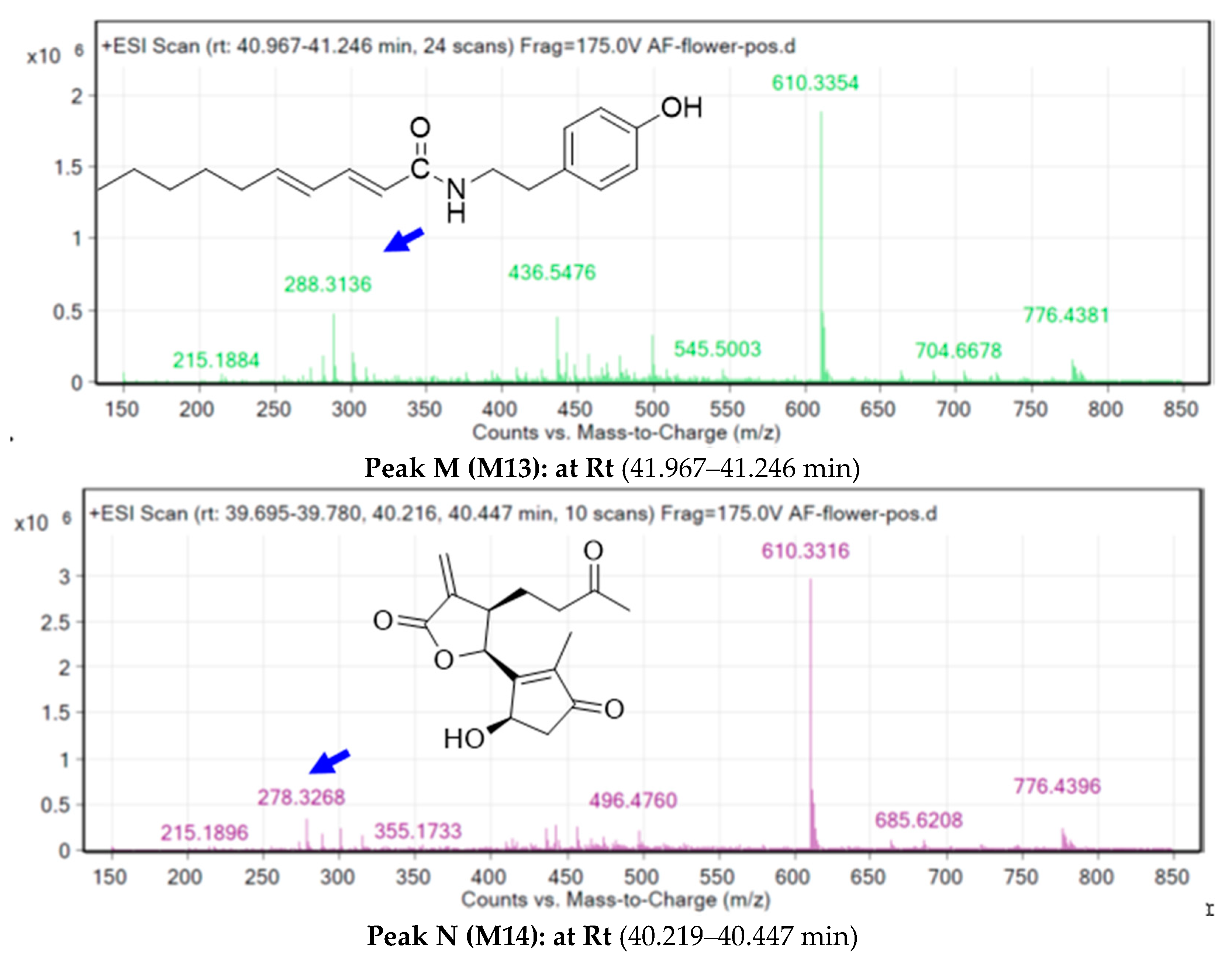
The crude extract of the A. fragrantissima flower dichloromethane was subjected to total ion current (TIC) spectra raw data and the data analysis program MassHunter (Agilent Technologies) qualitative and quantitative analysis software was used. After conducting a mass screening on the below spectrum, chemical features were extracted from the liquid chromatography–mass spectrometry (LC-MS) data using the Molecular Features Extraction (MFE) algorithm and the recursive analysis workflow (Figure 7). Features were extracted by screening the detected nodes at various retention times (Rt) per minute, with a minimum intensity of 6000 counts, and aligned with previously detected compounds considering adducts ([M + K]+, and [M − H]). Cosmosiin [21], Centaureidin [22], Apressin [22], Eupatilin [23], Lupeol [17], Oleic acid [24], Bisabolol, [22], Ethyl isoallocholate [25], Luteolin [26], Pseduosolasodine diacetate [25], Quercetin [27], Methylinosine [25], p-hydroxy-phenethylamine IV [28], and Tanaphillin, [29], Means m/z implies measured m/z.

2.4. PASS Online Anticancer Predictions

The anticancer activity of the identified metabolites in A. fragrantissima flower dichloromethane extract was predicted using the prediction of activity spectra for substances (PASS) online webserver. If the Pa value is greater than 0.3, it indicates a high probability that the metabolite will be active experimentally. As summarized in Table 2, all the 14 identified metabolites demonstrated high probabilities that were greater than 0.3 (M5, M7, M8, M10, M12, and M13), and some metabolites (M1M4, M6, M9, M11, and M14) were higher than 0.7 indicating a high probability that these metabolites would be active experimentally. Moreover, the inactivity scores determined for the metabolites were near to zero suggesting their promising anticancer activity.

2.5. Molecular Target Predictions

The possible molecular targets for the identified metabolites were predicted using the Molinspiration webserver. The webserver gives an activity prediction involving main five targets important for biological effects including the G protein-coupled receptor (GPCR), ion channel, kinase, nuclear receptor, protease, and enzyme. Our results showed that most of the metabolites demonstrated high bioavailability scores ranging from 0.13 to 0.81 as enzyme inhibitors (Table 3). Moreover, some of the metabolites exhibited high bioavailability scores as nuclear receptor ligands ranging from 0.01 to 0.81. These results suggest that the observed biological anticancer activity could involve several interactions and mechanisms.

2.6. Molecular Docking

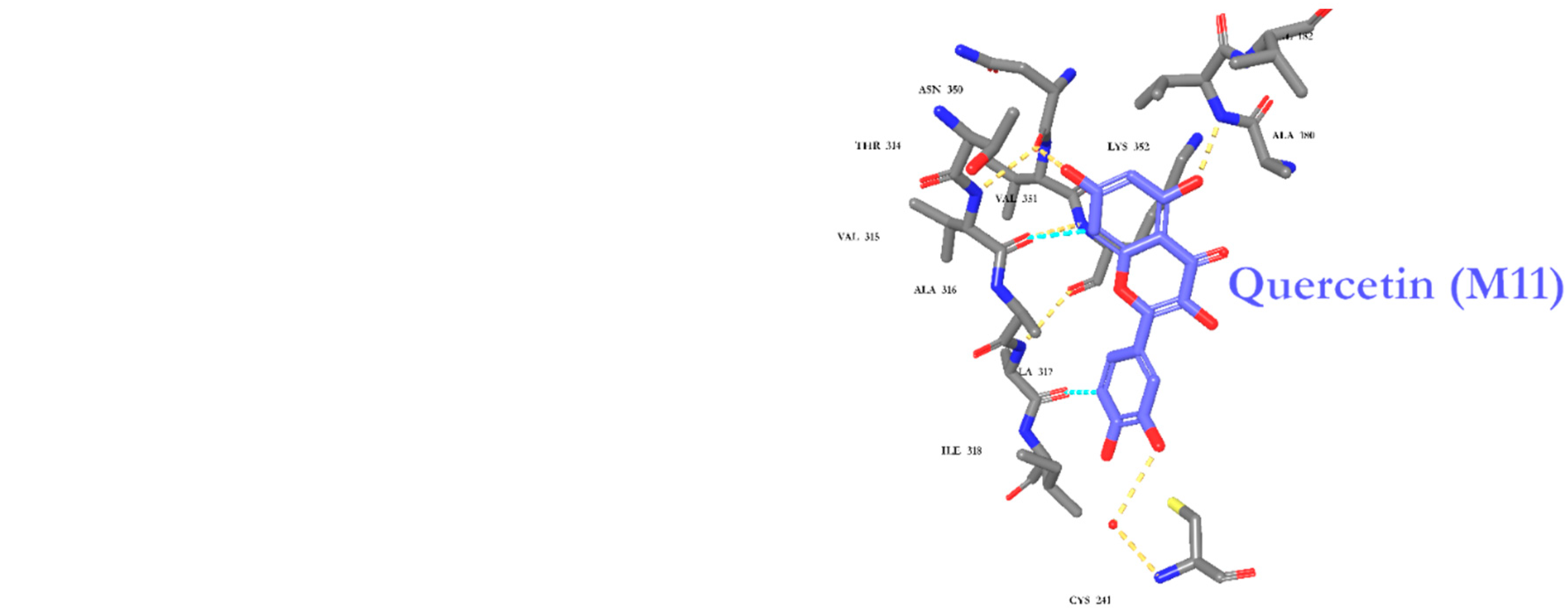
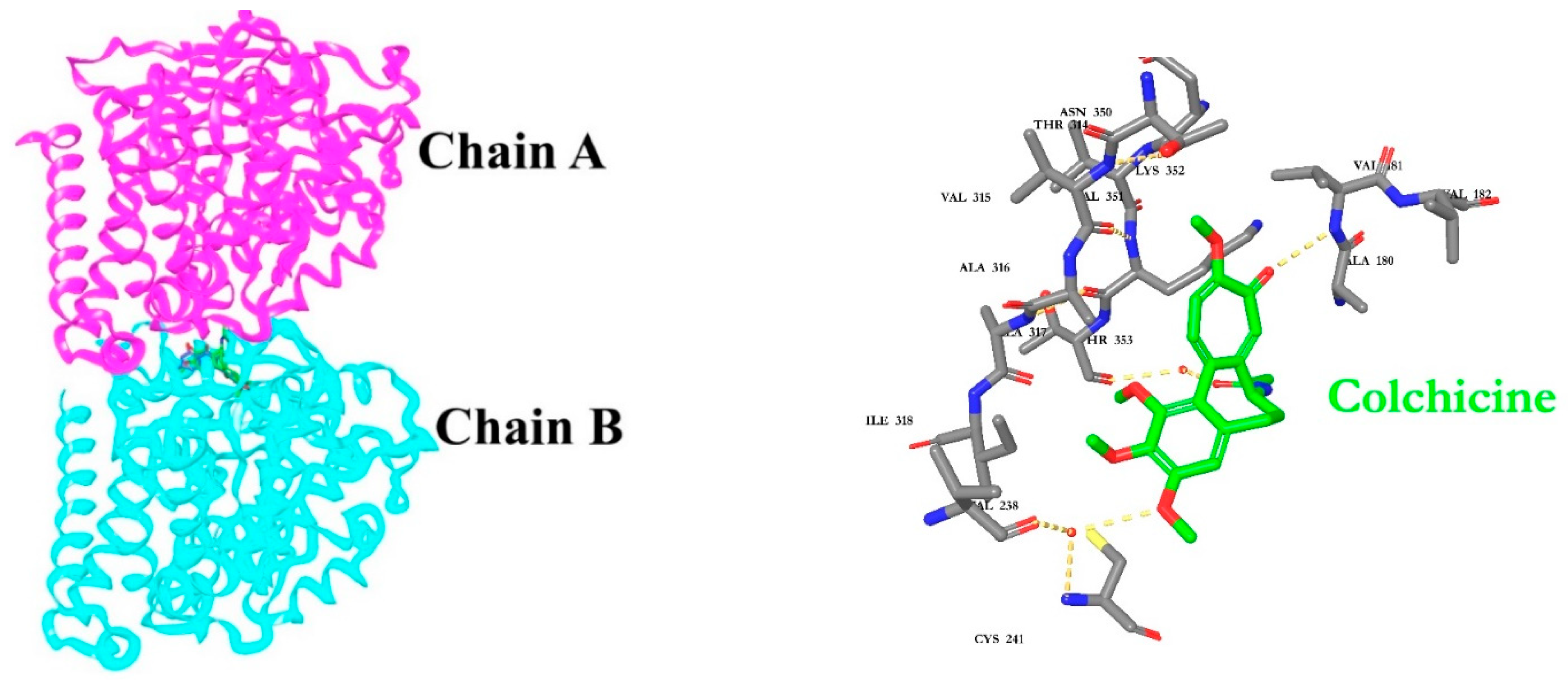
The fourteen identified metabolites from A. fragrantissima flower dichloromethane extract were docked into Tubulin [30,31,32] and Carbonic anhydrase (CA) IX enzyme [33,34], two main substrates reported to be inhibited by flavonoids, to investigate the molecular interactions using Maestro Schrödinger software. The crystal structures of Tubulin and CA IX enzyme were optimized and minimized, followed by grid generations for both binding pockets. For Tubulin docking, Colchicine (native ligand) was used as a positive control and the docking protocol was validated by docking the Colchicine into the binding site with a low root-mean-square deviation (RMSD) value relative to crystal structure. The docking scores for the fourteen identified metabolites range from −4.23 to −9.89 kcal/mol. As shown in Table 4, metabolite 11 (M11, Quercetin) demonstrated the highest docking score (−9.89 kcal/mol), followed by M1 (Cosmosiin, −9.59 kcal/mol) and M2 (Centaureidin, −9.16 kcal/mol). Moreover, M11 exhibited several interactions with the binding site including Cys241, Val181, and Asn350, and occupied a similar binding pose relative to Colchicine as illustrated in Figure 8. All the 2-D interaction maps for the metabolites with Tubulin are presented in Figure 8.
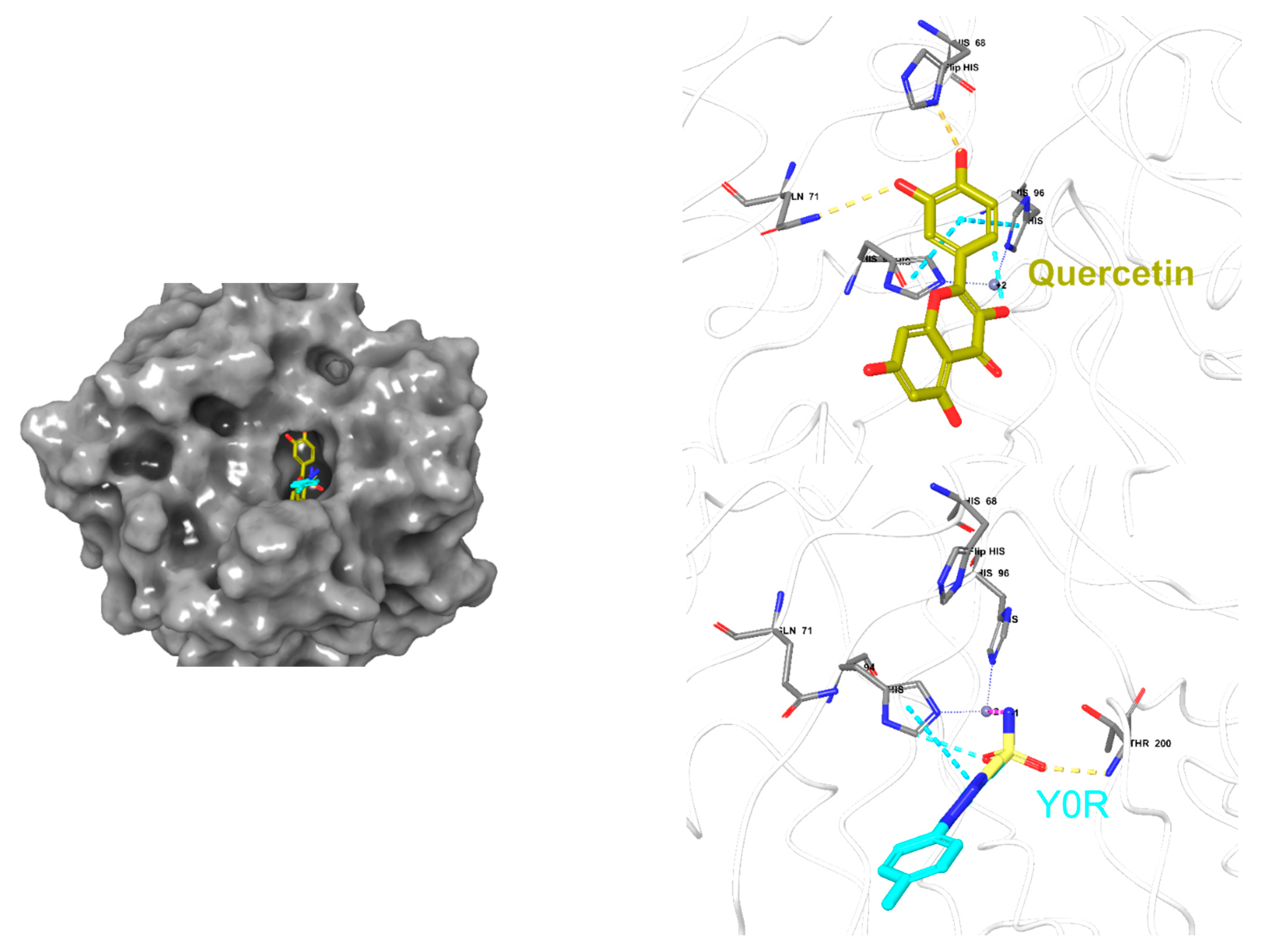
Moreover, the fourteen metabolites were also docked into the CA IX crystal structure and docking scores are summarized in Table 5. The docking scores ranged from −1.30 to −5.46 kcal/mol, and zinc coordination was maintained in the most docked metabolites. Interestingly, M11 demonstrated the highest docking score (−5.46 kcal/mol) followed by M4 (−5.25 kcal/mol) and M5 (−5.21 kcal/mol). Additionally, several hydrophilic and hydrophobic interactions were formed at the binding site as depicted in Figure 9. All the 2-D interaction maps for the metabolites with CA IX are depicted in Figure 9.

2.7. Absorption, Distribution, Metabolism, and Excretion (ADME) Predictions

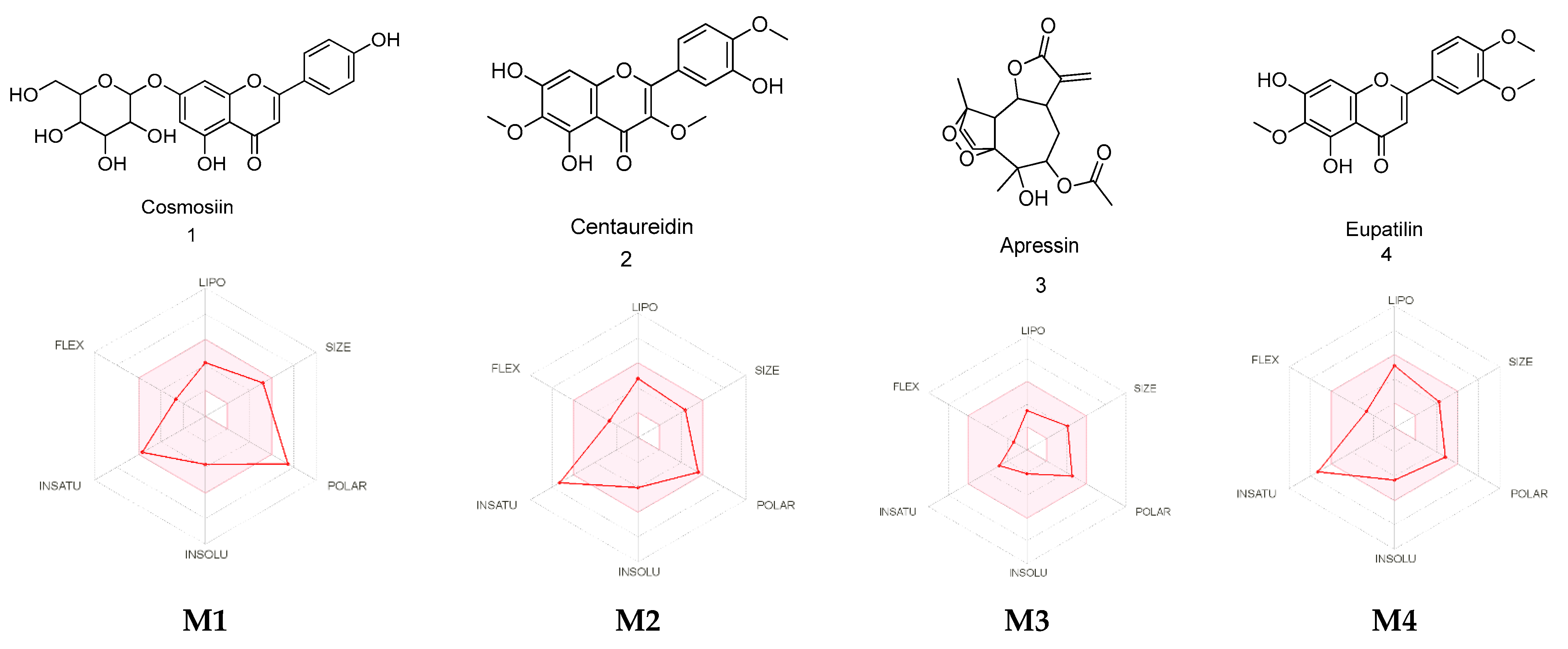
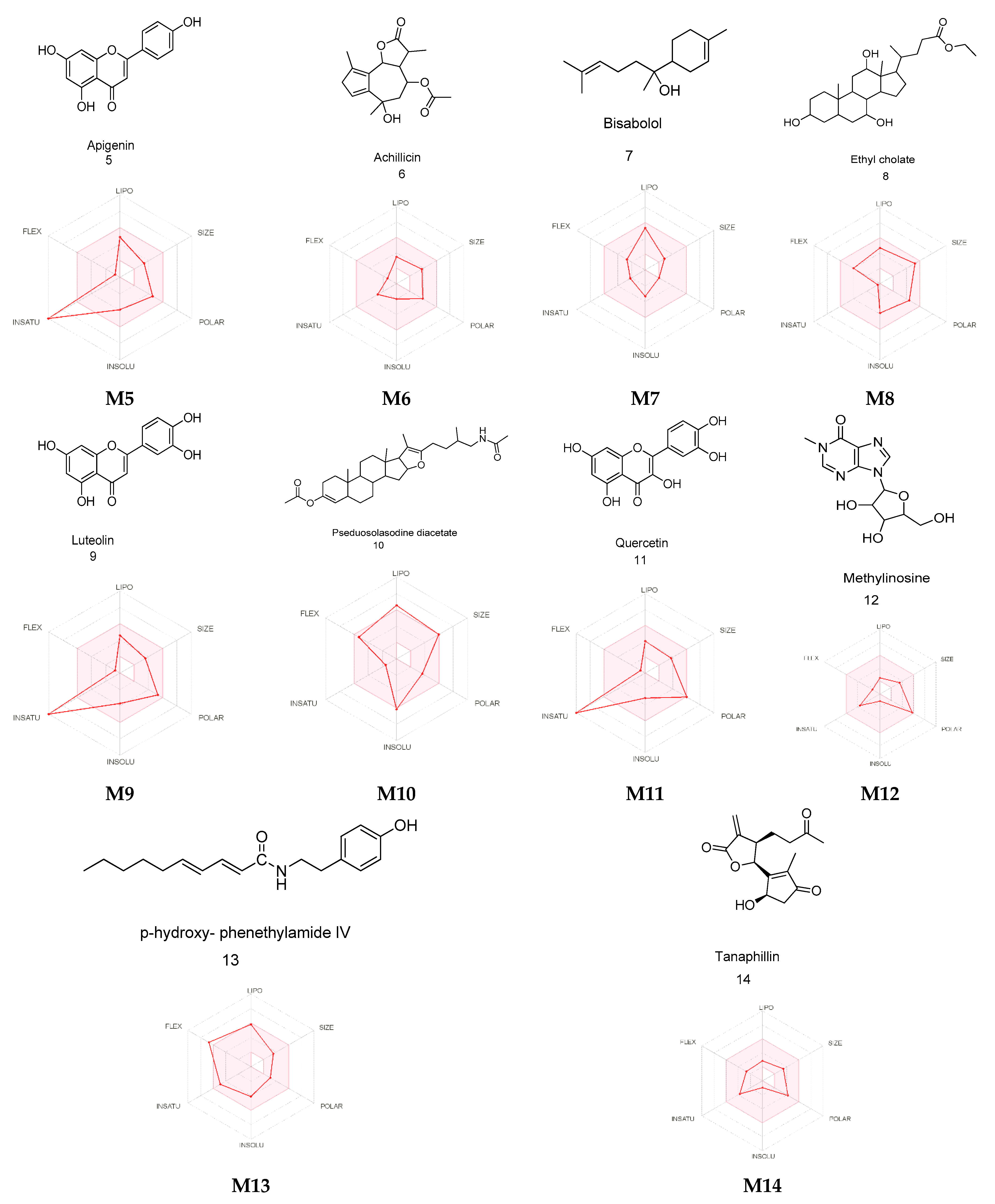
Several pharmaceutical properties important for drug discovery were calculated for the identified metabolites using the SwissADME web server and QikProp tool. These parameters include Molecular Weight (g/mol), Log Po/w (lipophilicity), Log S (solubility), blood–brain barrier (BBB) Permeability, and gastrointestinal (GI) Absorption. As shown in Table 6, all the metabolites exhibited acceptable pharmaceutical parameters (Figure 10) that were within Lipinski’s rule of five (ROF), except for metabolites M1 and M10 that violated the hydrogen bond donor/acceptor rule and molecular weight, respectively. Moreover, most of the metabolites were predicted to act peripherally with no central nervous system effects, except for M6, M7, and M13. Additionally, the majority of the identified metabolites were predicted to possess a high GI absorption, except for M1 and M12.

2.8. Cytochrome P450 (CYP) Enzyme Inhibition Profiling

The effects of the identified metabolites of A. fragrantissima flower dichloromethane extract on CYP isoenzymes were reported in Table 7 using the SwissADME webserver. Six out of fourteen of the evaluated metabolites were predicted to inhibit CYP1A2 and CYP3A4, while five metabolites were predicted to likely inhibit CYP2D6 and CYP2C9, and only one metabolite was predicted to inhibit CYP2C19, as summarized in Table 7.

2.9. Organ and Endpoint Toxicity Predictions

The toxicity of the identified metabolites was assessed using the ProTox-II webserver against hepatotoxicity, carcinogenicity, immunotoxicity, mutagenicity, and cytotoxicity. As summarized in Table 8, all the metabolites were inactive as hepatotoxic and cytotoxic. While six (M2M4, M6, M8, M10, and M13) out of fourteen were predicted to be immunotoxic, fewer were predicted to exhibit mutagenicity (M1, M3, M9, and M11) and carcinogenicity (M6, M9, and M11).

3. Discussions

Natural products, including bioactive molecules from plant, animal, fungal, and microbial origins, remain a vital source for therapeutic drug discovery. Since the last century, approximately more than fifty percent of pharmaceutical drugs for humans have been derived from natural sources [35,36]. The importance of natural products as a source of cancer treatment is well recognized and documented. It is estimated that forty-nine percent of approved small molecules for cancer treatment are derived from natural products in the time period between the 1940s and 2014 [37]. Endemic species of plants in the Middle Eastern region are among the least studied species and could potentially reveal novel lead compound(s). The medicinal plant species belonging to the Achillea genus found in the Middle East region are popular treatments for several illnesses, including respiratory diseases, fever, gastrointestinal disturbances, headache, fatigue, and diabetes [17,18,20]. This study explored one local species of A. fragrantissima for new therapies against TNBC using a series of in vitro experiments to determine its potential anti-proliferative, cytotoxic, and pro-apoptotic properties.
The investigation of this current study was initiated by chemical fingerprinting for the three plant parts of A. fragrantissima (i.e., flowers, leaves, and roots) using methanol, ethanol, dichloromethane, and chloroform. The solvents were chosen to represent the polar (i.e., methanol and ethanol), moderately polar (i.e., dichloromethane), and non-polar (i.e., chloroform) extractions that were reflected in the chromatograms. The chromatograms of methanol and ethanol extracts displayed more polar phytochemical constituents, which eluted first due to the separation using the reverse-phase C18 column while the least polar constituents appeared more abundant and eluted last in the chromatograms of dichloromethane and chloroform extracts. The numerous and abundant phytochemical constituents in the DMSO-reconstituted extracts also indicated the efficiency of the extraction method using the high-power probe sonicator, and that the abundant phytochemical constituents were retained after reconstitution with DMSO solvent. The rich phytochemical compositions observed in the chromatograms were consistent with studies that isolated and characterized numerous phytochemical constituents of A. fragrantissima plant’s parts using various techniques and identified several families of phytochemicals, including phenolic acids, flavonoids, lignans, terpenic lactones, and alkamides [18].
The anti-proliferative effects of A. fragrantissima plant parts-derived extracts on TNBC MDA-MB-231 cells were determined using the CellTiter-Glo™ assay. The bioassay determined the viability of cell populations by measuring the amount of adenosine triphosphate (ATP) generated by metabolically active cells. It is known that the bioassay is among the most sensitive cell viability bioassay in comparison to tetrazolium reduction, resazurin reduction, and protease activity bioassays [38]. The performance of the bioassay in this study was shown to be sensitive enough to establish the concentration–inhibition curves efficiently despite the relatively low cell seeding count. Furthermore, the variation in the inhibition concentrations values did reflect the chromatograms of the polar extracts displaying higher IC50 values in comparison to the least polar extracts, suggesting that the least bioactive polar compounds contributed to the weight of the extracts, resulting in higher inhibition values. The inhibition concentrations were used as a guideline to select the most potent anti-proliferative extract. The A. fragrantissima flower dichloromethane extract presenting the lowest mean IC50 value was chosen for further cell death-related molecular mechanism investigation.
Furthermore, additional confirmatory experiments were required to confirm apoptosis as the main mechanism of cell death [39]. Further experiments in this study include the observation of apoptosis-related cell morphological changes, the detection of caspase-3/7 activation, and abnormal exposure of phosphatidylserine on the outer cell membrane in treated MDA-MB-231 cells. The treatment of MDA-MB-231 cells with A. fragrantissima flower dichloromethane extract clearly led to a gradual increase in cell shrinkage, in a dose-dependent manner, with the highest concentrations (i.e., 100 and 200 µg/mL) displaying similar observations as the apoptosis inducer, STS, used as a positive control. For caspase-3/7 activation, the detection was based on the fluorescent inhibitor of caspases (FLICA) methodology. The method was based on the covalent binding of fluoromethyl ketone (FMK) moiety to the center of the activated caspase by a recognition sequence for capase-3/7 (i.e., aspartic acid–glutamic acid–valine–aspartic acid) [40]. The FLICA method allowed direct visualization of activated caspase-3/7 in treated MDA-MB-231 cells as red-fluorescence cells while cells without activated caspases display the nucleus only after use of the Hoechst 33342 (blue) staining. The A. fragrantissima flower dichloromethane extract clearly increased the number of activated caspase-3/7 cells to levels comparable to the protein kinase inhibitor, STS. The FACS analysis using the fluorescein isothiocyanate (FITC)-labeled Annexin V/phycoerythrin (PE)-labeled PI dual staining allowed the detection of viable, early apoptosis, late apoptosis, and dead/necrotic cell populations in MDA-MB-231 cells treated with STS or different A. fragrantissima extract concentrations. The experiment confirmed the occurrence of the early and late apoptotic status by Annexin V binding to the exposed phosphatidylserine on the outer leaf of the cell membrane, which is a hallmark characterization of programmed cell death [41]. Furthermore, the PI binding to nucleic acids is used to detect cells with lost cell membrane integrity as seen in late apoptotic and dead/necrotic cell populations, which is also considered an irreversible cell death status [42].
After confirming that caspase-dependent apoptosis as a cell death mechanism occurred in the MDA-MB-231 cells exposed to A. fragrantissima flower dichloromethane extract, a deeper investigation was conducted to determine the involvement of the intrinsic apoptosis pathway using the mitochondrial permeability transition pore activation assay. Indeed, apoptosis occurs in two pathways referred to as intrinsic and extrinsic. The intrinsic apoptosis pathway involves mitochondrial permeability transmembrane pore opening forming the apoptosome leading to caspase-9 activation, then subsequently to executioner caspases-3/7 activation [43]. The bioassay directly measured the mitochondrial permeability transition pore opening activity using acetoxymethyl ester of calcein dye, which accumulated in the cytosolic compartments and liberated into a highly-fluorescent calcein. In pro-apoptotic conditions, the cells with activated mitochondrial permeability transition pore opening appeared brightly green due to the redistribution of calcein from the cytosol into the mitochondria. The addition of the quencher, CoCl2, removed the remaining cytosolic fluorescence, while the mitochondrial fluorescence was maintained visible under the confocal microscopy. The treatment of MDA-MB-231 cells with A. fragrantissima flower dichloromethane extract increased the number of cells displaying activated mitochondrial permeability transition pore opening in a dose-dependent manner, confirming the involvement of the mitochondria-dependent intrinsic apoptosis pathway. Interestingly, our findings characterizing the main hallmarks of apoptosis, including caspase-3/7 detection, apoptosis status determination by FACS, and mitochondrial membrane transition pore opening activation, exhibited a similar pattern. The results clearly displayed an induction of apoptosis in TNBC MDA-MB-231 cells, which slightly and gradually increased in response to the 25 and 50 µg/mL treatment to reach the highest pro-apoptotic changes in response to 100 and 200 µg/mL of A. fragrantissima flower dichloromethane extract.
Numerous studies have reported the anticancer potencies of many species belonging to the Achillea genus, including the widely-consumed native North American species of A. millefolium [44]. Only a few studies reported A. fragrantissima crude extracts with anticancer properties using cancer cell lines. For instance, Sathiyamoorthy et al. tested A. fragrantissima arial part aqueous extract on sensitive and resistant melanoma cell lines and reported a significant cytotoxicity [45]. Alenad et al. tested A. fragrantissima leaves methanolic extract against K562 (myeloid leukemia), Jurkat (T-cell leukemia), and HepG2 (hepatocarcinoma) cells and reported drastic morphological changes and viability reduction in K562 cells [46]. Binbreak et al. treated HepG2, A549 (lung carcinoma), HCT-116 (colorectal carcinoma), and MCF-7 (hormone-dependent breast cancer) cells with methanolic extract of A. fragrantissima leaves and reported cytotoxic bioactivity of the extract against A549 cells with induction of apoptosis, including activation of caspase-3, increased p53 and Bax expression, and down-regulation of Bcl-2 expression [47]. Several studies reported the isolation of A. fragrantissima phytochemicals and performed further experiments for the evaluation of their anticancer properties using several cell lines. For example, Awad et al. isolated several phenolic compounds, including piceol, eupatilin 7-methyl ether, chrysosplenol D, cirsiliol, and cirsimaritin, from A. fragrantissima leaves methanolic extract tested on MCF-7, HepG2, A549, HeLa (cervix adenocarcinoma), and PC3 (prostatic adenocarcinoma) cell lines and presented IC50 values ranging from 3.2 to 28.3 µg/mL [48]. To the best of our knowledge, the present study is the first to determine the in vitro cytotoxicity of different A. fragrantissima plant parts (i.e., flowers, leaves, and roots) crude extracts against TNBC using MDA-MB-231 cell line and to investigate the pro-apoptotic effect of A. fragrantissima flower dichloromethane extract.
Our experimental observations suggested that A. fragrantissima possesses promising anticancer activity against TNBC. After chemical analysis of the A. fragrantissima flower dichloromethane extract, we attempted to correlate the observed experimental findings with the identified metabolites. Several computer-aided drug discovery (CADD) approaches were utilized to provide accurate and fast predictions of several biological and pharmaceutical properties required for the further development of novel drug entities [49,50]. Therefore, PASS online webserver was utilized to validate the anticancer activity of A. fragrantissima identified metabolites. Interestingly, all fourteen identified metabolites demonstrated moderate to high anticancer activity confirming the observed experimental results. Moreover, the most probable molecular targets for the identified metabolites were predicted and the analysis suggested that certain enzymes and several other targets could contribute to the observed activity.
An additional molecular docking study was performed to assess the possibility of the identified metabolites including flavonoids, to bind and inhibit Tubulin [30,31,32] and CA IX enzyme [33,34]. The selection of these two targets was based on the previously reported in vitro studies that confirmed the inhibition by flavonoids, particularly, Quercetin. It is worth noting that the CA IX enzyme is highly expressed in the TNBC MDA-MB-231 cell line, which is reported to contribute to the aggressiveness and dysfunction of this type of cancer [51,52,53]. Our docking results were consistent with the previously reported studies in which Metabolite 11 (M11, Quercetin) possesses the highest docking score among the other metabolites against the two targets, suggesting the validity of the protein selection. Moreover, it has been demonstrated by several studies that inhibition of Tubulin polymerization and CA IX enzyme serve as a new and novel mechanism for promising anticancer activity [54,55,56,57,58,59,60,61,62].
One of the main aspects of drug design and development is to assess and evaluate the pharmacokinetics or drug-likeness properties relative to Lipinski’s rule of five required for orally active drugs [63]. Twelve out of fourteen metabolites passed the rules indicating a good pharmaceutical profile. Additionally, one of the main mechanisms for drug–drug interactions has resulted from the inhibition of one or several CYP isoenzymes involved in the metabolism of drugs. Therefore, the inhibition profile for the fourteen metabolites was evaluated and our results demonstrated that not all the metabolites inhibited the CYP isoenzymes, except for a few that were predicted to inhibit CYP1A2, CYP3A4, CYP2D6, and CYP2C9. The toxicity of the identified metabolites demonstrated immunotoxicity for six metabolites, while none were predicted to exhibit hepatotoxicity and cytotoxicity.

4. Materials and Methods

4.1. Chemicals and Reagents

Dulbecco’s Modified Eagle Medium (DMEM) plus GlutaMax-1 (4.5 g/L D-Glucose, 25 mM HEPES, Pyruvate), fetal bovine serum (FBS), TrypLE™ Express, and Dulbecco’s phosphate-buffered saline (PBS) were provided by Gibco® (Waltham, MA, USA). Invitrogen NP-40 cell lysis buffer was purchased from Thermo Fisher Scientific (Carlsbad, CA, USA). Staurosporine (STS) was procured from Santa Cruz Biotechnology (Dallas, TX, USA). Methanol and ethanol were obtained from Honeywell Riedel-de Haen (Seelze, Germany) and Merck (Kenilworth, NJ, USA). Formic acid, dichloromethane, and chloroform were obtained from Sigma-Aldrich (St. Louis, MO, USA). Dimethyl sulfoxide (DMSO) was procured from Calbiochem (San Diego, CA, USA). The solvents are chromatography-grade or equivalent. Saudi Industrial Gas (Dammam, Saudi Arabia) provided purified carbon dioxide (CO2) gas. Ultra-pure water was produced using a Millipore (Billerica, MA, USA) system with a resistivity reading of 18.2 MΩ·cm at 25 °C.

4.2. Collection and Authentication of A. fragrantissima

The traditional herbal medicine of A. fragrantissima flowers, leaves, and roots was wildcrafted at Albatin (in Arabic, الباطن) dam area near Jalajil (in Arabic, جلاجل) city, Sudair (in Arabic, سدير) region, Kingdom of Saudi Arabia (coordinates; 25.6834° N, 45.4592° E), and authenticated by a Professor of Botany and Taxonomy. The plant parts were rinsed with filtered water and left to dry under a stream of slightly warm dry air. Once completely dried, the plant parts were fine powdered using an electric-motor grinder and kept in dark at room temperature until extraction. A voucher sample was deposited at King Saud University (Herbium, Department of Botany and Microbiology) with accession number 24575 dated on 17 June 2021 (see Supplementary Figure S1).

4.3. Extraction of A. fragrantissima Plant Parts and Endotoxin Removal

Approximately 500 mg of each dried A. fragrantissima flowers, leaves, and roots were extracted separately using 10 mL of high-purity methanol, ethanol, dichloromethane, and chloroform under high-power sonication using a Sonics (Newton, CT, USA) Vibra-Cell™ Ultrasonic Liquid Processor (Model GEX-130 probe-sonicator) for 30 min. The sonicated extract was filtered using a Sartorius stedim biotech (Göttingen, Germany) quantitative ashless paper filter under gravity-flow and dried in an incubator set at 40 °C. The remaining dried pellet residue was weighted and reconstituted with 100 to 300 µL of DMSO by vortex until completely dissolved. The reconstituted extract was stored at cool temperature in the dark until use. The averaged extraction yield percentage for the flowers-derived methanol, ethanol, dichloromethane, and chloroform solvent extracts were 30.3 (±4.2), 28.4 (±1.8), 13.4 (±0.6), and 22.5 (±1.3), respectively, the leaves-derived extracts were 34.3 (±5.1), 29.9 (±2.3), 18.9 (±4.0), and 30.2 (±2.1), respectively, and the roots-derived extracts were 9.5 (±1.1), 5.5 (±1.1), 2.4 (±0.2), and 7.7 (±0.9), respectively. The levels of endotoxins in the A. fragrantissima sonicated aqueous extracts were determined using the Thermo Scientific™ Pierce™ Chromogenic Endotoxin Quant kit as per the manufacturer’s instruction and as described in our previous publication [64].

4.4. Chromatographic Fingerprinting of A. fragrantissima Extracts

The fingerprinting of A. fragrantissima extracts was performed using our previously published method with modifications [65]. The major components of A. fragrantissima were separated using an Agilent (Agilent Technologies Inc., Santa Clara, CA) 1260 infinity HPLC-UVD and a C-18 Kinetex® (250 × 4.6 mm, 5 µm particle size) column (Phenomenex Inc., Torrance, CA, USA). The multi-wave UVD was set at wavelengths ranging from 225 to 375 nm at 25 nm segments without reference. The gradient programming of methanol and ultra-pure water was as follows: 5 to 100% methanol, (0 to 35 min), 100% methanol, (35 to 40 min), and 100 to 5% methanol, (40 to 45 min) at flow rate of 1 mL/min. The injection volume was 5 µL at ambient temperature and the total run time was 45 min.

4.5. TNBC MDA-MB-231 Cell Culture and Treatment

The TNBC MDA-MB-231 (catalog number, HTB-26) cell line was provided by American Type Culture Collection Inc. (Manassas, VA, USA). The cells were cultured in complete DMEM Glutamax-I medium containing 10% heat-inactivated FBS, 1% penicillin G (100 IU/mL), and streptomycin (100 µg/mL) solution. The incubation was set at 37 °C in 5% CO2 and 95% relative humidity. The culture medium was changed every two to three days and sub-cultured when the cell population density reached 70 to 80% confluency. The cells were exposed to various concentrations of A. fragrantissima extracts and the appropriate controls; including 1% DMSO (negative control) and 1 µM STS (potent apoptosis inducer) were used.

4.6. Determination of MDA-MB-231 Cell Viability Using the CellTiter-Glo™ Assay

The cell viability was determined using Promega (Madison, WI) CellTiter-Glo™ assay according to the manufacturer’s instructions as described in [66]. Briefly, the MDA-MB-231 cells (400/cm2) were seeded in opaque white bottom 96-well plates (Costar, Thermo Fisher Scientific, Waltham, MA, USA). The next day, the untreated cells and the cells treated with various concentrations (0.05–2000 µg dry extract weight/mL) of each solvent extract were incubated for 48 h. Then, the plate was left at room temperature for 30 min and 100 µL of CellTiter-Glo™ reagent were added to each well and mixed on a shaker for 2 min. Luminescence was read using the Perkin Elmer® (Waltham, MA, USA) EnVision® multilabel plate reader.

4.7. Evaluation of the Hallmarks of Apoptosis

The shrinkage of cells was observed using a ZEISS inverted phase-contrast microscope (Peine, Germany). The activity of pro-apoptotic executioner caspase-3/7 and of the mitochondrial permeability transition pore opening in the untreated and treated cells were assessed using Image-iT™ LIVE Red caspase-3/7 detection kit and Image-IT™ LIVE Mitochondrial Transition Pore Assay Kit (Invitrogen, Thermo Fisher Scientific, Waltham, MA, USA), respectively, according to the manufacturer’s instructions and as previously described in [67]. The cells with activated caspase-3/7 (red fluorescence) and apoptotic cells (whole green fluorescence) with mitochondrial outer membrane permeabilization were observed under the confocal microscopy. The abnormal phosphatidylserine exposure was determined using the Becton Dickinson (BD) Annexin V apoptosis detection kit (Becton Dickinson, San Jose, CA, USA) as described in [68]. The apoptotic status of 10,000 cells based on the dual staining of Annexin V conjugated to FITC and PI conjugated to PE was analyzed using BD FACSDiva™ 6.0 software on the BD FACSCanto™ II flow cytometer.

4.8. Metabolite Identification Using ESI-LC-QTOF

The method of separation and identification of A. fragrantissima flower metabolites in dichloromethane extract was performed using an Agilent (Santa Clara, CA, USA) 1260 Infinity HPLC system coupled to Agilent 6530 quadrupole time-of-flight (Q-TOF) as described earlier in [69]. Briefly, the separation was performed using an Agilent SB-C18 column (4.6 mm × 150 mm, 1.8 μm) with the following elution gradient; 0 to 2 min, 5% B; 2 to 17 min, 5 to 100% B; 17 to 21 min, 95% B; 21 to 25 min, 5% B, using mobile-phase A (0.1% formic acid in water) and mobile-phase B (0.1% formic acid in methanol). The flow rate was set at 250 μL/min and injection volume was 10 μL. The scanning range was set at 50–800 (m/z), gas temperature at 300 °C gas flow of 8 L/min, nebulizer pressure of 35 psi, sheath gas temperature at 350 °C, and sheath gas flow rate of 11 L/min. The data were generated by the Agilent MassHunter (version B. 06.00) qualitative analysis software.

4.9. Anticancer Activity Predictions

The activity spectra for substances (PASS) online service provides online predictions of a variety of biological activities including anticancer predictions (http://www.way2drug.com/passonline/info.php, accessed on 25 April 2020). The Simplified Molecular Input Line Entry System (SMILES) of the chemical structures of the identified metabolites was utilized to generate the predictions using a database containing 250,000 biological activities. The generated results are represented as Pa (probability of active molecule) and Pi (probability of inactive molecule), ranging from 0.00 to 1.00. The higher Pa value for a certain biological activity indicates the molecule is predicted to be highly active experimentally Pa > 0.7), while Pa ranging from 0.5 to 0.7 suggests a moderate probability for experimental activity, and Pa lower than 0.5 indicates negligible experimental activity.

4.10. Molecular Target Predictions

The biological targets for the identified metabolites were predicted using the Cheminformatics tool Molinspiration webserver (https://www.molinspiration.com/, accessed on 25 April 2020). Each metabolite was evaluated as GPCR Ligand, Ion Channel modulator, Kinase Inhibitor, Nuclear Receptor Ligand, Protease Inhibitor, and Enzyme Inhibitor. The bioactivity scores were classified into three main classifications, including active (score above 0.00), moderately active (−0.50 to 0.00), and inactive (score below −0.50).

4.11. Molecular Docking

The molecular docking study was performed using Maestro Schrödinger software (Release 2022-1). The crystal structures of the Tubulin-Colchicine complex (Protein Data Bank identifier, PDB ID 4O2B) and human CA IX in complex with 5-[1-(4-Methylphenyl)-1H-1,2,3-triazol-4yl]thiophene-2-sulfonamide (PDB ID Y0R) were downloaded from the protein data bank (https://www.rcsb.org/, accessed on 25 April 2020). The proteins were prepared using the protein preparation wizard followed by receptor grid generation using the Glide tool. The 2D structures of the identified metabolites were converted into 3D and minimized using the LigPrep tool. All the prepared metabolites were docked using the Glide tool and the 2-D interaction map with docking scores was used to rank the docked metabolites into the two targets.

4.12. ADME Properties Predictions

The online accessible webserver SwissADME (http://www.swissadme.ch/, accessed on 25 April 2020) and commercial QikProp (Schrödinger software, Release 2022–1) tools were utilized to predict the ADME properties for the identified metabolites. Several important pharmaceutical properties were selected for evaluation including Molecular Weight (g/mol), Log Po/w (lipophilicity), Log S (solubility), BBB Permeability, GI Absorption, and violations of ROF.

4.13. CYP Enzyme Inhibition Predictions

To evaluate if the identified metabolites are substrates for the CYP enzymes, the SwissADME tool was used to evaluate the inhibition profile against five main isoenzymes (CYP1A2, CYP2C19, CYP2C9, CYP2D6, and CYP3A4). The predicted results could enrich the literature regarding the possible drug–drug interactions, toxicity, or unwanted side effects of the metabolites that result from the inhibition of CYP enzymes.

4.14. Organ Toxicity and Safety Predictions

The prediction of various toxicity endpoints was performed using a freely accessible webserver ProTox-II (https://tox-new.charite.de/protox_II/, accessed on 25 April 2020). For each metabolite, several toxicity endpoints were evaluated including hepatotoxicity, cytotoxicity, carcinogenicity, mutagenicity, and immunotoxicity. The ProTox-II predicted results were based on reported in vitro and in vivo assays.

4.15. Data and Statistical Analyses

For the cell viability experiments, the extract concentration versus inhibition curves were obtained from plotting the log10 treatment concentrations versus the percentage inhibition of MDA-MB-231 cell viability. The IC50 values were extrapolated from 50% maximum viability inhibition. The inhibition and effective doses were measured using the log-inhibition variable-slop (three or four-parameter) model in GraphPad Prism (San Diego, CA, USA) software version 5.04. For the FLICA caspase-3/7 and mitochondrial membrane transition pore opening activation assay, several images were counted for each individual treatment and averaged. Student’s t-test and one-way ANOVA statistical analysis were used to determine if the treatment is statistically significant to no treatment when p value is less than 0.05. The measured variables in this study are expressed as mean ± SD.

5. Conclusions

This study demonstrated the anticancer properties of A. fragrantissima flower dichloromethane extract through apoptosis induction using a series of in vitro apoptosis-related experiments on TNBC MDA-MB-231 cells. The findings revealed caspase-dependent apoptosis as the mechanism of cell death with the involvement of the intrinsic mitochondrial apoptosis pathway. Using in silico approaches, fourteen metabolites were extensively evaluated for their anticancer activity, which could be correlated to several mechanisms involving CA IX and Tubulin polymerization inhibition. Molecular docking investigations revealed that Quercetin was the most active hit compared to other evaluated metabolites that were consistent with previously reported studies with moderate CYP enzyme inhibition and toxicity. These findings suggest that A. fragrantissima flower dichloromethane extract is a promising source containing anticancer agents that could be further structurally modified and optimized to discover novel drug entities. Further in vitro and in vivo studies are required to isolate and characterize the phytochemicals and determine the potential bioactive compound(s) against TNBC.

Supplementary Materials

The following supporting information can be downloaded at https://www.mdpi.com/article/10.3390/ph15091060/s1, Figure S1: Authentication of A. fragrantissima flower. Deposited voucher sample.

Author Contributions

Conceptualization, Z.A., S.A., R.S. and S.M.-N.; data curation, Z.A., S.A., R.S., R.A., T.B. and B.A.; formal analysis, S.A., R.S., T.B. and B.A.; funding acquisition, Z.A.; investigation, Z.A., S.A., R.S. and S.M.-N.; methodology, N.A., Z.A., H.A.-E., R.A., M.A. (Mansour Almutairi) and S.M.-N.; resources, M.A. (Mona Alwhibi), A.A. (Abir Alamro) and A.A. (Amani Alghamdi); software, S.A., R.S., T.B. and B.A.; supervision, Z.A., M.A. (Mona Alwhibi), A.A. (Abir Alamro), A.A. (Amani Alghamdi) and S.M.-N.; validation, N.A., Z.A., S.A., R.S., H.A.-E., R.A., T.B., B.A. and M.A. (Mansour Almutairi), visualization, N.A., Z.A., S.A., R.S., H.A.-E., R.A., T.B., B.A., M.A. (Mansour Almutairi) and S.M.-N.; writing-original draft, N.A., Z.A., S.A. and S.M.-N.; writing-review and editing, N.A., Z.A., S.A., R.S., H.A.-E., R.A., T.B., B.A., M.A. (Mansour Almutairi), M.A. (Mona Alwhibi), A.A. (Abir Alamro), A.A. (Amani Alghamdi) and S.M.-N. All authors have read and agreed to the published version of the manuscript.

Funding

This study was fully funded by King Abdullah International Medical Research Center (KAIMRC) (Riyadh, Saudi Arabia) under grant number: RC16/175/R. The funder had no role in study design, data collection and analysis, preparation of the manuscript or decision to publish.

Institutional Review Board Statement

The study was conducted in accordance with the Declaration of Helsinki, and approved by the Institutional Review Board of King Abdullah International Medical Research Center (IRB NCBE Registration no. IRBC-557-17, approved on 6 May 2017).

Informed Consent Statement

Not applicable.

Data Availability Statement

The data presented in this study are available on request from the corresponding author.

Acknowledgments

The authors would like to thank the Deanship of Scientific Research (DSR) at King Saud University (Riyadh, Saudi Arabia) for supporting this research through the initiative of DSR Graduate Students Research Support.

Conflicts of Interest

The authors declare that there are no conflict of interest.

Abbreviations

ADMEabsorption, distribution, metabolism, and excretion
BBBblood–brain barrier
BDBecton Dickinson
CA IXcarbonic anhydrase IX
CADDcomputer-aided drug discovery
CoCl2cobalt chloride
CO2carbon dioxide
CYPcytochrome P450
DNAdeoxyribonucleic acid
DMEMDulbecco’s Modified Eagle Medium
DMSOdimethyl sulfoxide
EC50half-maximal effective concentration
EUendotoxin unit
FACSfluorescence-activated cell sorting
FBSfetal bovine serum
FITCfluorescein isothiocyanate
FLICAfluorochrome-labeled inhibitors of caspases
FMKfluoromethyl ketone
GIgastrointestinal
GPCRG protein-coupled receptor
HPLC-UVDhigh-performance liquid chromatography and ultra-violet detector
IC50half-maximal inhibitory concentration
log10common logarithm (base 10)
PASSprediction of activity spectra for substances
PBSphosphate-buffered saline
PDB IDProtein data bank identifier
PEphycoerythrin
PIpropidium iodide
QTOFquadrupole time-of-flight
ROFrule of five
SDstandard deviation
THMtraditional herbal medicine
TNBCtriple-negative breast cancer

References

  1. Sung, H.; Ferlay, J.; Siegel, R.L.; Laversanne, M.; Soerjomataram, I.; Jemal, A.; Bray, F. Global cancer statistics 2020: GLOBOCAN estimates of incidence and mortality worldwide for 36 cancers in 185 countries. CA Cancer J. Clin. 2021, 71, 209–249. [Google Scholar] [CrossRef] [PubMed]
  2. Li, A.; Keck, J.M.; Parmar, S.; Patterson, J.; Labrie, M.; Creason, A.L.; Johnson, B.E.; Downey, M.; Thomas, G.; Beadling, C.; et al. Characterizing advanced breast cancer heterogeneity and treatment resistance through serial biopsies and comprehensive analytics. NPJ Precis. Oncol. 2021, 5, 28. [Google Scholar] [CrossRef] [PubMed]
  3. Jitariu, A.-A.; Cîmpean, A.M.; Ribatti, D.; Raica, M. Triple negative breast cancer: The kiss of death. Oncotarget 2017, 8, 46652–46662. [Google Scholar] [CrossRef] [PubMed]
  4. Chue, B.M.; La Course, B.D. Case report of long-term survival with metastatic triple-negative breast carcinoma: Treatment possibilities for metastatic disease. Medicine 2019, 98, e15302. [Google Scholar] [CrossRef] [PubMed]
  5. Kim, J.; Yu, D.; Kwon, Y.; Lee, K.S.; Sim, S.H.; Kong, S.-Y.; Lee, E.S.; Park, I.H.; Park, C. Genomic characteristics of triple-negative breast cancer nominate molecular subtypes that predict chemotherapy response. Mol. Cancer Res. 2020, 18, 253–263. [Google Scholar] [CrossRef]
  6. Bianchini, G.; De Angelis, C.; Licata, L.; Gianni, L. Treatment landscape of triple-negative breast cancer—Expanded options, evolving needs. Nat. Rev. Clin. Oncol. 2022, 19, 91–113. [Google Scholar] [CrossRef]
  7. Keene, K.S.; King, T.; Hwang, E.S.; Peng, B.; McGuire, K.P.; Tapia, C.; Zhang, H.; Bae, S.; Nakhlis, F.; Klauber-Demore, N.; et al. Molecular determinants of post-mastectomy breast cancer recurrence. NPJ Breast Cancer 2018, 4, 34. [Google Scholar] [CrossRef]
  8. Mitra, S.; Dash, R. Natural products for the management and prevention of breast cancer. Evid. Based Complement. Altern. Med. 2018, 2018, 8324696. [Google Scholar] [CrossRef]
  9. Sun, Z.; Zhang, B. High-throughput screening (HTS) of natural products with triple-negative breast cancer (TNBC) organoids. J. Clin. Oncol. 2019, 37, e12558. [Google Scholar] [CrossRef]
  10. Zhai, Z.; Zheng, Y.; Yao, J.; Liu, Y.; Ruan, J.; Deng, Y.; Zhou, L.; Zhao, P.; Yang, S.; Hu, J.; et al. Evaluation of adjuvant treatments for T1 N0 M0 triple-negative breast cancer. JAMA Netw. Open 2020, 3, e2021881. [Google Scholar] [CrossRef]
  11. Abad, M.N.; Calabuig-Fariñas, S.; de Mena, M.L.; de Bremond, M.J.G.S.; González, C.G.; Martínez, S.T.; García-García, J.; González-Cruz, V.I.; Herrero, C.C. Update on systemic treatment in early triple negative breast cancer. Ther. Adv. Med. Oncol. 2021, 13, 1758835920986749. [Google Scholar]
  12. Millimouno, F.M.; Dong, J.; Yang, L.; Li, J.; Li, X. Targeting apoptosis pathways in cancer and perspectives with natural compounds from mother nature. Cancer Prev. Res. 2014, 7, 1081–1107. [Google Scholar] [CrossRef] [PubMed]
  13. Luo, H.; Vong, C.T.; Chen, H.; Gao, Y.; Lyu, P.; Qiu, L.; Zhao, M.; Liu, Q.; Cheng, Z.; Zou, J.; et al. Naturally occurring anti-cancer compounds: Shining from Chinese herbal medicine. Chin. Med. 2019, 14, 48. [Google Scholar] [CrossRef] [PubMed]
  14. Cavalcante, G.C.; Schaan, A.P.; Cabral, G.F.; Santana-Da-Silva, M.N.; Pinto, P.; Vidal, A.F.; Ribeiro-Dos-Santos, A. A cell’s fate: An overview of the molecular biology and genetics of apoptosis. Int. J. Mol. Sci. 2019, 20, 4133. [Google Scholar] [CrossRef] [PubMed]
  15. Brentnall, M.; Rodriguez-Menocal, L.; De Guevara, R.L.; Cepero, E.; Boise, L.H. Caspase-9, caspase-3 and caspase-7 have distinct roles during intrinsic apoptosis. BMC Cell Biol. 2013, 14, 32. [Google Scholar] [CrossRef]
  16. Jiang, M.; Qi, L.; Li, L.; Li, Y. The caspase-3/GSDME signal pathway as a switch between apoptosis and pyroptosis in cancer. Cell Death Discov. 2020, 6, 112. [Google Scholar] [CrossRef]
  17. Saeidnia, S.; Gohari, A.; Mokhber-Dezfuli, N.; Kiuchi, F. A review on phytochemistry and medicinal properties of the genus Achillea. Daru 2011, 19, 173–186. [Google Scholar]
  18. Patocka, J.; Navratilova, Z. Achillea fragrantissima: Pharmacology review. Clin. Oncol. 2019, 4, 1601. [Google Scholar]
  19. Mansi, I.; Ali, N.A.A.; Mhaidat, N.M.; Hussain, K.; Al-Kaf, A.G.; Anwar, S.; Setzer, W.N. Chemical composition and biological activity of the essential oil isolated from the leaves of Achillea fragrantissima growing wild in Yemen. Pharmacogn. J. 2019, 11, 1077–1081. [Google Scholar] [CrossRef]
  20. Farouk, A.; Ali, H.; Al-Khalifa, A.R.; Mohsen, M.; Fikry, R. Comparative study for the volatile constituents and the antioxidant activity of the essential oils of dried Achillea fragrantissima cultivated in Madinah Monawara, Saudi Arabia and Egypt. Int. J. Food Prop. 2019, 22, 395–404. [Google Scholar] [CrossRef]
  21. Falk, A.J.; Smolenski, S.J.; Bauer, L.; Bell, C.L. Isolation and identification of three new flavones from Achillea millefolium L. J. Pharm. Sci. 1975, 64, 1838–1842. [Google Scholar] [CrossRef] [PubMed]
  22. Trifunović, S.; Vajs, V.; Juranić, Z.; Zizak, Z.; Tešević, V.; Macura, S.; Milosavljević, S. Cytotoxic constituents of Achillea clavennae from Montenegro. Phytochemistry 2006, 67, 887–893. [Google Scholar] [CrossRef] [PubMed]
  23. Abu-Niaaj, L.F.; Abu-Zarga, M.H.; Abdalla, S.S. Isolation and inhibitory effects of eupatilin, a flavone isolated from Artemisia Monosperma Del., on rat isolated smooth muscle. Pharm. Biol. 1996, 34, 134–140. [Google Scholar] [CrossRef]
  24. Zonuz, N.; Akpinar, A.E.; Akpinar, N.; Dag, S.; Akpinar, M.A. Fatty acids of the seed oils of three Achillea species. Chem. Nat. Compd. 2020, 56, 115–118. [Google Scholar] [CrossRef]
  25. Elsharkawy, E.R.; Alghanem, S.M.; Elmorsy, E. Effect of habitat variations on the chemical composition, antioxidant, and antimicrobial activities of Achillea fragrantissima (Forssk) Sch. Bip. Biotechnol. Rep. 2020, 29, e00581. [Google Scholar] [CrossRef]
  26. Kiumarsi, A.; Abomahboub, R.; Rashedi, S.M.; Parvinzadeh, M. Achillea millefolium, a new source of natural dye for wool dyeing. Prog. Color. Colorants Coat. 2009, 2, 87–93. [Google Scholar]
  27. Cirak, C.; Radusiene, J.; Raudone, L.; Vilkickyte, G.; Seyis, F.; Marksa, M.; Ivanauskas, L.; Yayla, F. Phenolic compounds and antioxidant activity of Achillea Arabica populations. S. Afr. J. Bot. 2022, 147, 425–433. [Google Scholar] [CrossRef]
  28. Si, X.T.; Zhang, M.L.; Shi, Q.W.; Kiyota, H. Chemical constituents of the plants in the genus Achillea. Chem. Biodivers. 2006, 3, 1163–1180. [Google Scholar] [CrossRef]
  29. Ghantous, A.; Nasser, N.; Saab, I.; Darwiche, N.; Saliba, N.A. Structure-activity relationship of seco-tanapartholides isolated from Achilleafalcata for inhibition of HaCaT cell growth. Eur. J. Med. Chem. 2009, 44, 3794–3797. [Google Scholar] [CrossRef]
  30. Olazarán-Santibañez, F.; Rivera, G.; Vanoye-Eligio, V.; Mora-Olivo, A.; Aguirre-Guzmán, G.; Ramírez-Cabrera, M.; Arredondo-Espinoza, E. Antioxidant and antiproliferative activity of the ethanolic extract of Equisetum myriochaetum and molecular docking of its main metabolites (Apigenin, Kaempferol, and Quercetin) on β-Tubulin. Molecules 2021, 26, 443. [Google Scholar] [CrossRef]
  31. Gupta, K.; Panda, D. Perturbation of microtubule polymerization by quercetin through tubulin binding: A novel mechanism of its antiproliferative activity. Biochemistry 2002, 41, 13029–13038. [Google Scholar] [CrossRef] [PubMed]
  32. Klimaszewska-Wiśniewska, A.; Hałas-Wiśniewska, M.; Izdebska, M.; Gagat, M.; Grzanka, A.; Grzanka, D. Antiproliferative and antimetastatic action of quercetin on A549 non-small cell lung cancer cells through its effect on the cytoskeleton. Acta Histochem. 2017, 119, 99–112. [Google Scholar] [CrossRef] [PubMed]
  33. Ma, T.; Liu, Y.; Wu, Q.; Luo, L.; Cui, Y.; Wang, X.; Chen, X.; Tan, L.; Meng, X. Quercetin-modified metal–organic frameworks for dual sensitization of radiotherapy in tumor tissues by inhibiting the carbonic anhydrase IX. ACS Nano 2019, 13, 4209–4219, Correction in ACS Nano 2020, 14, 2553. [Google Scholar] [CrossRef] [PubMed]
  34. Ekinci, D.; Karagoz, L.; Ekinci, D.; Senturk, M.; Supuran, C.T. Carbonic anhydrase inhibitors: In vitro inhibition of α isoforms (hCA I, hCA II, bCA III, hCA IV) by flavonoids. J. Enzym. Inhib. Med. Chem. 2013, 28, 283–288. [Google Scholar] [CrossRef]
  35. Clark, A.M. Natural products as a resource for new drugs. Pharm Res. 1996, 13, 1133–1144. [Google Scholar] [CrossRef]
  36. Atanasov, A.G.; Waltenberger, B.; Pferschy-Wenzig, E.-M.; Linder, T.; Wawrosch, C.; Uhrin, P.; Temml, V.; Wang, L.; Schwaiger, S.; Heiss, E.H.; et al. Discovery and resupply of pharmacologically active plant-derived natural products: A review. Biotechnol. Adv. 2015, 33, 1582–1614. [Google Scholar] [CrossRef]
  37. Newman, D.J.; Cragg, G.M. Natural products as sources of new drugs from 1981 to 2014. J. Nat. Prod. 2016, 79, 629–661. [Google Scholar] [CrossRef]
  38. Riss, T.L.; Moravec, R.A.; Niles, A.L.; Benink, H.A.; Worzella, T.J.; Minor, L. Cell viability assays. In Assay Guidance Manual [Internet]; Eli Lilly & Company: Bethesda, MD, USA; The National Center for Advancing Translational Sciences: Rockville, MA, USA, 2004. [Google Scholar]
  39. Sanders, E.J. Methods for detecting apoptotic cells in tissues. Histol. Histopathol. 1997, 12, 1169–1177. [Google Scholar]
  40. Grabarek, J.; Amstad, P.; Darzynkiewicz, Z. Use of fluorescently labeled caspase inhibitors as affinity labels to detect activated caspases. Hum. Cell 2002, 15, 1–12. [Google Scholar] [CrossRef]
  41. Vermes, I.; Haanen, C.; Steffens-Nakken, H.; Reutellingsperger, C. A novel assay for apoptosis. Flow cytometric detection of phosphatidylserine expression on early apoptotic cells using fluorescein labelled Annexin V. J. Immunol. Methods 1995, 184, 39–51. [Google Scholar] [CrossRef]
  42. Vermes, I.; Haanen, C.; Reutelingsperger, C. Flow cytometry of apoptotic cell death. J. Immunol. Methods 2000, 243, 167–190. [Google Scholar] [CrossRef]
  43. Elmore, S. Apoptosis: A review of programmed cell death. Toxicol. Pathol. 2007, 35, 495–516. [Google Scholar] [CrossRef] [PubMed]
  44. Applequist, W.L.; Moerman, D.E. Yarrow (Achillea millefolium L.): A Neglected Panacea? A Review of Ethnobotany, Bioactivity, and Biomedical Research. Econ. Bot. 2011, 65, 209–225. [Google Scholar] [CrossRef]
  45. Sathiyamoorthy, P.; Lugasi-Evgi, H.; Schlesinger, P.; Kedar, I.; Gopas, J.; Pollack, Y.; Golan-Goldhirsh, A. Screening for cytotoxic and antimalarial activities in desert plants of the Negev and Bedouin market plant products. Pharm. Biol. 1999, 37, 188–195. [Google Scholar] [CrossRef]
  46. Alenad, A.M.; Al-Jaber, N.A.; Krishnaswamy, S.; Yakout, S.M.; Al-Daghri, N.M.; Alokail, M.S. Achillea fragrantissima extract exerts its anticancer effect via induction of differentiation, cell cycle arrest and apoptosis in chronic myeloid leukemia (CML) cell line K562. J. Med. Plants Res. 2013, 7, 1561–1567. [Google Scholar]
  47. Bin Break, M.K.; Younes, K.M.; Elkahoui, S.; Unissa, R.; Alfahidat, S.A.; Alshawi, K.S.; Abouzied, A.S. Achillea fragrantissima (Forssk.) Sch. Bip. methanolic extract exerts potent antimicrobial activity and causes cancer cell death via induction of caspase-dependent apoptosis and S-phase arrest. Nat. Prod. Res. 2021, 1–6. [Google Scholar] [CrossRef] [PubMed]
  48. Awad, B.M.; Habib, E.S.; Ibrahim, A.K.; Wanas, A.S.; Radwan, M.M.; Helal, M.A.; Ahmed, S.A.; ElSohly, M.A. Cytotoxic activity evaluation and molecular docking study of phenolic derivatives from Achillea fragrantissima (Forssk.) growing in Egypt. Med. Chem. Res. 2017, 26, 2065–2073. [Google Scholar] [CrossRef]
  49. Macalino, S.J.Y.; Gosu, V.; Hong, S.; Choi, S. Role of computer-aided drug design in modern drug discovery. Arch. Pharm. Res. 2015, 38, 1686–1701. [Google Scholar] [CrossRef]
  50. Kapetanovic, I.M. Computer-aided drug discovery and development (CADDD): In silico-chemico-biological approach. Chem. Biol. Interact. 2008, 171, 165–176. [Google Scholar] [CrossRef]
  51. Li, Y.; Wang, H.; Oosterwijk, E.; Tu, C.; Shiverick, K.T.; Silverman, D.N.; Frost, S.C. Expression and activity of carbonic anhydrase IX is associated with metabolic dysfunction in MDA-MB-231 breast cancer cells. Cancer Investig. 2009, 27, 613–623. [Google Scholar] [CrossRef]
  52. Li, Y.; Tu, C.; Wang, H.; Silverman, D.N.; Frost, S.C. Catalysis and pH control by membrane-associated carbonic anhydrase IX in MDA-MB-231 breast cancer cells. J. Biol. Chem. 2011, 286, 15789–15796. [Google Scholar] [CrossRef] [PubMed]
  53. Li, Y.; Wang, H.; Tu, C.; Shiverick, K.T.; Silverman, D.N.; Frost, S.C. Role of hypoxia and EGF on expression, activity, localization and phosphorylation of carbonic anhydrase IX in MDA-MB-231 breast cancer cells. Biochim. Biophys. Acta 2011, 1813, 159–167. [Google Scholar] [CrossRef] [PubMed]
  54. Mukhtar, E.; Adhami, V.M.; Sechi, M.; Mukhtar, H. Dietary flavonoid fisetin binds to β-tubulin and disrupts microtubule dynamics in prostate cancer cells. Cancer Lett. 2015, 367, 173–183. [Google Scholar] [CrossRef] [PubMed]
  55. Choudhury, D.; Ganguli, A.; Dastidar, D.G.; Acharya, B.R.; Das, A.; Chakrabarti, G. Apigenin shows synergistic anticancer activity with curcumin by binding at different sites of tubulin. Biochimie 2013, 95, 1297–1309. [Google Scholar] [CrossRef] [PubMed]
  56. Kaur, H.; Bansal, T. Role of flavonoids in cancer prevention: Chemistry and mode of action. Eur. J. Mol. Clin. Med. 2020, 7, 3608–3625. [Google Scholar]
  57. Mboge, M.Y.; McKenna, R.; Frost, S.C. Advances in anti-cancer drug development targeting carbonic anhydrase IX and XII. Top. Anticancer Res. 2015, 5, 3–42. [Google Scholar] [PubMed]
  58. Winum, J.Y.; Scozzafava, A.; Montero, J.L.; Supuran, C.T. Inhibition of carbonic anhydrase IX: A new strategy against cancer. Anticancer Agents Med. Chem. 2009, 9, 693–702. [Google Scholar] [CrossRef] [PubMed] [Green Version]
  59. McDonald, P.C.; Winum, J.Y.; Supuran, C.T.; Dedhar, S. Recent developments in targeting carbonic anhydrase IX for cancer therapeutics. Oncotarget 2012, 3, 84–97. [Google Scholar] [CrossRef]
  60. Innocenti, A.; Beyza Öztürk Sarıkaya, S.; Gülçin, I.; Supuran, C.T. Carbonic anhydrase inhibitors. Inhibition of mammalian isoforms I–XIV with a series of natural product polyphenols and phenolic acids. Bioorg. Med. Chem. 2010, 18, 2159–2164. [Google Scholar] [CrossRef]
  61. Karioti, A.; Carta, F.; Supuran, C.T. Phenols and polyphenols as carbonic anhydrase inhibitors. Molecules 2016, 21, 1649. [Google Scholar] [CrossRef]
  62. Gülçin, I.; Beydemir, S. Phenolic compounds as antioxidants: Carbonic anhydrase isoenzymes inhibitors. Mini Rev. Med. Chem. 2013, 13, 408–430. [Google Scholar] [PubMed]
  63. Lipinski, C.A. Lead-and drug-like compounds: The rule-of-five revolution. Drug Discov. Today Technol. 2004, 1, 337–341. [Google Scholar] [CrossRef] [PubMed]
  64. Alehaideb, Z.; Alatar, G.; Nehdi, A.; Albaz, A.; Al-Eidi, H.; Almutairi, M.; Hawsa, E.; Alshuail, N.; Matou-Nasri, S. Commiphora myrrha (Nees) Engl. resin extracts induce phase-I cytochrome P450 2C8, 2C9, 2C19, and 3A4 isoenzyme expressions in human hepatocellular carcinoma (HepG2) cells. Saudi Pharm. J. 2021, 29, 361–368. [Google Scholar] [CrossRef] [PubMed]
  65. Alehaideb, Z.; Sheriffdeen, M.; Law, F.C.P. Furanocoumarin bioactives in the Apiceae and Rutaceae families of plants. Can. J. Pure Appl. Sci. 2017, 11, 4157–4167. [Google Scholar]
  66. Rameshbabu, S.; Messaoudi, S.A.; Alehaideb, Z.I.; Ali, M.S.; Venktraman, A.; Alajmi, H.; Al-Eidi, H.; Matou-Nasri, S. Anastatica hierochuntica (L.) methanolic and aqueous extracts exert antiproliferative effects through the induction of apoptosis in MCF-7 breast cancer cells. Saudi Pharm. J. 2020, 28, 985–993. [Google Scholar] [CrossRef] [PubMed]
  67. Matou-Nasri, S.; Rabhan, Z.; Al-Baijan, H.; Al-Eidi, H.; Yahya, W.B.; Abdulrahman, A.A.; Almobadel, N.; Alsubeai, M.; Ghamdi, S.A.; Alaskar, A.; et al. CD95-mediated apoptosis in Burkitt’s lymphoma B-cells is associated with Pim-1 down-regulation. Biochim. Biophys. Acta Mol. Basis Dis. 2017, 1863, 239–252. [Google Scholar] [CrossRef] [PubMed]
  68. Alehaideb, Z.; AlGhamdi, S.; Bin Yahya, W.; Al-Eidi, H.; Alharbi, M.; Alaujan, M.; Albaz, A.; Tukruni, M.; Nehdi, A.; Abdulla, M.-H.; et al. Anti-proliferative and pro-apoptotic effects of Calligonum comosum (L’Her.) methanolic extract in human triple-negative MDA-MB-231 breast cancer cells. J. Evid. Based Integr. Med. 2020, 25, 2515690X20978391. [Google Scholar] [CrossRef]
  69. Alehaideb, Z.I.; Venkatraman, A.; Kokane, M.; Mohamed, S.A.; Rameshbabu, S.; Suliman, R.S.; Alghamdi, S.S.; Al-Eidi, H.; Alghanem, B.; Abdulla, M.-H.; et al. Bursatella leachii purple ink secretion concentrate exerts cytotoxic properties against human hepatocarcinoma cell line (HepG2): In vitro and in silico studies. Molecules 2022, 27, 826. [Google Scholar] [CrossRef]
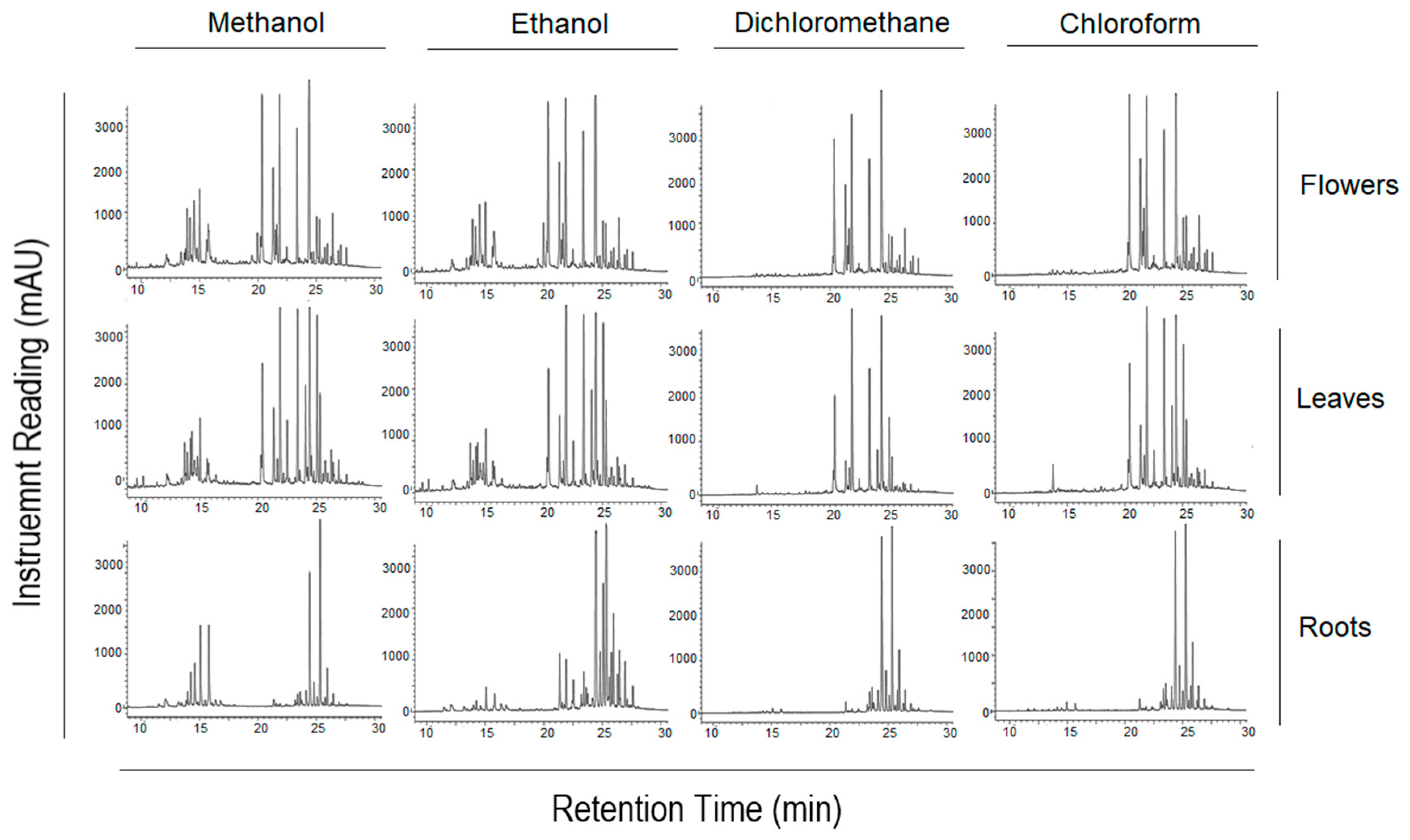
Figure 1. Fingerprinting chromatograms of A. fragrantissima flower-, leaf-, and root-derived solvent extracts using HPLC-UV. The chromatograms are based on a gradient method of methanol and water, and UV set at 275 nm with 5 µL injection volume. The separation of the dimethyl sulfoxide (DMSO)-dissolved compounds was performed using a Kinetex® C-18 column (250 × 4.6 mm, 5 µm) and revealed diverse polar and non-polar compositions for the different solvent extracts of the herbal medicine.
Pharmaceuticals 15 01060 g001
Figure 2. Inhibition of TNBC MDA-MB-231 cell viability by different solvent extracts of A. fragrantissima plant parts using the CellTiter-Glo™ assay.
Pharmaceuticals 15 01060 g002
Figure 3. Microscopic images of the MDA-MB-231 cells treated with increasing concentrations of A. fragrantissima flower dichloromethane extract displaying apoptosis-related morphological changes in cell shrinkage. The MDA-MB-231 cells display a dose-dependent increase in cell shrinkage, a morphological characterization related to apoptosis. The cell shrinkage that occurred in the MDA-MB-231 cells treated with 200 µg/mL of A. fragrantissima flower dichloromethane extract was similar to the cells treated with the strong apoptosis inducer STS, the positive control.
Pharmaceuticals 15 01060 g003
Figure 4. The increase in caspase-3/7 activation in the A. fragrantissima flower dichloromethane extract-treated TNBC MDA-MB-231 cells using confocal microscopy. The MDA-MB-231 cell treatment with the dichloromethane A. fragrantissima flower dichloromethane extract (25–200 µg/mL) and STS (1 µM) showed an increase in caspase-3/7 activity by more than 1.5-fold in comparison to the DMSO-treated cells, the negative control. The arrow is an example of MDA-MB-231 cells with activated caspase-3/7 (red fluorescence). Hoechst 33342 and fluoromethyl ketone-sulforhodamine (FMK-SR) were used as a nuclear dye and a cell-permeant red fluorescent probe labeling active caspase-3/7, respectively. * p < 0.05 vs. DMSO-treated cells.
Pharmaceuticals 15 01060 g004
Figure 5. Determination of the apoptotic status in the TNBC MDA-MB-231 cells exposed to A. fragrantissima flower dichloromethane extract. Representative scatter plots indicating the percentage of MDA-MB-231 cells in viability (V), in apoptotic status including early apoptosis (E APO) and late apoptosis (L APO), and in necrotic (N) status, based on Annexin V (-FITC) and PI (-PE) double staining measured by FACS. A. fragrantissima flower dichloromethane extract prompted apoptosis in TNBC MDA-MB-231 cells, in a dose-dependent manner, compared to DMSO-treated cells. STS was used as a positive control. The bar graph presents data as mean ± SD of three separate experiments. * p < 0.05 and ** p < 0.01 vs. DMSO-treated cells.
Pharmaceuticals 15 01060 g005
Figure 6. The increase in mitochondrial membrane potential opening activity in A. fragrantissima flower dichloromethane extract-treated TNBC MDA-MB-231 cells using confocal microscopy. The MDA-MB-231 cell treatment with A. fragrantissima flower dichloromethane extract (25–200 µg/mL) showed an increase in the activity of the opening of the mitochondrial transmission membrane pores, as indicated by the arrow. * p < 0.05 and ** p < 0.01 vs. DMSO-treated cells.
Pharmaceuticals 15 01060 g006
Figure 7. The identification of A. fragrantissima flower dichloromethane extract metabolites using an Agilent LC-QTOF in positive and negative modes.
Pharmaceuticals 15 01060 g007aPharmaceuticals 15 01060 g007bPharmaceuticals 15 01060 g007cPharmaceuticals 15 01060 g007dPharmaceuticals 15 01060 g007e
Figure 8. Docking poses for metabolite 11 (M11) and Colchicine into Tubulin crystal structure.
Pharmaceuticals 15 01060 g008aPharmaceuticals 15 01060 g008b
Figure 9. Docking poses for metabolite 11 (M11) and Y0R into CA IX crystal structure.
Pharmaceuticals 15 01060 g009
Figure 10. Radar diagrammatic representation of A. fragrantissima flower dichloromethane extract-derived identified metabolites (M1M14) using SwissADME webserver. LIPO: Lipophilicity, Size: Molecular weight, POLAR: solubility, INSOLU: insolubility, INSATU: insaturation, and FLEX: flexibility. The properties involved in the colored zone are preferred for orally active drugs.
Pharmaceuticals 15 01060 g010aPharmaceuticals 15 01060 g010b
Table 1. The half-maximal inhibitory concentrations (IC50) of A. fragrantissima plant part extracts on TNBC MDA-MB-231 cell viability using the ATP-based Promega CellTiter-Glo™ assay. Results are expressed as mean ± standard deviation (SD) from at least three separate experiments.
Plant PartExtraction SolventInhibition Values (IC50) (µg/mL)
Mean ± SD
Flowers
Methanol122.97 ± 24.50
Ethanol66.77 ± 31.11
Dichloromethane32.43 ± 3.41
Chloroform33.13 ± 7.20
Leaves
Methanol82.66 ± 3.88
Ethanol99.71 ± 9.24
Dichloromethane36.67 ± 3.94
Chloroform36.48 ± 3.02
Roots
Methanol103.85 ± 27.02
Ethanol161.77 ± 45.59
Dichloromethane120.50 ± 17.15
Chloroform52.24 ± 11.63
Table 2. The bioactivity scores of A. fragrantissima flower dichloromethane extract-derived metabolites using PASS online webserver.
Anti-Carcinogenic ActivityProbability of Being Active
(Pa)
Probability of Being Inactive
(Pi)
M10.9260.002
M20.8310.008
M30.9410.004
M40.8190.010
M50.6410.011
M60.8670.005
M70.6570.034
M80.6340.003
M90.7830.014
M100.4610.083
M110.7970.012
M120.5750.014
M130.4450.068
M140.9050.005
Table 3. Predicted biological targets of A. fragrantissima flower dichloromethane extract-derived metabolites using Molinspiration webserver.
Compound NameTarget Predictions Using Molinspiration
M1GPCR ligand
Ion channel modulator
Kinase inhibitor
Nuclear receptor ligand
Protease inhibitor
Enzyme inhibitor
0.10
−0.01
0.14
0.31
0.02
0.43
M2GPCR ligand
Ion channel modulator
Kinase inhibitor
Nuclear receptor ligand
Protease inhibitor
Enzyme inhibitor
−0.15
−0.28
0.14
0.01
−0.35
0.13
M3GPCR ligand
Ion channel modulator
Kinase inhibitor
Nuclear receptor ligand
Protease inhibitor
Enzyme inhibitor
0.31
0.17
−0.08
0.81
0.21
0.81
M4GPCR ligand
Ion channel modulator
Kinase inhibitor
Nuclear receptor ligand
Protease inhibitor
Enzyme inhibitor
−0.09
−0.23
0.20
0.13
−0.29
0.14
M5GPCR ligand
Ion channel modulator
Kinase inhibitor
Nuclear receptor ligand
Protease inhibitor
Enzyme inhibitor
−0.07
−0.09
0.18
0.34
−0.25
0.26
M6GPCR ligand
Ion channel modulator
Kinase inhibitor
Nuclear receptor ligand
Protease inhibitor
Enzyme inhibitor
−0.03
0.02
−0.48
0.34
0.00
0.33
M7GPCR ligand
Ion channel modulator
Kinase inhibitor
Nuclear receptor ligand
Protease inhibitor
Enzyme inhibitor
−0.06
0.26
−0.78
0.37
−0.38
0.43
M8GPCR ligand
Ion channel modulator
Kinase inhibitor
Nuclear receptor ligand
Protease inhibitor
Enzyme inhibitor
0.17
0.21
−0.38
0.65
0.18
0.58
M9GPCR ligand
Ion channel modulator
Kinase inhibitor
Nuclear receptor ligand
Protease inhibitor
Enzyme inhibitor
−0.02
−0.07
0.26
0.39
−0.22
0.28
M10GPCR ligand
Ion channel modulator
Kinase inhibitor
Nuclear receptor ligand
Protease inhibitor
Enzyme inhibitor
−0.06
−0.07
−0.62
0.09
−0.04
0.24
M11GPCR ligand
Ion channel modulator
Kinase inhibitor
Nuclear receptor ligand
Protease inhibitor
Enzyme inhibitor
−0.06
−0.19
0.28
0.36
−0.25
0.28
M12GPCR ligand
Ion channel modulator
Kinase inhibitor
Nuclear receptor ligand
Protease inhibitor
Enzyme inhibitor
0.78
0.17
0.27
−1.52
−0.28
0.86
M13GPCR ligand
Ion channel modulator
Kinase inhibitor
Nuclear receptor ligand
Protease inhibitor
Enzyme inhibitor
0.27
0.10
−0.12
0.22
0.14
0.20
M14GPCR ligand
Ion channel modulator
Kinase inhibitor
Nuclear receptor ligand
Protease inhibitor
Enzyme inhibitor
0.12
−0.04
−0.52
0.69
0.08
0.64
Table 4. Docking scores of identified metabolites with Tubulin crystal structure.
Compound NameDocking Score
(kcal/mol)
Interactions with Amino Acid Residues
M1−9.59Lys254, and Val181
M2−9.16Asp251, and Glu183
M3−6.90Thr353, Cys241, and Val238
M4−8.60Asp251, and Cys241
M5−8.10Asp251, Cys241, and Asn350
M6−7.07Lys254
M7−4.75Cys241, and Val238
M8--
M9−8.43Val181, Cys241
M10−6.38Asp251, Cys241
M11−9.89Cys241, Val181, and Asn350
M12−7.35Val181, Cys241
M13−4.23Val238
M14−7.40Thr179, Val181
Colchicine−11.35Val181, Cys241, Thr353, Val238
Table 5. Docking scores of identified metabolites with CA IX crystal structure.
Compound NameDocking Score
(kcal/mol)
Interactions with Amino Acid Residues
M1−5.01Thr200, Thr201, Gln92, Leu91, and zinc coordination
M2−4.08Asn66, and zinc coordination
M3−4.14Gln92
M4−5.25Asn66, Thr200, Zinc coordination
M5−5.21His68, Thr200, and zinc coordination
M6−4.07Gln92, and zinc coordination
M7−3.32Gln92
M8--
M9−5.20Zinc coordination
M10--
M11−5.46His68, Thr200, His94, Zinc coordination
M12−4.52His68, Thr201, and zinc coordination
M13−1.30Gln92, His94, Thr200, and zinc coordination
M14−4.40Gln71, Thr201, Gln92, and zinc coordination
Y0R−7.13Gln92, Thr200, and zinc coordination
Table 6. The predicted ADME properties of A. fragrantissima flower dichloromethane extract-derived metabolites using SwissADME webserver.
Compound NameMolecular Weight
(g/mol)
Log Po/wLog SBBB PermeantGI AbsorptionRule of Five (ROF)
SWISS ADMEQikPropSWISS ADMEQik PropSWISS ADMEQik PropSWISS ADMEQik PropSWISS ADMEQik Prop
(%)
SWISS ADME
M1432.38432.380.05−0.36−2.69
Soluble
−3.22NoNoLow29.50Yes; 1 violation: NHorOH > 5
M2360.31360.322.602.21−4.74
Moderately soluble
−3.92NoNoHigh82.42Yes; 0 violation
M3336.34336.340.821.66−1.52
Soluble
−3.466NoNoHigh82.33Yes; 0 violation
M4344.32344.322.902.79−5.33
Moderately soluble
−4.142NoNoHigh92.79Yes; 0 violation
M5270.24270.242.581.59−4.40
Moderately soluble
−3.297NoNoHigh73.11Yes; 0 violation
M6306.35306.351.902.34−2.27
Soluble
−3.888YesYesHigh92.97Yes; 0 violation
M7222.37222.374.234.60−3.00
Soluble
−4.95YesYesHigh100Yes; 0 violation
M8436.62436.633.933.80−3.39
Soluble
−5.759NoNoHigh95.21Yes; 0 violation
M9286.24286.242.280.91−3.82
Soluble
−3.026NoNoHigh61.10Yes; 0 violation
M10497.71497.716.546.40−6.30
Poorly soluble
−8.969NoNoHigh100Yes; 1 violation: MLOGP > 4.15
M11302.24302.241.990.353−3.24
Soluble
−2.882NoNoHigh51.56Yes; 0 violation
M12282.25282.25−2.58−1.680.59
Soluble
−1.693NoNoLow49.49Yes; 0 violation
M13287.40287.403.744.28−4.81
Moderately soluble
−5.433YesNoHigh100Yes; 0 violation
M14278.30278.301.100.28−2.52
Soluble
−2.155NoNoHigh68.622Yes; 0 violation
Table 7. The CYP-P450 enzyme inhibition profile of A. fragrantissima flower dichloromethane extract-derived metabolites using SwissADME webserver.
CompoundCYP1A2CYP2C19CYP2C9CYP2D6CYP3A4
M1NoNoNoNoNo
M2YesNoYesYesYes
M3NoNoNoNoNo
M4YesNoYesYesYes
M5YesNoNoYesYes
M6NoNoNoNoNo
M7NoNoYesNoNo
M8NoNoNoNoNo
M9YesNoNoYesYes
M10NoNoYesNoNo
M11YesNoNoYesYes
M12NoNoNoNoNo
M13YesYesYesNoYes
M14NoNoNoNoNo
Table 8. Organ and endpoint toxicity predictions of A. fragrantissima flower dichloromethane extract-derived metabolites using the ProTox-II webserver.
Compound NameClassification
Organ Toxicity (% Probability)Toxicity Endpoint (% Probability)
HepatotoxicityCarcinogenicityImmunotoxicityMutagenicityCytotoxicity
M10.82
(inactive)
0.86
(inactive)
0.93
(inactive)
0.59
(active)
0.69
(inactive)
M20.70
(inactive)
0.69
(inactive)
0.97
(Active)
0.82
(inactive)
0.75
(inactive)
M30.67
(inactive)
0.61
(inactive)
0.99
(Active)
0.55
(Active)
0.58
(inactive)
M40.70
(inactive)
0.69
(inactive)
0.84
(Active)
0.82
(inactive)
0.75
(inactive)
M50.68
(inactive)
0.62
(inactive)
0.99
(inactive)
0.57
(inactive)
0.87
(inactive)
M60.72
(inactive)
0.54
(Active)
0.94
(Active)
0.68
(inactive)
0.77
(inactive)
M70.78
(inactive)
0.70
(inactive)
0.97
(inactive)
0.83
(inactive)
0.75
(inactive)
M80.60
(inactive)
0.75
(inactive)
0.57
(Active)
0.73
(inactive)
0.75
(inactive)
M90.69
(inactive)
0.68
(Active)
0.97
(inactive)
0.51
(Active)
0.99
(inactive)
M100.81
(inactive)
0.52
(inactive)
0.98
(Active)
0.79
(inactive)
0.75
(inactive)
M110.69
(inactive)
0.68
(Active)
0.87
(inactive)
0.51
(Active)
0.99
(inactive)
M120.68
(inactive)
0.73
(inactive)
0.97
(inactive)
0.83
(inactive)
0.62
(inactive)
M130.83
(inactive)
0.62
(inactive)
0.84
(Active)
0.76
(inactive)
0.83
(inactive)
M140.71
(inactive)
0.63
(inactive)
0.71
(inactive)
0.84
(inactive)
0.64
(inactive)
Publisher’s Note: MDPI stays neutral with regard to jurisdictional claims in published maps and institutional affiliations.