Article

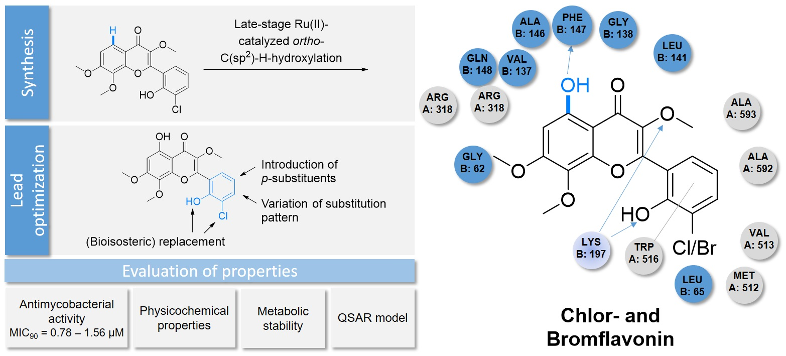
Total Synthesis of the Antimycobacterial Natural Product Chlorflavonin and Analogs via a Late-Stage Ruthenium(II)-Catalyzed ortho-C(sp2)-H-Hydroxylation

1
Institute of Pharmaceutical and Medicinal Chemistry, Heinrich Heine University Düsseldorf, Universitätsstraße 1, 40225 Düsseldorf, Germany
2
Institute of Pharmaceutical Biology and Biotechnology, Heinrich Heine University Düsseldorf, Universitätsstraße 1, 40225 Düsseldorf, Germany
3
South African Medical Research Council Drug Discovery and Development Research Unit, Department of Chemistry and Institute of Infectious Disease and Molecular Medicine, University of Cape Town, Rondebosch 7701, South Africa
4
Drug Discovery and Development Centre (H3D), University of Cape Town, Rondebosch 7701, South Africa
5
Institute of Clinical Pharmacy and Pharmacotherapy, Heinrich Heine University Düsseldorf, Universitätsstraße 1, 40225 Düsseldorf, Germany
6
Institut für Organische und Biomolekulare Chemie, Georg-August-Universität Göttingen, Tammannstraße 2, 37077 Göttingen, Germany
7
John-von-Neumann-Institute for Computing (NIC), Jülich Supercomputing Centre (JSC), Institute of Biological Information Processing (IBI-7: Structural Biochemistry), and Institute of Bio- and Geosciences (IBG-4: Bioinformatics), Forschungszentrum Jülich GmbH, 52428 Jülich, Germany
*
Author to whom correspondence should be addressed.
These authors contributed equally to this work.
Pharmaceuticals 2022, 15(8), 984; https://doi.org/10.3390/ph15080984
Submission received: 8 July 2022 / Revised: 1 August 2022 / Accepted: 8 August 2022 / Published: 10 August 2022
(This article belongs to the Special Issue C–H Functionalization Chemistry: Applications in Drug Synthesis)

Abstract

:
The continuous, worldwide spread of multidrug-resistant (MDR) and extensively drug-resistant (XDR) tuberculosis (TB) endanger the World Health Organization’s (WHO) goal to end the global TB pandemic by the year 2035. During the past 50 years, very few new drugs have been approved by medical agencies to treat drug-resistant TB. Therefore, the development of novel antimycobacterial drug candidates to combat the threat of drug-resistant TB is urgent. In this work, we developed and optimized a total synthesis of the antimycobacterial natural flavonoid chlorflavonin by selective ruthenium(II)-catalyzed ortho-C(sp2)-H-hydroxylation of a substituted 3′-methoxyflavonoid skeleton. We extended our methodology to synthesize a small compound library of 14 structural analogs. The new analogs were tested for their antimycobacterial in vitro activity against Mycobacterium tuberculosis (Mtb) and their cytotoxicity against various human cell lines. The most promising new analog bromflavonin exhibited improved antimycobacterial in vitro activity against the virulent H37Rv strain of Mtb (Minimal Inhibitory Concentrations (MIC90) = 0.78 μm). In addition, we determined the chemical and metabolic stability as well as the pKa values of chlorflavonin and bromflavonin. Furthermore, we established a quantitative structure–activity relationship model using a thermodynamic integration approach. Our computations may be used for suggesting further structural changes to develop improved derivatives.

Graphical Abstract

1. Introduction

In 2020, the World Health Organization estimated that over two billion people were infected with tuberculosis (TB) caused by the bacterial pathogen Mycobacterium tuberculosis (Mtb). The first-line therapy for drug-susceptible Mtb strains has remained unchanged for 50 years, despite a long treatment duration and, in part, severe side effects. During this period, only three new anti-TB drugs with novel modes of action, bedaquiline, delamanid, and pretomanid, have been clinically approved. However, these three drugs are only approved for the treatment of multidrug-(MDR-) and extensively drug-resistant-(XDR-)TB [1,2,3]. Therefore, there is a strong need for the development of novel lead structures, and (pre)clinical candidates with novel mechanisms of action for inclusion in novel drug regimens that can contribute to shortening treatment and delaying resistance. The naturally occurring flavonoid chlorflavonin (CF, Figure 1) was first isolated in 1969 from Aspergillus candidus. CF’s antimycotic properties against, e.g., Aspergillus amstelodami und Paecilomyces variotii were reported in the same year [4]. In 1970, the structure of CF was resolved [5]. Recently, Rehberg et al. discovered CF’s strong in vitro growth inhibition against virulent Mtb H37Rv (MIC90 = 1.56 μM) and Mtb XDR clinical isolates. CF exhibited no cytotoxicity against the human fetal lung fibroblast cell line MRC-5 and monocyte cell line THP-1. For mono treatment with CF, a bacteriostatic effect was observed. In addition, CF demonstrated synergistic efficacy with delamanid and the first-line anti-TB drug isoniazid, including intracellular activity against infected human macrophages. CF specifically inhibits the large catalytic subunit IlvB1 of the mycobacterial acetohydroxyacid synthase (AHAS), which is the first common enzyme in the de novo biosynthetic pathway of essential branched-chain amino acids (BCAA) and pantothenic acid [6,7,8,9,10,11,12,13]. The inhibition of the mycobacterial AHAS through chlorflavonin causes combined auxotrophies of BCAA and pantothenic acid [14]. In addition, the treatment of Mtb-infected mice with the AHAS inhibitor sulfometuron methyl, which is approved as a herbicide, led to a significantly reduced proliferation of the pathogen in the host’s lungs [15]. In the context of vaccine research, it was shown that BCAA-auxotrophic mutants of intracellular pathogens exhibited significantly reduced virulence in vivo [16]. The fact that AHAS is present in Mtb but not in the human host makes it a promising target for developing novel antituberculosis drugs [17].
None of the currently approved TB drugs inhibit AHAS nor any enzyme in the BCAA biosynthetic pathway [14]. CF’s novel mechanism of action, structure, and potent antimycobacterial in vitro activity make it a promising lead structure for developing a new class of anti-TB drugs. The eponymous chlorine substituent on the B-ring adjacent to the phenolic hydroxyl group is particularly noteworthy and unusual for a flavonoid [18,19]. In 1980, Tökés et al. described the only published synthesis of CF through an Allan–Robinson condensation of 2-(benzoyloxy)-3-chlorobenzoic anhydride (3) with a hydroxyacetophenone derivative 4 in a total yield of only 0.79% over seven steps (Scheme 1A) [20].

2. Results and Discussion

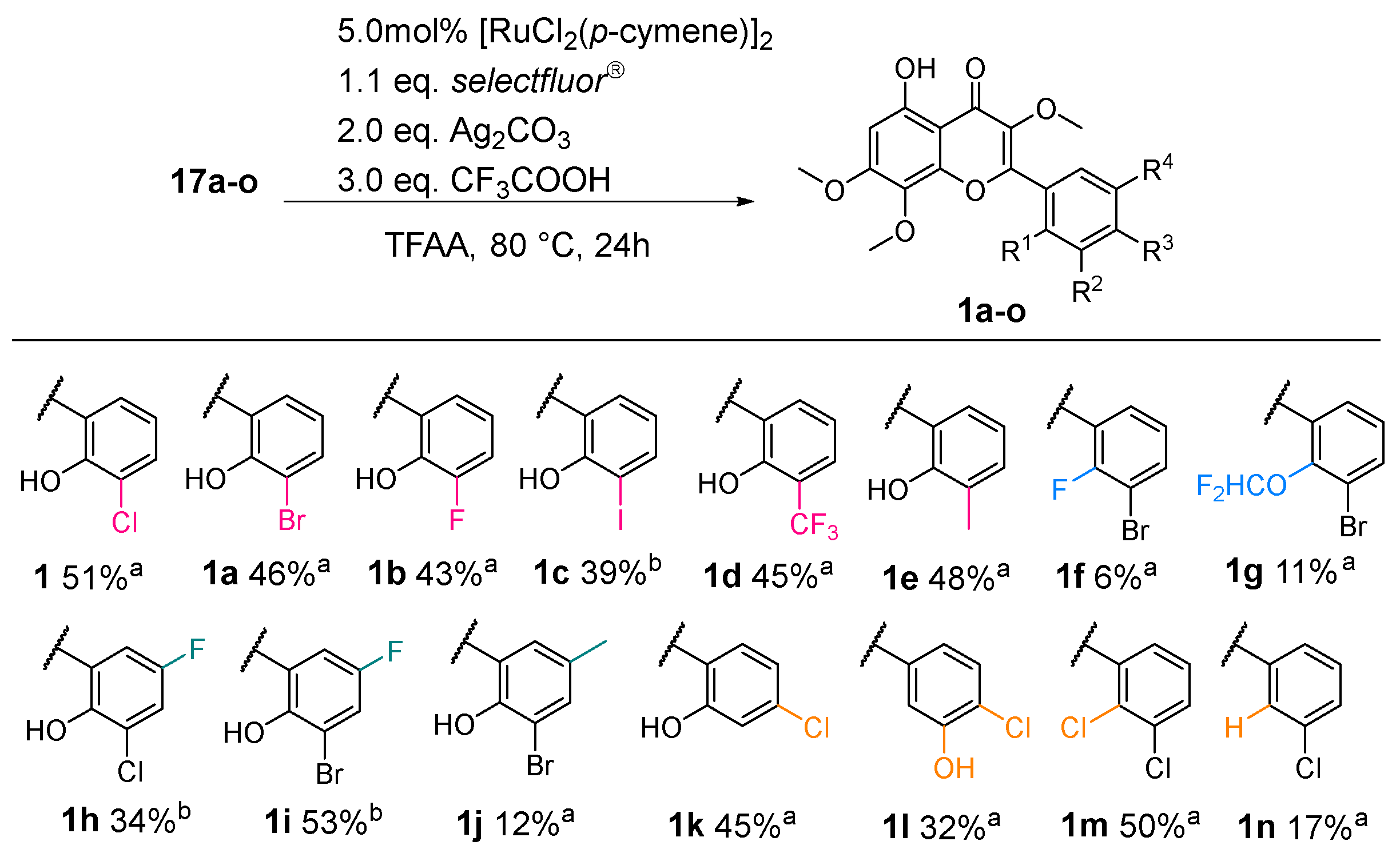
2.1. Synthesis of CF

In a first synthetic attempt, we tried to avoid the occasionally unfavorable Elbs persulfate oxidation by using the 2′-hydroxyacetophenon 7 with the natural substitution pattern of the A-ring [21]. Using this approach adapted from Tökés et al. [20], we planned to circumvent the difficult selective methylation of the phenolic hydroxyl groups in the 7- and 8-position (Scheme 1A). While intermediate 6 was successfully synthesized, the selective demethylation of 6 with BBr3 to provide 1-(2-hydroxy-3,4,6-trimethoxyphenyl)-2-methoxyethan-1-one (7) in sufficient yield was not feasible (Scheme 1B, see Supplementary Materials for details). In our second attempt, flavone 10 was synthesized by a Claisen–Schmidt condensation of the substituted 2′-hydroxyacetophenon 8 and the salicylic aldehyde derivative 9, followed by oxidative cyclo-dehydration of the chalcone intermediate with a catalytic amount of iodine in dimethyl sulfoxide (DMSO). Unfortunately, the oxidation of flavone 10 with dimethyldioxirane (DMDO), oxone®, or tert-butylhydroperoxid (TBHP) resulted in poor yields (Scheme 1C, see Supplementary Materials for details). To introduce the important phenolic hydroxyl group of the A-ring immediately and to forego selective demethylation in a separate step, the commonly used Algar–Flynn–Oyamada (AFO) reaction was employed to build the flavonol skeleton. However, the substrate scope of the AFO reaction was limited due to the competing formation of aurones when using substrates with electron-donating groups [21,22,23]. Based on this limitation, we planned to generate the essential substitution pattern of the A-ring via a transition metal-catalyzed ortho-C(sp2)-H-functionalization using the carbonyl oxygen of the 4-chromanone moiety as the coordinating directing group in the last step (Scheme 2). Therefore, the synthesis of the 3′-methoxyflavonol 16 should take place by methylation of the flavonol 15, which can be synthesized in advance by Claisen–Schmidt condensation of the commercially available starting materials 2 and 13. The oxidative ring closure of the chalcone intermediate should then be initiated with alkaline hydrogen peroxide in an ethanolic solution in a sequential one-pot process. Starting from 3-chloro-2-hydroxybenzaldehyde (2), the methoxymethyl ether (MOM) protecting group was introduced to the phenolic hydroxyl group by nucleophilic substitution with chloromethyl methyl ether in the presence of N,N-diisopropylethylamine (DIPEA) with an excellent yield of 97% (Scheme 3).
Next, the resulting MOM-protected salicylaldehyde 12 was condensed with the commercially available 2′-hydroxyacetophenone 13 by a base-mediated Claisen–Schmidt condensation in ethanolic solution. Afterward, hydrogen peroxide was added to initiate the oxidative Algar–Flynn–Oyamada ring closure to provide flavonol 15 with a moderate yield of 42% on a multigram scale. Effective cooling, a slow addition rate of hydrogen peroxide, and sufficient dilution of the reaction mixture were critical for avoiding side products and simplifying the purification process (Scheme 3). Next, the enolic hydroxyl group of flavonol 15 was methylated with iodomethane in the presence of Cs2CO3 in dimethylformamide (DMF). The MOM protecting group of 3′-methoxyflavonol 16 was then removed by acidolysis with methanolic hydrochloric acid at 50 °C to give the deprotected flavonoid 17 with a good yield. It was unnecessary to add a scavenger to deactivate in situ-generated formaldehyde, since no significant side reactions were observed (Scheme 3).
In the final step, we studied the introduction of the essential phenolic hydroxyl group in position 5 of the A-ring by a selective transition metal-catalyzed ortho-C(sp2)-H-hydroxylation using the carbonyl oxygen of the 4H-chromen-4-one skeleton as a coordinating directing group. In the last two decades, significant progress has been made for C-H-functionalization [24,25,26], especially for substrates with weakly coordinating directing groups such as the carbonyl oxygen of acetophenones and chromones [26,27,28,29,30,31,32,33] An extensive catalyst, oxidant, solvent, and temperature screening was performed to optimize our method. For this purpose, the conversion and selectivity of the ortho-C(sp2)-H-hydroxylation were monitored by HPLC-UV/Vis (Table 1). Inspired by the work of Shan et al. and others, we first studied palladium(II)-catalysts in combination with PhI(TFA)2 as the terminal oxidant in 1,2-dichlorethane (DCE) [34]. However, these reaction conditions provided only trace amounts of CF, and in the case of longer reaction times and an excess of oxidant, the substrate decomposed (entries 1–2). Replacing DCE with a mixture of trifluoroacetic acid (TFA) and trifluoroacetic anhydride (TFAA) (9:1, v/v) resulted in a higher conversion rate and moderate selectivity. Notably, reversing the solvent ratio significantly increased conversion and improved selectivity (entries 3–4). By combining [RuCl2(p-cymene)]2 as a catalyst and selectfluor as an oxidizing agent, the formation of byproducts could be suppressed to such an extent that purification by silica gel chromatography or recrystallization was successfully applied (entries 5–6). The addition of a silver(I) additive was beneficial because of its role as a halogen scavenger. Silver-cations abstract chloride from the neutral dimeric catalyst and form two monomeric, cationic catalyst species, which can interact with the partially negatively charged oxygen in a stronger fashion [29,35]. While an increased number of side reactions was observed with 2.0 equivalents of the oxidizing agent, virtually no side reaction occurred with a slight stoichiometric excess of 1.1 equivalents (entries 7–8). At the ideal reaction temperature of 80 °C, good conversions and superior selectivity were achieved, while only trace amounts of 1 were detected at 60 °C. In contrast, complete conversion of the starting material was observed at 120 °C, as well as the formation of several side products, necessitating purification with RP-flash chromatography (entries 9–10). Furthermore, no significant increase in conversion was observed when the reaction was carried out under argon atmosphere, nor with the continuous addition of the oxidants or without separate previous activation of the catalyst through the silver additive (see Supplementary Materials for complete optimization studies). Initially, the ortho-C(sp2)-H-hydroxylation yielded the 2,2,2-trifluoroacetate intermediate 18, which was converted into the desired product 1 through methanolysis. The structure of chlorflavonin was elucidated via 1H-and 13C-NMR, which agrees with the spectral data published and discussed by Rehberg et al. [14].

2.2. Lead Optimization of CF

After establishing the optimal reaction conditions, we designed a small compound library of CF analogs for preliminary structure–activity relationship (SAR) studies. Previously, we published a homology model of the Mtb H37Rv IlvB1 catalytic subunit of AHAS using the Saccharomyces cerevisiae and Arabidopsis thaliana (PDB ID 1T9C and 1YBH) AHAS proteins as templates [14]. Subsequent docking studies performed with Glide and AutoDock identified potential interactions between CF and amino acid residues within the putative active site of IlvB1, which provided a rational approach for the design of the analogs. Therefore, in this first series of analogs, we focused on modifications to the B-ring. Since the lipophilic chlorine substituent might interact with amino acid side chains inside a hydrophobic side pocket, we replaced the chlorine substituent with bioisosteric moieties and lipophilic substituents, e.g., Br and CF3 [14]. In addition, we wanted to increase the acidity of the phenolic hydroxyl group of the B-ring to facilitate its deprotonation and strengthen the possible salt bridge to Lys197. To analyze the importance of the 2′-hydroxy group, we replaced it with the bioisosteric difluoromethoxy group and a chlorine or fluorine substituent [36,37,38]. The salicylic aldehyde starting materials and several MOM-protected salicylic aldehydes were purchased or synthesized according to the literature [39,40,41,42,43,44,45]. Analogs 17ap were synthesized according to Scheme 3. For the preparation of 1h, an additional step was needed. According to a procedure of Li et al. for difluromethylation of phenols [46], the 2′-hydroxy group of intermediate 17b was treated with aqueous KOH solution in dichloromethane. Dropwise addition of (bromodifluormethyl)trimethylsiliane as difluorocarbene precursor gave intermediate 19 with a yield of 78%. The final ortho-C(sp2)-H-hydroxylation of 17ap afforded the CF analogs 1ap with yields of 6–53% (Scheme 4).
The SAR for the B-ring was quite narrow, and even small structural changes resulted in a loss of antimycobacterial activity. This activity loss was observed for the bioisosteric replacement of the 2′-hydroxy group with potential bioisosteric groups (1f,g), the introduction of a fluorine or a methyl substituent in the 5′-position of the B-ring (1hj), and the variation in the substitution pattern (1ko). An explanation for the inactivity of these analogs could be their poor water solubility. For a more comprehensive assessment of the antimycobacterial activity, a better understanding of the biological functions of the different mycobacterial AHAS isoenzymes is needed. For this purpose, we are currently developing a new robust AHAS enzyme assay.

2.3. Evaluation of Selected Physicochemical Properties, Microsomal Stability, and Computations

For further preclinical profiling of CF and BF, selected physicochemical properties (e.g., water solubility), chemical and microsomal metabolic stability were determined. The aqueous solubility of both compounds was determined to be <5 μM at a pH of 7.4 using a miniaturized shake-flask method (Table 2). While the chemical stability of CF and BF in a phosphate buffer at pH 2.0 and 7.4 was excellent (99% drug content after 48 h), the metabolic stability in human liver microsomes was moderate, and both compounds can be classified as high extraction drugs (hepatic extraction ratio EH ≥ 0.72) (Table 2).
To investigate the protonation state of CF and BF in physiological media (pH = 7.4) and these compounds’ ability to form the proposed salt bridge with Lys197, the pKa values were determined by 1H-NMR titration (see Supplementary Data File; Figures S1–S4). For determination of the pKa of the 5-hydroxy group, the shift of the singlet at 6.65 ppm, the triplet at 7.01 ppm for the 2′-hydroxyl group, and both double duplets at 7.42 and 7.59 ppm were used.
Furthermore, seven structurally diverse chlorflavonin derivatives (1, 1a, 1b, 1c, 1d, 1f, 1i) with substitutions at the B-ring were chosen to establish a quantitative structure–activity relationship model using the thermodynamic integration (TI) approach [49,50], as implemented in FEW [51] of Amber21 (see Supplementary Materials Data File) [52]. The computed relative free energies (ΔΔG) were generally in qualitative agreement with the changes in the minimal inhibitory concentrations (MIC90) (see Supplementary Materials Data File; Figure S7). This suggests that changes in MIC90 were predominantly determined by differences in the derivatives’ affinities. Furthermore, such computations may suggest further structural changes to obtain more active chlorflavonin derivatives.

3. Materials and Methods

3.1. Chemistry

The syntheses of chlorflavonin and chlorflavonin analogs are described in detail in the Supplementary Materials Data File. Commercially available reagents and solvents were purchased from Apollo Scientific, Sigma-Aldrich, TCI, BLDpharm, Carbolution, ABCR GmbH, Acros Organics, or Alfa Aesar and were used without further purification. Dry solvents were purchased from Acros Organics. Analytical thin-layer chromatography was performed using silica gel 60 F254 aluminium plates. Compound spots were visualized either by UV light (254 nm) or by staining with a solution of 1% FeCl3 in ethanol. Flash chromatography was performed on CombiFlash® Rf 200 using RediSep™ Rf-columns. 1H-, 13C-, and 19F-NMR spectra were recorded with Bruker Avance III—300, Bruker Avance III—600, or Bruker Avance DRX—500 spectrometers. 1H- and 13C-NMR signals were calibrated to the residual proton and carbon resonance of the solvent: CDCl3 (1H-NMR δ = 7.26 ppm, 13C-NMR δ = 77.2 ppm), DMSO (1H-NMR δ = 2.50 ppm, 13C-NMR δ = 39.5 ppm). The following abbreviations were used to describe peak splitting patterns when appropriate: s = singlet, d = doublet, t = triplet, q = quartet, h = hextet, m = multiplet, dd = doublet of doublet, td = doublet of triplet, ddd = doublet of doublet of doublet and brs = broad singlet. Coupling constants, J, were reported in the Hertz unit (Hz). ESI-MS data were recorded with UHR-QTOF maXis 4G. Melting points were measured on a Büchi M 565 instrument and were not corrected. Reverse-phase high-performance liquid chromatography data were measured with Varian ProStar 210 with a Phenomenex Luna C-18 (2) particle size 5 μm (250 × 4.6 mm) column. The detection took place with the UV detector Varian ProStar 330 at 220–254 nm, and eluents water/acetonitrile with 0.1% TFA were used.

3.2. Determination of Minimal Inhibitory Concentration (MIC)

To characterize the structure–activity-relationship, the MIC assay was employed as previously described. Briefly, M. tuberculosis H37Rv cells were precultured in Middlebrook 7H9 liquid medium supplemented with 0.5% (v/v) glycerol, 0.05% (v/v) tyloxapol, and 10% (v/v) ADS (0.81% NaCl, 5% BSA and 2% dextrose) at 37 °C to an OD600 nm of 0.5–0.8. Cells were diluted with fresh medium and seeded into 96-well plates to yield a final density of 105 cells in 100 µL medium per well. The compounds were tested in two-fold serial dilutions with a final concentration ranging from 100 to 0.05 µM. The microtiter plates were incubated at 37 °C, 5% CO2, and 80% humidity for 5 days. Then 10 µL of a 100 µg/mL resazurin solution were added to each well and incubated for 16 h at room temperature. After fixation of the bacteria for 30 min with a final concentration of 5% (v/v) formalin, the fluorescence was measured in a TECAN microplate reader (excitation of 540 nm, emission of 590 nm). The percentage of growth was calculated in comparison to dimethyl sulfoxide (DMSO)-treated cells (=100% growth) and the sterile control (=0% growth). All experiments were performed in triplicates.

3.3. Determination of Cytotoxcity

For the determination of the cytotoxicity of the compounds, different human cell lines were used. The human fetal lung fibroblast cell line MRC-5 (American Type Culture Collection) was incubated in Eagle’s Minimum Essential Medium (EMEM) containing 1% (v/v) Na-pyruvate, whereas the human embryonic kidney cell line HEK-293 (CLS Cell Lines Service GmbH) was cultivated in EMEM supplemented with 2 mM l-glutamine, 1% (v/v) non-essential amino acids, and 1 mM sodium pyruvate. The human monocyte cell line THP-1 (Deutsche Sammlung von Mikroogranismen und Zellkulturen GmbH) was cultured in RPMI 1640 medium. All media were supplemented with 10% (v/v) heat-inactivated fetal bovine serum (FBS).
The cell densities were quantified using a hemocytometer and then adjusted to 106 cells/mL. Next, 100 µL per well were seeded in 96-well microtiter plates to yield an inoculation cell density of 5 × 10⁵ cells per well. Compounds were added employing two-fold serial dilutions resulting in final concentrations ranging from 100 to 0.78 µM. Cells were incubated for two days at 37 °C and 5% CO2 in a humidified atmosphere. Subsequently, 10 µL of a 100 µg/mL resazurin solution was added to each well, mixed thoroughly, and incubated for an additional 3–4 h. Fluorescence was measured in a TECAN microplate reader (excitation of 540 nm, emission of 590 nm). The percentage of growth was calculated with respect to DMSO (100% growth) and Triton X-100 (0% growth) controls.

3.4. Evaluation of Solubility

Solubility was measured at pH 7.4 using an adapted miniaturized shake-flask method in 96-well plate format [53,54]. Briefly, 4 μL of a 10 mM stock in DMSO was added to a 96-well plate and evaporated using a GeneVac® system. Phosphate buffer pH 7.4 was then added to the wells, and the plate was incubated for 24 h at 25 °C with shaking. At the end of this incubation, the samples were centrifuged at 3500 g for 15 min then transferred to an analysis plate. A calibration curve in DMSO for each sample between 10 and 220 μM was prepared and included in the analysis plate. Analysis was then performed by HPLC-DAD, and the solubility of each sample was determined from the corresponding calibration curve. Reserpine and hydrocortisone were used as positive controls and treated similarly.

3.5. Evaluation of Compound Chemical Stability

Stability testing was performed in phosphate buffer pH 2.0 and 7.4 (prepared according to Ph. Eur. 10) at 20 °C over a period of 48 h. Therefore, 0.4 mg of substance was dissolved in Tween® 20 und ethanol (7/3 v/v) and diluted with the respective phosphate buffer. The solution was shaken with an IKA KS 260 basic (250 min−1) at 25 °C for 48 h. Analysis was performed after 1, 3, 6, 24, and 48 h by comparing the area under the curve (A.U.C.), which was measured with HPLC-UV at 254 nm (n = 1).

3.6. Evaluation of Compound Metabolic Stability

The metabolic stability assay was performed in Human Liver Microsomes (HLM) using a single-point metabolic stability assay. Briefly, the compounds were incubated at 1 μM in 0.4 mg/mL human mixed-gender liver microsomes (Xenotech, Kansas City, KS, USA, pool of 50) for 30 min at 37 °C. Reactions were quenched by adding ice-cold acetonitrile containing internal standard. The samples were then centrifuged and analyzed by LC-MS/MS for the disappearance of the parent compound. Half-life, clearance, and hepatic excretion ratios were determined using standard equations [47,48]. Propranolol and midazolam were used as positive controls and treated similarly.

3.7. Determination of Compound pKa Values

NMR experiments were performed at 298 K on a Bruker Avance III HD spectrometer operating at 600 MHz equipped with 5 mm triple resonance TCI (1H, 13C, 15N) cryoprobes and shielded z-gradients. For the pKa value determination, 23 samples of 200 µM concentration of CF and BF were prepared with pH ranges from 2 to 13 (pH in 0.5 log unit steps) in 50 mM sodium phosphate, 100 mM sodium chloride, 10% (v/v) DMSO-d6. 1D 1H-NMR experiments were performed using 128 or 256 scans for each sample, and the data were processed and analyzed by the TopSpin 3.2 software (TopSpin, v3.2, Bruker BioSpin GmbH, Rheinstetten, Germany). Sodium 2,2-dimethyl-2-silapentane-5-sulfonate (DSS) was used for chemical shift referencing. 1H chemical shift values were extracted for the reporter protons using the TopSpin software (see Supplementary Materials Data File Figures S1 and S3). The pKa values were calculated using the chemical shifts of the protonated and unprotonated forms and their corresponding mole fraction values applying the Henderson–Hasselbalch equation as explained by Gift et al. [55]. The data were plotted with the Origin software (Origin Pro 2019, v9.6.0.172, OriginLab Corporation, Northampton, MA, USA) (see Supplementary Data File Figures S2 and S4).

3.8. Analysis of a Quantitative Structure-Activity Relationship Model

Chlorflavonin derivates were prepared with the LigPrep module of the Schrödinger suite [56] and subsequently aligned with align_ligands of the Schrödinger suite to chlorflavonin docked into the homology model of the catalytic subunit of human AHAS [14]. The homology model [14] was prepared for calculations with the Protein Preparation Wizzard module of the Schrödinger suite. Files for relative free energy computations and analyses were prepared with the TI module of FEW [51], and AMBER version 21.1 [52] was used for simulations. The RESP charge model [57] was chosen for computing partial charges of the ligands.
Ligands in water and ligand–protein complexes in water were minimized with a three-step procedure, each with 2000 steps of steepest descent and 1000 steps of conjugate-gradient minimization in the presence of 25 kcal mol−1 A−2, 5 kcal mol−1 A−2, and no restraints on the solute, respectively. Subsequently, three replicas of each system were heated from 100 K to 300 K in the NVT (constant temperature and constant volume) ensemble during 50 ps of MD simulations followed by an adjustment of the density in the NPT (constant temperature and constant pressure) ensemble during a 50 ps MD run.
The transition map was based on Kruskal’s algorithm [58] with a modification introduced to close thermodynamic cycles for convergence control. As “cost” of the edges, 103 × (2—TanimotoCombo) was calculated, where the TanimotoCombo score was computed with the ROCS module of OpenEye [56,59]. Production phase MD simulations were carried out at 300 K in the NVT ensemble for 9 λ windows, λ = 0.1, 0.2, …, 0.9, respectively, with lengths of 10 ns per λ window for ligands in solvent and 30 ns per λ window for complexes in solvent. The impact of the simulation time per λ window (10 ns and 30 ns) on the precision of the computations in the case of complex simulations was estimated by calculating the standard deviation over three replicas (Figure S5). The convergence of the computations was confirmed in three ways: First, the precision of average dV/dλ for each λ window was determined by employing the Student’s distribution for each replica separately as implemented in the FEW workflow (convergence_check_method 2) [51]; second, the average dV/dλ values were checked with respect to their consistency across the three replicas (Figure S6); third, the cycle-closure hysteresis of closed thermodynamic cycles was computed (Table S3).

3.9. Solubility and Partition Coefficient Predictions

QikProp [60] of the Schrödinger suite (version 6.7), Schrödinger, LLC, New York, NY, USA, 2015 was used to compute the solubility (QPlogS) and octanol/water partition coefficient (QPlogPo/w) for chlorflavonin derivates included in relative free energy computations (Table S4).

4. Conclusions

In conclusion, we developed an efficient five-step synthesis of the antimycobacterial natural flavonoid chlorflavonin (1) and 14 structural analogs, demonstrating the broad applicability of our method. The overall yield of CF (13.5%) was significantly improved compared to the original synthesis by Tökés et al. The SAR for the B-ring was surprisingly narrow, as small structural changes resulted in a loss of antimycobacterial activity. We will therefore extend the future optimization to rings A and C. The most active compound, BF, exhibited submicromolar antimycobacterial activity with no cytotoxicity toward human cells. The bromine substituent appeared to fill the proposed lipophilic sub-pocket ideally. While the chemical stability of CF and BF was excellent, the metabolic stability needs improvement through future lead optimization. The initially derived SARs should encourage and guide the synthesis of further analogs. For a successful and more comprehensive structure optimization of CF and BF, we are currently developing a robust AHAS enzyme assay. In addition, our quantitative structure-activity relationship model will be further developed to effectively support this lead optimization program.

Supplementary Materials

The following Supplementary data files can be downloaded at: https://www.mdpi.com/article/10.3390/ph15080984/s1. References [55,56,57,58,59,60,61] are cited in the Supplementary Materials. In this file, we describe the synthesis, characterization, and analysis of chlorflavonin 1 and chlorflavonin analogs 1an. Spectral copies of 1H-, 13C-, and 19F-NMR data of chlorflavonin 1 and chlorflavonin analogs 1an. Table S1. Literature for known compounds. Table S2. Reaction optimization. Table S3. Relative free energy of binding calculated with the FEW free energy workflow. All values of cycle closure hysteresis are below the threshold of chemical accuracy (1 kcal mol−1). Figure S1: 1H-NMR-Spectra of chlorflavonin’s aromatic protons (6–8 ppm) at pH of 2.0–12.5. A–D are the four aromatic protons. Chemical shift in ppm (parts per million). Figure S2: Titration curves (chemical shift in 1H-NMR spectra vs. pH value) of chlorflavonin’s four aromatic protons plotted with Origin software (OriginLab Corporation, Northampton, MA, USA). Figure S3: 1H-NMR-Spectra of bromflavonin’s aromatic protons (6–8 ppm) at pH of 2.0–12.5. A–D are the four aromatic protons. Chemical shift in ppm (parts per million). Figure S4: Titration curves (chemical shift in 1H-NMR spectra vs. pH value) of bromflavonin’s four aromatic protons plotted with Origin software (OriginLab Corporation, Northampton, MA, USA). Table S4. Predicted solubility and octanol/water partition coefficient computed with QikProp for chlorflavonin derivatives included in the relative free energy computations. Figure S5. Average dV/dλ over three replicas of free energy calculations of complexes with respect to the simulation time (10 ns per λ step and 30 ns per λ step). Error bars denote the standard deviation from the three replicas. Figure S6. Ensemble-averaged dV/dλ after 10 ns per λ step (ligand in solvent) and 30 ns per λ step (complex in solvent) of sampling time for transitions of chlorflavonin derivatives. The standard error of the mean in all cases was < 0.1 kcal mol−1. Figure S7. Comparison of predicted relative free energy of binding (ΔΔG predicted) with the corresponding relative free energy of binding calculated from difference in minimal inhibitory concentration (ΔΔG calculated from MIC90) for the transitions V0 → V1 shown on the x-axis.

Author Contributions

Conceptualization, A.B., T.K. (Talea Knak) and T.K. (Thomas Kurz); Methodology, A.B., T.K. (Talea Knak), K.M., M.G., V.S., M.N. and A.-L.K.-D.; Software, K.M., H.G. and M.G.; Validation, A.B., T.K. (Talea Knak), A.-L.K.-D., K.M., M.G. and B.L.; Formal Analysis, A.B., T.K. (Talea Knak), A.-L.K.-D., K.M. and M.G.; Investigation, A.B., T.K. (Talea Knak), K.M., A.-L.K.-D., M.G. and B.L.; Resources, H.G., K.C., R.K. and T.K. (Thomas Kurz); Data Curation, H.G., R.K., K.C. and T.K. (Thomas Kurz); Writing—Original Draft Preparation, A.B., T.K. (Talea Knak), K.M., A.-L.K.-D., M.G., V.S. and M.N.; Writing—Review & Editing, L.A., B.B.B., A.B., K.C., H.G., R.K., T.K. (Talea Knak) and T.K. (Thomas Kurz); Visualization, A.B., T.K. (Talea Knak), M.G. and K.M.; Supervision, T.K. (Thomas Kurz); Project Administration, T.K. (Talea Knak) and T.K. (Thomas Kurz), Funding Acquisition, H.G., R.K., K.C. and T.K. (Thomas Kurz). All authors have read and agreed to the published version of the manuscript.

Funding

This work was financially supported by the Deutsche Forschungsgemeinschaft—GRK 2158/2—270650915 (Research Training Group GRK 2158, TP4a to H.G., TP3a to R.K., TP2c to T.K.) and GO 1367/4-3 (to H.G.). The University of Cape Town, South African Medical Research Council, and South African Research Chairs Initiative of the Department of Science and Innovation, administered through the South African National Research Foundation, are gratefully acknowledged for support (K.C.).

Institutional Review Board Statement

Not applicable.

Informed Consent Statement

Not applicable.

Data Availability Statement

Data are contained within the article and Supplementary Materials.

Acknowledgments

Thanks to the CeMSA@HHU (Center for Molecular and Structural Analytics @ Heinrich Heine University) for recording the mass-spectrometric and the NMR-spectroscopic data. M.G. and H.G. acknowledge access to the Jülich-Düsseldorf Biomolecular NMR Center and are thankful to Manuel Etzkorn for the discussion of NMR data. H.G. is grateful to OpenEye for a Free Public Domain Research License and for computational support by the “Zentrum für Informations und Medientechnologie” at the Heinrich Heine University Düsseldorf and the computing time provided by the John von Neumann Institute for Computing (NIC) to H.G. on the supercomputer JUWELS at Jülich Supercomputing Centre (JSC) (user ID: HKF7, VSK33). We would also like thank Carolyn Glasser, University of Michigan for editing and proofreading this manuscript.

Conflicts of Interest

The authors declare no conflict of interest.

References

  1. Gandhi, N.R.; Brust, J.C.M.; Shah, N.S. A new era for treatment of drug-resistant tuberculosis. Eur. Respir. J. 2018, 52, 1801350. [Google Scholar] [CrossRef] [PubMed]
  2. Ignatius, E.H.; Dooley, K.E. New Drugs for the Treatment of Tuberculosis. Clin. Chest Med. 2019, 40, 811–827. [Google Scholar] [CrossRef] [PubMed]
  3. Li, Y.; Sun, F.; Zhang, W. Bedaquiline and delamanid in the treatment of multidrug-resistant tuberculosis: Promising but challenging. Drug Dev. Res. 2018, 80, 98–105. [Google Scholar] [CrossRef]
  4. Richards, M.; Bird, A.E.; Munden, J.E. Chlorflavonin, A New Antifungal Antibiotic. J. Antibiot. 1969, 22, 388. [Google Scholar] [CrossRef] [Green Version]
  5. Bird, A.E.; Marshall, A.C. Structure of chlorflavonin. J. Chem. Soc. Perkin 1 1969, 18, 2418–2420. [Google Scholar] [CrossRef] [PubMed]
  6. Sampson, S.L.; Mansfield, K.G.; Carville, A.; Magee, D.M.; Quitugua, T.; Howerth, E.W.; Bloom, B.R.; Hondalus, M.K. Extended safety and efficacy studies of a live attenuated double leucine and pantothenate auxotroph of Mycobacterium tuberculosis as a vaccine candidate. Vaccine 2011, 29, 4839–4847. [Google Scholar] [CrossRef] [PubMed] [Green Version]
  7. Sampson, S.L.; Dascher, C.C.; Sambandamurthy, V.K.; Russell, R.G.; Jacobs, W.R.; Bloom, B.R.; Hondalus, M.K. Protection Elicited by a Double Leucine and Pantothenate Auxotroph of Mycobacterium tuberculosis in Guinea Pigs. Infect. Immun. 2004, 72, 3031–3037. [Google Scholar] [CrossRef] [Green Version]
  8. Hondalus, M.K.; Bardarov, S.; Russell, R.; Chan, J.; Jacobs, W.R.; Bloom, B.R. Attenuation of and protection induced by a leucine auxotroph of Mycobacterium tuberculosis. Infect. Immun. 2000, 68, 2888–2898. [Google Scholar] [CrossRef] [Green Version]
  9. Radhakrishnan, A.N.; Wagner, R.P.; Snell, E.E. Biosynthesis of Valine and Isoleucine: III. α-Keto-β-hydroxy acid reductase and α-hydroxy-β-keto acid reductoisomerase. J. Biol. Chem. 1960, 235, 2322–2331. [Google Scholar] [CrossRef]
  10. Leaviti, R.I. Isoleucine and Valine Metabolism in Escherichia coli XII. J. Bacteriol. 1964, 88, 172–178. [Google Scholar] [CrossRef] [Green Version]
  11. Steinmetz, A.; Vyazmensky, M.; Meyer, D.; Barak, Z.E.; Golbik, R.; Chipman, D.M.; Tittmann, K. Valine 375 and Phenylalanine 109 Confer Affinity and Specificity for Pyruvate as Donor Substrate in Acetohydroxy Acid Synthase Isozyme II from Escherichia coli. Biochemistry 2010, 49, 5188–5199. [Google Scholar] [CrossRef] [PubMed]
  12. Choi, K.-J.; Yu, Y.G.; Hahn, H.G.; Choi, J.-D.; Yoon, M.-Y. Characterization of acetohydroxyacid synthase from Mycobacterium tuberculosis and the identification of its new inhibitor from the screening of a chemical library. FEBS Lett. 2005, 579, 4903–4910. [Google Scholar] [CrossRef] [PubMed] [Green Version]
  13. Umbarger, H.E.; Brown, B. Isoleucine and Valine Metabolism in Escherichia coli: VIII. The Formation of Acetolactate. J. Biol. Chem. 1958, 233, 1156–1160. [Google Scholar] [CrossRef]
  14. Rehberg, N.; Akone, H.S.; Ioerger, T.R.; Erlenkamp, G.; Daletos, G.; Gohlke, H.; Proksch, P.; Kalscheuer, R. Chlorflavonin Targets Acetohydroxyacid Synthase Catalytic Subunit IlvB1 for Synergistic Killing of Mycobacterium tuberculosis. ACS Infect. Dis. 2018, 4, 123–134. [Google Scholar] [CrossRef] [PubMed]
  15. Grandoni, J.A.; Marta, P.T.; Schloss, J.V. Inhibitors of branched-chain amino acid biosynthesis as potential antituberculosis agents. J. Antimicrob. Chemother. 1998, 42, 475–482. [Google Scholar] [CrossRef] [Green Version]
  16. Awasthy, D.; Gaonkar, S.; Shandil, R.K.; Yadav, R.; Bharath, S.; Marcel, N.; Subbulakshmi, V.; Sharma, U. Inactivation of the ilvB1 gene in Mycobacterium tuberculosis leads to branched-chain amino acid auxotrophy and attenuation of virulence in mice. Microbiology 2009, 155, 2978–2987. [Google Scholar] [CrossRef] [Green Version]
  17. Gokhale, K.; Tilak, B. Mechanisms of bacterial acetohydroxyacid synthase (AHAS) and specific inhibitors of Mycobacterium tuberculosis AHAS as potential drug candidates against tuberculosis. Curr. Drug Targets 2015, 16, 689–699. [Google Scholar] [CrossRef]
  18. Martins, B.T.; Correia da Silva, M.; Pinto, M.; Cidade, H.; Kijjoa, A. Marine natural flavonoids: Chemistry and biological activities. Nat. Prod. Res. 2018, 33, 3260–3272. [Google Scholar] [CrossRef]
  19. Veitch, N.C.; Grayer, R.J. Flavonoids and their glycosides, including anthocyanins. Nat. Prod. Rep. 2011, 28, 1626–1695. [Google Scholar] [CrossRef]
  20. Toekes, A.L.; Bognar, R. Synthesis of chlorflavonin. Acta Chim. Acade. Sci. Hung. 1981, 107, 365–368. [Google Scholar]
  21. Zhao, X.; Liu, J.; Xie, Z.; Li, Y. A One-Pot Synthesis of Aurones from Substituted Acetophenones and Benz aldehydes: A Concise Synthesis of Aureusidin. Synthesis 2012, 44, 2217–2224. [Google Scholar] [CrossRef] [Green Version]
  22. Ferreira, D.; Brandt, E.V.; du Volsteedt, F.R.; Roux, D.G. Parameters regulating the α- and β-cyclization of chalcones. J. Chem. Soc. Perkin Trans. 1 1975, 1437–1446. [Google Scholar] [CrossRef]
  23. Serdiuk, I.E.; Roshal, A.D.; Błażejowski, J. Quantum-Chemical Analysis of the Algar–Flynn–Oyamada Reaction Mechanism. Chem. Heterocycl. Compd. 2014, 50, 396–403. [Google Scholar] [CrossRef]
  24. Wencel-Delord, J.; Dröge, T.; Liu, F.; Glorius, F. Towards mild metal-catalyzed C–H bond activation. Chem. Soc. Rev. 2011, 40, 4740–4761. [Google Scholar] [CrossRef]
  25. Gensch, T.; Hopkinson, M.N.; Glorius, F.; Wencel-Delord, J. Mild metal-catalyzed C–H activation: Examples and concepts. Chem. Soc. Rev. 2016, 45, 2900–2936. [Google Scholar] [CrossRef] [Green Version]
  26. Chen, S.; Ranjan, P.; Voskressensky, L.G.; Van der Eycken, E.V.; Sharma, U.K. Recent Developments in Transition-Metal Catalyzed Direct C-H Alkenylation, Alkylation, and Alkynylation of Azoles. Molecules 2020, 25, 4970. [Google Scholar] [CrossRef]
  27. Kim, J.; Kim, J.; Chang, S. Ruthenium-catalyzed direct C-H amidation of arenes including weakly coordinating aromatic ketones. Chemistry 2013, 19, 7328–7333. [Google Scholar] [CrossRef]
  28. Shin, Y.; Han, S.; De, U.; Park, J.; Sharma, S.; Mishra, N.K.; Lee, E.-K.; Lee, Y.; Kim, H.S.; Kim, I.S. Ru(II)-Catalyzed Selective C–H Amination of Xanthones and Chromones with Sulfonyl Azides: Synthesis and Anticancer Evaluation. J. Org. Chem. 2014, 79, 9262–9271. [Google Scholar] [CrossRef]
  29. Kim, K.; Choe, H.; Jeong, Y.; Lee, J.H.; Hong, S. Ru(II)-Catalyzed Site-Selective Hydroxylation of Flavone and Chromone Derivatives: The Importance of the 5-Hydroxyl Motif for the Inhibition of Aurora Kinases. Org. Lett. 2015, 17, 2550–2553. [Google Scholar] [CrossRef]
  30. Da Silva Júnior, E.N.; Jardim, G.A.M.; Gomes, R.S.; Liang, Y.-F.; Ackermann, L. Weakly-coordinating N-oxide and carbonyl groups for metal-catalyzed C–H activation: The case of A-ring functionalization. Chem. Commun. 2018, 54, 7398–7411. [Google Scholar] [CrossRef] [Green Version]
  31. Das, R.; Kumar, G.S.; Kapur, M. Amides as Weak Coordinating Groups in Proximal C–H Bond Activation. Eur. J. Org. Chem. 2017, 2017, 5439–5459. [Google Scholar] [CrossRef]
  32. Leitch, J.A.; Wilson, P.B.; McMullin, C.L.; Mahon, M.F.; Bhonoah, Y.; Williams, I.H.; Frost, C.G. Ruthenium(II)-Catalyzed C–H Functionalization Using the Oxazolidinone Heterocycle as a Weakly Coordinating Directing Group: Experimental and Computational Insights. ACS Catal. 2016, 6, 5520–5529. [Google Scholar] [CrossRef] [Green Version]
  33. De Sarkar, S.; Liu, W.; Kozhushkov, S.I.; Ackermann, L. Weakly Coordinating Directing Groups for Ruthenium(II)-Catalyzed C-H Activation. Adv. Synth. Catal. 2014, 356, 1461–1479. [Google Scholar] [CrossRef]
  34. Shan, G.; Yang, X.; Ma, L.; Rao, Y. Pd-Catalyzed C-H Oxygenation with TFA/TFAA: Expedient Access to Oxygen-Containing Heterocycles and Late-Stage Drug Modification. Angew. Chem. Int. Ed. 2012, 51, 13070–13074. [Google Scholar] [CrossRef] [PubMed]
  35. Sambiagio, C.; Schönbauer, D.; Blieck, R.; Dao-Huy, T.; Pototschnig, G.; Schaaf, P.; Wiesinger, T.; Zia, M.F.; Wencel-Delord, J.; Besset, T.; et al. A comprehensive overview of directing groups applied in metal-catalysed C–H functionalisation chemistry. Chem. Soc. Rev. 2018, 47, 6603–6743. [Google Scholar] [CrossRef] [PubMed] [Green Version]
  36. Meanwell, N.A. Fluorine and Fluorinated Motifs in the Design and Application of Bioisosteres for Drug Design. J. Med. Chem. 2018, 61, 5822–5880. [Google Scholar] [CrossRef] [PubMed]
  37. Zafrani, Y.; Sod-Moriah, G.; Yeffet, D.; Berliner, A.; Amir, D.; Marciano, D.; Elias, S.; Katalan, S.; Ashkenazi, N.; Madmon, M.; et al. CF2H, a Functional Group-Dependent Hydrogen-Bond Donor: Is It a More or Less Lipophilic Bioisostere of OH, SH, and CH3? J. Med. Chem. 2019, 62, 5628–5637. [Google Scholar] [CrossRef]
  38. Zafrani, Y.; Yeffet, D.; Sod-Moriah, G.; Berliner, A.; Amir, D.; Marciano, D.; Gershonov, E.; Saphier, S. Difluoromethyl Bioisostere: Examining the “Lipophilic Hydrogen Bond Donor” Concept. J. Med. Chem. 2017, 60, 797–804. [Google Scholar] [CrossRef]
  39. Kobayashi, S.; Arai, K.; Shimizu, H.; Ihori, Y.; Ishitani, H.; Yamashita, Y. A Novel Dinuclear Chiral Niobium Complex for Lewis Acid Catalyzed Enantioselective Reactions: Design of a Tridentate Ligand and Elucidation of the Catalyst Structure. Angew. Chem. Int. Ed. 2005, 44, 761–764. [Google Scholar] [CrossRef]
  40. Daly, A.M.; Gilheany, D.G. The synthesis and use in asymmetric epoxidation of metal salen complexes derived from enantiopure trans-cyclopentane- and cyclobutane-1,2-diamine. Tetrahedron Asymmetry 2003, 14, 127–137. [Google Scholar] [CrossRef]
  41. Christensen, H. Preparation of Salicylaldehydes via the Ortho-Lithio Derivatives of Methoxymethyl-Protected Phenols. Synth. Commun. 1975, 5, 65–78. [Google Scholar] [CrossRef]
  42. Takeuchi, D.; Chiba, Y.; Takano, S.; Osakada, K. Double-Decker-Type Dinuclear Nickel Catalyst for Olefin Polymerization: Efficient Incorporation of Functional Co-monomers. Angew. Chem. Int. Ed. 2013, 52, 12536–12540. [Google Scholar] [CrossRef] [PubMed]
  43. Bischof, D.; Tripp, M.W.; Hofmann, P.E.; Ip, C.-H.; Ivlev, S.I.; Gerhard, M.; Koert, U.; Witte, G. Regioselective Fluorination of Acenes: Tailoring of Molecular Electronic Levels and Solid-State Properties. Chemistry 2022, 28, e202103653. [Google Scholar] [CrossRef] [PubMed]
  44. Badetti, E.; Lloveras, V.; Amadio, E.; Di Lorenzo, R.; Olivares-Marín, M.; Tesio, A.Y.; Zhang, S.; Pan, F.; Rissanen, K.; Veciana, J.; et al. Organic Polyradicals as Redox Mediators: Effect of Intramolecular Radical Interactions on Their Efficiency. ACS Appl. Mater. Interfaces 2020, 12, 45968–45975. [Google Scholar] [CrossRef] [PubMed]
  45. Ohno, S.; Qiu, J.; Miyazaki, R.; Aoyama, H.; Murai, K.; Hasegawa, J.-Y.; Arisawa, M. Ni-Catalyzed Cycloisomerization between 3-Phenoxy Acrylic Acid Derivatives and Alkynes via Intramolecular Cleavage and Formation of the C–O Bond To Give 2,3-Disubstituted Benzofurans. Org. Lett. 2019, 21, 8400–8403. [Google Scholar] [CrossRef]
  46. Li, L.; Wang, F.; Ni, C.; Hu, J. Synthesis of gem-Difluorocyclopropa(e)nes and O-, S-, N-, and P-Difluoromethylated Compounds with TMSCF2Br. Angew. Chem. Int. Ed. 2013, 52, 12390–12394. [Google Scholar] [CrossRef]
  47. Di, L.; Kerns, E.H.; Gao, N.; Li, S.Q.; Huang, Y.; Bourassa, J.L.; Huryn, D.M. Experimental Design on Single-Time-Point High-Throughput Microsomal Stability Assay. J. Pharm. Sci. 2004, 93, 1537–1544. [Google Scholar] [CrossRef]
  48. Obach, R.S. Prediction of Human Clearance of Twenty-Nine Drugs from Hepatic Microsomal Intrinsic Clearance Data: An Examination of In Vitro Half-Life Approach and Nonspecific Binding to Microsomes. Drug Metab. Dispos. 1999, 27, 1350–1359. [Google Scholar]
  49. Kaus, J.W.; Pierce, L.; Walker, R.C.; McCammont, J.A. Improving the Efficiency of Free Energy Calculations in the Amber Molecular Dynamics Package. J. Chem. Theory Comput. 2013, 9, 4131–4139. [Google Scholar] [CrossRef]
  50. Homeyer, N.; Stoll, F.; Hillisch, A.; Gohlke, H. Binding Free Energy Calculations for Lead Optimization: Assessment of Their Accuracy in an Industrial Drug Design Context. J. Chem. Theory Comput. 2014, 10, 3331–3344. [Google Scholar] [CrossRef]
  51. Homeyer, N.; Gohlke, H. FEW: A workflow tool for free energy calculations of ligand binding. J. Comput. Chem. 2013, 34, 965–973. [Google Scholar] [CrossRef] [PubMed]
  52. Case, D.A.; Cheatham, T.E., III; Darden, T.; Gohlke, H.; Luo, R.; Merz, K.M., Jr.; Onufriev, A.; Simmerling, C.; Wang, B.; Woods, R.J. The Amber biomolecular simulation programs. J. Comput. Chem. 2005, 26, 1668–1688. [Google Scholar] [CrossRef] [PubMed] [Green Version]
  53. Alelyunas, Y.W.; Liu, R.; Pelosi-Kilby, L.; Shen, C. Application of a Dried-DMSO rapid throughput 24-h equilibrium solubility in advancing discovery candidates. Eur. J. Pharm. Sci. 2009, 37, 172–182. [Google Scholar] [CrossRef] [PubMed]
  54. Zhou, L.; Yang, L.; Tilton, S.; Wang, J. Development of a high throughput equilibrium solubility assay using miniaturized shake-flask method in early drug discovery. J. Pharm. Sci. 2007, 96, 3052–3071. [Google Scholar] [CrossRef] [PubMed]
  55. Gift, A.D.; Stewart, S.M.; Kwete Bokashanga, P. Experimental Determination of pKa Values by Use of NMR Chemical Shifts, Revisited. J. Chem. Educ. 2012, 89, 1458–1460. [Google Scholar] [CrossRef]
  56. ROCS, 3.4.2.1; OpenEye Scientific Software: Santa Fe, NM, USA, 2020.
  57. Wang, J.; Cieplak, P.; Kollman, P.A. How well does a restrained electrostatic potential (RESP) model perform in calculating conformational energies of organic and biological molecules? J. Comput. Chem. 2000, 21, 1049–1074. [Google Scholar] [CrossRef]
  58. Kruskal, J.B. On the shortest spanning subtree of a graph and the traveling salesman problem. Proc. Am. Math. Soc. 1956, 7, 48–50. [Google Scholar] [CrossRef]
  59. Hawkins, P.C.D.; Skillman, A.G.; Nicholls, A. Comparison of Shape-Matching and Docking as Virtual Screening Tools. J. Med. Chem. 2006, 50, 74–82. [Google Scholar] [CrossRef]
  60. QikProp, Release 2021-4; Schrödinger: New York, NY, USA, 2021.
  61. Ioannidis, H.; Drakopoulos, A.; Tzitzoglaki, C.; Homeyer, N.; Kolarov, F.; Gkeka, P.; Freudenberger, K.; Liolios, C.; Gauglitz, G.; Cournia, Z.; et al. Alchemical Free Energy Calculations and Isothermal Titration Calorimetry Measurements of Aminoadamantanes Bound to the Closed State of Influenza A/M2TM. J. Chem. Inf. Modeling 2016, 56, 862–876. [Google Scholar]
Figure 1. Structure and nomenclature of CF (1).
Pharmaceuticals 15 00984 g001
Scheme 1. Synthetic approaches towards chlorflavonin (1).
Pharmaceuticals 15 00984 sch001
Scheme 2. Retrosynthetic approach.
Pharmaceuticals 15 00984 sch002
Scheme 3. Synthesis of 3′-methoxyflavonol 17.
Pharmaceuticals 15 00984 sch003
Scheme 4. Scope of ortho-C(sp2)-H-hydroxylation. a Reactions were performed on a 1.0 mmole scale. b 0.3–0.8 mmole reaction scales.
Pharmaceuticals 15 00984 sch004
Table 1. Optimization of ortho-C(sp2)-H-hydroxylation of 3′-methoxyflanonol 17.
Pharmaceuticals 15 00984 i001
Entry aCatalystOxidant
(Equiv.)
Additive
(Equiv.)
Solvent
(0.07 M)
T
(°C)
Time
(h)
Conversion
(%)
1Pd(TFA)2PhI(TFA)2 (1.2)-DCE808trace
2Pd(TFA)2PhI(TFA)2 (2.0)-DCE8016decomp.
3Pd(TFA)2PhI(TFA)2 (1.2)-9:1 TFA/TFAA80848
4Pd(TFA)2PhI(TFA)2 (1.2)-1:66 TFA/TFAA b80852
5[RuCl2(p-cymene)]2PhI(TFA)2 (1.5)Ag2CO3 (1.5)1:66 TFA/TFAA b802443
6[RuCl2(p-cymene)]2selectfluor (1.5)Ag2CO3 (1.5)1:66 TFA/TFAA b801653
7[RuCl2(p-cymene)]2selectfluor (2.0)Ag2CO3 (2.0)1:66 TFA/TFAA b801661
8[RuCl2(p-cymene)]2selectfluor (1.1)Ag2CO3 (2.0)1:66 TFA/TFAA b802472
9[RuCl2(p-cymene)]2selectfluor (1.5)Ag2CO3 (1.5)1:66 TFA/TFAA b6016trace
10[RuCl2(p-cymene)]2selectfluor (1.2)Ag2CO3 (1.2)1:66 TFA/TFAA b120274
a Reaction conditions: Flavone 8 (0.5 mmole, 1.0 equiv.), 5 mol% catalyst, 1.1–2.0 equiv oxidant, 1.2–2.0 equiv additive. b 3.0 equiv. TFA were used.
Table 2. Determination of physicochemical properties.
CFBF
Water solubility at pH 7.4 a<5 μM<5 μM
pKa1 (2′-OH) b
pKa2 (5-OH) b
6.80 ± 0.07
10.40 ± 0.03
6.74 ± 0.04
10.30 ± 0.04
remaining after 30 min c48%60%
CLint, app d76.3 mL/min/mg53.4 mL/min/mg
EH d0.780.72
a 1 mg/mL compound was dissolved in phosphate buffer pH 7.4. The solubility was determined using the shake-flask method and measured with HPLC at 254 nm after 4 and 24 h. b pKa values were determined by 1H-NMR titration. c The compounds were incubated at 1 μM in human liver microsomes (0.4 mg/mL) for 30 min at 37 °C. The samples were analyzed by LC-MS/MS for the disappearance of the parent compound. d Predicted in vivo intrinsic clearance (CLint, app) and hepatic extraction ratio (EH) were determined using standard equations established by Di et al. and Obach et al. [47,48].
Publisher’s Note: MDPI stays neutral with regard to jurisdictional claims in published maps and institutional affiliations.

