Review

Pharmacological Potential of Lathyrane-Type Diterpenoids from Phytochemical Sources

by
Fátima Vela
1,
Abdellah Ezzanad
1,
Alan Christy Hunter
2,
Antonio José Macías-Sánchez
1 and
Rosario Hernández-Galán
1,*
1
Departamento de Química Orgánica and Instituto de Investigación en Biomoléculas [INBIO], Facultad de Ciencias, Universidad de Cádiz, Puerto Real, 11510 Cadiz, Spain
2
School of Pharmacy, College of Science, University of Lincoln, Lincolns LN6 7DL, UK
*
Author to whom correspondence should be addressed.
Pharmaceuticals 2022, 15(7), 780; https://doi.org/10.3390/ph15070780
Submission received: 12 May 2022 / Revised: 16 June 2022 / Accepted: 17 June 2022 / Published: 23 June 2022
(This article belongs to the Special Issue Privileged Structures as Leads in Medicinal Chemistry)

Abstract

:
Lathyrane diterpenoids are one of the primary types of secondary metabolites present in the genus Euphorbia and one of the largest groups of diterpenes. They are characterized by having a highly oxygenated tricyclic system of 5, 11 and 3 members. These natural products and some synthetic derivatives have shown numerous interesting biological activities with clinical potential against various diseases, such as cytotoxic activity against cancer cell lines, multi-drug resistance reversal, antiviral properties, anti-inflammatory activity and their capability to induce proliferation or differentiation into neurons of neural progenitor cells. The structure of the lathyrane skeleton could be considered privileged because its framework is able to direct functional groups in a well-defined space. The favorable arrangement of these makes interaction possible with more than one target. This review aims to highlight the evidence of lathyranes as privileged structures in medicinal chemistry. Chemical structures of bioactive compounds, the evaluation of biological properties of natural and semisynthetic derivatives, and the exploration of the mechanisms of action as well as target identification and some aspects of their targeted delivery are discussed.

Graphical Abstract

1. Introduction

Lathyrane diterpenoids are one of the main chemical components present in the genus Euphorbia and one of the largest groups of diterpenes. They are characterized by a twenty carbon skeleton that has a highly oxygenated tricyclic system of 5, 11 and 3 members [1]. As shown in Figure 1, its structural diversity mainly arises from the modifications (redox, etherification or esterification) of the 3, 5 and 11-membered rings [2]. These compounds are usually substituted with various acyl groups; some of the most frequently found ones, from natural sources, are acetyl, benzoyl and phenylacetyl groups. Methoxyl, tiglyl or cinnamoyl groups are also quite widespread in lathyranes from natural sources. Specific functionalization patterns can be found in the Supplementary Material section (Sections S1–S11, including Tables S1–S11), where bioactive lathyranes described in the literature (up to March 2022) have been arranged according to specific structural classes. Discussions on the structure–activity relationships of these compounds are the main topic of this review.
Therapeutic applications of herbal remedies containing lathyranes trace back over thousands of years, especially in traditional Chinese medicine. They find use for the treatment of different medical disorders, such as skin diseases, migraine, edema, intestinal parasites and gonorrhea [1,3]. These natural products and some synthetic derivatives have shown numerous interesting biological activities with clinical potential such as cytotoxicity, multidrug resistance reversal (MDR) ability, antiviral properties, anti-inflammatory activity and capability to induce neural progenitor cell (NPC) proliferation or differentiation into neurons [3,4,5].
The structure of lathyrane could be considered privileged because its framework is able to direct functional groups in a well-defined space [6]. The favorable arrangement of the functional groups that decorate the lathyrane skeleton makes interaction with more than one target possible. For example, the acylation pattern is a critical factor in the reversal of MDR, where aromatic moieties are of fundamental importance [7,8]. Nevertheless, other factors such as lipophilicity or the presence of a free hydroxyl group at C-3 appear to be of significance [9,10]. Furthermore, some additional structural features, such as the fused epoxy ring, also appear to play an essential role. On the other hand, the gem-dimethylcyclopropane subunit contained within the lathyrane framework is important for substrate–target biological interactions, as is frequently found in bioactive diterpenes [2]; nevertheless, no clear evidence exists in full discerning its function. Therefore, there is not a single factor that determines the activity of these compounds, which depends on the balance between a set of factors.
Several reviews have been published covering diterpenes from Euphorbia species [11,12,13]. In 2014, Vasas and Hohmann’s review article covered all diterpenoids isolated from Euphorbia between 2008 and 2012, including 48 lathyrane diterpenes and their biological activities [3]. In the same year, Ferreira et al. posted a review covering metabolites from Euphorbia and Momordica that could overcome multidrug resistance [14]. The optimal activity was obtained with macrocyclic diterpenes containing jatrophane and lathyrane scaffolds. Also in 2014, Durán-Peña et al. published a review covering occurrence and biological activity of diterpenes containing a gem-dimethylcyclopropane subunit of specific bioactive diterpenes [2]. They demonstrated that antiviral activity, cytotoxicity against cancer cell lines and modulation of multidrug resistance (MDR) were the principal activities showed by lathyrane diterpenoids. No reviews to date have covered the phytochemical potential of lathyranes. In addition, none of these reviews was published recently. This paper aims to highlight the evidence of lathyranes as privileged structures in medicinal chemistry. Multiple aspects of the lathyranes are summarized, including the isolation of new biologically active derivatives, the evaluation of biological properties of natural and semisynthetic derivatives, and the exploration of the underlying mechanisms of action as well as target identification. Some aspects of their targeted delivery for the treatment of specific diseases are also discussed. To compose the manuscript, the authors performed a systematic search in Scifinder, Scopus, PubMed, Google Scholar and Web of Science databases. In addition, the keywords that were combined and used in the search were: lathyranes, lathyrol, jolkinol, laurifolioside, jatrogrossidion, ingol, biological activity and Euphorbia.

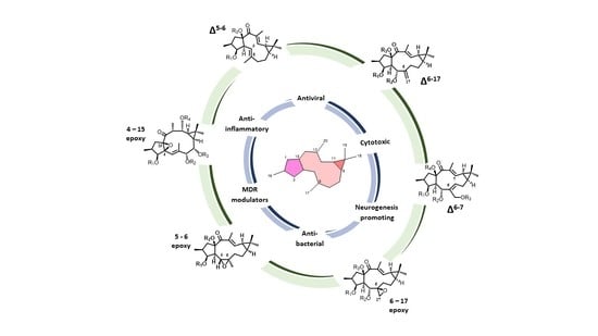
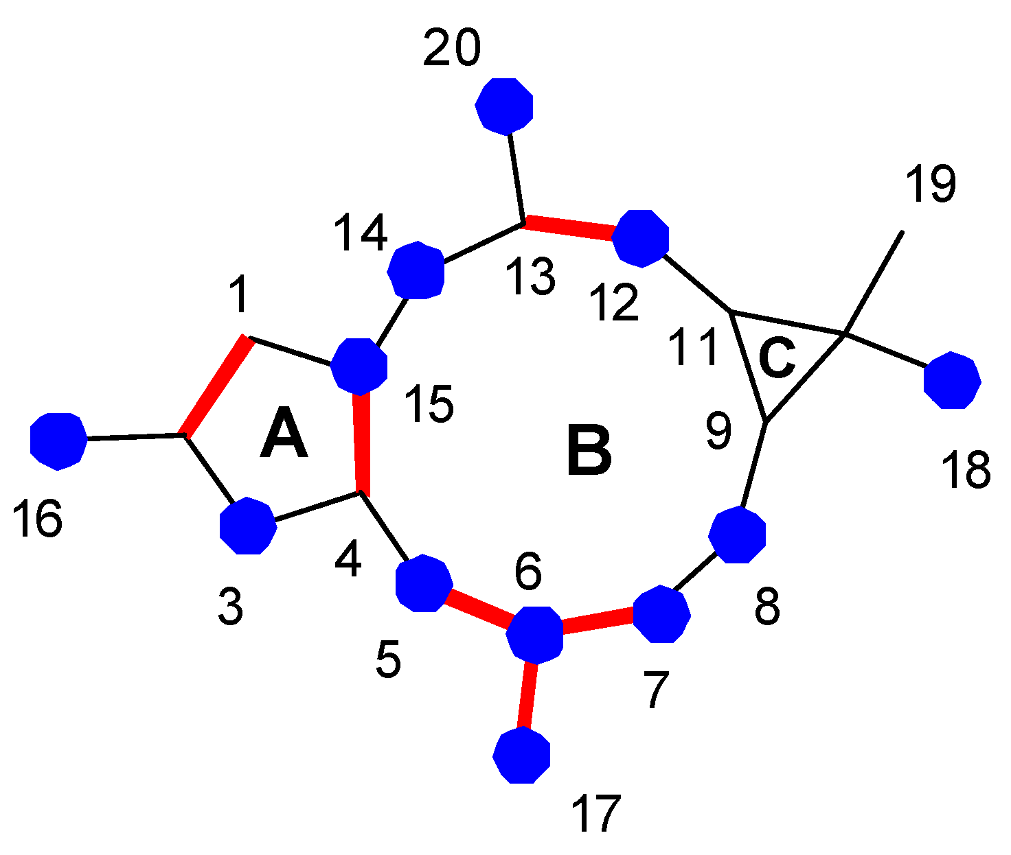
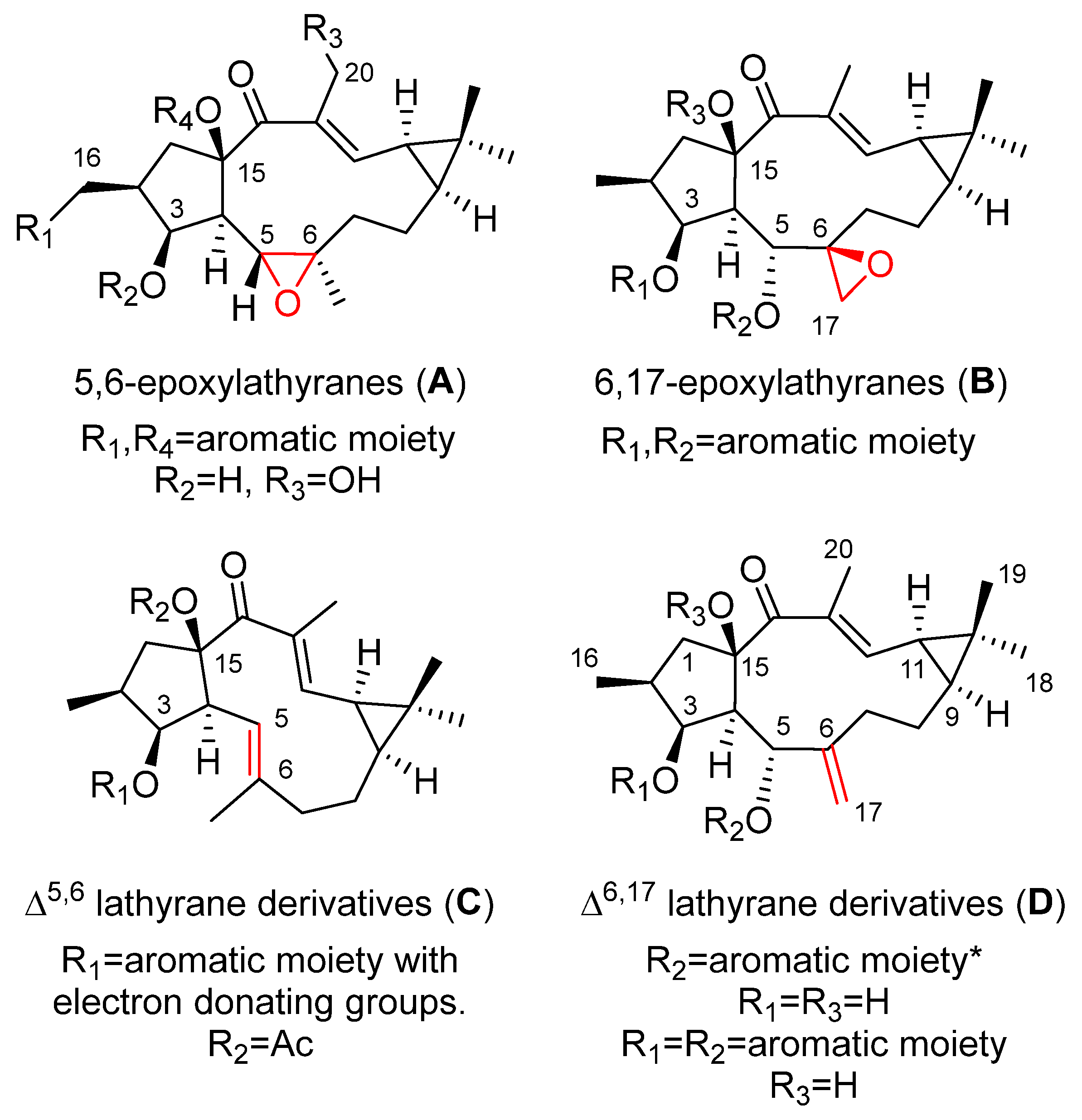
2. Bioactive Lathyranes

Lathyrane diterpenes have primarily been isolated from Euphorbia species and they accumulate in all parts of the plant. Procedures describe their extraction at room temperature by maceration and isolation through multistep separation protocols [13]. Among the biological activities shown by this group of compounds, their capability to modulate MDR [14], their cytotoxicity against cancer cell lines [13], their anti-inflammatory activity [15,16,17,18,19] and their capability to induce proliferation or differentiation of NPC stand out with potential clinical application [4,5,20]. The relevance of the activities shown by these compounds, as well as their high content in natural sources, has allowed the development of libraries of compounds through molecular derivatization, these have been directed toward the evaluation of structure–activity relationships (SAR).
Physicochemical properties of bioactive lathyranes show a tendency toward intermediate values of molecular weight (MW = 476 ± 24.9), molecular volume (MV = 497.0 ± 30.4), logarithm of octanol/water partition coefficient (log P = 6.5 ± 0.9) and topological polar surface area (TPSA = 85.4 ± 17.6) [14]. It is important to note that their relatively high lipophilicity (logP > 5, out of the range of Lipinsky’s rule-of-five) [20] constitutes a potential handicap for their use as therapeutic agents, so the use of adequate vehicles for delivery may be needed, as is discussed later.
Tables showing the chemical structures of bioactive lathyrane diterpenoids, including those described in this review, can be found in the Supplementary Material (Sections S1–S11, including Tables S1–S11). This compilation, which covers literature published until March 2022, is organized on the basis of certain structurally characteristic deacylated derivatives, their biological activities and effects, and the molecular target on which they act (where available). The compounds described in the text are labeled in the SM with the same number with which they appear in it, while those not included have been numbered consecutively as S-1 to S-100. Where available, CAS registry numbers for compounds described in Sections S1 to S11 in the Supplementary Material have been included.

3. Biological Activities

3.1. Modulation of Multidrug Resistance (MDR)

Reversion of MDR in cancer cells by lathyrane diterpenoids has been extensively studied by investigation of their ability to modulate the transport activity of P-glycoprotein (P-gp) on different tumoral cell lines [3,7,10,14,21]. Studies have shown that not only are optimal physicochemical features, particularly lipophilicity, of major importance for MDR reversal activity, but also other factors, such as specific structural characteristics, contribute to their P-gp modulating activity [14].
Four main sets of compounds have been used to perform SAR studies: the molecules are characterized by the existence of a 5,6 or a 6,17-epoxy ring (latilagascenes A–F and jolkinol B derivatives and epoxyboetiranes, respectively; 5,6-epoxylathyranes) and 6,17-epoxylathyranes, Figure 2A,B), and those possessing an endocyclic Δ5,6 (jolkinol D derivatives; Δ5,6 lathyrane derivatives Figure 2C) or exocyclic Δ6,17 double bond (lathyrol derivarives; Δ6,17 lathyrane derivatives Figure 2D).
Latilagascenes A–I (19) (Figure 3), main representatives of the 5,6-epoxylathyranes (Figure 2A), showed to be strong modulators of P-gp efflux and exhibited concentration dependence [14]. Among them, latilagascenes D–F (46) were found to be the strongest modulators of P-gp when tested in human MDR1 gene-transfected mouse lymphoma cells using the rhodamine 123 exclusion test with verapamil (VRP) as the positive control [10]. Compounds 4 and 6 showed a fluorescence activity ratio R = 168.5 and 216.8 at 4 µg/mL, respectively, indicating the concentration-dependent activity of these compounds. Latilagascene E (5) exhibited the highest effect with R = 15.3 (4 µg/mL) while VRP showed R = 2.8 at 10 µg/mL. Comparison of the activity data demonstrated by these compounds indicated that the presence of a free hydroxyl group at C-3 is very important for this activity (Figure 3). This was demonstrated by the marked increase in activity for the latilagascene D (4) when compared to that of latilagascene G (7), which only differ by the presence of a free hydroxyl group at C-3. The substitution pattern of the pentacyclic ring also affects activity, especially the presence at C-16 of an ester group, uniquely if it is aromatic (Figure 3). For example, comparison of latilagascene B (2), which has two free hydroxyl groups at C-3 and C-16, and latilagacenes D (4) and A (1), which differ in the replacement of the benzoyl group at C-16 present in latilagascene D (4) by an acetyl group in latilagascene A (1), results in a decrease of activity. The presence of a hydroxyl group at C-20 also seems to be relevant for the modulation of MDR, as can be deduced from the higher activity shown by latilagacene E (5) when compared to that of latilagacene D (4). The most active 5,6-epoxylathyrane in this study was 5, which contains two aromatic moieties at C-16 and C-15 and a free hydroxyl groups at C-3 and C-20 (Figure 3) [10]. Interestingly, a synergistic interaction was observed between doxorubicin and latilagascene B (2) (the most abundant of this class of diterpenes), which also bears a free OH group at C-3 and an aromatic moiety at C-15 (Figure 3). Both compounds were tested on human MDR1 gene-transfected mouse lymphoma cells, where ID50 for doxorubicin is 0.35 μg/mL, ID50 for 2 is 4.58 μg/mL, and ID50 for combination of both is 0.095 μg/mL, with a fractional inhibitory index of 0.292 [10].
Different behavior was observed for the 6,17-epoxylathyranes (Figure 2B; see also Figure 4) in comparison with the 5,6-epoxyderivatives, where acylation pattern was of key importance, especially the presence of aromatic residues at C-3 and C-5, in P-gp modulation (Figure 2B). Interestingly, among compounds bearing aromatic moieties at C-3 and C-5, when benzoyl was replaced with phenylacetyl groups, epoxyboetiranes E (14), F (15) and H (16), respectively (Figure 4), increased activity was observed, suggesting that the extra methylene group may enhance the interaction with P-gp binding sites [21]. On the other hand, two of the most active compounds, epoxyboetiranes J (18) and L (20) (Figure 4), do not contain aromatic residues at these positions, which shows that, although this is important, it is not an essential factor for activity. Interaction studies with doxorubicin showed synergism for combinations with compounds 1120, with the maximum observed for epoxyboetirane K (19), which presented no aromatic substituents [21].
Trying to optimize epoxylathyranes as active MDR reversal agents, several compounds were isolated from the aerial parts of E. boetica [23]. The one obtained in greater quantity was subjected to various chemical transformations, generating a library of 16 compounds, epoxylathyrol (10), epoxyboetiranes (14, 2128) and epoxycarbamoylboetiranes B and C (2930) (Figure 4), which were evaluated as P-gp-mediated MDR reversers at non-cytotoxic doses in L5178Y ABCB1 transfected mouse T-lymphoma cells, by the accumulation of rhodamine-123 assay [24]. Most of the tested compounds showed a strong P-gp-modulating activity and were also able to synergistically increase the cytotoxicity of doxorubicin, restoring its sensitivity by reversion of the ABCB1-MDR phenotype. Structure–activity relationship studies indicated that the presence of an aromatic ring on these structures significantly enhances the inhibition of rhodamine-123 efflux (see Figure 2B) [24].
Similar results were obtained for the jolkinol D derivatives (Figure 5), that are representative of lathyranes with a Δ5,6 endocyclic double bond (Figure 2C). Here, the esterification of the hydroxyl at C-3 produced an improvement in the modulating effect. For acylated alkanoyl derivatives, this increasing activity could be correlated with increasing molecular weight. However, the compounds that showed the highest activity were those with an aromatic moiety at C-3 [7], probably due to the establishment of additional hydrophobic interactions within the drug-binding pocket [7,23]. In order to investigate the effect of the presence of an aromatic residue in position C-3 of the derivatives of jolkinol D, Reis et al. generated a second generation of derivatives. Two sets of compounds were prepared, those with a free hydroxyl group at C-15 (jolkinolates A–D, 3235) (Figure 5) and those possessing an acetyl group in this position (jolkinoates N–T, 3642) (Figure 5) [8]. In both sets of compounds, electron-donating and -withdrawing groups were added to the aromatic moiety. In general, 15-acetylderivatives were more active than those possessing the C-15 free hydroxyl group and those with electron-donating groups at the aromatic moieties were the most active (Figure 2C). No significant correlations were found between the calculated physicochemical properties for these compounds and the P-gp-modulating activity, suggesting a stronger role of the sum of other structural aspects, rather than the contribution of a single physicochemical property [8]. These results reinforce the conclusion of in silico 2D and 3D QSAR studies that pointed to the conformation of the lathyrane macrocyclic scaffold and its substitution pattern (related steric and electrostatic factors) as the main determinant for MDR activity [22].
Several studies on the SAR of lathyrol derivatives, which contain a Δ6,17 exocyclic double bond (Figure 2D), have been carried out. Jiao et al. generated a library of five series of mono or diacylated lathyranes by modifying the hydroxyl moiety of C-3, C-5 or C-15 of Euphorbia Factor L3 (EFL3, 43) (Figure 6). This approach generated 37 compounds that were tested against breast cancer multidrug-resistant MCF-7/ADR cells that overexpress P-gp [25]. Eight derivatives 4449 (Figure 6) exhibited more potent chemo-reversal ability than the positive control (VRP), and three of them (45, 46 and 49) were more active than the parent structure, EFL3 (43). Comparison of the monoacylated derivatives to determine the effect of the substituent at C-5 resulted in no observed correlation between the size of aliphatic and aromatic substituents and reversal activity. Compounds with an aliphatic substituent showed poor activity profiles, while those bearing a 1-naphtylacyl or a phenylacetyl substituent at this position exhibited higher reversal fold (Figure 2D). The evaluation of lathyrol derivatives containing two ester groups at C-3 and C-5 resulted in five compounds 4650 (Figure 6) exhibiting higher MDR-modulating activities than VRP. The most active compound contained two benzyl groups (46), resulting in 4.8 times greater effectiveness than VRP in MDR-reversal activity in MCF-7/ADR cells. It also had greater activity than all E. lathyris diterpenes previously evaluated [25]. A combination of propionyl and aromatic groups at C-3 and C-5, respectively, led to the second highest activity compounds (47, 48, 49). However, two bulky aromatic groups at these two positions resulted in low activity (50) (Figure 6). Comparing compounds with the same side chain, the activity of aliphatic substituted derivatives increased by the epoxidation of C-15 and C-12 (51) and decreased when the epoxy group is between C-15 and C-11 (52) (Figure 7) [25].
An additional study into the influence of the acylation pattern on lathyrane with an exocyclic Δ6,17 double bond (Figure 2D) has been undertaken by Neto et al. [26]. This group conducted a phytochemical study of E. boetica in which euphoboetiranes A (54) and B (55) (Figure 6) were isolated in large amounts. Starting from euphoboetirane B (55), 13 new derivatives, lathyrol (53), euphoboetiranes C–I (5662), 12-hydroxyboetiranes A–D (6972) and 14β-hydroxylathyrane (73) were prepared (Figure 6). Euphoboetirane A (54), euphoboetiranes C–G (5660) and 12-hydroxyboetiranes A–C (6971) were found to be strong P-gp modulators on L5178Y-MDR mouse lymphoma cells, when tested at 20 μM. In general, acylation of one or two of the hydroxyl groups at C-3 and C-5 (Figure 2D) of lathyrol (53) led to a 6 to 42-fold increase of the activity (euphoboetiranes C–G (5660) and euphoboetirane I (62)). The strongest effects were exhibited by euphoboetiranes C, D and E (5658). Conversely, reduction of carbonyl group at C-14 of lathyrol, 14β-hydroxylathyrane (73), led to a total loss of activity [26].
In a previous study, euphoboetiranes J–O (6368) (Figure 6) besides epoxylathyrol (10) and eleven epoxyboetiranes, A (11), C–F (1215), H–L (1620) (Figure 4), were evaluated for their ability to inhibit the drug-efflux activity of Cdr1p and Mdr1p transporters of Candida albicans that were overexpressed in a Saccharomyces cerevisiae strain [27]. The products tested could be divided into three groups depending on their main structural differences: (i) those derived from epoxylathyrol, characterized by having a 6,17-epoxy function (1020) (Figure 2B); (ii) those derived from lathyrol that contain two double bonds, Δ6,17 (Figure 2D) and Δ12,13 (6365, 68) (Figure 6); (iii) and those characterized by the absence of the endocyclic Δ12,13 double bond with an extra hydroxyl function at C-12, euphoboetiranes M and N (6667) (Figure 6). Euphoboetiranes J–O (6368) showed the strongest inhibitory activity of Cdr1p efflux pump, while the most active compounds in S. cerevisiae cells overexpressing Mdr1p were epoxylahyrol (10) > epoxyboetirane J (18) > epoxyboetirane E (14) > epoxiboetirane D (13) > epoxyboetirane A (11). According to the results of the study, the inhibitory activity on both proteins seemed to be associated to the type of substituent at C-6 [27]. Therefore, euphoboetiranes J–O (6368) showed a variable inhibitory potential. The highest activity was achieved for euphoboetirane J (63), which has an unsubstituted benzoyl moiety, while the substitution of the benzene ring with a strong electron-withdrawing effect (euphoboetirane K, 64) seems to be detrimental to the inhibitory activity. On the other hand, steric hindrance seems to be the reason why euphoboetirane O (68), bearing a cinnamoyl group at C-5, showed low activity, probably due to a poor interaction with the protein binding sites. Finally, the presence of an extra hydroxyl group at C -12, euphoboetiranes M and N (6667), did not have a significant effect on the activity. Comparison of general physicochemical properties and the inhibitory activity for the three groups of compounds indicated that a preferential log P value, between 3.11 and 4.16, is required for a good inhibitory activity of the ABC-transporter Cdr1p. Nevertheless, no significant correlations were found between the calculated physicochemical properties and AD-MDR1 inhibitory activity, suggesting that other factors, such as the particular structural features of the compound, played the strongest role [27].
A study comparing the MDR inhibition activity of different types of lathyrane diterpenoids, all of them isolated from E. lathyris, was conducted by Jiao et al. [28]. Six compounds, belonging to jolkinol and isolathyrol groups (EFL7a, 74 and EFL7b, 75, respectively, Figure 8) characterized for an endocyclic double bond, Δ5,6 and Δ6,7, respectively, to the epoxylathyrol group with a 6,17-epoxy ring (EFL1, 76), and to 7-hydroxylathyrol (EFL2, 77) and lathyrol groups (EFL3, 43 and 79), which possess an exocyclic Δ6,17 double bond, (Figure 6 and Figure 8, respectively), were tested as modulators of multidrug resistance using MCF-7/ADM cell lines in vitro. According to the results, the position of the double bond at C-5 (EFL7a, 74) or C-6 (EFL7b, 75) and endo or exocyclic (75 or 43 and 79, respectively) is the key element responsible for inhibitory activity. Furthermore, when the double bond changes to epoxide (43 to 76), the effect decreases, although the influence of the change of position of the double bond is bigger. An additional very important factor is the substitution of C-7. The approximate sequence of different skeletons as MDR modulator was 7-hydroxylathyrol, jokinol > lathyrol >epoxylathyrol > isolathyrol [28].
Recently, Yang et al. also evaluated the reversing MDR activity of HepG2/ADR cells of twenty-three diterpenoids isolated from the seed of Euphorbia lathyris L. [29]. The study again included three of the four sets of characteristic lathyrane derivatives: 6,17-epoxylathyranes, and lathyranes with endocyclic Δ5,6 or exocyclic Δ6,17 double bond (Figure 8). Three of them (5,15-diacetoxy-3-nicotinoyloxylathyra-6 (17),12-dien-14-one (79), 5,15-diacetoxy-3-benzoyloxi-7-nicotinoyloxylathyra-6 (17),12-dien-14-one (80) and 3,12-O-diacetyl-8-O-(2-methyl)butyrilingol (euphorantin N, 81)) were more potent than positive control VRP [29]. Loss of activity is observed if a 6,17-epoxide is present, as shown by the comparison of compound 79 (most potent component in above series), and its epoxide at Δ6,17 double bond (compound 82), confirming the trend observed by Jiao et al. [28]. Conversely, loss of activity is also observed when the nicotinyl subtituent on C-3 in compound 79 is replaced by a cinnanoyl (compound 78) [29].

Mode of Action

Resistance to anticancer drugs is a complex process that can include more than one MDR mechanism [30]. One of the most significant is the overexpression of ATP-binding cassette (ABC) transporters, a family of proteins that mediate MDR via ATP-dependent drug efflux pumps [31]. P-gp is the most typical efflux pump in the cell membrane. The principal strategy used to overcome MDR is the development of P-gp modulators that, when co-administered with an anticancer drug, avoid its efflux and prevent chemotherapy failure [32].
P-gp inhibition may proceed through competitive (direct interaction with drug-binding sites), non-competitive or allosteric (indirect inhibition of P-gp through conformational changes which inhibit activity and translocation of protein) mechanisms. For instance, EM-E-11-4 (jolkinol B, 83) (Figure 9) does not change P-gp expression levels, but suppresses ATPase activity, which indicates a non-competitive inhibition mechanism for this lathyrane [33].
On the other hand, a recent study on the paclitaxel resistance-reversing activity of the 5,6-epoxylathyrane 83 (jolkinol B, Figure 9) has pointed at the presence of multiple mechanisms of action, where not only P-gp is involved, but also inhibition of class III β-tubulin [33]. The relevance of β-III-tubulin overexpression in connection with resistance to paclitaxel in tumors has been described [34]. β-III-tubulin has the ability to counteract the stabilizing effect of paclitaxel and other microtubule interacting agents on the microtubules dynamic nature [35], avoiding the mitotic arrest (G2/M phase arrest) which leads to cell apoptosis associated with the action of microtubule stabilizing agents such as paclitaxel [36,37]. β-III-Tubulin is capable of induction of resistance to paclitaxel and other drugs, resulting in promotion of tumor survival [36]. Compound 83 worked synergistically with paclitaxel, promoting tubulin from soluble to insoluble states and increased binding of paclitaxel to microtubules. The authors suggested that 83 may bind in the proximity of the paclitaxel binding domain, resulting in protein conformation change and enhancing paclitaxel-mediated tubulin polymerization and its binding to microtubules [33].
The potential mechanism by which compound 79 (Figure 8) regulates P-gp-dependent MDR was also studied. The results show that 79 did not influence the P-gp expression and did not inhibit the transcription and translation process, but it induced the amount of P-gp monomer in a time-dependent style [29].
In a recent study aimed at considering other potential anti-MDR mechanisms of action, the epoxylathyrane derivatives, epoxylathyrol A (10), epoxiboetiranes (14, 2128), epoxycarbamoylboetiranes B and C (2930) (Figure 4) and methoxyboetiranes A–C (8486) (Figure 10) were investigated for their potential as collateral sensitizing compounds. This was achieved using drug-sensitive and drug-resistant sublines of human tumor gastric (EPG85-257), pancreatic (EPP-181) and colon (HT-29) cell models [38]. The compounds tested were found to be more effective against the resistant gastric cell line, resulting in epoxyboetirane P (26) and methoxyboetiranes B (85) and C (86) being the most promising compounds, which were additionally investigated as apoptosis inducers. The collateral sensitivity effect elicited by methoxyboetiranes 85 and 86 seemed to be due to the induction of apoptosis via caspase-3 activation [38].

3.2. Cytotoxic

The cytotoxic activity of 5,6-epoxylathyranes (Figure 11A) has been extensively studied, the substitution pattern of the A ring influences the antiproliferative activities for this group of lathyranes, as well as the necessity for the presence of a hydroxyl group at C-20.
This was observed in studies with latilagascenes B (2), C (3) and D (4) (Figure 3) and jolkinol B (83) (Figure 9) undertaken on several human cancer cell lines that were derived from three different tumor entities: gastric (EPG85-257), pancreatic (EPP85-181) and colon cancer cells (HT-29) [39]. The activity demonstrated against gastric carcinoma was found to be dependent on the individual drug-resistant phenotype. Latilagascenes C (3) and D (4) were found to be more effective than the positive control etoposide in the drug-resistant subline EPG85-257RDB. This is associated with the overexpression of the ABC transporter MDR1/Pgp, and latilagascene B (2) also exhibited a significant activity; however, jolkinol B (83) showed a moderate activity. On the other hand, latilagascenes B–D (24) had moderate activity in MRD EPG85-257RNOV cells associated with altered topoisomerase II expression, while jolkinol B (83) exhibited a significant antineoplastic activity. None of these compounds showed significant activity against any of the three sublines of the pancreatic carcinoma cells (EPP85-181), resulting in latilagascene D (4) being inactive. Comparable results were obtained for the colon carcinoma cells [39]. Compounds 24 and jolkinol B (83) only differed in their substitution pattern of the pentacyclic ring (ring A, Figure 1). Latilagascenes B (2) and D (4) and jolkinol B (83) have a free hydroxyl group at C-3, which is acetylated in latilagascene C (3). Jolkinol B (83) is not oxidized at C-16, while latilagascene B (2) has a free hydroxyl group at this position that in latilagascenes C (3) and D (4) is esterified with an acetate and benzoylate, respectively. Comparison of the results obtained in EPG85-257 cells for these compounds suggested that the esterification of hydroxyl groups at C-3 and C-16 is important for the cytotoxic activity in multidrug-resistant EPG85-257RDB cells (Figure 11), since the presence of free hydroxyl groups at these positions decreased the activity. Additionally, oxidation at C-16 seems to also be a relevant structural requirement for the activity, as Jolkinol B (83), containing a methyl group at C-16, was the most active compound tested in multidrug-resistant EPG85-257RNOV cells, but the least active in multidrug-resistant EPG85- 257RDB cells [39].
The effect of Latilagacenes A–E (15) (Figure 3) and jolkinol B (83) (Figure 9) on human cytomegalovirus (CMV) IE antigen expression in lung cancer cells has also been investigated. Latilagascene E (5) was found to have the highest activity, while latilagascene D (4) was inactive [40]. Comparison of the activity of latilagascene D (4) and A (1), whose structures differ only at the ester group at C-16, suggests that the presence of the benzoyl moiety has a negative effect on the inhibitory of IE antigen expression of CMV. Comparatively, the activity of latilagascene D (4) and E (5) confirmed that the presence of a hydroxyl group at C-20 appears to be important in the antitumor promoter activity of these compounds [40]. However, the opposite result was obtained when the cytotoxic activity of another set of 5,6-epoxylathyranes, the euphofischers A (87) and B (88), jolkinol A (89) (Figure 12) and B (83) (Figure 9) and ebracteolata C (90) (Figure 12) was tested. They showed moderate activities against human prostate cancer cell lines C4-2B, as well as the enzalutamide-resistant cell line C4-2B/ENZR. Weak activity was observed against the human breast cancer cell line MDA-MB-231. Euphorfischer A (87), a rare example of a lathyrane diterpenoid featuring a 15-p-coumaroyl moiety and the only one that does not have a hydroxyl group at C-20, was found to be the most active, exhibiting significant toxicity against C4-2B cell line with an IC50 value of 11.3 μM [41].
In a previous study with diterpenes from E. fischeriana, jolkinol A (89) (Figure 12), the inhibitory activity on the formation of mammospheres in human breast cancer MCF-7 cells was observed [42]. Furthermore, jolkinol B (83) (Figure 9) was isolated from the roots of E. ebracteolata Hayata, and its cytotoxic activity tested against five cancer cell lines: HL-60 (human promyelocytic leukemia cell line), SMMC-7721 (human hepatocellular carcinoma cell line), A-549 (human lung cancer cell line), MCF-7 (human breast cancer cell line) and SW480 (colorectal cancer cell line), where it exhibited moderate cytotoxic effects [43].
Lathyrane diterpenoids isolated from E. lathyris, named Euphorbia factors (EF), have been subjected to several anticancer studies. Thus, among the cytotoxicity against cancer cell lines A549, MDA-MB231, KB and MCF-7, and the MDR cancer cell line KB-VIN of six compounds with different structural features, EFL1–3 (76, 77 and 43) (Figure 6 and Figure 8), EFL8–9 (91, 80) (Figure 8 and Figure 13) and the tetraol derivarive of 91 (compound 92) (Figure 13), EFL9 (80) exhibited the strongest activity against all tested cell lines and EFL2 (77) was found to be selective against KB-VIN, while 76 and 92 were inactive. The SAR studies revealed that the substitutions at C-3, C-5, C-7 and C-15 are critical for cytotoxicity, as well as cell type-selectivity [44]. A combination of acetate groups at C-5 and C-15 and benzoate groups at C-3 and C-7 seems to be required for selective cytotoxicity against KB-VIN (Figure 11D).
In comparison, the cytotoxic activity of EFL1–3 (76, 77 and 43) (Figure 6 and Figure 8), EFL7a (74) (Figure 8), EFL8 (91) and 7-hydroxylathyrol (92) (Figure 13), EFL9 (80) (Figure 8) and EFL26 (93) (Figure 13), together with other lathyrane-type diterpenes isolated from E. lathyris, EFL27 (94) and EF28 (95) (Figure 13), which are characterized for an α-orientation of their substituent at C-3, was also evaluated against several breast cancer cell lines. Results showed a strong cytotoxicity associated with 95 against the 786-0 and HepG2 cell lines. This indicated that not only the variety but also the configuration of substituent groups in this kind of compounds are of great importance for their bioactivity [45].
In a recent study looking at the composition of stems of Jatropha podagrica, a set of lathyrane-type diterpenoids were isolated and their antitumor activities in two human osteosarcoma cell lines (MG-63 and Sais-2) were evaluated [46]. Only one, jatropodagin A (96, Figure 14), had significant cytotoxic activity with IC50 values of 8.08 and 14.64 μM, against Saos-2 and MG63 cell lines, respectively.
A study into the composition of the perennial herb Euphorbia stracheyi, two new lathyrane diterpenoids, euphstrachenols A and B (9798) and nine analogues were isolated and identified from the methanol extract of its roots (Figure 15). The evaluation of their cytotoxicity against four human cancer cell lines, HGC-27 (stomach cancer), MV4-11 (leukemia), H460 (lung cancer), Skvo3 (ovarian cancer) and a murine cell line BaF3 (lymphocyte), indicated that all of them showed cytotoxicity against H460 and Skvo3 cell lines, but only six of them, euphstracehnols A (97) and B (98), 99 (Figure 15), EFL15 (euphoboetirane A, 54) (Figure 6), jolkinoate I (100) (Figure 15) and jolkinol B (83) (Figure 9) indicated moderate cytotoxicity against MV4-11 cell lines [47].
The cytotoxic activity of ingol-type diterpenes has also been evaluated. 3,12-diacetyl-7-angeloyl-8-methoxyingol (101), 7-angeloyl-12-acetyl-8-methoxyingol (102) and 3,12-diacetyl-7-hydroxy-8-methoxyingol (103) (Figure 16), isolated from E. nivulia, showed significant cytotoxic activity against Colo 205, MT2 and CEM cell lines, although some ingol derivatives had little or no activity [48].

Mode of Action

It is well documented that there is a direct relationship between resistance to cancer chemotherapy and P-gp expression [49,50,51]. One of the approaches that is being developed to overcome resistance to anticancer drugs is development of alternative drugs without cross-resistance in cancer cells exhibiting a drug-resistant phenotype [32].
Several macrocyclic diterpenes with a latilagascene skeleton (Figure 11A) have been shown to have very strong modulation of P-gp activity in resistant cancer cells, as well as apoptosis induction activity in human MDR1 gene-transfected mouse lymphoma cells [10,40,52]. Latilagascenes A (1), B (2) and C (3) (Figure 3), isolated from E. lagascae, had the ability to inhibit rhodamine 123 efflux of human MDR1 gene-transfected mouse lymphoma cells. Latilagascene B (2) was tested in combination with doxorubicine and this showed a synergistic interaction in the same resistant cell line [53]. In contrast, latilagascenes C (3) and D (4) were highly effective against the drug-resistant subline EPG85-257RDB (associated with the overexpression of the ABC transporter MDR1/P-gp) derived from gastric carcinoma. These showed moderate activity in multidrug-resistant EPG85-257RNOV cells associated with altered topoisomerase II expression [39]. However, the macrocyclic lathyrane diterpene jolkinol B (83) (Figure 9) showed significant antineoplastic activity against this multidrug-resistant variant, suggesting that the activity of these compounds depends from the individual drug-resistant phenotype.
The mechanism of action in KB-VIN cells of EFL1–3 (76, 77 and 43) (Figure 6 and Figure 8) and EFL8–9 (91, 80) (Figure 8 and Figure 13) was determined. Two different modes of action seem to be present. EFL3 (43) and EFL9 (80) acted disrupting normal cell cycle progression, whereas EFL2 (77) and EFL8 (91) induced both actin filament aggregation, as well as partial disruption of microtubule networks [44].
A study into the reversal activities of EFL1 (76) (Figure 8) against ABCB1-mediated MDR and apoptosis sensitization in K562/ADR cell was conducted by Zhang et al. in 2013 [52]. EFL1 (76) elevated sensitivity to chemotherapeutical drugs in ABCB1-mediated MDR K562/ADR cells and did not affect the sensitivity of K562, KB and MCF-7 cells to chemotherapeutic agents. The results suggest that 76 combined with chemotherapeutic drugs might be useful to overcome multidrug resistance. The study of the mechanism of action revealed that the mitochondrial pathway is involved in the apoptosis induced by EFL1 (76) [52,54].
Studies on the cytotoxic activity of EFL2 (77) (Figure 8) and EFL3 (43) (Figure 6) revealed that the antiproliferative activity in vitro shown by these compounds against lung cancer A549 cells was also mediated by apoptosis induction, via a mitochondrial pathway [55,56]. Besides, treatment of A549 cells with 43 induced release of cytochrome c in a time-dependent manner, indicating a mitochondrially mediated pathway resulting in apoptosis, presumably via Caspase 9 activation that produces the activation of the executioner Caspase 3 [55]. Similar results were obtained in a study with 77, in which it was also found that there was an increase in ROS generation, activation of caspase-9 and caspase-3 and the cleavage of FF, reinforcing the hypothesis that apoptosis of A549 cells is produced through a mitochondrial pathway [56]. On the other hand, EFL2 (77) has a potent effect on hepatocellular carcinoma (HCC) and the study of its mode of action suggested that 77 inhibited TGF-β-induced migration and proliferation in HCC cells through the inhibition of phosphorylation of AKT and STAT3 [57].
The apoptosis-inducing activity of latilagascene A–D (14, Figure 3) and jolkinol B (83) (Figure 9) in human MDR1 gene-transfected mouse lymphoma cells has been tested. Analysis of the observed effect allowed the conclusion that these lathyrane diterpenes can not only be considered effective anti-MDR agents, but also as apoptosis inducers, reinforcing the importance of them as antitumor agents [10].
Apoptosis also turned out to be the mode of action that mediates the antiproliferative effect of jatropodagin A (96) (Figure 14). This was confirmed by analyzing the morphological changes observed in saos-2 cells treated with this compound. Jatropodagin A (96) treatment caused significant morphological changes, including the appearance of membrane blebbing and granular apoptotic bodies [46].
A lathyrane designated EFL713283 (104) (Figure 17), isolated from E. lathyris, showed a strong anticancer activity. Using integrated in silico methods, the possible targets of compound 104 were explored. These studies indicated that the potential target of EFL713283 (104) might be β-tubulin, suggesting an anticancer mechanism similar to that of Taxol. Compound 104 binds to β-tubulin favoring the formation of α, β-tubulin dimmer [58].
A different mode of cytotoxic activity was found with laurifolioside (105) (Figure 17). This was found to be active against human prostate cancer (PC-3) and human breast adenocarcinoma (MCF-7) cell lines. A Drug Affinity Responsive Target Stability (DARTS) strategy showed that Clathrin heavy chain 1, a protein mainly involved in selective uptake of proteins, viruses and other macromolecules at the plasma membrane of cells, is the main target for laurifolioside (105) [59].

3.3. Anti-Inflammatory Activity

Inflammation is considered to be the body’s normal response to defend itself against pathogens and injuries, but excessive inflammation can affect the normal function of tissues and organs, leading to chronic diseases and sometimes the development of cancer [60,61]. To mitigate its effects, the body activates the immune system by recruiting immune cells and antibodies [62]. Nitric oxide is a critical signaling molecule and is considered to be the regulator of many physiological mechanisms [63]. When the immune system is chronically or overly activated, NO and inflammatory mediator cytokines such as IL-1β and IL-6 are released, which have been shown to be closely related to inflammation [64]. Pharmacological research has proven that overproduction of nitric oxide (NO] is indicative of an inflammatory process. For example, NO is over-produced and secreted out of mouse macrophages in response to bacterial lipopolysaccharide (LPS) [65]. Consequently, the most general way to prove the anti-inflammatory activity of a compound is to measure its ability to inhibit NO production.
Twenty-one compounds, 11 (Figure 4), 43 (Figure 6), 7477 and 80 (Figure 8), 91 and 93 (Figure 13), 89 (Figure 12) and 106117 (Figure 18), belonging to several groups of lathyranes, were found to inhibit the nitric oxide production in LPS-induced RAW 264.7 macrophages; nevertheless, no significant SAR could be established [18,66]. A subsequent study by Zhang et al. did allow the establishment of some essential structural characteristics for the NO production inhibitory activity. In their study, they used three different sets of compounds bearing a distinct substitution pattern: exocyclic Δ6,17 double bond (see Figure 19D), endocyclic Δ5,6 (Figure 19C) or Δ6,7 double bond (isolathyrol, (S47, Supplementary Material, Section S6)) and endocyclic (Figure 2A) or exocyclic (Figure 19B) epoxy function [16]. Those compounds with an exocyclic Δ6,17 double bond (43, 113116, 80) (Figure 6, Figure 18 and Figure 8, respectively) were the most active, showing a significant inhibitory effect higher than those showed for those with a 5α,6β-epoxy (Figure 2A) or Δ5,6 (Figure 19C) or Δ6,7 (S47, Supplementary Material, Section S6) double bonds. In contrast, compounds similar to 80 without a nicotinoyl group on C-7 were inactive, indicating a nitrogen-containing aromatic group at C-7 is probably critical for the inhibition of NO production. Furthermore, the acetylation at C-15 in 80 canceled the inhibitory effect. Comparing the activity of the compounds with a Δ5,6 double bond, only those bearing an aliphatic moiety at C-3 and a free hydroxyl group at C-17, such as 117, showed significant activity. Finally, compound 76 with a 6,17-epoxy moiety showed moderate inhibitory activity [16]. Compound 91, described as active by Lee el al. [18], was found to be inactive.
In a recent study, Zuo et al. evaluated the inhibitory activity against NO production induced by LPS in BV-2 microgial cell of twenty-one lathyrane diterpenoids [17]. Seventeen compounds, 43 (Figure 6), 74, 75 and 77 (Figure 8), 91 (Figure 13), 107110 and 113 (Figure 18) and 118125 (Figure 20), that demonstrated low cytotoxicity were found to be significantly active against LPS-induced NO overproduction in BV-2 microglial cells at 10 µM. Compounds 120 and 43 were the most potent (respectively, 6,17-epoxylathyrane (B) and Δ6,17 lathyrane (D) derivatives, Figure 19), showing an inhibitory effect approximately twice as active as the positive control resveratrol (20 µM) and reducing markedly the mRNA levels of pro-inflammatory cytokines IL6 and IL1β in LPS-stimulated BV2-cells. Structures 93 (Figure 13), 118 and 124 (Figure 20) were effective at the non-toxic concentration 1 µM, indicating that their effective concentration was lower than other tested compounds. Following the comparison of the activity shown by compounds 118, 124, 125 and EFL2 (77) (Figure 20), which are isomeric compounds in C-9 or/and C-11, it can be deduced that the anti-inflammatory activity depends on the configurations at C-9 and C-11 [17].
A systematic study of the anti-inflammatory activity of different types of lathyrane has been carried out by Wang et. al. [19]. Eleven new lathyranes along with ten known analogues, isolated from E. lathyris, were evaluated for their inhibitory activities against NO production induced by LPS in RAW264.7 macrophage cells. The tested compounds possess different patterns of substitution on the macrocyclic diterpene skeletons, including 17-hydroxyjolkinols, 17-hydroxyisolathyrols (two of them with an unusual trans-gem-dimethylcyclopropane unit), lathyrols (Figure 19D) and epoxylathyrols (Figure 19B). The most active compounds were found to be four 17-hydroxyisolathyrol derivatives, euplarisan A, B and D (126128) (Figure 21), EFL17 (108) (Figure 18) and two lathyrols, EFL28 (95) (Figure 13) and EFL32 (129) (Figure 21). Comparison of the activity showed for all the tested compounds that the diterpenoids with an endocyclic Δ5,6 double bond (108 and 128) and a benzoate at C-3 have the highest inhibitory effect (Figure 19C) [19].
The influence of different positions and type of substitution on anti-inflammatory efficacy of EFL3 (43) (Figure 6) was explored, building a library of compounds through the allylic hydroxylation at C-7 with subsequent esterification with fatty acids, substituted benzoic acids, cinnamic acid and heterocyclic acids. In addition, compounds bearing one or two of these chains at C-3 and/or C-5 were also synthesized [15]. In the first set of compounds, when the C-7 hydroxyl group was esterified, the inhibitory activity demonstrated by many derivatives was weaker than those showed by 43, being comparable or even better in those compounds with a nicotinic (80) (Figure 8), isonicotinic acids (130) or glycine at C-7 (131) (Figure 22). The second set of compounds displayed better activity than the first one. These were compounds 46 (Figure 6) and 132 (Figure 22) (Δ6,17 lathyrane, Figure 19D). The preliminary SAR showed that the esterification of the C-5 with aromatic groups is important for improving the anti-inflammatory activity of the lathyrol scaffold. When the benzene ring is substituted or changed into a heterocycle, the inhibitory activity is decreased. Higher activity is observed when both hydroxyl groups at C-3 and C-5 are esterified, particularly with a benzoyl or nicotinoyl group (Figure 19) [15].
Lathyrane-type diterpene glycosides have demonstrated anti-inflammatory activity. Kansuingol A and B (133134) (Figure 23), diterpenes isolated from the roots of Euphorbia kansui, were shown to be, at least in part, responsible for the anti-inflammatory effect shown by the butanol-soluble extract of the roots of this plant. This is because they potently inhibited the IL-6 production in HMC-1 cells stimulated by a combination of PMA and ionophore. Furthermore, 133 inhibited TNFα and IL6 mRNA expression level [67].
In a study for determining the anti-inflammatory potential of diterpenoids from E. antiquorum, three ingol-type diterpenoids (135137) (Figure 23), ingol derivatives (Figure 19E), displayed a strong NO-inhibitory effect. Euphorin D (135) differs from 136 and 137 in the configuration of the methyl group at C-2 [68].
Jatrocurcasenones H (138) and I (139) (Figure 24), two lathyrane diterpenoids containing an 7,14-oxygen bridged, were recently isolated from the roots of Jatropha curcas L., along with a number of lathyrane (jatrocurcasenones F and G and 4Z and 4E-jatrograrossidentation), which do not possess anti-inflammatory activity. Compounds 138 and 139 showed potent inhibitory activities against LPS-induced NO production in RAW264.7 cells, which suggests that the epoxy ring may play an important role in the activity [69].

Mode of Action

The ability to produce inflammatory cytokines, including IL-1β and IL-6, when cells were treated with EFL30 (113) (Figure 18) was evaluated; the results showed a significantly elevated production of them in the cell supernatant of LPS-induced RAW264.7. The investigation of the mechanism of modulation of pro-inflammatory cytokine response showed that 113 reduced the expression level of inducible nitric oxide synthase (iNOS) and NF-κB in a dose-dependent manner. In addition, compound 113 reduced the phosphorylation of IκBα and eliminates LPS-induced nuclear translocation of NF-κB. These results indicate that 113 exerts its anti-inflammatory activity by interfering with the phosphorylation of IκBα, thereby blocking the expression and nuclear translocation of NF-κB and reducing the expression of iNOS [16].
Similar results were obtained in the investigation of mode of action of EFL2 (77) (Figure 8), which showed robust inhibitory effects on the production of IL-1β and IL-6, tumor necrosis factor-α (TNF-α) and IL-8 released from LPS-stimulated RAW264.7 cells in vitro. Consistently, experiments in vivo showed that 77 exerted a potent anti-inflammatory effect by decreasing the levels of IL-1β and IL-6, TNF-α and IL-8 and myeloperoxidase (MPO) in the lung and bronchioalveolar lavage fluid. EFL2 (77) inhibition appeared to be mediated by NF-κB signaling activation, but not the MAPK pathway. Additionally, 77 decreased phosphorylation of IKK α/β and IκBα levels, and significantly suppressed p65 translocation and its DNA-binding activity [56]. In the same way, euplarisan A (126) (Figure 21) inhibited the generation of inflammatory cytokines, such as IL-1β and IL-6 and TNF-α. Additionally, 126 decreased the expression of the crucial proteins of inflammatory signaling pathway oxide synthase (iNOS), cyclooxygenase-2 (COX-2) and IκBα, further blocking the expression of NF-κB and nuclear translocation [19].
To explore the possible mode of NO inhibition, the binding interaction of compounds 135137 (Figure 23) with iNOS and COX-2 were investigated through molecular docking studies, which revealed that the three compounds had strong interactions with the protein. Compound 136 was selected to test on iNOS/COX-2 protein expression. Treatment of LPS-stimulated BV2-2 cells with 136 produced significant decrease of iNOS and COX-2 levels, indicating that these compounds may exert their anti-inflammatory effects by down-regulation of iNOS and COX-2 protein levels [68]. The same effects were observed in the study of mode of action of compounds 138 and 139 (Figure 24), where a further differential gene expression (DGE) analysis was conducted to investigate the underlying genes targeted by jatrocurcasenone I (139) in LPS-induced RAW264.7 macrophages [69]. The results demonstrated that 139 has a regulatory effect on the 587 DEGs, mainly related to immune diseases, immune systems, signaling molecules and interaction and signal transduction. 24 of those DEGs were associated with the inflammatory responses, including: interkeukin and interleukin-related genes (IL1α, 1L1β, IL1f6, IL-1rn and IL-27); chemokines (Ccl2, Ccl5, Ccl7, Ccl9, Ccl22 and CXcl10); intracellular signaling (Trim25; Bcl2a1a, Dusp1, Dusp2, Ptgs2 and End1); and transcription factor (Nr4a1). The molecular mechanism underlying the protection of RAW264.7 cells from inflammation could be due to the regulation of these genes.

3.4. Antiviral and HIV-1 Reactivation Activities

The activity of three highly functionalized ingol-type diterpenes, characterized for the presence of a phenylacetate group between their substituents, was tested on HIV-LTR transactivation by measuring the levels of GFP as a marker of HIV promoter activation. Only one of them, 8-methoxyingol 7,12-diacetate-3-phenylacetate (140) (Figure 25) induced cell-cycle arrest in Jukart-LTR-GFP cells and HIV-1-LTR promoter activation [70]. Other ingol-type diterpene, 3,12-di-O-acetyl-8-O-tigloylingol (ELAC, 141) (Figure 25), also showed the ability to reactivate HIV-1 latency in a concentration-dependent manner [71]. The study of its mechanism of action revealed that this activity could be mediated by PKC activation. 141 induced IκBα phosphorylation and its subsequent degradation suggesting that the activation can be induced by NF-κB. Furthermore, probably other transcription factors can probably contribute to the reactivation of HIV-1 from latency, as JNK and ERK were also phosphorylated [71].
On the other hand, some lathyrane diterpenoids have shown activity against HIV-1 replication. In the study on the activity shown by the diterpenes isolated from the roots of E. Micractina, 15-cinnamoyloxy-3-hydroxylathyra-5,12-dien-14-one (142) (Figure 25) exhibited weak activity, with an IC50 value of 8.2 μM, while the positive control zidovudine gave 0.05 μM [72]. With these precedents and encouraged by the activity displayed by related diterpenes [73], the anti-HIV activity of ethanolic extracts from Euphorbia lathyris seeds of different origins was evaluated [74]. Although the results showed a significant activity of all the tested extracts, the isolated diterpenoids, most of them lathyrane types, were inactive against HIV viral replication, indicating a possible synergetic effect [74].
Activity of this kind of diterpenes against other viruses has also been investigated. For example, in the phytochemical study of the buds of Wikstroemia chamaedaphne Meisn, it was observed that only laurifolioside A (143) (Figure 25), one of the six lathyrane diterpenes isolated, exhibited potential anti-hepatitis B virus activity [75].
In an investigation to determine the antifeedant and antiviral activity of diterpenoids from the fresh roots of E. jolkinii resulted in jolkinol A (89) (Figure 12) showing significant anti-respiratory syncytial virus (RSV) activity [76].
The ethyl acetate extract of the trunk bark of Sandwithia guyanensis showed a strong anti-chikungunya virus (CHIKV) activity. The study of the most active fractions led to the identification of 19 diterpenoids with different carbon skeletons. Only one of them, possessing a lathyrane framework and was named jatrointelone K (144) (Figure 25), showed a moderate anti-CHIKV activity [77].

3.5. Neurogenesis Promoting

Three studies have been carried out on the neurogenesis promotion activity of lathyrane-type diterpenes. In the first one, four lathyranes, 140, ELAC (141) (Figure 25) and 145146 (Figure 26) were tested [5]. The culture in a bFGF-supplement medium with the different lathyranes produces a significant increase in neurosphere size when cultured with 141, without modifying neurosphere size. No effect was observed when treated with 145, 146 or the acetylated derivative of 141 on C-7-OH (AcELAC, 147) (Figure 26). Studies on mechanism of action showed that classical PKCs, especially PKCβ, are responsible for ELAC-dependent NPC proliferation. The lathyrane ELAC (141) also stimulated NPC proliferation in vivo [5].
In a second study, the effects of epoxyboetirane A (11) (Figure 4) and euphoboetirane A (54) (Figure 6) on NPC proliferation was evaluated. Epoxyboetirane A (11) resulted in an increase in the size of neurospheres in cultures stimulated with a combination of the growth factors EGF and bFGF, while no statistically significant effect was observed in cultures stimulated with either EGF or bFGF alone. Euphoboetirane A (54) induced a smaller increase in the neurosphere size and in the presence of both growth factors. On the other hand, the capacity to form neurospheres was not affected by any of the two compounds, since no changes were found in their number [78].
Finally, the capacity to activate PKC, to facilitate neuregulin 1 release and to promote neuroblast differentiation and survival in cultures of subventricular zone of lathyrane diterpene EOF2 (146) (Figure 26) was proved. Local infusion of 146 in mechanical cortical injuries induced neuroblast enrichment within the perilesional area and, when it was administered intranasally, promoted migration of neuroblasts from the subventicular zone toward the injury. The results show that the neural differentiation of NPC in neuroblasts promoted by 146 was mediated by novel PKCs, especially PKCϴ [4].
The comparison of the structures of 140, 141 and 145147 demonstrates the important role of the substituents on C-3-OH, C-7-OH and C-8-OH in the interaction with the PKCs of this class of diterpenes. Further studies are needed to determine the essential structural elements in each type of activity and how these influence the interaction with PKCs.

3.6. Others

3.6.1. Anticholestasis

Recent studies have suggested that lathyrane diterpenoids could serve as a new type of human pregnane X receptor (hPXR) agonist for future anticholestasis drug development. In a bioassay-guided isolation on E. lathyris extract looking for an hPXR agonistic compound, 16 lathyrane diterpenoids were isolated [79]. Five of them were novel compounds and were named euphlathyrinoid A–E (148152) (Figure 27). Known compounds EFL1–2 (76, 77) (Figure 8), EFL3 (43) (Figure 6), EFL8–9 (91, 80) (Figure 8 and Figure 13), EFL11 (116) (Figure 18), EFL30 (113) (Figure 18) and EFL31 (121) (Figure 20) were identified. The major components, EFL2 (77) and EFL3 (43), were subjected to chemical transformations, generating a lathyrane library containing 34 compounds that were used for a systematic hPXR agonistic activity investigation. The main structural modification included the Δ6,17 terminal double bond, the α,β-unsaturated ketone, the cyclopropane ring, as well as the substituents on C-3, C-5, C-7, and C-15. Three natural products, euphlathyrinoid C (150), EFL30 (113) and EFL31 (121), and a synthetic compound, were excluded from the hPXR agonistic screening because of their cytotoxicity on HEF293T cells. The remaining compounds were subjected to the hPXR agonistic screening, most of them showing a potent activity at the concentration of 10 μM. The most active of them were subjected to further assays, showing a dose–response-dependent activity. EFL9 (80) (Figure 8), was found to be the most active compound, this could significantly activate hPXR, as evidenced by the hPXR reporter gene activity (6.9-fold), and up-regulate the expressions of hPXR downstream key genes CYP3A4, CYP2B6, and MDR1. SAR studies revealed that acyloxy substituents on C-7, specifically a nicotinoyl group, and the presence of 14-carbonyl were essential to activity [79].

3.6.2. Antibacterial

Japodagrin (153) (Figure 28) is a compound with an unusual epoxy function in ring A of the lathyrane skeleton. It was isolated from the root of Jatropha podagrica Hook [80], a plant that had shown a range of biological activities, including antibacterial. Its antibacterial activity and that of four other known diterpenoids, 4Z-jatrogrossidentadion (154), 15-epi-4Z-jatrogrossidentadion (155), 2-hydroxyisojatrogrossidion (156), and 2-epi-hydroxyisojatrogrossidion (157) (Figure 28), also isolated from this plant, were tested against Bacillus subtilis (ATCC 6051), Staphylococcus aureus (ATCC 25923) Escherichia coli (ATCC 25922) and Pseudomonas aeruginosa (ATCC 27853). All of which resulted in different levels of microbial killing against gram-negative bacteria (B. subtillus and S. aureus), but were found to be inactive in disk assays against the gram-positive ones (E.coli and P. aeruginosa) [80]. All compounds were tested in standard disk assays, at 20 μg/disk doses, with streptomycin and gentamycin as positive controls. The most active compound was 2-epi-hydroxyisojatrogrossidion (157), which gave the same inhibition against B. subtillus and S. aureus than streptomycin (35 and 26 mm inhibition zones, respectively), and similar to the inhibition exerted by gentamycin against B. subtillus and S. aureus (34 and 28 mm, respectively). Compounds 156, 155, 154 and 153 were less active, with respective inhibition zones of 31, 17, 20 and 16 mm against B. subtillus and of 21, 9, 10 and 12 mm versus S. aureus.

3.6.3. Vascular-Relaxing Activity

Remarkably, vasodilatory activity was observed using a phenylephrine-induced vasoconstriction model with lathyrane diterpenoids isolated from E. micractina [72]. Vascular relaxation was found in compounds containing a benzyloxy group, primarily at C-15, (99) (Figure 15) and 158162 (Figure 29), indicating an important role of this group in vascular-relaxing activity.

3.6.4. Gastrointestinal Toxicity

E. lathyris L. is a traditional Chinese medicine. It produces several side effects, including irritation of the gastrointestinal tract, which manifests as severe diarrhea [81]. The studies carried out to identify the components responsible for this effect indicated that the diterpene EFL1 (76) (Figure 8) could be the main cause of the diarrhea [82]. The intestinal toxicity of 76 and the underlying mechanisms were studied using nematode Caenorhabditis elegans as the model. The results show that toxicity was related to intestinal oxidative damage, disorder transportation, down-regulated cell junctions, enhanced rhythm behavior, muscle contraction and injured GABAergic neurons [83]. The gastric cytotoxicity of 76 and the underlying mechanism in human gastric mucosa epithelium cells was also investigated. EFL1 (76) induced oxidative stress, activation of mitochondrial-mediated apoptosis in GES-1 cells and authophagy via inhibition of the PI3K/AKT/mTOR pathway [84].

3.6.5. Osteoclastogenesis Inhibition

In another study carried out on the biological activity of the diterpene EFL1 (76) (Figure 8), compound 76 was proposed as a potential therapeutic agent to prevent or treat bone-related diseases caused by an excess of osteoclast, since it inhibited osteoclast differentiation by regulating cellular redox status and induced Fas-mediated apoptosis in osteoclast [85].

3.6.6. Inhibition of 11β-HDS1

11β-Hydroxysteroid dehydrogenase Type 1 (11β-HDS1) is an attractive therapeutic target for the treatment of a number of diseases such as obesity and metabolic and cardiovascular disease [86]. Ingol type diterpenes were investigated for an inhibitory effect on human and mouse 11β-HDS1 from a set of compounds isolated from Euphorbia antiquorum [87]. Unfortunately, none showed inhibitory effects on human 11β-HDS1 and only three, euphorantins A (163) (Figure 30) and N (81) (Figure 8) and 3,12-diacetyl-7-benzoyl-8-nicotinylingol (164) (Figure 30), inhibited mouse 11β-HDS1. The cytotoxicity of these compounds against HL-60 (human premyelocytic leukemia) and A-549 (human lung adenocarcinoma) cells was also evaluated, as well as their inhibitory ability against the PTP1B enzyme, but none of them were active [87].

3.6.7. Induction of Lysosomal Biosynthesis

A set of ingol-type diterpenoids, isolated from E. resinifera, named euphorblins A–Q and EFRL4 (EOF2, 146) (Figure 26) were evaluated as inducing agents of lysosomal biosynthesis. Between them, EFRL4 (146), euphorblin B (165) and D (166) (Figure 30) proved to be promising compounds for the development of drugs for the treatment of lysosome-related diseases, as they showed high capacities to induce lysosome biosynthesis at different doses and concentrations [88].

3.6.8. PGE2 Inhibition

E. nivulia, a succulent Euphorbiaceae found in the tropics, is known for its therapeutic properties against diseases like bronchitis and rheumatism [81]. The phytochemical study of the latex of this plant afforded five ingol-type diterpenes which were tested for the lipo-polysaccharide (LPS)-induced PGE2 inhibition activity. Only that with a free hydroxyl group on C-3, 7-angeloyl-12-acetyl-8-methoxyingol (102) (Figure 17) showed significant PGE2 inhibition, the IC50 value (0.003 µM) being less than that of the reference compound celecoxib (0.050 μM) [89].

4. Drug Delivery

As discussed above, lathyranes are a class of diterpenes which have a range of intriguing biological activities that are ripe for exploitation, yet, in striking contrast with other well developed diterpenoids such as taxanes, very few have made it into the clinic. This may be due to the side effect profiles of the drugs, the complexity of formulating them into dosage forms that are compatible with a range of routes for drug delivery, as well as the $1400 million [90] cost of bringing a small molecule drug to market. That said, the potential of these compounds in the treatment of cancer and neurological disease suggests a significantly untouched resource in commercially rich fields of clinical treatment.
The oral route for delivery of pharmaceuticals is the most widely used and accepted [91]. In order to get an idea of the druglikeness of lathyranes, Lipinski’s rule of 5 [20], originally based on physicochemical profiles of phase 2 drugs, can be applied. This rule (of thumb) predicts that poor drug absorption or permeation for a molecule across the gastrointestinal tract is less likely if there are more than 5 hydrogen bond donors, 10 hydrogen bond acceptors, the molecular weight is greater than 500 and the calculated Log P (ClogP) is greater than 5. This clearly indicates that for oral drug delivery, many diterpenoid lathyranes have good druglikeness, as they are relatively small and lipophilic. However, the rule does not take into account gut anatomy and physiology, which includes, for example, the coverage of the gastrointestinal wall lining with a barrier of mucous that has to be diffused through, or the presence of active transporters, as well as the efficiency, first pass metabolism in the liver, all of which directly affect the pharmacokinetic profile of the drug. This led to a preference for clinically used diterpenoids, such as the anticancer drugs Paclitaxel and Docetaxel, to be formulated for the intravenous route. However, new approaches are being considered to avoid the invasive nature of the intravenous route, for example, currently in clinical trial is a novel oral formulation of the anticancer drug, Docetaxel. It is co-administered with cytochrome P450 3A4 and the P-gp inhibitor ritonavir; this strategy has demonstrated increased oral bioavailability [92].
Diterpenoid formulation to a medicinal product can be complex and lengthy. Paclitaxel was first isolated from the Pacific Yew (Taxus brevifolia) in 1971, yet was not approved for medical use until 1993. Originally solubilized in 75% polyethylene glycol, it was found that activity of Paclitaxel was significantly reduced. This was resolved using cremophor EL, a polyethoxylated caster oil as a non-ionic solubilizer and emulsifier. In aqueous solution and by implication in serum, Cremophor EL forms star shaped micelles. These macromolecular structures have been problematic in triggering a part of the human innate immune system called the Complement system [93]. Activation of the complement system causes hypersensitivity reactions, which at worst can result in patient death [94]. Mechanistically, it is the excipient cremophor EL and not the drug that causes the clinically relevant toxicity. Mechanistically, these side effects are driven by portions of the cremophor EL’s hydrophobic components incorporating into lipoproteins [95,96]. This results in loss of hydrophobic character in the micelles, resulting in the formation of droplets of 100–300 nm in size and it is these that are believed to trigger the complement system. These types of reactions have brought focus onto the immunological interactions of nano-carrier vehicles with the immune system [97]. This research is enabling safer design and has produced a range of carriers with far greater performance in terms of stealth characteristics for avoidance of the immune system, thus providing safer intravenous systems for potential inclusion of lathyranes. These include a wide range of formulations, including pegylated liposomes [98], cubosomes [99], dendrimers [100], albumin nanoparticles [101], as well as complement inhibitors [102].
Recently, the phytochemical lathyrane ELAC (141) (Figure 25) has been found to promote endogenous neurogenesis in adult CD1 mouse brains [5]. As the active pharmacophores do not fit into the ‘rule of 5′ in terms of excess hydrogen bond donors, it is unlikely that it is absorbed across the gastrointestinal tract readily. Also, its lipophilicity is such that it is not high enough to diffuse across the barrier, and structurally would also be potentially prone to the effects of efflux pumps such as P-gp. A number of strategies have recently been developed that may facilitate the uptake of lathyranes, most likely using the intravenous route for drug delivery to the brain via the blood brain barrier (BBB). For example, self-assembled nanoligand drug carriers based on phage display peptide target cerebral endothelial cells through transferrin receptor-reaching neurons and microglial cells [100], without damage to the BBB. Significantly, more invasive methods have been developed to open the BBB in situ to drugs. This includes techniques such as magnetic resonance-guided focused ultrasound. This technique transiently permeabilises the BBB and was applied in a recent human trial for opening in eloquent primary motor cortex [103]. Another intriguing method for drug delivery to the brain has been reported where transient breakdown of the BBB can be achieved via modulation of the UNC5B receptor [104] that controls BBB integrity. If the time frame of the opening of the brain endothelial cells can be effectively controlled, this may facilitate a less invasive method for opening the BBB to small molecules such as lanthyranes. An alternative route is the intra-nasal, where the drug non-invasively bypasses the BBB following transport along the olfactory and trigeminal nerves [105]. The primary process of drug absorption is through the mucus and is ideal for lipophilic and low molecular weight drugs (<400 Da), even with poor stability in fluids [106]. Significant advances are being made in this area of drug delivery, which seems well suited to the diversity of centrally acting lathyranes and may offer a much safer route that does not affect the homeostatic condition of the brain.
Clearly, there is significant potential for lathyranes to be used orally. The problem is that this results in the non-site-specific delivery of the drug to the whole body. Nanocarriers have the advantage in the case of current solid tumour treatment of site-specific targeting. This reduces the side-effect profile of the drug, such as cardiotoxicity associated with administration of free doxorubicin. Therefore, nano-carriers offer an excellent targeting system for administration of lathyranes with anti-cancer activity in the future.

5. Conclusions

This review demonstrates the importance of lathyranes as privileged structures in drug design. The ability of the lathyrane framework to direct the functional groups that decorate its skeleton in a well-defined space makes the interaction of these compounds with various targets possible, turning them in potential therapeutic agents to prevent or treat different types of diseases, even though no clinical applications have yet been described.
Modulation of multidrug resistance (MDR) has enough studies to draw some generalized structure/activity correlations. Four groups of lathyrane derivatives have been shown to promote reversal of MDR (Figure 2), where functionalization patterns are relevant. For 5,6-epoxy-lathyranes (see A in Figure 2), presence of aromatic moieties at C-15 and C-16, as well as free hydroxyl groups at C-3 and C-20 is required. On the other hand, active 6,17-epoxy lathyranes (see B in Figure 2) and Δ6,17 double bond lathyranes (see D in Figure 2) bear aromatic moieties at C-5 and C-20. Presence of an aromatic moiety at C-3 also plays a key role in activity in Δ5,6 double bond lathyranes (see C in Figure 2). The mechanism of action for most of the above-mentioned compounds seems to involve inhibition of P-gp drug efflux pump, but it is not the only one involved, as shown by jolkinol B (83).
For cytotoxic activity, less clear-cut tendencies can be drawn. 5,6-epoxylathyranes Figure 11A) with ester groups at C3, C-15 and C-16 have shown activity against multidrug-resistant EPG85-257RDB cells, through apoptosis induction. On the other hand, Δ6,17 double bond lathyrane derivatives (Figure 11D) with aromatic moieties at C-3 and C-7 and acetate groups at C-5 and C-15 have been described as active against KB-VIN cells. For these compounds, two different modes of action seem to be present. EFL3 (43) and EFL9 (80) acted disrupting normal cell cycle progression, whereas EFL2 (77) and EFL8 (91) induced both actin filament aggregation, as well as partial disruption of microtubule networks.
Several structural variants of the lathyrane skeleton have been evaluated for anti-inflammatory activity, inhibition of NO production out of mouse macrophages in response to bacterial lipopolysaccharide (LPS) being the standard test. 6,7-Epoxy-lathyranes (Figure 19B) with aromatic moieties at C-3 and C-5 induce this effect, as well as Δ5,6 double bond lathyrane derivatives (Figure 19C) with a wide variety of substituents. Compounds with a Δ6,17 double bond (19D) have also proved to be active, such as EFL30 (113). Regulation of iNOS and COX-2 protein levels seems to be involved in the mode of action of the above compounds, as well as in ingol derivative 136 (Figure 19E).
Further biological activities, such as antiviral, antibacterial, vascular-relaxing, gastrointestinal toxicity, osteoclastogenesis inhibition, inhibition of 11β-HDS1, induction of lysosomal activity and PGE2 inhibition have been described, with very few results to draw general conclusions. On the other hand, neurogenesis-promoting experiments have shown that ingol derivatives with a free hydroxyl group at C-7, such as ELAC (141), promote neural progenitor cell (NPC) proliferation in a classical PKC-dependent manner. Finally, anticholestasis evaluation of lathyrane derivatives, through agonist interaction with the human pregnane X receptor (hPXR), shows that Δ6,17 double bond lathyrane derivatives were active as long as they bear acyloxy groups at C-7 and a carbonyl group at C-14 is present.
Currently, a preference for intravenous administration is observed in clinically used diterpenoids. However, significant advances are being made in the area of drug delivery, which may offer a much safer and non-invasive route for administration of lathyrane-type diterpenes.

6. Forward-Looking Outlook and Recommendations

The current trend for publications (Web of Science) on the subject of lathyranes is increasing. Continuation of this upward trend will reveal new and as of yet unexpected biological activity. For example, induction of neurogenesis for potential application in brain disorders. Much of the current work is focused on inhibition of efflux pumps and it is expected that these molecules are very likely to have other biological activities as well. A combination of drug efflux pump inhibition and cytotoxic activity could prove a viable clinical tool against chemotherapeutically resistant brain tumours such as glioma. Alternatively, one can readily envisage a delivery system having two molecules to bring about efflux inhibition and the other to invoke cytotoxicity.
The lack of lathyranes in clinical use, in spite of extraordinary potential, suggests that there is a significant rate-limiting step to the clinic. In part, this may be due to toxicity; however, in the case of those molecules that have acceptable profiles for use in humans, formulation to a medicinal form is problematic. It would therefore be worthwhile to apply the nanotechnology that has made paclitaxel so effective in treating metastatic breast cancer and HIV Karposi sarcoma. Although the majority of pharmaceutical companies may not have used carriers such as liposomes, micelles and albumin, application of this technology to the delivery of lathyranes may circumvent problems of solubility that was inherent in the 30-year time frame to formulate paclitaxel. Embracing the nano approach may now be economically viable because of the unique biological activity inherent in many of the lathyrane diterpenes and open up the potential of these compounds for medicinal use.

Supplementary Materials

The following supporting information can be downloaded at: https://www.mdpi.com/article/10.3390/ph15070780/s1: Section S1: Bioactive lathyrol derivatives; Table S1: Bioactive lathyrol derivatives: biological activities, observed effects and molecular targets; Section S2: Bioactive 7-hydroxylathyrol and 7-oxylathyrol derivatives; Table S2: Bioactive 7-hydroxylathyrol and 7-oxolathyrol derivatives: biological activities, observed effects and molecular targets; Section S3: Bioactive 12-hydroxylathyrol derivatives; Table S3: Bioactive 12-hydroxylathyrol derivatives: biological activities, observed effects and molecular targets; Section S4: Bioactive 12,15-epoxylathyrol derivatives; Table S4: Bioactive 12,15-epoxylathyrol derivatives: biological activities, observed effects and molecular targets; Section S5: Bioactive 6,17-epoxylathyrol derivatives; Table S5: Bioactive 6,17-epoxylathyrol derivatives: biological activities, observed effects and molecular targets; Section S6: Bioactive isolathyrol derivatives; Table S6: Bioactive isolathyrol derivatives: biological activities, observed effects and molecular targets; Section S7: Bioactive jolkinol derivatives; Table S7: Bioactive jolkinol derivatives: biological activities, observed effects and molecular targets; Section S8: Bioactive 15-deacyljolkinol B derivatives; Table S8: Bioactive 15-deacyljolkinol B derivatives: biological activities, observed effects and molecular targets; Section S9: Bioactive laurifolioside derivatives; Table S9: Bioactive laurifolioside derivatives: biological activities, observed effects and molecular targets; Section S10: Bioactive jatrogrossidion derivatives; Table S10: Bioactive jatrogrossidion derivatives: biological activities, observed effects and molecular targets; Section S11: Bioactive ingol derivatives; Table S11: Bioactive ingol derivatives: biological activities, observed effects and molecular targets. References [107,108,109,110,111,112,113,114,115,116,117,118,119,120,121,122,123,124,125,126,127] are cited in the supplementary materials.

Author Contributions

Conceptualization, supervision, writing, original draft preparation, R.H.-G.; literature review, figures, SM, F.V. and A.E.; literature review, writing review and editing tables and figures, A.J.M.-S.; writing drug delivery section, A.C.H. All authors have read and agreed to the published version of the manuscript.

Funding

This research was funded by Ministerio de Ciencia, Innovación y Universidades, (grant RTI2018-099908-B-C22) and Consejeria de transformación económica, industria, conocimiento y Universidades, Junta de Andalucía (grant P18-RT-2655).

Institutional Review Board Statement

Not applicable.

Informed Consent Statement

Not applicable.

Data Availability Statement

No new data were created or analyzed in this study. Data sharing is not applicable to this article.

Acknowledgments

Administrative and technical support was provided by the Faculty of Science, University of Cádiz. F.V. thanks the University of Cádiz for the financing of a predoctoral contract.

Conflicts of Interest

The authors declare no conflict of interest.

References

  1. Shi, Q.W.; Su, X.H.; Kiyota, H. Chemical and Pharmacological Research of the Plants in Genus Euphorbia. Chem. Rev. 2008, 108, 4295–4327. [Google Scholar] [CrossRef] [PubMed]
  2. Durán-Peña, M.J.; Botubol Ares, J.M.; Collado, I.G.; Hernández-Galán, R. Biologically Active Diterpenes Containing a Gem-Dimethylcyclopropane Subunit: An Intriguing Source of PKC Modulators. Nat. Prod. Rep. 2014, 31, 940–952. [Google Scholar] [CrossRef] [PubMed]
  3. Vasas, A.; Hohmann, J. Euphorbia Diterpenes: Isolation, Structure, Biological Activity, and Synthesis (2008–2012). Chem. Rev. 2014, 114, 8579–8612. [Google Scholar] [CrossRef] [PubMed] [Green Version]
  4. Domínguez-García, S.; Geribaldi-Doldán, N.; Gómez-Oliva, R.; Ruiz, F.A.; Carrascal, L.; Bolívar, J.; Verástegui, C.; Garcia-Alloza, M.; Macías-Sánchez, A.J.; Hernández-Galán, R.; et al. A Novel PKC Activating Molecule Promotes Neuroblast Differentiation and Delivery of Newborn Neurons in Brain Injuries. Cell Death Dis. 2020, 11, 262. [Google Scholar] [CrossRef] [PubMed]
  5. Murillo-Carretero, M.; Geribaldi-Doldán, N.; Flores-Giubi, E.; García-Bernal, F.; Navarro-Quiroz, E.A.; Carrasco, M.; Macías-Sánchez, A.J.; Herrero-Foncubierta, P.; Delgado-Ariza, A.; Verástegui, C.; et al. ELAC (3,12-Di-O-Acetyl-8-O-Tigloilingol), a Plant-Derived Lathyrane Diterpene, Induces Subventricular Zone Neural Progenitor Cell Proliferation through PKCβ Activation. Br. J. Pharmacol. 2017, 174, 2373–2392. [Google Scholar] [CrossRef] [PubMed] [Green Version]
  6. Evans, B.E.; Rittle, K.E.; Bock, M.G.; DiPardo, R.M.; Freidinger, R.M.; Whitter, W.L.; Lundell, G.F.; Veber, D.F.; Anderson, P.S.; Chang, R.S.L.; et al. Methods for Drug Discovery: Development of Potent, Selective, Orally Effective Cholecystokinin Antagonists. J. Med. Chem. 1988, 31, 2235–2246. [Google Scholar] [CrossRef]
  7. Reis, M.; Ferreira, R.J.; Santos, M.M.M.; Dos Santos, D.J.V.A.; Molnár, J.; Ferreira, M.J.U. Enhancing Macrocyclic Diterpenes as Multidrug-Resistance Reversers: Structure-Activity Studies on Jolkinol D Derivatives. J. Med. Chem. 2013, 56, 748–760. [Google Scholar] [CrossRef]
  8. Reis, M.A.; Ahmed, O.B.; Spengler, G.; Molnár, J.; Lage, H.; Ferreira, M.J.U. Exploring Jolkinol D Derivatives to Overcome Multidrug Resistance in Cancer. J. Nat. Prod. 2017, 80, 1411–1420. [Google Scholar] [CrossRef]
  9. Duarte, N.; Járdánházy, A.; Molnár, J.; Hilgeroth, A.; Ferreira, M.J.U. Synergistic Interaction between P-Glycoprotein Modulators and Epirubicine on Resistant Cancer Cells. Bioorganic Med. Chem. 2008, 16, 9323–9330. [Google Scholar] [CrossRef]
  10. Duarte, N.; Varga, A.; Cherepnev, G.; Radics, R.; Molnár, J.; Ferreira, M.J.U. Apoptosis Induction and Modulation of P-Glycoprotein Mediated Multidrug Resistance by New Macrocyclic Lathyrane-Type Diterpenoids. Bioorganic Med. Chem. 2007, 15, 546–554. [Google Scholar] [CrossRef]
  11. Jian, B.; Zhang, H.; Han, C.; Liu, J. Anti-Cancer Activities of Diterpenoids Derived from Euphorbia Fischeriana Steud. Molecules 2018, 23, 387. [Google Scholar] [CrossRef] [PubMed] [Green Version]
  12. Jian, B.; Zhang, H.; Liu, J. Structural Diversity and Biological Activities of Diterpenoids Derived from Euphorbia Fischeriana Steud. Molecules 2018, 23, 935. [Google Scholar] [CrossRef] [PubMed] [Green Version]
  13. Vasas, A.; Rédei, D.; Csupor, D.; Molnár, J.; Hohmann, J. Diterpenes from European Euphorbia Species Serving as Prototypes for Natural-Product-Based Drug Discovery. Eur. J. Org. Chem. 2012, 1, 5115–5130. [Google Scholar] [CrossRef]
  14. Ferreira, M.J.U.; Duarte, N.; Reis, M.; Madureira, A.M.; Molnár, J. Euphorbia and Momordica Metabolites for Overcoming Multidrug Resistance. Phytochem. Rev. 2014, 13, 915–935. [Google Scholar] [CrossRef]
  15. Wang, W.; Wu, Y.; Li, C.; Yang, Y.; Li, X.; Li, H.; Chen, L. Synthesis of New Lathyrane Diterpenoid Derivatives from Euphorbia Lathyris and Evaluation of Their Anti-Inflammatory Activities. Chem. Biodivers. 2020, 17, 531. [Google Scholar] [CrossRef] [PubMed]
  16. Zhang, C.Y.; Wu, Y.L.; Zhang, P.; Chen, Z.Z.; Li, H.; Chen, L.X. Anti-Inflammatory Lathyrane Diterpenoids from Euphorbia Lathyris. J. Nat. Prod. 2019, 82, 756–764. [Google Scholar] [CrossRef]
  17. Zuo, Q.; Mu, H.-Y.; Gong, Q.; Ding, X.; Wang, W.; Zhang, H.-Y.; Zhao, W.-M. Diterpenoids from the Seeds of Euphorbia Lathyris and Their Effects on Microglial Nitric Oxide Production. Fitoterapia 2021, 150, 104834. [Google Scholar] [CrossRef]
  18. Lee, J.W.; Jin, Q.; Jang, H.; Kim, J.G.; Lee, D.; Kim, Y.; Hong, J.T.; Lee, M.K.; Hwang, B.Y. Lathyrane-Type Diterpenoids from the Seeds of Euphorbia lathyris L. with Inhibitory Effects on NO Production in RAW 264.7 Cells. Chem. Biodivers. 2018, 15, 144. [Google Scholar] [CrossRef]
  19. Wang, Y.; Song, Z.; Guo, Y.; Xie, H.; Zhang, Z.; Sun, D.; Li, H.; Chen, L. Diterpenoids from the Seeds of Euphorbia Lathyris and Their Anti-Inflammatory Activity. Bioorganic Chem. 2021, 112, 104944. [Google Scholar] [CrossRef]
  20. Lipinski, C.A.; Lombardo, F.; Dominy, B.W.; Feeney, P.J. Experimental and Computational Approaches to Estimate Solubility and Permeability in Drug Discovery and Development Settings. Adv. Drug Deliv. Rev. 1997, 23, 3–25. [Google Scholar] [CrossRef]
  21. Vieira, C.; Duarte, N.; Reis, M.A.; Spengler, G.; Madureira, A.M.; Molnár, J.; Ferreira, M.J.U. Improving the MDR Reversal Activity of 6,17-Epoxylathyrane Diterpenes. Bioorganic Med. Chem. 2014, 22, 6392–6400. [Google Scholar] [CrossRef] [PubMed] [Green Version]
  22. Baptista, R.; Ferreira, R.J.; Dos Santos, D.J.; Fernandes, M.; Ferreira, M. Optimizing the Macrocyclic Diterpenic Core toward the Reversal of Multidrug Resistance in Cancer. Future Med. Chem. 2016, 8, 629–645. [Google Scholar] [CrossRef] [PubMed]
  23. Ferreira, R.J.; Dos Santos, D.J.V.A.; Ferreira, M.J.U.; Guedes, R.C. Toward a Better Pharmacophore Description of P-Glycoprotein Modulators, Based on Macrocyclic Diterpenes from Euphorbia Species. J. Chem. Inf. Model. 2011, 51, 1315–1324. [Google Scholar] [CrossRef]
  24. Matos, A.M.; Reis, M.; Duarte, N.; Spengler, G.; Molnár, J.; Ferreira, M.J.U. Epoxylathyrol Derivatives: Modulation of ABCB1-Mediated Multidrug Resistance in Human Colon Adenocarcinoma and Mouse T-Lymphoma Cells. J. Nat. Prod. 2015, 78, 2215–2228. [Google Scholar] [CrossRef] [PubMed]
  25. Jiao, W.; Wan, Z.; Chen, S.; Lu, R.; Chen, X.; Fang, D.; Wang, J.; Pu, S.; Huang, X.; Gao, H.; et al. Lathyrol Diterpenes as Modulators of P-Glycoprotein Dependent Multidrug Resistance: Structure-Activity Relationship Studies on Euphorbia Factor L3 Derivatives. J. Med. Chem. 2015, 58, 3720–3738. [Google Scholar] [CrossRef]
  26. Neto, S.; Duarte, N.; Pedro, C.; Spengler, G.; Molnár, J.; Ferreira, M.J.U. Effective MDR Reversers through Phytochemical Study of Euphorbia Boetica. Phytochem. Anal. 2019, 30, 498–511. [Google Scholar] [CrossRef] [Green Version]
  27. Mónico, A.; Nim, S.; Duarte, N.; Rawal, M.K.; Prasad, R.; Di Pietro, A.; Ferreira, M.J.U. Lathyrol and Epoxylathyrol Derivatives: Modulation of Cdr1p and Mdr1p Drug-Efflux Transporters of Candida Albicans in Saccharomyces Cerevisiae Model. Bioorganic Med. Chem. 2017, 25, 3278–3284. [Google Scholar] [CrossRef]
  28. Jiao, W.; Dong, W.; Li, Z.; Deng, M.; Lu, R. Lathyrane Diterpenes from Euphorbia Lathyris as Modulators of Multidrug Resistance and Their Crystal Structures. Bioorganic Med. Chem. 2009, 17, 4786–4792. [Google Scholar] [CrossRef]
  29. Yang, T.; Wang, S.; Li, H.; Zhao, Q.; Yan, S.; Dong, M.; Liu, D.; Chen, X.; Li, R. Lathyrane Diterpenes from Euphorbia Lathyris and the Potential Mechanism to Reverse the Multi-Drug Resistance in HepG2/ADR Cells. Biomed. Pharmacother. 2020, 121, 109663. [Google Scholar] [CrossRef]
  30. Gottesman, M.M.; Lavi, O.; Hall, M.D.; Gillet, J.-P. Toward a Better Understanding of the Complexity of Cancer Drug Resistance. Annu. Rev. Pharmacol. Toxicol. 2016, 56, 85–102. [Google Scholar] [CrossRef]
  31. Sarkadi, B.; Homolya, L.; Szakács, G.; Váradi, A. Human Multidrug Resistance ABCB and ABCG Transporters: Participation in a Chemoimmunity Defense System. Physiol. Rev. 2006, 86, 1179–1236. [Google Scholar] [CrossRef]
  32. Ozben, T. Mechanisms and Strategies to Overcome Multiple Drug Resistance in Cancer. FEBS Lett. 2006, 580, 2903–2909. [Google Scholar] [CrossRef] [PubMed] [Green Version]
  33. Liu, Q.; Cai, P.; Guo, S.; Shi, J.; Sun, H. Identification of a Lathyrane-Type Diterpenoid EM-E-11-4 as a Novel Paclitaxel Resistance Reversing Agent with Multiple Mechanisms of Action. Aging 2020, 12, 3713–3729. [Google Scholar] [CrossRef]
  34. Sève, P.; Dumontet, C. Is Class III β-Tubulin a Predictive Factor in Patients Receiving Tubulin-Binding Agents? Lancet Oncol. 2008, 9, 168–175. [Google Scholar] [CrossRef]
  35. Moktan, S.; Ryppa, C.; Kratz, F.; Raucher, D. A Thermally Responsive Biopolymer Conjugated to an Acid-Sensitive Derivative of Paclitaxel Stabilizes Microtubules, Arrests Cell Cycle, and Induces Apoptosis. Invest. New Drugs 2012, 30, 236–248. [Google Scholar] [CrossRef] [PubMed]
  36. Gan, P.P.; Pasquier, E.; Kavallaris, M. Class III β-Tubulin Mediates Sensitivity to Chemotherapeutic Drugs in Non–Small Cell Lung Cancer. Cancer Res. 2007, 67, 9356–9363. [Google Scholar] [CrossRef] [PubMed] [Green Version]
  37. Stepien, A.; Grzanka, A.; Grzanka, D.; Andrzej Szczepanski, M.; Helmin-Basa, A.; Gackowska, L. Taxol-Induced Polyploidy and Cell Death in CHO AA8 Cells. Acta Histochem. 2010, 112, 62–71. [Google Scholar] [CrossRef]
  38. Reis, M.A.; Matos, A.M.; Duarte, N.; Ahmed, O.B.; Ferreira, R.J.; Lage, H.; Ferreira, M.J.U. Epoxylathyrane Derivatives as MDR-Selective Compounds for Disabling Multidrug Resistance in Cancer. Front. Pharmacol. 2020, 11, 599. [Google Scholar] [CrossRef]
  39. Lage, H.; Duarte, N.; Coburger, C.; Hilgeroth, A.; Ferreira, M.J.U. Antitumor Activity of Terpenoids against Classical and Atypical Multidrug Resistant Cancer Cells. Phytomedicine 2010, 17, 441–448. [Google Scholar] [CrossRef]
  40. Pusztai, R.; Ferreira, M.J.U.; Duarte, N.; Engi, H.; Molnar, J. Macrocyclic Lathyrane Diterpenes as Antitumor Promoters. Anticancer Res. 2007, 27, 201–206. [Google Scholar]
  41. Li, J.; He, J.; Yang, C.; Yan, X.; Yin, Z. Cytotoxic Lathyrane Diterpenoids from the Roots of Euphorbia Fischeriana. Rec. Nat. Prod. 2020, 14, 286–291. [Google Scholar] [CrossRef]
  42. Kuang, X.; Li, W.; Kanno, Y.; Yamashita, N.; Kikkawa, S.; Azumaya, I.; Nemoto, K.; Asada, Y.; Koike, K. Euphorins A–H: Bioactive Diterpenoids from Euphorbia Fischeriana. J. Nat. Med. 2016, 70, 412–422. [Google Scholar] [CrossRef]
  43. Yuan, W.J.; Yang, G.P.; Zhang, J.H.; Zhang, Y.; Chen, D.Z.; Li, S.L.; Di, Y.T.; Hao, X.J. Three New Diterpenes with Cytotoxic Activity from the Roots of Euphorbia Ebracteolata Hayata. Phytochem. Lett. 2016, 18, 176–179. [Google Scholar] [CrossRef] [Green Version]
  44. Teng, Y.N.; Wang, Y.; Hsu, P.L.; Xin, G.; Zhang, Y.; Morris-Natschke, S.L.; Goto, M.; Lee, K.H. Mechanism of Action of Cytotoxic Compounds from the Seeds of Euphorbia Lathyris. Phytomedicine 2018, 41, 62–66. [Google Scholar] [CrossRef] [PubMed]
  45. Wang, J.X.; Wang, Q.; Zhen, Y.Q.; Zhao, S.M.; Gao, F.; Zhou, X.L. Cytotoxic Lathyrane-Type Diterpenes from Seeds of Euphorbia Lathyris. Chem. Pharm. Bull. 2018, 66, 674–677. [Google Scholar] [CrossRef] [Green Version]
  46. Yuan, H.T.; Li, Q.F.; Tian, T.; Zhang, C.Y.; Huang, Z.Q.; Fan, C.X.; Mei, K.; Zhou, J.; Zhai, X.X.; Li, S.B.; et al. Lathyrane Diterpenoids from Jatropha Podagrica and Their Antitumor Activities in Human Osteosarcoma Cells. Nat. Prod. Res. 2020, 35, 5089–5095. [Google Scholar] [CrossRef]
  47. Ye, Y.; Liu, G.H.; Dawa, D.; Ding, L.S.; Cao, Z.X.; Zhou, Y. Cytotoxic Diterpenoids from the Roots of Euphorbia Stracheyi. Phytochem. Lett. 2020, 36, 183–187. [Google Scholar] [CrossRef]
  48. Ravikanth, V.; Lakshmi Niranjan Reddy, V.; Vijender Reddy, A.; Ravinder, K.; Prabhakar Rao, T.; Siva Ram, T.; Anand Kumar, K.; Prakesh Vamanarao, D.; Venkateswarlu, Y. Three New Ingol Diterpenes from Euphorbia Nivulia: Evaluation of Cytotoxic Activity. Chem. Pharm. Bull. 2003, 51, 431–434. [Google Scholar] [CrossRef] [Green Version]
  49. Kröger, N.; Achterrath, W.; Hegewisch-Becker, S.; Mross, K.; Zander, A.R. Current Options in Treatment of Anthracycline-Resistant Breast Cancer. Cancer Treat. Rev. 1999, 25, 279–291. [Google Scholar] [CrossRef]
  50. Penson, R.T.; Oliva, E.; Skates, S.J.; Glyptis, T.; Fuller, A.F.; Goodman, A.; Seiden, M.V. Expression of Multidrug Resistance-1 Protein Inversely Correlates with Paclitaxel Response and Survival in Ovarian Cancer Patients: A Study in Serial Samples. Gynecol. Oncol. 2004, 93, 98–106. [Google Scholar] [CrossRef]
  51. Pokharel, D.; Roseblade, A.; Oenarto, V.; Lu, J.; Bebawy, M. Proteins Regulating the Intercellular Transfer and Function of P-Glycoprotein in Multidrug-Resistant Cancer. Ecancermedicalscience 2017, 11, 768. [Google Scholar] [CrossRef] [PubMed] [Green Version]
  52. Zhang, J.Y.; Lin, M.T.; Yi, T.; Tang, Y.N.; Fan, L.L.; He, X.C.; Zhao, Z.Z.; Chen, H.B. Apoptosis Sensitization by Euphorbia Factor L1 in ABCB1-Mediated Multidrug Resistant K562/ADR Cells. Molecules 2013, 18, 12793–12808. [Google Scholar] [CrossRef] [PubMed]
  53. Zhang, J.Y.; Mi, Y.J.; Chen, S.P.; Wang, F.; Liang, Y.J.; Zheng, L.S.; Shi, C.J.; Tao, L.Y.; Chen, L.M.; Chen, H.B.; et al. Euphorbia Factor L1 Reverses ABCB1-Mediated Multidrug Resistance Involving Interaction with ABCB1 Independent of ABCB1 Downregualtion. J. Cell. Biochem. 2011, 112, 1076–1083. [Google Scholar] [CrossRef]
  54. Duarte, N.; Gyémánt, N.; Abreu, P.M.; Molnár, J.; Ferreira, M.J.U. New Macrocyclic Lathyrane Diterpenes, from Euphorbia Lagascae, as Inhibitors of Multidrug Resistance of Tumour Cells. Planta Med. 2006, 72, 162–168. [Google Scholar] [CrossRef]
  55. Zhang, J.Y.; Liang, Y.J.; Chen, H.B.; Zheng, L.S.; Mi, Y.J.; Wang, F.; Zhao, X.Q.; Wang, X.K.; Zhang, H.; Fu, L.W. Structure Identification of Euphorbia Factor L3 and Its Induction of Apoptosis through the Mitochondrial Pathway. Molecules 2011, 16, 3222–3231. [Google Scholar] [CrossRef] [Green Version]
  56. Zhang, Q.; Zhu, S.; Cheng, X.; Lu, C.; Tao, W.; Zhang, Y.; William, B.C.; Cao, X.; Yi, S.; Liu, Y.; et al. Euphorbia Factor L2 Alleviates Lipopolysaccharide-Induced Acute Lung Injury and Inflammation in Mice through the Suppression of NF-ΚB Activation. Biochem. Pharmacol. 2018, 155, 444–454. [Google Scholar] [CrossRef] [PubMed]
  57. Lu, F.; Huayun, Z.; Weiwei, T.; Li, L.; Xin, S.; Ming, Z.; Dongdong, S. Euphorbia Factor L2 Inhibits TGF-β-Induced Cell Growth and Migration of Hepatocellular Carcinoma through AKT/STAT3. Phytomedicine 2019, 62, 152931. [Google Scholar] [CrossRef]
  58. Chang, S.; He, H.Q.; Kong, R.; Xie, Z.J.; Hu, J.P. Study on Molecular Recognition between Euphorbia Factor L713283 and β -Tubulin via Molecular Simulation Methods. J. Chem. 2015, 2015, 9238. [Google Scholar] [CrossRef] [Green Version]
  59. Dal Piaz, F.; Vera Saltos, M.B.; Franceschelli, S.; Forte, G.; Marzocco, S.; Tuccinardi, T.; Poli, G.; Nejad Ebrahimi, S.; Hamburger, M.; De Tommasi, N.; et al. Drug Affinity Responsive Target Stability (DARTS) Identifies Laurifolioside as a New Clathrin Heavy Chain Modulator. J. Nat. Prod. 2016, 79, 2681–2692. [Google Scholar] [CrossRef]
  60. Medzhitov, R. Origin and Physiological Roles of Inflammation. Nature 2008, 454, 428–435. [Google Scholar] [CrossRef]
  61. Atsumi, T.; Singh, R.; Sabharwal, L.; Bando, H.; Meng, J.; Arima, Y.; Yamada, M.; Harada, M.; Jiang, J.J.; Kamimura, D.; et al. Inflammation Amplifier, a New Paradigm in Cancer Biology. Cancer Res. 2014, 74, 8–14. [Google Scholar] [CrossRef] [PubMed] [Green Version]
  62. Baummann, H.; Gauldie, J. The Acute Phase Response. Inmunol. Today 1994, 75, 74–80. [Google Scholar] [CrossRef]
  63. Edwards, A.D. The Pharmacology of Inhaled Nitric Oxide. Arch. Dis. Child. 1995, 72, F127–F130. [Google Scholar] [CrossRef] [PubMed] [Green Version]
  64. Mario Scuri, M.; Samsell, L.; Piedimonte, G. The Role of Neurotrophins in Inflammation and Allergy. Inflamm. Allergy-Drug Targets 2010, 9, 173–180. [Google Scholar] [CrossRef] [PubMed]
  65. Liu, L.; Xu, D.; Liu, P.; Liu, F.; Dai, L.; Yan, H.; Wen, F. Effects of Calcium Gluconate on Lipopolysaccharide-Induced Acute Lung Injury in Mice. Biochem. Biophys. Res. Commun. 2018, 503, 2931–2935. [Google Scholar] [CrossRef]
  66. Lee, J.W.; Lee, C.; Jin, Q.; Jang, H.; Lee, D.; Lee, H.-J.; Shin, J.W.; Han, S.B.; Hong, J.T.; Kim, Y.; et al. Diterpenoids from the Roots of Euphorbia Fischeriana with Inhibitory Effects on Nitric Oxide Production. J. Nat. Prod. 2016, 79, 126–131. [Google Scholar] [CrossRef]
  67. Kim, Y.M.; Ahn, J.; Chae, H.S.; Choi, Y.H.; Kim, J.; Chin, Y.W. Two New Lathyrane-Type Diterpenoid Glycosides with IL-6 Production Inhibitory Activity from the Roots of Euphorbia Kansui. Bioorganic Med. Chem. Lett. 2018, 28, 1207–1210. [Google Scholar] [CrossRef]
  68. An, L.; Liang, Y.; Yang, X.; Wang, H.; Zhang, J.; Tuerhong, M.; Li, D.; Wang, C.; Lee, D.; Xu, J.; et al. NO Inhibitory Diterpenoids as Potential Anti-Inflammatory Agents from Euphorbia Antiquorum. Bioorganic Chem. 2019, 92, 103237. [Google Scholar] [CrossRef]
  69. Huang, J.-D.; Zhang, C.; Xu, W.-J.; Lian, C.-L.; Liu, X.-M.; Wang, C.-F.; Liu, J.-Q. New Lathyrane Diterpenoids with Anti-Inflammatory Activity Isolated from the Roots of Jatropha curcas L. J. Ethnopharmacol. 2021, 268, 113673. [Google Scholar] [CrossRef]
  70. Daoubi, M.; Marquez, N.; Mazoir, N.; Benharref, A.; Hernández-Galán, R.; Muñoz, E.; Collado, I.G. Isolation of New Phenylacetylingol Derivatives That Reactivate HIV-1 Latency and a Novel Spirotriterpenoid from Euphorbia Officinarum Latex. Bioorganic Med. Chem. 2007, 15, 4577–4584. [Google Scholar] [CrossRef]
  71. Avila, L.; Perez, M.; Sanchez-Duffhues, G.; Hernández-Galán, R.; Muñoz, E.; Cabezas, F.; Quiñones, W.; Torres, F.; Echeverri, F. Effects of Diterpenes from Latex of Euphorbia Lactea and Euphorbia Laurifolia on Human Immunodeficiency Virus Type 1 Reactivation. Phytochemistry 2010, 71, 243–248. [Google Scholar] [CrossRef] [PubMed]
  72. Tian, Y.; Xu, W.; Zhu, C.; Lin, S.; Li, Y.; Xiong, L.; Wang, S.; Wang, L.; Yang, Y.; Guo, Y.; et al. Lathyrane Diterpenoids from the Roots of Euphorbia Micractina and Their Biological Activities. J. Nat. Prod. 2011, 74, 1221–1229. [Google Scholar] [CrossRef]
  73. Yan, S.L.; Li, Y.H.; Chen, X.Q.; Liu, D.; Chen, C.H.; Li, R.T. Diterpenes from the Stem Bark of Euphorbia Neriifolia and Their in Vitro Anti-HIV Activity. Phytochemistry 2018, 145, 40–47. [Google Scholar] [CrossRef] [PubMed]
  74. Wang, S.; Li, H.; Liu, D.; Zhao, Q.; Yang, T.; Li, R.; Chen, X. Diterpenoids from the Seeds of Euphorbia Lathyris and Their in Vitro Anti-HIV Activity. Chem. Nat. Compd. 2020, 56, 78–85. [Google Scholar] [CrossRef]
  75. Li, S.F.; Jiao, Y.Y.; Zhang, Z.Q.; Chao, J.B.; Jia, J.; Shi, X.L.; Zhang, L.W. Diterpenes from Buds of Wikstroemia Chamaedaphne Showing Anti-Hepatitis B Virus Activities. Phytochemistry 2018, 151, 17–25. [Google Scholar] [CrossRef] [PubMed]
  76. Huang, C.-S.; Luo, S.-H.; Li, Y.-L.; Li, C.-H.; Hua, J.; Liu, Y.; Jing, S.-X.; Wang, Y.; Yang, M.-J.; Li, S.-H. Antifeedant and Antiviral Diterpenoids from the Fresh Roots of Euphorbia Jolkinii. Nat. Prod. Bioprospect. 2014, 4, 91–100. [Google Scholar] [CrossRef] [Green Version]
  77. Remy, S.; Olivon, F.; Desrat, S.; Blanchard, F.; Eparvier, V.; Leyssen, P.; Neyts, J.; Roussi, F.; Touboul, D.; Litaudon, M. Structurally Diverse Diterpenoids from Sandwithia Guyanensis. J. Nat. Prod. 2018, 81, 901–912. [Google Scholar] [CrossRef]
  78. Flores-Giubi, E.; Geribaldi-Doldán, N.; Murillo-Carretero, M.; Castro, C.; Durán-Patrón, R.; MacÍas-Sánchez, A.J.; Hernández-Galán, R. Lathyrane, Premyrsinane, and Related Diterpenes from Euphorbia Boetica: Effect on in Vitro Neural Progenitor Cell Proliferation. J. Nat. Prod. 2019, 82, 2517–2528. [Google Scholar] [CrossRef] [Green Version]
  79. Huang, D.; Wang, R.-M.; Li, W.; Zhao, Y.-Y.; Yuan, F.-Y.; Yan, X.-L.; Chen, Y.; Tang, G.-H.; Bi, H.-C.; Yin, S. Lathyrane Diterpenoids as Novel HPXR Agonists: Isolation, Structural Modification, and Structure–Activity Relationships. ACS Med. Chem. Lett. 2021, 12, 1159–1165. [Google Scholar] [CrossRef]
  80. Aiyelaagbe, O.O.; Adesogan, K.; Ekundayo, O.; Gloer, J.B. Antibacterial Diterpenoids from Jatropha Podagrica Hook. Phytochemistry 2007, 68, 2420–2425. [Google Scholar] [CrossRef]
  81. Chopra, R.N.; Nayar, S.L.; Chopra, I.C. Glossary of Indian Medicinal Plants; CSIR: New Delhi, India, 1956. [Google Scholar]
  82. Zhu, A.; Zhang, T.; Wang, Q. The Phytochemistry, Pharmacokinetics, Pharmacology and Toxicity of Euphorbia Semen. J. Ethnopharmacol. 2018, 227, 41–55. [Google Scholar] [CrossRef] [PubMed]
  83. Zhu, A.; Ji, Z.; Zhao, J.; Zhang, W.; Sun, Y.; Zhang, T.; Gao, S.; Li, G.; Wang, Q. Effect of Euphorbia Factor L1 on Intestinal Barrier Impairment and Defecation Dysfunction in Caenorhabditis Elegans. Phytomedicine 2019, 65, 3102. [Google Scholar] [CrossRef] [PubMed]
  84. Zhu, A.; Sun, Y.; Zhong, Q.; Yang, J.; Zhang, T.; Zhao, J.; Wang, Q. Effect of Euphorbia Factor L1 on Oxidative Stress, Apoptosis, and Autophagy in Human Gastric Epithelial Cells. Phytomedicine 2019, 64, 152929. [Google Scholar] [CrossRef] [PubMed]
  85. Hong, S.E.; Lee, J.; Seo, D.H.; In Lee, H.; Ri Park, D.; Lee, G.R.; Jo, Y.J.; Kim, N.; Kwon, M.; Shon, H.; et al. Euphorbia Factor L1 Inhibits Osteoclastogenesis by Regulating Cellular Redox Status and Induces Fas-Mediated Apoptosis in Osteoclast. Free Radic. Biol. Med. 2017, 112, 191–199. [Google Scholar] [CrossRef] [PubMed]
  86. Han, M.L.; Shen, Y.; Wang, G.C.; Leng, Y.; Zhang, H.; Yue, J.M. 11β-HSD1 Inhibitors from Walsura Cochinchinensis. J. Nat. Prod. 2013, 76, 1319–1327. [Google Scholar] [CrossRef] [PubMed]
  87. Qi, W.-Y.; Zhang, W.-Y.; Shen, Y.; Leng, Y.; Gao, K.; Yue, J.-M. Ingol-Type Diterpenes from Euphorbia Antiquorum with Mouse 11β-Hydroxysteroid Dehydrogenase Type 1 Inhibition Activity. J. Nat. Prod. 2014, 77, 1452–1458. [Google Scholar] [CrossRef]
  88. Zhao, N.D.; Ding, X.; Song, Y.; Yang, D.Q.; Yu, H.L.; Adelakun, T.A.; Qian, W.D.; Zhang, Y.; Di, Y.T.; Gao, F.; et al. Identification of Ingol and Rhamnofolane Diterpenoids from Euphorbia Resinifera and Their Abilities to Induce Lysosomal Biosynthesis. J. Nat. Prod. 2018, 81, 1209–1218. [Google Scholar] [CrossRef]
  89. Ravikanth, V.; Niranjan Reddy, V.L.; Prabhakar Rao, T.; Diwan, P.V.; Ramakrishna, S.; Venkateswarlu, Y. Macrocyclic Diterpenes from Euphorbia Nivulia. Phytochemistry 2002, 59, 331–335. [Google Scholar] [CrossRef]
  90. Maxmen, A. Busting the Billion-Dollar Myth: How to Slash the Cost of Drug Development. Nature 2016, 536, 389–390. [Google Scholar] [CrossRef] [Green Version]
  91. Hunter, A.C.; Elsom, J.; Wibroe, P.P.; Moghimi, S.M. Polymeric Particulate Technologies for Oral Drug Delivery and Targeting: A Pathophysiological Perspective. Maturitas 2012, 73, 5–18. [Google Scholar] [CrossRef]
  92. Vermunt, M.; Marchetti, S.; Beijnen, J. Pharmacokinetics and Toxicities of Oral Docetaxel Formulations Co-Administered with Ritonavir in Phase I Trials. Clin. Pharmacol. Adv. Appl. 2021, 13, 21–32. [Google Scholar] [CrossRef] [PubMed]
  93. Szebeni, J.; Alving, C.R.; Savay, S.; Barenholz, Y.; Priev, A.; Danino, D.; Talmon, Y. Formation of Complement-Activating Particles in Aqueous Solutions of Taxol: Possible Role in Hypersensitivity Reactions. Int. Immunopharmacol. 2001, 1, 721–735. [Google Scholar] [CrossRef]
  94. Hunter, A.C.; Moghimi, S.M. Therapeutic Synthetic Polymers: A Game of Russian Roulette? Drug Discov. Today 2002, 7, 998–1001. [Google Scholar] [CrossRef]
  95. Kessel, D.; Woodburn, K.; Decker, D.; Sykes, E. Fractionation of Cremophor EL Delineates Components Responsible for Plasma Lipoprotein Alterations and Multidrug Resistance Reversal. Oncol. Res. 1995, 7, 207–212. [Google Scholar] [PubMed]
  96. Woodburn, K.; Sykes, E.; Kessel, D. Interactions of Solutol HS 15 and Cremophore EL with Plasma Lipoproteins. Int. J. Biochem. Cell Biol. 1995, 27, 693–699. [Google Scholar] [CrossRef]
  97. Moghimi, S.M.; Andersen, A.J.; Ahmadvand, D.; Wibroe, P.P.; Andresen, T.L.; Hunter, A.C. Material Properties in Complement Activation. Adv. Drug Deliv. Rev. 2011, 63, 1000–1007. [Google Scholar] [CrossRef]
  98. Gabizon, A.; Shmeeda, H.; Tahover, E.; Kornev, G.; Patil, Y.; Amitay, Y.; Ohana, P.; Sapir, E.; Zalipsky, S. Development of Promitil®, a Lipidic Prodrug of Mitomycin c in PEGylated Liposomes: From Bench to Bedside. Adv. Drug Deliv. Rev. 2020, 154–155, 13–26. [Google Scholar] [CrossRef]
  99. Yu Helvig, S.; Woythe, L.; Pham, S.; Bor, G.; Andersen, H.; Moein Moghimi, S.; Yaghmur, A. A Structurally Diverse Library of Glycerol Monooleate/Oleic Acid Non-Lamellar Liquid Crystalline Nanodispersions Stabilized with Nonionic Methoxypoly(Ethylene Glycol) (MPEG)-Lipids Showing Variable Complement Activation Properties. J. Colloid Interface Sci. 2021, 582, 906–917. [Google Scholar] [CrossRef]
  100. Wu, L.P.; Ahmadvand, D.; Su, J.; Hall, A.; Tan, X.; Farhangrazi, Z.S.; Moghimi, S.M. Crossing the Blood-Brain-Barrier with Nanoligand Drug Carriers Self-Assembled from a Phage Display Peptide. Nat. Commun. 2019, 10, 12554. [Google Scholar] [CrossRef] [Green Version]
  101. Pilati, D.; Howard, K.A. Albumin-Based Drug Designs for Pharmacokinetic Modulation. Expert Opin. Drug Metab. Toxicol. 2020, 16, 783–795. [Google Scholar] [CrossRef]
  102. Gifford, G.; Vu, V.P.; Banda, N.K.; Holers, V.M.; Wang, G.; Groman, E.V.; Backos, D.; Scheinman, R.; Moghimi, S.M.; Simberg, D. Complement Therapeutics Meets Nanomedicine: Overcoming Human Complement Activation and Leukocyte Uptake of Nanomedicines with Soluble Domains of CD55. J. Control. Release 2019, 302, 181–189. [Google Scholar] [CrossRef] [PubMed]
  103. Abrahao, A.; Meng, Y.; Llinas, M.; Huang, Y.; Hamani, C.; Mainprize, T.; Aubert, I.; Heyn, C.; Black, S.E.; Hynynen, K.; et al. First-in-Human Trial of Blood–Brain Barrier Opening in Amyotrophic Lateral Sclerosis Using MR-Guided Focused Ultrasound. Nat. Commun. 2019, 10, 12426. [Google Scholar] [CrossRef] [Green Version]
  104. Boyé, K.; Geraldo, L.H.; Furtado, J.; Pibouin-Fragner, L.; Poulet, M.; Kim, D.; Nelson, B.; Xu, Y.; Jacob, L.; Maissa, N.; et al. Endothelial Unc5B Controls Blood-Brain Barrier Integrity. Nat. Commun. 2022, 13, 28785. [Google Scholar] [CrossRef] [PubMed]
  105. Crowe, T.P.; Greenlee, M.H.W.; Kanthasamy, A.G.; Hsu, W.H. Mechanism of Intranasal Drug Delivery Directly to the Brain. Life Sci. 2018, 195, 44–52. [Google Scholar] [CrossRef]
  106. Xu, J.; Tao, J.; Wang, J. Design and Application in Delivery System of Intranasal Antidepressants. Front. Bioeng. Biotechnol. 2020, 8, 6882. [Google Scholar] [CrossRef] [PubMed]
  107. Li, X.; Hu, J.; Wang, B.; Sheng, L.; Liu, Z.; Yang, S.; Li, Y. Inhibitory effects of herbal constituents on P-glycoprotein in vitro and in vivo: Herb–drug interactions mediated via P-gp. Toxicol. Appl. Pharmacol. 2014, 275, 163–175. [Google Scholar] [CrossRef]
  108. Wang, Q.; Zhen, Y.Q.; Gao, F.; Huang, S.; Zhou, X.L. Five New Diterpenoids from the Seeds of Euphorbia lathyris. Chem. Biodivers. 2018, 15, 9–16. [Google Scholar] [CrossRef]
  109. Lu, J.; Li, G.; Huang, J.; Zhang, C.; Zhang, L.; Zhang, K.; Li, P.; Lin, R.; Wang, J. Lathyrane-type diterpenoids from the seeds of Euphorbia lathyris. Phytochemistry 2014, 104, 79–88. [Google Scholar] [CrossRef]
  110. Sousa, I.J.; Ferreira, M.J.U.; Molnár, J.; Fernandes, M.X. QSAR studies of macrocyclic diterpenes with P-glycoprotein inhibitory activity. Eur. J. Pharm. Sci. 2013, 48, 542–553. [Google Scholar] [CrossRef]
  111. Engi, H.; Vasas, A.; Rédei, D.; Molnár, J.; Hohmann, J. New MDR modulators and apoptosis inducers from Euphorbia species. Anticancer Res. 2007, 27, 3451–3458. [Google Scholar] [PubMed]
  112. Lin, M.; Tang, S.; Zhang, C.; Chen, H.; Huang, W.; Liu, Y.; Zhang, J. Euphorbia factor L2 induces apoptosis in A549 cells through the mitochondrial pathway. Acta Pharm. Sin. B 2017, 7, 59–64. [Google Scholar] [CrossRef] [PubMed] [Green Version]
  113. Li, L.; Huang, J.; Lyu, H.; Guan, F.; Li, P.; Tian, M.; Xu, S.; Zhao, X.; Liu, F.; Paetz, C.; et al. Two lathyrane diterpenoid stereoisomers containing an unusual: Trans-gem -dimethylcyclopropane from the seeds of Euphorbia lathyris. RSC Adv. 2021, 11, 3183–3189. [Google Scholar] [CrossRef] [PubMed]
  114. Xu, J.; Jin, D.Q.; Song, H.; Guo, Y.; He, Y. Lathyrane diterpenes from Euphorbia prolifera and their inhibitory activities on LPS-induced NO production. Fitoterapia 2012, 83, 1205–1209. [Google Scholar] [CrossRef] [PubMed]
  115. Zhang, J.Y.; Zhang, C.; Chen, H.B.; Fu, L.W.; Tao, Y.W.; Zheng, X.Q.; Cao, Z.M.; Zhong, Y.F.; Yu, L.H. Assignments of 1H and 13C NMR signals of Euphorbia factor L1 and investigation of its anticancer activity in vitro. J. Med. Plants Res. 2010, 4, 335–338. [Google Scholar] [CrossRef]
  116. Nabatchian, F.; Moradi, A.; Aghaei, M.; Ghanadian, M.; Jafari, S.M.; Tabesh, S. New 6(17)-epoxylathyrane diterpene: Aellinane from Euphorbia aellenii induces apoptosis via mitochondrial pathway in ovarian cancer cell line. Toxicol. Mech. Methods 2017, 27, 622–630. [Google Scholar] [CrossRef] [PubMed]
  117. Gao, J.; Chen, Q.B.; Liu, Y.Q.; Xin, X.L.; Yili, A.; Aisa, H.A. Diterpenoid Constituents of Euphorbia Macrorrhiza. Phytochemistry 2016, 122, 246–253. [Google Scholar] [CrossRef] [Green Version]
  118. Appendino, G.; Porta, C.D.; Conseil, G.; Sterner, O.; Mercalli, E.; Dumontet, C.; Di Pietro, A. A New P-Glycoprotein Inhibitor from the Caper Spurge (Euphorbia Lathyris). J. Nat. Prod. 2003, 66, 140–142. [Google Scholar] [CrossRef]
  119. Valente, C.; Pedro, M.; Ascenso, J.R.; Abreu, P.M.; Nascimento, M.S.J.; Ferreira, M.J.U. Euphopubescenol and Euphopubescene, Two New Jatrophane Polyesters, and Lathyrane-Type Diterpenes from Euphorbia Pubescens. Planta Med. 2004, 70, 244–249. [Google Scholar] [CrossRef]
  120. Ricciotti, E.; FitzGerald, G.A. Prostaglandins and Inflammation. Arterioscler. Thromb. Vasc. Biol. 2011, 31, 986–1000. [Google Scholar] [CrossRef]
  121. Zhang, J.-S.; Zhang, Y.; Li, S.; Ahmed, A.; Tang, G.-H.; Yin, S. Cytotoxic Macrocyclic Diterpenoids from Jatropha Multifida. Bioorg. Chem. 2018, 80, 511–518. [Google Scholar] [CrossRef] [PubMed]
  122. Chianese, G.; Fattorusso, E.; Aiyelaagbe, O.O.; Luciano, P.; Schröder, H.C.; Müller, W.E.G.; Taglialatela-Scafati, O. Spirocurcasone, a Diterpenoid with a Novel Carbon Skeleton from Jatropha curcas. Org. Lett. 2011, 13, 316–319. [Google Scholar] [CrossRef] [PubMed]
  123. Liu, J.Q.; Yang, Y.F.; Xia, J.J.; Li, X.Y.; Li, Z.R.; Zhou, L.; Qiu, M.H. Cytotoxic Diterpenoids from Jatropha curcas cv. Nigroviensrugosus CY Yang Roots. Phytochemistry 2015, 117, 462–468. [Google Scholar] [CrossRef] [PubMed]
  124. Yin, Z.-Y.; Dai, Y.; Hua, P.; Sun, Z.-J.; Cheng, Y.-F.; Yuan, S.-H.; Chen, Z.-Y.; Gu, Q. Discovery of diverse diterpenoid scaffolds from Euphorbia antiquorum and their activity against RANKL-induced osteoclastogenesis. Bioorg. Chem. 2019, 92, 103292. [Google Scholar] [CrossRef]
  125. Baloch, I.B.; Baloch, M.K.; Saqib, Q.N.U. Cytotoxic macrocyclic diterpenoid esters from Euphorbia cornigera. Planta Med. 2006, 72, 830–834. [Google Scholar] [CrossRef]
  126. Marco, J.A.; Sanz-Cervera, J.F.; Yuste, A. Ingenane and lathyrane diterpenes from the latex of Euphorbia canariensis. Phytochemistry 1997, 45, 563–570. [Google Scholar] [CrossRef]
  127. Miranda, F.J.; Alabadi, J.A.; Orti, M.; Centeno, J.M.; Pión, M.; Yuste, A.; Sanz-Cervera, J.F.; Marco, J.A.; Alborch, E. Comparative Analysis of the Vascular Actions of Diterpenes Isolated from Euphorbia canariensis. J. Pharm. Pharmacol. 1998, 50, 237–241. [Google Scholar] [CrossRef] [PubMed]
Figure 1. General layout of bioactive lathyrane derivatives, with no stereochemical detail. Carbocyclic rings are labeled with the letters A, B and C. Solid blue dots indicate the positions where an oxygenated functional group has been described (alcohol, ether/epoxide, ester, ketone). Broadened red lines show positions where double bonds have been described.
Pharmaceuticals 15 00780 g001
Figure 2. Functionalization patterns in active compounds evaluated for MDR. (A) Functionalization patterns in 5,6-epoxylathyranes; (B) Functionalization patterns in 6,17-epoxylathyranes; (C) Functionalization patterns in Δ5,6 lathyranes; (D) Functionalization patterns in Δ6,17 lathyranes. As discussed in the text, these are not absolute rules, as exceptions are observed. Both conformation of lathyrane macrocyclic scaffold and substitution patterns are determinants for MDR activity [22]. * Electron withdrawing groups and steric hindrance is detrimental for the activity of compounds evaluated against drug efflux transporters in Candida albicans.
Pharmaceuticals 15 00780 g002
Figure 3. Chemical structures of latilagascene derivatives (compounds 19) with MDR-modulating activity.
Pharmaceuticals 15 00780 g003
Figure 4. Chemical structures of 6,17-epoxyboetirane derivatives (compounds 1030) with MDR-modulating activity.
Pharmaceuticals 15 00780 g004
Figure 5. Chemical structures of jolkinol D derivatives (compounds 3142) with MDR-modulating activity.
Pharmaceuticals 15 00780 g005
Figure 6. Chemical structures of bioactive lathyrol derivatives, euphoboetiranes and 12-hydroxy-boetiranes (compounds 4350, 5373) with MDR-modulating activity.
Pharmaceuticals 15 00780 g006
Figure 7. Chemical structures of compounds 5152.
Pharmaceuticals 15 00780 g007
Figure 8. Chemical Structures of lathyranes from E. lathyris L. with MDR-modulating activity (compounds 7482).
Pharmaceuticals 15 00780 g008
Figure 9. Chemical Structure of jolkinol B or EM-E-11-4 (83).
Pharmaceuticals 15 00780 g009
Figure 10. Chemical Structures of methoxyboetiranes A–C (8486).
Pharmaceuticals 15 00780 g010
Figure 11. Selected functionalization patterns in active Ingol tupe diterpenes evaluated for their activity against cancer cell lines. (A) Functionalization pattern in 5,6-epoxylathyranes; (D) Functionalization pattern in Δ6,17 lathyranes. Due to the variety of the cell lines investigated and the reduced number of compounds explored against each cell line, no further comparison can be drawn. * Patterns for active compounds evaluated against multidrug-resistant EPG85-257RDB cells. ** Patterns for active compounds evaluated against KB-VIN cells.
Pharmaceuticals 15 00780 g011
Figure 12. Cytotoxic lathyranes diterpenoids (8790) from the roots of E. fischeriana.
Pharmaceuticals 15 00780 g012
Figure 13. Chemical structures of compounds 9195.
Pharmaceuticals 15 00780 g013
Figure 14. Structure of jatropodagin A (96).
Pharmaceuticals 15 00780 g014
Figure 15. Cytotoxic lathyrane-diterpenes (97100) from E. stracheyi.
Pharmaceuticals 15 00780 g015
Figure 16. Ingol-type diterpenes (101103) with cytotoxic activity.
Pharmaceuticals 15 00780 g016
Figure 17. Structure of EFL713283 (104) and laurifolioside (105).
Pharmaceuticals 15 00780 g017
Figure 18. Structure of compounds 106117.
Pharmaceuticals 15 00780 g018
Figure 19. Selected functionalization patterns in biologically active compounds evaluated for their anti-inflammatory effects. (B) Functionalization pattern in 6,17-epoxylathyranes; (C) Functionalization pattern in Δ5,6 lathyranes; (D) Functionalization pattern in Δ6,17 lathyranes; (E) Functionalization pattern in ingol derivatives.
Pharmaceuticals 15 00780 g019
Figure 20. Structure of compounds 77, 118125.
Pharmaceuticals 15 00780 g020
Figure 21. Structure of compounds 126129.
Pharmaceuticals 15 00780 g021
Figure 22. Structure of compounds 130132.
Pharmaceuticals 15 00780 g022
Figure 23. Structure of compounds 133137.
Pharmaceuticals 15 00780 g023
Figure 24. Structure of Jatrocurcasenones H and I.
Pharmaceuticals 15 00780 g024
Figure 25. Structure of lathyranes with antiviral or HIV-1 reactivation activity.
Pharmaceuticals 15 00780 g025
Figure 26. Structure of lathyrane tested on neurogenesis promotion activity.
Pharmaceuticals 15 00780 g026
Figure 27. Compounds with anticholestasis activity.
Pharmaceuticals 15 00780 g027
Figure 28. Structure of diterpenes isolated from the root of J. podagrica Hook.
Pharmaceuticals 15 00780 g028
Figure 29. Lathyrane diterpenoids from E. micractina with vascular-relaxing activity.
Pharmaceuticals 15 00780 g029
Figure 30. Ingol-type diterpenoids with the abilities to inhibit mouse 11β-HDS1 (163164) and to induce lysosomal biosynthesis (165166).
Pharmaceuticals 15 00780 g030
Publisher’s Note: MDPI stays neutral with regard to jurisdictional claims in published maps and institutional affiliations.

