
Recent Advances in Green Synthesis, Characterization, and Applications of Bioactive Metallic Nanoparticles

 , ,
, ,  and
and
Abstract
:1. Introduction
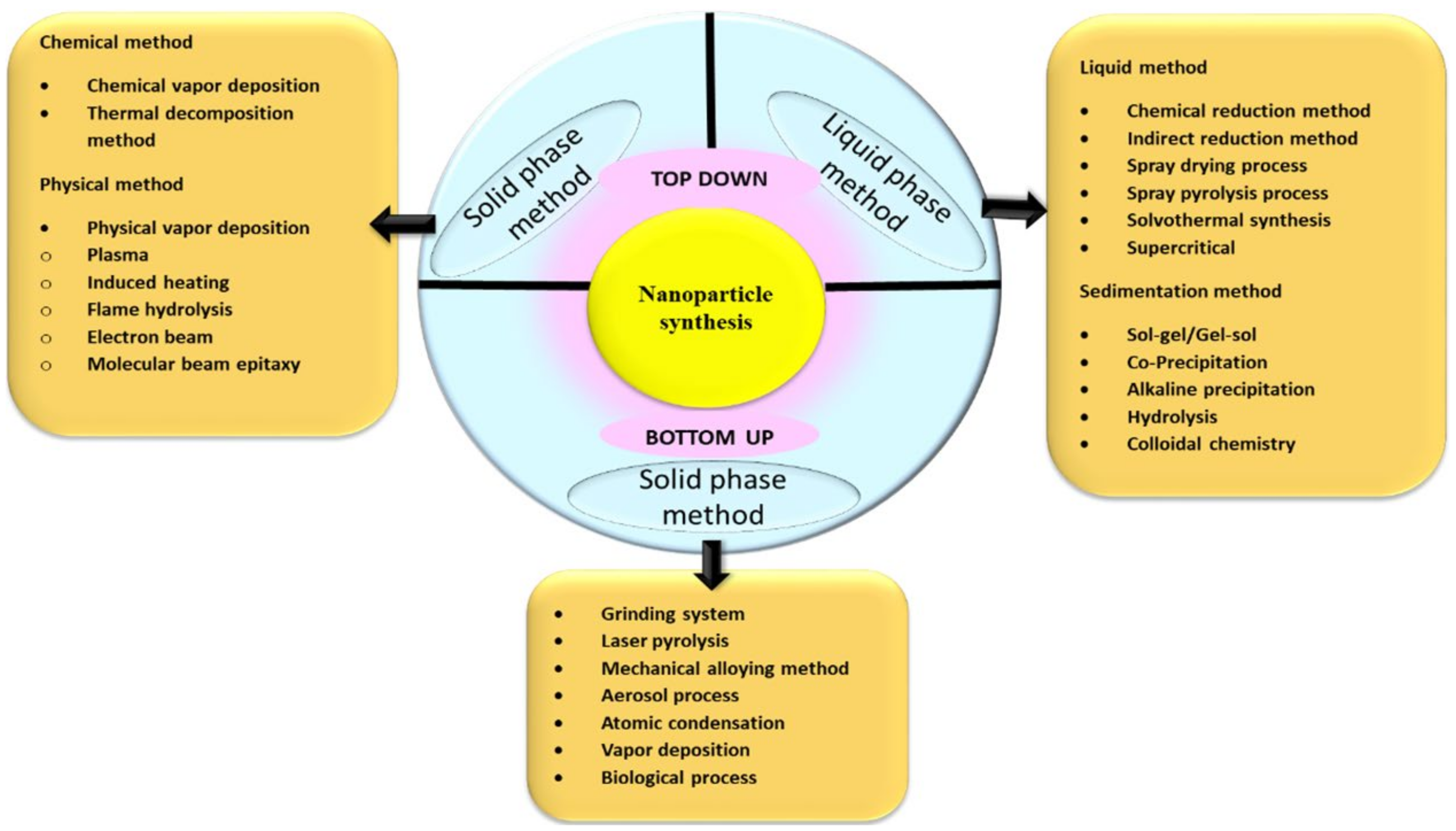
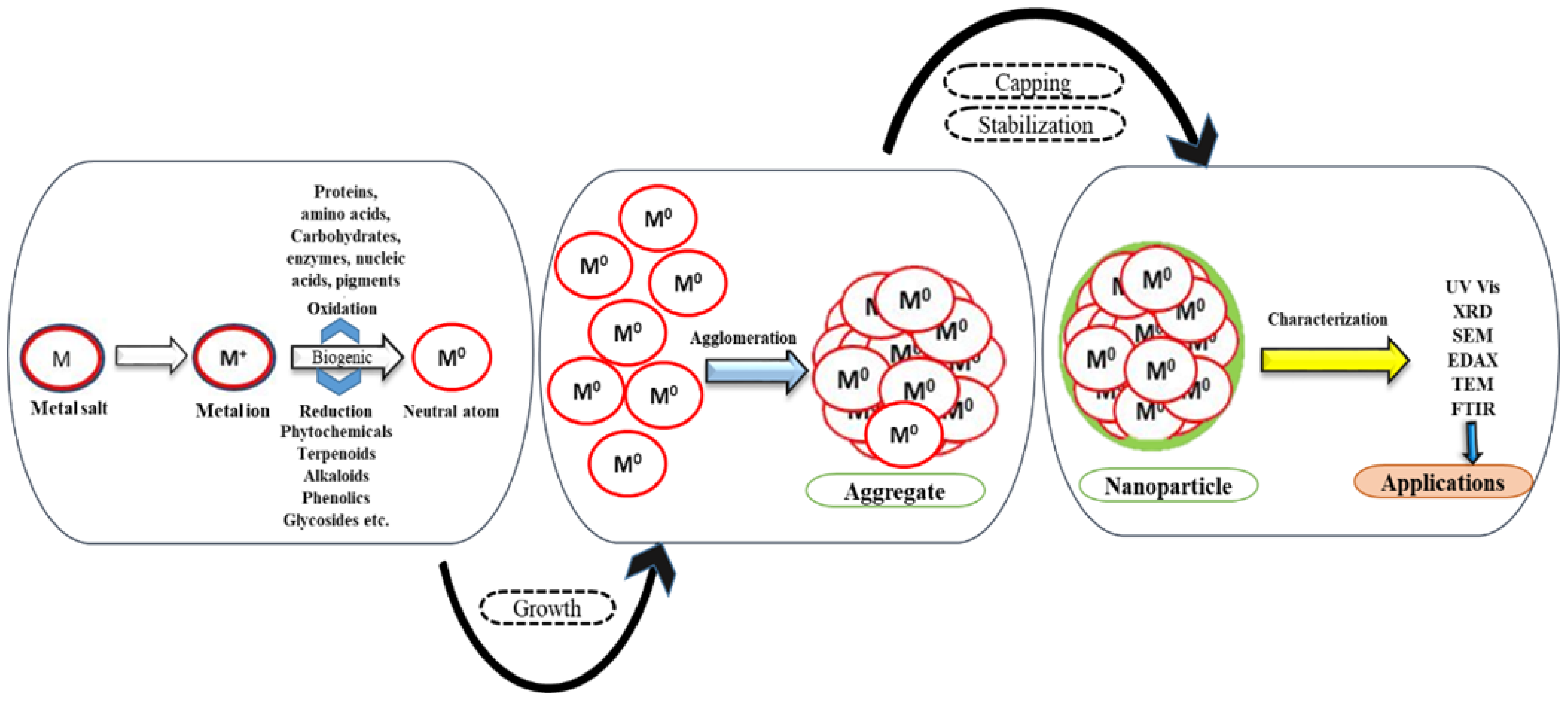
2. Synthesis of NPs
2.1. Perspectives of Nanoparticle Synthesis
2.2. Secondary Biomolecules for Capping and Stabilization
3. Structural Analysis of NPs
4. Biofunctionalization of NPs
4.1. Gold NPs (Au NPs)
4.2. Silver NPs (Ag NPs)
4.3. Platinum Group of Metals
4.4. Metallic Oxide NPs
4.4.1. Zinc Oxide NPs (ZnO NPs)
4.4.2. Magnesium Oxide NPs (MgO NPs)
4.4.3. Copper Oxide NPs (CuO NPs)
4.4.4. Titanium Dioxide NPs (TiO2 NPs)
4.4.5. Lanthanide NPs

| Sr. No. | Botanical Names of Plants | Part Used | Size Range (nm) (SEM/TEM) | Characterization Tools | Bio-Functionalization | Ref. |
|---|---|---|---|---|---|---|
| Zinc oxide NPs | ||||||
| 1. | Trianthema portulacastrum | Extract | 25–90 | UV–Vis, XRD, FTIR, SEM, TEM, XPS |
| [117] |
| 2. | Matricaria chamomilla L., Lycopersicon esculentum M., Olea europaea | Extract | 40.5–124 | UV–Vis, XRD, FTIR, SEM, TEM, EDS |
| [118] |
| 3. | Punica granatum | Extract | 32.98–81.84 | UV–Vis, XRD, FTIR, SEM, TEM |
| [119] |
| 4. | Rheum turketanicum | Extract | 17–20 | UV–Vis, XRD, FTIR, SEM, TEM |
| [120] |
| 5. | Tecoma castanifolia | Extract | 70–75 | UV–Vis, XRD, FTIR, SEM, TEM |
| [121] |
| 6. | Silybum marianum | Extract | 31.2 | UV–Vis, XRD, FTIR, SEM, TEM |
| [122] |
| 7. | Anchusa italic | Flower | ~8–~14 | UV-Vis, EDX XRD, FT-IR, FESEM, TEM |
| [107] |
| 8. | Aloe vera | Leaves | 8–20 | UV-Vis, EDX, XRD, FT-IR, GC-MS, SEM TEM |
| [3] |
| 9. | Rosa canina | Fruit | 50–400 | XRD, EDX, DLS, FT-IR, SEM |
| [36] |
| 10. | Boswellia ovalifoliata | Bark | 20 | UV-Vis, DLS, ZP, FTIR, SEM, TEM |
| [108] |
| Magnesium oxide NPs | ||||||
| 1. | Emblica officinalis | Fruit | 27 | UV-Vis, XRD, EDX, FT-IR, SEM |
| [110] |
| 2. | Clitoria ternatea | Whole plant | 50–400 nm | UV-Vis, XRD, PL, FTIR, EDS, FESEM |
| [42] |
| Copper oxide NPs | ||||||
| 1. | Ocimum tenuiflorum | Extract | 20–30 nm | UV–Vis, XRD, FTIR, SEM, TEM |
| [123] |
| 2. | Moringa oleifera | Extract | 35–95 nm | UV–Vis, XRD, FTIR, SEM, TEM |
| [124] |
| 3. | Eichhornia crassipes | Leaves | 28 ± 4 | UV-Vis, XRD, FT-IR, FESEM |
| [111] |
| 4. | Gloriosa superba | Leaves | 5–10 | UV-Vis, PXRD, SEM TEM |
| [112] |
| Titanium dioxide NPs | ||||||
| 1. | Artocarpus heterophyllus | Extract | 15–20 nm | UV–Vis, XRD, FTIR, SEM, TEM |
| [125] |
| 2. | Citrus sinensis | Fruit peel | 20–50 nm | UV–Vis, XRD, FTIR, SEM, EDAX, TEM |
| [126] |
| 3. | Musa alinsanaya | Fruit peel | 31.5 nm | UV–Vis, XRD, FTIR, SEM, EDAX, TEM |
| [114] |
| 4. | Psidium guajava | Leaves | 32.58 | XRD, EDX, FT-IR, FESEM |
| [113] |
| 5. | Vitex negundo | Leaves | 93.33 | UV-Vis, XRD, EDX, FTIR, SEM |
| [4] |
| Samarium NPs | ||||||
| 1. | Medicago sativa | leaves | 10 | UV-Vis |
| [115] |
| Neodymium NPs | ||||||
| 1. | Medicago sativa | Leaves | 10 | UV-Vis, RS, PSD, DLS, EDAX, XRD, FT-IR, SEM |
| [116] |
5. Applications of Phytofabricated NPs
5.1. In Agriculture
5.2. Applications of Phytofabricated NPs as Nanoantibiotics
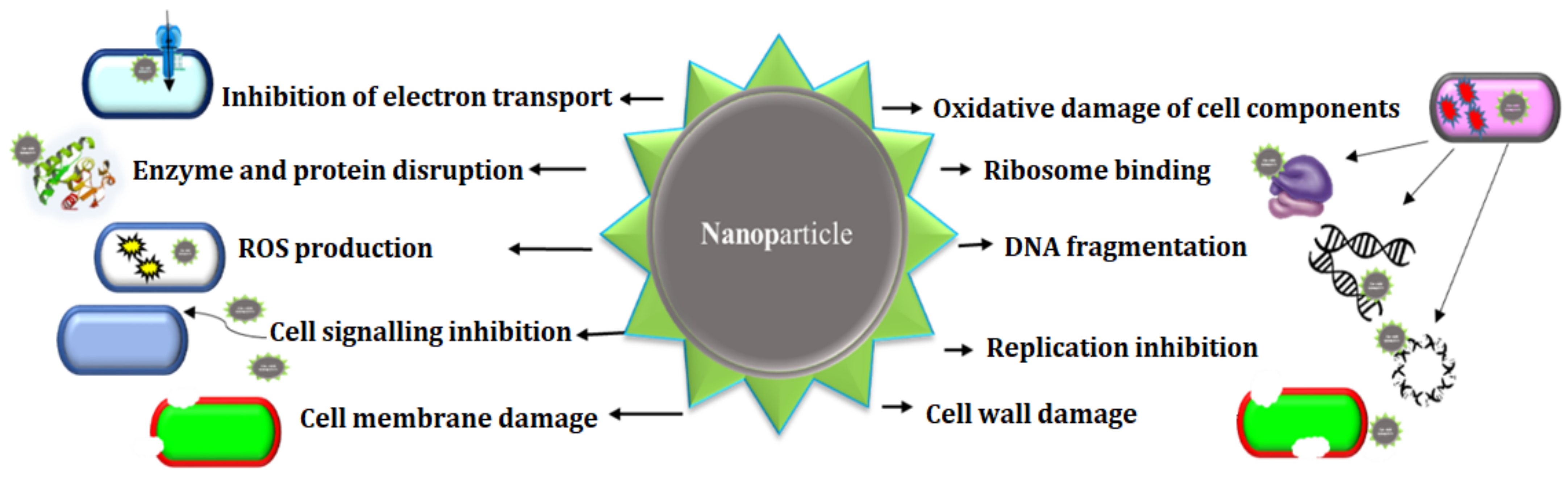
6. Mode of Action of NPs
7. Discussion
8. Conclusions and Future Perspectives
Author Contributions
Funding
Institutional Review Board Statement
Informed Consent Statement
Data Availability Statement
Conflicts of Interest
References
- Ahmad, S.; Munir, S.; Zeb, N.; Ullah, A.; Khan, B.; Ali, J.; Ali, S. Green nanotechnology: A review on green synthesis of silver nanoparticles—an ecofriendly approach. Int. J. Nanomed. 2019, 14, 5087. [Google Scholar] [CrossRef] [Green Version]
- Ahmed, M.J.; Murtaza, G.; Mehmood, A.; Bhatti, T.M. Green synthesis of silver nanoparticles using leaves extract of Skimmia laureola: Characterization and antibacterial activity. Mater. Lett. 2015, 153, 10–13. [Google Scholar] [CrossRef]
- Ali, K.; Dwivedi, S.; Azam, A.; Saquib, Q.; Al-Said, M.S.; Al-Khedhairy, A.; Musarrat, J. Aloe vera extract functionalized zinc oxide nanoparticles as nanoantibiotics against multi-drug resistant clinical bacterial isolates. J. Colloid Interface Sci. 2016, 472, 145–156. [Google Scholar] [CrossRef] [PubMed]
- Ambika, S.; Sundrarajan, M. [EMIM] BF4 ionic liquid-mediated synthesis of TiO2 nanoparticles using Vitex negundo Linn extract and its antibacterial activity. J. Mol. Liquids 2016, 221, 986–992. [Google Scholar]
- Amini, S.M. Preparation of antimicrobial metallic nanoparticles with bioactive compounds. Mater. Sci. Eng. C 2019, 103, 109809. [Google Scholar] [CrossRef]
- Anand, K.; Tiloke, C.; Phulukdaree, A.; Ranjan, B.; Chuturgoon, A.; Singh, S.; Gengan, R. Biosynthesis of palladium nanoparticles by using Moringa oleifera flower extract and their catalytic and biological properties. J. Photochem. Photobiol. B Biol. 2016, 165, 87–95. [Google Scholar] [CrossRef] [PubMed]
- Anuradha, J.; Abbasi, T.; Abbasi, S.A. An eco-friendly method of synthesizing gold nanoparticles using an otherwise worthless weed pistia (Pistia stratiotes L.). J. Adv. Res. 2014, 6, 711–720. [Google Scholar] [CrossRef] [Green Version]
- Arruda, S.C.C.; Silva, A.L.D.; Galazzi, R.; Azevedo, R.A.; Arruda, M.A.Z. Nanoparticles applied to plant science: A review. Talanta 2015, 131, 693–705. [Google Scholar] [CrossRef]
- Ascencio, J.A.; Canizal, G.; Medina-Flores, A.; Bejar, L.; Tavera, L.; Matamoros, H.; Liu, H.B. Neodymium Nanoparticles: Biosynthesis and Structural Analysis. J. Nanosci. Nanotechnol. 2006, 6, 1044–1049. [Google Scholar] [CrossRef]
- Ascencio, J.A.; Rincon, A.C.; Canizal, G. Synthesis and Theoretical Analysis of Samarium Nanoparticles: Perspectives in Nuclear Medicine. J. Phys. Chem. B 2005, 109, 8806–8812. [Google Scholar] [CrossRef]
- Azizi, S.; Ahmad, M.B.; Namvar, F.; Mohamad, R. Green biosynthesis and characterization of zinc oxide nanoparticles using brown marine macroalga Sargassum muticum aqueous extract. Mater. Lett. 2014, 116, 275–277. [Google Scholar] [CrossRef]
- Babu, P.J.; Das, R.K.; Kumar, A.; Bora, U. Microwave-Mediated Synthesis of Gold Nanoparticles Using Coconut Water. Int. J. Green Nanotechnol. 2011, 3, 13–21. [Google Scholar] [CrossRef]
- Baharara, J.; Namvar, F.; Ramezani, T.; Hosseini, N.; Mohamad, R. Green Synthesis of Silver Nanoparticles using Achillea biebersteinii Flower Extract and Its Anti-Angiogenic Properties in the Rat Aortic Ring Model. Molecules 2014, 19, 4624–4634. [Google Scholar] [CrossRef] [PubMed] [Green Version]
- Baker, S.; Rakshith, D.; Kavitha, K.S.; Santosh, P.; Kavitha, H.U.; Rao, Y.; Satish, S. Plants: Emerging as Nanofactories towards Facile Route in Synthesis of Nanoparticles. BioImpacts 2013, 3, 111–117. [Google Scholar]
- Bar, H.; Bhui, D.K.; Sahoo, G.P.; Sarkar, P.; Pyne, S.; Misra, A. Green synthesis of silver nanoparticles using seed extract of Jatropha curcas. Colloids Surf. A Physicochem. Eng. Asp. 2009, 348, 212–216. [Google Scholar] [CrossRef]
- Basavegowda, N.; Sobczak-Kupiec, A.; Malina, D.; Yathirajan, H.S.; Keerthi, V.R.; Dinkar, S. Plant mediated synthesis of gold nanoparticles using fruit extracts of Ananas comosus (L.) (pineapple) and evaluation of biological activities. Adv. Mater. Lett. 2013, 4, 332–337. [Google Scholar] [CrossRef] [Green Version]
- Bayda, S.; Adeel, M.; Tuccinardi, T.; Cordani, M.; Rizzolio, F. The History of Nanoscience and Nanotechnology: From Chemical–Physical Applications to Nanomedicine. Molecules 2020, 25, 112. [Google Scholar] [CrossRef] [Green Version]
- Das, J.; Das, M.P.; Velusamy, P. Sesbania grandiflora leaf extract mediated green synthesis of antibacterial silver nanoparticles against selected human pathogens. Spectrochim. Acta Part A Mol. Biomol. Spectrosc. 2013, 104, 265–270. [Google Scholar] [CrossRef]
- Chandra, H.; Kumari, P.; Bontempi, E.; Yadav, S. Medicinal plants: Treasure trove for green synthesis of metallic nanoparticles and their biomedical applications. Biocatal. Agric. Biotechnol. 2020, 24, 101518. [Google Scholar] [CrossRef]
- Crisan, C.M.; Mocan, T.; Manolea, M.; Lasca, L.I.; Tăbăran, F.-A.; Mocan, L. Review on Silver Nanoparticles as a Novel Class of Antibacterial Solutions. Appl. Sci. 2021, 11, 1120. [Google Scholar] [CrossRef]
- Das, S.; Parida, U.K.; Bindhani, B.K. Green biosynthesis of silver nanoparticles using Moringa oleifera L. leaf. Int. J. Nanotechnol. Appl. 2013, 3, 51–62. [Google Scholar]
- De Oliveira, P.F.M.; Torresi, R.M.; Emmerling, F.; Camargo, P.H.C. Challenges and opportunities in the bottom-up mechanochemical synthesis of noble metal nanoparticles. J. Mater. Chem. A 2020, 8, 16114–16141. [Google Scholar] [CrossRef]
- Deb, S. Synthesis of silver nano particles using Murraya koenigii (Green Curry Leaves), Zea mays (baby corn) and its antimicrobial activity against pathogens. Int. J. PharmTech Res. 2014, 6, 91–96. [Google Scholar]
- Devi, G.D.; Murugan, K.; Selvam, C.P. Green synthesis of silver nanoparticles using Euphorbia hirta (Euphorbiaceae) leaf extract against crop pest of cotton bollworm, Helicoverpa armigera (Lepidoptera: Noctuidae). J. Biopestic. 2014, 7, 54. [Google Scholar]
- Dikshit, P.; Kumar, J.; Das, A.; Sadhu, S.; Sharma, S.; Singh, S.; Gupta, P.; Kim, B. Green Synthesis of Metallic Nanoparticles: Applications and Limitations. Catalysts 2021, 11, 902. [Google Scholar] [CrossRef]
- Dipankar, C.; Murugan, S. The green synthesis, characterization and evaluation of the biological activities of silver nanoparticles synthesized from Iresine herbstii leaf aqueous extracts. Colloids Surf. B Biointerfaces 2012, 98, 112–119. [Google Scholar] [CrossRef] [PubMed]
- El-Seedi, H.R.; El-Shabasy, R.M.; Khalifa, S.A.M.; Saeed, A.; Shah, A.; Shah, R.; Iftikhar, F.J.; Abdel-Daim, M.M.; Omri, A.; Hajrahand, N.H.; et al. Metal nanoparticles fabricated by green chemistry using natural extracts: Biosynthesis, mechanisms, and applications. RSC Adv. 2019, 9, 24539–24559. [Google Scholar] [CrossRef] [Green Version]
- Chopade, B.A.; Ghosh, S.; Patil, S.; Ahire, M.; Kitture, R.; Jabgunde, A.; Kale, S.; Pardesi, K.; Cameotra, S.S.; Bellare, J.; et al. Synthesis of silver nanoparticles using Dioscorea bulbifera tuber extract and evaluation of its synergistic potential in combination with antimicrobial agents. Int. J. Nanomed. 2012, 7, 483–496. [Google Scholar] [CrossRef] [Green Version]
- Gnanajobitha, G.; Paulkumar, K.; Vanaja, M.; RajeshKumar, S.; Malarkodi, C.; Annadurai, G.; Kannan, C. Fruit-mediated synthesis of silver nanoparticles using Vitis vinifera and evaluation of their antimicrobial efficacy. J. Nanostruct. Chem. 2013, 3, 67. [Google Scholar] [CrossRef] [Green Version]
- Gopinath, K.; Gowri, S.; Arumugam, A. Phytosynthesis of silver nanoparticles using Pterocarpus santalinus leaf extract and their antibacterial properties. J. Nanostruct. Chem. 2013, 3, 68. [Google Scholar] [CrossRef]
- Gour, A.; Jain, N.K. Advances in green synthesis of nanoparticles. Artif. Cells Nanomed. Biotechnol. 2019, 47, 844–851. [Google Scholar] [CrossRef] [PubMed] [Green Version]
- Hassanisaadi, M.; Bonjar, G.; Rahdar, A.; Pandey, S.; Hosseinipour, A.; Abdolshahi, R. Environmentally Safe Biosynthesis of Gold Nanoparticles Using Plant Water Extracts. Nanomaterials 2021, 11, 2033. [Google Scholar] [CrossRef] [PubMed]
- Hussain, I.; Singh, N.B.; Singh, A.; Singh, H.; Singh, S.C. Green synthesis of nanoparticles and its potential application. Biotechnol. Lett. 2016, 38, 545–560. [Google Scholar] [CrossRef] [PubMed]
- Ibrahim, H.M. Green synthesis and characterization of silver nanoparticles using banana peel extract and their antimicrobial activity against representative microorganisms. J. Radiat. Res. Appl. Sci. 2015, 8, 265–275. [Google Scholar] [CrossRef] [Green Version]
- Jadoun, S.; Arif, R.; Jangid, N.K.; Meena, R.K. Green synthesis of nanoparticles using plant extracts: A review. Environ. Chem. Lett. 2020, 19, 355–374. [Google Scholar] [CrossRef]
- Jafarirad, S.; Mehrabi, M.; Divband, B.; Kosari-Nasab, M. Biofabrication of zinc oxide nanoparticles using fruit extract of Rosa canina and their toxic potential against bacteria: A mechanistic approach. Mater. Sci. Eng. C 2016, 59, 296–302. [Google Scholar] [CrossRef]
- Jagtap, U.B.; Bapat, V.A. Green synthesis of silver nanoparticles using Artocarpus heterophyllus Lam. seed extract and its antibacterial activity. Ind. Crop. Prod. 2013, 46, 132–137. [Google Scholar] [CrossRef]
- Jahan, I. Phyto-Nanofabrication: Plant-Mediated Synthesis of Metal and Metal Oxide Nanoparticles. In Handbook of Research on Green Synthesis and Applications of Nanomaterials; IGI Global: Hershey, PA, USA, 2022; pp. 51–76. [Google Scholar]
- Jamkhande, P.G.; Ghule, N.W.; Bamer, A.H.; Kalaskar, M.G. Metal nanoparticles synthesis: An overview on methods of preparation, advantages and disadvantages, and applications. J. Drug Deliv. Sci. Technol. 2019, 53, 101174. [Google Scholar] [CrossRef]
- Jayaseelan, C.; Ramkumar, R.; Rahuman, A.A.; Perumal, P. Green synthesis of gold nanoparticles using seed aqueous extract of Abelmoschus esculentus and its antifungal activity. Ind. Crops Prod. 2013, 45, 423–429. [Google Scholar] [CrossRef]
- Jeevanandam, J.; Kiew, S.F.; Boakye-Ansah, S.; Lau, S.Y.; Barhoum, A.; Danquah, M.K.; Rodrigues, J. Green approaches for the synthesis of metal and metal oxide nanoparticles using microbial and plant extracts. Nanoscale 2022, 14, 2534–2571. [Google Scholar] [CrossRef]
- Sushma, N.J.; Prathyusha, D.; Swathi, G.; Madhavi, T.; Raju, B.D.P.; Mallikarjuna, K.; Kim, H.-S. Facile approach to synthesize magnesium oxide nanoparticles by using Clitoria ternatea—characterization and in vitro antioxidant studies. Appl. Nanosci. 2015, 6, 437–444. [Google Scholar] [CrossRef] [Green Version]
- Jyoti, K.; Pattnaik, P.; Singh, T. Green Synthesis of Silver Nanoparticles Using Sustainable Resources and their Use as Antibacterial Agents: A Review. Curr. Mater. Sci. Former. Recent Pat. Mater. Sci. 2021, 14, 40–52. [Google Scholar] [CrossRef]
- Mathew, S.; Victorio, C.P.; Sidhi, J.; Thanzeela, B.H.B. Biosynthesis of silver nanoparticle using flowers of Calotropis gigantea (L.) WT Aiton and activity against pathogenic bacteria. Arab. J. Chem. 2020, 13, 9139–9144. [Google Scholar] [CrossRef]
- Kalimuthu, K.; Cha, B.S.; Kim, S.; Park, K.S. Eco-friendly synthesis and biomedical applications of gold nanoparticles: A review. Microchem. J. 2019, 152, 104296. [Google Scholar] [CrossRef]
- Suresh, J.; Yuvakkumar, R.; Sundrarajan, M.; Hong, S.I. Green synthesis of magnesium oxide nanoparticles. In Advanced Materials Research; Trans Tech Publications Ltd: Bäch, Switzerland, 2014; Volume 952, pp. 141–144. [Google Scholar]
- Karaiskos, I.; Giamarellou, H. Multidrug-resistant and extensively drug-resistant Gram-negative pathogens: Current and emerging therapeutic approaches. Expert Opin. Pharmacother. 2014, 15, 1351–1370. [Google Scholar] [CrossRef] [PubMed]
- Komal, R.; Arya, V. Biosynthesis and characterization of silver nanoparticles from aqueous leaf extracts of Carica papaya and its antibacterial activity. Int. J. Nanomater. Biostruct. 2013, 3, 17–20. [Google Scholar]
- Kumara, P.P.N.V.; Pammib, S.V.N.; Kollu, P.; Satyanarayana, K.V.V.; Shameema, U. Green synthesis and characterization of silver nanoparticles using Boerhaavia diffusa plant extract and their anti-bacterial activity. Ind. Crop. Prod. 2014, 52, 562–566. [Google Scholar] [CrossRef]
- Lade, B.D.; Shanware, A.S. Phytonanofabrication: Methodology and factors affecting biosynthesis of nanoparticles. In Smart Nanosystems for Biomedicine, Optoelectronics and Catalysis; IntechOpen: London, UK, 2020. [Google Scholar]
- Farhadi, S.; Ajerloo, B.; Mohammadi, A. Low-cost and eco-friendly phyto-synthesis of Silver nanoparticles by using grapes fruit extract and study of antibacterial and catalytic effects. Int. J. Nano Dimens. 2017, 8, 49–60. [Google Scholar]
- Roopan, S.M.; Madhumitha, G.; Rahuman, A.A.; Kamaraj, C.; Bharathi, A.; Surendra, T.V. Low-cost and eco-friendly phyto-synthesis of silver nanoparticles using Cocos nucifera coir extract and its larvicidal activity. Ind. Crop. Prod. 2013, 43, 631–635. [Google Scholar] [CrossRef]
- Fahmy, S.A.; Preis, E.; Bakowsky, U.; Azzazy, H.M.E.-S. Palladium Nanoparticles Fabricated by Green Chemistry: Promising Chemotherapeutic, Antioxidant and Antimicrobial Agents. Materials 2020, 13, 3661. [Google Scholar] [CrossRef]
- Luna, C.; Chávez, V.; Barriga-Castro, E.D.; Núñez, N.O.; Mendoza-Reséndez, R. Biosynthesis of silver fine particles and particles decorated with nanoparticles using the extract of Illicium verum (star anise) seeds. Spectrochim. Acta Part A Mol. Biomol. Spectrosc. 2015, 141, 43–50. [Google Scholar] [CrossRef] [PubMed]
- Qiao, J.; Qi, L. Recent progress in plant-gold nanoparticles fabrication methods and bio-applications. Talanta 2020, 223, 121396. [Google Scholar] [CrossRef] [PubMed]
- Zhang, Y.; Zhang, C.; Xu, C.; Wang, X.; Liu, C.; Waterhouse, G.; Wang, Y.; Yin, H. Ultrasmall Au nanoclusters for biomedical and biosensing applications: A mini-review. Talanta 2019, 200, 432–442. [Google Scholar] [CrossRef]
- Xiao, T.; Huang, J.; Wang, D.; Meng, T.; Yang, X. Au and Au-Based nanomaterials: Synthesis and recent progress in electrochemical sensor applications. Talanta 2019, 206, 120210. [Google Scholar] [CrossRef] [PubMed]
- Shahriari, M.; Hemmati, S.; Zangeneh, A.; Zangeneh, M.M. Biosynthesis of gold nanoparticles using Allium noeanum Reut. ex Regel leaves aqueous extract; characterization and analysis of their cytotoxicity, antioxidant, and antibacterial properties. Appl. Organomet. Chem. 2019, 33, e5189. [Google Scholar] [CrossRef]
- Gharehyakheh, S.; Ahmeda, A.; Haddadi, A.; Jamshidi, M.; Nowrozi, M.; Zangeneh, M.M.; Zangeneh, A. Effect of gold nanoparticles synthesized using the aqueous extract of Satureja hortensis leaf on enhancing the shelf life and removing Escherichia coli O157:H7 and Listeria monocytogenes in minced camel’s meat: The role of nanotechnology in the food industry. Appl. Organomet. Chem. 2020, 34, e5492. [Google Scholar]
- Valsalam, S.; Agastian, P.; Esmail, G.A.; Ghilan, A.-K.M.; Al-Dhabi, N.A.; Arasu, M.V. Biosynthesis of silver and gold nanoparticles using Musa acuminata colla flower and its pharmaceutical activity against bacteria and anticancer efficacy. J. Photochem. Photobiol. B Biol. 2019, 201, 111670. [Google Scholar] [CrossRef] [PubMed]
- Zhaleh, M.; Zangeneh, A.; Goorani, S.; Seydi, N.; Zangeneh, M.M.; Tahvilian, R.; Pirabbasi, E. In vitro and in vivo evalution of cytotocicity, antioxidant, antibacterial, antifungal, and cutaneous wound healing properies of gold nanoparticles produced via a green chemistry synthesis using Gundelia tournefortii L. as acapping and reducing agent. Appl. Organomet. Chem. 2019, 33, e5015. [Google Scholar] [CrossRef]
- Jeyarani, S.; Vinita, N.M.; Puja, P.; Senthamilselvi, S.; Devan, U.; Velangani, A.J.; Biruntha, M.; Pugazhendhi, A.; Kumar, P. Biomimetic gold nanoparticles for its cytotoxicity and biocompatibility evidenced by fluorescence-based assays in cancer (MDA-MB-231) and non-cancerous (HEK-293) cells. J. Photochem. Photobiol. B Biol. 2020, 202, 111715. [Google Scholar] [CrossRef]
- Hemmati, S.; Joshani, Z.; Zangeneh, A.; Zangeneh, M.M. Green synthesis and chemical characterization of Thymus vulgaris leaf aqueous extract conjugated gold nanoparticles for the treatment of acute myeloid leukemia in comparison to doxorubicin in a leukemic mouse model. Appl. Organomet. Chem. 2019, 34, e5267. [Google Scholar] [CrossRef]
- Ansari, S.; Bari, A.; Ullah, R.; Mathanmohun, M.; Veeraraghavan, V.P.; Sun, Z. Gold nanoparticles synthesized with Smilax glabra rhizome modulates the anti-obesity parameters in high-fat diet and streptozotocin induced obese diabetes rat model. J. Photochem. Photobiol. B Biol. 2019, 201, 111643. [Google Scholar] [CrossRef] [PubMed]
- Ismail, E.H.; Saqer, A.M.A.; Assirey, E.; Naqvi, A.; Okasha, R.M. Successful Green Synthesis of Gold Nanoparticles using a Corchorus olitorius Extract and Their Antiproliferative Effect in Cancer Cells. Int. J. Mol. Sci. 2018, 19, 2612. [Google Scholar] [CrossRef] [Green Version]
- Filip, G.A.; Moldovan, B.; Baldea, I.; Olteanu, D.; Suharoschi, R.; Decea, N.; Cismaru, C.M.; Gal, E.; Cenariu, M.; Clichici, S.; et al. UV-light mediated green synthesis of silver and gold nanoparticles using Cornelian cherry fruit extract and their comparative effects in experimental inflammation. J. Photochem. Photobiol. B Biol. 2018, 191, 26–37. [Google Scholar] [CrossRef]
- Ahmeda, A.; Zangeneh, A.; Zangeneh, M.M. Green synthesis and chemical characterization of gold nanoparticle synthesized using Camellia sinensis leaf aqueous extract for the treatment of acute myeloid leukemia in comparison to daunorubicin in a leukemic mouse model. Appl. Organomet. Chem. 2020, 34, e5290. [Google Scholar] [CrossRef]
- Liu, R.; Pei, Q.; Shou, T.; Zhang, W.; Hu, J.; Li, W. Apoptotic effect of green synthesized gold nanoparticles from Curcuma wenyujin extract against human renal cell carcinoma A498 cells. Int. J. Nanomed. 2019, 14, 4091–4103. [Google Scholar] [CrossRef] [PubMed] [Green Version]
- Liu, Y.; Kim, S.; Kim, Y.J.; Perumalsamy, H.; Lee, S.; Hwang, E.; Yi, T.H. Green synthesis of gold nanoparticles using Euphrasia officinalis leaf extract to inhibit lipopolysaccharide-induced inflammation through NF-kappa B and JAK/STAT pathways in RAW 264.7 macrophages. Int. J. Nanomed. 2019, 14, 2945–2959. [Google Scholar] [CrossRef] [PubMed] [Green Version]
- Park, S.Y.; Yi, E.H.; Kim, Y.; Park, G. Anti-neuroinflammatory effects of Ephedra sinica Stapf extract-capped gold nanoparticles in microglia. Int. J. Nanomed. 2019, 14, 2861–2877. [Google Scholar] [CrossRef] [Green Version]
- Zhang, T.; Dang, M.; Zhang, W.; Lin, X. Gold nanoparticles synthesized from Euphorbia fischeriana root by green route method alleviates the isoprenaline hydrochloride induced myocardial infarction in rats. J. Photochem. Photobiol. B Biol. 2019, 202, 111705. [Google Scholar] [CrossRef]
- Ahmeda, A.; Zangeneh, M.M. Novel green synthesis of Boswellia serrata leaf aqueous extract conjugated gold nanoparticles with excellent anti-acute myeloid leukemia property in comparison to mitoxantrone in a leukemic mice model: Introducing a new chemotherapeutic drug. Appl. Organomet. Chem. 2019, 34, e5344. [Google Scholar] [CrossRef]
- Yun, Z.; Chinnathambi, A.; Alharbi, S.A.; Jin, Z. Biosynthesis of gold nanoparticles using Vetex negundo and evaluation of pro-apoptotic effect on human gastric cancer cell lines. J. Photochem. Photobiol. B Biol. 2019, 203, 111749. [Google Scholar] [CrossRef]
- Siddiqi, K.S.; Husen, A. Recent advances in plant-mediated engineered gold nanoparticles and their application in biological system. J. Trace Elements Med. Biol. 2017, 40, 10–23. [Google Scholar] [CrossRef] [PubMed]
- Acharya, D.; Mohanta, B.; Pandey, P. Green synthesis of Silver and Silver-gold core-shell nanoparticles using Pineapple leaf extract (Ananas comosus) and study of their antibacterial properties. Int. J. Nano Dimens. 2021, 12, 203–210. [Google Scholar]
- Francis, G.; Thombre, R.; Parekh, F.; Leksminarayan, P. Bioinspired synthesis of gold nanoparticles using Ficus benghalensis (Indian Banyan) leaf extract. Chem. Sci. Trans. 2014, 3, 470–474. [Google Scholar]
- Wani, K.; Choudhari, A.; Chikate, R.; Kaul-Ghanekar, R. Synthesis and characterization of gold nanoparticles using Ficus religiosa extract. Carbon Sci. Technol. 2013, 5, 203–210. [Google Scholar]
- Reddy, G.R.; Morais, A.B.; Gandhi, N.N. Green Synthesis, Characterization and in vitro Antibacterial Studies of Gold Nanoparticles by Using Senna siamea Plant Seed Aqueous Extract at Ambient Conditions. Asian J. Chem. 2013, 25, 8541–8544. [Google Scholar] [CrossRef]
- Ghramh, H.A.; Khan, K.A.; Ibrahim, E.H.; Setzer, W.N. Synthesis of Gold Nanoparticles (AuNPs) Using Ricinus communis Leaf Ethanol Extract, Their Characterization, and Biological Applications. Nanomaterials 2019, 9, 765. [Google Scholar] [CrossRef] [Green Version]
- Sunderam, V.; Thiyagarajan, D.; Lawrence, A.V.; Mohammed, S.S.S.; Selvaraj, A. In-vitro antimicrobial and anticancer properties of green synthesized gold nanoparticles using Anacardium occidentale leaves extract. Saudi J. Biol. Sci. 2018, 26, 455–459. [Google Scholar] [CrossRef]
- Chahardoli, A.; Karimi, N.; Fattahi, A.; Salimikia, I. Biological applications of phytosynthesized gold nanoparticles using leaf extract of Dracocephalum kotschyi. J. Biomed. Mater. Res. Part A 2018, 107, 621–630. [Google Scholar] [CrossRef]
- Ghramh, H.A.; Khan, K.A.; Ibrahim, E.H. Biological Activities of Euphorbia peplus Leaves Ethanolic Extract and the Extract Fabricated Gold Nanoparticles (AuNPs). Molecules 2019, 24, 1431. [Google Scholar] [CrossRef] [Green Version]
- Ghosh, S.; Patil, S.; Ahire, M.; Kitture, R.; Gurav, D.D.; Jabgunde, A.M.; Kale, S.; Pardesi, K.; Shinde, V.; Bellare, J.; et al. Gnidia glauca flower extract mediated synthesis of gold nanoparticles and evaluation of its chemocatalytic potential. J. Nanobiotechnol. 2012, 10, 17. [Google Scholar] [CrossRef] [Green Version]
- Suman, T.; Rajasree, S.R.; Ramkumar, R.; Rajthilak, C.; Perumal, P. The Green synthesis of gold nanoparticles using an aqueous root extract of Morinda citrifolia L. Spectrochim. Acta Part A Mol. Biomol. Spectrosc. 2013, 118, 11–16. [Google Scholar] [CrossRef] [PubMed]
- Nagaraj, B.; Malakar, B.; Divya, T.K.; Krishnamurthy, N.B.; Liny, P.; Dinesh, R. Environmental benign synthesis of gold nanoparticles from the flower extracts of Plumeria alba Linn, (Frangipani) and evaluation of their biological activities. Int. J. Drug Dev. Res. 2012, 4, 144–150. [Google Scholar]
- Valsalam, S.; Agastian, P.; Arasu, M.V.; Al-Dhabi, N.A.; Ghilan, A.-K.M.; Kaviyarasu, K.; Ravindran, B.; Chang, S.W.; Arokiyaraj, S. Rapid biosynthesis and characterization of silver nanoparticles from the leaf extract of Tropaeolum majus L. and its enhanced in-vitro antibacterial, antifungal, antioxidant and anticancer properties. J. Photochem. Photobiol. B Biol. 2018, 191, 65–74. [Google Scholar] [CrossRef] [PubMed]
- Göl, F.; Aygün, A.; Seyrankaya, A.; Gür, T.; Yenikaya, C.; Şen, F. Green synthesis and characterization of Camellia sinensis mediated silver nanoparticles for antibacterial ceramic applications. Mater. Chem. Phys. 2020, 250, 1–2. [Google Scholar] [CrossRef]
- Uttu, A.J.; Sallau, M.S.; Iyun, O.R.A.; Ibrahim, H. Antimicrobial Efficacy of Selected Strychnos Species: A Mini Review. J. Chem. Rev. 2022, 4, 59–62. [Google Scholar]
- Ibrahim, E.; Kilany, M.; Ghramh, H.A.; Khan, K.; Islam, S.U. Cellular proliferation/cytotoxicity and antimicrobial potentials of green synthesized silver nanoparticles (AgNPs) using Juniperus procera. Saudi J. Biol. Sci. 2018, 26, 1689–1694. [Google Scholar] [CrossRef]
- Kumar, V.; Singh, S.; Srivastava, B.; Bhadouria, R.; Singh, R. Green synthesis of silver nanoparticles using leaf extract of Holoptelea integrifolia and preliminary investigation of its antioxidant, anti-inflammatory, antidiabetic and antibacterial activities. J. Environ. Chem. Eng. 2019, 7, 103094. [Google Scholar] [CrossRef]
- Salari, S.; Bahabadi, S.E.; Samzadeh-Kermani, A.; Yosefzaei, F. In-vitro Evaluation of Antioxidant and Antibacterial Potential of Green Synthesized Silver Nanoparticles Using Prosopis farcta Fruit Extract. Iran. J. Pharm. Res. IJPR 2019, 18, 430–455. [Google Scholar]
- Yadav, R.; Saini, H.; Kumar, D.; Pasi, S.; Agrawal, V. Bioengineering of Piper longum L. extract mediated silver nanoparticles and their potential biomedical applications. Mater. Sci. Eng. C 2019, 104, 109984. [Google Scholar] [CrossRef]
- Botha, T.L.; Elemike, E.E.; Horn, S.; Onwudiwe, D.C.; Giesy, J.P.; Wepener, V. Cytotoxicity of Ag, Au and Ag-Au bimetallic nanoparticles prepared using golden rod (Solidago canadensis) plant extract. Sci. Rep. 2019, 9, 4169. [Google Scholar] [CrossRef] [Green Version]
- Nazeruddin, G.; Prasad, N.; Shaikh, Y.; Waghmare, S.; Adhyapak, P. Coriandrum sativum seed extract assisted in situ green synthesis of silver nanoparticle and its anti-microbial activity. Ind. Crop. Prod. 2014, 60, 212–216. [Google Scholar] [CrossRef]
- Sreekanth, T.; Ravikumar, S.; Eom, I.-Y. Green synthesized silver nanoparticles using Nelumbo nucifera root extract for efficient protein binding, antioxidant and cytotoxicity activities. J. Photochem. Photobiol. B Biol. 2014, 141, 100–105. [Google Scholar] [CrossRef] [PubMed]
- Rout, Y.; Behera, S.; Ojha, A.K.; Nayak, P.L. Green synthesis of silver nanoparticles using Ocimum sanctum (Tulashi) and study of their antibacterial and antifungal activities. J. Microbiol. Antimicrob. 2012, 4, 103–109. [Google Scholar] [CrossRef]
- Raut, R.W.; Mendhulkar, V.D.; Kashid, S.B. Photosensitized synthesis of silver nanoparticles using Withania somnifera leaf powder and silver nitrate. J. Photochem. Photobiol. B Biol. 2014, 132, 45–55. [Google Scholar] [CrossRef]
- Bindhu, M.; Umadevi, M. Synthesis of monodispersed silver nanoparticles using Hibiscus cannabius leaf extract and its antimicrobial activity. Spectrochim. Acta Part A Mol. Biomol. Spectrosc. 2013, 101, 184–190. [Google Scholar] [CrossRef]
- Attar, A.; Yapaoz, M.A. Biosynthesis of palladium nanoparticles using Diospyros kaki leaf extract and determination of antibacterial efficacy. Prep. Biochem. Biotechnol. 2018, 48, 629–634. [Google Scholar] [CrossRef]
- Mavukkandy, M.O.; Chakraborty, S.; Abbasi, T.; Abbasi, S.A. A Clean-Green Synthesis of Platinum Nanoparticles Utilizing a Pernicious Weed Lantana (Lantana Camara). Am. J. Eng. Appl. Sci. 2016, 9, 84–90. [Google Scholar] [CrossRef] [Green Version]
- Narendhran, S.; Manikandan, M.; & Shakila, P. Antibacterial, antioxidant properties of Solanum trilobatum and sodium hydroxide-mediated magnesium oxide nanoparticles: A green chemistry approach. Bull. Mater. Sci. 2019, 42, 1–8. [Google Scholar] [CrossRef] [Green Version]
- Tahir, K.; Nazir, S.; Li, B.; Ahmad, A.; Nasir, T.; Khan, A.U.; Shah, S.A.A.; Khan, Z.U.H.; Yasin, G.; Hameed, M.U. Sapium sebiferum leaf extract mediated synthesis of palladium nanoparticles and in vitro investigation of their bacterial and photocatalytic activities. J. Photochem. Photobiol. B Biol. 2016, 164, 164–173. [Google Scholar] [CrossRef]
- Sathishkumar, M.; Sneha, K.; Yun, Y.S. Palladium nanocrystal synthesis using Curcuma longa tuber extract. Int. J. Mater. Sci. 2009, 4, 11–17. [Google Scholar]
- Surendra, T.; Roopan, S.M.; Arasu, M.V.; Al-Dhabi, N.A.; Rayalu, G.M. RSM optimized Moringa oleifera peel extract for green synthesis of M. oleifera capped palladium nanoparticles with antibacterial and hemolytic property. J. Photochem. Photobiol. B Biol. 2016, 162, 550–557. [Google Scholar] [CrossRef] [PubMed]
- Basnet, P.; Chanu, T.I.; Samanta, D.; Chatterjee, S. A review on bio-synthesized zinc oxide nanoparticles using plant extracts as reductants and stabilizing agents. J. Photochem. Photobiol. B Biol. 2018, 183, 201–221. [Google Scholar] [CrossRef] [PubMed]
- Akbarian, M.; Mahjoub, S.; Elahi, S.M.; Zabihi, E.; Tashakkorian, H.; Elahi, M. Appraisal of the biological aspect of Zinc oxide nanoparticles prepared using extract of Camellia sinensis L. Mater. Res. Express 2019, 6, 095022. [Google Scholar] [CrossRef]
- Azizi, S.; Mohamad, R.; Bahadoran, A.; Bayat, S.; Rahim, R.A.; Ariff, A.; Saad, W.Z. Effect of annealing temperature on antimicrobial and structural properties of bio-synthesized zinc oxide nanoparticles using flower extract of Anchusa italica. J. Photochem. Photobiol. B Biol. 2016, 161, 441–449. [Google Scholar] [CrossRef]
- Supraja, N.; Prasad, T.N.V.K.V.; Krishna, T.G.; David, E. Synthesis, characterization, and evaluation of the antimicrobial efficacy of Boswellia ovalifoliolata stem bark-extract-mediated zinc oxide nanoparticles. Appl. Nanosci. 2015, 6, 581–590. [Google Scholar] [CrossRef] [Green Version]
- Ringe, E. Shapes, plasmonic properties, and reactivity of magnesium nanoparticles. J. Phys. Chem. C 2020, 124, 15665–15679. [Google Scholar] [CrossRef]
- Ramanujam, K.; Sundrarajan, M. Antibacterial effects of biosynthesized MgO nanoparticles using ethanolic fruit extract of Emblica officinalis. J. Photochem. Photobiol. B Biol. 2014, 141, 296–300. [Google Scholar] [CrossRef]
- Vanathi, P.; Rajiv, P.; Sivaraj, R. Synthesis and characterization of Eichhornia-mediated copper oxide nanoparticles and assessing their antifungal activity against plant pathogens. Bull. Mater. Sci. 2016, 39, 1165–1170. [Google Scholar] [CrossRef]
- Naika, H.R.; Lingaraju, K.; Manjunath, K.; Kumar, D.; Nagaraju, G.; Suresh, D.; Nagabhushana, H. Green synthesis of CuO nanoparticles using Gloriosa superba L. extract and their antibacterial activity. J. Taibah Univ. Sci. 2015, 9, 7–12. [Google Scholar] [CrossRef] [Green Version]
- Santhoshkumar, T.; Rahuman, A.A.; Jayaseelan, C.; Rajakumar, G.; Marimuthu, S.; Kirthi, A.V.; Velayutham, K.; Thomas, J.; Venkatesan, J.; Kim, S.-K. Green synthesis of titanium dioxide nanoparticles using Psidium guajava extract and its antibacterial and antioxidant properties. Asian Pac. J. Trop. Med. 2014, 7, 968–976. [Google Scholar] [CrossRef] [Green Version]
- Kirthi, A.V.; Jayaseelan, C.; Rahuman, A. Biosynthesis and characterization of different nanoparticles and its larvicidal activity against human disease vectors. Mar. Biomater. 2013, 25, 273–288. [Google Scholar]
- Hu, R.; Beguiristain, T.; De Junet, A.; Leyval, C. Bioavailability and transfer of elevated Sm concentration to alfalfa in spiked soils. Environ. Sci. Pollut. Res. 2020, 27, 44333–44341. [Google Scholar] [CrossRef] [PubMed]
- Rezaee, A. Accumulation and Toxicity of Lanthanum and Neodymium in Horticultural Plants. Ph.D. Thesis, University of Guelph, Guelph, ON, Canada, 2018. [Google Scholar]
- Khan, Z.U.H.; Sadiq, H.M.; Shah, N.S.; Khan, A.U.; Muhammad, N.; Hassan, S.U.; Tahir, K.; Safi, S.Z.; Khan, F.U.; Imran, M.; et al. Greener synthesis of zinc oxide nanoparticles using Trianthema portulacastrum extract and evaluation of its photocatalytic and biological applications. J. Photochem. Photobiol. B Biol. 2019, 192, 147–157. [Google Scholar] [CrossRef] [PubMed]
- Ogunyemi, S.O.; Abdallah, Y.; Zhang, M.; Fouad, H.; Hong, X.; Ibrahim, E.; Masum, M.I.; Hossain, A.; Mo, J.; Li, B. Green synthesis of zinc oxide nanoparticles using different plant extracts and their antibacterial activity against Xanthomonas oryzae pv. oryzae. Artif. Cells Nanomed. Biotechnol. 2019, 47, 341–352. [Google Scholar] [CrossRef] [PubMed] [Green Version]
- Sukri, S.N.A.M.; Shameli, K.; Wong, M.M.-T.; Teow, S.-Y.; Chew, J.; Ismail, N.A. Cytotoxicity and antibacterial activities of plant-mediated synthesized zinc oxide (ZnO) nanoparticles using Punica granatum (pomegranate) fruit peels extract. J. Mol. Struct. 2019, 1189, 57–65. [Google Scholar] [CrossRef]
- Nemati, S.; Hosseini, H.A.; Hashemzadeh, A.; Mohajeri, M.; Sabouri, Z.; Darroudi, M.; Oskuee, R.K. Cytotoxicity and photocatalytic applications of biosynthesized ZnO nanoparticles by Rheum turketanicum rhizome extract. Mater. Res. Express 2019, 6, 125016. [Google Scholar] [CrossRef]
- Sharmila, G.; Thirumarimurugan, M.; Muthukumaran, C. Green synthesis of ZnO nanoparticles using Tecoma castanifolia leaf extract: Characterization and evaluation of its antioxidant, bactericidal and anticancer activities. Microchem. J. 2018, 145, 578–587. [Google Scholar] [CrossRef]
- Hameed, S.; Khalil, A.T.; Ali, M.; Numan, M.; Khamlich, S.; Shinwari, Z.K.; Maaza, M. Greener synthesis of ZnO and Ag–ZnO nanoparticles using Silybum marianum for diverse biomedical applications. Nanomedicine 2019, 14, 655–673. [Google Scholar] [CrossRef]
- Altikatoglu, M.; Attar, A.; Erci, F.; Cristache, C.M.; Isildak, I. Green synthesis of copper oxide nanoparticles using Ocimum basilicum extract and their antibacterial activity. Fresenius Environ. Bull. 2017, 25, 7832–7837. [Google Scholar]
- Pagar, K.; Ghotekar, S.; Pagar, T.; Nikam, A.; Pansambal, S.; Oza, R.; Sanap, D.; Dabhane, H. Antifungal activity of biosynthesized CuO nanoparticles using leaves extract of Moringa oleifera and their structural characterizations. Asian J. Nanosci. Mater. 2020, 3, 15–23. [Google Scholar]
- Ullah, A.M.; Tamanna, A.N.; Hossain, A.; Akter, M.; Kabir, M.F.; Tareq, A.R.; Kibria, A.F.; Kurasaki, M.; Rahman, M.M.; Khan, M.N. In vitro cytotoxicity and antibiotic application of green route surface modified ferromagnetic TiO2 nanoparticles. RSC Adv. 2019, 23, 13254–13262. [Google Scholar] [CrossRef] [Green Version]
- Rueda, D.; Arias, V.; Zhang, Y.; Cabot, A.; Agudelo, A.C.; Cadavid, D. Low-cost tangerine peel waste mediated production of Titanium Dioxide Nanocrystals: Synthesis and characterization. Environ. Nanotechnol. Monit. Manag. 2020, 13, 100285. [Google Scholar] [CrossRef]
- Küünal, S.; Rauwel, P.; Rauwel, E. Plant extract mediated synthesis of nanoparticles. In Emerging Applications of Nanoparticles and Architecture Nanostructures; Elsevier: Amsterdam, The Netherlands, 2018; Volume 12, pp. 411–446. [Google Scholar]
- Parham, S.; Wicaksono, D.H.B.; Bagherbaigi, S.; Lee, S.L.; Nur, H. Antimicrobial Treatment of Different Metal Oxide Nanoparticles: A Critical Review. J. Chin. Chem. Soc. 2016, 63, 385–393. [Google Scholar] [CrossRef]
- Qamar, S.U.R.; Ahmad, J.N. Nanoparticles: Mechanism of biosynthesis using plant extracts, bacteria, fungi, and their applications. J. Mol. Liq. 2021, 334, 116040. [Google Scholar] [CrossRef]
- Saka, R.; Chella, N. Nanotechnology for delivery of natural therapeutic substances: A review. Environ. Chem. Lett. 2020, 19, 1097–1106. [Google Scholar] [CrossRef]
- Sharma, V.; Kaushik, S.; Pandit, P.; Dhull, D.; Yadav, J.P.; Kaushik, S. Green synthesis of silver nanoparticles from medicinal plants and evaluation of their antiviral potential against chikungunya virus. Appl. Microbiol. Biotechnol. 2018, 103, 881–891. [Google Scholar] [CrossRef]
- Singh, A.; Gautam, P.K.; Verma, A.; Singh, V.; Shivapriya, P.M.; Shivalkar, S.; Sahoo, A.K.; Samanta, S.K. Green synthesis of metallic nanoparticles as effective alternatives to treat antibiotics resistant bacterial infections: A review. Biotechnol. Rep. 2020, 25, e00427. [Google Scholar] [CrossRef]
- Singh, P.; Kim, Y.-J.; Zhang, D.; Yang, D.-C. Biological Synthesis of Nanoparticles from Plants and Microorganisms. Trends Biotechnol. 2016, 34, 588–599. [Google Scholar] [CrossRef]
- Sunny, N.E.; Kaviya, A.; Kumar, S.V. Mechanistic approach on the synthesis of metallic nanoparticles from microbes. In Agri-Waste and Microbes for Production of Sustainable Nanomaterials; Elsevier: Amsterdam, The Netherlands, 2022; pp. 577–602. [Google Scholar]
- Nweze, J.A.; Mbaoji, F.N.; Huang, G.; Li, Y.; Yang, L.; Zhang, Y.; Huang, S.; Pan, L.; Yang, D. Antibiotics Development and the Potentials of Marine-Derived Compounds to Stem the Tide of Multidrug-Resistant Pathogenic Bacteria, Fungi, and Protozoa. Mar. Drugs 2020, 18, 145. [Google Scholar] [CrossRef] [Green Version]


| Techniques | Instruments | Mass Number | Number | Size Distribution | Agglomeration State | Shape | Surface Area | Chemical Composition |
|---|---|---|---|---|---|---|---|---|
| Spectroscopy techniques | UV visible Spectroscopy | ✓ | ✓ | |||||
| X-ray Diffraction | ✓ | ✓ | ||||||
| FT-IR spectroscopy | ✓ | |||||||
| RAMAN spectroscopy | ✓ | |||||||
| Atomic absorption/optical emission spectroscopy | ✓ | |||||||
| Mass spectroscopy | ✓ | ✓ | ||||||
| X-ray photoelectron | ✓ | |||||||
| Dynamic light Scattering | ✓ | ✓ | ||||||
| Zeta potential | ✓ | |||||||
| Microscopy techniques | Scanning electron Microscopy | ✓ | ✓ | ✓ | ✓ | |||
| Transmission electron microscopy | ✓ | ✓ | ✓ | ✓ | ||||
| Scanning probe microscopy | ✓ | ✓ | ✓ | ✓ |
| Sr.No. | Botanical Names of Plants | Part Used | Size Range (nm) (SEM/TEM) | Characterization Tools | Bio-Functionalization | Ref. |
|---|---|---|---|---|---|---|
| 1. | Ricinus communis | Leaf | 40–80 | UV-Vis, XRD, FT-IR, TEM, HRTEM |
| [79] |
| 2. | Anacardium occidentale | Bark | 10–60 | XRD, FT-IR, UV-Vis, TEM |
| [80] |
| 3. | Dracocephalum kotschyi | Flower | 5–21 | XRD, UV-Vis, FT-IR, TEM |
| [81] |
| 4. | Euphorbia peplus | Fruit | 50 | XRD, FT-IR, TEM, UV-Vis |
| [82] |
| 5. | Gnidia glauca L. | Flower | 5–20 | XRD, SEM, FT-IR, UV-Vis, TEM |
| [83] |
| 6. | Abelmoschus esculentus (L.) Moench | Seed | 45–75 | UV-Vis, EDX XRD, FT-IR, AFM, FESEM |
| [40] |
| 7. | Ananas comosus (L.) | Fruit | 10 ± 5 | UV-Vis, EDX, XRD, FT-IR, SEM |
| [16] |
| 8. | Cocos nucifera Linn. | Liquid endosperm | 0.22 | UV-Vis, XRD, FT-IR, AFM, TEM, HRTEM |
| [12] |
| 9. | Ficus religiosa L. | Bark | 20–30 | UV-Vis, XRD, FT-IR, TEM |
| [77] |
| 10. | Morinda citrifolia L. | Root | 12.17–38.26 | UV-Vis, XRD, EDX, FT-IR, FE-SEM, TEM |
| [84] |
| 11. | Pistia stratiotes L. | Aerial and submerged parts | 2–40 | UV-Vis, XRD, EDAX, FT-IR, SEM, HR-SEM, TEM |
| [7] |
| 12. | Senna siamea (Lam.) | Leaf | 70 | UV-Vis, XRD, FT-IR, HRTEM |
| [78] |
| 13. | Plumeria alba Linn. | Flower | 20–30 | UV-Vis, HRTEM |
| [85] |
| 14. | Euphorbia fischeriana | Root | UV-Vis EDX XRD, FT-IR, FESEM |
| [71] |
| Sr.No. | Botanical Names of Plants | Part Used | Size Range (nm) (SEM/TEM) | Characterization Tools | Bio-Functionalization | Ref. |
|---|---|---|---|---|---|---|
| 1. | Tropaeolum majus L | Flower | 38–82 | UV–Vis, XRD, FTIR, SEM, TEM |
| [86] |
| 2. | Camellia Sinensis | Leaf | 4–50 | UV-Vis, XRD, FT-IR, TEM |
| [87] |
| 3. | Ferula gumosa, Ferula latisecta, Teucrium polium, Trachomitum venetum | Leaf | 20–80 | UV-Vis, XRD, EDX, AFM, FT-IR, SEM |
| [88] |
| 4. | Juniperus procera | Fruit | 30–90 | UV-Vis, XRD, EDX, FT-IR, SEM |
| [89] |
| 5. | Holoptelea integrifolia | Unripe fruits | 32–38 | UV-Vis, XRD, EDX, FT-IR, SEM, TEM |
| [90] |
| 6. | Prosopis farcta | Leaf | 11–15 | UV-Vis, XRD, FT-IR, SEM, TEM |
| [91] |
| 7. | Piper longum L. | Leaf | 28.8 | UV-Vis, XRD, EDX, FT-IR, TEM |
| [92] |
| 8. | Solidago canadensis | Latex | 100–300 | UV-Vis, XRD, EDAX, FT-IR, FESEM, TEM |
| [93] |
| 9. | Cocos nucifera Linn. | Coir | 23 ± 2 | UV-Vis, XRD, GC-MS, TEM |
| [52] |
| 10. | Coriandrum sativum L. | Seed | 13.09 | UV-Vis, XRD, PDS, SEM, TEM |
| [94] |
| 11. | Euphorbia hirta L. | Leaf | 30–60 | UV-Vis, EDS, FT-IR, SEM |
| [24] |
| 12. | Musa acuminata colla L. | flower | Nanoclusters | UV-Vis, XRD, FT-IR, EDS, SEM |
| [60] |
| 13. | Nelumbo nucifera Gaertn. | Root | ~16.7 | UV-Vis, XRD, FT-IR, SEM, TEM |
| [95] |
| 14. | Ocimum sanctum Linn. | Leaf | 50 | UV-Vis, XRD, SEM |
| [96] |
| 15. | Withania somnifera Linn. | Leaf | 5–30 | UV-Vis, IR, EDS, CV, NTA, ZP, FT-IR, TEM |
| [97] |
| Sr. No. | Botanical Names of Plants | Part Used | Size Range (nm) (SEM/TEM) | Characterization Tools | Bio-Functionalization | Ref. |
|---|---|---|---|---|---|---|
| Platinum NPs | ||||||
| 1. | Diospyros kaki | Leaves | 2–12 | UV-Vis, XRS, FT-IR, HRTEM |
| [99] |
| 2. | Lantana camara (L.) | Leaves | 35 | UV-Vis, RS, PSD, DLS, EDAX, XRD, FT-IR, SEM |
| [100] |
| Palladium NPs | ||||||
| 1. | Sapium sebiferum | Leaves | 2–12 | UV-Vis |
| [102] |
| 2. | Moringa oleifera | Leaves | 35 | UV-Vis, RS, PSD, DLS, EDAX, XRD, FT-IR, SEM |
| [104] |
Publisher’s Note: MDPI stays neutral with regard to jurisdictional claims in published maps and institutional affiliations. |
© 2022 by the authors. Licensee MDPI, Basel, Switzerland. This article is an open access article distributed under the terms and conditions of the Creative Commons Attribution (CC BY) license (https://creativecommons.org/licenses/by/4.0/).
Share and Cite
Begum, S.J.P.; Pratibha, S.; Rawat, J.M.; Venugopal, D.; Sahu, P.; Gowda, A.; Qureshi, K.A.; Jaremko, M. Recent Advances in Green Synthesis, Characterization, and Applications of Bioactive Metallic Nanoparticles. Pharmaceuticals 2022, 15, 455. https://doi.org/10.3390/ph15040455
Begum SJP, Pratibha S, Rawat JM, Venugopal D, Sahu P, Gowda A, Qureshi KA, Jaremko M. Recent Advances in Green Synthesis, Characterization, and Applications of Bioactive Metallic Nanoparticles. Pharmaceuticals. 2022; 15(4):455. https://doi.org/10.3390/ph15040455
Chicago/Turabian StyleBegum, Shabaaz J. P., S. Pratibha, Janhvi M. Rawat, Divya Venugopal, Prashant Sahu, Abhilash Gowda, Kamal A. Qureshi, and Mariusz Jaremko. 2022. "Recent Advances in Green Synthesis, Characterization, and Applications of Bioactive Metallic Nanoparticles" Pharmaceuticals 15, no. 4: 455. https://doi.org/10.3390/ph15040455
APA StyleBegum, S. J. P., Pratibha, S., Rawat, J. M., Venugopal, D., Sahu, P., Gowda, A., Qureshi, K. A., & Jaremko, M. (2022). Recent Advances in Green Synthesis, Characterization, and Applications of Bioactive Metallic Nanoparticles. Pharmaceuticals, 15(4), 455. https://doi.org/10.3390/ph15040455