Article

Methods for the Determination of Transition Metal Impurities in Cyclotron-Produced Radiometals

1
Department of Medical Imaging, Division of Nuclear Medicine and Translational Imaging, Faculty of Medicine, University of Debrecen, Nagyerdei krt. 98., H-4032 Debrecen, Hungary
2
Faculty of Science and Technology, Doctoral School of Chemistry, University of Debrecen, Egyetem tér 1, H-4032 Debrecen, Hungary
*
Author to whom correspondence should be addressed.
Pharmaceuticals 2022, 15(2), 147; https://doi.org/10.3390/ph15020147
Submission received: 29 December 2021 / Revised: 21 January 2022 / Accepted: 25 January 2022 / Published: 26 January 2022

Abstract

:
Cyclotron-produced radiometals must be separated from the irradiated target and purified from other metal impurities, which could interfere with the radiolabeling process. We compared different chromatographic and colorimetric methods to determine the amount of transition metals in radioactive samples. Besides commercially available colorimetric tests, 4-(2-pyridylazo)resorcinol and xylenol orange were used as a non-selective metal reagents, forming water-soluble chelates with most of the transition metals immediately. We compared the applicability of pre- and post-column derivatization, as well as colorimetric determination without separation. The studied chromatographic and colorimetric analyses are not suitable to completely replace atomic spectroscopic techniques for the determination of metal contaminants in radioactive samples, but they may play an important role in the development of methods for the purification of radiometals and in their routine quality control.

Graphical Abstract

1. Introduction

With the increasing application of cyclotron-produced radiometals, the determination of non-radioactive metal impurities is becoming more important, since they can reduce the radiolabeling efficiency. Most cyclotron sites do not have dedicated ICP or AAS instruments for the in-house measurement of radioactive samples. External laboratories are not prepared to accept radioactive samples and measurement after total decay of the radioactivity, as it is too slow to provide practical feedback for process optimization. For the routine quality control of radiometal solutions, simple and fast limit tests are desired.
From the broad pool of cyclotron-produced radiometals, gallium-68 is receiving the highest attention. Nowadays, the positron-emitting gallium-68 isotope is obtained mainly from 68Ge/68Ga generators for the synthesis of radiopharmaceuticals, but the recent increase of generator price and availability issues have motivated the development of different cyclotron production methods using liquid- [1,2,3] and solid targets [4,5,6,7,8,9,10,11]. For some well-established gallium radiotracers, it can be observed that the 68Ga-labeled compound is replaced by the 18F-labeled analogue due to the higher isotope production yield and longer half-life of the latter in large-scale applications, resulting in a logistical and cost-effective advantage. However, the simple labeling chemistry of gallium-68 facilitates the development of new specific radiopharmaceuticals based on receptor–ligand interaction for different diseases and the broadening application of receptor-targeted radionuclide therapy necessitates the use of 68Ga-labeled tracers for therapy monitoring and dosimetry.
The longer half-life (4.042 h) and the lutetium-like coordination chemistry of positron-emitting scandium-44 makes it a potential competitor of gallium-68. Scandium-44 tracers were shown to have more similar biodistribution to lutetium-labelled compounds than gallium tracers [12]. Scandium-44 and gallium-68 can be produced with the proton irradiation of appropriate metal targets (44Ca(p,n)44Sc [13]; 68Zn(p,n)68Ga [14]). Several authors developed purification methods for scandium-44 [15] using various resins, and Kilian et al. compared some of these methods [16,17]. Transition metal concentration in the purified samples was determined using ICP-OES or ICP-MS techniques after the decay of the radioactivity.
A great advantage of using ICP for metal determination in radiometal samples is the simultaneous detection of all metals in the low concentration (ppm to ppb) range, as the effect transition metals have on labeling efficiency is cumulative in the case of a non-selective chelator (e.g.,: DOTA). However, not all metals are created equal in complex formation reactions. Oehlke et al. compared the influence of numerous metal ions on the radiolabeling reaction of DOTA with gallium-68 isotope. The most interfering metal ions are Cu2+, Fe3+, Ga3+ and Zn2+, similar to [44Sc]Sc3+ [18].
The main limitation of using atomic spectroscopic techniques is that the measurements have to be performed off-site with significant delay. The high sample volume consumption (up to several mL, depending on the method of sample injection) is a further significant drawback. The sample must be inactive at the time of measurement and must not contain long-lived isotopes that could accumulate on the parts of the instrument. European Pharmacopoea describes GFAAS method for the determination of Zn2+ and Fe3+, with a limit of 10 µg/GBq for cyclotron-produced gallium-68 [19]. Most PET centers are not equipped to perform these measurements. One approach is to determine the typical cold metal content of radiometal samples during the validation of the isotope production and purification process. This can be acceptable if the produced radiometal is used for labeling in a continuous process. But if centralized supply of cyclotron-produced radiometal is performed by a manufacturer and the radiometal for labeling is used elsewhere, the produced solution must be tested for metal content. Countless sources of metals can be found in every laboratory, and trace metal contamination can easily get into the radiometal production process at various points. Non-filtered laboratory air contains a significant amount of rust- and metal-containing dust particles, while powdered gloves and the rubber plungers of the syringes contain zinc, as well as nylon parts and highly pigmented plastics that are sources of trace metals [20]. Even traces of decorative cosmetics can be sources of Bi3+ and Sb2+ [21]. Without regular checking of metal content, it is hard to identify and avoid these sources. For the determination of metal content, in-house methods would be preferable.
Besides the well-known atomic spectroscopic techniques, transition metals can also be determined by converting to colored complexes with spectrophotometric or colorimetric techniques with or without chromatographic separation. Complex formation can occur both before and after the column. One of the most widely used complexing agents is 4-(2-pyridylazo)resorcinol (PAR) [22,23] (Figure 1). This tridentate ligand forms colorful water-soluble chelates with most of the transition metals immediately at room temperature. The aqueous solution of PAR is itself colored: red at pH 5.5 or below; orange between pH 6–12.5; and deep red above pH 13.
Post-column derivatization is often used after ion chromatographic separation to quantify metal ions in environmental samples. Transition metals (Fe3+, Cu2+, Ni2+, Zn2+, Co2+, Cd2+, Mn2+ and Pb2+) can be separated as anionic complexes with pyridine-2,6-dicarboxylic acid (PDCA) containing eluent [24,25,26]. Most often, pyridylazo derivatives (e.g., PAR and 5-Br-PADAP (2-(5-bromo-2-pyridylazo)-5-diethylaminophenol)) are used as complexing agents. The main drawback of the post-column method is the relative complexity of the chromatographic system. Two metal-free HPLC pumps and an injector (autosampler) is necessary. The post-column reagent slowly decomposes in air and contaminates hardware (connections, UV cell). The reagent has to be freshly prepared and flushed through the system prior to measurement and cleared out with careful rinsing of the system.
Besides the post-column method, pre-column complexation is also broadly used. The most important transition metals were examined with: 2-(2-quinolinylazo)-5-diethylaminophenol (QADEAP) in drinking water [27]; with 2-(2-quinolinylazo)-5-dimethylaminophenol (QADMAP) [28] and 2-(8-quinolylazo)-4,5-diphenylimidazole (QAI) [29] in tobacco; with 2-(5-nitro-2-pyridylazo)-5-[N-propyl-N-(3-sulfopropyl)amino]phenol (nitro-PAPS) [30] in milk powder, wine and drinking water; and with PAR in river water [31].
Transition metal impurities could also be determined by colorimetric tests; for example, xylenol-orange [32,33], triazine [34,35] and Chromeazurol S [36,37] reagents (Figure 1). Colorimetric detection of transition metals can be performed quickly and with high sensitivity in the ppm concentration range, because a complex formation is accompanied by intense color change [38,39].
For the quality control (QC) of radiopharmaceuticals, it is essential to use methods that are reliable and fast. In this report we described the comparison of different chromatographic and colorimetric methods to determine the amount of transition metals in radioactive samples.

2. Results and Discussion

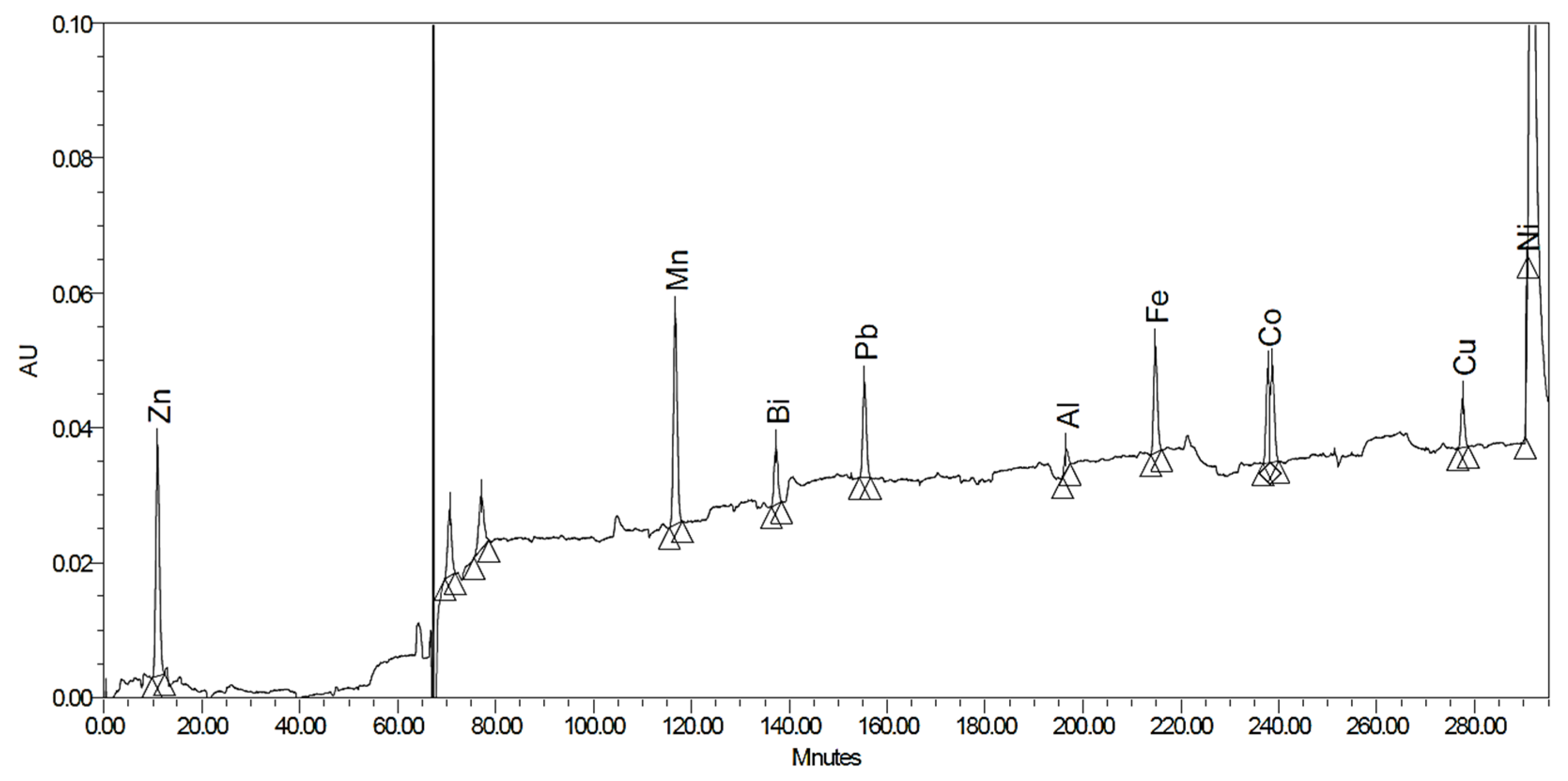
2.1. Post-Column Derivatization with PAR

Transition metals were separated on a Dionex IonPac CS5A column. The PAR reagent solution was mixed to the effluent to form a light-absorbing complex with the metals. Fe3+ and Zn2+, considered to be the major contaminants of cyclotron-produced [44Sc]Sc3+, were baseline separated. Residual zinc ions can be determined down to 0.16 ppm and iron to 0.26 ppm. Besides the two mentioned transition metals, Cu2+ ions are also a strong competitor in DOTA-labeling reactions [18]. It cannot be separated from Zn2+ ions with the current method, but the presence of both metal ions can be excluded if no peak is observed between 8 and 9 min. Many transition metal ions can be detected with good LOQ values (Table 1). However, the long-term observation of the baseline (Figure 2) shows significant noise, which could be decreased by careful equilibration of the system with the eluents, but it could not be completely avoided.
Thus, small differences in retention times, together with the baseline instability, do not allow unambiguous identification of each peak, however the absence of peaks is a good indication of sample purity.
This method has an acceptable resolution for Fe3+ and Zn2+ ions (Rs = 1.43, Figure 3) under the applied conditions, but for other potentially interfering metal ions (e.g.,: Cu2+, Al3+) the separation is not appropriate. Therefore, it is able to verify the low metal content of the radiometal samples but does not allow accurate quantification at low concentrations.
The post-column derivatization method is sensitive, but time-consuming to use. In addition, reagents should be freshly prepared by the excluding air before measurement and the system must be carefully equilibrated with the eluents. Despite prolonged washing, significant baseline drift and stability problems were often observed. Based on our results, the use of this method for routine quality control of short-lived isotopes is not recommended.

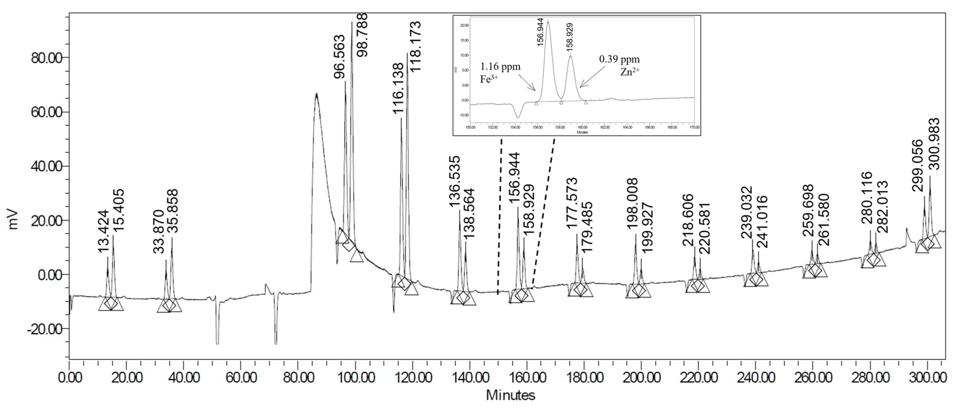
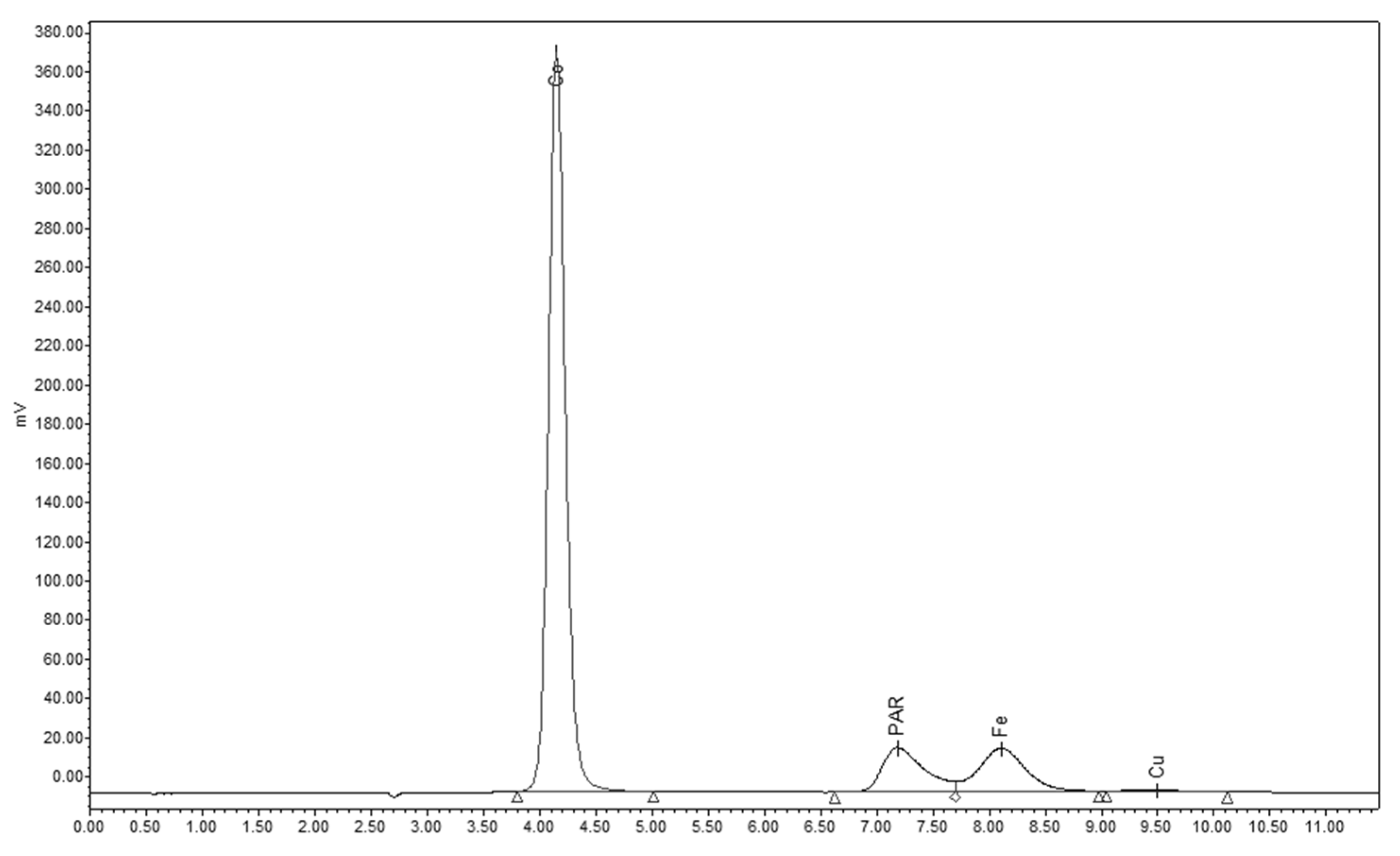
2.2. Pre-Column Derivatization with PAR

To avoid the difficulties observed during the post-column derivatization, we applied the same derivatization reaction before sample injection for analysis of the samples. Determination of Co2+, Fe3+, Cu2+ and Zn2+ ions were investigated. In the case of Co2+, Fe3+ and Cu2+, the formed metal-PAR complexes were retained on reversed phase column (LiChrospher 100 RP18) and eluted with phosphate buffer–methanol eluent. The unreacted PAR reagent elutes at 6.1 min, and the Fe3+-PAR complex was baseline separated (RS = 3.09) at 7.8 min with detection at 530 nm (Figure 4). The calibration curve was linear (R2 = 0.998) in the 0.5–10 ppm range. Limit of quantitation was found to be 0.1 ppm.
Zn2+ ions could not be determined with pre-column derivatization, as a solid precipitate was formed in the sample which was irreversibly retained on the column. However, this method was suitable for the determination of Fe3+ with good sensitivity and in the case of Cu2+ contaminants the sensitivity was moderate (Table 2). Results of iron determination was in good agreement with ICP data (Table 3).

2.3. Colorimetric Metal Determination

The determination of the small amount of transition metal ions (Fe3+, Zn2+, Al3+) was also investigated by commercially available colorimetric tests. These tests are based on the selective formation of a metal–dye complex and the selectivity of a particular metal ion is often enhanced by masking of interfering metal ions. Therefore, the tests contain several reagents to be added to the sample before the color formation reaction. The main disadvantage of commercially available colorimetric tests is that they require a large volume of sample, which must be reduced when testing radioactive samples. The original color comparison method does not allow the decrease of test solution volume without reducing the sensitivity. This could be overcome by changing the method of color determination from visual to instrumental, using UV-VIS detection. Thus, we decreased the sample and reagent volumes (Table 4) and injected the samples to the HPLC-UV detector, without the presence of a column.
Besides the colorimetric tests, we investigated the use of PAR- and xylenol orange reagents without separation to determine the metal content of the radioactive samples. The formation and the color of the metal–PAR complex was pH-dependent; thus, the addition of buffer was essential. Accordingly, 1 M ammonium acetate buffer (pH = 6.5) was used to adjust the pH of the samples. The strong orange color of the PAR reagent did not allow the observation of a color change when the PAR concentration of the sample solution was 200 µM, so it was optimized. Using a 10 µM PAR solution, a color change from light yellow to pink was clearly visible (Figure 5). The LOQ was 0.21 ppm for Fe3+ with approximately 10 µM PAR, using photometric detection at 490 nm. The calibration curve was linear (R2 = 0.9993) up to 10 ppm Fe3+ concentration.
Xylenol orange was successfully used for Zn2+ determination in samples containing 0.01 M HCl solution (LOQ 0.61 ppm, R2 = 0.9996), but not in the case of high hydrochloric acid concentration (2–5 M HCl). Despite the neutralization of samples by large excess of the buffer, the spectra of xylenol orange was shifted, causing a decrease of absorbance on the examined wavelength. This was probably due to the presence of chloride anions. This limits the use of this test for only low HCl-containing samples, so it can be used to test the residual Zn2+ content of purified gallium-68 samples, but is not applicable for checking the intermediate samples of the purification. Similar disturbance was observed in the presence of other transition metal ions in the sample (Fe3+, Cu2+) with a concentration higher than 1 ppm. This should be also taken into consideration when attempting to use xylenol orange for zinc determination. Accordingly, to exclude the presence of Fe3+- and other interfering transition metal ions, the PAR test (pre-column derivatization or colorimetric) should be used first, followed by the xylenol orange test to determine Zn2+ content of radioactive samples.
The tested colorimetric reagents (Table 5) can be used in the low ppm range for metal determination, but based on our results, the typical Fe3+- and Zn2+ content of the purified gallium-68 solutions was under the LOQ of the examined methods due to the use of ultrapure chemicals and careful purification.

3. Materials and Methods

3.1. Chemicals and Reagents

All chemicals (buffers, 2-dimethylaminoethanol, pyridine-2,6-dicarboxylic acid, potassium hydroxide, formic acid, potassium sulfate, ammonium hydroxide, sodium bicarbonate, natural calcium) were obtained from Sigma-Aldrich (Budapest, Hungary) and used without further purification. 4-(2-pyridylazo)resorcinol (PAR) was purchased from Alfa Aesar (Kandel, Germany). DGA, Zr and TK200 resins were purchased from TrisKem (Bruz, France). Enriched zinc-68 (98.60%) was obtained from NeonestAB (Stockholm, Sweden). Ultrapure hydrochloric acid (35%) and ultrapure nitric acid (69%) were supplied by Carl Roth GmbH (Karlsruhe, Germany). Metal ion standard solutions (1000 mg/L Fe3+, Co2+, Cu2+, Ni2+, Bi3+ in nitric acid for AAS), colorimetric tests (MColortest Zinc Test 1.14412.0001, Mqant Aluminium Test 1.14413.0001, Mcolortest Iron Test 1.14403.0001) and xylenol orange (XO) were supplied by Merck (Budapest, Hungary). HPLC-grade solvents were purchased from VWR. Ultrapure water was obtained from a Merck Simplicity Water Purification System.

3.2. HPLC Equipment and Conditions

3.2.1. Post-Column Complexation

The measurements were performed with a chromatographic system, consisting of a Jasco PU-2080i (metal free) pump, a waters Acquity UPLC BSM pump, a modified Knauer 3800 autosampler (equipped with plastic injector valve, needle, and 10 µL loop) and a Waters 2487 dual λ absorbance detector. A photomultiplier tube (Hamamatsu Photonics H10493-001), equipped with plastic scintillator, was used for the detection of radioactivity. Data were evaluated by Empower 3 chromatography software. Dionex IonPac CS5A (2 × 250 mm) column was used for the separation of transition metals [40]. The eluent contained 7.0 mM PDCA (pyridine-2,6-dicarboxylic acid), 66 mM Potassium hydroxide, 74 mM Formic acid, 5.6 mM Potassium sulfate, delivered with 0.3 mL/min flowrate by the Jasco metal-free pump. The post-column reagent contained 0.5 mM PAR, 1.0 M 2-dimethylaminoethanol, 0.50 M ammonium hydroxide and 0.30 M sodium bicarbonate. It was pumped by the Waters BSM pump to the column effluent with 0.15 mL/min flowrate. Both solutions were prepared and used under an inert atmosphere. Samples were injected without any sample preparation. The separation took 15 min. UV chromatograms were integrated at λ = 530 nm (Table 6).

3.2.2. Pre-Column Complexation

The measurements were performed with a Jasco PU-2080i (metal free) pump, a modified Knauer 3800 autosampler (equipped with plastic valve, needle, and 10 µL loop) and a Waters 2487 dual λ absorbance detector. Flow rate of 0.8 mL/min on a LiChrospher 100 RP18 column (75 × 4 mm, 5 µm). The mobile phase was 65% 0.1 M; pH 6.5 NH4H2PO4/(NH4)2HPO4 buffer and 35% methanol. Samples were mixed in 1:1 ratio with PAR reagent solution, prepared by dissolving 6 mg of solid PAR in 3.5 mL methanol and 6.5 mL NH4H2PO4/(NH4)2HPO4 buffer (0.1 M; pH 6.5). Formation of the PAR complex was accompanied by an immediate color change (Table 6).

3.2.3. Colorimetry

The measurements were performed with a Jasco PU-2080i (metal free) pump, a modified Knauer 3800 autosampler (equipped with plastic valve, needle and 10 µL loop) and a Waters 2487 dual λ absorbance detector. The injector was directly connected to the detector—without column. Eluent was water with 0.8 mL/min flowrate. UV detection was performed at 490 nm (Table 6). Table 4 shows the sample preparation methods for different colorimetric analyzes.

3.3. Production and Purification of Scandium-44 and Gallium-68 Radionuclides

Scandium-44 and gallium-68 isotopes were produced in a GE PETtrace cyclotron with proton irradiation of solid calcium and zinc metal targets (44Ca(p,n)44Sc), (68Zn(p,n)68Ga) using a shuttle-type solid target system developed in-house [15], equipped with 50 µm HAVAR- and 500 µm aluminum energy degrader foils. The targets were prepared by pressing approximately 120 mg natural calcium metal or 40 mg enriched zinc-68 powder to form a pellet and these pellets were pressed into aluminum target holders. Targets were irradiated with 30–50 µA proton beam for 10–180 min.
Purification of [44Sc]Sc3+: Irradiated target (with 100–500 MBq activity) was placed in a lead pot and pressed against a plastic dissolution block equipped with an O-ring seal and two Teflon capillaries ending at the target metal surface. The irradiated metal was dissolved in 4 mL 3 M u.p. HCl at room temperature, pumped at 1 mL/min with a syringe pump. Target solution was loaded onto 70 mg DGA resin and washed with 3 mL 3 M HCl (removal of Ca2+) and 3 mL 1 M HNO3 (removal of Fe3+, Zn2+, Ni2+). [44Sc]Sc3+ was eluted fractionally with 1 mL 0.1 M HCl. [44Sc]Sc3+ recovery was 75–85%.
Purification of [68Ga]Ga3+: Irradiated target (2–60 GBq) was dissolved in 10 mL 5 M u.p. HCl at room temperature in the same dissolution block and loaded to Zr resin, washed with 10 mL 5 M HCl and eluted to TK200 resin with 5 mL 2 M HCl. Final elution from the second resin was performed with 0.05 M HCl.

4. Conclusions

We have compared various metal determination methods, utilizing colored complex formations with different dyes.
Based on our studies, chromatographic separation with post-column derivatization is a more selective and sensitive method for trace metal analysis, but the complexity of the instrumental setup and the observed baseline instabilities prohibit its use for routine QC. Furthermore, commercially available or in-house-developed colorimetric tests can be useful for the detection of higher (more than few ppm) concentrations of contaminating metals, but is not selective enough to use in the sub-ppm range, where the typical metal content of radiometal samples can be found. We have found that the reliability and documentation of colorimetric testing can be improved by injecting the samples to the HPLC-UV detector without a column. In addition, the use of HPLC detection enables the use of much smaller sample volumes (30 µL instead of 5–20 mL), which significantly reduces the material loss of short-lived radiometals during QC. The most important advantage of colorimetric methods is the possibility to obtain information about the metal content of a sample without delay, which together with labeling tests (e.g., apparent molar activity determination) can facilitate method development, help in troubleshooting and may enable some level of process control in routine radiotracer production.
The examined colorimetric tests cannot replace atom spectroscopic techniques for the determination of trace metal impurities in radioactive samples but can find their place in purification method development and routine QC of radiometal solutions for labeling.

Author Contributions

V.F. performed method development and data analysis, A.F., B.G., D.S. (Dániel Szücs) and G.T. contributed to the design of the experiments, D.S. (Dezső Szikra) provided radiometal samples. All authors discussed the results and contributed to the final manuscript. All authors have read and agreed to the published version of the manuscript.

Funding

This research received no external funding.

Institutional Review Board Statement

Not applicable.

Informed Consent Statement

Not applicable.

Data Availability Statement

Data is contained within the article.

Acknowledgments

This research was partially supported by the IAEA Coordinated Research Program No. F22073. We thank for Zsófi Sajtos and Edina Baranyai for assistance with ICP measurements.

Conflicts of Interest

The authors declare no conflict of interest.

References

  1. Pandey, M.K.; DeGrado, T.R. Cyclotron Production of PET Radiometals in Liquid Targets: Aspects and Prospects. Curr. Radiopharm. 2020, 13, 325–339. [Google Scholar] [CrossRef] [PubMed]
  2. Rodnick, M.E.; Sollert, C.; Stark, D.; Clark, M.; Katsifis, A.; Hockley, B.G.; Parr, D.C.; Frigell, J.; Henderson, B.D.; Abghari-Gerst, M.; et al. Cyclotron-Based Production of 68Ga, [68Ga]GaCl3, and [68Ga]Ga-PSMA-11 from a Liquid Target. EJNMMI Radiopharm. Chem. 2020, 5, 25. [Google Scholar] [CrossRef] [PubMed]
  3. Alves, V.; do Carmo, S.; Alves, F.; Abrunhosa, A. Automated Purification of Radiometals Produced by Liquid Targets. Instruments 2018, 2, 17. [Google Scholar] [CrossRef] [Green Version]
  4. Siikanen, J.; Jussing, E.; Milton, S.; Steiger, C.; Ulin, J.; Jonsson, C.; Samén, E.; Tran, T.A. Cyclotron-Produced 68Ga from Enriched 68Zn Foils. Appl. Radiat. Isot. 2021, 176, 109825. [Google Scholar] [CrossRef]
  5. Thisgaard, H.; Kumlin, J.; Langkjær, N.; Chua, J.; Hook, B.; Jensen, M.; Kassaian, A.; Zeisler, S.; Borjian, S.; Cross, M.; et al. Multi-Curie Production of Gallium-68 on a Biomedical Cyclotron and Automated Radiolabelling of PSMA-11 and DOTATATE. EJNMMI Radiopharm. Chem. 2021, 6, 1. [Google Scholar] [CrossRef] [PubMed]
  6. Lin, M.; Paolillo, V.; Ta, R.T.; Damasco, J.; Rojo, R.D.; Carl, J.C.; Melancon, M.P.; Ravizzini, G.C.; Le, D.B.; Santos, E.B. Fully Automated Preparation of 68Ga-PSMA-11 at Curie Level Quantity Using Cyclotron-Produced 68Ga for Clinical Applications. Appl. Radiat. Isot. 2020, 155, 108936. [Google Scholar] [CrossRef]
  7. Nelson, B.J.B.; Wilson, J.; Richter, S.; Duke, M.J.M.; Wuest, M.; Wuest, F. Taking Cyclotron 68Ga Production to the next Level: Expeditious Solid Target Production of 68Ga for Preparation of Radiotracers. Nucl. Med. Biol. 2020, 80–81, 24–31. [Google Scholar] [CrossRef]
  8. Svedjehed, J.; Pärnaste, M.; Gagnon, K. Demystifying Solid Targets: Simple and Rapid Distribution-Scale Production of [68Ga]GaCl3 and [68Ga]Ga-PSMA-11. Nucl. Med. Biol. 2022, 104–105, 1–10. [Google Scholar] [CrossRef]
  9. Becker, K.V.; Chernysheva, M.; Barnhart, T.E.; Gagnon, K.; Engle, J.W. A Review of Accelerator-Produced Ga-68 with Solid Targets. Curr. Radiopharm. 2021, 14, 315–324. [Google Scholar] [CrossRef]
  10. Lin, M.; Waligorski, G.J.; Lepera, C.G. Production of Curie Quantities of 68Ga with a Medical Cyclotron via the 68Zn(p,n)68Ga Reaction. Appl. Radiat. Isot. 2018, 133, 1–3. [Google Scholar] [CrossRef]
  11. Talip, Z.; Favaretto, C.; Geistlich, S.; van der Meulen, N.P. A Step-by-Step Guide for the Novel Radiometal Production for Medical Applications: Case Studies with 68Ga, 44Sc, 177Lu and 161Tb. Molecules 2020, 25, 966. [Google Scholar] [CrossRef] [PubMed] [Green Version]
  12. Umbricht, C.A.; Benešová, M.; Schmid, R.M.; Türler, A.; Schibli, R.; van der Meulen, N.P.; Müller, C. 44Sc-PSMA-617 for Radiotheragnostics in Tandem with 177Lu-PSMA-617—Preclinical Investigations in Comparison with 68Ga-PSMA-11 and 68Ga-PSMA-617. EJNMMI Res. 2017, 7, 9. [Google Scholar] [CrossRef] [PubMed] [Green Version]
  13. Krajewski, S.; Cydzik, I.; Abbas, K.; Bulgheroni, A.; Simonelli, F.; Holzwarth, U.; Bilewicz, A. Cyclotron Production of 44Sc for Clinical Application. Radiochim. Acta 2013, 101, 333–338. [Google Scholar] [CrossRef]
  14. Pandey, M.K.; Byrne, J.F.; Jiang, H.; Packard, A.B.; DeGrado, T.R. Cyclotron Production of 68Ga via the 68Zn(p,n)68Ga Reaction in Aqueous Solution. Am. J. Nucl. Med. Mol. Imaging 2014, 4, 303–310. [Google Scholar] [PubMed]
  15. Mikolajczak, R.; Huclier-Markai, S.; Alliot, C.; Haddad, F.; Szikra, D.; Forgacs, V.; Garnuszek, P. Production of Scandium Radionuclides for Theranostic Applications: Towards Standardization of Quality Requirements. EJNMMI Radiopharm. Chem. 2021, 6, 19. [Google Scholar] [CrossRef]
  16. Pyrzyńska, K.; Kilian, K.; Pęgier, M. Separation and Purification of Scandium: From Industry to Medicine. Sep. Purif. Rev. 2019, 48, 65–77. [Google Scholar] [CrossRef]
  17. Kilian, K.; Cheda, Ł.; Sitarz, M.; Szkliniarz, K.; Choiński, J.; Stolarz, A. Separation of 44Sc from Natural Calcium Carbonate Targets for Synthesis of 44Sc-DOTATATE. Molecules 2018, 23, 1787. [Google Scholar] [CrossRef] [Green Version]
  18. Oehlke, E.; Le, V.S.; Lengkeek, N.; Pellegrini, P.; Jackson, T.; Greguric, I.; Weiner, R. Influence of Metal Ions on the 68Ga-Labeling of DOTATATE. Appl. Radiat. Isot. 2013, 82, 232–238. [Google Scholar] [CrossRef]
  19. Pharmeuropa 30.4. Reference: PA/PH/Exp. 14/T (18) 13 ANP.
  20. U.S. Environmental Protection Agency’s (EPA’s) Office of Water (OW). Method 1640 Determination of Trace Elements in Water by Preconcentration and Inductively Coupled Plasma-Mass Spectrometry; U.S. Environmental Protection Agency: Washington, DC, USA, 1997. [Google Scholar]
  21. Rodushkin, I.; Engström, E.; Baxter, D.C. Sources of Contamination and Remedial Strategies in the Multi-Elemental Trace Analysis Laboratory. Anal. Bioanal. Chem. 2010, 396, 365–377. [Google Scholar] [CrossRef]
  22. Geary, W.J.; Nickless, G.; Polland, F.H. The Metal Complexes of Some Azo and Azomethine Dyestuffs. Anal. Chim. Acta 1962, 26, 575–582. [Google Scholar] [CrossRef]
  23. Sommer, L. Complexation of Aluminium, Yttrium, Lanthanum and Lanthanides with 4-(2-Pyridylazo)Resorcinol (Par). Talanta 1967, 14, 457–471. [Google Scholar] [CrossRef]
  24. Ding, X.; Mou, S.; Liu, K.; Siriraks, A.; Riviello, J. Ion Chromatography of Heavy and Transition Metals by On- and Post-Column Derivatizations. Anal. Chim. Acta 2000, 407, 319–326. [Google Scholar] [CrossRef]
  25. Horstkotte, B.; Jarošová, P.; Chocholouš, P.; Sklenářová, H.; Solich, P. Sequential Injection Chromatography with Post-Column Reaction/Derivatization for the Determination of Transition Metal Cations in Natural Water Samples. Talanta 2015, 136, 75–83. [Google Scholar] [CrossRef] [PubMed]
  26. Ahmed, S.H.; El Gammal, E.M. Chromatographic Separation and Determination of Some Heavy Metals and the Sum of the Rare Earth Elements in Some Geological Materials. Chem. Pap. 2020, 74, 3491–3501. [Google Scholar] [CrossRef]
  27. Hu, Q.; Yang, G.; Yang, J.; Yin, J. Study on Determination of Iron, Cobalt, Nickel, Copper, Zinc and Manganese in Drinking Water by Solid-Phase Extraction and RP-HPLC with 2-(2-Quinolinylazo)-5-Diethylaminophenol as Precolumn Derivatizing Reagent. J. Environ. Monitor. 2002, 4, 956–959. [Google Scholar] [CrossRef] [PubMed]
  28. Li, Z.; Yang, G.; Wang, B.; Jiang, C.; Yin, J. Determination of Transition Metal Ions in Tobacco as Their 2-(2-Quinolinylazo)-5-Dimethylaminophenol Derivatives Using Reversed-Phase Liquid Chromatography with UV–VIS Detection. J. Chromatogr. A 2002, 971, 243–248. [Google Scholar] [CrossRef]
  29. Zhang, C.; Miura, J.; Nagaosa, Y. Determination of Cadmium, Zinc, Nickel and Cobalt in Tobacco by Reversed-Phase High-Performance Liquid Chromatography with 2-(8-Quinolylazo)-4,5-Diphenylimidazole as a Chelating Reagent. Anal. Sci. 2005, 21, 1105–1110. [Google Scholar] [CrossRef] [Green Version]
  30. Burakham, R.; Srijaranai, S.; Grudpan, K. High-Performance Liquid Chromatography with Sequential Injection for Online Precolumn Derivatization of Some Heavy Metals. J. Sep. Sci. 2007, 30, 2614–2619. [Google Scholar] [CrossRef]
  31. Roston, D.A. Precolumn Chelation with 4-(2-Pyridylazo)Resorcinol for Simultaneous Determination of Metal Ions by Liquid Chromatography. Anal. Chem. 1984, 56, 241–244. [Google Scholar] [CrossRef]
  32. Martínez-Zepeda, D.L.; Meza-González, B.; Álvarez-Hernández, M.L.; Bazany-Rodríguez, I.J.; Vilchis Néstor, A.R.; Cortés-Guzmán, F.; Gómez-Espinosa, R.M.; Valdes-García, J.; Dorazco-González, A. Efficient Naked Eye Sensing of Tartrate/Malate Based on a Zn-Xylenol Orange Complex in Water and Membrane-Based Test Strips. Dye. Pigment. 2021, 188, 109239. [Google Scholar] [CrossRef]
  33. Študlar, K.; Janoušek, I. The Photometric Determination of Zinc with Xylenol Orange. Talanta 1961, 8, 203–208. [Google Scholar] [CrossRef]
  34. Smith, F.E.; Herbert, J.; Gaudin, J.; Hennessy, D.J.; Reid, G.R. Serum Iron Determination Using Ferene Triazine. Clin. Biochem. 1984, 17, 306–310. [Google Scholar] [CrossRef]
  35. Dahlén, J.; Karlsson, S. Determination of Iron(II) in Natural Waters by Capillary Zone Electrophoresis Using on-Capillary Complexation with 2,4,6-Tri(2′-Pyridyl)-1,3,5-Triazine. J. Chromatogr. A 1999, 848, 491–502. [Google Scholar] [CrossRef]
  36. Kennedy, J.A.; Powell, H.K.J. Colorimetric Determination of Aluminium(III) with Chrome Azurol S and the Reactivity of Hydrolysed Aluminium Species. Anal. Chim. Acta 1986, 184, 329–333. [Google Scholar] [CrossRef]
  37. Pakalns, P. Spectrophotometric Determination of Aluminium with Chrome Azurol s. Anal. Chim. Acta 1965, 32, 57–63. [Google Scholar] [CrossRef]
  38. Kim, D.; Jo, A.; Seo, B.-K.; Lee, K.-W.; Park, W.H.; Lee, T.S. Colorimetric Detection of Transition Metal Ions with Azopyridine-Based Probing Molecule in Aqueous Solution and in PMMA Film. Fibers Polym. 2013, 14, 1993–1998. [Google Scholar] [CrossRef]
  39. McCall, K.A.; Fierke, C.A. Colorimetric and Fluorimetric Assays to Quantitate Micromolar Concentrations of Transition Metals. Anal. Biochem. 2000, 284, 307–315. [Google Scholar] [CrossRef]
  40. Thermo Scientific Technical Note 10 Ic-Transition-Metals Tn72544. Available online: https://assets.thermofisher.com/TFS-Assets/CMD/Technical-Notes/tn-10-ic-transition-metals-tn72544-en.pdf (accessed on 20 January 2022).
Figure 1. Structure of the applied reagents: triazine, 4-(2-pyridylazo)resorcinol, xylenol-orange, Chromeazurol S.
Pharmaceuticals 15 00147 g001
Figure 2. Separation of transition metals with post-column derivatization in test solutions.
Pharmaceuticals 15 00147 g002
Figure 3. Analysis of radioactive scandium-44 samples on Dionex IonPac CS5A column with PDCA (pyridine-2,4-dicarboxylic acid) eluent.
Pharmaceuticals 15 00147 g003
Figure 4. Separation of PAR and its Co2+, Fe3+ and Cu2+ complexes on reversed phase column in test sample.
Pharmaceuticals 15 00147 g004
Figure 5. Color change of PAR and xylenol orange solutions in the presence of various amounts of Fe3+ and Zn2+ ions. (a) Colors with PAR at 0, 5 and 10 ppm Fe3+; (b) Colors with XO at 0, 5 and 10 ppm Zn2+.
Pharmaceuticals 15 00147 g005
Table 1. Quantitation limits for the examined non-radioactive metals with post-column derivatization.
Metal IonsLOQ (ppm)tR (min.)c (ppm)Rs
Bi3+8.855.321.0-
Fe3+0.266.560.50.89
Zn2+0.168.520.51.43
Cu2+0.138.870.30.26
Ni2+1.399.481.00.80
Al3+1.179.691.00.25
Pb2+0.319.751.00.04
Co2+0.139.961.00.15
Cd2+0.3310.410.30.33
Mn2+0.1017.571.05.17
Table 2. Separation of transition metal ions in test solution.
MetalLOQ (ppm)tR (min.)c (ppm)Rs
Co2+0.0044.144.91-
PAR-7.19-6.54
Fe3+0.18.123.31.33
Cu2+22.339.5052.961.78
Table 3. Comparison of Fe3+ content of gallium-68 samples, determined with ICP (inactive samples) and pre-column derivatization (radioactive samples).
Samplesc(Fe3+) ppm
ICPHPLC Pre-Column Derivatization with PAR
07.020.390.41
07.01/20.410.30
07.01/10.390.24
Table 4. Sample preparation methods of colorimetric tests.
ReagentOriginal Method 1Sample Preparation for HPLC Detection
PAR-Reagent: 6 mg PAR was dissolved in 3.5 mL methanol and 6.5 mL buffer 2.
Sample preparation: 5 µL of PAR solution, 1465 µL of buffer 1 and 30 µL sample.
Xylenol orange-Reagent: 14.3 mg xylenol orange was dissolved in 10 mL buffer 1. Sample preparation: 10 µL of XO solution, 1460 µL of buffer 1 and 30 µL sample.
Zinc test5 mL of sample, 4 drops of reagent 1 (160 µL), 1 dosing spoon of reagent 2 (188 mg) and 1 microspoon of reagent 3 (10.3 mg). Leave to stand for exactly 5 min (reaction time). And add 4 drops of reagent 4 (160 µL).45 µL of reagent 1, 53 mg of reagent 2, 9 mg of reagent 3, 1380 µL of buffer 1, 30 µL sample, leave to stand for exactly 5 min (reaction time), and 45 µL of reagent 4.
Iron test20 mL of sample and 5 drops of reagent 1 (200 µL). Leave to stand for 3 min (reaction time).15 µL of reagent, 1455 µL of buffer 1 and 30 µL sample. Leave to stand for 3 min (reaction time).
Aluminum test5 mL of sample, 1 microspoon of reagent 1 (147.6 mg), 1.2 mL of reagent 2 and 4 drops of reagent 3 (160 µL). Leave to stand for 7 min (reaction time).34.8 mg of reagent 1, 282 µL of reagent 2, 1150 µL of buffer 1, 30 µL sample and 38 µL of reagent 3. Leave to stand for 7 min (reaction time).
1 Original methods and reagent 1, 2, 3 were provided by Merck with colorimetric test. 2 1 M ammonium acetate buffer, pH 6.5.
Table 5. Comparison of the tested methods.
ReagentsMethodsMetal DeterminedRange (ppm)LOQ (ppm)Interfering Metal Ions
PARpost-columnFe3+0.5–1.00.08-
pre-columnFe3+1.95–10.00.10-
colorimetryFe3+1.50–11.00.21Cu2+, Ni2+, Ga3+, Bi3+, Co2+, Cd2+
Triazine derivate (Merck)colorimetryFe3+1.50–13.00.62Co2+, Cr3+, Cu2+, Ni2+, Pb2+
Xylenol OrangecolorimetryZn2+1.50–20.00.61Cu2+, Ni2+, Fe3+, Co2+, Al3+
Thiocyanate (Merck)colorimetryZn2+1.0–20.00.20Cu2+, Fe3+, Ni2+, Pb2+,
Chromazurol S (Merck)colorimetryAl3+-0.04Ag+, Co2+, Cr3+, Cu2+, Fe3+, Mn2+, Pb2+, Sn2+, Zn2+
Table 6. Summary of HPLC methods.
SystemColumnEluentFlow RateReagentDetection
Post-column complexationDionex IonPac CS5A (2 × 250 mm)Eluent A: 7.0 mM PDCA, 66 mM Potassium hydroxide, 74 mM Formic acid, 5.6 mM Potassium sulfate
Eluent B: 0.5 mM PAR, 1.0 M 2-dimethylaminoethanol, 0.50 M ammonium hydroxide and 0.30 M sodium bicarbonate
A: 0.3 mL/min
B: 0.15 mL/min
PAR530 nm
Pre-column complexationLiChrospher 100 RP18 column (75 × 4 mm, 5 µm)65% 0.1 M; pH 6.5 NH4H2PO4/(NH4)2HPO4 buffer and 35% methanol0.8 mL/minPAR530 nm
Colorimetry-water0.8 mL/minPAR490 nm
XO570 nm
Zinc test435 nm
Iron test560 nm
Aluminium test590 nm
Publisher’s Note: MDPI stays neutral with regard to jurisdictional claims in published maps and institutional affiliations.

