Review

Recent Development in Optical Chemical Sensors Coupling with Flow Injection Analysis

Department of Analytical Chemistry, Faculty of Sciences, University of Málaga, Campus Teatinos s/n, 29071 Málaga, Spain
*
Author to whom correspondence should be addressed.
Sensors 2006, 6(10), 1245-1307; https://doi.org/10.3390/s6101245
Submission received: 4 May 2006 / Accepted: 5 October 2006 / Published: 10 October 2006
(This article belongs to the Special Issue Sensors in Flow Analysis)

Abstract

:
Optical techniques for chemical analysis are well established and sensors based on these techniques are now attracting considerable attention because of their importance in applications such as environmental monitoring, biomedical sensing, and industrial process control. On the other hand, flow injection analysis (FIA) is advisable for the rapid analysis of microliter volume samples and can be interfaced directly to the chemical process. The FIA has become a widespread automatic analytical method for more reasons; mainly due to the simplicity and low cost of the setups, their versatility, and ease of assembling. In this paper, an overview of flow injection determinations by using optical chemical sensors is provided, and instrumentation, sensor design, and applications are discussed. This work summarizes the most relevant manuscripts from 1980 to date referred to analysis using optical chemical sensors in FIA.

1. Introduction

A chemical sensor is a device which responds to a particular analyte in a selective way through a chemical reaction and can be used for the qualitative or quantitative determination of the analyte. Chemical sensors are categorized into the following groups according to the transducer type: Electrochemical, optical, mass sensitive and heat sensitive.
In optical sensors there is a spectroscopic measurement associated with the chemical interactions. Optical sensors are often referred to as “optodes” and the use of optical fibres is a common feature. Many biosensors make use of optical measurements. Absorbance, reflectance, photoluminescence and chemiluminescence measurements are used in the different types of optical sensors.
The aim of flow analysis was progressively extended to the automation of the whole analytical procedure via manipulation of a sample zone in a properly designed flow network that ended with a flow-through detector.
This is a review on the development and applications of FI-optical chemical sensors, including the sensors for the detection of inorganic species, organic and biological compounds and drugs.

2. Absorbance

Molecular absorption spectroscopy is no doubt the most frequently used detection technique in analytical laboratories due to its high flexibility for adaptation to a wide variety of analytical problems.
Optical chemical sensors (optodes) have played an important role in industrial, environmental and clinical monitoring since their introduction more than two decades ago as a result of their low cost, possibility for miniaturisation and great flexibility. In general, these sensors are comprised of various reagents immobilised within suitable membranes. A number of the existing optodes utilises colour complexion reactions between immobilised ligands and heavy metal ions. The utilisation of colour redox reactions has been largely ignored despite the fact that numerous substances of analytical interest are electroactive.
In these sense, Ferroin, tris(1,10-phenanthroline) iron(II), is a well-known redox indicator that changes colour from red to blue when oxidised to Ferriin, tris(1,10-phenanthroline) iron(III); analytical applications of the redox couple associated with Ferroin other than its utilisation as a redox indicator include both batch and flow injection determinations of various reductants and oxidants; also, previous applications of Ferroin and its derivatives in the preparation of ion-selective electrodes are known and involve the immobilisation of the reagent as an ion associate into various membrane compositions. As a cation, Ferroin can easily be immobilised within cation exchange membranes without the necessity of preliminary modification of the membrane (e.g. using lipophilic anions or plasticising agents as in the case of neutral PVC membranes). A suitable membrane of this kind which was successfully employed for the construction of an optode incorporating the colorimetric reagent 1-(2′-pyridylazo)-2-naphthol (PAN) is Nafion® 117. The redox properties of the optode membrane were studied under batch and flow conditions using acidified bromate or cerium(IV) solutions as oxidants and ascorbic acid or sodium ascorbate solutions as reductants. The experiments under flow conditions were performed in a computer-controlled sequential flow injection system incorporating a flow-through measuring cell where the optode membrane was mounted. Optical fibre technology was used to monitor the membrane absorbance with time [1]. Other application is the determination of copper [2]; the optrode is based on a Nafion membrane and an immobilized organic ligand coupled with a flow injection (FI) system. The FI system includes a flow-through removable measuring cell and a simple spectrophotometer. Owing to the miniature size of the system and the efficient use of optical fibers, this optrode is well suited for monitoring environmental water samples. The success of the described optrode system depends on the effectiveness of the FI reagent delivery system. Optimum contact time with the membrane (as determined by the reagent flow rates) and the injected sample volume are critical. Environmental water samples were analyzed for copper content using the optimized optrode system.
Solid phase spectrophotometry (SPS) is a very sensitive technique based on the direct measurement of the absorbance of an active solid support (usually an ion-exchanger) that previously has sorbed a sample component from a sample solution (batch methodology). It combines the steps of preconcentration, separation and determination, and provides (both) high selectivity and a sensitivity several orders of magnitude higher than those obtained with solution phase spectrophotometry. SPS has also been integrated with flow injection analysis (FIA) systems: a solid support is placed in a flow-cell in a photometric detector, which monitors the signal obtained from the sorption of the species of interest on the support. The direct sorption of the analyte without previous derivatization considerably simplifies the FI manifold used, provides a higher sample frequency and does not require additional reagents. Various applications of these procedures are described in table 1.
For many pharmaceutical and biomedical analyses various dry colorimetric tests are developed. There are paper or plastic strips, plastic cuvettes and microtitration-plates coated with reagents layers that selectively react with an analyte forming color products detectable spectrophotometrically. For such tests no additional reagents are necessary. Recently, they have developed cuvette tests for selected redox species, having integrated sensing layer made of Prussian blue (PB). PB film plays double role in the developed determination system, first as an immobilized reagent (oxidant), and second as an optical sensing element. However, the tests are useful only as single-use, disposable devices. Application of PB film for construction of flow-through detector is presented by Lenarczuk et al. [3]. Utilization of the PB film-based reaction–detection system in flow-injection analysis (FIA) conditions enables both, spectrophotometric determination of analyte and regeneration of detector. The presented optical PB-based-FIA system has been used for determination of vitamin C, cysteine and hydrogen peroxide in pharmaceutical products.
On the other hand, theoretical aspects of bulk optode membranes based on the extraction of the analyte into a thin plasticized poly(vinyl chloride) membrane have been described in the literature. Various ionophores and appropriate lipophilic pH indicator dyes introduced into the membranes have been used to design optical cation sensing systems. This pH indicator approach was also adapted to design optical anion optode membranes; for example, optical chemical sensors for nitrate and chloride were based on polymeric membranes doped with lipophilic pH indicator dyes and quaternary ammonium exchangers. When bathed in a sample solution, H+/Xn- ion exchange equilibrium occurs between the two phases, resulting in a change in the optical absorbance of the optode membrane. This type of optode displays Hofmeister selectivity, with the largest response to lipophilic anions (perchlorate, thiocyanate, etc). Previously, García et al. developed a new type of flow-ion sensitive bulk optode membrane that incorporates a chromogenic reagent (metallochromic indicator) in a plasticized poly (vinyl) chloride membrane entrapped in a cellulose support, which was applied to determining various contaminant metallic ions. The authors also described and tested the performance of a flow-optode membrane, which was constructed using a similar immobilization procedure, for the FI-spectrophotometric determination of perchlorate anion [4]. The optode incorporates the lipophilic pH indicator 5-octadecanoyloxy-2-(4-nitrophenylazo) phenol and methyltridodecylammonium as the active constituents. The optode is coupled into a flow-cell in a continuous configuration. As is known, bulk optodes have gained considerably in practical reliability, and can be considered as inexpensive alternatives to certain conventional analytical methods. The use of chemical sensors in flow-injection analysis by far simplifies the procedure and may provide higher selectivity than the common photometric mode of operation. The FI mode of operation automates measurements with the optode membrane and permits the sensor layer of the optode to be regenerated quickly with the same carrier solution.
Recently, for the first time, a bead injection spectroscopy–flow injection analysis (BIS–FIA) system with spectrophotometric detection in a commercially available flow cell (Hellma 138-OS) is developed [5]. The flow cell is filled by injecting in the flow system a homogeneous bead suspension of an appropriate solid support previously loaded with the chromogenic reagent. The solid beads work as a flow-through chemical sensing microzone integrating on-line separation/reaction/detection, developing the analytical signal when the injected sample containing the analyte reaches the beads. At the end of the analysis, the beads are discarded by reversing the flow and instantaneously transported out of the system. These systems can be used in those flow-through optosensors in which the species of interest is so strongly retained on the solid sensing microzone that the regeneration of the solid beads becomes (extraordinarily) difficult. They can be considered as belonging to the third generation of FI microanalytical techniques which use renewable sensing solid surface, avoiding the need for a reversible sensing mechanism. To demonstrate the utility of this technique, a single BIS–FIA system is developed to determine: (a) directly Fe(II); (b) Fe(III) or total iron (Fe(II)+Fe(III)), using a previous reduction step; and (c) indirectly ascorbic acid. Ferrozine (Fz) is the bead-loaded chromogenic reagent used. The analytical signal corresponds to the absorbance of the complex [Fe(II)Fz3]4−. The system was applied to the determination of (a) iron in wine; water and pharmaceuticals; and (b) ascorbic acid in fruit juices, pharmaceuticals and conservative liquids, obtaining satisfactory results.

3. Reflectance

A novel optical fibre reflectance sensor coupled to a multisyringe flow injection system (MSFIA) for the determination and speciation analysis of iron at trace level using chelating disks (iminodiacetic groups) is proposed [52]. Once iron(III) has been retained onto a chelating disk, an ammonium thiocyanate stream is injected in order to form the iron(III)-thiocyanate complex which is spectrophotometrically detected at 480 nm. Iron(III) is eluted with 2 M hydrochloric acid so that the chelating disk is regenerated for subsequent experiments. The determination of total iron is achieved by the on-line oxidation of iron(II) to iron(III) with a suitable hydrogen peroxide stream. A mass calibration was feasible in the range from 0.001 to 0.25 μg. The detection limit was 0.001 μg. The applicability of the proposed methodology in fresh and seawater samples has been proven. The proposed technique has been validated by replicate analysis of certified reference materials of water with satisfactory results. Also, a combination of MSFIA technique with an optical fibre reflectance sensor for the determination of iron in water samples has been developed [53]. Anion-exchange solid phase extraction (SPE) disks have been used as solid phase. Ammonium thiocyanate has been chosen as chromogenic reagent for Fe(III). The complex Fe[SCN]63- is retained onto the SPE disk and spectrophotometrically detected at 480 nm. The complex is eluted with 0.25 mol L-1 hydrochloric acid in 75% ethanol. Total iron can be determined by oxidising Fe(II) to Fe(III) with hydrogen peroxide. A mass calibration was run within the range of 0.4-37.5 ng. The detection limit was 0.4 ng. An injection throughput of 7 injections per hour for a sampling volume of 1 ml has been achieved. The applicability of the proposed methodology in natural water samples has been proven. The properties of anion-exchange and chelating SPE disks have been studied and compared.
A flow-through optical fibre reflectance sensor for the determination of lead at trace level using immobilised gallocynine as the reagent phase is proposed [54]. Gallocynine is physically adsorbed onto XAD-7. This method provided a wide linear range of 1×10⁻¹ to 1×10³ ppm and detection limit of 0.06 ppm. The response towards lead was also reversible using acidified KNO3 as the regenerating solution. Interference studies showed that Hg(II) and Ag(I) significantly interfered during the determination.
A potassium optical-sensing layer based on reflectance change measurements of the Takagi reagent (4′-picryl-3′-nitrobenzo-18-crown-6) immobilized on a plasticized PVC membrane has been developed [55]. The analytical characteristics of this sensing-layer design have been studied in detail and are critically compared with those observed by immobilization (adsorption) of the same reagent on non-ionic resin beads packed at the end of an optical fiber (optrode). The immobilization of the indicator on the resin surface and its use in the optrode form has proved to be clearly superior to PVC immobilization and its use in connection with a typical FIA system. A probable recognition-process mechanism for both sensing phases of potassium is proposed. In the case of the sensing layer using a PVC membrane, a very limited operational lifetime is observed due to leaching of the reagent and water absorption in the membrane. Detection limits are in the range of 10-15 ppm K+ and the precision observed is around ±2% for both devices. The optrode type of sensor has been further developed for the determination of potassium in urine, milk and sea water.
A novel, versatile and sensitive flow-through optical fiber diffuse reflectance sensor to implement disk-based solid-phase extraction in a FIA system is presented [56]. Nitrite optosensing at trace levels is chosen as a model of chemistry to demonstrate its applicability. The methodology is based on on-line nitrite derivatization with Shinn reagent to form a moderately polar azo dye, whose preconcentration on to octadecyl covalently bonded silica gel particles tightly bound to an inert matrix (C18 disk) is continuously monitored using a plug-in diode-array spectrophotometer. After the analytical signal has been recorded, fast sensor regeneration is achieved with a methanolic eluent, rendering the system ready for the next extraction. Selection of the solid disk support and the suitable flow-through cell configuration to reduce back-pressure effects are discussed in detail. By matching the illumination and retention zones, concentrations of nitrite as low as 1 ng mL-1 are easily determined using 2.5 mL of sample. A detection limit of 0.1 ng mL-1 of nitrite, repeatability and reproducibility better than 3.2%, an analytical throughput of 11 h-1 and an enrichment factor of 140 are the figures of merit of the proposed optrode. The utility of the flow-through optosensing system, wherein minimization of additive matrix interferences is feasible, was testified by the satisfactory results obtained in its application to tap, ground, harbor and aquarium water samples.
A software-controlled flow-through optical fiber diffuse reflectance sensor capitalized on the implementation of disk-based solid-phase pre-concentration schemes in a MSFIA set-up is proposed for the trace determination of sulfide in environmental waters and wastewaters [57]. The fully automated flowing methodology is based on Fischer's coupling reaction of sulfide with N,N-dimethyl-p-phenylenediamine (DMPD) in the presence of Fe(III) as oxidizing reagent in a 0.5 M HCl medium. The on-line generated methylene blue dye is subsequently delivered downstream to a dedicated optode cell furnished with an octadecyl-chemically modified (C18) disk, while continuously recording the diffuse reflectance spectrum of the pre-concentrated compound. A double regeneration protocol is finally executed to warrant minimum background noise and negligible baseline. Under the optimized chemical and hydrodynamic conditions, the optosensing MSFIA method features coefficients of variation better than 0.7% (n = 10) at 50 μg L-1 concentration, a linear working range of 20-200 μg L-1 sulfide, a 3σ blank detection limit of 2.9 μg L-1 sulfide and an injection throughput of 8 h-1 for a pre-concentration sample volume of 2.9 mL. The interfacing of the robust and versatile multisyringe flow injection-based optode with a plug-in spectrophotometer furnished with a light emitting diode assures the miniaturization of the overall flow analyzer, which is, thus, readily adaptable to real-time monitoring schemes. The potential of the multisyringe flow method was assessed via the determination of sulfide traces in water samples of different complexity (namely, freshwater, seawater and wastewater). Also, a MSFIA system coupling a flow-through optical fiber diffuse reflectance sensor with in-line gas-diffusion (GD) separation is proposed for the isolation, preconcentration and determination of traces of volatile and gas-evolving compounds in samples containing suspended solids, with no need for any preliminary batch sample treatment [58]. The flowing methodology overcomes the lost of sensitivity of the in-line separation technique, when performed in a uni-directional continuous-flow mode, through the implementation of disk-based solid-phase extraction schemes. The high selectivity and sensitivity, the low reagent consumption and the miniaturization of the whole assembly are the outstanding features of the automated set-up. The proposed combination of techniques for separation, flow analysis, preconcentration and detection was applied satisfactorily to sulfide determination in environmental complex matrices. The method based on multicommutation flow analysis involves the stripping of the analyte as hydrogen sulfide from the donor channel of the GD-module into an alkaline receiver segment, whereupon the enriched plug merges with well-defined zones of the chormogenic reagents (viz., N,N-dimethyl-p-phenylenediamine (DMPD) and Fe(III)). The in-line generated methylene blue dye is subsequently delivered downstream to the dedicated optrode cell furnished with a C18 disk, while recording continuously the diffuse reflectance spectrum of the pre-concentrated compound. This procedure provides a linear working range of 20-500 μg L-1 sulfide with a relative standard deviation of 2.2% (n = 10) at the 200 μg L-1 level, and a detection limit of 1.3 μg L-1.
An optical fiber biosensor for the determination of the pesticides propoxur (Baygon®) and carbaryl, two of the most commonly used carbamate insecticides in vegetable crops, is described [59]. A pH indicator, chlorophenol red, is used as optical transducer of the inhibition of the enzyme acetylcholinesterase by the analytes. The biorecognition element is covalently immobilized onto controlled pore glass beads (CPG) and packed in a thermostatized bioreactor connected to a flow-through cell that contains CPG- immobilized chlorophenol red placed at the common end of a bifurcated fiber optic bundle. In the presence of a constant acetylcholine concentration, the colour of the pH sensitive layer changes and the measured reflectance signal can be related to the carbamate concentration in the sample solution. The performance of the biosensor has been optimized using a flow injection system. The linear dynamic range for the determination of carbaryl and propoxur spans from 0.8 to 3.0 mg L-1 and from 0.03 to 0.50 mg L-1, respectively. The detection limit of the biosensor for propoxur (0.4 ng) is lower than that measured for carbaryl (25 ng). The biosensor has been applied to the determination of propoxur in spiked vegetables (onion and lettuce) using ultrasound extraction.
Finally, a novel optical fiber reflectance sensor is coupled to a MSFIA for the preconcentration and determination of 1-naphthylamine (NPA) in water samples using C18 disks (octadecyl groups) [60]. NPA, being a first-class carcinogen, is important from a toxicological point of view and, therefore, its quantification is of considerable interest. In this study, the Griess reaction is used for sensitive and selective spectrophotometric determination of NPA. The reaction involves conversion of nitrite into nitrous acid in acidic medium followed by diazotization of sulphanilic acid and formation of a diazonium salt. The diazonium salt is then combined with NPA to form 4-(sulphophenylazo)-1-naphthylamine, an azo dye. This compound is subsequently retained onto a C18 disk followed by spectrophotometric detection at 540 nm, and it is then eluted with methanol in water (80%, v/v), so that the C18 disk is regenerated for subsequent experiments. Under the established optimum conditions, a calibration graph for NPA was constructed. Good linearity was observed within a concentration range from 10 to 160 μg L−1. The detection and quantification limits were 1.1 and 3.7 μg L−1. A sampling throughput of 14 injections per hour is achieved. The proposed technique has been validated by replicate analysis of several water samples with spiked NPA.

4. Fluorescence

4.1. Inorganic analysis

A novel, single and robust solid surface fluorescence-based sensing device assembled in a continuous flow system has been developed for the determination of trace amounts of aluminium in water samples [61]. The proposed method is based on the transient immobilization of the target species on an appropriate active solid sensing zone (C18 silica gel). The target species was the fluorogenic chelate, formed as a result of the on-line complexation of Al(III) with chromotropic acid at pH 4.1. The fluorescence of the complex is continuously monitored at an emission wavelength of 390 nm upon excitation at 361 nm. After selecting the most suitable conditions, the sensing system was calibrated in the range 10-500 μg L-1, obtaining a detection limit of 2.6 μg L-1, and a R.S.D. of 2.2%, with a sampling frequency of 24 h -1. The method was satisfactorily applied to different water samples. The simplicity, low cost and easy operation are the main advantages of this procedure.
For the first time a new, sensitive, and simple bead injection spectroscopy-flow injection analysis (BIS-FIA) system with spectrofluorimetric detection is described for the sequential determination of two metals [62]. The sensor is based on the alternate use of two carriers and a commercially available flow cell (Hellma 176-QS). The flow cell is filled by injecting in the flow system 500 μL of a homogeneous bead suspension of an appropriate solid support (Sephadex QAE A-25) previously loaded with the fluorogenic reagent morin. A sequential reaction of Al(III) and Be(II) with morin (immobilized on beads) to form their fluorescent complexes is performed on the bead sensing support and their respective fluorescence emission monitored, after doing two successive injections from the mixture solution. Firstly, Al(III) could be determined in the sample using 0.5M NaCl/HCl, pH 6 as carrier. Then, the carrier solution was changed (0.3M NaCl/NaOH, pH 12) making possible the elution of Al(III) and the restoration of the baseline, then allowing the reaction of Be(II). At the end of the analysis, beads are automatically discarded from the flow cell, by reversing the flow, and transported out of the system. The analytical signals are measured at an excitation wavelength of 440nm and an emission wavelength of 520nm. Using a sample volume of 600 μL, the analytical signal showed a very good linearity in the range 0.1-8 ng mL-1 and 0.1-1 μg mL-1 with detection limits of 0.024 ng mL-1 and 0.010 μg mL-1 for Be(II) and Al(III), respectively. The sensor was satisfactorily applied to the determination of these metals in waters and simulated alloy samples.
A novel synchronous fluorimetric and absorptiometric technique based on dynamic liquid drops coupled with flow injection is described for the determination of Cr(VI) in aqueous solutions [63]. Drops formed continuously at the end of a quartz capillary tube. The capillary tube serves as reactor and optical cell. The reaction between chromium(VI) and 3,3′,5,5′-tetramethylbenzidine dichloride (TMB-d) results in a significant decrease in fluorescence and transmitted light intensity, proportional to the logarithm of the concentration of chromium(VI). The transmitted light and fluorescence emission are detected in counter and perpendicular positions of the excitation light by a photomultipler tube and photodiode, respectively.
A sensitive fluorescence optosensing method for the determination of Hg(II) in water samples is described [64]. The method, using a flow injection technique, is based on the immobilization on a non-ionic-exchanger solid support (packed in a flow cell placed in a conventional fluorimeter) of the thiochrome formed by the oxidation of thiamine with Hg(II). (in a continuous flow carrier at pH 8.1, can be removed}. Experimental parameters such as the solid support, the carrier pH, the thiamine concentration and the flow-rate were investigated to select the optimum operating conditions. The proposed optosensor showed a R.S.D. of ± 3.0% for ten replicates analysis of 100 ng mL-1 of mercury(II). A detection limit of 3 ng mL-1 for mercury(II) was achieved for 4 mL sample injections. A detailed study of interferences (possible elements present in natural waters) demonstrated that this optosensing method is virtually specific for this metal, because it allows the determination of mercury in the presence of relatively large amounts of other heavy metals and compounds present in natural waters. The method was successfully applied to the determination of Hg(II) in spiked samples of mineral, tap and sea water. Also, the design and characteristics of a novel drop-based fluorescence-detection technique for the determination of mercury(II) are described [65]. The method, using a flow injection technique, is based on the renewable-drops of 3,3′,5,5′-tetramethylbenzidine(TMB), which are formed at the bottom tip of a silica capillary tube connected to the end of the flow system. An excitation beam from a high-pressure Hg lamp directly illuminates the drops; the fluorescence emission is conducted to a photodiode (PD) to convert the photocurrent into a voltage signal (mV). Optimum analytical conditions for Hg(II) assays have been established. In NaAc/HAc buffer at pH 3.09 this assay has a wide linear range for Hg(II) from 8.0×10⁻⁸ to 2.0×10⁻⁵ mol L-1 with a detection limit of 2.0×10⁻⁸ mol L-1. The use of renewable drops allowing a fresh reaction surface for each sample is of particular value to solving the problems of irreversible reactions. Besides its high sensitivity, the method permits a simple, fast, and inexpensive measurement with only micro-quantities of reagent consumption. The technique described provides a simple and sensitive way to fabricate sensors of feasible prospects and commercial advantages.
The development of a fibre optic sensor system is described for the on-line detection of heavy metal ions in water [66]. This is based on laser-induced fluorescence spectroscopy of suitable metal-ligand complexes. The sensor system is designed to measure heavy metal ions in the field. FIA is coupled with the sensor system, to overcome problems of a slow diffusion rate of heavy metals through the membrane of an in situ sensor head. Preliminary experiments show the new FIA system has good reproducibility, a high sample analysis rate and it can measure heavy metal ions, (Cu(II), Ni(II), Cd(II) and Zn(II)) at the ppb level, when using the appropriate ligands.
V(V) is determined by a simple bead injection spectroscopy-flow- injection analysis (BIS-FIA) system with spectrofluorimetric detection using a commercially available flow cell (Hellma 176-QS) [67]. The 500 μL of a homogeneous bead suspension of an anionic resin (Sephadex QAE A-25) previously loaded with the fluorogenic reagent 1,2-dihydroxyanthraquinone-3-sulfonic acid (Alizarin Red S) was injected to fill the flow cell. Next, V(V) is injected into the carrier and reacts with the immobilized reagent on the active solid support placed in the flow cell to form a fluorescent chelate in the absence of surfactant agents. The complex is so strongly retained on the beads that the regeneration of the solid support becomes difficult, making it necessary to improve the sensing surface which is achieved by means of bead injection. At the end of the analysis, beads are automatically discarded from the flow cell and transported out of the system by reversing the flow. The measurement of fluorescence is continuously performed at an excitation wavelength of 521 nm and an emission wavelength of 617 nm. Using a low sample volume of 800 μL, the analytical signal showed a very good linearity in the range 2-60 ng mL -1, with a detection limit of 0.45 ng mL-1 and a R.S.D. (%) of 4.22 for 50 ng mL-1 of V(V) concentration (n = 10). The sensor showed both a good selectivity, which could also be increased by the addition of EDTA and F- as masking agents, and applicability to the determination of V(V) in waters, physiological samples (serum and urine) and mussel tissues.
FIA system incorporating a gas-diffusion membrane was fabricated for the detection of cyanide anion in aqueous samples [68]. The principle of measurement is based on the reaction of o- phthalaldehyde and cyanide in the presence of glycine to produce a fluorescent isoindole derivative. The cyanide concentration of the samples is thus proportional to the observed fluorescence intensity. Although extremely low levels of cyanide could be determined using this system (lower detection limit 0.4 ng mL-1 of CN-), measurements were affected by the presence of sulfite ion and thiols. Therefore, a gas-diffusion membrane was incorporated into the system to separate gaseous hydrogen cyanide from interferents in the sample. Consequently, this system displayed high selectivity for cyanide. The sensor was then used for the rapid (150 s per measurement) detection of cyanide in samples of river water.
A rapid, sensitive and selective method is described for the determination of traces of fluoride in real samples based on the integration of retention and fluorescence detection (λ(ex) = 335 nm, λ(em) 405 nm) of a ternary complex [zirconium(IV)-Calcein Blue-fluoride] using a conventional flow cell packed with an anion-exchange resin [69]. A study of a large number of experimental variables (flow-injection configuration, type of support, eluting carrier, sample pH, etc.) allowed the development of an optimized, highly selective determination of fluoride with an analytical concentration range of 1-40 ng mL-1 (R.S.D. 1%) with a sampling frequency of 30 h-1. A critical comparison with a probe sensor using the same chemical system showed the described flow-through sensor to be clearly superior.
An optical sensor for nitric oxide (NO) is described [70]. The sensor involves FIA system coupled with an evanescent wave (EW) sensor employing total internal reflection of fluorescence radiation (TIRF). The detection is based on the measurement of the decrease in fluorescence response in the presence of NO of a highly fluorescent product which is the complex of glutathione and 7-diethylamino-3-(4′-maleimidylphenyl)-4-methylcoumarin (CPM). CPM forms a highly fluorescent product with thiol containing compound i.e. cysteine/glutathione which can be detected using an evanescent fluorosensor. NO form a stable complex with glutathione/cysteine which then does not react with CPM to form fluorescent product. Thus there is a decrease in the fluorescence signal on increasing concentration of NO with constant concentration of glutathione. The lowest detection limit of NO is found to be in the order of 0.10 μM.
A fluorescence-based chemistry has been developed for the detection of nitrite and nitrate (as excess nitrite following reduction of nitrate to nitrite) [71]. Detection limits are 4.6 and 6.9 nM, respectively. The technique capitalizes on the triple bond between the two nitrogen atoms within the diazonium ion formed via the well-known reaction between an acidified nitrite sample and an aromatic primary amine. Fluorescence of π-electrons within this bond allows this reaction to be probed with standard fluorescence spectroscopy. Reverse Flow Injection Analysis (rFIA) is used to correct for background fluorescence from leachates and naturally occurring dissolved organic matter.
A new fluorimetric sensor incorporated with FI technique has been developed for water vapour quantification [72]. The sensor was fabricated by immobilising rhodamine 6G in an organogel deposited on an overhead transparency film. The organogel was prepared from a solution of gelatine, rhodamine 6G and sodium bis(2-ethylhexyl)sulphosuccinate (AOT) in water-isooctane. Isooctane was employed as the organic solvent and AOT as the surfactant, thus forming water-in-oil microemulsions with a high capacity for water solubilisation. The organogel-based optode membrane showed a strong fluorescence at 564nm when it was excited at 540nm. The fluorescence intensity of the optode membrane at 564nm decreased upon exposure to water vapour. The sensor had good repeatability, photostability and long-term stability. Oxygen, carbon dioxide gases, acetone, toluene, ethanol, chloroform, acetic acid vapours and NO(x) did not cause any interference. The addition of AOT to the organogel was shown to enhance the sensitivity for the detection of water vapour and also shortened the exposure and recovery times in which 1 cycle of FI was completed within 1min.

4.2. Organic analysis

A flow-through optosensor for cinchona alkaloids with C18 silica gel as a substrate is proposed [73]. The sensor is developed in conjunction with a flow-injection analysis system and is based on the retention of the cinchona alkaloids on a C18 column and the enhancement of their fluorescence. The analytical performance characteristics of the proposed sensor for the detection and quantification of these alkaloids were as follows: the detection limits for quinine, cinchonine, quinidine and cinchonidine were 2.3, 31.6, 2.3 and 31.6 ng mL-1, respectively, with R.S.D of 0.9% for quinine and quinidine (20 ng mL-1, n = 7) and 1.1% for cinchonine and cinchonidine (4.0 μg mL-1, n = 7), respectively. Most of the common species did not interfere. The recommended method has been successfully tested for determination of quinine in pharmaceutical preparations and soft drinks.
A rapid and simple flow-through solid phase spectrofluorimetric system is described for the determination of the diuretic amiloride in physiological fluid (serum) and pharmaceuticals [74]. The sensor was developed in conjunction with a monochannel flow-injection analysis system with fluorimetric transduction. Amiloride was temporarily retained on cationic exchanger gel Sephadex SP-C25 placed in the detection area into the cell. The determination is carried out without any derivatization reaction, by measuring directly the intrinsic fluorescence of the analyte and using the peak height as analytical signal. The wavelengths of excitation and emission were 291 and 419 nm, respectively. Amiloride could be determined in the concentration ranges of 10-600 μg L-1 at a sampling rate of 24 h-1 and 4-250 μg L-1 at 30 h-1, with detection limits of 0.92 and 0.33 μg L-1 for 100, and 600 μL of sample volume injected, respectively. The relative standard deviations for ten independent determinations were better than 0.65%. The method was satisfactorily applied to the determination of amiloride in spiked biological fluids (serum) and pharmaceutical preparations without any pretreatment of the samples.
A flow injection analysis system coupled with an evanescent wave biosensor employing total internal reflection of fluorescence radiation for the detection of the compounds that intercalate within DNA is reported [75]. A highly fluorescent intercalator, ethidium bromide, has been used as the reference compound for the detection. The evanescent wave biosensor was developed using immobilized doublestrand DNA (dsDNA) over the surface of a cylindrical wave guide. The response of the DNA-modified fiber is significantly higher than the response obtained with an unmodified fiber. The response of the biosensor at a constant concentration of ethidium bromide increases on increasing the concentration of immobilized dsDNA. At the steady-state response of the biosensor, obtained at a constant concentration of ethidium bromide, there is a decrease in the response to the injection of another DNA intercalator that competes for the intercalation sites on the dsDNA, displacing the ethidium bromide. This is immediately followed by recovery of the steady-state response. The decrease in the sensor response is a linear function of the concentrations of injected intercalator. Response curves for 9,10-anthraquinone-2,6-disulfonic acid, remazol brillant blue, decacyclene, and 4′,6-diamidino-2-phenylindole dihydrochloride are reported. Also, a method for detecting and quantifying the highly carcinogenic polycyclic aromatic hydrocarbon (PAH) benzo[a]pyrene (BaP) in water, based on a flow-through optical sensor is developed [76]. The technique is fast (response time of 40s) and simple and at the same time meets the standards of sensitivity and selectivity required by the European Guidelines on Water for Human Consumption. The optosensor is based on the on-line immobilization of BaP on a non-ionic resin (Amberlite XAD-4) solid support in a continuous-flow system. BaP was analyzed in a 15mM H2PO4-/HPO42- buffer solution with 25% (v/v) 1,4-dioxane at pH 7. Fluorescence intensity was measured at λex/em=392/406nm. Optosensor was sensitive to a linear concentration range of 3.0 and 250.0 ng L-1 with a detection limit of 3.0 ng L-1. The optosensor was applied to the quantification of BaP in drinking and waste water samples.
A novel, versatile and sensitive continuous-flow on-line solid phase fluorescence based system is proposed for the simultaneous determination of benomyl and carbendazim [77]. The continuous-flow system is based on the on-line preconcentration and resolution of the pesticides on a solid sensing zone, followed by the sequential measure of their native fluorescence, monitored at 235/306 and 293/398nm (λexcem for carbendazim and benomyl, respectively), and later desorption of these analytes (from the flow-through cell filled with C18 silica gel) using aqueous methanol mixtures as carrier and eluent solutions. A double discrimination is used for the simultaneous monitoring of these analytes: (1) the usage of two pair of excitation/emision wavelengths, performed by the use of a multiwavelength fluorescence detection mode and (2) a temporary sequentiation in the arrival of the analytes to the sensing system by on-line separation due to the different kinetics showed by the analytes in the sorption-desorption process performed just in the solid support placed in the flow-through cell. Carbendazim is determined the first, because it shows a weaker retention in the C18 bonded phase silica beads, while benomyl is strongly fixed. Then, benomyl is conveniently eluted from the flow-through sensing zone and its native fluorescence signal is measured (at 398nm). The sensor was calibrated for two different injection volumes: 400 and 2000 μL. Using a 2000 μL sample volume, the analytical signal showed linearity in the range 0.050-1.0 and 0.020-0.50 μg mL-1 with detection limits of 3.0 and 7.5 ng mL-1 for carbendazim and benomyl, respectively, and R.S.D. values smaller than 2% for both analytes. A recovery study was performed on four different spiked environmental water samples at concentration levels from 0.05 to 0.35 μg mL-1. The same authors report a novel approach for the simultaneous determination of three widely used pesticides (namely, fuberidazole (FBZ), carbaryl (CBL) and benomyl (BNM)) [78]. The proposed method is based on a single continuous-flow solid surface fluorimetric multi-optosensor implemented with the use of a minicolumn placed just before the flow-through cell and filled with C18 silica gel. The three pesticides are determined from a single injection only (simultaneous determination): the minicolumn strongly retains two of them while the third develops a transitory signal when passing through the sensing solid microzone. Then, two alternate eluting solutions appropriately selected perform the sequential elution of the two pesticides from the minicolumn, achieving the detection zone and developing their transitory signals. The proposed optosensor works under optimal sensitivity conditions for all the three analytes because of the use of multi-wavelength fluorescence detection mode, so recording three different signals corresponding at three pairs of optima excitation/emission wavelengths. Using a sample volume of 2100 μL, the system was calibrated in the range 0.5-15, 40-800 and 50-1000 μg L-1 with detection limits of 0.09, 6 and 9 μg L-1 for FBZ, CBL and BNM, respectively. Also, a rapid and selective method was developed for the simultaneous determination of carbendazim (CBZ), carbofuran (CF), and benomyl (BNM) [79]. The method utilized a single continuous-flow, solid surface fluorometric multioptosensor implemented with a previous separation of the analytes on a minicolumn, placed just before the sensor, that was packed with the same solid support (C18 silica gel) as the flow-through cell. The separation was achieved because of the different kinetics of retention/elution of the pesticides on the solid support in the minicolumn, enabling the sequential arrival of the analytes at the sensing zone. With a single injection of the mixture, 2 of them were more strongly retained in the minicolumn (CF and BNM) while the other (CBZ) passed through the system towards the sensing material where it developed its fluorescence transitory signal. Then, CF and BNM were successively eluted from the solid support using 2 different eluting solutions, and they sequentially reached the sensing zone and developed their respective signals. A multiwavelength fluorescence detection mode was used, recording the signals of each pesticide at its maximum excitation/emission wavelength; therefore, the sensitivity was increased. The system was calibrated using a sample volume of 2000 μL. The linear dynamic range was 80-1400, 250-2400, and 150-2000 ng mL-1 with detection limits of 15, 68, and 35 ng mL-1 and R.S.D. values of 3.5, 3.2, and 2.4% for CBZ, CF, and BNM, respectively. A recovery study was applied to spiked environmental water samples, and recoveries ranged from 96 to 104%.
A single, rapid flow-through optosensor spectrofluorometric system is proposed for the determination of diphenhydramine in different pharmaceutical preparations [80]. This sensor was developed in conjunction with a monochannel flow-injection analysis system with fluorometric solid-phase transduction. Diphenhydramine was directly injected into a carrier stream of ethanol/water, 50% (v:v), and transitorily retained on a sorption gel Sephadex G-15 placed in the detection area into the cell. The determination was carried out without any derivatization reaction by directly measuring the intrinsic fluorescence of the analyte and using the peak height as an analytical signal. Diphenhydramine could be determined in the concentration ranges of 0.5-8 μg mL-1 and 0.1-1.2 μg mL-1 with detection limits of 0.088 and 0.019 μg mL-1 at sampling rates of 30 and 19 h-1 for 200 and 800 μL of the sample volume, respectively.
A flow-through optosensor with fluorimetric transduction has been prepared for sensitive and selective determination of dipyridamole in aqueous solutions and biological fluids [81]. The method is based on a monochannel flow-injection analysis system using Sephadex QAE A-25 resin, placed into a Hellma 176-QS fluorimetric flow-through cell, as an active sorbing substrate. The native fluorescence of dipyridamole fixed on the solid sorbent is continuously monitored at wavelengths of 305 and 490 nm for excitation and emission, respectively. After obtaining the maximum fluorescence intensity, the eluent solution (KH2PO4/NaOH buffer solution, cT=0.05 mol L-1, pH 6.0) is allowed to reach the flow cell, the analyte is removed, and the resin support is regenerated. When an NaOH (10-4 mol L-1)/NaCl (0.1 mol L-1) solution is used as carrier solution, at a flow-rate of 1.56 mL min-1, the sensor responds linearly in the measuring range of 10-500 μg L-1 with a detection limit of 0.94 μg L-1 and a throughput of 22 samples per hour (300 μL of sample volume). The relative standard deviation for ten independent determinations (200 μg L-1) is less than 0.82%. The method was satisfactorily applied to the determination of dipyridamole in pharmaceutical preparations and human plasma.
A flow injection renewable drops method is introduced for quantitative assay of micro amounts of DNA based on the fluorescence quenching of 3,3′,5,5′-tetramethylbenzidine dihydrochloride (TMB-d) [82]. The dynamically growing and falling drops formed at the end of a silanized silica capillary tube serve as reactors for a chromogenic reaction and windowless optical cells as well. The design and characteristics of the sensor system are described. An excitation beam directly illuminates the drops; the fluorescence emission is conducted to a photodiode (PD) to convert the photocurrent into voltage signal (mv). The optimum analytical conditions for DNA assay have been established. In NaAc/HAc buffer at pH 3.29, this assay has a wide linear range for calf thymus DNA from 0.03 to 8.4 μg mL-1 with a detection limit of 10 ng mL-1.
A flow-injection system for the fibre-optical determination of L-glutamate in foods and pharmaceutical preparations that makes use of two kinds of fibre-optic biosensors is proposed [83]. In the first type, an oxygen-sensitive optrode was covered with a membrane onto which was immobilized L-glutamate oxidase. The decrease in oxygen partial pressure in the presence of glutamate as a result of enzymatic reaction was determined via dynamic quenching of the fluorescence of an oxygen-sensitive indicator dye. In the second type, a carbon dioxide-sensitive optrode was covered with a membrane of immobilized L-glutamate decarboxylase. The production of carbon dioxide in the presence of substrate was determined via the changes in the pH of a carbon dioxide sensor consisting of a membrane-covered pH-sensitive fluorescent pH indicator dye entrapped in a hydrogencarbonate buffer. The oxygen optrode-based glutamate biosensor shows a linear response from 0.02 to 1.0 mM gutamate with an R.S.D. of 3% at the 2 mM level (5 measurements). The carbon dioxide optrode-based glutamate biosensor shows a linear response from 0.1 to 2.5 mM glutamate, with an R.S.D. of 3% at the 2.5 mM level (5 measurements). The application of both biosensor optrodes to determinations of L-glutamate in food and pharmaceutical samples is demonstrated. The advantages and disadvantages of both the oxygen and carbon dioxide optrodes are discussed in terms of sensitivity, selectivity and response time.
A FIA system based on fibre optic detection of oxygen consumption using immobilized glucose oxidase (GOD) and lactate oxidase (LOD) is described for the on-line monitoring of glucose and lactate concentrations in animal cell cultures [84]. The consumption of oxygen was determined via dynamic quenching by molecular oxygen of the fluorescence of an indicator. GOD and LOD were immobilized on controlled pore glass (CPG) in enzyme reactors which were directly linked to a specially designed fibre optic flow-through cell covering the oxygen optrode. The system is linear for 0-30 mM lactate, with an R.S.D. of 5% at 30 mM (five measurements). The enzyme reactors used were stable for more than 4 weeks in continuous operation, and it was possible to analyse up to 20 samples per hour. The system has been successfully applied to the on-line monitoring of glucose and lactate concentrations of an animal cell culture designed for the production of recombinant human antithrombine III.
The monitoring of insulin is of great relevance for the management of diabetes, the detection of pancreatic islet-cell malfunction, the definition of hypoglycemia, and the diagnosis of insulinoma. An on-line competitive immunoassay for insulin has been developed and applied to monitoring insulin concentration in a flowing stream [85]. In the assay, solutions of fluorescein-labeled insulin (FITC-insulin), monoclonal anti-insulin, and sample containing insulin are pumped into a cross where they begin to mix. The mixture flows through a fused silica reactor capillary to a flow-gated interface. During transfer to the interface, insulin and FITC-insulin compete to form a complex with the antibody. At the interface, a plug of the mixture is injected into a separation capillary, where the bound and free FITC-insulin are separated and detected by capillary electrophoresis with laser-induced fluorescence detection. The amount of bound FITC-insulin, amount of free FITC-insulin, or bound/free ratio can be used to quantify insulin concentration. Typical relative standard deviations of bound over free ratio are 5%. The detection limit of the immunoassay in the on-line mode is <0.3 nM. Each separation requires as little as 3 s, and over 1600 consecutive assays can be acquired with no need to rinse the separation capillary. Thus, the system can be used to monitor insulin in a flowing stream for flow injection analysis or for sensor-like monitoring. Dilution and zone broadening during transfer of sample to the interface limit the response time of the on-line system to about 25 s. Also, a liposomal immunosensing system for the determination of insulin was developed by Ho et al. [86]. The insulin sensor was constructed by the immobilization of anti-insulin antibodies on the inner wall of the microcapillary immunoseparator. Liposomes tagged with anti-insulin and encapsulating a fluorescent dye were used as the detectable label. In the presence of insulin, sandwich immunocomplexes were formed between the immobilized antibodies in the column, the sample of insulin, and the antibody-tagged sulforhodamine B-dye-loaded liposomes. Signals generated by lysing the bound liposomes with 30 mM n-octyl-β-d-glucopyranoside were measured by a fluorescence detector. The detected signal was directly proportional to the amount of insulin in the test sample. MeOH (30%) was used for the regeneration of antibody-binding sites in the microcapillary after each measurement, which allowed the immunoseparator to be used for at least 70 repeated assays. The antibody activity in this proposed microcapillary immunoseparator could be well maintained for at least 1 week. The calibration curve for insulin in Tris-buffered saline had a linear dynamic range of 10 pM-10 nM, and the total assay time was less than 30 min.
A single automatic method for continuous flow determination of β-naphthol based on the enhancement of its native fluorescence once the analyte was temporarily retained on-line on a solid support (QAE A-25 resin) is reported [87]. So, a flow-through optosensor was developed using a flow-injection analysis system with solid phase fluorimetric transduction. KCl (0.15 mol L-1) at pH 12.0 was used as carrier solution. To obtain the optimum fluorescence signal the wavelengths chosen were 245 nm (excitation) and 420 nm (emission). The response of the sensor was directly proportional to the sample volume injected in the studied range 40-1500 μL. Approximately one higher order of magnitude is achieved in sensitivity when 1500 μL are used with respect to the use of 40 μL of sample. The sensor was calibrated for three different injection volumes: 40, 600 and 1500 μL, responding linearly in the measuring range of 2-60, 0.5-15 and 0.2-5 μg L-1 with detection limits of 0.5, 0.09 and 0.05 μg L-1, respectively. The relative standard deviation for ten independent determinations is 0.6% (40 μL), 0.9% (600 μL) and 2.3% (1500 μL). A recovery study was performed onto three different spiked water samples at concentration levels from 1 to 2.5 μg L-1 and the recovery percentage from the experimental data ranged between 101±2 and 105±5. The same authors proposed a single flow-through optosensor spectrofluorimetric system for the resolution of mixtures of α- and βnaphthol at μg L-1 levels using a partial least-squares (PLS) calibration approach [88]. The sensor was developed in conjunction with a monochannel flow-injection analysis system with fluorimetric detection using Sephadex QAE A-25 resin as an active sorbent substrate in the flow cell and the second derivative of the native synchronous fluorescence spectra of analytes as analytical signal. In the manifold, the solutions of naphthol (at pH 10.0) were injected in a carrier stream of KCl (0.15 M)/NaOH (10-2 M). Because of the strong spectral overlap, the mixture could not be resolved by conventional spectrofluorimetry. The non-additive behaviour of the fluorescence signals revealed an interaction in the system, which was not found by working in the solution only (without the sorbent support). This interaction, probably due to the environment of the analytes on the solid phase, made impossible their simultaneous determination. So, the use of synchronous fluorescence spectroscopy or even its derivative signal could not resolve satisfactorily the mixture. The simultaneous determination of both naphthol has been carried out by recording the signal of the second-derivative synchronous fluorescence (Δλ = 170 nm) spectra between 200 and 450 nm and a PLS multivariate calibration treatment. The optimum number of factors was selected by using the cross-validation method. After validating the proposed method, it was applied to the determination of these compounds in natural waters with different amounts of each chemical.
A flow-injection system for monitoring of nitrofurantoin (NFT) in rabbit urine that makes use of a kind of fiber-optic chemical sensor is proposed by Li and Chen [89]. The NFT-sensitive optrode was combined with a membrane onto which was immobilized pyrenebutyric acid (PBA) as a fluoroprobe. The NFT concentration was determined via dynamic quenching of the fluorescence of PBA. The detection limit of this method was 0.74 (00181)g mL-1.
A single continuous-flow method for the determination of quinine (QN) and quinidine (QD) based on the enhancement of their native fluorescence by on-line transitory retention on a solid support placed in a flow cell is described by Ruedas Rama et al. [90]. KCl solution was used as carrier/self-eluting solution. The active solid surface is regenerated by the carrier itself which also acts as eluting solution, thus making the microsensing zone reusable for subsequent measurements. In the range of 40 to 1260 μL, the response of the sensor (λexcem=250/450nm) was directly proportional to the sample volume injected. The sensor was calibrated for three injection volumes: 40, 600 and 1000 μL, responding linearly in the range of 40-800, 2-40 and 0.4-20 μg L-1 of QN and 20-600, 5-40 and 0.9-20 μg L-1 of QD with detection limits of 2.2, 0.2 and 0.1 μg L-1 (QN) and 3.9, 0.4 and 0.2 μg L-1 (QD), respectively. The relative standard deviation for ten independent determinations is 1.0% (QN) and 3.9% (QD). The sampling frequency ranges between 40 and 22h-1 depending on the sample volume injected. This sensor was satisfactorily applied to the determination of QN in soft drink samples and a shampoo, and to the determination of QD in pharmaceutical preparations with equally satisfactory results.
A method for the quantification of succinate by flow injection analysis was developed using an immobilized-enzyme reactor and fluorescence detection [91]. Succinate was quantified using a co-immobilized isocitrate lyase (ICL) and isocitrate dehydrogenase (ICDH) reactor. Succinate was converted to isocitrate by ICL in the presence of glyoxylate, and then the produced isocitrate was oxidized with NADP+ by ICDH. The NADPH produced by the ICL-ICDH reactor was monitored fluorometrically at 455nm (excitation at 340nm). A linear relationship between sensor responses and concentration of succinate was obtained in the range of 5-200 μM. The relative standard deviation for 10 successive injections was 1.01% at the 200 μM level. This analytical method was applied to the quantification of succinate in shellfishes and Japanese sakes.
An immunochemical flow-injection system for the determination of triazine herbicide based on principles of immunoaffinity chromatography was developed [92]. Triazine herbicide derivatives immobilized on oxirane acrylic beads serve as the affinity column. They are saturated with fluorescently labelled monoclonal anti-herbicide antibodies prior to the assay. The label used is the fluorescent Eu(III) chelate. A fraction of the fluorescent antibodies is replaced when exposed to analyte and this fluorescence is detected in a postcolumn mode by means of a special laser-based fluorimeter. With the reusable affinity column a detection limit of 1 μg L-1 for the herbicide atrazine was obtained.
To assist in explosives detection for airport security and environmental remediation efforts, a biosensor has been developed at the Naval Research Laboratory based on immunoassays for explosives which are inexpensive, sensitive, and specific for the compound in question [93]. Recently, an analog and antigen-protein complex of the plastic explosive hexahydro-1,3,5-trinitro-1,3,5-triazine (RDX) became commercially available. In this case, it presents a synthetic scheme for the preparation of a dye-labeled analog of RDX and demonstrate its use in the detection of explosives in environmental samples. The RDX analog is linked to the succinimidyl ester of a sulfoindocyanine dye via two amide bonds using 1,5-diaminopentane. This dye conjugate is then allowed to bind to antibodies against RDX which are immobilized on plastic beads inside a 100 μL column. Upon injection of samples into the stream of buffer flowing through the column, the fluorescent analog is displaced from the column in amounts which are proportional to the concentration of the analyte. Concentrations of RDX as low as 15 ppb are easily measurable in laboratory samples using this system. The continuous-flow immunosensor is also able to detect quantitatively RDX in environmental samples. Also, a continuous flow fluorescence based immunosensor has been developed at the Naval Research Laboratory as an inexpensive, field portable device to detect environmental pollutants [94]. Detection of environmental pollutants such as explosives [e.g. trinitrotoluene (TNT) and hexahydro-1,3,5 trinitro-1,3,5-triazine (RDX)] and polychlorinated biphenyls (PCBs) have been achieved at low level concentrations. The continuous flow immunosensor (CFI) employs antibodies as recognition elements for specific antigens. Antibodies specific for the environmental pollutants of interest are covalently immobilized on a solid support matrix. Subsequent saturation of the antibody-support complex with a fluorescence analog (ie. cyanine dye) of the pollutant completes the sensor matrix. The derivatized matrix is prepacked into a micro column with a continuous flow stream of buffer that removes nonspecifically bound fluorescent analog. After a stable baseline is obtained sample injections of the desired pollutant (PCBs, TNT, RDX, etc.) into the flow stream displaces the fluorescence analog from the immobilized antibody on the solid support. A signal response over background from the displaced fluorescence analog is measured and integrated by an in-line fluorometer. Dose response curves reveal the lowest limit of detection for TNT and RDX is 20 ppb. Detection limits for PCBs is slightly higher at 1.0 ppm.
For the first time, flow-through solid phase spectroscopic transduction has been implemented with photochemically induced fluorescence [95]. The system developed allows the sensitive, selective and rapid determination of thiamine in the presence of the rest of B-complex vitamins. By UV-irradiation in a single-line flow injection device, thiamine is converted on-line into a strongly fluorescent photoproduct, which is retained on C18 silica gel filling the flow cell placed in the detector. The fluorescence of the photoproduct retained on C18 is measured at excitation and emission wavelengths of 367 and 443 nm, respectively. The sorption of the photoproduct on the solid support improves noticeably sensitivity and selectivity with regard to previously reported photochemical-fluorimetric methods. Two calibration lines are constructed for 300 and 600 μL of injection volume. Concentrations of the vitamin of 0.18-80 ng mL-1 (300 μL) and 0.09-50 ng mL-1 (600 μL) are determined; the detection limits are 55 and 28 ng L-1 and the R.S.D.s are 1.4 and 2.4%, respectively. The method allows the analysis of 52 and 36 samples per hour, respectively. The applicability of the method for the determination of thiamine in pharmaceuticals and human serum and urine was demonstrated by investigating the effect of potential interferences and by the analysis of real samples. Also, a novel optical chemical sensor based on the dynamic liquid drops combined with flow injection-solid phase has been developed for continuous and sequential determinations of the mixture of Vitamin B1, Vitamin B2 and Vitamin B6 [96]. The dynamically growing and falling drops serve as windowless optical cells. The adsorption, desorption and quantitative determinations of VB1, VB2 and VB6 were carried out based on selective adsorption of Sephadex CMC-25 and different fluorescence characteristics of VB2, VB6 in the alkaline solutions. Linear calibration curves were obtained over the range of 0.01–8.00, 0.01–10.00 and 0.01–3.00 μg mL-1 for VB1, VB2 and VB6 with the limits of determination of 0.008, 0.005 and 0.006 μg mL-1, respectively. This method has been applied in the determination of synthetic mixture of VB1, VB2, VB6 and Vitamin B compound tablets with satisfactory results. The technique, which has been described, provides a simple, effective and sensitive method to assay biological samples.
A flow-through optosensor for warfarin is described [97]. The sensor is developed in conjunction with flow analysis systems and uses a commercial bound β-cyclodextrin material as the sensing phase. A strong fluorescence signal was observed as a result of the formation of an inclusion complex between warfarin and β-cyclodextrin. The analytical performance characteristics of the proposed sensor for analysis of low levels of warfarin were as follows: the detection limits for continuous and flow injection analysis systems were 2 and 19 ppb, respectively; the observed relative standard deviations at 0.5 ppm warfarin level were less than 2.3%. The continuous flow method was satisfactorily applied to the determination of the rodenticide in natural waters.

5. Phosphorescence

The metal chelates of 8-hydroxy-7-iodo-5-quinolinesulfonic acid (ferron) with Al(III) exhibit strong room-temperature phosphorescence (RTP) when adsorbed on different solid supports. Moisture is a strong quencher of the solid-surface RTP emitted due to weakening of the bonds between the metal chelates and the solid support by water molecules. This effect can be exploited for moisture sensing. Different materials including anion-exchange resins and membranes or xerogel are described to immobilize the Al-ferron chelate. The resulting active phases are compared in terms of the intensity of the corresponding RTP emissions and their phosphorescent lifetimes, and the effect of moisture is studied over a wide range of humidities. Both argon and air gaseous media have been investigated for relative humidity (RH) sensing by RTP-quenching measurements [98]. Results demonstrate that the nature of the solid support plays a critical role in the efficiency of the humidity quenching and so in the RTP sensing response. Anion-exchange resins and membranes require lifetime measurements for reliable sensing instead of RTP intensity measurements. Linear dynamic ranges, however, are wider using anion-exchange resins or membranes as solid supports of the RTP metal chelates. The sensing phases prepared using sol-gel techniques show better potential for the RTP analytical quantification of humidity in gaseous media in both continuous and flow-injection systems. Detection limits observed are around 0.09% RH in air, while the precision is ± 3.2% at 8% RH levels using the sol-gel active phase in a flow-cell system.

5.1. Oxygen

Oxygen sensors are of great importance in environmental monitoring (e.g., the amount of oxygen dissolved in water is an indicator of the quality of the water because a decrease in this amount usually indicates the presence of organic waste), in industrial process control (e.g., careful controls of parts per billion oxygen levels is vital to avoid oxidative corrosion of pipelines in industry) and in medical and biological applications (e.g., assisting in anaesthesiology, in blood analysis and in respiratory monitoring).
In principle, RTP quenching-based oxygen sensors offer several advantages over fluorescent sensors. The longer lifetime of phosphorescence, τ0, greatly enhances the chance of an encounter between the reactive species. Moreover, the analytical signal is a low-noise phosphorescent emission measured after any short-lived background luminescence has ceased. Consequently, phosphorescence techniques can detect oxygen with greater sensitivity than fluorescence.
The development of an optical oxygen sensor, based on the RTP quenching of the Al–ferron chelate phosphorescence when immobilized in a sol–gel-derived silica matrix, is reported for oxygen sensing in gas mixtures, aqueous solutions and organic solvents [99]. The sensor's analytical performance is evaluated using continuous flow and flow injection (FIA) systems and using both intensity and triplet lifetime measurements. This highly sensitive oxygen probe has been successfully applied to dissolved oxygen determinations in river and tap waters. The possibility of coupling this active phase to fiber has also been evaluated.
A simple system for enzymatic FIA of metabolites is described, which is based on the phosphorescence lifetime based detection of molecular oxygen using phase-modulation techniques and a simple instrument- phosphorescence phase detector equipped with a fibre-optic probe [100]. The phase detector is connected to the oxygen sensor membrane and allows real-time continuous monitoring of the phosphorescence phase shift. This parameter is related to the phosphorescence lifetime of the oxygen probe, therefore giving a measure of the dissolved oxygen concentration, and its changes as a result of the enzymatic oxidative reaction with the substrate. The sensor membrane is positioned in a compact integrated flow-through cell and exposed to the flow stream. Using glucose as a test analyte and glucose oxidase enzyme, two different sensor setups were tested: 1) the membrane type biosensor in which the enzyme is immobilized directly on the oxygen sensor membrane; 2) the microcolumn type biosensor in which the enzyme is immobilized separately, on a microparticle sorbent (controlled pore glass) and put into a microcolumn with the oxygen sensor membrane placed at the column outlet. In either case a new type of oxygen sensitive material was used, which provides a number of advantages over the existing materials. In this material the oxygen-sensitive coating was applied on a microporous scattering support, the latter comprised of a layer of cellulose particles on polyester support. Performance and main working characteristics for the two setups and the new oxygen sensor membranes were investigated and compared.
The effects of three experimental factors (pH, precursors, alcohol) on the characteristics of sensing materials for oxygen were screened by means of two- level factorial designs [101]. The resulting materials turned out to be useful as luminescent probes for the measurement of dissolved and gaseous oxygen. The photochemical properties and the analytical performance of the RTP sensing phases have been studied by using both gas FIA and continuous liquid flow-through systems. The proposed sensing materials were particularly suitable for measuring dissolved oxygen in natural waters. The detection limit attained was 0.004 mg·ml-1 and a typical precision of ± 1.0 % at a 0.6 mg·mL-1 oxygen level was achieved.
A portable fibre optic instrument for oxygen sensing based on luminescence lifetime is described [102]. The instrument is based on measurement of the quenching by oxygen of the RTP emitted by aluminium tris(8-hydroxy-7-iodo-5-quinolinesulfonic acid) chelate incorporated in an inorganic matrix by sol-gel technology. The comparatively long RTP lifetime of this sensing material (450 μs) and the large singlet-triplet splitting (λexc 390 nm, λem 590 nm) allow the use of simple opto-electronic circuits and low-cost processing electronics. Two optical sensor configurations, “flow-through cell” and “probe”, have been designed and evaluated for the determination of very low levels of oxygen, in gaseous argon streams and in waters. Also, the same authors present the design and analytical characterization of a fiber-optic instrument for simultaneous multiposition water-dissolved oxygen monitoring by RTP measurements [103]. The sensing principle is based on the RTP quenching by oxygen of the phosphorescent light emitted by Al-ferron trapped in a sol-gel solid support. Four RTP oxygen sensor flow-cells are assembled in order to measure the oxygen content in four different water streams. A xenon flash-lamp is used as the single excitation source for the four sensing regions, while multichannel phosphorescence detection is achieved by using a cooled intensified charge-coupled device (ICCD). Four bifurcated optical fibers are used to carry the light from the excitation source to the sensing active surface in the flow-cell and to bring the emitted RTP to the detector. The RTP light coming from each of the optical fibers was focused onto different sites (rows of pixels) of the ICCD. In this way, the RTP signals from each of the four sensing materials packed in the flow-cells can be differentiated and measured. Simultaneous dissolved oxygen monitoring in several water streams containing different dissolved oxygen levels is demonstrated by using the proposed instrumentation. Also, the relatively long phosphorescence lifetime of the materials used allow the use of the developed instrumentation to perform lifetime room-temperature phosphorescence measurements.
The dye palladium-meso-tetra (trimethylaminophenyl) porphyrin (Pd-TAPP) was covalently bound to Dowex50X2-100 resin by the electrostatic interaction. The phosphorescence of the resulting material is significantly quenched by oxygen. The same material can be used as a useful probe for the measurement of oxygen in gas mixture [104]. The photochemical properties and the analytical performance of the RTP probe were studied by using a gas FIA system, which incorporated a convenient exponential dilution chamber for gas sample introduction. It showed that the sensor had merits of rapid response (typical response times were 18 s from N2 to O2 stream and 86 s from O2 to N2 stream for 95% signals change), high precision (2.7% at 0. 6% O2 (V/V) level) and low detection limit (0.09% O2 (V/V)).
Finally, inorganic organic hybrid sol-gel processing has been utilized to fabricate porous chemically selective silicate particles for oxygen recognition in organic solvents [105]. The materials operate on the principle of RTP quenching of a triplet probe which has been entrapped in the silica network. A comprehensive investigation was carried out in order to establish optimal particle-processing parameters, and a simple continuous flow system for oxygen sensing in a heptane/chloroform mixture is described. Other proposed methods based on the flow-through phosphorescence sensors are summarised in Table 2.

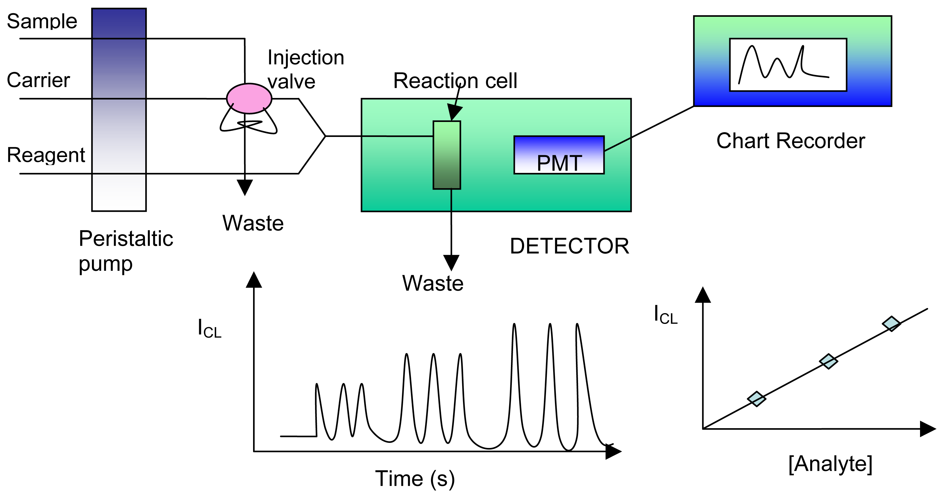
6. Chemiluminescence

There are two basic measurement methodologies using liquid-phase chemiluminescence (CL) reactions: static samples and flowing streams. Flowing stream methods involve delivery and mixing of the CL reagent with the analyte stream or column effluent and the use of a flow-cell for the detection of the CL emission at a fixed time after mixing. FIA includes relatively simple methodology offering acceptable robustness, feasibility and precision, and its rapid measuring response makes it suitable for monitoring liquid-phase CL reactions. This technique allows the sample to undergo on-line chemical and physical treatment to obtain species suitable for CL detection. Figure 1 shows a simple FIA-CL manifold in which the sample is injected into a flowing stream and mixed with the reagent in close proximity to the detector. Emission occurs in a cell placed in front of the optical window of a detector such as solid-state devices, scintillation counters or modified fluorimeters, although photomultiplier tubes (PMT) are most commonly used. The sensitivity of detection can be optimized by controlling some experimental variables such as dimensions of the mixing and detector coils, flow rates, temperature, pH and reagent concentrations. Using this approach and adapting different reaction conditions mentioned above, metal ions, catalysts, ligands, oxidants and reductants, hydrogen peroxide, inorganic compounds and related compounds have been determined; the production of reproducible peaks assures an adequate relation between the recorded CL intensity and the concentration of the analyte.
Important applications have been carried out using flow injection in pharmaceutical quality control. More recently, the development of immobilization techniques has provided the introduction of enzyme reactors which can be positioned before the CL reaction takes place, thus avoiding the lack of selectivity that may occur when a given CL reagent yields emission for a variety of compounds. In this alternative procedure, the analyte is the substrate of the enzymatic reaction and one of the products will sensitively participate in the CL reaction. Substrates that have been detected in this way include glucose, cholesterol, choline, uric acid, amino acids, aldehydes and lactate which generate H2O2 when flowing through a selective column reactor with immobilized oxidase enzymes in the presence of the necessary oxidant, usually O2, present in the samples. Luminol, in the presence of a peroxidase catalyst seems to be one of the best systems for this post-column H2O2 determination.
Recent developments and applications of CL sensors are reviewed in various publications [117-123]. The combination of the FI method with CL detection is becoming increasingly important in clinical, biological and environmental analyses because of its high sensitivity, wide linear dynamic range, reproducibility, simplicity, rapidity and feasibility. However, a serious limitation to widespread application of FI-CL analysis is the requirement to continuously deliver the CL reagent into the reaction zone because the CL reagent is consumed during the CL reaction, which is undesirable not only for the simplification of the detection device, but also for the cost, environment and resources considerations. An effective approach to solve this problem is to employ CL reagents in immobilized or solid-state format, which would eventually direct toward a new construction of FI-CL sensors. In these systems, analytes are detected by the CL reactions either with the immobilized reagents directly or with the dissolved reagents which are released from the immobilized substrates or the solid-state forms by some appropriate eluents or are electrogenerated on line by the electrodes [124-127].
Some basic requirements must be met by a substrate with immobilized reagent for application in a FI-CL sensor system [128]: (1) the substrate should have high mechanical stability and do not swell in aqueous solutions; (2) there would be low band broadening coupled with minimum back-pressure in a packed bed reactor; (3) for a long lifetime, a substrate with a high surface reagent coverage is preferable; (4) when the immobilized substrate is packed in a flow cell placed in front of the detector, a favourable transparency is essential for high sensitivity; (5) the substrate should exhibit good chemical stability, even at elevated pH conditions, which are frequently required to enable the light-producing reactions.
Until several years ago, CL flow sensors were relatively rare as compared to other type of optical flow sensors and most of them were limited to biosensors based on immobilized oxidases with dissolved CL reagents which react with hydrogen peroxide released from the enzymatic reactions to produce a CL light signal [129]. In recent years, CL flow injection systems with immobilized or solid-state reagents have received much attention and many analytical applications have appeared in the literature [119, 121, 130].
New fibre-optic probes with both high selectivity and sensitivity have been designed, based on light-emitting reactions (LER) [131]. Chemiluminescence reactions involving luminol and requiring hydrogen peroxide are particularly interesting, as they can be enzymatically catalysed by peroxidase, as well as bioluminescence reactions catalysed by luciferases either from the firefly or from marine bacteria, which provide a light emission correlated to the concentration of ATP or NAD(P)H, respectively. Co-immobilization of other enzymes like dehydrogenases used as auxiliary enzymes enabled the performance of the probe to be extended to other target molecules. In parallel, in the presence of suitable substrates, dehydrogenases activities can be monitored using the bacterial bienzyme system. The design of a reagentless probe has been investigated using the combination, in a multifunction sensing layer, of covalent immobilization technique for enzymes associated with loose embedding for co-reactants. Flavin mononucleotide can be trapped in a polyvinyl alcohol matrix, allowing about 35–40 consecutive accurate measurements to be performed. Promising results have also been obtained by connecting such probes to flow-injection-analysis manifolds for the monitoring of biotechnological processes. The potential of chemiluminescence and bioluminescence is especially attractive, since no light source or monochromator is required and since their sensitivity, selectivity and polyvalence appear particularly promising for extended development.
Trends in biosensor research and development are described by Karube and Yokoyama [132]. Micromachining techniques have been applied to construct biosensor systems. Several detection units for flow injection analysis have been fabricated. An electrochemical flow cell was fabricated, and both an enzyme immobilized column and an electrochemical detector were integrated. A chemiluminescence detector was also fabricated. A quartz crystal oscillator was used for protein detection, and a thermistor was employed as a thermal detector resulting from the enzyme reaction. An injectable biosensor for rapid measurement has been developed, and the determination of sugars in fruits was carried out. Glucose oxidase was immobilized for the determination of glucose, and three enzymes, invertase, mutarotase and glucose oxidase, were immobilized for the sucrose sensor. Microbiosensors using carbon fiber microelectrodes have been applied to neuroscience. Micro acetylcholine sensors based on acetylcholine esterase and choline oxidase were fabricated and characterized. Micro glutamate sensors were also fabricated using platinized carbon fiber electrodes which were modified with glutamate oxidase. The micro glutamate sensor was applied to the detection of glutamate released from the neurons.
An especially active field of scientific research in photoluminescence nowadays is its use to study microheterogeneous systems and, conversely, to investigate how the emission properties of a lumiphor can be improved by manipulating the physico-chemical properties of its microenvironment. Applications of such basic chemical knowledge to improve analytical detection, particularly in flowing systems, are stimulating a great deal of interest. A major breakthrough in this vein was the use of “organized media” to improve luminescence quantum yields in solution. An alternative to the use of “ordered media”to further enhance the rigidity of the lumiphor is by adsorbing or binding it to a solid matrix, as in solid surface luminescence (SSL). Unfortunately SSL is a rather discontinuous technique and so less suited than fluid “organized media” for detection in a flow system. An elegant way-out to such a limitation is the coupling of SSL with FIA techniques. Solid surface photoluminescence and flow analysis have had a most fortunate encounter, as the merging of the two techniques is providing new avenues for optical sensor transduction and SSL techniques improvement. Fluorimetric advantages of such combinations for new optical sensor development are dealth with and illustrated with the development of a very sensitive and selective optosensor for low aluminium levels in samples of utmost importance in renal failure disease control. However, the potential advantages and increased scope of SSL–FIA combinations are more fully realised when using room temperature phosphorescence (RTP) detection in an aqueous flow system. It is shown how new possibilities for fundamental and applied studies on the SS-RTP phenomenon are opened. SS-RTP—FIA analytical applications to sensing of cations (e.g., by using ferron reagent to bind the cation and retaining this RTP chelate in a flow-through cell), of anions (e.g., an optically “active” phase with the complex Al-8-hydroxyquinoline becomes phosphorescent in the flow-through cell only when iodide is bound to it) or oxygen (which quenches the RTP signal of a phosphorescent “active” phase) which can be analyzed in gas mixtures or dissolved in solutions. Finally, basic measurements carried out will be used to explain the amazingly strong RTP signals obtained in aqueous solutions by using anion-exchange resins to retain the studied phosphors [133].
Various computer programs for large-scale bioprocess control and optimization have been developed as well as software for simple laboratory routine analysis [134]. In comparison, software can hardly be found that works on laboratory scale and provides the control of complex FIA systems, multisubstrate determination, data evaluation as well as minimal process control abilities. The sensors applied can be of different type (luminometric or other optical as well as electrochemical biosensors). The development of such software may be very helpful for the transfer of FIA/biosensor systems from the state of development to industrial processes. A computer-controlled FIA system for research level based on the software FIACRE is presented. Five FIA/(bio)sensor systems can be controlled simultaneously. Additionally, common temperature and pH recordings are possible. Determinations of substrate concentrations are performed by means of calibration curves which can be recorded at different times. This allows supervising the activities of the sensors during a cell cultivation and controlling the bioprocess, e.g. by adding substrate to a cell culture. The automated monitoring of the degradation of glucose and urea by two different optical sensing principles during cell cultivation under the control of one microcomputer is presented for the first time. For this purpose, already well examined biosensors (a urease optode and a luminometric glucose sensor) were employed and their properties discussed under the aspect of working in real cultivation media. It is also shown that substrates of interest for bioprocess control can be detected by slight modifications of known reactions. For example, substrates of NADH-dependent enzymatic reactions can be detected by the luminol chemiluminescence system, and optodes can be employed for pH, penicillin and glucose determination.
A new flow cell design for spectroscopic measurements of suspensions, the jet ring cell, is introduced [135]. This cell exploits radial flow through a narrow ring-shaped gap to retain suspended particles within the detection region. This ring constitutes a detection volume of well-defined area from which the trapped particles can be instantaneously removed at will. The bed of particles thus forms a renewable surface, which can be probed by reflectance, fluorescence, or chemiluminescence using a microscope or optical fiber. This device should prove useful for microscopic study of cells, for automated immunoassays, and for preconcentration of analytes on sorbents with in situ spectroscopic detection. In conjunction with a fiber optic detection system, the jet ring cell becomes a component of a renewable chemical sensor system.
By taking advantage of both the semiconductor properties and the advanced micromachining possibilities of silicon, integration of back-side photodiodes with a microfluidic channel network on a single substrate is accomplished [136]. The resulting microsystem has all fluidic and electrical connections on one side of the chip. Hermetic sealing by anodic bonding is greatly facilitated by the simple topography on the front-side of the chip. To test the chemical functionality and performance of the device a chemiluminescence-based detection scheme is implemented. The introduction and control of the necessary chemical solutions is achieved using a flow injection analysis approach.
Multisyringe flow injection analysis (MSFIA) is a recently developed flowing technique, which was conceived with the aim of coupling the overall advantages of the parent flow injection analysis, sequential injection analysis and multicommutation flow analysis. MSFIA is presented as a powerful and promising tool for automated liquid-phase CL assays [137]. The capability of operation under discontinuous forward flow regime while handling minute, well-defined volumes of sample and reagents in a multicommuted format offers unrivalled analytical features. As opposed to the parent flow injection (FI) and sequential injection (SI) analysis, CL reactions with divergent pH and kinetic demands can be easily implemented in a single protocol sequence. MSFIA is proven extremely suitable for accommodating enzymatic CL assays in a renewable fashion by exploiting soluble enzymes; with no need for the typical immobilization procedures used in FI and SI systems. Solid-phase CL sensors have also gained full advantage of the benefits of MSFIA. In this context, a novel optosensor devised for on-line monitoring of trace levels of orthophosphate is described. Similar configurations are proposed for the selective and sensitive determination of metals, Vitamins, nutrients and saccharides in environmental and biological samples as well as beverages, which denote the versatility of this automated flow technique. Special emphasis is also given to the simple instrumental set-up, including a dedicated flow-through luminometer, arranged for a multitude of CL assays.

6.1. Analytical applications

6.1.1. FIA-CL sensors for inorganic compounds

6.1.1.1. H2O2

Hydrogen peroxide can be detected chemiluminometrically in the presence of luminol at cobalt and copper foils [138]. The chemiluminescence signal can also be induced electrochemically. The linear determination ranges are 0.1–200 μM on cobalt and 5–2000 μM on copper under flow injection conditions. To improve the selectivity the chemiluminescence detector was combined with a thin layer gas dialysis cell. Hydrogen peroxide was detected in the range between 0.5 and 100 mM.
Other sensing system is prepared by electrostatically immobilizing the analytical reagents, luminol and cobalt(II), on a strongly basic anion-exchange resin and a weakly acid cation-exchange resin, respectively [139]. Hydrogen peroxide is sensed by the CL reaction with luminol and cobalt(II) bleeding from the ion-exchange column with immobilized reagents by hydrolysis. The calibration graph is linear in the range 4×10⁻⁸ to 1×10⁻⁵ mol l−1, and the detection limit is 1.2×10⁻⁸ mol l−1. A complete analysis could be performed in 1 min with a relative standard deviation of <5%. The system could be used for 50 h and has been applied successfully to the determination of hydrogen peroxide in rainwater and glucose in serum by measuring the formation of hydrogen peroxide from a packed bed reactor with immobilized glucose oxidase.
H2O2 is sensed by the CL reaction with luminol and Co2+ bleeding from the ion exchange column with immobilized reagents by hydrolysis [140]. The calibration graph is linear and falls between the ranges of 4×10⁻⁸ to 1×10⁻⁵ mol/L. A complete analysis could be performed in 1 min. The system was reused for over 50 hours.
A peroxidase modified membrane, stabilized by thin plasma polymer layer was deposited using the monomers allylalcohol or hexamethyldisilazane for H2O2 detection [141]. In comparison to the uncoated enzyme membrane, there is a significant operational stability improvement of the hydrogen peroxide detection. H2O2 can be detected in the range between 4 and 1000 μM. The sensor is stable for more than 1000 detections under flow injection conditions.
A micromachined flow cell (overall size; 25 × 25 × 1 mm3) was designed for the fast determination of hydrogen peroxide, based on a luminol-H2O2 chemiluminescence reaction catalyzed by immobilized peroxidase (POD) [142]. The flow cell consisted of a sandwich of anisotropically etched silicon and glass chips and contained a spiral channel (20 turns, 50 cm long, 150 μm wide, 20 μm depth, channel volume 1.4 μL) and two holes (1 mm diameter). POD was covalently immobilized with 3-(trimethoxysilyl)propyldietylenetriamine and glutaraldehyde on the inner surface of the channel. The chip was placed in front of a window of a photomultiplier tube and used as a flow cell in a single-line flow-injection analysis system using a luminol solution as a carrier solution. The sample volume for one measurement was 0.2 μL. The maximal sampling rate was 315 h-1 at a carrier solution flow rate of 10 μL min-1. A calibration graph for H2O2 was linear for 5 nM - 5 μM; the detection limit was 1 nM. The H2O2 concentration in rainwater was determined using this sensor system.
The oxidation reaction between periodate and polyhydroxyl compounds was studied. A strong CL emission was observed when the reaction took place in a strong alkaline solution without any special CL reagent. It was interesting to find that in the presence of carbonate the CL signal was significantly enhanced. When O2 gas and N2 gas were bubbled into the reagent solutions, both background and CL signals of the sample were enhanced by O2 and decreased by N2. The spectral distribution of the CL emission showed two main bands (λ = 436-446 and 471-478 nm). Based on the studies of the spectra of CL, fluorescence and UV-visible, a possible CL mechanism was proposed [143]. In strongly alkaline solution, periodate reacts with the dissolved oxygen to produce superoxide radical ions. A microamount of singlet oxygen (1O2*) could be produced from the superoxide radicals. A part of the superoxide radicals acts on carbonates and/or bicarbonates leading to the generation of carbonate radicals. Recombination of carbonate radicals may generate excited triplet dimers of two CO2 molecules ((CO2)2*). Mixing of periodate with carbonate generated were very few 1O2* and (CO2)2*. These two emitters contribute to the CL background. The addition of polyhydroxyl compounds or H2O2 caused enhancement of the CL signal. It may be due to the production of 1O2* during the oxidized decomposition of the analytes in periodate solution. This reaction system has been established as a flow injection analysis for H2O2, pyrogallol, and α- thioglycerol and their detection limits were 5 × 10⁻⁹, 5 × 10⁻⁹, and 1 × 10⁻⁸ M, respectively. Considering the effective reaction ions, IO4-, CO32-, and OH- could be immobilized on a strongly basic anion-exchange resin. A highly sensitive flow CL sensor for H2O2, pyrogallol, and α- thioglycerol was also prepared.

6.1.1.2. Ammonium

A novel CL sensor for NH4+ combined with flow injection analysis is presented by Li et al. [144]. It is based on the inhibition effect of NH4+ on the CL reaction between luminol, immobilized electrostatically on an anion-exchange column, and hypochlorous acid electrogenerated on-line. The sensor responds linearly to NH4+ concentration in 1.0×10⁻⁶-4.0×10⁻⁹ g mL-1 range. A complete analysis could be performed in l min. The system is stable for 200 determinations.

6.1.1.3. Cyanide

Based on the chemiluminescence reaction of luminol immobilized on Amberlyst A-27 anion-exchange resin and copper ion immobilized on D151 large-pore cation-exchange resin with CN- in alkaline solution, the sensor can be used for CN- monitoring with a wide linear range, high sensitivity as well as simplicity of instrumentation [145]. The sensor response to the concentration of cyanide is linear in the range of 5.0 × 10⁻⁹ to 2.0 × 10⁻⁶ g mL-1 with a relative standard deviation of < 5% (n = 7). The detection limit is 2.0 × 10⁻⁹ g mL-1. The results can be obtained within 1 min for each measurement. The column with immobilized chemiluminescence reagents can be used 200 times. The sensor has been used for CN- monitoring in tap water and industrial waste water.

6.1.1.4. Chlorine

The analytical reagent luminol was immobilized on an anion exchange resin column. While a volume of sodium hydroxide passed through the column, luminol was eluted from the resin in alkaline aqueous solution and then mixed with a sample stream to react and produce CL. The CL emission intensity was correlated with the standard ClO- concentration in the range 1×10⁻⁸ to 4×10⁻⁵ g mL-1, and the detection limit was 8x10⁻⁹ g mL-1 ClO-. Interfering metal ions present in water were effectively separated by a pre-column cation exchanger. A complete analysis, including sampling and washing, could be performed in 1 min with a relative standard deviation of less than 5%. The sensor was stable for over 200 times and has been applied successfully to the determination of ClO- in tap water [146,147].

6.1.1.5. Copper

A novel CL flow-through sensor based on immobilizing all the ingredients involved in the analytical reaction for the determination of copper is proposed by Qin [148]. The analytical reagents including luminol and cyanide were coimmobilized permanently on an anion-exchange column, while the analyte copper was retained temporarily by electrochemical preconcentration on a gold electrode placed in an anodic stripping voltammetric flow cell. By injection of a volume of sodium hydroxide through the column with immobilized reagents, luminol and cyanide were eluted from the resins in alkaline aqueous solution and then reacted with copper stripped from the gold electrode to produce a CL signal, by means of which copper could be sensed. The sensor was not susceptible to interference by other metal ions associated with the CL reaction. The response to the concentration of copper was linear in the range of 0.01-10 μg L-1 and an extremely low detection limit of 8.0 × 10⁻³ μg L-1 was achieved. A complete analysis could be performed in 4 min with a relative standard deviation of less than 8%. The column with immobilized reagents was readily prepared and could be reused over 200 times. The sensor was applied successfully to the determination of copper in human serum and natural water samples.
An all-plastic micro-sensor system for remote measurement of copper (II) ions in the aqueous environment has been developed [149]. The sensing structure was designed for ease of milling and fabricated in poly (methyl methacrylate) (PMMA) using a hot-embossing technique. Issues of sealing the structure were studied extensively and an efficient protocol has been established. The detection system comprises a compact photo-multiplier tube and integrated photon counting system. This method has advantages of low sample volume, (creating a minimal volume of waste), low exposure to contaminants due to the closed system, no moving parts and employs a robust polymer material which is resistant to the environment of intended use. The sensor operates on the principle of flow injection analysis and has been tested using a FIA-CL reaction arising from the complexation of copper with 1,10-phenanthroline and subsequent oxidation by hydrogen peroxide.

6.1.1.6. Nitric oxide

An on-line nitric oxide (NO) monitoring method is described employing a flow injection manifold with CL detection [150]. The method is tailored for monitoring the NO from its complexes with porphyrin ring-bearing (PRB) biochemical materials. The method is quite sensitive, simple, rapid, and precise. A sensitivity of 2.63 × 10⁵ mV mol-1 NO, a range of 2.0 × 10⁻⁵ up to 1.0 × 10⁻² mol NO (under the conditions used) a reproducibility of 1.0% over the range 1.0 × 10⁻³ up to 1.0 × 10⁻² mol NO, a frequency of ca. five samples per minute and a detection limit of 1.0 × 10⁻⁵ mol NO are the analytical figures of merit for the proposed method.
Peroxynitrite is an important derivative made by nitric oxide in vivo. It can make damages in many kinds of tissues and cells. Its research value in heart diseases and cancer is very high. A sensitive, specific method for analysis of peroxynitrite is described [151]. In this method, chemiluminescence reaction between peroxynitrite and luminol was used to detect with flow injection system. The assay has a detection limit of 2 × 10⁻⁸ mol L-1, and linear range of 5 × 10⁻⁸mol L-1 to 5 × 10⁻⁵ mol L-1. The application of flow injection system offer possibility to establish biosensor for real-time detection of peroxynitrite.

6.1.1.7. Oxygen

An indicator phase, luminol immobilized on a strongly basic anion-exchange resin, was evaluated for the chemiluminescence sensing of O2 in N2 gas [152]. The base catalyzed luminol chemiluminescence was monitored with a silicon photodiode installed in a flow cell, into which the luminol-loaded resin was packed. The signal was dependent significantly on resins and solvents used for the immobilization. Oxygen gas concentrations at ppm levels in a N2 gas and a city gas were determined by means of 1 mL sample injection.

6.1.1.8. Phosphate

Nakamura et al. [153] constructed an automatic phosphate ion sensing system for the quality control of drinking water. The analyte was detected using the phosphate ion-dependent pyruvate oxidase reaction and the hydrogen peroxide produced was detected by luminol chemiluminescence catalyzed by Arthromyces ramosus peroxidase. It obtained a detection limit of 0·16 μM phosphate ion and it was possible to detect 0·32 μM phosphate ion for 48 days using pyruvate oxidase immobilized on Chitopearl BCW-2601 beads. Also, an automated flow-injection sensor by combining a pyruvate oxidase reaction and CL reaction for the detection of phosphate ion in river water has been developed by the same authors [154]. The detection limit was 96 nM phosphate ions. This sensor was sufficient to determine the maximal permissible phosphate-ion concentration in the environmental waters of Japan. The same authors also examined the possibilities for the construction of sensors using the combinations of several enzymes without the need for coenzymes, and developed a phosphate ion biosensor based on a maltose phosphorylase, mutarotase, and glucose–oxidase (MP–MUT–GOD) reaction combined with an arthromyces ramosus peroxidase–luminol reaction system. The response provided by this system was linear, with a wide range between 10 and 30 nM phosphate ion [155,156]. Morais et al. [157] described a CL sensor for the trace determination of orthophosphate in waters. The proposed sensor relies upon the in-line derivatisation of the analyte with ammonium molybdate in the presence of vanadate, and the transient immobilization of the resulting heteropolyacid in a copolymer packed spiral shape flow-through cell. This sensor avoided drawbacks of the excess of molybdate anion, which causes high background signals due to its self-reduction and accommodate reactions with different pH requirements and the ability to determine trace levels of orthophosphate in high silicate content samples.

6.1.1.9. Sulfite

Tris-(2, 2′-bipyridyl)ruthenium(II) complex, Ru(bpy)32+, was immobilized on the Dowex-50 W cationic ion-exchange resin. The chemiluminescent characteristics of Ru(bpy)32+ in solution and in resin form were compared by using batch and flow injection methods. A strong chemiluminescence was observed during the reaction of Ru(bpy)32+ both in solution and in resin with KMnO4 or Ce(SO4)2 under acidic or basic conditions. The Ru(bpy)32+ immobilized resin is stable, which can be used at least for 6 months when it reacts with the dilute KMnO4 solution. Based on this property, Ru(bpy)32+ immobilized in the resin phase Lin et al. [158] developed as a flow-through chemiluminescent sensor that could be used to determine oxalate, sulfite and ethanol chemically or electronically with (Ru(bpy)33+ generation on the surface of resin. The limits of detection were 1×10⁻⁶ M for oxalate, 0.5% (v/v) for ethanol and 1×10⁻⁷ M for sulfite. The method has been successfully applied to determine sulfite in sugar.
By designing a novel flow-through electrolytic cell, the Mn3+ was firstly electrochemically generated in situ on the near surface of the platinum electrode by constant current oxidizing MnSO4 in H2SO4 medium. It was then found that this Mn3+ could oxidize sulfite, which was injected into the electrolytic cell, to produce the strong CL emission signal. Based on this observation, a novel CL method for sulfite is developed [159]. Under the optimum conditions developed, the CL emission intensity was linear with sulfite concentration in the range 3.0 × 1.0-7 to 1.0 × 10⁻⁴ mol L-1. The detection limit was 8.0 × 10⁻⁸ mol L-1 original concentration. The method was successfully used for the determination of SO2 in the air sample.

6.1.2. FIA-CL sensors for organic and biological compounds

The potential of chemiluminescence and bioluminescence in organic analysis has been reinvented by García-Campaña et al. [160]. The rapid development of immobilization techniques has considerably enhanced the applications of CL, especially in flow injection analysis (FIA), in immunoassay and in the development of CL-based sensors.

6.1.2.1. Immunoassay

A simple flow enzyme system for real-time continuous monitoring of interaction of biological molecules has been developed [161]. It relies upon a thin-layer flow-through cell placed directly into the measuring compartment of the luminometer. One ligand (antibody) is immobilized on the inner surfaces of the flow cuvette, and a second ligand (antigen) labeled with a peroxidase molecule moves through the flow cell. The quantity of the complex on the surface of the cell may be monitored by measuring the intensity of chemiluminescence after the reaction of peroxidase label with the substrates (p-iodophenol, luminol and hydrogen peroxide). Consequently, the kinetics of association (or dissociation) of the complex labeled ligand-receptor on the surface of the cuvette can be detected in a real time regime. Due to the small thickness of the flow cell the diffusion limitations of interaction for two kinds of biomolecules (soluble and immobilized) are negligible, so the resulting intensity of chemiluminescent signal reflects the kinetics of interaction between soluble and immobilized components. The system may be successfully used for molecular recognition studies, analyzing the kinetics of bimolecular interaction and for concentration determination.
Tris(2,2′-bipyridyl)ruthenium(II) (Ru(bpy)32+) and dehydrogenase enzymes are immobilized in cation exchange polymer films to create electrogenerated chemiluminesccnce (ECL) biosensors [162]. Eastman AQ and Nafion polymers were used as immobilization support in a regenerable ECL sensor for NADH and NADPH and in biosensors for glucose, lactate and ethanol. The biosensor design places an enzyme-loaded polymer film adjacent to a Ru(bpy)32+loaded polymer film covering a platinum electrode. The modified electrode was used in a flow injection analysis system as an ECL detector. The CL response to samples containing enzyme substrate and cofactor (NAD+) result from the Ru(bpy)33+ ECL reaction with NADH produced by the enzyme. The analytical response to NADH using an AQ film modified with Ru(bpy)32+ was similar to that seen for Nafion films modified with Ru(bpy)32+. The coimmobilization of Ru(bpy)32+ and dehydrogenase enzymes in the same film showed reduced enzyme activity which led to various biosensor designs with adjacent layers. The ECL biosensor concept was shown applicable for both NAD+- and NADP+-dependent enzymes, where NADPH detection ranged from 0.01 to 100 μM NADPH.
A DNA optical sensor system is proposed based on the combination of sandwich solution hybridization, magnetic bead capture, flow injection and chemiluminescence for rapid detection of DNA hybridization [163]. Bacterial alkaline phosphatase (phoA) gene and Hepatitis B virus (HBV) DNA were used as target DNA. A biotinylated DNA probe was used to capture the target gene onto the streptavidin-coated magnetic beads and a calf intestine alkaline phosphatase (CAP)-labelled DNA probe was used for subsequent enzymatic chemiluminescence detection. The detection cycle was less than 30 min, excluding the DNA hybridization time, which was about 100 min. Both the phoA gene and HBV DNA could be detected at picogramme or femtomole level. No response signal was obtained when target DNA did not exist in the sample. Successive sample detection could be made by removing the magnetic field and a washing step.
A chemiluminescent immunosensor integrated in a flow injection analysis system was developed for the detection of the ‘diarrheic shellfish poisoning’ (DSP) toxin okadaic acid (OA). Anti-OA monoclonal antibodies were labelled with horseradish peroxidase for their use in a competitive assay, in which the free antigen of the sample competes with immobilised OA [164]. Based on commercially available polyethersulfone membranes, this bioanalytical system exhibits low non-specific binding of antibodies in the presence of mussel homogenate.
The immunosensor was used in a semi-automated analysis procedure in which the free OA containing sample was injected in the flow system concomitantly with the labelled antibodies. With an overall measurement time of 20min, the immunosensor has a detection limit of 0.2μg OA/100g mussel homogenate. The operational stability of the sensor for the detection of critically contaminated mussels (40μg/100g of mussel homogenate) has been investigated over 38 OA determination cycles. A stable response was obtained under the 34 first measurements with a CV of 11.7%. Moreover, no significant change in the immunosensor performances was observed during short and long term storage periods of the membranes (1 month). The performances of five immunosensors (five different membranes) showed a good repeatability with a variation of the negative control and the critically contaminated signal equal to 7% and 12.6%, respectively.

6.1.2.2. Amino acids

An enzymatic FIA procedure was developed for the chemiluminometric determination of L-aspartate in a medium for mammalian cell cultivation [165]. Packed bed flow reactors containing aspartate:aminotransferase and L-glutamate oxidase, respectively, immobilized on sieved porous glass beads were combined with a peroxidase modified fibre optic sensor to detect the produced hydrogen peroxide. Peroxidase from Arthromyces ramosus was immobilized on a preactivated microporous nylone membrane and catalyzes the light generating oxidation of luminol by hydrogen peroxide. To improve the selectivity of the L-aspartate determination an L-glutamate eliminating reactor was prepared by coimmobilization of L-glutamate oxidase and catalase in a packed bed enzyme reactor. Under FIA conditions at least 0.5 mM L-glutamate can be quantitatively eliminated from the preconditioned sample solution. L-Aspartate can be determined in the range between 5 and 1000 μM under FIA conditions. The determination of L-aspartate was optimized with respect to pH, cosubstrate concentration and residence time in the packed bed enzyme reactors. The proposed procedures show high recoveries between 96 and 100% for the L-aspartate determination in a medium for mammalian cell cultivations after the elimination of L-glutamate.
Glutamate and glutamine were determined by luminol chemiluminescence with FIA based on immobilized L-glutamate oxidase and glutaminase coupled with peroxidase [166]. The laboratory-made flow-through cell of the detector has a measured volume of only 15 μL. The hydrogen peroxide produced in the first reaction is detected by luminol chemiluminescence catalysed by peroxidase. A membrane sensor and enzyme reactor based on immobilized hydrogen peroxidase is used for the determination of hydrogen peroxide. It was observed that Arthromyces ramosus peroxidase produced a 100 times stronger luminescence signal than horseradish peroxidase. By immobilization of the microbial peroxidase on a membrane inside the flow cell, simplification could be achieved with regard to apparatus, reagents and operation. The sensitivity of detection was considerably improved. In addition, the concept of a hydrogen peroxide biosensor was realized. The membrane sensor shows a detection limit of 1 × 10⁻⁷ M for L-glutamate and 1 × 10⁻⁶ M for L-glutamine. The calibration graphs were approximately linear in the range of 1 × 10⁻⁷-6 × 10⁻⁵ M for L-glutamate and 1 × 10⁻⁶-2.5 × 10⁻³ M for L-glutamine. The membrane sensor was stable over a period of 10 weeks (>1000 analyses).
Chemiluminometric monoenzyme sensors were developed for the FIA of hydrogen peroxide, xanthine and hypoxanthine in the concentration range between 10-3 and 10-6 M [167]. Both a fibre optic set-up with a photomultiplier tube (PMT) and a photodiode with an integrated preamplifier were investigated to detect the chemiluminescence. Microbial peroxidase or xanthine oxidase was immobilized covalently on a preactivated membrane, which was placed in the 7.5 μL flow detector cell. The photodiode and the PMT based H2O2 sensor achieve detection limits of 10-6 and 10-7 M, respectively. To develop bienzyme sensors for glutamate, lysine and xanthine the microbial peroxidase was coimmobilized with the corresponding oxidases on the sensor membrane. These analytes can be detected in the range between 10-3 and 10-6 M. The fibre optic H2O2 sensor was combined with packed bed enzyme reactors to determine, e.g., glucose, lactate, glutamine, glutamate, ammonia, xanthine, hypoxanthine and phosphate in the range between 10-3 and 10-7 M. On this basis a fully automated FIA set-up with 5 parallel arranged enzyme reactor channels was developed for the on-line monitoring of animal cell cultures.
CL-FIA procedures were developed for the enzymatic determination of L-alanine, α-ketoglutarate and L-glutamate in the cultivation medium of mammalian cells [168]. A packed bed flow microreactor containing alanine aminotransferase and glutamate oxidase immobilized on sieved porous glass beads was combined with a chemiluminescence detector for the generated hydrogen peroxide. To catalyze the indicator reaction between luminol and hydrogen peroxide both Co(II) ions and immobilized peroxidase from Arthromyces ramosus were used in a fibre optic detector cell. L-alanine, α-ketoglutarate and L-glutamate can be detected over two decades of concentration magnitudes with detection limits of 2, 5 and 1 μM, respectively. The FIA procedure was applied to determine L-alanine and α-ketoglutarate in cell cultivation media.
A selective and sensitive chemiluminometric flow sensor for the determination of l-glutamate in serum, based on immobilized oxidases such as glutamate oxidase (GOD), uricase (UC) and peroxidase (POD), is described [169]. The principle for the selective chemiluminometric detection for l-glutamate is based on coupled reactions of four sequentially aligned immobilized oxidases, UC/POD/GOD/POD in a flow cell. The immobilized UC was employed to decompose urate, which is one of the major interfering components in serum for a luminol-H2O2 chemiluminescence reaction. The H2O2 produced from the UC reaction readily reacted with reducing components, such as ascorbate and glutathione, and then the excess H2O2 was decomposed by the immobilized POD. L-Glutamate in the sample plug was enzymatically converted to H2O2 with immobilized GOD. Subsequently, the peroxide reacts with luminol on the immobilized POD to produce chemiluminescence, proportional to glutamate concentration. The enzymes were immobilized on tresylated poly(vinyl alcohol beads). The immobilized enzymes were packed into a TPFE tube (1.0 mm i.d. × 60 cm), in turn, and used as a flow cell. The sampling rate was 30 h-1. The calibration graph for l-glutamate is linear in the range of 20 nM - 5 μM. The procedure exhibited a detection limit of 10 nM.
Another chemiluminometric flow-through sensor for simultaneous determination of L-glutamate (Glu) and L-lysine (Lys) in a single sample has been developed [170]. Immobilized uricase, immobilized peroxidase, support material, coimmobilized glutamate oxidase/peroxidase, support material, and coimmobilized lysine oxidase/peroxidase were packed sequentially in a transparent PTFE tube, and the tube was placed in front of a photomultiplier tube as a flow cell. A three-peak recording was obtained by one injection of the sample solution. The peak height of the first peak was due to the concentrations of urate and other reductants in the sample; the immobilized uricase was used to decompose urate, and the hydrogen peroxide produced was decomposed with a luminol-hydrogen peroxide reaction by immobilized peroxidase. The peak heights of the second and third peaks were free from the interferences from the reductants and were dependent only on the concentrations of Glu and Lys, respectively. Calibration graphs for Glu and Lys were linear at 40-1000 and 50-1200 nM, respectively. The sampling rate was 11 h-1 without carryover. The sensor was stable for two weeks. The sensor system was applied to the simultaneous determination of Glu and Lys in serum.
A chemiluminometric flow injection analytical system for the quantitation of L-histidine is described by Kiba et al. [171]. Histidine oxidase from Brevibacillus borstelensis KAIT-B-022 was immobilized on tresylated poly(vinyl alcohol) beads and packed into a stainless-steel column. The hydrogen peroxide produced was detected chemiluminometric ally by a flow-through sensor containing immobilized peroxidase. The maximum sample throughput was 10 h-1. The calibration graph was linear from 0.05 to 5 mM; the detection limit was 0.01 mM. The activity of immobilized histidine oxidase was reduced to 65% of the initial value after 350 injections. The system was applied to the determination of L-histidine in fish meat, such as salmon, tunny, bonito, and mackerel.
Finally, a bienzyme fiberoptic sensor for the chemiluminescent flow injection analysis of L-lactate was developed [172]. Lactate oxidase and peroxidase were covalently immobilized on preactivated polyamide membrane either separately on different membranes or randomly coimmobilized on the same membrane. Hydrogen peroxide, generated by the lactate oxidase reaction in the presence of L-lactate, was the substrate of the second reaction, catalyzed by peroxidase, in the course of which light was emitted in the presence of luminol. Compartmentalization of the enzyme layer was obtained by stacking a peroxidase membrane on a lactate oxidase membrane at the sensing tip of the fiberoptic sensor. When using such a sensing tip, the response of the fiberoptic sensor for lactate was 22 times higher than the response obtained when using a membrane bearing the two enzymes randomly coimmobilized. With the compartmentalized sensor tip, the detection limit was 250 pmol lactate and the coefficient of variation was 1.7% for 10 replicates of 6.25 nmol lactate. The method was applied for lactate determination in reconstituted whey solutions, and the results were in good agreement with the analysis by an enzyme electrode.

6.1.2.3. Adrenaline and isoprenaline

A novel integrated CL flow sensor for the determination of adrenaline and isoprenaline is developed based on the enhancing effect of analytes on CL emission of luminol oxidized by periodate in alkaline solution [173]. The analytical reagents luminol and periodate are immobilized on anion exchange resins, and packed in a glass tube to construct a reagentless sensor. The proposed sensor allows the determination of adrenaline and isoprenaline over the range from 2.0×10⁻⁸ to 1.0×10⁻⁵g mL-1 and 2.0×10⁻⁷ to 5.0×10⁻⁵g mL-1, respectively. The detection limits are 7.0×10⁻⁹g mL-1 for adrenaline and 5.0×10⁻⁸g mL-1 for isoprenaline. The sample throughput was 60 samples h-1. The sensor has been successfully applied to the determination of adrenaline and isoprenaline in pharmaceutical preparations.

6.1.2.4. Cholesterol

A chemiluminescence biosensor combined with FIA has been developed for determining cholesterol [174]. Cholesterol oxidase was immobilized onto amine-modified silica gel via glutaraldehyde activation and packed in a column. The analytical reagents, including luminol and ferricyanide, were electrostatically coimmobilized on an anion-exchange column. Cholesterol was sensed by the CL reaction between hydrogen peroxide released from the enzymatic reaction and luminol and ferricyanide, which were released from a column with immobilized reagents by elution. The calibration graph was linear over the range 5x10⁻⁶ to 1x10⁻⁴ g mL-1 and the detection limit was 5x10⁻⁶ g mL-1. The proposed method has been successfully applied to the determination of cholesterol in human serum.

6.1.2.5. Ethanol

A new approach for the determination of ethanol in beverages is presented [175]. The hydrogen peroxide generated in the enzymatically catalyzed oxidation of ethanol is measured by the luminol chemiluminescence reaction using potassium hexacyanoferrate(III) as a catalyst. Alcohol oxidase is immobilized on aminopropyl glass beads packed in a glass column. The chemiluminescence measurement is made using an optical fiber to transport the luminescence from the flow cell to the detector in conjunction with a flow system. This assay system responds linearly in the ethanol concentration range 3 × 10⁻⁶-7.5 × 10⁻⁴ M with a precision of 2.4% and an analysis time of 1.5 min.

6.1.2.6. Formaldehyde

A rapid and sensitive chemiluminescence flow sensor for the determination of formaldehyde was proposed by Song and Hou [176]. The analytical reagents involved in chemiluminescence (CL) reaction, luminol and KIO4, were both immobilized on an anion-exchange column. The CL signal produced by the reaction between luminol and KIO4, which were eluted from the column through water injection, was decreased in the presence of formaldehyde. Formaldehyde was sensed by measuring the decrement of CL intensity, which was observed linear over the logarithm of formaldehyde concentration range of 5.0-1000.0 ng mL-1, and the limit of detection is 1.8 ng mL-1. At a flow rate of 2.0 mL min-1 including sampling and washing, could be performed in 0.5 min with a relative standard deviation of less than 3.0%. The flow sensor offered reagentless procedures and remarkable stability in determination of formaldehyde, and could be easily re-used over 80h. The proposed flow microsensor was applied successfully in the determination of formaldehyde in artificial water samples and air.

6.1.2.7. Glucose

A FIA method for glucose is presented which is based on oxidation of glucose by glucose dehydrogenase with concomitant conversion of NAD+ to NADH followed by chemiluminescent detection of NADH [177]. The glucose dehydrogenase is immobilized via glutaraldehyde crosslinking to controlled pore glass to form an immobilized enzyme reactor. The chemiluminescent reagent, tris(2,2′-bipyridyl)ruthenium(II) [Ru(bpy)32+] is immobilized in a Nafion film coated on a platinum electrode to form a chemiluminescent sensor that can be regenerated. The immobilized Ru(bpy)32+ is oxidized to Ru(bpy)33+ which then reacts with NADH produced by the enzyme reactor to yield light and Ru(bpy)32+. Ru(bpy)32+ is thus recycled and made available again. Conditions for optimum enzyme reactor efficiency and chemiluminescent detection are determined and reported for pH (about 6.5), flow-rate (2 mL min-1), and NAD+ concentration (1-2.5 mM). At the optimum conditions a working curve is constructed where the upper limit for glucose detection is dependant on NAD+ concentration and lower detection limit is 10 μM glucose. Signal reproducibility is 1-2% relative standard deviation. The method is very selective for glucose; some interference is seen from uric acid, ascorbic acid and catechol as well as species (such as oxalate and aliphatic amines) already known to chemiluminesce with the Ru(bpy)32+ sensor.
A chemiluminescence fiber optic system coupled to FIA and ion exchange chromatography has been developed for determining glucose in blood and urine [178]. Immobilized glucose oxidase acted on β-D-glucose to produce hydrogen peroxide, which was then reacted with luminol in the presence of ferricyanide to produce a light signal. Endogenous ascorbic acid and uric acid present in urine or blood samples were effectively retained by an upstream acetate anion exchanger. In addition, acetaminophen could also be adsorbed by this ion exchanger. Immobilized glucose oxidase was reused for over 500 analyses without losing its original activity. A conservative estimate for the reuse of the acetate ion exchange column was about 100 analyses.
Also, a fiberoptic sensor for the CL-FIA of glucose in a one-step procedure was developed [179]. Glucose oxidase and peroxidase were coimmobilized on a preactivated polyamide membrane. Hydrogen peroxide, generated by the glucose oxidase reaction in the presence of glucose, was detected by the chemiluminescence reaction of luminol catalyzed by peroxidase. The detection limit for hydrogen peroxide was 2.5 pmol at pH 8.5 and 0.25 pmol at pH 9.0. For 10 replicates of 0.1 nmol H2O2, the coefficient of variation was 2.5%. For glucose analysis, the detection limit was 0.25 nmol and the coefficient of variation was 3.8% for seven measurements of 2.5 nmol glucose. The method was applied for glucose analysis in soft drinks, and the results were in good agreement with the analysis by a standard spectrophotometric method. The analytical throughput was 40 samples per hour for hydrogen peroxide and 25 samples per hour for glucose.
Glucose oxidase was covalently attached to aminosilanized indium tin oxide coated glass wafers and these were used as working electrodes in an electrochemiluminescence flow injection analyser as a model for enzyme immunoassays [180]. Light from the chemiluminescent reaction between enzymatically produced hydrogen peroxide and electrochemically oxidized luminol was detected and the effect of pH and luminol concentration on this reaction was determined. Repetitive assays for glucose were carried out to investigate the surface chemistry. Results suggested that electrochemiluminescence enzyme immunoassays with glucose oxidase as the antibody label could be carried out.
Also, glucose oxidase (GOD) was immobilized in the course of gelation of the sol of tetraelhylorthosilicate (TEOS), and immobilized GOD columns were easily prepared with different carriers (silica gel and molecular sieve) using this system [181]. It was demonstrated that the activity of GOD remained high in sol-gel matrix. With the application of the GOD column prepared with sol-gel method to the flow injection chemiluminescent system, glucose could be effectively determined in the range of 3.5-70 μmol L-1 and the detection limit was 0.6 μmol L-1.
A chemiluminometric flow-through sensor for the simultaneous determination of glucose (Glu) and 3-hydroxybutyrate (HB) in a single sample has been developed [182]. Coimmobilized 3-hydroxybutyrate dehydrogenase/NADH oxidase/peroxidase, a support material, and coimmobilized glucose dehydrogenase/NADH oxidase/peroxidase were packed sequentially in a transparent PTFE tube. The tube was then placed in front of a photomultiplier tube as a flow cell. A two-peak recording was obtained by one injection of the sample solution. The peak heights of the first and second peaks were dependent on the concentrations of HB and Glu, respectively. The calibration graphs for HB and Glu were linear at 0.05-10 and 0.1-30 μM, respectively. The maximum sample throughput was 30 h-1. The sensor was stable for two weeks.
Finally, the chemiluminescence of luminol in the presence of H2O2 has been exploited to develop fiberoptic biosensors associated with FIA systems [183]. A chlorophenol sensor was developed based on the ability of certain halophenols to enhance the peroxidase-catalyzed luminol chemiluminescence. Horseradish peroxidase immobilized on a collagen membrane was used. Ten chlorophenols have been tested with this chemiluminescent-based sensor. The lower detection limit was obtained with 4-chloro-3methylphenol and was equal to 0.01 μM. Electrochemiluminescent-based fiberoptic biosensors for glucose and lactate were also developed using glucose oxidase or lactate oxidase immobilized on polyamide membranes. In the presence of oxidase-generated H2O2, the light emission was triggered electrochemically by means of a glassy carbon electrode polarized at +425 mV vs a platinum pseudo-reference electrode. The detection limits for glucose and lactate were 150 and 60 pmol, respectively.

6.1.2.8. Histamine

A flow sensor with immobilized oxidases is proposed for the determination of histamine in fish meat [184]. Chemiluminometric measurement of histamine was based on the luminol reaction with hydrogen peroxide produced by immobilized histamine oxidase and peroxidase within a flow cell. Histamine oxidase was found in cells of Arthrobacter crystallopoietes KAIT-B-007 isolated from soil. The oxidase and peroxidase were coimmobilized covalently on tresylated hydrophilic vinyl polymer beads and packed into transparent PTFE; the tubing was used as the flow cell. One assay for histamine was done at intervals of 2 min without carryover. The calibration curve for histamine was linear from 0.1 μM to 50 μM. The response was reproducible within 1.25% of the relative standard deviation for 115-replicate injections of 50 μM histamine.

6.1.2.9. Hydrazine

A novel CL sensing system for hydrazine combined with flow-injection technique is presented by Song et al. [185]. The analytical reagents, luminol and hexacyanoferrate(III), were both immobilized on an anion-exchange column. The CL signal produced by the reaction between luminol and hexacyanoferrate(III), which were eluted from the column through sodium phosphate injection, was decreased in the presence of hydrazine. The decrease in CL intensity is linear with hydrazine concentration in the range from 0.1 to 100.0 ng mL-1; and the detection limit was 0.04 ng mL-1. A complete analysis could be performed in 2 min with a relative standard deviation of less than 5.0%. The flow sensor was stable for over 400 analyses and was applied successfully to the determination of hydrazine in artificial samples.

6.1.2.10. Hydroxylamine

A novel CL sensor, which can he used for hydroxylamine determination in combination withFIA, was developed by electrostatically immobilizing luminol and periodate on an-ion-exchange resin respectively [186]. Hydroxylamine was sensed by its enhancing effect on the weak CL reaction between luminol and periodate, which were eluted from the ion exchange column. The response of the sensor to hydroxylamine was linear in the concentration range of 8.0×10⁻⁸-2.0×10⁻⁶ mol L-1 with a detection limit of 4.0×10⁻⁸ mol L-1. The sensor could be reused for over 400 times with a good reproducibility and was used to determine hydroxylamine in wastewater.

6.1.2.11. Oxalate

The development of a detection method based on the electrogenerated chemiluminescence of tris(2,2′-bipyridine)ruthenium(II), (Ru(bpy)32+), immobilized in a Nafion film coated on an electrode is discussed [187]. Control of the electrode potential controls creation of the reactive reagent Ru(bpy)33+ which reacts with certain analytes to yield chemiluminescence emission of intensity proportional to the analyte concentration. The reaction results in Ru(bpy)33+ being converted to Ru(bpy)32+, which then is recycled to Ru(bpy)33+ again at the electrode. This sensor has been used in flow injection to determine oxalate, alkylamines, and NADH. Detection limits are 1 μM, 10 nM, and 1 μM, respectively, with working ranges extending over 4 decades in concentration. The sensor remains stable for several days with suitable storage conditions.

6.1.2.12. Resorcinol

A new chemosensor based on the inhibition of the CL intensity in a flow injection system is developed for the determination of resorcinol [188]. The CL was generated by the reaction of K3Fe(CN)6 and luminol in alkaline medium while the reagents were immobilized on an anion exchange resin and eluted by sodium phosphate. The CL intensity was inhibited by resorcinol and the decrease of CL intensity is linear over the resorcinol concentration range of 9.2-920 ng mL-1. The method was applied successfully to the determination of resorcinol in pharmaceutical tinctures.

6.1.2.13. Uric acid

A novel CL flow sensor for the determination of uric acid in human urine and serum has been developed by using controlled-reagent- release technology [189]. The reagents involved in the CL reaction, luminol and periodate, are immobilized on anion-exchange resin packed in a column. After injection of water, chemiluminescence generated by released luminol and periodate in alkaline media is inhibited in presence of uric acid. By measuring the decreased CL intensity the uric acid is sensed. The decreased response is linear in the 5.0-500.0 ng mL-1 range, with a detection limit of 1.8 ng mL-1. The flow sensor showed remarkable operational stability and could be easily reused for over 80h with sampling frequency of 100 h-1. The proposed sensor was applied to the determination of uric acid in human urine and serum, and monitoring metabolic uric acid in human urine with R.S.D. less than 3.0%.

6.1.2.14. Vitamins

A novel flow sensor based on CL for the determination of ascorbic acid has been proposed [190]. The analytical reagents, luminol and ferricyanide, were both immobilized on an anion-exchange resin column. The CL signal produced by the reaction between luminol and ferricyanide, which were eluted from the column through sodium phosphate injection, was decreased in the presence of ascorbic acid. The CL emission intensity was linear with ascorbic acid concentration in the range 0.01-0.8 μg mL-1; the detection limit was 5.5×10⁻³ μg mL-1. The whole process, including sampling and washing, could be completed in l min with a relative standard deviation of less than 5%. The sensor could be reused more than 100 times and has been applied successfully to the analysis of ascorbic acid in pills and vegetables. Other FI-CL sensor for the determination of ascorbic acid has been proposed by Wang et al. [191]. The analytical reagents, luminol and permanganate, were both immobilized on an anion-exchange resin column and were eluted from the column through hydrochloric acid injection. The CL signal produced by the reaction between luminol and permanganate decreased in the presence of ascorbic acid. The CL emission intensity is linear with ascorbic acid concentration in the range of 1.0×10⁻⁵-4.0×10⁻³ g L-1. The whole process, including sampling and washing, can be completed in l min with a relative standard deviation of 2.3%. The sensor can be reused more than 100 times and has been applied successfully to the analysis of ascorbic acid in beverage.
A continuous flow sensor for the determination of thiamine was constructed by using controlled-reagent-release technology in a FIA-CL system [192]. The analytical reagents, luminol and KIO4, were both immobilized on an anion-exchange column. The CL signal produced by the reaction between luminol and KIO4, which were eluted from the column through H2O injection, was decreased in the presence of thiamine. The decreased CL intensity was linear with thiamine concentration in the range 3.3 pmol mL-1-6.7 nmol mL-1; and the limit of detection was 1.0 pmol mL-1. The whole process, including sampling and washing, could be completed in 0.5 min with a relative standard deviation of less than 3.0%. The flow sensor showed remarkable stability and could be easily reused over 80 h. The sensor proposed was tested in determination of thiamine in pharmaceutical preparation and human urine samples. The same authors present other CL sensor for thiamine combined FI technology [193]. The analytical reagents, luminol and ferricyanide, were both immobilized on an anion exchange column. The CL signal produced by the reaction between luminol and ferricyanide, which were eluted from the column with the sodium phosphate injection, was decreased in the presence of thiamine. The decreased CL intensity was linear for thiamine concentration in the range from 0.2 to 4.0 nmol mL-1; and the limit of detection was 66 pmol mL-1. The whole process, including sampling and washing, could be completed in 2 min with a relative standard deviation of less than 3.0%. The sensor could be reused more than 400 times and has been applied for the determination of thiamine in pharmaceutical preparations and human urine samples.
Vitamin B2 was determined by a CL sensor combined with FI technology [194]. The analytical reagents involved in the CL reaction, luminol and hexacyanoferrate (III), were both immobilized on an anion-exchange resin column in FI system. The CL signal produced by the reaction between luminol and hexacyanoferrate (III), which were eluted from the column through sodium phosphate injection, decreased in the presence of vitamin B2. The decreased CL intensity was linearly correlated with the vitamin B2 concentration in the range of 0.01-1.0 μg mL-1 the detection limit was 4.0 ng mL-1. At a flow rate of 2.0 mL min-1, the procedure including sampling and washing could be performed in 2 min with a relative standard deviation of less than 3.0%. The flow sensor exhibited both good sensitivity and stability. It could be reused more than 450 times and has been applied successfully to the analysis of vitamin B2 in pharmaceutical preparations.
A novel CL sensor for vitamin B12 combined with FIA is proposed by Qin et al. [195]. It is based on the catalytic effect of cobalt(II), liberated from vitamin B12 by acidification, on the CL reaction between luminol, immobilized electrostatically on an anion-exchange column, and hydrogen peroxide electrochemically generated on-line via a negatively-biased electrode from dissolved oxygen in the flow cell. The sensor responds linearly to vitamin B12 concentration in the 1.0 × 10⁻³-10 mg L-1 range, and the detection limit is 3.5 × 10⁻⁴ mg L-1. A complete analysis, including sampling and washing, could be performed in 1 min with a relative standard deviation of < 3.5%. The system is stable for over 500 determinations and has been applied successfully to the determination of vitamin B12 in pharmaceutical preparations.
A chemiluminescence biosensor responding to amygdalin was prepared by covalently coupling β-glucosidase to the controlled pore glass as a molecular recognition element and electrostatically immobilizing luminol on the ion exchange resin as a transduction element in a flow injection system [196]. The analyte was injected into a continuous stream of simple medium flowing through an enzyme reactor containing β-glucosidase to produce the cyanide, which reacted with the soluble O2 to produce superoxide anion O2+. The luminol was eluted by NaOH solution from the ion exchange column, and then reacted with superoxide anion O2+ to produce chemiluminescence. The linear range was from 1.0 μg to 200 μg, the detection limit was 0.3 μg of amygdailn. The sensor was stable in 6 months and it has been applied successfully to the determination of amygdalin in amygdalloid nucleus.
A CL sensor for vitamin K3 (menadione sodium bisulfite, MSB) combined with a flow-injection system is described by Huang et al. [197]. It is based on the auto-oxidation of bisulfite liberated from MSB in alkaline media in the presence of Tween 80 sensitized by Rhodamine 6G immobilized on a cation-exchange column. The sensor responds linearly to the MSB concentration in the range of 0.5-10 μg mL-1 with a detection limit of 2.6 μg L-1. The analysis can be performed within 1 min with a relative standard deviation of <5%. The sensor is stable for over 250 determinations and has been successfully applied to the determination of MSB in injections and tablets.
A novel CL sensor for folic acid combined FI technology was proposed by Song and Zhou [198]. The analytical reagents involved in the CL reaction, including luminol and hexacyanoferrate(III), were both immobilized on an anion-exchange column in FI system. The CL signal produced by the reaction between luminol and hexacyanoferrate(III), which were eluted from the column through sodium phosphate injection, was decreased in the presence of folic acid. The CL emission was correlated with the folic acid concentration in the range from 0.01 to 15 μg mL-1, and the detection limit was 3.5 ng mL-1. At a flow rate of 2.0 mL min-1, including sampling and washing, could be performed in 2 min with a relative standard deviation of <2.5%. The flow sensor could be reused more than 300 times and has been applied to the analysis of folic acid in pharmaceutical preparations.
Finally, a novel continuous-flow sensor based on CL detection was developed for the determination of rutin in pharmaceutical preparations and human urine by controlled-reagent-release technology [199]. The analytical reagents involved in the CL reaction, including luminol and hexacyanoferrate(III), were both immobilized on an anion-exchange column in a FI system. The CL signal produced by the reaction between luminol and hexacyanoferrate(III), which were eluted from the column through sodium phosphate injection, was decreased in the presence of rutin. CL intensity was inhibited by rutin; the decrement of CL intensity was linear over the logarithm of the rutin concentration range of 1.0-400 ng mL-1, and the detection limit was 0.35 ng mL-1. The whole process, including sampling and washing, could be completed in 1.5 min with a relative standard deviation of <3.5%. The flow sensor showed remarkable stability and could be easily reused >450 times; the sensor proposed was successfully applied to the determination of rutin in pharmaceutical preparations and human urine.

6.1.3. FIA-CL sensors for drug analysis

6.1.3.1. Analgin

CL flow-through sensor [200] for the determination of analgin was developed based on the auto-oxidation of analgin by O2 in the presence of Tween 80 sensitized by Rh6G immobilized on a cation-exchange column. Based on the inhibition effect of analgin on the CL signals produced by the reaction between luminol and periodate. Also, a selective and sensitive as well as rapid CL flow sensor for the determination of analgin is described by Song and Zhang [201]. The analytical reagents involved in chemiluminescence reaction, luminol and periodate, were both immobilized on an anion-exchange column. The CL signals produced by the reaction between luminol and periodate, which were eluted from the column through water injection, were decreased in the presence of analgin. Analgin was sensed by measuring the decrement of CL intensity, and which was observed linear over the logarithm of analgin concentration range of 0.1 to 50.0 ng mL-1, and the limit of detection was 0.04 ng mL-1. At a flow rate of 2.0 mL min-1, including sampling and washing, the detection could be performed in 0.5 min with a relative standard deviation of less than 3.0%. The proposed procedure was applied successfully in the monitoring of analgin in human urine samples without any pre-treatment process. It was found that the analgin concentration reached its maximum after being orally administrated for 4 h, and the analgin metabolism ratio in 10 h was 9.28% in the body of volunteers. The flow sensor offered reagentless procedures and remarkable stability in determination of analgin, and could be easily reused over 80 h. Finally, a CL flow-through sensor for the determination of analgin was based on the direct oxidation of analgin by manganese dioxide to produce a weak CL in the absence of luminescence reagent [202]. The CL intensity in acidic medium was enhanced by the addition of Rhodamine B (RhB). The solid-phase manganese dioxide was immobilized on the sponge rubber inside the CL flow cell by a very simple means. The calibration graph is linear in the range 4 × 10⁻⁵ to 1 × 10⁻³ g mL-1 with a detection limit of 2.7 × 10⁻⁵ g mL-1. The sensor was successfully used for automated dissolution testing of analgin tablets and sampling frequency was 120 h-1.

6.1.3.2. Berberine

A sensitive CL sensor for berberine combined with FI technology is described by Song et al. [203]. The analytical reagents involved in the CL reaction, including luminol and potassium ferricyanide, were both immobilized on an anion-exchange resin column. While a volume of sodium phosphate was passed through the column, the two CL reagents were eluted from the resin and then mixed with a berberine stream under alkaline conditions. By means of the fast oxidation reaction between berberine and potassium ferricyanide, potassium ferrocyanide was generated, which then inhibited the CL reaction of luminol and potassium ferricyanide. The decreased CL intensity was correlated with the berberine concentration in the range from 0.05 to 300 ng mL-1 with a relative standard deviation of less than 4.3%, and a limit of detection of 0.02 ng mL-1 at a flow rate of 2.0 mL min-1. It was shown that the flow sensor could greatly improve the selectivity and sensitivity of the determination of berberine. The determination of analyte could be performed in 2 min, including sampling and washing. The sensor is stable for over 300 analyses. Preliminary studies were done on two herbal medicines commonly found in compound prescriptions, rutin and baicalin. The method has been applied to the determination of berberine in pharmaceutical preparations.

6.1.3.3. Carbohydrate antigen

A novel chemiluminescent immunosensor for carbohydrate antigen 19-9 (CA19-9) based on the immobilization of CA19-9 on the cross-linked chitosan membrane was developed [204]. The different membranes were characterized by atomic force microscopy (AFM) and infrared spectroscopy, respectively. Based on a noncompetitive immunoassay format, this proposed chemiluminescent immunosensor enabled a low-cost, flexible and rapid determination for CA19-9 in combination with FIA. After an off-line incubation of the analyte CA19-9 with horseradish peroxidase (HRP)-labeled anti-CA19-9, the mixture was injected into the immunosensor, which led to the trapping of free HRP-labeled anti-CA19-9 by the immobilized antigen in the immunosensor. The trapped HRP-labeled antibody was detected by chemiluminescence due to its catalytic activity following the reaction of luminol and H2O2.

6.1.3.4. Ergonovine

Hexacyanoferrate(III) was immobilized electrostatically on an anion-exchange resin column and was eluted from the column using sodium phosphate solution as eluent. The sensor responds linearly to ergonovine maleate concentration in the range of 5.0×10⁻³ to 1.0 mg L-1 with a detection limit of 2.6 μg L-1. The sensor is stable for 200 determinations and has been successfully applied to the determination of ergonovine maleate in injections and urine samples [205].

6.1.3.5. Heroin

A novel method for rapid, inexpensive, sensitive and selective determination of heroin was proposed by flow injection electrogenerated chemiluminescence (ECL) [206]). Zeolite Y sieves were used for the preparation of an ECL sensor by immobilizing tris(2,2′-bipyridyl)ruthenium(II) (Ru(bpy)32+) in their supercages, which was achieved through the ion exchange properties of the sieves. The electrochemical and ECL behaviors of Ru(bpy)32+ immobilized in zeolite Y modified carbon paste electrode was investigated. The modified electrode showed an electrocatalytic response to the oxidation of heroin, producing a sensitized ECL signal. The ECL sensor showed a linear response to flow injection of heroin in the range of 2.0-80 μmol L-1 with a detection limit of 1.1 μmol L-1. Its surface could be renewed quickly and reproducibly by a simple polishing step.

6.1.3.6. Hydralazine

A novel FI-CL sensor for hydralazine determination using molecularly imprinted polymer (MIP) as recognition element is reported by Xiong et al. [207]. Hydralazine–MIP was prepared through non-covalent copolymerization using methacrylic acid (MAA) monomer, hydralazine template and ethylene glycol dimethacrylate (EGDMA) cross-linker. Particles of the MIP were packed into a v-shape glass tube for on-line adsorption of the analyte of hydralazine. The adsorbed hydralazine could be sensed by its great enhancing effect on the CL reaction between luminol and periodate. The CL intensity is linear to hydralazine concentration in the range from 2×10⁻⁹ to 8×10⁻⁷ g mL-1. The selective experiment showed that the selectivity and sensitivity of the CL method could be greatly improved when MIP was used as recognition element in the flow-injection CL sensor. The sensor was reversible and reusable. It could be used for more than 100 times. It has been used directly to determine the hydralazine in human urine.

6.1.3.7. Isoniazid

By electrostatically immobilizing the CL reagent luminol and periodate on anion exchange resin separately, a novel CL sensor for isoniazid combined with FIA was developed [208]. Isoniazid was sensed by its enhancing effect on the weak CL reaction between luminol and periodate, which were eluted from the ion exchange column. The calibration graph is linear in the range 8.0×10⁻⁹ to 1.0×10⁻⁶ mol L-1 and the detection limit is 4.2×10⁻⁹ mol L-1. A complete analysis could be performed in 1 min with a relative standard deviation 2.0%. The sensor could be reused for over 400 times and has been applied successfully to the determination of isoniazid in pharmaceutical preparations. Other CL sensor for isoniazid combined with flow-injection technology is also presented by Song et al. [209]. The analytical reagents, luminol and ferricyanide, were both immobilized on an anion-exchange column. The CL signal produced by the reaction between luminol and ferricyanide, which were eluted from the column through sodium phosphate injection, was decreased in the presence of isoniazid. The decreased CL intensity was linear with isoniazid concentration in the range 0.001-1.0 μg mL-1; and the detection limit was 0.35 ng mL-1. The whole process, including sampling and washing, could be completed in 2 min with a relative standard deviation of less than 4.1%. The sensor could be reused more than 400 times and has been applied for the determination of isoniazid in pharmaceutical preparations. Finally, molecularly imprinted polymer (MIP) of isoniazid is synthesized through thermal radical copolymerization of metharylic acid (MAA) and ethylene glycol dimethacrylate (EGDMA) in the presence of isoniazid template molecules and a novel FI-CL for isoniazid determination is developed by packing the isoniazid-MIP into the flow cell as recognition elements [210]. Isoniazid could be selectively adsorbed by the MIPs and the adsorbed isoniazid is sensed by its great enhancing effect on the weak CL reaction between luminol and periodate which were mixed in the flow cell. The enhanced CL intensity is linear in the range 2×10⁻⁹ to 2×10⁻⁷g mL-1 and the detection limit is 7×10⁻¹⁰ g mL-1 isoniazid with a relative standard deviation 2.8%. The sensor is reversible and reusable. The MIP technique has greatly improved the sensitivity and selectivity of CL analysis. As a result, the sensor has been successfully applied to the determination of isoniazid in human urine. At the same time, the binding characteristic of the polymer to isoniazid was evaluated by batch method and the dynamic method.

7. Conclusions

The successful operation of a modern analytical laboratory requires accurate analysis of samples in the shortest time possible after receipt. Recent advances in instrumentation have led to increased interest in the development of automated chemical analyses. Flow systems have been widely developed in routine analysis as they allow fast analysis with a minimal sample handling in many cases. Furthermore, other advantages such as minimal reagent consumption in some flow applications and the possibility of adapting these systems in the field should be highlighted. Development and applications of chemical and biochemical flow-through sensors have shown fast growth over the past two decades. The growing number of published papers in last few years clearly indicates the vitality of the flow-through optosensing technique.
One of the most promising key areas in recent studies in this field is the research efforts devoted to the development of flow-through optosensors for simultaneous determination of several active principles in pharmaceuticals (multioptosensors). New strategies exploiting other possibilities the sensing detection can offer were studied.
Other interesting techniques is multisyringe flow injection analysis (MSFIA). This technique has been developed combining the advantages presented by flow injection analysis (FIA), sequential injection analysis (SIA) and multicommuted flow injection analysis.
In recent years, the technique of membrane extraction has proven to be a successful strategy in the determination of concentrations at trace levels, in matrix simplification and as an ionic exchange medium, among other applications. Membrane extraction usually implies the elution of the retained analyte after preconcentration and subsequent detection. This technique has been developed by means of optochemical sensors, such as the optical fiber reflectance sensor or optosensor, due to their great flexibility, easy miniaturization, low cost and robustness. It is usually based on detection of the change in optical properties of a reactive phase when exposed to either the analyte or to an active derivative of the analyte, by means of an instrument with a bifurcated optical fiber.

References and Notes

  1. Newcombe, D.T.; Cardwell, T.J.; Cattrall, R.W.; Kolev, S.D. An optical redox chemical sensor based on ferroin immobilised in a NafionR membrane. Analytica Chimica Acta 1999, 401, 137–144. [Google Scholar]
  2. Coo, L.dlC.; Belmonte, C.J. Nafion-PAN optical chemical sensor: optimisation by FIA. Talanta 2002, 58, 1063–1069. [Google Scholar]
  3. Lenarczuk, T.; Glab, S.; Koncki, R. Application of Prussian blue-based optical sensor in pharmaceutical analysis. Journal of Pharmaceutical and Biomedical Analysis 2001, 26, 163–169. [Google Scholar]
  4. García, S.; Albero, I.; Ortuño, J.A.; Sánchez-Pedreño, C.; Expósito, R. Flow-through bulk optrode for the spectrophotometric determination of perchlorate. Microchimica Acta 2003, 143, 59–63. [Google Scholar]
  5. Ruedas Rama, M.J.; Ruiz Medina, A.; Molina Díaz, A. bead injection spectroscopic flow-through renewable surface sensors with commercial flow cells as an alternative to reusable flow-through sensors. Analytica Chimica Acta 2003, 482, 209–217. [Google Scholar]
  6. Trinkel, M.; Trettnak, W.; Reininger, F.; benes, R.; O'Leary, P.; Wolfbeis, O.S. Study of the performance of an optochemical sensor for ammonia. Analytica Chimica Acta 1996, 320, 235–243. [Google Scholar]
  7. Yoshimura, K. On-line concentration and flow analysis of trace amounts of bismuth with anion-exchange method and ion-exchanger spectrophotometry. Bunseki Kagaku 1987, 36, 656–661. [Google Scholar]
  8. Zhu, C.; Hieftje, G.M. On-line determination of bromide ion in spent brine. Analytica Chimica Acta 1992, 256, 97–103. [Google Scholar]
  9. Vereda Alonso, E.; Cano Pavón, J.M.; Rios, A.; Valcárcel, M. Automatic determination of cobalt at the submicrogram per millilitre level using a flowthrough spectrophotometric sensor. Talanta 1996, 42, 1941–1947. [Google Scholar]
  10. Matsuoka, S.; Shiota, N.; Yoshimura, K. Determination of trace amounts of cobalt(II) by flow injection-solid phase spectrometry (FI-SPS) with 5-Br-PADAB. Analytical Sciences 2006, 22, 177–181. [Google Scholar]
  11. Vereda, E.; Rios, A.; Valcárcel, M. Simultaneous automatic determinationof trace amounts of copper and cobalt by use of a flow-through sensor and firt-derivative spectrometry. Analyst 1997, 122, 85–88. [Google Scholar]
  12. Yoshimura, K.; Matsuoka, S. 4,7-diphenyl-2,9-dimethyl-1,10-phenanthroline disulphonate and its application to the study of karst groundwater storm runoff. Analytica Chimica Acta 1992, 268, 225–233. [Google Scholar]
  13. Colsa Herrera, J.M.; Sánchez Rojas, F.; Bosch ojeda, C.; García de Torres, A.; Cano Pavón, J.M. Determination of trace amounts of copper using a flow-through spectrophotometric sensor. Laboratory Robotic Automation 2000, 12, 241–245. [Google Scholar]
  14. Jerónimo, P.C.A.; Araújo, A.N.; Montenegro, M.C.B.S.M.; Pasquini, C.; Raimundo, I.M., Jr. Direct determination of copper in urine using a sol-gel optical sensor coupled to a multicommutated flow system. Analytical and Bioanalytical Chemistry 2004, 380, 108–114. [Google Scholar]
  15. Ruedas Rama, M.J.; Ruiz Medina, A.; Molina Díaz, A. Resolution of biparametric mixtures using bead injection spectroscopic flow-through renewable surface sensors. Analytical Sciences 2005, 21, 1079–1084. [Google Scholar]
  16. Yoshimura, K. Application of ion-exchanger phase absorptiometry to flow analysis. Determination of trace amounts of chromium(VI) in water. Analyst 1988, 113, 471–474. [Google Scholar]
  17. Matsuoka, S.; Tennichi, Y.; Takehara, K.; Yoshimura, K. Flow analysis of micro amounts of chromium(III) and (VI) in natural water by solid phase spectrophotometry using diphenylcarbazide. Analyst 1999, 124, 787–791. [Google Scholar]
  18. Górski, L.; Malinowska, E. Fluoride-selective sensors based on polyurethane membranes doped with Zr(IV)-porphyrins. Analytica Chimica Acta 2005, 540, 159–165. [Google Scholar]
  19. Lázaro, F.; Luque de Castro, M.D.; Valcárcel, M. Integrated retention/spectrophotometric detection in flow-injection analysis. Determination of iron in water and wine. Analytica Chimica Acta 1989, 219, 231–238. [Google Scholar]
  20. Hase, U.; Yoshimurs, K. Determination of trace amounts of iron in highly purified water by ion-exchanger phase absorptiometry combined with flow analysis. Analyst 1992, 117, 1501–1506. [Google Scholar]
  21. Ayora Cañada, M. J.; Pascual reguera, M. I.; Molina Díaz, A. An integrated reaction-retention and spectrophotometric detection flow system for determination of nickel. Fresenius' Journal of Analytical Chemistry 1999, 363, 59–63. [Google Scholar]
  22. Amini, M. K.; Momeni-Isfahani, T.; Khorasani, J.H.; Pourhossein, M. development of an optical chemical sensor based on 2-(5-bromo-2-pyridylazo)-5-(diethylamino)phenol in Nafion for determination of nickel ion. Talanta 2004, 63, 713–720. [Google Scholar]
  23. Coo, L.dlC.; Martínez, I.S. nafion-based optical sensor for the determination of selenium in water samples. Talanta 2004, 64, 1317–1322. [Google Scholar]
  24. Ayora Cañada, M.J.; Pacual Reguera, M.I.; Molina Díaz, A. Continuous flow-through solid phase spectrophotometric determination of trace amounts of zinc. Analytica Chimica 1998, 375, 71–80. [Google Scholar]
  25. Schepers, D.; Schulze, G.; Frenzel, W. Spectrophotometric flow-through gas sensor for the determination of atmospheric nitrogen dioxide. Analytica Chimica Acta 1995, 308, 109–114. [Google Scholar]
  26. Sanz Martínez, A.; Rios, A.; Valcárcel, M. Determination of dissolved oxygen by use of a spectrophotometric flow-through sensor. Analytica Chimica Acta 1993, 284, 189–193. [Google Scholar]
  27. Capitán Vallvey, L.F.; Valencia, M.C.; Arana Nicolás, E. Monoparameter sensors for the determination of the antioxidants butylated hydroxyanisole and n-propyl gallate in foods and cosmetics by flow injection spectrophotometry. Analyst 2001, 126, 897–902. [Google Scholar]
  28. Capitán Vallvey, L.F.; Valencia, M.C.; Arana Nicolás, E. Simple resolution of butylated hydroxyanisole and n-propyl gallate in fatty foods and cosmetics samples by flow-injection solid-phase spectrophotometry. Journal of Food Science 2003, 68, 1595–1599. [Google Scholar]
  29. Capitán Vallvey, L.F.; Valencia, M.C.; Arana Nicolás, E. Flow-through sensor for determination of butylated hydroxytoluene in cosmetics. Analytical Letters 2002, 35, 65–81. [Google Scholar]
  30. Capitán Vallvey, L.F.; Valencia, M.C.; Arana Nicolás, E. Solid-phase ultraviolet absorbance spectrophotometric multisensor for the simultaneous determination of butylated hydroxytoluene and co-existing antioxidants. Analytica Chimica Acta 2004, 503, 179–186. [Google Scholar]
  31. Portela, D.C.; Pereira, I.M.F.; Paíga, P.; Deleure-Matos, C.; Vaz, M.C.V.F. Amperometric and spectrophotometric determination of carbaryl in natural waters and commercial formulations. Analytical and Bioanalytical Chemistry 2003, 377, 356–361. [Google Scholar]
  32. Agudo, M.; Rios, A.; Valcárcel, M. Automatic continuous-flow determination of paraquat at the subnanogram per millilitre level. Analytica Chimica Acta 1993, 281, 103–109. [Google Scholar]
  33. Cañizares, M.P.; Tena, M.T.; Luque de Castro, M.D. On line coupling of a liquid-liquid extraction flow-reversal system to a spectrophotometric flow-through sensor for the determination of polyphenols in olive oil. Analytica Chimica Acta 1996, 323, 55–62. [Google Scholar]
  34. Sánchez-Cabezudo, M.; Fernández-Romero, J.M.; Luque de Castro, M.D. Flow-through biosensor for sequential determination of total and prostatic acid phosphatase activity. Sensors and Actuators, B: Chemical 1995, B23, 9–15. [Google Scholar]
  35. Capitán-Vallvey, L.F.; Valencia, M.C.; Nicolás, E.A. Flow-through spectrophotometric sensor for the determination of sacchar in in low-calorie products. Food Additives and Contaminants 2004, 21, 32–41. [Google Scholar]
  36. Domínguez Vidal, A.; García Reyes, J.F.; Ortega Barrales, P.; Molina Díaz, A. UV spectrophotometric flow-through multiparameter sensor for the simultaneous determination of acetaminophen, acetylsalicylic acid, and caffeine. Analytical Letters 2002, 35, 2433–2447. [Google Scholar]
  37. Ruiz Medina, A.; Fernández de Córdova, M.L.; Ortega barrales, P.; Molina Díaz, A. International Journal of Pharmaceutics 2001, 216, 95–104.
  38. Ruiz Medina, A.; Fernández de Córdova, M.L.; Molina Díaz, A. sensitive determination of adrenaline by means of a flow-through solid phase UV spectrophotometric sensing device. Mikrochimica Acta 2000, 134, 101–105. [Google Scholar]
  39. Ruedas Rama, M.J.; Ruiz Medina, A.; Molina Díaz, A. A Prussian blue-based flow-through renewable surface optosensor for analysis of ascorbic acid. Microchemical Journal 2004, 78, 157–162. [Google Scholar]
  40. Ruiz Medina, A.; Fernández de Córdova, M.L.; Ayora Cañada, M.J.; Pascual Reguera, M.I.; Molina Díaz, A. A flow-through solid phase UV spectrophotometric biparameter sensor for the sequential determination of ascorbic acid and paracetamol. Analytica Chimica Acta 2000, 404, 131–139. [Google Scholar]
  41. Pascual Reguera, M.I.; Pérez Parras, G.; Molina Díaz, A. a single spectroscopic flow-through sensing device for determination of ciprofloxacin. Journal of Pharmaceutical and Biomedical Analysis 2004, 35, 689–695. [Google Scholar]
  42. Ortega Barrales, P.; Ruiz Medina, A.; Fernández de Córdova, M.L.; Molina Díaz, A. sensitive and simple determination of ciclofenac sodium by use of a continuous flow-injection procedure with solid-phase spectroscopic detection. Analytical Sciences 1999, 15, 985–989. [Google Scholar]
  43. Llorent Martínez, E.J.; García Reyes, J.F.; Ortega Barrales, P.; Molina Díaz, A. Solid-phase ultraviolet sensing system for determination of methylxanthines. Analytical and Bioanalytical Chemistry 2005, 382, 158–163. [Google Scholar]
  44. Ruiz Medina, A.; Fernández de Córdova, M.L.; Molina Díaz, A. A integrated flow-injection-solid phase spectrophotometric determination of minoxidil. Talanta 1999, 50, 277–282. [Google Scholar]
  45. Ruiz Medina, A.; Fernández de Córdova, M.L.; Molina Díaz, A. A very simple resolution of the mixture paracetamol and salicylamide by flow injection-solid phase spectrophotometry. Analytica Chimica Acta 1999, 394, 149–158. [Google Scholar]
  46. Ayora Cañada, M.J.; Pascual Reguera, M.I.; Molina Díaz, A. selective determination of pyridoxine in the presence of hydrosoluble vitamins using a continuous-flow solid phase sensing device with UV detection. International Journal of Pharmaceutics 2000, 202, 113–120. [Google Scholar]
  47. Pascual Reguera, M.I.; Ayora Cañada, M.J.; Castro Ruiz, M.S. determination of piroxicam by solid-phase spectrophotometry in a continuous flow system. European Journal of Pharmaceutical Sciences 2002, 15, 179–183. [Google Scholar]
  48. Hassan, S.S.M.; Mahmoud, W.H.; Othman, A.H.M. Determination of ranitidine in pharmaceutical preparations using manual and flow injection potentiometry and spectrophotometry. Analytica Chimica Acta 1996, 332, 39–48. [Google Scholar]
  49. Fernández de Córdova, M.L.; Ortega Barrales, P.; Rodríguez Torne, G.; Molina Díaz, A. a flow injection sensor for simultaneous determination of sulfamethoxazole and trimethoprim by using Sephadex SP C-25 for continuous on-line separation and solid phase UV transduction. Journal of Pharmaceutical and Biomedical Analysis 2003, 31, 669–677. [Google Scholar]
  50. Tena, M.T.; Luque de Castro, M.D.; Valcárcel, M. Flow-through photometric sensor for determination of sulfonamides. Analyst 1994, 119, 1625–1628. [Google Scholar]
  51. Ortega Barrales, P.; Fernández de Córdova, M.L.; Molina Díaz, A. A selective optosensor for UV spectrophotometric determination of thiamine in the presence of other vitamins B. Analytica Chimica Acta 1998, 376, 227–233. [Google Scholar]
  52. Pons, C.; Forteza, R.; Cerdá, V. Optical fibre reflectance sensor for the determination and speciation analysis of iron in fresh and seawater samples coupled to a multisyringe flow injection system. Analytica Chimica Acta 2005, 528, 197–203. [Google Scholar]
  53. Pons, C.; Forteza, R.; Cerdá, V. The use of anion-exchange disks in an optrode copules to a multi-syringe flow-injection system for the determination and speciation analysis of iron in natural water samples. Talanta 2005, 66, 210–217. [Google Scholar]
  54. Yusof, N.A.; Ahmad, M. A flow-through optical fibre reflectance sensor for the detection of lead ion based on immobilised gallocynine. Sensors and Actuators, B: Chemical 2003, 94, 201–209. [Google Scholar]
  55. Alava-Moreno, F.; Pereiro-García, R.; Díaz-García, M.E.; Sanz-Medel, A. Comparative study of two different approaches for active optical sensing of potassium with a chromoionophore. Sensors and Actuators, B: Chemical 1993, B11, 413–419. [Google Scholar]
  56. Miró, M.; Frenzel, W.; Estela, J.M.; Cerdá, V. A novel flow-through disk-based solid-phase extraction diffuse reflectance optrode. Application to preconcentration and determination of trace levels of nitrite. Analyst 2001, 126, 1740–1746. [Google Scholar]
  57. Ferrer, L.; De Armas, G.; Miró, M.; Estela, J.M.; Cerdá, V. Flow-through optical fiber sensor for automatic sulfide determination in waters by multisyringe flow injection analysis using solid-phase reflectometry. Analyst 2005, 130, 644–651. [Google Scholar]
  58. Ferrer, L.; De Armas, G.; Miró, M.; Estela, J.M.; Cerdá, V. Interfacing in-line gas-diffusion separation with optrode sportive preconcentration exploiting multisyringe flow injection analysis. Talanta 2005, 68, 343–350. [Google Scholar]
  59. Xavier, M.P.; Vallejo, B.; Marazuela, M.D.; Moreno-Bondi, M.C.; Baldini, F.; Falai, A. Fiber optic monitoring of carbamato pesticides using porous glass with covalently bound chlorophenol red. Biosensors and Bioelectronics 2000, 14, 895–905. [Google Scholar]
  60. Guzmán Mar, J.L.; López Martínez, L.; López de Alba, P.L.; Castrejón Durán, J.E.; Cerdá Martín, V. Optical fiber rwflectance sensor coupled to a multisyringe flow injection system for preconcentration and determination of 1-naphthylamine in water samples. Analytica Chimica Acta 2006, 573-574, 406–412. [Google Scholar]
  61. Reyes, J.F.G.; Barrales, P.O.; Díaz, A.M. Development of a solid surface fluorescence-based sensing system for aluminium monitoring in drinking water. Talanta 2005, 65, 1203–1208. [Google Scholar]
  62. Ruedas Rama, M.J.; Ruiz Medina, A.; Molina Díaz, A. Implementation of flow-through multi-sensors with bead injection spectroscopy: Fluorimetric renewable surface biparameter sensor for determination of berillium and aluminium. Talanta 2004, 62, 879–886. [Google Scholar]
  63. Xiao, D.; Wang, K.; Xiao, W. Synchronous fluorescence and absorbance dynamic liquid drop sensor for Cr(VI) determination at the femtomole level. Analyst 2001, 126, 1387–1392. [Google Scholar]
  64. Segura-Carretero, A.; Costa-fernández, J.M.; Pereiro, R.; Sanz-medel, A. Low-level mercury determination with thiamine by fluorescence optosensing. Talanta 1999, 49, 907–913. [Google Scholar]
  65. Yang, R.; Wang, K.; Xiao, D.; Luo, K.; Yang, X. Determination of low-level mercury based on a renewable-drops sensing technique. Fresenius' Journal of Analytical Chemistry 2000, 368, 797–802. [Google Scholar]
  66. Zhang, J.; Prestel, H.; Gahr, A.; Niessner, R. Development of a FIA system for the measurement of heavy metals using a fibre optic chemical sensor based on laser-induced fluorescence. Proceding of SPIE- The international Society for Optical Engineering 2000, 4077, 32–39. [Google Scholar]
  67. Ruedas Rama, M.J.; Ruiz Medina, A.; Molina Díaz, A. a flow-injection renewable surface sensor for the fluorimetric determination of vanadium(V) with Alizarin red S. Talanta 2005, 66, 1333–1339. [Google Scholar]
  68. Nomura, Y.; Nagakubo, K.; Ji, G.S.; Watanabe, A.; akimoto, T.; Mcniven, S.; Hayashi, K.; Karube, I. Continuous in situ cyanide monitoring using a highly sensitive and selective FIA system. Environmental science and Technology 2000, 34, 2618–2622. [Google Scholar]
  69. Chen, D.; Luque de castro, M.D.; Valcarcel, M. Fluorimetric sensor for the determination of fluoride at the nanograms per millilitre level. Analytica Chimica Acta 1990, 234, 345–352. [Google Scholar]
  70. Pandey, P.C. Detection of nitric oxide using an optical sensor. Indian Journal of Chemical Technology 1998, 5, 402–404. [Google Scholar]
  71. Masserini, R.T., Jr; Fanning, K.A. a sensor package for the simultaneous determination of nanomolar concentrations of nitrite, nitrate, and ammonia in seawater by fluorescence detection. Marine Chemistry 2000, 68, 323–333. [Google Scholar]
  72. Choi, M.M.F.; Tse, O.L. Flow injection analysis of water vapour based on a fluorosensor. Analytica Chimica Acta 2000, 423, 229–238. [Google Scholar]
  73. Gong, Z.; Zhang, Z.; Yang, X. Optosensor for cinchona alkaloids with C18 silica gel as substrate. Analyst 1997, 122, 283–285. [Google Scholar]
  74. Domínguez-Vidal, A.; Ortega-Barrales, P.; Molina-Díaz, A. Fast flow-injection fluorimetric determination of amiloride by using a solid sensing zone. Talanta 2002, 56, 1005–1013. [Google Scholar]
  75. Pandey, P.C.; Weetall, H.H. Detection of aromatic compounds based on DNA intercalation using an evanescent wave biosensor. Analytical Chemistry 1995, 67, 787–792. [Google Scholar]
  76. Fernández-Sánchez, J.F.; Segura Carretero, A.; Cruces-Blanco, C.; Fernández-Gutiérrez, A. Highly sensitive and selective fluorescence optosensor to detect and quantify benzo[a]pyrene in water samples. Analytica Chimica Acta 2004, 506, 1–7. [Google Scholar]
  77. Reyes, J.F.G.; Barrales, P.O.; Díaz, A.M. Gel-surface enhanced fluorescence sensing system coupled to a continuous-flow assembly for simultaneous monitoring of benomyl and carbendazim. Analytica Chimica Acta 2003, 493, 35–45. [Google Scholar]
  78. García Reyes, J.F.; Llorent Martínez, E.J.; Ortega Barrales, P.; Molina Díaz, A. Multiwavelength fluorescence based optosensor for simultaneous determination of fuberidazole, carbaryl and benomyl. Talanta 2004, 64, 742–749. [Google Scholar]
  79. Llorent-Martínez, E.J.; García-Reyes, J.F.; Ortega-Barrales, P.; Molina-Díaz, A. Flow-through fluorescence-based optosensor with on-line solid-phase separation for the simultaneous determination of a ternary pesticide mixture. Journal of AOAC International 2005, 88, 860–865. [Google Scholar]
  80. Reguera, I.P.; Rubio, M.G.; Díaz, A.M. Native fluorescence flow-through optosensor for the fast determination of diphenhydramine in pharmaceuticals. Analytical Sciences 2004, 20, 799–803. [Google Scholar]
  81. Ruiz-Medina, A.; Fernández de Córdova, M.L.; Molina-Díaz, A. A flow-through optosensing device with fluorimetric transduction for rapid and sensitive determination of dipyridamole in pharmaceuticals and human plasma. European Journal of Pharmaceutical Sciences 2001, 13, 385–391. [Google Scholar]
  82. Yang, R.H.; Wang, K.M.; Xiao, D.; Luo, K.; Yang, X.H. Flow injection renewable drops technique for assay of micro-amounts of DNA. Analytica Chimica Acta 2001, 432, 135–141. [Google Scholar]
  83. Dremel, B.A.A.; Schmid, R.D.; Wolfbeis, O.S. Comparison of two fibre-optic L-glutamate biosensors based on the detection of oxygen or carbon dioxide, and their application in combination with flow-injection analysis to the determination of glutamate. Analytica Chimica Acta 1991, 248, 351–359. [Google Scholar]
  84. Dremel, B.A.A.; Li, S.Y.; Schmid, R.D. On-line determination of glucose and lactate concentrations in animal cell culture based on fibre optic detection of oxygen in flow-injection analysis. Biosensors and Bioelectronics 1992, 7, 133–139. [Google Scholar]
  85. Tao, L.; Kennedy, R.T. on-line competitive immunoassay for insulin based on capillary electrophoresis with laser-induced fluorescence detection. Analytical Chemistry 1996, 68, 3899–3906. [Google Scholar]
  86. Ho, J.A.A.; zeng, S.C.; Huang, M.R.; Kuo, H.Y. Development of liposomal immunosensor for the measurement of isulin with femtomole detection. Analytica Chimica Acta 2006, 556, 127–132. [Google Scholar]
  87. Algar, S.O.; Martos, N.R.; Díaz, A.M. A continuous flow system combined with a sensing fluorimetric transductor for determination of α-naphthol. Microchemical Journal 2002, 73, 279–285. [Google Scholar]
  88. Ortega-Algar, S.; Ramos-Martos, N.; Molina-Díaz, A. A flow-through fluorimetric sensing device for determination of α- and β-naphthol mixtures using a partial least-squares multivariate calibration approach. Talanta 2003, 60, 313–323. [Google Scholar]
  89. Li, W.; Chen, J. on-line monitoring of nitrofurantoin in urine of rabbit by a flow-injection system with a fiber-optic chemical sensor. Acta Pharmaceutica Sinica 1995, 30, 599–604. [Google Scholar]
  90. Ortega-Algar, S.; Ramos-Martos, N.; Molina-Díaz, A. Fluorimetric flow-through sensing of quinine and quinidine. Microchimica Acta 2004, 147, 211–217. [Google Scholar]
  91. Tsukatani, T.; Matsumoto, K. Flow-injection fluorometric quantification of succinate in foodstuffs based on the use of an immobilized enzyme reactor. Analytica Chimica Acta 2000, 416, 197–203. [Google Scholar]
  92. Wortberg, M.; Midendorf, C.; Katerkamp, A.; Rump, T.; Krause, J.; Cammann, K. Flow-injection immunosensor for triazine herbicides using Eu(III) chelate label fluorescence detection. Analytica Chimica Acta 1994, 289, 177–186. [Google Scholar]
  93. Bart, J.C.; Judd, L.L.; Kusterbeck, A.W. Environmental immunoassay for the exploxive RDX using a fluorescent dye-labeled antigen and the continuous-flow immunosensor. Sensors and Actuators, B: Chemical 1997, 39, 411–418. [Google Scholar]
  94. Charles, P.T.; Bart, J.C.; Judd, L.L.; Gauger, P.R.; Ligler, F.S.; Kusterbeck, A.W. Continuous flow fluorescence based immunosensor for the detection of explosives and environmental pollutants. Proceeding of SPIE- The international Society for Optical Engineering 1997, 3105, 80–87. [Google Scholar]
  95. López-Flores, J.; Fernández de Córdova, M.L.; Molina-Díaz, A. Implementation of flow-through solid phase spectroscopic transduction with photochemically induced fluorescence: Determination of thiamine. Analytica Chimica Acta 2005, 535, 161–168. [Google Scholar]
  96. Feng, F.; Wang, K.; Chen, Z.; Chen, Q.; Lin, J.; Huang, S. Flow injection renewable drops spectrofluorimetry for sequential determinations of Vitamins B1, B2 and B6. Analytica Chimica Acta 2004, 527, 187–193. [Google Scholar]
  97. Badía, R.; Díaz-García, M.E. Cyclodextrin-based optosensor for the determination of wafarin in waters. Journal of Agricultural and Food Chemistry 1999, 47, 4256–4260. [Google Scholar]
  98. Costa-Fernández, J.M.; Díaz-García, M.E.; Sanz-Medel, A. A critical comparison of different solid supports to develop room-temperature phosphorescence sensing phases of air moisture. Sensors and Actuators, B: Chemical 1997, 38, 103–109. [Google Scholar]
  99. Costa-Fernández, J.M.; Díaz-García, M.E.; Sanz-Medel, A. Sol-gel immobilized room-temperature phosphorescent metal-chelate as luminescent oxygen sensing material. Analytica Chimica Acta 1998, 360, 17–26. [Google Scholar]
  100. Ovchinnikov, A.N.; Ogurtsov, V.I.; Trettnak, W.; Papkovsky, D.B. Enzymatic flow-injection analysis of metabolites using new type of oxygen sensor membranes and phosphorescence phase measurements. Analytical Letters 1999, 32, 701–716. [Google Scholar]
  101. Badía, R.; Díaz García, M.E. Tuning the performance of room temperature phosphorescence sensing materials for oxygen using factorial designs. Analytical Letters 2000, 33, 307–322. [Google Scholar]
  102. Costa-Fernández, J.M.; Bordel, N.; Campo, J.C.; Ferrero, F.J.; Pérez, M.A.; Sanz-Medel, A. Portable fibre optic oxygen sensor based on room-temperature phosphorescence lifetime. Mikrochimica Acta 2000, 134, 145–152. [Google Scholar]
  103. Díaz-García, J.; Costa-Fernández, J.M.; Bordel-García, N.; Sanz-Medel, A. Room-temperature phosphorescence fiber-optic instrumentation for simultaneous multiposition analysis of dissolved oxygen. Analytica Chimica Acta 2001, 429, 55–64. [Google Scholar]
  104. Duan, Y.; Chang, H.; Li, G.; Jin, W. Oxygen sensor based on palladium-porphyrin room-temperature phosphorescence quenching. Fenxi Huaxue 2003, 31, 1069–1072. [Google Scholar]
  105. Díaz-García, M.e.; Noval Gutiérrez, B.; Badía, R. Tailoring room-temperature phosphorescent ormosil particles for oxygen recognition in organic solvents. Sensors and Actuators B 2005, 110, 66–72. [Google Scholar]
  106. Gong, Z.; Zhang, Z. Room temperature phosphorescenc optosensing for gadolinium. Mikrochimica Acta 1997, 126, 117–121. [Google Scholar]
  107. San Vicente de la Riva, B.; Costa-Fernández, J.M.; Pereiro, R.; Sanz-Medel, A. Flow-through room temperature phosphorescence optosensing for the determination of lead in sea water. Analytica Chimica Acta 1999, 395, 1–9. [Google Scholar]
  108. Liu, Y.M.; garcía, R.P.; Díaz García, M.E.; Sanz-Medel, A. Room-temperature luminescence optosensing based on immobilized metal chelates: Application to iodide determination. Analytica Chimica Acta 1991, 255, 245–251. [Google Scholar]
  109. Traviesa-Álvarez, J.M.; Costa-Fernáncez, J.M.; Pereiro, R.; Sanz-medel, A. Flow-through solid-phase energy transfer-room temperature phosphorescence for orthophosphate determinations at trace levels. Talanta 2004, 62, 827–833. [Google Scholar]
  110. Papkovsky, D.; uskova, M.A.; Ponomarev, G.V.; Korpela, T.; Kulmala, S.; Guilbault, G.G. Optical sensing of sulfite with a phosphorescent probe. Analytica Chimica Acta 1998, 374, 1–9. [Google Scholar]
  111. Álava-Moreno, F.; Valencia-González, M.J.; Díaz-García, M.E. Room temperature phosphorescence optosensor for anthracyclines. Analyst 1998, 123, 151–154. [Google Scholar]
  112. Cuenca-Trujillo, R.M.; Ayora-Cañada, M.J.; Molina-Díaz, A. Determination of ciprofloxacin with a room-temperature phosphorescence flow-through sensor based on lanthanide-sensitized luminescence. Journal of AOAC International 2002, 85, 1268–1272. [Google Scholar]
  113. Casado-Terrones, S.; Fernández-Sánchez, J.F.; Segura-Carretero, A.; Fernández-Gutiérrez, A. The development and comparison of a fluorescence and a phosphorescence optosensors for determining the plant growth regulator 2-naphthoxyacetic acid. Sensors and Actuators, B: Chemical 2005, 107, 929–935. [Google Scholar]
  114. Salinas-Castillo, A.; Fernández-Sánchez, J.F.; Segura-Carretero, A.; Fernández-Gutiérrez, A. A facile flow-through phosphorimetric sensing device for simultaneous determination of naptalam and its metabolite 1-naphthylamine. Analytica Chimica Acta 2004, 522, 19–24. [Google Scholar]
  115. Salinas-Castillo, A.; Fernández-Sánchez, J.F.; Segura-Carretero, A.; Fernández-Gutiérrez, A. Solid-phase phosphorescence characterization of polycyclic aromatic hydrocarbons and selective determination of benzo(a)pyrene in water samples. Analytica Chimica Acta 2005, 550, 53–60. [Google Scholar]
  116. Álava-Moreno, F.; Díaz-García, M.E.; Sanz-Medel, A. Room temperature phosphorescence optosensor for tetracyclines. Analytica Chimica Acta 1993, 281, 637–644. [Google Scholar]
  117. García-Campaña, A.M.; Baeyens, W.R.G.; Zhang, X.R.; Smet, E.; Van Der Weken, G.; Nakashima, K.; Calokerinos, A.C. Detection in the liquid phase applying chemiluminescence. Biomedical Chromatography 2000, 14, 166–172. [Google Scholar]
  118. García-Campaña, A.M.; Baeyens, W.R.G. Principles and recent analytical applications of chemiluminescence. Analusis 2000, 28, 686–698. [Google Scholar]
  119. Aboul-Enein, H.Y.; Stefan, R.I.; Van Standen, J.F.; Zhang, X.R.; García-Campaña, A.M.; Baeyens, W.R.G. Recent developments and applications of chemiluminescence sensors. Critical Reviews in Analytical Chemistry 2000, 30, 271–289. [Google Scholar]
  120. García-Campaña, A.M.; Baeyens, W.R.G. Chemiluminescence in Analytical Chemistry; Marcel Dekker: NY, 2001; pp. 567–591. [Google Scholar]
  121. Zhang, X.R.; Baeyens, W.R.G.; García-Campaña, A.M.; Ouyang, J. Recent developments in chemiluminescence sensors. Trac-Trends Analytical Chemistry 1999, 18, 384–391. [Google Scholar]
  122. Zhang, Z.; Zhang, S.; Zhang, X. Recent developments and applications of chemiluminescence sensors. Analytica Chimica Acta 2005, 541, 37–47. [Google Scholar]
  123. Qin, W. Flow injection chemiluminescence-bases chemical sensors. Analytical Letters 2002, 35, 2207–2220. [Google Scholar]
  124. Hool, K.; Nieman, T.A. Chemiluminescence analysis in flowing streams with luminol immnobilized on silica and controlled-pore glass. Analytical Chemistry 1987, 59, 869–872. [Google Scholar]
  125. Nieman, T.A. Immobilized and solid-state reagent systems for luminol chemiluminescence in flow systems. Mikrochimica Acta 1988, 3, 239–247. [Google Scholar]
  126. Downey, T.M.; Nieman, T.A. Chemiluminescence detection using regenerable tris(2,2′-bypyridyl)ruthenium(II) immobilized in nafion. Analytical Chemistry 1992, 64, 261–268. [Google Scholar]
  127. Kuniyoshi, A.; Hatta, K.; Suzuki, T.; Masuda, A.; Yamada, M. Chemiluminiscence sensor with Mn(II)-tetrakis(4-sulfonatophenyl)-porphyrin immobilized on dioctadecyldimethylammonium chloride bilayer membranas incorporated into PVC film. Analytical Letter 1996, 29, 673–685. [Google Scholar]
  128. Ponten, E.; glad, B.; stigbrand, M.; Sjogren, A.; Irgum, K. Non-porous spherical polymer particles for luminescent solid phase detection. Analytica Chimica Acta 1996, 320, 87–97. [Google Scholar]
  129. Zhang, Z.J.; Qin, W. Fiber optic chemical sensors based on chemiluminescence and bioluminescence. Fenxi Kexue Xuebao 1997, 13, 72–77. [Google Scholar]
  130. Aboul-Enein, H.Y.; Stefan, R.I.; Van Standen, J.F. Chemiluminescence based (bio)sensors- An overview. Critical Review Analytical Chemistry 1999, 29, 323–331. [Google Scholar]
  131. Coulet, P.R.; Blum, L.J.; Gautier, S.M. Luminescence-based fibre-optic probles. Sensors and Actuators, B: Chemical 1993, B11, 57–61. [Google Scholar]
  132. Karube, I.; Yokoyama, K. trends in biosensor research and development. Sensors and Actuators, B: Chemical 1993, B13, 12–15. [Google Scholar]
  133. Sanz-Medel, A. Solid surface photoluminescence and flow analysis: A happy marriage. Analytica Chimica Acta 1993, 283, 367–378. [Google Scholar]
  134. Busch, M.; Hobel, W.; Polster, J. Sofware FIACRE: Bioprocess monitoring on the basis of flow injection analysis using simultaneously a ures optrode and a glucose luminescence sensor. Journal of Biotechnology 1993, 31, 327–343. [Google Scholar]
  135. Ruzicka, J.; Pollema, C.H.; Scudder, K.M. Jet ring cell: A tool for flow injection spectroscopy and microscopy on a renewable solid support. Analytical Chemistry 1993, 65, 3566–3570. [Google Scholar]
  136. Jorgensen, A.M.; Mogensen, K.B.; Kutter, J.P.; Geschke, O. A biochemical microdevice with an integrated chemiluminescence detector. Sensors and Actuators B 2003, 90, 15–21. [Google Scholar]
  137. Miró, M.; Estela, J.M.; Cerdá, V. Potentials of multisyringe flor injection analysis for chemiluminescence detection. Analytica Chimica Acta 2005, 541, 57–68. [Google Scholar]
  138. Janasek, D.; Spohn, U.; Beckmann, D. Novel chemiluminometric H2O2 sensors for the selective flow injection analysis. Sensors and actuators B 1998, 51, 107–113. [Google Scholar]
  139. Qin, W.; Zhang, Z.; Li, B.; Liu, S. Chemiluminescence flow-sensing system for hydrogen peroxide with immobilized reagents. Analytica Chimica Acta 1998, 372, 357–363. [Google Scholar]
  140. Li, B.X.; Qin, W.; Zhang, Z.J. Flow-injection chemiluminescence sensor for hydrogen peroxide with immobilized reagents. Chinese Chemical Letters 1998, 9, 471–472. [Google Scholar]
  141. Janasek, D.; Spohn, U.; Kiesow, A.; Heilmann, A. Enzyme modified membranes protected by plasma polymer layers. Sensors and actuators B 2001, 78, 228–231. [Google Scholar]
  142. Kiba, N.; Tokizawa, T.; Kato, S.; Tachibana, M.; Tani, K.; Koizumi, H.; Edo, M.; Yonezawa, E. Flow-through micro sensor using immobilized peroxidise with chemiluminometric FIA system for determining hydrogen peroxide. Analytical Sciences 2003, 19, 823–827. [Google Scholar]
  143. Lin, J.M.; yamada, M. Oxidation reaction between periodate and polyhydroxyl compounds and its application to chemiluminescence. Analytical Chemistry 1999, 71, 1760–1766. [Google Scholar]
  144. Li, B.X.; Qin, W.; Zhang, Z.J. Chemiluminescence flow sensor for determination of the ammonium ion. Chinese Chemical Letters 1998, 9, 743–745. [Google Scholar]
  145. Lu, J.; Qin, W.; Zhang, Z.; Feng, M.; Wang, Y. A flow-injection type chemiluminescence-based sensor for cyanide. Analytica Chimica Acta 1995, 304, 369–373. [Google Scholar]
  146. Liu, S.N.; Qin, W.; Zhang, Z.J. Flow-injection chemiluminescence sensor for the determination of free chlorine in tap water. Chinese Chemical Letters 1996, 7, 1023–1026. [Google Scholar]
  147. Qin, W.; Zhang, Z.; Liu, S. Flow-injection chemiluminescence sensor for the determination of free chlorine in tap water. Analytical Letters 1997, 30, 11–19. [Google Scholar]
  148. Qin, W.; Zhang, Z.; Liu, H. Chemiluminescence flow-through sensor for copper based on an anodic stripping voltammetric flow cell and ion-exchange column with immobilized reagents. Analytical Chemistry 1998, 70, 3579–3584. [Google Scholar]
  149. Gibson, C.; Byrne, P.; Gray, D.; MacCraith, B.; Paul, B.; Tyrrell, E. Design of a micro-fluidic sensor for high sensitivity copper(II) sensing applications. Proceeding of SPIE-The international Society for Optical Engineering 2002, 4876, 615–622. [Google Scholar]
  150. Yao, D.; Vlessidis, A.G.; Evmiridis, N.P. On-line monitoring of nitric oxide complexed with porphyrine-bearing biochemical materials by using flow injection with chemiluminescence detection. Analytica Chimica Acta 2001, 435, 273–280. [Google Scholar]
  151. Dai, K.; Evmiridis, N.P.; Vlessidis, A.; Zhou, Y. Chemiluminescence detection of peroxynitrite with flow injection. Proceeding of SPIE-The international Society for Optical Engineering 2001, 4414, 104–107. [Google Scholar]
  152. Okamoto, T.; Tanaka, K.; Goto, H.; Lin, J.M.; Yamada, M.; Asano, Y. Luminol immobilized anion-exchange resin as an indicador phase for a chemiluminescence oxigen gas sensor. Analytical Communication 1999, 36, 181–183. [Google Scholar]
  153. Nakamura, H.; Ikebukuro, K.; McNiven, S.; Krube, I.; Yamamoto, H.; Hayashi, K.; Suzuki, M.; Kubo, I. a chemiluminescent FIA biosensor for phosphate ion monitoring using pyruvate oxidase. Biosensors & Bioelectronics 1997, 12, 959–966. [Google Scholar]
  154. Nakamura, H.; Tanaka, H.; Hasegawa, M.; Masuda, Y.; Arikawa, Y.; Nomura, Y.; Ikebukuro, K.; Karube, I. An automatic flor-injection análisis system for determining phosphate ion in river water using pyruvate oxidase G (from Aerococcus viridans). Talanta 1999, 50, 799–807. [Google Scholar]
  155. Nakamura, H.; Hasegawa, M.; Nomura, Y.; Arikawa, Y.; Matsukawa, R.; Ikebukuro, K.; Karube, I. Development of a highly sensitive chemiluminescence flow-injection analysis sensor for phosphate-ion detection using maltose phosphorylase. Journal of Biotechnology 1999, 75, 127–133. [Google Scholar]
  156. Nakamura, H.; Hasegawa, M.; Nomura, Y.; Ikebukuro, K.; Arikawa, Y.; Karube, I. Improvement of a CL-FIA system using maltose phosphorylase for the determination of phosphate-ion in freshwater. Analytical Letters 2003, 36, 1805–1817. [Google Scholar]
  157. Morais, I.P.A.; Miró, M.; Manera, M.; Estela, J.M.; Cerdá, V.; Souto, M.R.S.; Rangel, A.O.S.S. Flor-through solid-phase based optical sensor for the multisyringe flor injection trace determination of orthophosphate in waters with chemiluminescence detection. Analytica Chimica Acta 2004, 506, 17–24. [Google Scholar]
  158. Lin, J.M.; Qu, F.; Yamada, M. Chemiluminescent investigation of tris(2,2′-bipyridyl)ruthenium(II) immobilized on a cationic ion-exchange resin and its application to analysis. Analytical and Bioanalytical Chemistry 2002, 374, 1159–1164. [Google Scholar]
  159. Zheng, X.; Zhang, Z. Flow-injection chemiluminescence detecting sulfite with in situ electrogenerated Mn3+ as the oxidant. Sensors and Actuators B 2002, 84, 142–147. [Google Scholar]
  160. García-Campaña, A.M.; Baeyens, W.R.G.; Cuadros-Rodríguez, L.; Alés Barrero, F.; Bosque-Sendra, J.M.; Gámiz-Gracia, L. Potencial of chemiluminescence and bioluminescence in organic analysis. Current Organic Chemistry 2002, 6, 1–20. [Google Scholar]
  161. Osipov, A.P.; Zaitseva, N.V.; Egorov, A.M. Chemiluminescent immunoenzyme biosensor with a thin-layer flow-through cell. Application for study of a real-time biomolecular antigen-antibody interaction. Biosensor & Bioelectronics 1996, 11, 881–887. [Google Scholar]
  162. Martin, A.F.; Nieman, T.A. Chemiluminescence biosensors using tris(2,2′-bipyridyl)ruthenium(II) and dehydrogenases immobilized in cation exchange polymers. Biosensor & Bioelectronics 1997, 12, 479–489. [Google Scholar]
  163. Chen, X.; Zhang, X.E.; Chai, Y.Q.; Hu, W.P.; Zhang, Z.P.; Zhang, X.M.; Cass, A.E.G. DNA optical sensor: a rapid method for the detection of DNA hybridization. Biosensor & Bioelectronics 1998, 13, 451–458. [Google Scholar]
  164. Marquette, C.A.; Coulet, P.R.; Blum, L.J. Semi-automated membrane based chemiluminescent immunosensor for flow injection analysis of okadaic acid in mussels. Analytica Chimica Acta 1999, 398, 173–182. [Google Scholar]
  165. Janasek, D.; Spohn, U. A chemiluminometric FIA procedure for the enzymatic determination of L-aspartate. Sensors and Actuators B 2001, 74, 163–167. [Google Scholar]
  166. Blankestein, G.; preuschoff, F.; Spohn, U.; Mohr, K.H.; Kula, M.R. Determination of L-glutamate and L-glutamine by flow-injection analysis and chemiluminescence detection: Comparison of an enzyme column and enzyme membrane sensor. Analytica Chimica Acta 1993, 271, 231–237. [Google Scholar]
  167. Spohn, U.; Preuschoff, F.; Blankestein, G.; Janasek, D.; Kula, M.R.; Hacker, A. Chemiluminometric enzyme sensors for flow-injection analysis. Analytica Chimica Acta 1995, 303, 109–120. [Google Scholar]
  168. Janasek, D.; Spohn, U. Chemiluminometric flow injection analysis procedures for the enzymatic determination of L-alanine, α-ketoglutarate and L-glutamate. Biosensors & Bioelectronics 1999, 14, 123–129. [Google Scholar]
  169. Kiba, N.; Ito, S.; Tachibana, M.; Tani, K.; Koizumi, H. Flow-through chemiluminescence sensor using immobilized oxidases for the selective determination of L- glutamate in flow-injection system. Analytical Sciences 2001, 17, 929–933. [Google Scholar]
  170. Kiba, N.; Miwa, T.; Tachibana, M.; Tani, K.; Koizumi, H. Chemiluminometric sensor for simultaneous determination of L-glutamate and L-Lysine with immobilized oxidases in a flow injection system. Analytical Chemistry 2002, 74, 1269–1274. [Google Scholar]
  171. Kiba, N.; Koga, A.; Tachibana, M.; Tani, K.; Koizumi, H.; kayama, T.; Yamamura, A.; Matsumoto, K.; Okuda, T.; Yokotsuka, K. Flow-injection determination of L-histidine with an immobilized histidine oxidase from Brevibacillus borstelensis KAIT-B-=22 and chemiluminescence detection. Analytical Sciences 2006, 22, 95–98. [Google Scholar]
  172. Berger, A.; Blum, L.J. Enhancement of the response of a lactate oxidase/peroxidise-based fiberoptic sensor by compartmentalization of the enzyme layer. Enzyme and Microbial technology 1994, 16, 979–984. [Google Scholar]
  173. Zhou, G.J.; Zhang, G.F.; Chen, H.Y. Development of integrated chemiluminescence flow sensor for the determination of adrenaline and isoprenaline. Analytica Chimica Acta 2002, 463, 257–263. [Google Scholar]
  174. Huang, Y.; Zhang, C.; Zhang, Z. Chemiluminescence flow biosensor system for cholesterol with immobilized reagents. Analytical Sciences 1999, 15, 867–870. [Google Scholar]
  175. Xie, X.; Suleiman, A.A.; Guilbault, G.G. Flow-injection determination of ethanol by fiber-optic chemiluminescence measurement. Analytica Chimica Acta 1992, 266, 325–329. [Google Scholar]
  176. Song, Z.; Hou, S. On-line monitoring of formaldehyde in water and air using chemiluminescence detection. International Journal Environmental Analytical Chemistry 2003, 83, 807–817. [Google Scholar]
  177. Martin, A.F.; Nieman, T.A. Glucose quantitation using an immobilized glucose dehydrogenase enzyme reactor and a tris(2,2(000B4)-bipyridyl)ruthenium(II) chemiluminescent sensor. Analytica Chimica Acta 1993, 281, 475–481. [Google Scholar]
  178. Cattaneo, M.V.; Luong, J.H.T. On-line chemiluminescence assay using FIA and fiber optics for urinary and blood glucose. Enzyme and Microbial Technology 1993, 15, 424–428. [Google Scholar]
  179. Blum, L.J. Chemiluminescence sensor flow injection analysis of the glucose in drinks with a bienzyme fiberoptic biosensor. Enzyme and Microbial Technology 1993, 15, 407–411. [Google Scholar]
  180. Wilson, R.; Kremeskotter, J.; Schiffrin, D.J. Electrochemiluminescence detection of glucose oxidase as a model for flow injection immunoassays. Biosensors & Bioelectronics 1996, 11, 805–810. [Google Scholar]
  181. Li, Q.; Wang, Y.; Zhang, X.; Luo, G. preparation of immobilized glucose oxidase column by sol-gel technique and its application to the chemiluminescent glucose sensor. Fenxi Huaxue 1999, 27, 1274–1277. [Google Scholar]
  182. Kiba, N.; Inoue, Y.; Tachibana, M.; Tani, K.; Koizumi, H. Simultaneous determination of d-glucose and 3-hydroxybutyrate by chemiluminescence detection with immobilized encimes in a flow injection system. Analytical Sciences 2003, 19, 1203–1206. [Google Scholar]
  183. Marquette, C.A.; Degiuli, A.; Blum, L.J. Fiberoptic biosensors based on chemiluminescent reactions. Applied Biochemistry and Biotechnology-Part A Enzyme Engieriering and Biotechnology 2000, 89, 107–115. [Google Scholar]
  184. Sekiguchi, Y.; Nishikawa, A.; Makita, H.; Yamamura, A.; Matsumoto, K.; Kiba, N. Flow-through chemiluminescence sensor using immobilized histamine oxidase from Arthrobacter crystallopoietes KAIT-B-007 and preoxidase for selective determination of histamine. Analytical Sciences 2001, 17, 1161–1164. [Google Scholar]
  185. Song, Z.; Wang, L.; Zhao, T. Chemiluminescence flow sensor for hydrazine with immobilized reagents. Analytical Letters 2001, 34, 399–413. [Google Scholar]
  186. Zhang, S.C.; Zhou, G.J.; Ju, H.X. Chemiluminescence sensor for the determination of hydroxilamine by electrostatically immobilizing luminol and periodate. Chemical Research in Chinese Universities 2003, 19, 155–159. [Google Scholar]
  187. Downey, T.M.; Nieman, T.A. Chemiluminescence detection using regenerable tris(2,2′-bipyridyl)ruthenium(II) immobilized in Nafion. Analytical Chemistry 1992, 64, 261–268. [Google Scholar]
  188. Song, Z.; Wang, L. Resorcinol chemosensor based on detection of chemiluminescence with immobilized reagents. Microchemical Journal 2001, 68, 47–52. [Google Scholar]
  189. Song, Z.H.; Hou, S. Chemiluminescence assay for uric acid in human serum and urine using flow-injection with immobilized reagents technology. Analytical Bioanalytical Chemistry 2002, 372, 327–332. [Google Scholar]
  190. Zhang, Z.; Qin, W. Chemiluminescence flow sensor for the determination of ascorbic acid with immobilized reagents. Talanta 1996, 43, 119–124. [Google Scholar]
  191. Wang, F.; Qin, W.; Zhang, Z. Flow-injection chemiluminescence sensor for the determination of ascorbic acid. Fénix Huaxue 1997, 25, 1257–1258. [Google Scholar]
  192. Song, Z.; Hou, S. Determination of picomole amounts of thiamine through flow-injection analysis based on the supression of luminol-KIO4 chemiluminescence system. Journal of Pharmaceutical and Biomedical Analysis 2002, 28, 683–691. [Google Scholar]
  193. Song, Z.; Hou, S. Flow through sensor for the chemiluminescence determination of thiamine. Chemia Analityczna 2002, 47, 747–757. [Google Scholar]
  194. Wang, L.; Song, Z.H. Chemiluminescence flow-through sensor for the determination of vitamin B2 using controlled-reagent-release technology. Yaoxue Xuebao 2002, 37, 793–797. [Google Scholar]
  195. Qin, W.; Zhang, Z.; Liu, H. Chemiluminescence flow sensor for the determination of vitamin B12. Analítica Chimica Acta 1997, 357, 127–132. [Google Scholar]
  196. Song, Z.; Zhang, Z. A chemiluminescence biosensor for amygdalin. Fenxi Huaxue 2000, 28, 966–967. [Google Scholar]
  197. Huang, Y.; Chen, Z.; Zhang, Z. Flow-injection determination of vitamin K3 by a chemiluminescence sensor. Analytical Sciences 1999, 15, 1227–1230. [Google Scholar]
  198. Song, Z.; Zhou, X. Chemiluminescence flow sensor for folic acid with immobilized reagents. Spectrochimica Acta Part A 2001, 57, 2567–2574. [Google Scholar]
  199. Song, Z.; Wang, L. Chemiluminescence investigation of detection of rutin in medicine and human urine using controlled-reagent-release technology. Journal of Agricultural and Food Chemistry 2001, 49, 5697–5701. [Google Scholar]
  200. Huang, Y.M.; Zhang, C.; Zhang, X.R.; Zhang, Z.J. A novel chemiluminescence flow-through sensor for the determination of analgin. Fresenius Journal Analytical Chemistry 1999, 365, 381–383. [Google Scholar]
  201. Song, Z.; Zhang, N. In vitro monitoring sub-nanogram amounts analgin in human urine by its inhibitory of the luminol-periodate chemiluminescence reaction using reagent immobilization release technique. Bioorganic & Medicinal Chemistry 2002, 10, 2091–2097. [Google Scholar]
  202. Zhao, L.; baoxin, L.; Zhang, Z.; Lin, J.M. Chemiluminescent flow-through sensor for automated dissolution testing of analgin tablets using manganese dioxide as oxidate. Sensors and actuators B 2004, 97, 266–271. [Google Scholar]
  203. Song, Z.; Zhao, T.; Wang, L.; Xiao, Z. Chemiluminescence flow sensor for berberine with immobilized reagents. Bioorganic & Medicinal Chemistry 2001, 9, 1701–1705. [Google Scholar]
  204. Lin, J.; Yan, F.; Hu, X.; Ju, H. Chemiluminescent immunosensor for CA19-9 based on antigen immobilization on a cross-linked chitosan membrane. Journal of Immunological Methods 2004, 291, 165–174. [Google Scholar]
  205. Gong, Z.; Huang, Y.; Zhang, Z. Flow injection chemiluminescence sensor for the determination of ergonovine maleate. Fenxi Huaxue 2004, 32, 641–643. [Google Scholar]
  206. Zhuang, Y.; Zhang, D.; Ju, H. Sensitive determination of heroin based on electrogenerated chemiluminescence of tris(2,2′-bipyridyl)ruthenium(II) immobilized in zeolite Y modified carbon paste electrode. Analyst 2005, 130, 534–540. [Google Scholar]
  207. Xiong, Y.; Zhou, H.; Zhang, Z.; He, D.; He, C. Determination of hydralazine with flow injection chemiluminescence sensor using molecularly imprinted polymer as recognition element. Journal of Pharmaceutical and Biomedical Analysis 2006, 41, 694–700. [Google Scholar]
  208. Zhang, S.; Li, H. Flow-injection chemiluminescence sensor for the determination of isoniazid. Analítica Chimica Acta 2001, 444, 287–294. [Google Scholar]
  209. Song, Z.; Lü, J.; Zhao, T. Chemiluminescence sensor for isoniazid with controlled-reagent-release technology. Talanta 2001, 53, 1171–1177. [Google Scholar]
  210. Xiong, Y.; Zhou, H.; Zhang, Z.; He, D.; He, C. Flow-injection chemiluminescence sensor for determination of isoniazid in urine sample based on molecularly imprinted polymer. Spectrochimica Acta Part A Molecular and Biomolecular Spectroscopy 2006, in press Available on-line 7 March. [Google Scholar]
Figure 1. Schematic representation of a typical FIA-CL manifold.
Sensors 06 01245f1
Table 1. Methods based on flow-through UV/VIS sensors.
AnalyteRemarksRef.
Inorganic species
AmmoniaA pH-sensitive dye (bromophenol blue) was immobilized as an ion pair with cetyltrimethylammonium in a silicone matrix[6]
BismuthBy forming iodide complexes and retaining them in a flow-through cell containing Sephadex QAE A-25 anion exchanger as support; in rocks and metals[7]
BromideFiber-optic sensor; selective oxidation to bromine in a flow reactor containing a packed bed of chloramine-T; in spent brine[8]
CobaltUsing pyridoxal-4-phenylthiosemicarbazone; in pharmaceutical preparations[9]
CobaltThe complex formed with 5-Br-PADAB was on-line protonated and concentrated on an AG 50W-X2 cation-exchanger in a flow through cell; in water samples[10]
Cobalt and copperUse of a diode-array detector accommodated in a flow-through sensor and pyridoxal-4-phenylthiosemicarbazone as reagent immobilized in C18; in steels[11]
CopperThe increase in the absorbance of the coloured complex with 4,7-diphenyl-2,9-dimethyl-1,10-phenanthroline disulphonate, which was concentrated on-line on to an ion exchanger packed in a flow-through cell, could be measured continuously with a spectrophotometer at 484 nm; in water[12]
CopperBased on retention of Cu(I)-1,2 ciclohexanedione thiosemicarbazone complex on C18 bonded phase beads packed in a flow-cell; in catalysts and alloys[13]
CopperMulticommutated flow system incorporating a sol–gel optical sensor; by physical entrapment of 4-(2-pyridylazo)resorcinol (PAR) in sol–gel thin films by means of a base-catalysed process; at 500 nm; optical transduction was based on a dual-colour light-emitting diode (LED) (green/red) light source and a photodiode detector; in urine[14]
Copper and zincThe flow-cell (Hellma 138-OS) is filled by injecting in the flow system 300 μL of a homogeneous bead suspension of an anion exchanger gel (Sephadex QAE A-25) previously loaded with the chromogenic reagent 2-carboxyl-2-hydroxy-5-sulfoformazylbenzene (Zincon); a sequential reaction of Cu(II) and Zn(II) with Zincon to form two complexes is performed on the bead sensing support and the absorbance is monitored at 627 nm, after two successive injections from the mixture solution; the sample containing these metal ions is injected into the first carrier (deionized water, pH 5.9), and Cu(II) selectively reacts with Zincon on the beads, developing the analytical signal, then, 600 μL of 2 M HCl is injected to decompose the complex, and the carrier solution is changed; at pH 11 (second carrier) both Cu(II) and Zn(II) react with the chromogenic reagent, the absorbance now corresponding to both analytes. The eluent is again injected to descompose both complexes; in waters, pharmaceuticals, soils and human hair samples[15]
Chromium(VI)The product of the reaction with 1,5-diphenylcarbazide was introduced into a carrier solution stream in the flow system; the increase in absorption by the coloured complex sorbed on a cation exchanger, with which the light-path of a flow-through cell had been partly filled, could be measured directly with high precision; in natural waters[16]
Chromium(III) and (VI)The chromium in sample solution was concentrated on cation-exchange resin packed in a flow-through cell as a reaction product of Cr(VI) with diphenylcarbazide; the absorbance increase caused by the accumulation of the complex on cation-exchange resin was continuously measured; by using peroxodisulfate as an oxidizing agent, the chromium(III) in sample solution was completely oxidized to Cr(VI). The Cr(III) concentration was calculated by the difference; in natural water[17]
FluorideUsing Tecoflex polyurethane as a polymeric matrix for fluoride-selective membranes doped with Zr(IV)-octaethyl-(OEP) or Zr(IV)-tetraphenylporphyrins (TPP)[18]
IronBy forming Fe(III)-thiocyanate complex and retention on Dowex 1-X2-200 anion exchanger located in the flow cell; in water and wine[19]
IronAfter the sample had been introduced onto a small cation-exchange column, the iron concentrated on the column was eluted with an acetate carrier solution, and then mixed with the other carrier stream which contained a pulse of 4,7-diphenyl-1,10-phenanthroline disulfonate (DPPS) reagent solution; the coloured iron–DPPS complex, formed in the stream, was introduced into a flow-through cell, the light path of which had been partly filled with anion exchanger. The increase in attenuance due to the sorption of the coloured complex was measured continuously; in highly purified water[20]
NickelBased on the reaction with 1-(2-pyridylazo)-2-naphtol immobilized on a cationic resin; at 566 nm[21]
NickelBased on immobilization of 2-(5-bromo-2- pyridylazo)-5-(diethylamino)phenol (Br-PADAP) in Nafion membrane; in vegetable oil and chocolate samples[22]
SeleniumOptical chemical sensor responsive to selenium (SeO32−); its matrix was nafion membrane suffused with an organic ligand p-amino-p'-methoxydiphenylamine or variamine blue; the method of analysis was flow injection where in the membrane was fixed in a flow-through demountable measuring cell and connected to a computer-controlled simple spectrophotometer; in water samples[23]
ZincUsing 1-(2-pyridylazo)-2-naphtol immobilized on a Dowex cation exchanger placed in a flow-through cell; in human hair, pharmaceutical and cosmetic preparations and waters[24]
Atmospheric nitrogen dioxideRelies on membrane based analyte collection and trapping in a temporally halted liquid absorber and in situ spectrophotometric detection of the reaction product formed in solution[25]
Dissolved oxygenMethylene Blue immobilized on a cationic exchange resin packed into a flow-cell located in the spectrophotometer, where the leuco form of the dye is formed by flowing a dithionate solution; in waters[26]
Organic species
Antioxidants: butylated hydroxyanisole and n-propyl gallateBased on the transient retention behaviour of these compounds in a flow-through cell packed with C-18 silica using ethanol–water mixtures as a carrier, and on the intrinsic absorbance monitored at 290 and 283 nm, respectively; in several food and cosmetic samples[27][28]
Butylated hydroxytolueneBased on transient retention of this compound in a flow-through cell packed with C18 silica using ethanol:water mixture as a carrier; at 274 nm; in cosmetics[29]
Butylated hydroxytoluene and co-existing antioxidantsButylated hydroxytoluene (BHT)/n-propyl gallate (n-PG) and butylated hydroxytoluene (BHT)/butylated hydroxyanisole (BHA), in food and cosmetics samples; based on the different residence times of each antioxidant when the flow cell is packed to a height of 25mm with silica C18 using methanol-water 50:50% (v/v) as a carrier with a flow rate of 1.25 and 1.10 mL min-1, respectively. The determination of each antioxidant is based on the measurement of its absorbance at its maximum wavelengths using a DAD detector at 30 and 180s for the mixture n-PG-BHT and 90 and 220s for BHA-BHT.[30]
CarbarylSamples are injected directly into the system where they are subjected to alkaline hydrolysis thus forming 1-naphthol; based on the coupling of 1-naphthol with phenylhydrazine hydrochloride to produce a red complex which has maximum absorbance at 495 nm; in spiked natural waters and commercial formulations[31]
ParaquatBased on integration of preconcentration, reaction and detection in the flow cell; in water samples and soils[32]
PolyphenolsCoupling of a liquid-liquid extraction approach based on flow-reversal to a low-through sensor; the basis of the sensor is the use of Folin-Ciocalteau reagent immobilized on an anionic exchange resin packed in a flow cell; in olive oil[33]
Total and prostatic acid phosphatase activityHydrolysis of p-nitrophenyl-phosphate catalysed by the analyte and monitoring at 405 nm; in serum[34]
SaccharinBased on the transient adsorption of the sweetener on Sephadex G-25 solid phase packed to a height of 20 mm in the flow cell. The optimal transient retention of the synthetic sweetener, in terms of sensitivity and sampling frequency, was obtained when pH 2.75 citric acid-sodium citrate buffer 5 × 10⁻³ M was used as a carrier at a flow-rate of 1.5 mL min-1; measuring its intrinsic absorbance at 217 nm; in low-calorie products[35]
Pharmaceutical products
Acetylsalicylic acid, caffeine and paracetamolThe use of a solid phase (C18 silica gel) placed in an on-line microcolumn provides the sequential arrival of analytes to the detection solid zone (also C18 silica gel beads placed in a flow cell); in pharmaceutical preparations[36]
Acetylsalicylic acid and salicylic acidSalicylic acid: By monitoring of its intrinsic absorbance at 297 nm sorbed on Sephadex QAE A-25 resin; indirect determination of acetylsalicylic acid previous hydrolysis on-line to salicylic acid; in pharmaceutical preparations[37]
AdrenalineBases of direct fixation on a solid support, Sephadex QAE A-25, and continuous monitoring of its intrinsic absorbance at 287 nm; different medical formulations[38]
Ascorbic acidBy a simple Bead Injection Spectroscopy-Flow Injection Analysis (BIS-FIA) system with spectrophotometric detection; based on the decrease of absorbance obtained (720 nm) when Prussian blue (PB) is reduced by ascorbic acid; commercial available flow-cell (Hellma 138-OS) is used and an appropriate volume of homogeneous bead suspension of Sephadex QAE A-25 was injected to fill this flow-cell for each measurement; PB is injected into the carrier and immobilized on beads, when sample is injected, reaching the bead surface where PB is sorbed, ascorbic acid converts it to Prussian white form, which is transparent, producing the discoloration of the detection zone; at the end of the analysis, beads are discarded by reversing the flow and instantaneously transported out of the system; in fruit juices, pharmaceuticals, sweets and conservative liquids[39]
Ascorbic acid and paracetamolBased on alternate use of two carrier/self-eluting agents; the selective and sequential sorption of both on an active support (Sephadex QAE A-25) is performed and their respective UV intrinsic absorbances monitored; in pharmaceuticals[40]
CiprofloxacinBased on its transient retention/concentration on Sephadex SP C-25 cation-exchange gel beads packed in the flow cell and the continuous monitoring of its native absorbance on the solid phase at 277 nm. The procedure is carried out without any derivatisation. Formic acid/NaOH 1.75 M at pH 2.2 is used as carrier solution in a simple monochannel FIA manifold. When the analytical signal reached the maximum value, ciprofloxacin was eluted from the solid support by the carrier solution itself; in pharmaceuticals[41]
Diclofenac sodiumBased on retention on a Sephadex QAE A-25 anion-exchange resin packed in a flow-through cell of 1 mm of optical path length; at 281 nm; in pharmaceutical preparations[42]
MethylxanthinesTwo methods were developed to determine caffeine (CF) and theophylline (TP) in pharmaceuticals and CF and theobromine (TB) in food and beverages. The sensor is based on transient and sequential retention of the analytes on a hydrophobic sensing solid zone (octadecyl silane C18 gel) and detection of their intrinsic UV absorbance. Temporary sequencing of the arrival of the analytes at the sensing zone is achieved by on-line separation of one of the analytes using a pre-column of the same particulate material, placed just before the flow cell. After TB or TP had been carried toward the sensing zone (by the appropriate carrier solution), produced its transitory signal, and been eluted by the carrier, an appropriate eluting solution (25% MeOH) was used to elute CF, which was strongly retained on the minicolumn, so that its transient signal could be recorded[43]
MinoxidilConcentrated on Sephadex SP-C25 ion-exchanger packed in a flow cell; at 282 nm; in pharmaceutical preparations[44]
Paracetamol and salicylamideUsing anionic exchanger (Sephadex QAE A-25) packed in a flow-through cell; at 300 nm; in pharmaceutical preparations[45]
PyridoxineContinuously monitored at 290 nm when it is transiently retained on Sephadex SP-C25 cation exchanger gel beads placed in the detection area of a flow cell; in pharmaceuticals[46]
PiroxicamBased on its transient retention and concentration on Sephadex DEAE-A-25 anion-exchange gel beads packed in the flow cell and the continuous monitoring of its native absorbance on the solid phase at 354 nm; in formulations[47]
RanitidineFormation of a yellow di(N-nitroso)ranitidine chromophore by reaction with excess nitrite in acetate of pH 4.8 and in the presence of Cu2+/Br- or micelles as catalyst; in pharmaceutical dosage forms[48]
Sulfamethoxazole and trimethoprimUsing the different kinetics of retention/elution of analytes on Sephadex SP C-25; in pharmaceuticals[49]
SulfonamidesBased on integration of spectrophotometric detection and retention of the product of the Bratton-Marshall reaction for the determination of sulphanilamide, sulfamethazine, sulfaquinoxaline and sulfathiazole; in spiked milks and waters[50]
ThiamineIn the presence of riboflavin, pyridoxine and hydroxocobalamine; the active solid phase of the optosensor was the cationic resin Sephadex CMC-25 placed in a quartz cell of 1 mm of optical path length; monitored at 247 nm; in pharmaceuticals[51]
Table 2. Methods based on flow-through phosphorescence sensors.
AnaliteRemarksRef.
GadoliniumRTP behaviour in aqueous solution induced by the transient adsorption of the complex formed by 1,4-bis (1′-phenyl-3′-methyl-5′-pyrazolone-4′-)butanedione (1,4) with Gd (III) on the chelating resin Chelex 100 (packed in a flow cell); in synthetic sample.[106]
LeadThe chelates formed between Pb(II) and 8-hydroxy-5-quinolinesulphonic acid, 8-hydroxy-7-quinolinesulphonic acid and 8-hydroxy-7-iodo-5-quinolinesulphonic acid exhibit strong RTP if retained on the surface of anion exchange resin beads; based on the on-line formation, in a FI system, of such RTP lead chelates and their transient immobilization on an anion exchange resin, three flow-through optosensing systems are investigated for lead in sea water.[107]
IodideThe chelate of Al(III) with quinolin-8-ol-5-sulphonic acid immobilized on an anion-exchange resin is the sensing phase; both RTP and RTF measurements provided excellent calibration linearity. The detection limits for iodide were 10 and 5 μg mL-1 for RTP and RTF measurement modes, respectively. RTP optosensing allows the determination of iodide in the presence of relatively large amounts of other halides (Br-, Cl-), and hence it overcomes the classical problem of fluorescence quenching-based iodide optical sensors.[108]
OrthophosphateFlow-through solid-phase RTP method based on the energy transfer from a phosphor molecule (acting as a donor) to an orthophosphate dye-indicator (acting as an acceptor); injection in a flow system of 1 mL sample treated to form phosphomolybdenum blue from the orthophosphate; after injection, the phosphomolybdenum blue is on-line co-immobilised onto a polymeric resin containing adsorbed erythrosine B. This selected donor molecule exhibits strong RTP in a de-oxygenated aqueous media when retained on the surface of polymeric resin beads.[109]
SulfiteSpectral-luminescent properties and quenching behavior of the covalent conjugate of the Pt(II) complex of coproporphyrin-I and bovine serum albumin. Quenching of phosphorescence by sulfite was studied in detail in an acidic range as a function of pH; FI system based on the PtCP-BSA conjugate immobilized on a preactivated Biodyne ABC membrane and placed in a flow-cell, with an acidic carrier buffer and fiber-optic phosphorescent detector.[110]
AnthracyclinesBased on the anthracycline-europium chelate RTP energy transfer. The sensor is based on the transient immobilization on a non-ionic resin (packed in a flow-through cell) of the anthracycline-europium chelate; in clinical samples (urine and pharmaceutical preparations).[111]
CiprofloxacinDirect measurement of the sensitized luminescence of the europium-ciprofloxacin chelate immobilized on a cationic exchanger; flow-through RTP optosensor; in pharmaceutical formulations.[112]
2-naphthoxyacetic acid (β-NOA)Two luminescence methods based on a flow-through optical sensor:The fluorescence optosensor is based on the on-line immobilization of β-NOA on a non-ionic resin (Amberlite XAD-7) solid support in a continuous-FI and the phosphorescence one on the on-line immobilization of β-NOA on silica gel solid support in a continuous-FI. Fluorescence and phosphorescence intensities were measured at λexc/em = 328/348 and 276/516 nm, respectively.[113]
NaptalamSimultaneous determination of pesticide N-1-naphthylphthlamic acid and its metabolite 1-naphthylamine; the system works as a rapid phosphorimetry-biparameter sensor. It is based in the on-line immobilization of the analytes onto a non-ionic resin solid support (Amberlite XAD 7) in a continuous flow system, followed by the measurement of their native phosphorescence; in drinking and mineral waters[114]
Polycyclic aromatic hydrocarbons (PAHs)PAHs immobilized onto diferent non-ionic resins solids supports (Amberlite XAD2, Amberlite XAD4, AmberliteXAD7, Silica Gel) coupled to a continuous flow system and the applications for the selective determination of benzo(a)pyrene (BaP). The phosphorescent characterization of 15 PAHs, described as major pollutants by the EPA (naphthalene, acenaphthylene, acenaphthene, fluorene, phenanthrene, anthracene, fluoranthene, pyrene, chrysene, benzo(a)anthracene, benzo(k)fluoranthene, benzo(b)fluoranthene, benzo(a)pyrene, indeno(1,2,3-cd) pyrene, benzo(g,h,i)perylene and dibenzo(a,h)anthracene) has been carried out.[115]
TetracyclinesBased on the tetracycline-europium chelate RTP energy transfer. The sensor is developed in conjunction with a FIA system and is based on the transient immobilization on a non-ionic resin (packed in a flow-through cell) of the tetracycline-europium chelate. The analytical performance characteristics of the proposed sensor for semiautomated analysis and control of very low levels of tetracycline were as follows: the detection limits for tetracycline, oxytetracycline and chlortetracycline were 0.25, 0.30 and 0.40 ng ml-1, respectively, with a relative standard deviation of 1% for determination of 0.24 μg ml-1 of each antibiotic; in clinical samples (urine and pharmaceutical preparations).[116]

