Article

DNA Electrochemical Behaviors, Recognition and Sensing by Combining with PCR Technique

Department of Chemistry, State Key Laboratory of Coordination Chemistry, Institute of Chemical Biology, Nanjing University, Nanjing 210093, China
*
Author to whom correspondence should be addressed.
Sensors 2003, 3(6), 128-145; https://doi.org/10.3390/s30600128
Submission received: 15 April 2003 / Accepted: 21 May 2003 / Published: 28 June 2003

Abstract

:
The electrochemical studies on the behaviors and recognition of DNA have attracted considerable attention. DNA biosensors based on nucleic acid hybridization process are rapidly being developed towards the goal of rapid and inexpensive diagnosis of genetic and infectious diseases. This brief review focuses on the current state of the DNA electrochemical sensors with emphasis on recent advances, challenges and trends. The works on DNA electrochemical behaviors, recognition and detection in our group in the last three years are also introduced.

Introduction

Nucleic acids have been extensively studied with a number of different techniques in last decades due to their important roles in life. Since Paleček discovered the electrochemical activity of nucleic acids [1], the electrochemical studies on the behaviors and recognition of DNA have attracted considerable attention, which have led to a series of voltammetric approach for rapid and inexpensive assays of DNA concentration and changes in structure. In recent years, wide-scale genetic testing has made the traditional methods, such as gel electrophoresis or membrane blots, inadequate to meet the demand for detecting DNA sequence. Biosensors offer a promising alternative for fast, inexpensive and simple nucleic acid assays. DNA sensors based on nucleic acid hybridization process have promoted the advancement of the diagnosis of genetic diseases, the detection of infectious agents, the measurement of differential genetic expression, forensic science, drug screening, and environmental monitoring [2]. The DNA electrochemical sensors combine the analytical power of electrochemical methods with the specificity of nucleic acid recognition process and thus have received considerable attention in connection to the detection of DNA hybridization. This technique represents a new research field with many advantages such as low cost, simple design, small dimensions and low power requirements [3]. Although systematic research in this field started only at the beginning of 1990’s, several reviews have already discussed this topic [4,5,6,7,8,9,10]. Table 1 summarizes several parts involved in the DNA electrochemical sensors.
Table 1. DNA sensors.
DNA SensorsCited References
Electrode materialsMercuryDropping mercury11-13
Hanging mercury drop14-24
Mercury film25,26
Mercury amalgam27-29
Carbon-basedCarbon paste29-42
Graphite23,43-52
Carbon fiber53-55
Glassy carbon56-71
Screen-printed72-75
Pencil76
Gold77-96
Platinum97,98
Silver99-101
Indium tin oxide102,103
C nanotube/nano-Au modified57,61,62
Others104-106
Probes Immobilization methodsCovalently attachment43,44,62,66,89,104
Adsorption21,32,39,46-48,52,57,65,67,68,80,86,
87,103,107
Sol-gel embedding71
Self-assembled monolayer77,78,82,84,85,96,108
IndicatorsMetal complexes14,19,21,22,28,29,31,34,35,39,42,45,
48,51,54,57,59-61,63,69,71,73,77,80,
82-86,88,90,92,97,102,107-110,113
Dyes and fluoresceins44,47,78,79,81,106,111
Biomolecules (cytochrome c,
enzyme)
33,70,89,91,93,94,104,114,
Pharmic molecules11,12,17,18,20,30,40,43,49,52,58,62,
87,105
Others37,66,107
UsageSensors for DNA hybridization
Sensors for DNA damage15,16,21,32,51,73,115
Sensors for screening toxicants112
One main problem in detection of DNA hybridization at physiological levels is that the amount of DNA to be detected is in the femtomolar or attomolar range, usually lower than the detection limit of general analytical technique. There are two alternatives to solve this problem: to amplify the sample or to amplify the signal. Obviously, it is difficult to detect electrochemically DNA at an attomolar level by only amplifying the signal. Thus, the amplification of sample by the polymerase chain reaction (PCR) is the best selection for this purpose. PCR technique stating with only one copy of a DNA strand amplify exponentially the amount of DNA by repetitive cycles and thus can be used for the detection of femtomolar or attomolar DNA by combining with the DNA electrochemical sensors.
Our works in DNA analysis contain three parts: DNA electrochemical behaviors and concentration determination, DNA biosensors for sequence detection and new method for DNA detection by combining electrochemical method with PCR technique. This brief review focuses on the current state of the DNA electrochemical sensors with emphasis on recent advances, challenges and trends. The progress in this field made in our group in the last three years is also introduced.

Materials and Methods

Electrode Materials

Since the electrochemical activity of nucleic acids was discovered at mercury electrodes, the electrochemical behaviors of DNA at mercury and mercury amalgam electrodes have been studied systematically by Paleček’s group [7]. Many works have been carried out at various mercury electrodes [11,12,13,14,15,16,17,18,19,20,21,22,23,24,25,26,27,28,29]. Recently, Paleček et al [27,28,29] introduced a new approaches in development of DNA sensors by detecting hybridization and electrochemical signals of DNA and RNA at two different surfaces: mercury or solid mercury amalgam electrode. Mercury electrodes have shown a remarkable sensitivity for small changes in the DNA structure and produced an early evidence of DNA premelting and ploymorphy of the DNA double helix. Up to now, carbon materials including carbon paste, graphite, glassy carbon, carbon fiber, carbon nanotube and screen-printed electrodes been widely used for the nucleic acid research and electrochemical detection due to their several advantages such as easy handle, renewable, cheap, convenient and disposable in coming commercial uses [29,30,31,32,33,34,35,36,37,38,39,40,41,42,43,44,45,46,47,48,49,50,51,52,53,54,55,56,57,58,59,60,61,62,63,64,65,66,67,68,69,70,71,72,73,74,75,76]. Various metals or metal oxides such as gold [77,78,79,80,81,82,83,84,85,86,87,88,89,90,91,92,93,94,95,96], platinum [97,98], silver [99,100,101] and indium tin oxide [102,103] etc have also been used in this filed.

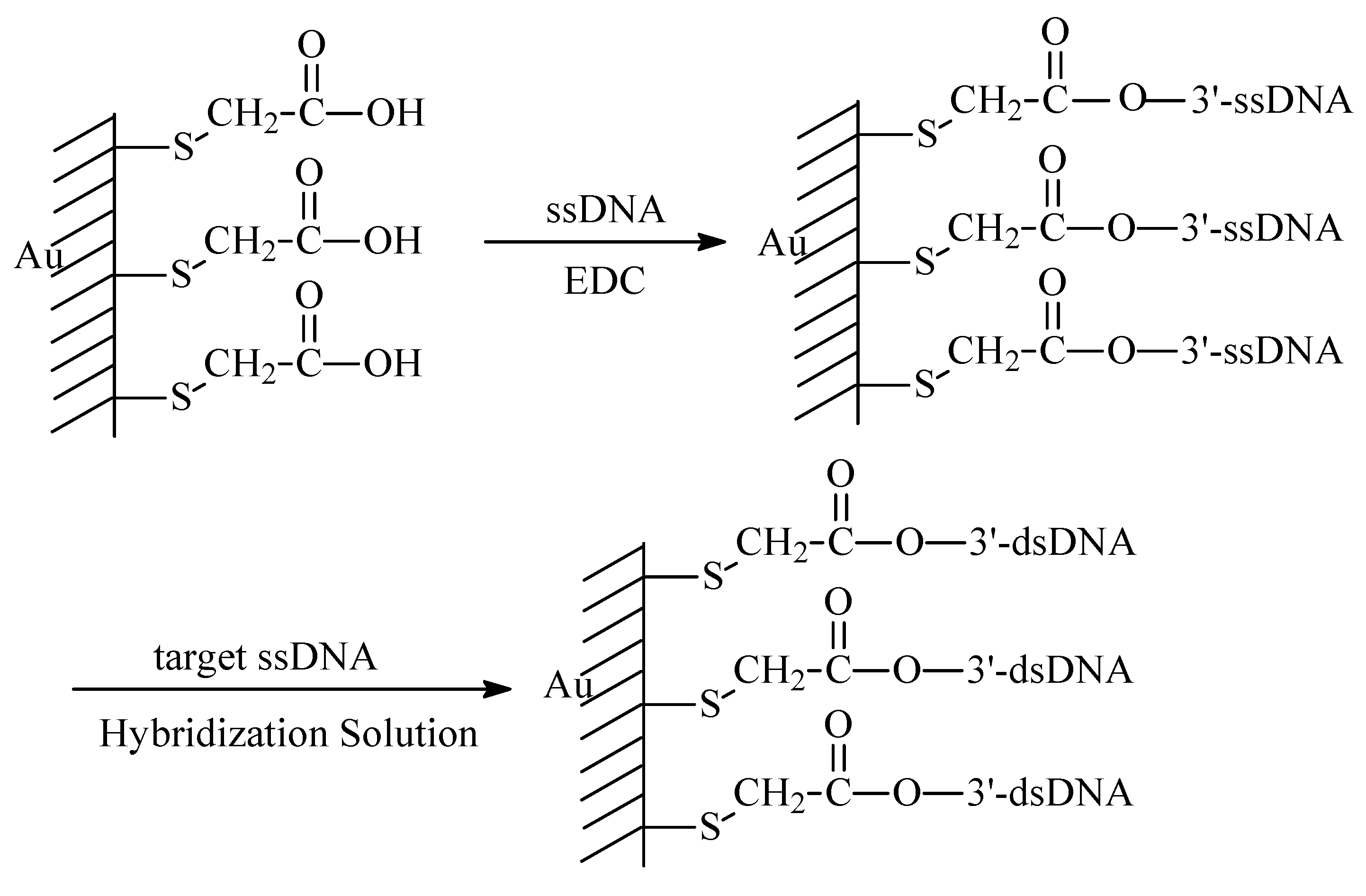
Probe and Its Immobilization

Single-strand DNA (ssDNA) and oligonucleotides are common probes used in DNA sensors. Messenger RNA is commonly revere-transcribed into the more stable form of complementary DNA (cDNA). Peptide nucleic acid (PNA) recognition layers have also been used for preparation of the electrochemical biosensors [4,34]. The distances between the bases in the PNA are very similar to the distances in DNA. The DNA electrochemical biosensors rely on the immobilization of a ssDNA, oligonucleotides or PNA probe onto the electrode surface to recognize through base pairing the complementary DNA strand or oligonucleotides in a sample solution. The distribution, packing density and orientation of the attached probe may affect the performance of DNA biosensors. Several references review the design of DNA biosensors with aspect of transducer surface and probe immobilization [8,116,117,118,119,120,121,122,123,124]. Immobilization methods vary depending on the kind of transducer surface and the application. Some of the most representative immobilization techniques are covalent attachment on a functional surface, adsorption on surface, embedding in sol-gel or polymeric matrix, affinity immobilization and self-assemble monolayer method (see Table 1). Scheme 1 shows one process of covalent attachment on a self-assembled thioglycolic acid (TGA) monolayer modified gold electrode by using water soluble N-(3-dimethylaminopropyl)- N'-ethylcarbodiimide hydrochloride (EDC) as a linker [125,126].

Electroactive Indicators and Labels

The formation double-stranded DNA (dsDNA) upon the DNA hybridization process is commonly being detected in connection with the use of an appropriate electroactive hybridization indicator [38]. Many kinds of substances such as pharmaceutical molecules, cationic metal complexes, biomolecules such as cytochrome c and some enzymes, organic compounds such as dyes and fluoresceins, C60 [66,107] and Doyle catalyst [37] can be employed as the electroactive indicators in monitoring the hybridization events or DNA damage and give electrochemical signals for concentration detection and study on the electron transfer mechanism. These indicators interact in a different way with ss- and dsDNA, which results in a change in the electrochemical response such as amperometric current, electrode potential, Faradaic impedance or capacitance due to the indicator association with the surface duplex. Recently, electrochemical enzyme-linked immunoassay has been used in a DNA hybridization sensor [28].
Scheme 1. Immobilization of ssDNA on TGA monolayer modified Au electrode and hybridization of complementary target ssDNA.
Sensors 03 00128 g010
On the other hand, the indicator-free [38] and label-free [76] methods have also been proposed. These electrochemical biosensors can be used for direct monitoring of DNA hybridization without the use of an external redox indicator.

Detection of DNA Hybridization or Damage

The immobilized DNA probe can recognize its cDNA sequence due to the base pairing [8]. Thus, the sensors may be conveniently used for monitoring DNA hybridization. The redox indicators or intercalators used generally for DNA electrochemical biosensors cannot destroy the double-helical structure of DNA, while the interaction of DNA with chemicals such as acids, radiation or ultrasonication can result in various types of DNA mutation and damage and changes in DNA structure, which may cause serious disturbance of the cell functions. The detection of such a process usually requires a highly sensitive analytical technique. The sensitivity of electrochemical methods to various kinds of DNA damage has been reported [127]. Paleček briefly reviewed the development of DNA biosensors for the detection of DNA damage [5]. The changes in the redox signals of residues in DNA immobilized at electrode the surface can be used as a sign of the damage of DNA base. The damage of cDNA structure in solution by chemical agents will affect its hybridization with immobilized ssDNA.

Discussion

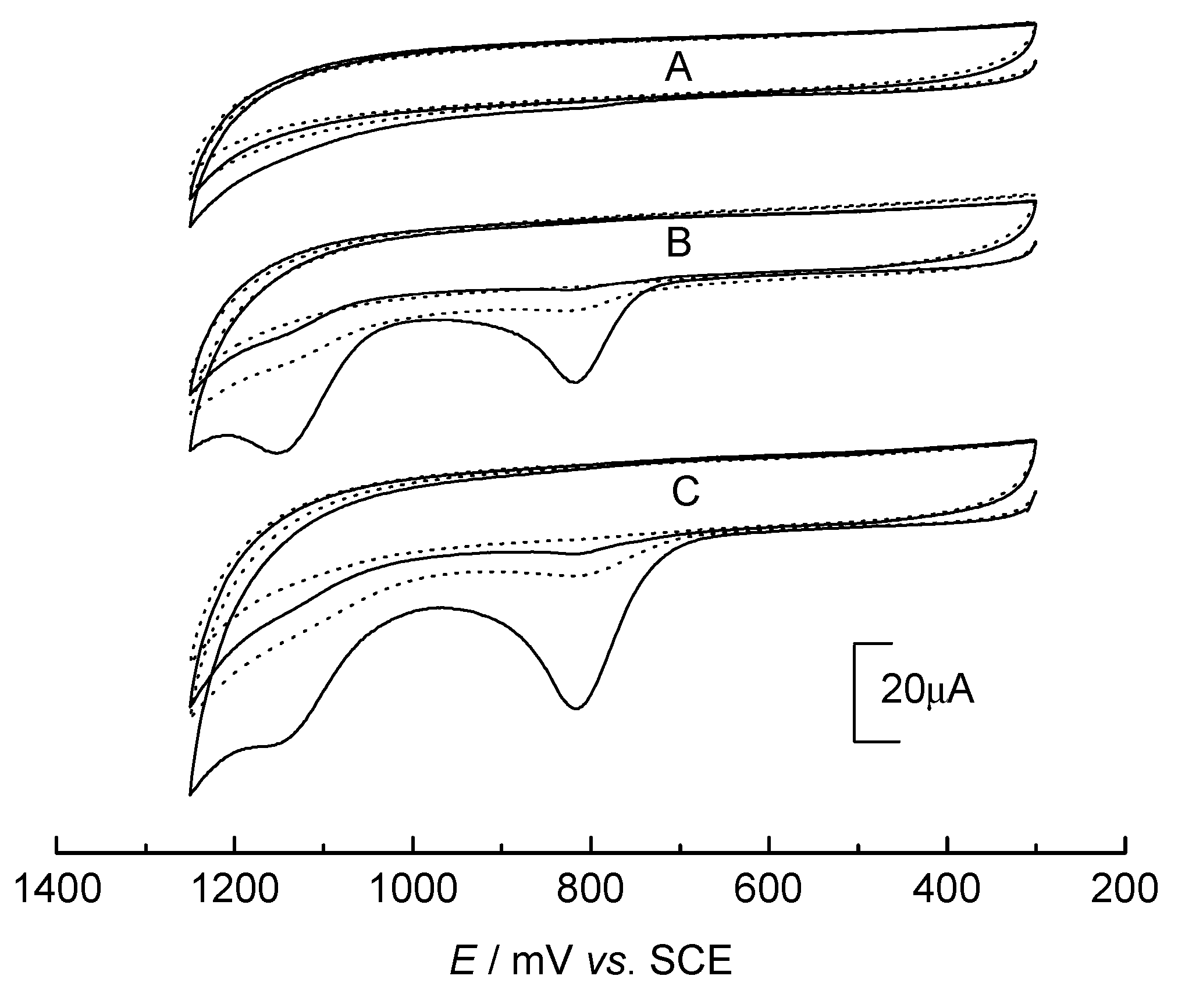
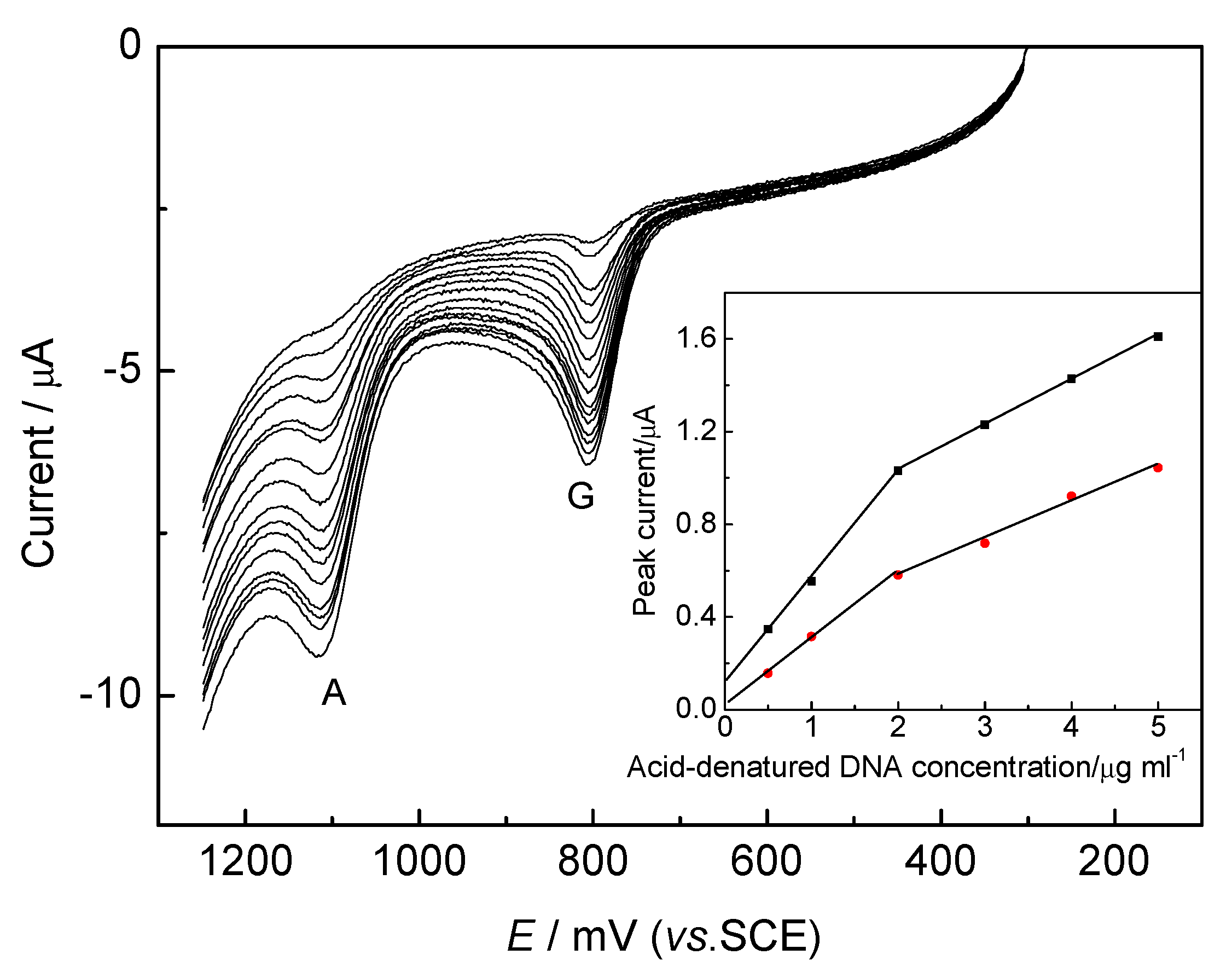
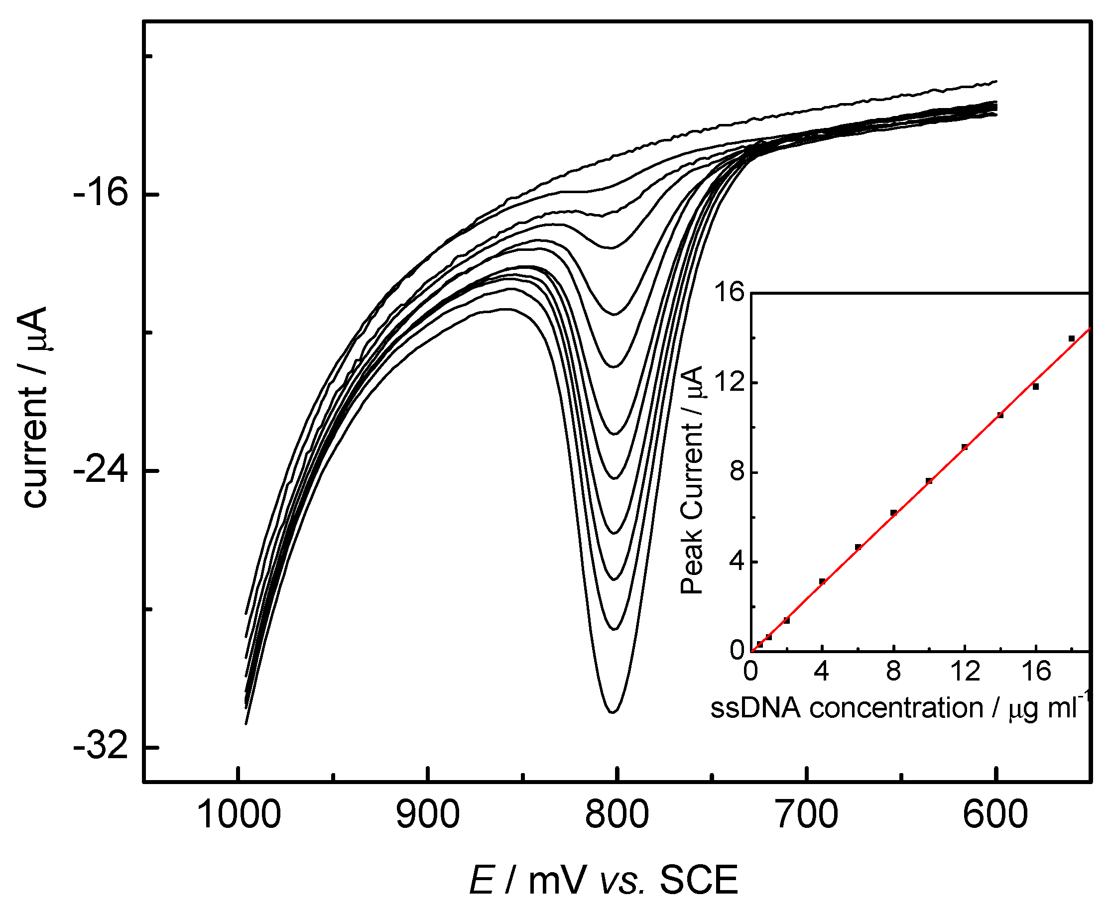
DNA Electrochemical Behaviors and Concentration Determination

There have been many reports on the electrochemical study of DNA over the past few decades. The studies of direct electrochemistry of DNA at mercury electrodes led to a deep understanding of DNA double helix [18,19]. The main objects of this field were focused on the reduction of DNA at mercury electrodes and the interaction between mercury electrode and native or denatured DNA. Different pulse polarography, one of the most sensitive electrochemical methods, can sensitively reflect small damage to the DNA double helix induced by physical and chemical agents. However, it requires tens of micrograms of ssDNA and hundreds of micrograms of dsDNA, which is too low for the analysis of plasmid or viral DNAs, oligonucleotides and other nucleic acid samples. The use of adsorptive stripping voltammetry for the measurements of the reduction signals of adenine (A) and cytosine (C) has improved the sensitivity by several orders of magnitude. Jelen et al. [16] studied the electrochemistry of ssDNA by square-wave voltammetry (SWV) with an HMDE and developed an adsorptive stripping SWV of DNA with a detection limit down to tens of nanograms per milliliter. ssDNA produced a cathodic peak due to the reduction of adenine and an anodic peak due to the oxidation of the guanine reduction product. Wang et al. [128] reported a method to quantitate nanogram quantities of ssDNA using adsorptive stripping potentiometry at electrochemically pretreated carbon paste electrode (CPE). The cyclic voltammogram of 2 mg l-1 ssDNA at the pretreated CPE gave one anodic peak at about +1.05 V corresponding to the oxidation of the guanine residue. Some techniques based on nucleotide acid-pretreated electrodes have been developed [129,130,131,132]. The DNA-modified electrode can be used as a sensor for monitoring the DNA damage [36,51,112].
In our laboratory, a cyclic voltammetric study on the oxidation of DNA at a pre-electrochemically oxidized glassy carbon electrode (GCE) was performed [133]. The pretreatment was carried out by electrochemically oxidizing at +1.75 V (vs. SCE) for 300 s under stirring with followed by cyclic sweep between +0.3 V and +1.25 V in pH 5.0 phosphate buffer until a steady-state current-voltage curve was observed. The electrode modification improved greatly the sensitivity for detection of thermally denatured and acid-denatured DNA and resulted in a 10-fold increase in the current signal (as shown in Figure 1). At the pretreated GCE the thermally denatured DNA and acid-denatured DNA gave two well-defined oxidation peaks in the first anodic sweep at about +0.80V and +1.11 V, while the response was very poor at the unpretreated GCE. The two anodic peaks at about +0.80 V and +1.11 V were corresponding to electrochemical oxidation of guanine (G) and A residues in denatured DNA molecules, respectively. The native DNA almost did not appear any response. Thus the pretreated GCE had a high sensitivity to the changes in DNA double helix. The electrochemical oxidation of denatured DNA on pretreated GCE was an entirely irreversible process. Based on the two oxidation peaks a convenient quantitation of low level of denatured DNA was proposed. The detection limits of thermally denatured and acid-denatured DNA were 2.0 and 0.10 μg ml-1 when the accumulation time was 150 s at the potential of +0.3 V.
Figure 1. Cyclic voltammograms of 1.0 mg ml-1 native DNA (A), 1.0 mg ml-1 thermally denatured DNA (B) and 0.30 mg ml-1 acid-denatured DNA (C) at unpretreated GCE (dot line) and pretreated GCE (solid line) in 0.1 mol l-1 pH 5.0 PBS at 100 mV s-1.
Sensors 03 00128 g001
In view of the equal importance of the measurement of G and A groups or their ratio in DNA to the determination of DNA concentration itself, a simple and reliable method for the simultaneous determination of G and A in DNA was proposed by accumulating denatured DNA on the pretreated electrode surface at a suitable potential (+0.3 V) or in open circuit [134]. The detection limits for individual measurement of G and A were 4.5 and 4.0 ng ml-1, respectively. The proposed method could be used to estimate the G and A contents in DNA using the calibration graph obtained previously for simultaneous determination of G and A (Figure 2) or a standard addition method. The detection limits of G and A in DNA calculated were 0.1 and 0.2 μg ml-1. The molar ratio of (G+C)/(A+T) for HCl and HClO4 denatured calf thymus DNA were 0.74 and 0.82, respectively, which coincided to the standard value of 0.77.
The modification of GCE, carried out by electrochemically oxidizing at +1.80 V (vs. SCE) for 10 min in pH 5.0 PBS and followed with scanning the electrode in the potential range of 0.3 V – -1.3 V for 20 circles, resulted in the improvement of sensitivity for ssDNA detection for about 100 fold. The modified GCE showed a high affinity towards ssDNA through hydrogen bond (specific static adsorption). By means of adsorptive stripping voltammetry, the electrochemical detection of thermal denatured DNA at electrochemically modified GCE was further developed (Figure 3) [135]. The detection limit of thermally denatured DNA was ca. 0.2 μg ml-1 when the accumulation time is 8 min in open circuit in pH 5.0 phosphate buffer.
Figure 2. Linear sweep stripping voltammograms at 100 mV s-1 for the simultaneous determination of G and A in acid-denatured DNA with increasing concentration from 0.50 to 25 μg ml-1 at the accumulation time of 120 s at + 0.3 V. Inset: plots of the peak currents vs. acid-denatured DNA concentrations.
Sensors 03 00128 g002
Figure 3. Linear sweep voltammograms of ssDNA in 0.1 mol l-1 pH 5.0 PBS at 100 mV s-1 with increasing concentration from 0 – 18.0 μg ml-1 at a preconcentration time of 5 min with stirring at open circuit. Inset: calibration plot.
Sensors 03 00128 g003

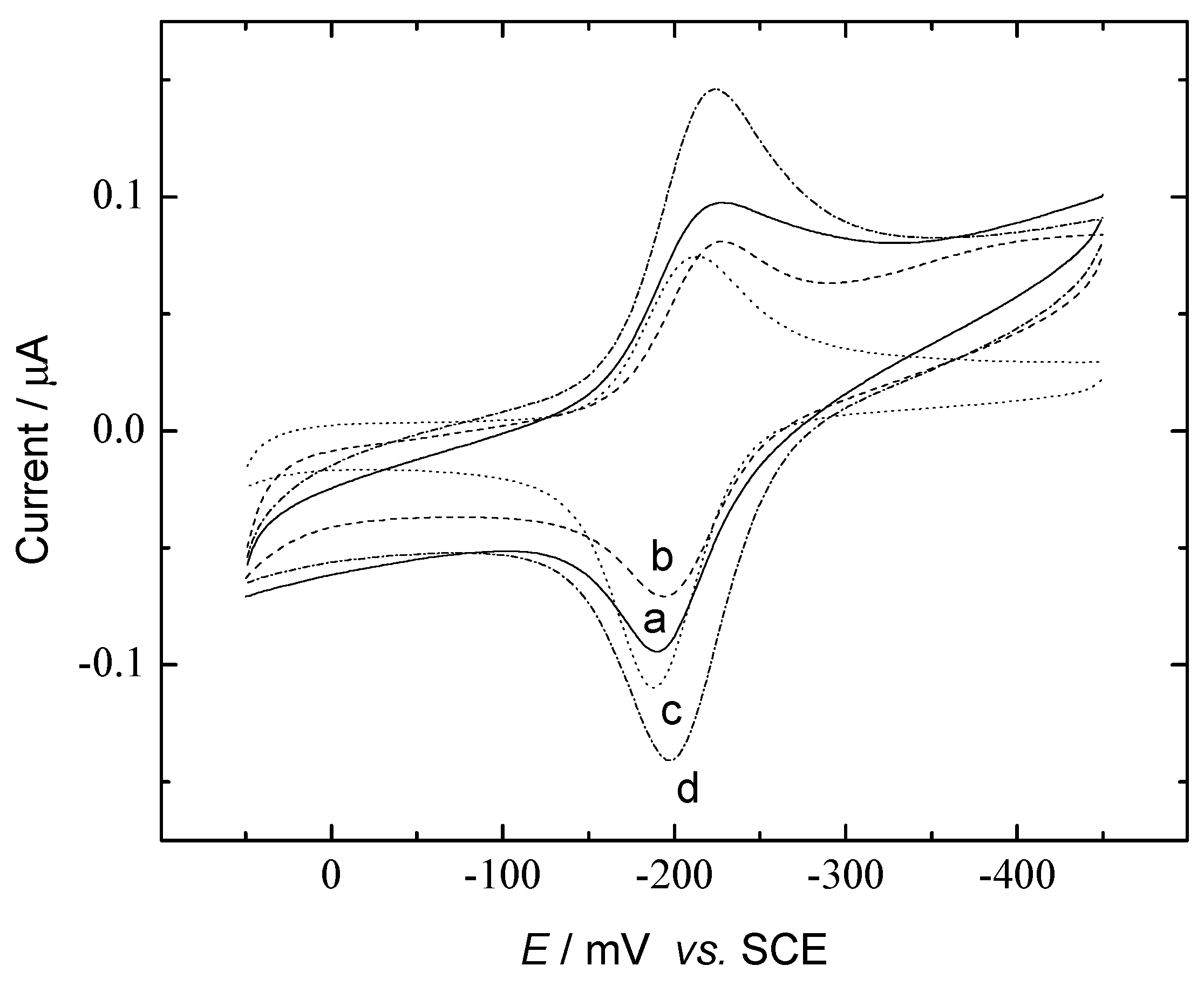
DNA Sensors for Sequence Recognition

The selection of a suitable molecular interface for detecting electrochemical transduction of a DNA hybridization event is a key step in most DNA sensors design. The studies on the interactions between DNA and other molecules provide an opportunity to employ these molecules as hybridization indicators. Recently Paleček et al. extended the choice of hybridization surfaces and detection electrodes [28,29,136]. Although label-free strategies for DNA recognition [137] and mediated electron-transfer for DNA detection [138,139] have become a promising aspect, the study of DNA hybridization biosensors using simple intercalators, threading intercalators or bisintercalators as redox indicators is still an important area [11,12,17,59,62,78,79,80,85,87,92,107,110,140]. Besides the intercalating, the interaction between DNA and electroactive indicator may involve the electrostatic [30,78,79,91,107] or groove [110] mechanism.
Figure 4. Cyclic voltammograms of 1.0×10⁻⁵ mol l-1 NB in 0.01 mol l-1 pH 7.4 Tris-HCl buffer at bare gold (a), calf-thymus ssDNA (b) and dsDNA (c) modified gold electrodes at 400 mV s-1 after soaked for 10 min.
Sensors 03 00128 g004
Our works studied the electrochemical interactions of methylene blue (MB) with yeast DNA in solution [125] and nile blue (NB) with calf-thymus dsDNA in solution, ssDNA and dsDNA adsorbed on gold electrodes [126]. The binding of NB with dsDNA in solution showed an electrostatic interaction mechanism with the binding constant of (5.9±0.2)×104 mol-1 l in 0.01 mol l-1 pH 7.4 Tris-HCl, while an electrostatic interaction mechanism between NB and adsorbed ssDNA and an action mechanism containing both electrostatic and intercalative bindings of NB to adsorbed dsDNA were observed. The presence of adsorbed dsDNA resulted in a great increase in the peak currents of NB in comparison with those obtained at a bare or ssDNA adsorbed gold electrode (Figure 4). The Langmuir adsorption constants of NB at ssDNA/Au and dsDNA/Au electrodes were (1.6±0.2)×105 and (4.2±0.6)×105 mol-1 l, respectively. The electron transfer rate constants of nile blue adsorbed on bare gold and bound to ss- and dsDNA/Au electrodes were 1.4±0.1, 1.2±0.02 and 2.9±0.2 s-1, respectively. The difference between interactions of NB with adsorbed ss- and dsDNA could be used for hybridization recognition of DNA. As an example, hepatitis B virus (HBV) DNA fragment was used for the application study. The interactions between NB and covalently immobilized HBV ssDNA fragment and its hybridization product showed the mechanisms similar to those of adsorbed calf-thymus DNA. A slight increase of NB peak current was observed at HBV ssDNA/Au electrode when comparing with that at bare gold electrode, while a great increase in both cathodic and anodic peak currents was observed at HBV dsDNA/Au electrode. The cathodic peak current of NB at HBV dsDNA/Au electrode increased by about 71% when comparing with that at HBV ssDNA/Au electrode. As control, no increase in peak current of the voltammogram of NB was observable after the covalently immobilized HBV ssDNA was treated with the same process in the hybridization solution containing denatured calf-thymus ssDNA as a substitute for HBV cDNA. Thus, NB was demonstrated to be a good electroactive indicator for recognition of DNA hybridization.
Figure 5. Cyclic voltammograms of 50 μmol l-1 MB in pH 7.2 50 mmol l-1 Tris + 20 mmol l-1 NaCl solution at bare gold (a, solid), SAM/Au (b, dash), ssDNA/Au (c, dot) and dsDNA/Au (d, dash dot) electrodes at 100 mV s-1.
Sensors 03 00128 g005
Based on the significant increase in peak current of MB upon the hybridization of immobilized ssDNA with cDNA in the solution, another DNA electrochemical sensor for sequence detection and monitoring hybridization of native yeast ssDNA was constructed (Figure 5) [125]. The interaction of immobilized ssDNA and dsDNA with MB also resulted in a transition of electrode process from diffusion-controlled to surface-controlled process. The adsorption constants of MB on ssDNA and dsDNA modified gold electrode surface were found to be (3.3±0.3)×103 mol-1 l and (6.6±0.4)×103 mol-1 l, respectively.

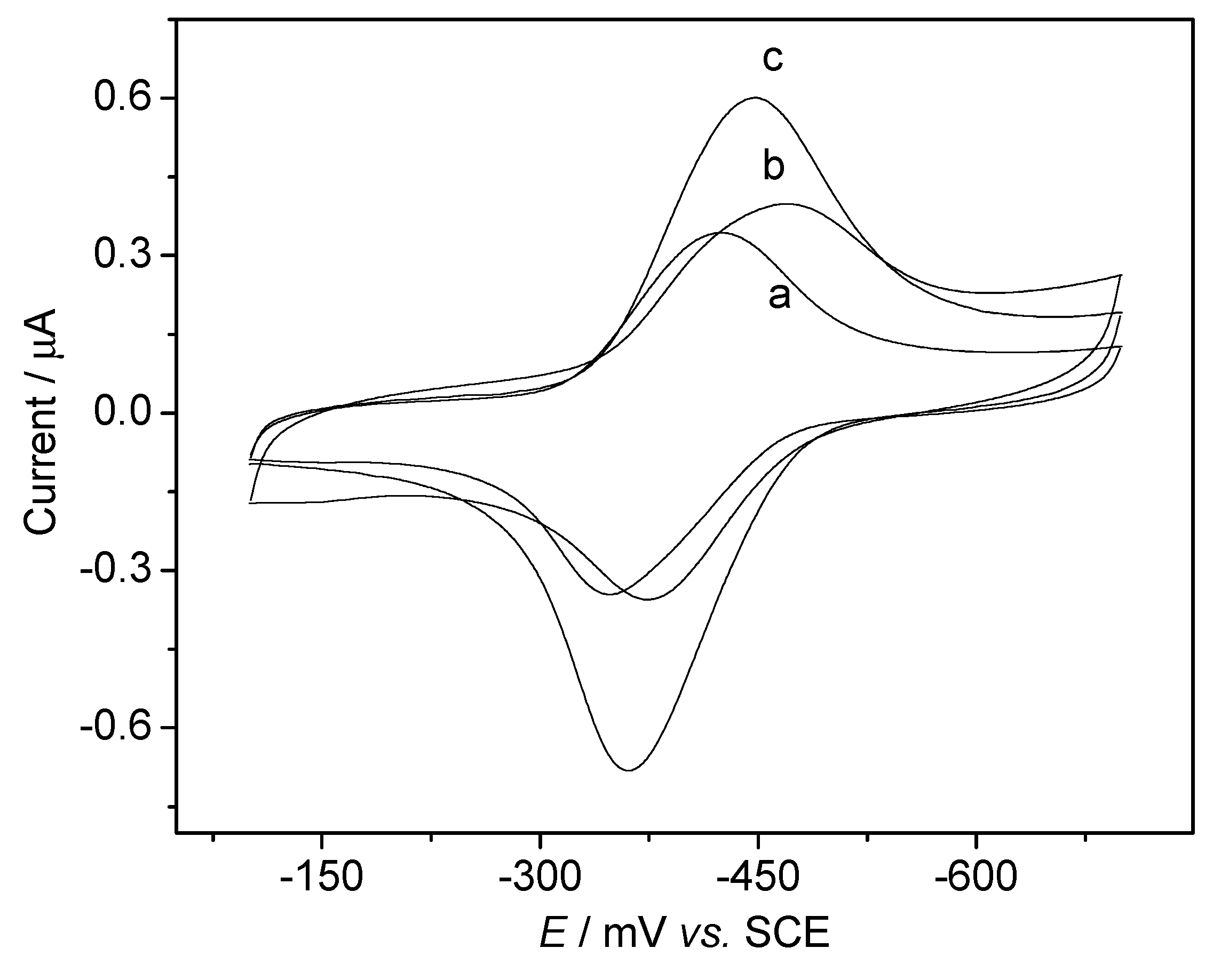
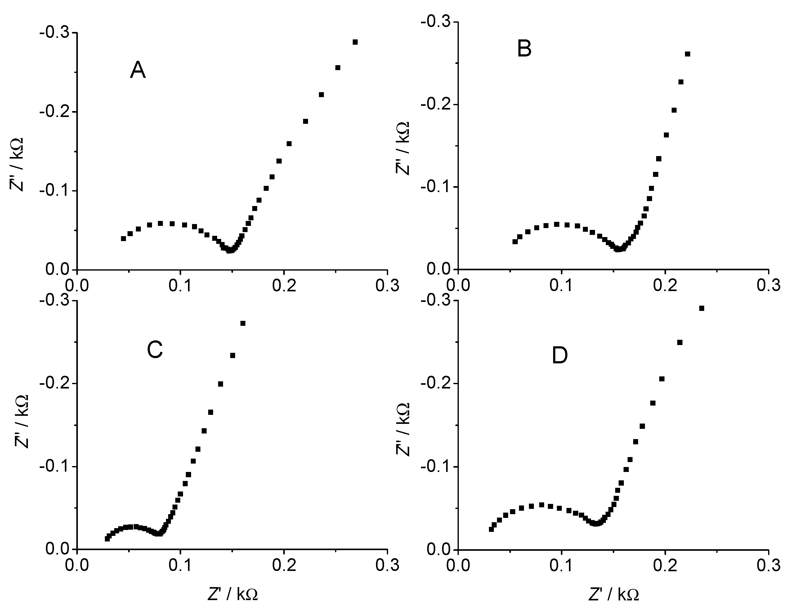
DNA Electrochemical Biosensors Combining With PCR Technique

PCR is a well-known technique for the amplification of sample. It allows enormous amplification of any specific sequence of DNA with three steps: denaturation, primer annealing and elongation. The electrochemically enriched nucleic acids can be amplified with a subsequent PCR step and detected by electrochemical methods. The sample preparation, PCR and detection modules can be integrated in an efficient and easy to use device with low-price electrodes [54], which makes electrochemical molecular methods more suitable for routine laboratory diagnosis or even decentralized models like point-of-care diagnosis. This is made possible by the great advantage of electrochemical biosensors, which, unlike many other types of biosensors, can be integrated in small and inexpensive devices offering high sensitivity.
Figure 6. AC impedance spectra of 8.0 × 10⁻⁴ mol l-1 FcPF6 in 0.01 mol l-1 TE solution at bare (a), TGA/Au (b), ssDNA/Au (c) and dsDNA/Au (d) electrodes.
Sensors 03 00128 g006
As an attempt, our works used PCR method to amplify a sample containing short-strand HBV DNA fragments. The ssDNA PCR product was then immobilized on a self-assembled monolayer modified gold electrode surface [126,141,142]. The binding of HBV cDNA with the immobilized ssDNA resulted in the formation of dsDNA monolayer on the surface. The immobilization of ssDNA PCR product and the surface hybridization of immobilized ssDNA with cDNA could be characterized with ac impedance and XPS techniques [45]. The AC impedance spectra were shown in Figure 6 [141]. The Rs values obtained at the four electrodes were very close. The value of Rct at the ssDNA/Au electrode was only half of those obtained at other electrodes. The interaction between Fc+ unit and DNA chain was an electrostatic mechanism. Thus it was easier aggregated on immobilized ssDNA surface due to the more naked negative-charged groups on the surface, resulting in a lower value of Rct and a faster electron transfer rate. After hybridization of immobilized ssDNA with cDNA, most of the negative charges on DNA chain were buried in dsDNA helix, which made the electrostatic interaction between Fc+ and DNA surface weaker. The Cd value obtained from the Rct and the characteristic angular frequency ω* at the TGA/Au electrode was slightly smaller than that at bare gold electrode, indicative of the presence of TGA monolayer on the electrode surface. The Cd value at the ssDNA/Au electrode was obviously larger than other three systems because of more negative charges on the ssDNA/Au surface, which possessed higher dielectric constant. The Cd value at the dsDNA/Au electrode was larger than those at bare and the TGA/Au electrodes. The dsDNA/Au electrode gave a smaller Cd value than the ssDNA/Au electrode due to the burial of the negatively charged phosphate groups in dsDNA helix.
Figure 7. XPS spectra of TGA/Au (A) and ssDNA /Au (B) from 0 to1200 eV.
Sensors 03 00128 g007
The component of immobilized biomolecules on electrode surface can be analyzed with XPS technique. The XPS spectra of the TGA/Au and ssDNA/Au electrodes were showed in Figure 7 [142]. The XPS spectra exhibited the peaks of C, O and S elements at the TGA/Au electrode and the peaks of C, O, S, N and P elements at the ssDNA/Au electrode. The difference in peak intensity of S2p at the TGA/Au and ssDNA/Au electrodes resulted from their different structures. At the ssDNA/Au electrode (curve B) the peak of S2p was lower, which was attribute to the burying of S atom in DNA fragment. The peak of P2p was the best evidence that ssDNA was covalently immobilized on the TGA self-assembled monolayer gold surface. The peak of N element also indicated the presence of ssDNA on the electrode surface. From the peak areas of curve 2, the molar ratio of N to O was calculated to be 1:1.82 which was near the theoretical value of 1:1.87 in the HBV ssDNA fragment.
The bindings of the electroactive indicators, ferrocenium [141] and [Os(bpy)2Cl2]+ [142], to immobilized ss and dsDNA showed different interaction mechanisms. As well known, the interaction mechanism between electroactive indicator and DNA helix depends on its type and structure. An electrostatic interaction of an electroactive indicator with ss or dsDNA makes its reduction at the surface with negative charge more difficult, thus results in a shift of formal potential in negative direction [80,83]. On the contrary, the intercalative interaction produces a positive shift of formal potential of the intercalator [80,83]. Our works indicated the cationic Fc+ molecules electrostatically associated with the anionic phosphate groups in DNA backbone. Furthermore, the electrostatic interaction between Fc+ and the dsDNA/Au electrode was weaker than that between Fc+ and the ssDNA helix, which resulted in a decrease in the redox peak currents or the amount of Fc+ cation electrostatically bound to the anionic DNA backbone upon the hybridization procedure [141]. However, [Os(bpy)2Cl2]+ showed the same E0 value at ssDNA/Au electrode as that at dsDNA /Au electrode [142]. Thus, the binding properties of [Os(bpy)2Cl2]+ to immobilized ss or dsDNA was not a pure electrostatic or intercalative interaction. Motonaka et al [143] used different length DNA probes to hybridize with their targets and studied the recognition of [Os(bpy)3]2+/3+ ion to the hybridization process. Their results showed that the osmium complex interacted to major or minor grooves of the dsDNA helix. However, the change of peak potential upon the major or minor groove interaction has not been reported. The same E0 value might also resulted from the contrary changes produced by the electrostatic and intercalative interactions. At present it was difficult to describe exactly the interaction mechanism between [Os(bpy)2Cl2]+ and DNA helix due to the particularity of Os, py complexes.
Figure 8. Anodic (A) and cathodic (B) differential pulse voltammograms of 8.0 × 10⁻⁴ M FcPF6 in 0.01 mol l-1 TE solution at bare (a), TGA/Au (b), ssDNA/Au (c) and dsDNA/Au (d) electrodes at 20 mV s-1.
Sensors 03 00128 g008
Based on the decrease in the redox peak currents of Fc+ upon hybridization of immobilized HBV ssDNA fragments with HBV cDNA fragments, a sequence detection method for about 104 copies (about 1.7 × 10⁻²⁰ mol or 0.7 fg) of original genomic HBV DNA fragments was proposed [141]. As shown in Figure 8, the anodic peak currents of FcPF6 at bare gold, TGA/Au, ssDNA/Au and dsDNA/Au electrodes were 3.86, 4.54, 16.18 and 6.60 µA, respectively, while the cathodic peak currents were 6.61, 7.45, 24.24 and 16.41 µA, respectively. The hybridization of the immobilized ssDNA with cDNA resulted in the decreases of anodic and cathodic peak currents by 59.3% and 32.3%, respectively. As control, no observable change in peak currents was observed after the covalently immobilized HBV ssDNA was treated with the same process in the solution containing denatured calf-thymus ssDNA or yeast DNA samples, indicated that they did not hybridize with the immobilized HBV ssDNA.
Following the further treatment of the electrode with HVB ssDNA fragments the redox peak currents of [Os(bpy)2Cl2]+ continuously increased and the peak potentials retained at constant values (Figure 9) [142]. The peak currents of [Os(bpy)2Cl2]+ at HBV ssDNA/Au electrode increase slightly in comparison with those at bare gold electrode, indicating a very weak binding of the complex to ssDNA fragment. However, the peak currents of [Os(bpy)2Cl2]+ at HBV dsDNA/Au electrode obtained with a hybridization process between immobilized HBV ssDNA fragment with its cDNA obviously increased. When comparing with those at bare gold electrode, the anodic and catodic peak currents at HBV dsDNA/Au electrode increase by about 23% and 25%, respectively. The enhancement of peak currents indicated more [Os(bpy)2Cl2]+ molecules were preconcentrated on the dsDNA/Au electrode surface due to the binding interaction between [Os(bpy)2Cl2]+ and immobilized HBV dsDNA. These results indicated the combination of DNA electrochemical biosensors with PCR techniques provided a platform for electrochemical sequence detection of very low content of DNA.
Figure 9. Cyclic voltammograms of 0.50 mM [Os(bpy)2Cl2]+ at bare gold (a), HBV ssDNA/Au (b) and HBV dsDNA/Au (c) electrodes and at HBV ssDNA/Au electrode treated with calf-thymus ssDNA (d) at 400 mV s-1.
Sensors 03 00128 g009

Conclusions

Based on the highly specific affinity interaction between immobilized DNA probe and the target to form a stable double helix and the advantages of rapid, convenient, cheap and miniaturization characters, the DNA electrochemical biosensors have received considerable attention and become an important research field. In recent decades their research has made a marvelous progress. However, the commercialization of this field has not been sufficiently developed. Before the DNA electrochemical biosensors become commercially available and widely used, a great deal of basic research will be necessary to improve the DNA sensor technologies [7]. Firstly, the fundamental studies on the mediated and electron transfer theory and new sensing principles will direct the DNA sensing design strategies. Secondly, the development of fast, reliable and biocompatible DNA sensing interface is the base of the application. Thirdly, association with other technologies such as immunological and DNA microarrays and chips etc will provide us further bio-information on the interaction between DNA and proteins or drugs, multiple DNA sequences and so on. The amplification methods such as PCR have been used to amplify the target DNA [54,72,74,81,87,104,111]. With this technique an approach for sensitive detection of femtogram original genomic target DNA can be achieved [126,141,142]. For the practical requirements of genetic diagnosis and routine applications of DNA electrochemical biosensors in medical, environmental and forensic sciences, an on-line or in-situ miniaturized and easy-to-use new technologies combining with bio/medicine-statistics will be the major research projects of this field.

Acknowledgements

This project was supported by the National Natural Science Foundation of China (29835110, 29975013 and 20275017), the Specialized Research Funds for the Doctoral Program of Higher Education (200028403) and the Excellent Young Teachers from Chinese Ministry of Education, and the Science Foundation of Jiangsu (No. BK99030 and BS2001063).

References

  1. Paleček, E.; Davidson, J.N.; Cohn, W.E. (Eds.) Progress in Nucleic Acids Research and Molecular Biology; Vol. 9, Academic Press: New York, 1969; p. 31.
  2. Paleček, E.; Fojta, M. Anal. Chem. 2001, 73, 74A.
  3. Bakker, E.; Telting-Diaz, M. Anal. Chem. 2002, 74, 2781, and references therein.
  4. Wang, J. Biosen. & Bioelectron. 1998, 13, 757, and references therein.
  5. Paleček, E.; Fojta, M.; Tomschik, M; Wang, J. Biosen. & Bioelectron. 1998, 13, 621, and references therein.
  6. Wang, J. J. of Pharm. and Biomed. Anal. 1999, 19, 47, and references therein.
  7. Paleček, E. Talanta 2002, 56, 809, and references therein.
  8. Homs, W.C.I. Anal. Lett. 2002, 35, 1875, and references therein.
  9. Wang, J. Anal. Chim. Acta 2002, 469, 63, and references therein.
  10. Lu, X.J.; Ju, H.X. Chin. J. of Anal. Chem. 2003, 31, 110, and references therein.
  11. Zhang, J.L.; Fan, H.Z.; Pan, J.H. Acta Pharm. Sinica 1997, 32, 314.
  12. Wang, H.F.; Yang, P.; Li, Q.S.; Wu, Q. Chem. J. Chin. Univ. 1997, 18, 671.
  13. Wang, J.; Grant, D.H.; Ozsoz, M.; Cai, X.H.; Tian, B.M.; Fernandes, J.R. Anal. Chim. Acta 1997, 349, 77.
  14. Li, M.X.; Li, N.Q. Chem. J. Chin. Univ. 2001, 22, 1230.
  15. Fojta, M.; Staňková, V.; Paleček, E.; Koscielniak, P.; Mitáš, J. Talanta 1998, 46, 155. [PubMed]
  16. Jelen, F.; Tomschik, M.; Paleček, E. J. Electroanal. Chem. 1997, 423, 141.
  17. Jelen, F.; Erdem, A.; Paleček, E. Bioelectrochem. 2002, 55, 165.
  18. Teijeiro, C.; Perez, P.; Marín, D.; Paleček, E. Bioelectrochem. and Bioenerg. 1995, 38, 77.
  19. Kizek, R.; Havran, L.; Fojta, M.; Paleček, E. Bioelectrochem. 2002, 55, 119.
  20. Zhang, H.M.; Li, N.Q. J. of Pharm. and Biomed. Anal. 2000, 22, 67.
  21. Fojta, M.; Harran, L.; Kubiěárová, T.; Paleček, E. Bioelectrochem. 2002, 55, 25.
  22. Fojta, M.; Kubiěárová, T.; Paleček, E. Biosen. & Bioelectron. 2000, 15, 107.
  23. Tomschik, M.; Jelen, F.; Harran, L.; Trnková, L.; Nielsen, P.E.; Paleček, E. J. Electroanal. Chem. 1999, 476, 71.
  24. Jelen, F.; Fojta, M.; Paleček, E. J. Electroanal. Chem. 1997, 427, 49.
  25. Wu, J.T.; Zhou, J.Z.; Huang, Y.; Luo, J.; Lin, Z.H. Chin. J. Anal. Chem. 1998, 26, 819.
  26. Wu, J.T.; Huang, Y.; Zhou, J.Z.; Luo, J.; Lin, Z.H. Bioelectrochem. and Bioenerg. 1997, 44, 151.
  27. Jelen, F.; Yosypchuk, B.; Kourilová, A.; Novotný, L.; Paleček, E. Anal. Chem. 2002, 74, 4788. [PubMed]
  28. Paleček, E.; Kizek, R.; Havran, L.; Billova, S.; Fojta, M. Anal. Chim. Acta. 2002, 469, 73.
  29. Paleček, E.; Fojta, M.; Jelen, F. Bioelectrochem. 2002, 55, 85.
  30. Luo, J.W.; Zhang, M.; Zhang, R.Y.; Pang, D.W. J. Anal. Sci. 2002, 18, 1.
  31. Millan, K. M.; Saraullo, A.; Mikkelsen, S.R. Anal. Chem. 1994, 66, 2943. [PubMed]
  32. Wang, J.; Rivas, G.; Ozsoz, M.; Grant, D.H.; Cai, X.H.; Parrado, C. Anal. Chem. 1997, 69, 1457. [PubMed]
  33. Ikebukuro, K.; Kohiki, Y.; Sode, K. Biosen. & Bioelectron. 2002, 17, 1075.
  34. Wang, J.; Rivas, G.; Cai, X.H.; Chicharro, M.; Parrado, C.; Dontha, N.; Begleiter, A.; Mowat, M.; Paleček, E.; Nielsen, P.E. Anal. Chim. Acta 1997, 344, 111.
  35. Wang, J.; Rivas, G.; Parrado, C.; Cai, X.H.; Flair, M.N. Talanta 1997, 44, 2003. [PubMed]
  36. Lucarelli, F.; Kicela, A.; Palchetti, I.; Marrazza, G.; Mascini, M. Bioelectrochem. 2002, 58, 113.
  37. Gil, E.S.; Kubota, L.T. Bioelectrochem. 2000, 51, 145.
  38. Wang, J.; Rivas, G.; Fernandes, J.R.; Paz, J.L.L.; Jiang, M; Waymire, R. Anal. Chim. Acta. 1998, 375, 197.
  39. Motonaka, J.; Mishima, Y.; Maruyama, K.; Minagawa, K.; Ikeda, S. Sensors and Actuators B. 2000, 66, 234.
  40. Wang, J.; Ozsoz, M.; Cai, X.H.; Rivas, G.; Shiraishi, H.; Grant, D.H.; Chicharro, M.; Fernandes, J.; Paleček, E. Bioelectrochem. and Bioenerg. 1998, 45, 33.
  41. Wang, J.; Kawde, A.N. Electrochem. Comm. 2002, 4, 349.
  42. Wang, J.; Rivas, G.; Cai, X.H.; Dontha, N.; Shiraishi, H.; Luo, D.B.; Valera, F.S. Anal. Chim. Acta 1997, 337, 41.
  43. Sun, X.Y.; Xu, C.; Liu, S.H.; He, P.G.; Fang, Y.Z. Chem. J. Chin. Univ. 1998, 19, 1393.
  44. Sun, X.Y.; He, P.G.; Xu, C.; Liu, X.P.; Liu, S.H.; Fang, Y.Z. J. Anal. Sci. 1999, 15, 279.
  45. Xu, C.; Cai, H.; He, P.G.; Fang, Y.Z. Chem. J. Chin. Univ. 2001, 22, 1492.
  46. Zhou, J.Z.; Wu, L.L.; Luo, J.; Lin, Z.H. Electrochem. 1999, 5, 186.
  47. Xu, C.; He, P.G.; Fang, Y.Z. Chem. J. Chin. Univ. 2000, 21, 1187.
  48. Xu, C.; He, P.G.; Fang, Y.Z. Anal. Chim. Acta. 2000, 411, 31.
  49. Hashimoto, K.; Ito, K.; Ishimori, Y. Anal. Chim. Acta 1994, 286, 219.
  50. Wang, J.; Zhang, X.J.; Parrado, C.; Rivas, G. Electrochem. Comm. 1999, 1, 197.
  51. Korbut, O.; Bučková; Tarapčik, P.; Labuda, J.; Gründler, P. J. Electroanal. Chem. 2001, 506, 143.
  52. Brabec, V. Electrochim. Acta 2000, 45, 2929.
  53. Wang, J.; Cai, X.H.; Fernandes, J.R.; Grant, D.H.; Ozsoz, M. J. Electroanal. Chem. 1998, 441, 167.
  54. Hassmann, J.; Misch, A.; Schülein, J.; Krause, J.; Graβl, B.; Müller, P.; Bertling, W.M. Biosen. & Bioelectron. 2001, 16, 857.
  55. Schülein, J.; Graβl, B.; Krause, J.; Schulze, C.; Kugler, C.; Müller, P.; Bertling, W.M.; Hassmann, J. Talanta 2002, 56, 875.
  56. Cheng, X.D.; Wang, H.E.; Sun, J.; Wen, X.M.; Chen, Z.D.; Hou, S.F.; Sun, Q.S. J. Jining Med. College 1999, 22, 7.
  57. Miao, Q.; Jin, B.K.; Lin, X.Q. Chem. J. Chin. Univ. 2000, 21, 27.
  58. Bao, X.Y.; Chen, J.G.; Chen, X.; Dang, Y.L. Chin. J. Anal. Lab. 2001, 20, 1.
  59. Carter, M.T.; Rodriguez, M.; Bard, A.J. J. Am. Chem. Soc. 1989, 111, 8901.
  60. Rodriguez, M.; Bard, A.J. Anal. Chem. 1990, 62, 2658. [PubMed]
  61. Lin, X.Q.; Miao, Q.; Jin, B.K. Chin. Chem. Lett. 1999, 10, 157.
  62. Cai, H.; Cao, X.; Jiang, Y.; He, P.G.; Fang, Y.Z. Anal. Bioanal. Chem. 2003, 375, 287. [PubMed]
  63. Xu, C.; Cai, H.; He, P.G.; Fang, Y.Z. Analyst 2001, 126, 62. [PubMed]
  64. Lee, T.Y.; Shim, Y.B. Anal. Chem. 2001, 73, 5629.
  65. Brett, C.M.A.; Brett, A.M.O.; Serrano, A.H.P. Electrochim. Acta 1999, 44, 4233.
  66. Li, M.X.; Li, N.Q.; Gu, Z.N.; Zhou, X.H.; Sun, Y.L.; Wu, Y.Q. Microchem. J. 1999, 61, 32.
  67. Siontorou, C.G.; Brett, A.M.O.; Nikolelis, D.P. Talanta. 1996, 43, 1137.
  68. Pedano, M.L.; Rivas, G.A. Biosen. & Bioelectron. 2003, 18, 269.
  69. Mahadevan, S.; Palaniandavar, M. Inorg. Chim. Acta. 1997, 254, 291.
  70. Chen, S.M.; Chen, S.V. Electrochim. Acta. 2003, 48, 513.
  71. Liu, S.Q.; Xu, J.J.; Chen, H.Y. Bioelectrochem. 2002, 57, 149.
  72. Marrazza, G.; Tombelli, S.; Mascini, M.; Manzoni, A. Clin. Chim. Acta 2001, 307, 241. [PubMed]
  73. Buckova, M.; Labuda, J.; Sandula, J.; Krizkova, L.; Stepanek, I.; Durackova, Z. Talanta 2002, 56, 939. [PubMed]
  74. Azek, F.; Grossiord, C.; Joannes, M.; Limoges, B.; Brossier, P. Anal. Biochem. 2000, 284, 107. [PubMed]
  75. Wang, J.; Cai, X.H.; Rivas, G.; Shiraishi, H.; Dontha, N. Biosen. & Bioelectron. 1997, 12, 587.
  76. Wang, J.; Kawde, A.N. Anal. Chim. Acta 2001, 431, 219.
  77. Zhou, J.Z.; Wu, L.L.; Dong, L.Q.; Lin, Z.Y.; Yan, J.W.; Dong, P.; Bao, Y.; Lin, Z.H. Electrochem. 2001, 7, 276.
  78. Liu, J.; Li, Q.W.; Luo, G.A.; Sun, H.W.; Feng, J. J. Instru. Anal. 2002, 21, 13.
  79. Zhou, J.H.; Jiang, H.J.; Feng, Y.Y.; Yang, H.; Xing, W.; Lu, T.H. Chin. J. Appl. Chem. 2001, 18, 994.
  80. Zhao, Y.D.; Pang, D.W.; Wang, Z.L.; Cheng, J.K.; Qi, Y.P. J. Electroanal. Chem. 1997, 431, 203.
  81. Hashimoto, K.; Ito, K.; Ishimori, Y. Sensors and Actuators B 1998, 46, 220.
  82. Zhao, Y.D.; Pang, D.W.; Hu, S.; Wang, Z.L.; Cheng, J.K.; Dai, H.P. Talanta 1999, 49, 751. [PubMed]
  83. Pang, D.W.; Adruna, H.D. Anal. Chem. 1998, 70, 3162. [PubMed]
  84. Maruyama, K.; Mishima, Y.; Minagawa, K.; Motonaka, J. J. Electroanal. Chem. 2001, 510, 96.
  85. Takenaka, S.; Uto, Y.; Takagi, M.; Kondo, H. Chem. Lett. 1998, 989.
  86. Zhao, Y.D.; Pang, D.W.; Zhang, M.; Cheng, J.K. F’ J. Anal. Chem. 1999, 363, 708.
  87. Yang, M.; Yau, H. C.M.; Chan, H.L. Langmuir 1998, 14, 6121.
  88. Zhao, Y.D.; Pang, D.W.; Hu, S.; Wang, Z.L.; Cheng, J.K.; Qi, Y.P.; Dai, H.P.; Mao, B.W.; Tian, Z.Q.; Luo, J.; Lin, Z.H. Anal. Chim. Acta. 1999, 388, 93.
  89. Xu, D.K.; Huang, K.; Liu, Z.H.; Liu, Y.Q.; Ma, L.R. Electroanlysis 2001, 13, 882.
  90. Maruyama, K.; Motonaka, J.; Mishima, Y.; Matsuzaki, Y.; Nakabayashi, I.; Nakabayashi, Y. Sensors and Actuators B 2001, 76, 215.
  91. Liu, H.H.; Lu, J.L.; Zhang, M.; Pang, D.W.; Abruna, H.D. J. Electroanal. Chem. 2003, 544, 93.
  92. Zhang, Q.L.; Liu, J.G.; Liu, j.; Xue, G.Q.; Li, H.; Liu, J.Z.; Zhou, H.; Qu, L.H.; Ji, L.N. J. Inorg. Biochem. 2001, 85, 291. [PubMed]
  93. Shao, Y.; Jin, Y.D.; Dong, S.J. Electrochem. Comm. 2002, 4, 773.
  94. Lisdat, F.; Ge, B.; Scheller, F.W. Electrochem. Comm. 1999, 1, 65.
  95. Neumann, E.; Tonsing, K.; Siemens, P. Bioelectrochem. 2000, 51, 125.
  96. Kertesz, V.; Whitemore, N.A.; Chambers, J.Q.; McKinney, M.S.; Baker, D.C. J. Electroanal. Chem. 2000, 493, 28.
  97. Mishima, Y.; Motonaka, J.; Maruyama, K.; Minagawa, K.; Ikeda, S. Sensors and Actuators B 2000, 65, 340.
  98. Moser, I.; Schalkhammer, T.; Pittner, F.; Urban, G. Biosen. & Bioelectron. 1997, 12, 729.
  99. Trnkova, L. Talanta 2002, 56, 887. [PubMed]
  100. Trnkova, L.; Kizek, R.; Dracka, O. Bioelectrochem. 2002, 55, 131.
  101. Fan, C.H.; Song, H.Y.; Hu, X.F.; Li, G.X.; Zhu, J.Q.; Xu, X.X.; Zhu, D.X. Anal. Biochem. 1999, 271, 1. [PubMed]
  102. Johnson, D.H.; Throp, H.H. J. Phys. Chem. 1996, 100, 13837.
  103. Xu, J.Z.; Zhu, J.J.; Huang, Q.; Chen, H.Y. Electrochem. Comm. 2001, 3, 665.
  104. Yang, Q.J.; Xu, D.K.; Jiang, X.P.; He, W.; Liu, Z.H.; Wang, Y.; Ma, L.R. Chin. J. Anal. Chem. 2002, 30, 56.
  105. Hu, J.B.; Shang, J.; Li, Q.L. Chem. J. Chin. Univ. 2001, 22, 749.
  106. Pike, A.R.; Lie, L.H.; Eagling, R.A.; Ryder, L.C.; Patole, S.N.; Connolly, B.A.; Horrocks, B.R.; Houlton, A. Angew. Chem. Int. Ed. 2002, 41, 615.
  107. Fang, P.F.; Zhao, Y.D.; Pang, D.W.; Chen, Y.Y. Acta Sci. Natu. Univ. Peking 2001, 37, 153.
  108. Zhao, Y.D.; Pang, D.W.; Zhang, M.; Cheng, J.K.; Dai, H.P. Chem. J. Chin. Univ. 2001, 22, 744.
  109. Zhang, L.R.; Zhu, J.J.; Zhao, G.C.; Chen, H.Y. Chem. J. Chin. Univ. 1998, 19, 1363.
  110. Stemp, E. D.A.; Holmlin, R.E.; Barton, J.K. Inorg. Chim. Acta 2000, 297, 88.
  111. Meric, B.; Kerman, K.; Ozkan, D.; Kara, P.; Erensoy, S.; Akarca, U.S.; Mascini, M.; Ozsoz, M. Talanta 2002, 56, 837. [PubMed]
  112. Lucarelli, F.; Palchetti, I.; Marrazza, G.; Mascini, A. Talanta 2002, 56, 949. [PubMed]
  113. Fojta, M.; Havran, L.; Kizek, R.; Billova, S. Talanta 2002, 56, 867. [PubMed]
  114. Wang, J.; Xu, D.K.; Erdem, A.; Polsky, R.; Salazar, M.A. Talanta 2002, 56, 931. [PubMed]
  115. Kawanishi, S.; Hiraku, Y.; Oikawa, S. Mutation Res. 2001, 488, 65. [PubMed]
  116. Henke, L.; Krull, U.J. Canadian J. of Anal. Sci. and Spec. 1999, 44, 61, and references therein.
  117. Wang, B.Q.; Cheng, G.J.; Dong, S.J. Chin. J. of Anal. Chem. 1999, 27, 982, and references therein.
  118. Walcarius, A. Electroanalysis 1998, 10, 1217, and references therein.
  119. Alegret, S. Analyst 1996, 121, 1751, and references therein.
  120. Tess, M.E.; Cox, J.A. J. of Pharm. and Biomed. Anal. 1999, 19, 55.
  121. Lin, J.; Brown, C.W. Trac-Trends in Anal. Chem. 1997, 16, 200.
  122. Tien, H.T.; Barish, R.H.; Gu, L.Q.; Ottova, A.L. Anal. Sci. 1998, 14, 3.
  123. Wink, T.; van Zuilen, S.J.; Bult, A.; van Bennekom, W.P. Analyst 1997, 122, R43.
  124. Paleček, E.; Jelen, F. Critical Rev. in Anal. Chem. 2002, 32, 261, and references therein.
  125. Gu, J.Y.; Lu, X.J.; Ju, H.X. Electroanalysis 2002, 14, 949.
  126. Ju, H.X.; Ye, Y.K.; Zhu, Y.L. Bioconj. Chem. in reviewed.
  127. Paleček, E. Electroanalysis 1996, 8, 7.
  128. Wang, J.; Cai, X. H.; Jonsson, C.; Balakrishnan, M. Electronalysis 1996, 8, 20.
  129. Paleček, E. Bioelectrochem. Bioenerg. 1988, 20, 171.
  130. Paleček, E. Anal. Biochem. 1988, 170, 421.
  131. Paleček, E.; Jelen, F.; Teijeiro, C.; Fucik, V.; Jovin, T. M. Anal. Chim. Acta 1993, 272, 175.
  132. Jelen, F.; Tomschik, M.; Paleček, E. J. Electroanal. Chem. 1997, 423, 141.
  133. Wang, H.S.; Ju, H.X.; Chen, H.Y. Electroanalysis 2001, 13, 1105.
  134. Wang, H.S.; Ju, H.X.; Chen, H.Y. Anal. Chim. Acta 2002, 461, 243.
  135. Wang, H.S.; Ju, H.X.; Chen, H.Y. Electroanalysis 2002, 14, 1615.
  136. Paleček, E.; Billová, S.; Havran, L.; Kizek, R.; Miculková, A.; Jelen, F. Talanta 2002, 56, 919.
  137. Souteyrand, E.; Cloarec, J.; Martin, J.R.; Wilson, C.; Lawrence, I.; Mikkelsen, S.; Lawrence, M.F. J. Phys. Chem. B 1997, 101, 2980.
  138. Napier, M.E.; Loomis, C.R.; Sistare, M.F.; Kim, J.; Eckhardt, A.E.; Thorp, H.H. Bioconjugate Chem. 1997, 8, 906.
  139. Welch, T.W.; Ciftan, S.A.; White, P.S.; Thorp, H.H. Inorg. Chem. 1997, 36, 4812. [PubMed]
  140. Paleček, E.; Fojta, M. Anal. Chem. 2001, 73, 74A.
  141. Ye, Y.K.; Zhao, J.H.; Yan, F.; Zhu, Y.L.; Ju, H.X. Biosen. & Bioelectron. in press.
  142. Ju, H.X.; Ye, Y.K.; Zhao, J.H.; Zhu, Y.L. Anal. Biochem. 2003, 312, 255.
  143. Mishima, Y.; Motonaka, J.; Ikeda, S. Anal. Chim. Acta. 1997, 345, 45.
  • Sample Availability: Available from the authors.

