Article

Copper Nanoclusters Anchored on Crumpled N-Doped MXene for Ultra-Sensitive Electrochemical Sensing

by
Hanxue Yang
1,2,3,
Chao Rong
1,2,3,
Shundong Ge
1,2,3,
Tao Wang
1,2,3,*,
Bowei Zhang
1,2,3,* and
Fu-Zhen Xuan
1,2,3,*
1
Shanghai Key Laboratory of Intelligent Sensing and Detection Technology, East China University of Science and Technology, Shanghai 200237, China
2
Key Laboratory of Pressure Systems and Safety of Ministry of Education, East China University of Science and Technology, Shanghai 200237, China
3
School of Mechanical and Power Engineering, East China University of Science and Technology, Shanghai 200237, China
*
Authors to whom correspondence should be addressed.
Sensors 2025, 25(8), 2508; https://doi.org/10.3390/s25082508
Submission received: 19 March 2025 / Revised: 9 April 2025 / Accepted: 14 April 2025 / Published: 16 April 2025

Abstract

:

Highlights

What are the main findings?
  • 3D crumpled MXene with strain wrinkles enhances conductivity and prevents restacking.
  • Cu nanoclusters (3.0 wt%) synergize N-doping for noble metal-like catalytic activity.
What is the implication of the main finding?
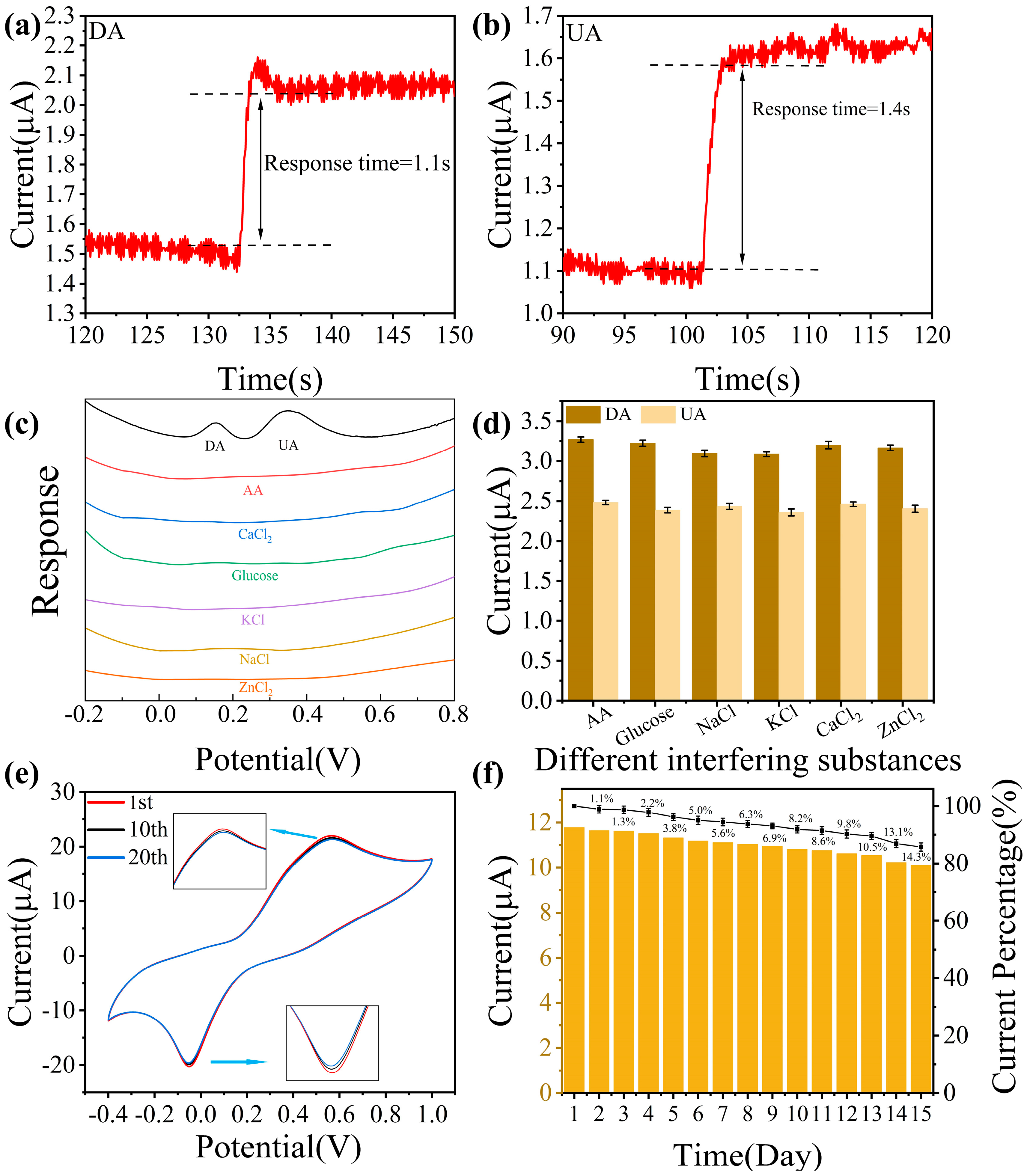
  • Ultrasensitive dual-analyte non-enzymatic detection of DA/UA with anti-interference capability and rapid response.
  • Novel material design strategies provide new approaches for the development of future high-performance sensors.

Abstract

Simultaneous detection of dopamine (DA) and uric acid (UA) is essential for diagnosing neurological and metabolic diseases but hindered by overlapping electrochemical signals. We present an ultrasensitive electrochemical sensor using copper nanoclusters anchored on nitrogen-doped crumpled Ti3C2Tx MXene (Cu-N/Ti3C2Tx). The engineered 3D crumpled architecture prevents MXene restacking, exposes active sites, and enhances ion transport, while Cu nanoclusters boost electrocatalytic activity via accelerated electron transfer. Structural analyses confirm uniform Cu dispersion (3.0 wt%), Ti-N bonding, and strain-induced wrinkles, synergistically improving conductivity. The sensor achieves exceptional sensitivity (1958.3 and 1152.7 μA·mM−1·cm−2 for DA/UA), ultralow detection limits (0.058 and 0.099 μM for DA/UA), rapid response (<1.5 s), and interference resistance (e.g., ascorbic acid). Differential pulse voltammetry enables independent linear detection ranges (DA: 2–60 μM; UA: 5–100 μM) in biofluids, with 94.4% stability retention over 7 days. The designed sensor exhibits excellent capabilities for DA and UA detection. This work provides a novel design strategy for developing high-performance electrochemical sensors.

1. Introduction

Dopamine (DA) and uric acid (UA) are vital biomarkers for disease diagnosis [1]. DA, a neurotransmitter linked to psychiatric diseases, regulates neurological functions [2,3]. UA, a purine metabolite, protects vascular health but induces gout and cardiovascular disorders at high concentrations [4,5,6]. Their coexistence in biological fluids and correlated concentration changes (e.g., reduced DA with elevated UA in Parkinson’s disease) underscore the need for simultaneous detection [7]. However, accurate detection is hampered by the similarity of the signals of DA and UA and the presence of other interferences during the detection process [8]. Developing high-performance sensors is crucial for advancing clinical diagnostics and mechanistic studies [9]. Current methods for DA/UA detection, such as high-performance liquid chromatography-mass spectrometry (HPLC-MS) and enzyme-linked immunosorbent assays (ELISA), face limitations such as high cost, lengthy procedures, and poor portability, limiting their clinical utility [10,11,12,13,14]. Electrochemical sensing emerges as a promising alternative due to rapid response, cost efficiency, multi-target capability, and portability, making it ideal for clinical diagnostics and dynamic disease studies [15,16,17]. However, existing sensing materials (carbon nanomaterials, MOFs, etc.) suffer from narrow detection ranges, low sensitivity, high detection limits, and poor selectivity against interferents like ascorbic acid [18,19,20,21,22]. To address these challenges, designing advanced nanomaterials with enhanced selectivity, sensitivity, and ultralow detection limits is crucial for enabling real-time clinical monitoring and precise disease progression analysis.
Transition metal carbide/nitride (MXene) is a diverse family of two-dimensional (2D) nanomaterials with excellent electrical conductivity, outstanding sensing performance, tunable surface chemistry, and layered structures, which have versatile applications in energy and sensing fields [23,24]. Ti3C2Tx MXene demonstrates exceptional electrochemical sensing capabilities due to its high conductivity and rich surface functional groups [25,26,27]. However, van der Waals-driven restacking of nanosheets reduces active site exposure and ion diffusion efficiency [28]. To address this, constructing a 3D crumpled structure through strain engineering effectively inhibits stacking, creating hierarchical pores and interconnected channels that shorten ion diffusion distances and enhance interfacial electron transfer [29,30,31]. Existing studies have shown that Cu nanoclusters have good sensing performance [32,33]. But the poor stability of Cu nanoclusters and the concomitant formation of Cu2O nanoparticles while anchoring Cu NCs limit the high atomic utilization efficiency of Cu NCs, affecting their sensitivity [34,35,36,37]. The crumpled surface of MXene can provide abundant anchor sites for loading Cu NCs and enhance the electrochemical catalytic performance through metal-carrier synergy [38,39,40]. Therefore, anchoring non-precious metal nanoclusters on crumpled MXene can achieve ultra-sensitive detection with noble metal-like properties at a low cost.
In this work, we loaded non-precious metal Cu nanoclusters onto crumpled N-doped Ti3C2Tx nanosheets (Cu-N/Ti3C2Tx) and used them for the electrochemical sensing detection of DA and UA. The introduction of surface-strained crumpled structures and anchoring of Cu nanoclusters not only enhanced material stability, created a more active specific surface area, and provided additional reactive sites, but also improved electrical conductivity and electrochemical performance. The Cu-N/Ti3C2Tx modified electrodes exhibited high sensitivity, superior selectivity, and rapid response towards DA and UA as tested by cyclic voltammetry (CV) and differential pulse voltammetry (DPV) experiments.

2. Experimental Section

2.1. Materials

Melamine (C3H6N6, ≥99%), Copper (II) chloride dihydrate (CuCl2·2H2O, ≥99.0%), Lithium fluoride (LiF, 99.99%) and Hydrochloric acid (HCl, 36.0~38.0%) were supplied by Macklin. Nafion solution (5%), ascorbic acid (AA, ≥99.0%), dopamine (DA, 98%), and uric acid (UA, 99%) were supplied by Aladdin. Phosphate-buffered saline (PBS, PH = 7.4, 0.1 M) was bought from Shanghai Yuanye Bio-Technology Co., Ltd. (Shanghai, China). Ti3AlC2 MAX (400 mesh, 99.5 wt.%) was bought from Foshan XinXi Technology Co., Ltd. (Foshan, China). Argon (Ar, 99.99%) was obtained from Shanghai HaoQi Gas Co., Ltd. (Shanghai, China).

2.2. Synthesis of Monolayer Ti3C2Tx Nanosheets

Ti3C2Tx MXene was obtained by etching away the Al layer of Ti3AlC2 MAX using hydrogen fluoride (HF). Firstly, HF was prepared by adding 1.6 g of LiF to 9 M 20 mL of HCl and stirring for 15 min to fully dissolve. Next, 1 g of Ti3AlC2 MAX phase was slowly added and stirred in a water bath at 40 °C for 48 h. The etchant was rinsed twice with 1 M HCl and then centrifuged several times (1 min each time) with deionized water (DIW) at 5000 rpm at high speed until the supernatant did not appear in the centrifuge tube after a certain number of centrifugations. When the liquid supernatant was blackened, the tube was shaken well, and centrifugation was continued, repeating the above steps one to three times. Next, pass Ar gas, sonicate in an ice bath for 30 min, and centrifuge at 3500 rpm for 30 min to obtain a monolayer Ti3C2Tx MXene colloidal solution. After suction filtration of the colloid for several hours, the obtained Ti3C2Tx MXene film was dried in a vacuum drying oven at 60 °C for 12 h, and finally the monolayer Ti3C2Tx MXene solid was obtained.

2.3. Synthesis of Cu-N/Ti3C2Tx, N/Ti3C2Tx, and Cu-Ti3C2Tx Nanosheets

Using ice bath sonication and stirring, 60 mg of the prepared monolayer Ti3C2Tx MXene was completely dissolved into 15 mL DIW. 180 mg of protonated melamine was taken and completely dissolved into 0.1 M 50 mL hydrochloric acid. Then, the two mixes were stirred continuously for 1 h. 3 mg of CuCl2·2H2O was added and stirred for 1 h. The obtained mixed precursor was freeze-dried for 72 h and then placed in a tube furnace and annealed with argon gas at 400 °C for 2 h. The final Cu-N/Ti3C2Tx sample was obtained.
The same prepared procedure of Cu/N-Ti3C2Tx was conducted for the synthesis of N/Ti3C2Tx without the addition of metal precursors. For Cu-Ti3C2Tx, the same steps are synthesized without adding positively charged melamine.

2.4. Preparation of Three Modified Electrodes

The prepared three materials were dispersed in DIW and shaken well. Next, the preparation of the modified electrode was completed by dropping 12 μL of the solution onto the glassy carbon electrode (GCE, φ = 3 mm).

2.5. Material Characterizations and Electrochemical Measurements

The morphology and microstructure of the materials were investigated using field emission scanning electron microscopy (FESEM) (JSM-IT800HL, Jeol, Tokyo, Japan). The chemical structure and elemental composition of the materials were further analyzed with X-ray diffractometer (XRD) (Cu-Kα radiation source, λ = 1.54059 Å, Rigaku D/max 2550VB/PC), X-ray Photoelectron Spectroscopy (XPS) (ESCALAB 250Xi, Thermo Fisher Scientific-CN, Shanghai, China), and the atomic-resolution high-angle annular dark-field scanning transmission electron microscopy (HAADF-STEM) (200 kV ThermoFisher Talos F200 X FETEM, Thermo Fisher, Waltham, MA, USA). Energy-dispersive X-ray spectroscopy (EDS) was performed using four array super X detectors. The copper loading in the material was determined using inductively coupled plasma atomic emission spectroscopy (ICP-AES, Agilent, 167–785 nm/725, Santa Clara, CA, USA).
Complete all electrochemical testing experiments with the CHI660E electrochemical workstation and three-electrode system. In which glassy carbon electrodes modified with Cu-N/Ti3C2Tx, N/Ti3C2Tx, and Cu-Ti3C2Tx were used as the working electrode, while the counter and reference electrodes were Pt wire and Ag/AgCl chloride reference electrodes, respectively. Electrochemical tests were completed by dissolving DA and UA using 0.1 M PBS (pH = 7.4).

3. Results and Discussion

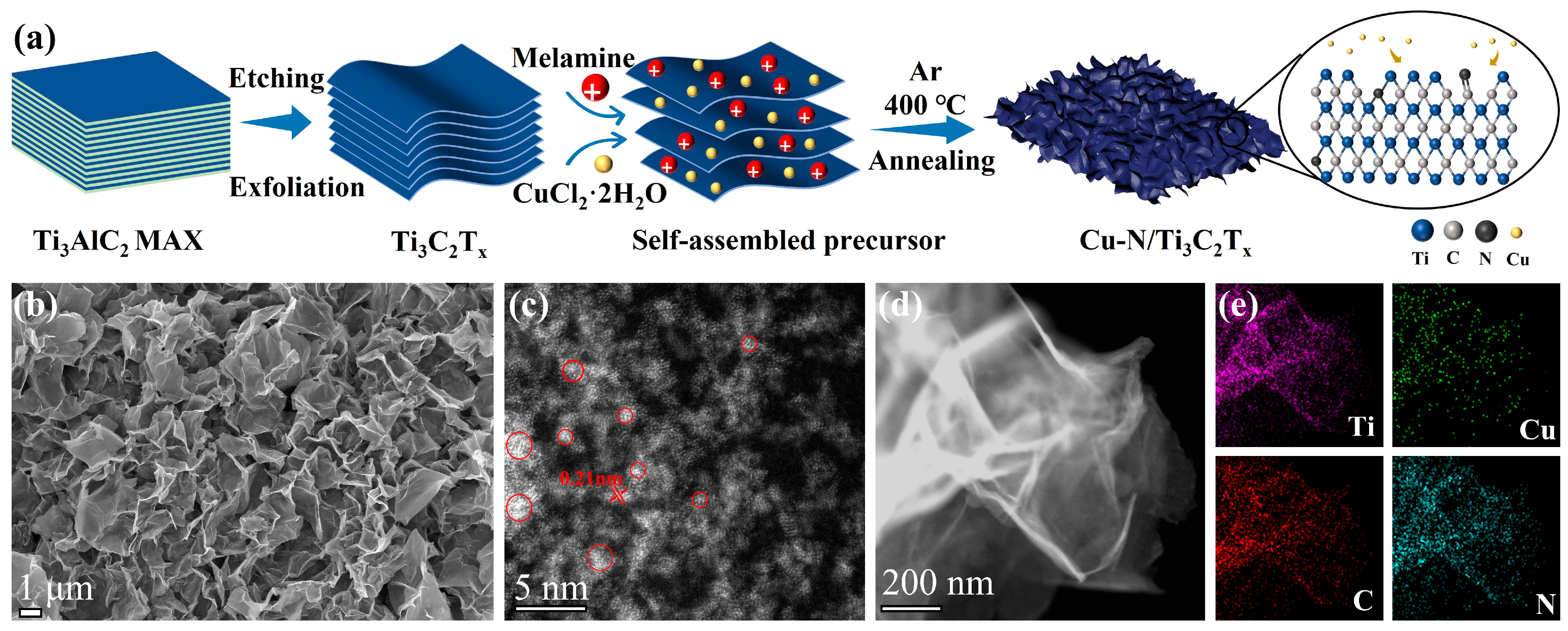
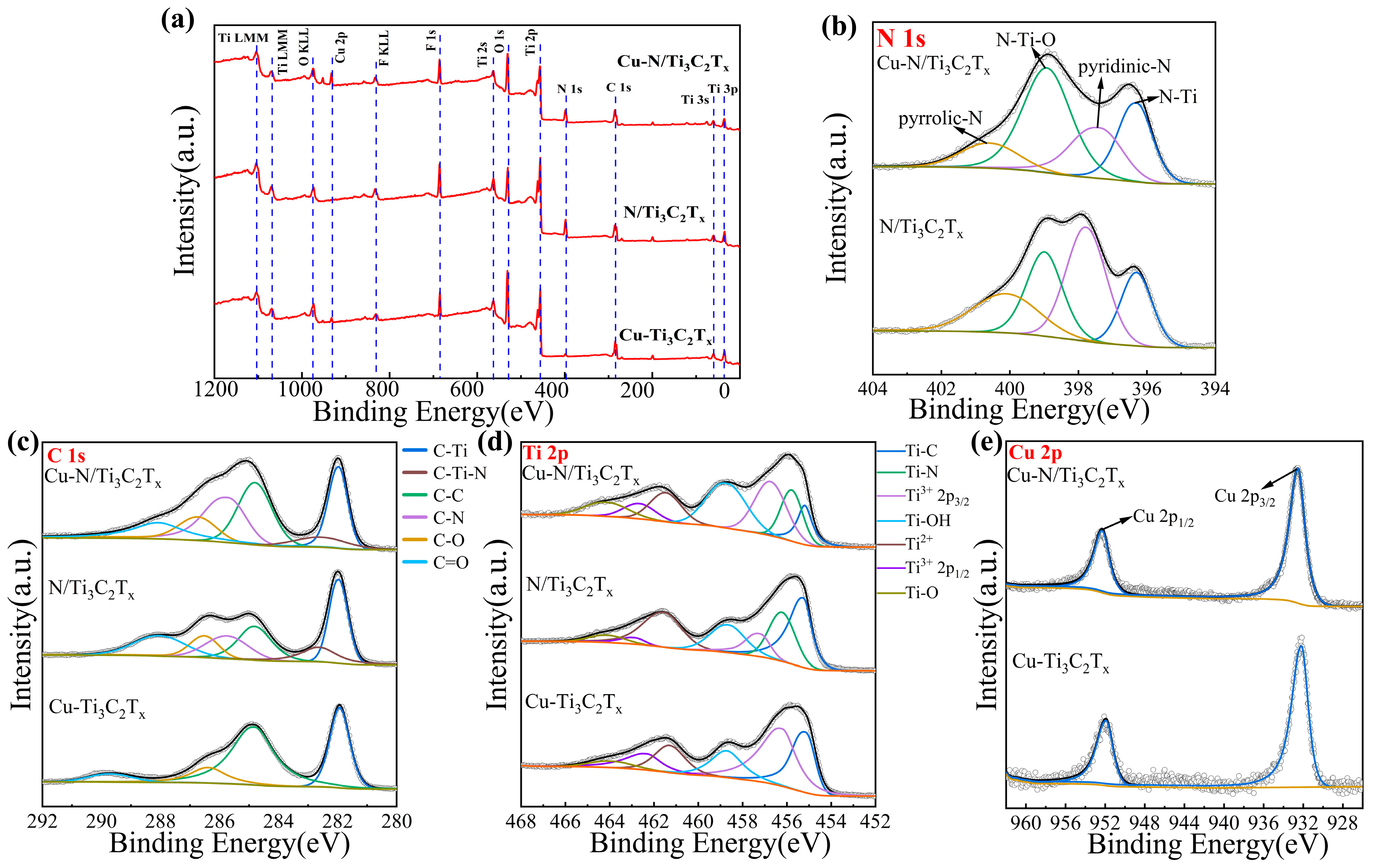
3.1. Characterization of Cu-N/Ti3C2Tx

The synthesis of crumpled N-doped Cu nanoclusters-modified Ti3C2Tx MXene was carried out in a three-step process, as shown in Figure 1a. Firstly, the Al layer in the Ti3AlC2 MAX phase was selectively etched away by HF generated by mixing LiF/HCl, and an oligo- or monolayer Ti3C2Tx colloidal suspension was formed by ultrasonication and high-speed centrifugation [28,41]. Subsequently, CuCl2·2H2O was added to the Ti3C2Tx suspension. Attributed to the presence of two low-valence Ti species (Ti2+/Ti3+) of Ti3C2Tx, a self-reduction process of copper (II) ions occurred during continuous stirring [42,43,44]. Due to the negative charge of Ti3C2Tx MXene, the added protonated melamine, which acts as a nitrogen source and separator, adsorbed onto MXene nanosheets to undergo electrostatic self-assembly [45]. The precursor was freeze-dried and then annealed at high temperature. And the thermal decomposition of melamine could introduce N doping and result in a crumpled structure to avoid the stacking of MXene, which finally leads to obtaining Cu-N/Ti3C2Tx. As a comparison experiment, only CuCl2·2H2O and protonated melamine were added to the Ti3C2Tx, respectively, and after the same treatment, Cu cluster-modified Ti3C2Tx (Cu-Ti3C2Tx) and surface strain-modulated wrinkled N-doped Ti3C2Tx (N/Ti3C2Tx) were obtained by annealing.
The synthetic materials were characterized using an X-ray diffractometer (XRD). Figure S1a shows that the sharp characteristic peak (002) of the diffractogram is moved to a lower angle, and the peak of Ti3AlC2 at 2θ = 39° disappears. This indicates that the Al layer was removed and the Ti3AlC2 MAX phase etching was successful in forming the Ti3C2Tx crystal structure [46,47]. The XRD patterns of Cu-N/Ti3C2Tx and Cu-Ti3C2Tx (Figure S1b) show a similar crystal structure to Ti3C2Tx, with no corresponding peaks in Cu-associated microcrystals observed, suggesting that the Cu species are well dispersed. The surface strain of Cu-N/Ti3C2Tx was observed by using FESEM. And Figure 1b shows the 3D crumpled structure after the introduction of strain, which is favorable to provide more active sites along with improved electronic conductivity. The HAADF-STEAM images confirmed the presence of Cu nanoclusters dispersed on the Ti3C2Tx (Figure 1c). Cu nanoclusters show sharp bright spots because Cu has a larger atomic number than Ti. Also, the lattice spacing of the Cu nanoclusters on Ti3C2Tx is 0.21 nm, which is attributed to the (111) face of metallic Cu [48]. EDS images (Figure 1d,e) showed that Cu and N elements were homogeneously distributed on the Ti3C2Tx substrate. The Cu loading of 3.0 and 2.6 wt% for Cu-N/Ti3C2Tx and Cu-Ti3C2Tx, respectively, as measured by ICP-AES, was attributed to the introduction of surface strain to expose more active sites.
The elemental composition and structure of the three synthetic materials were further investigated through XPS characterization. As shown in Figure 2a, the survey spectra of XPS show the presence of C, N, Ti, and Cu, indicating that Cu and N are doped onto the Ti3C2Tx MXene. Figure 2b shows the N 1s spectra of Cu-N/Ti3C2Tx and N/Ti3C2Tx, revealing pyridinic N and pyrrolic N, which are two exemplary N-doped structures [49]. Due to the interaction between Cu nanoclusters and lattice N resulting in a positive shift in binding energy, pyrrolic N of Cu-N/Ti3C2Tx (400.6 eV) is 0.5 eV larger than pyrrolic N of N/Ti3C2Tx (400.1 eV) [50]. And the binding energy peaks at 396.3 eV and 398.9 eV correspond to N-Ti and N-Ti-O bonds, respectively, demonstrating the successful substitution of N for lattice carbon. The C 1s spectra of Cu-N/Ti3C2Tx, N/Ti3C2Tx, and Cu-Ti3C2Tx are shown in Figure 2c. The binding energy peaks of C-Ti, C=C, C-O, and C=O bonds of Cu-Ti3C2Tx are 281.9, 284.8, 286.4, and 289.7 eV, respectively. The formation of C-Ti-N bonds (282.6 eV) and C-N bonds (285.7 eV) results in two more characteristic peaks for Cu-N/Ti3C2Tx and N/Ti3C2Tx than for Cu-Ti3C2Tx [51]. The O 1s spectra (Figure S2) indicate that the binding energy peaks of the C-Ti-Ox bond and the Ti-OH bond are 530.1 and 531.6 eV, respectively [45]. Figure 2d shows the Ti 2p spectra of Cu-N/Ti3C2Tx, revealing the binding energy peaks of the Ti-C bond (455.2 eV), Ti3+ 2p3/2 (456.7 eV), Ti3+ 2p1/2 (462.7 eV), the Ti-OH bond (458.7 eV), Ti2+ (461.5 eV), and the Ti-O bond (464.1 eV) [52]. Furthermore, the additional binding energy peak of 455.8 eV compared to Cu-Ti3C2Tx is attributed to the Ti-N bond, which corresponds to the results observed in the N 1s spectra. The Cu 2p spectra of Cu-N/Ti3C2Tx and Cu-Ti3C2Tx (Figure 2e) detected Cu 2p3/2 (932.5 eV) and Cu 2p1/2 (952.3) peaks, which are attributed to metallic Cu (Cu0), demonstrating the successful anchoring of Cu nanoclusters [53]. Meanwhile, the characterization results can prove the absence of CuO, as there are no obvious Cu2+ satellite peaks between the binding energy peaks 940 and 950 eV [50].

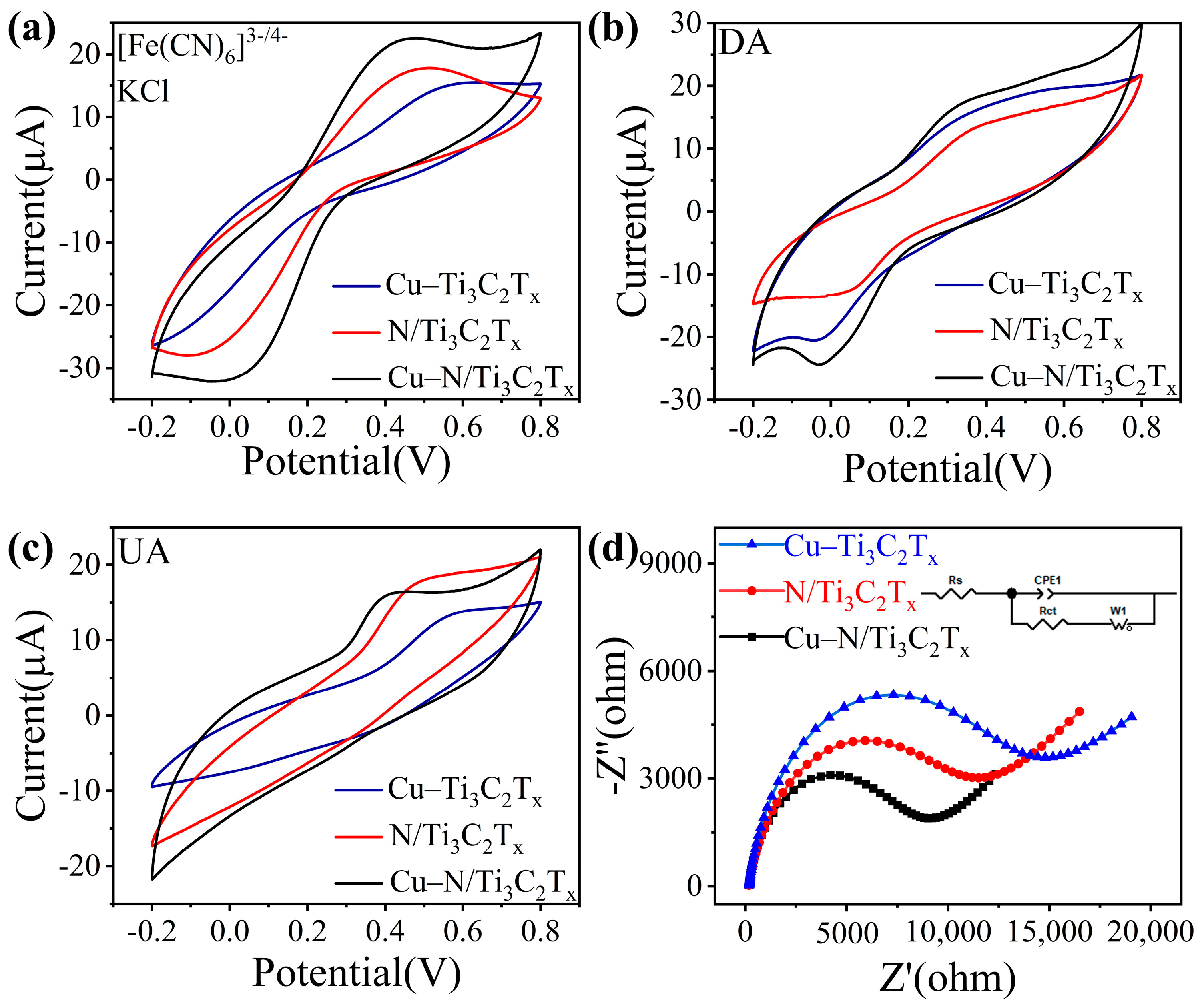
3.2. Analysis of Electrochemical Sensing Responses to DA and UA

Firstly, the CV responses of Cu-N/Ti3C2Tx, Cu-Ti3C2Tx, and N/Ti3C2Tx modified electrodes were tested in 5 mM [Fe(CN)6]3-/4- and 0.1 M KCl solutions. As shown in Figure 3a, the intensities of peak currents exhibited the following trend: Cu-N/Ti3C2Tx > N/Ti3C2Tx > Cu-Ti3C2Tx. Due to the doping of Cu nanoclusters and more active sites exposed by the surface strain-modulated 3D crumpled structure, Cu-N/Ti3C2Tx had higher peak currents, suggesting improved electrochemical activity. The effective active area of its electrochemistry was calculated based on the CV curves (Figure S3) with variable sweep speed and the Randles-Sevick equation:
I P = 2.69 × 10 5 A D 1 / 2 n 3 / 2 γ 1 / 2 C
where A is the calculated electrochemical active area (cm2) of the electrode, Ip and γ are the peak current and scan rate of the CV curves in Figure S3, and the parameters D, n, and C are the diffusion coefficient of the redox probe (6.7 × 10⁻⁶ cm2·s−1), the number of electrons transferred during the reaction, and the concentration of the redox probe (5 × 10⁻⁶ mol·cm−3) [54]. Subsequently, the three Cu-N/Ti3C2Tx, Cu-Ti3C2Tx, and N/Ti3C2Tx modified electrodes were compared by CV response in phosphate-buffered saline (PBS) (pH = 7.4, 0.1 M) containing 0.5 mM DA and 0.5 mM UA. The redox mechanisms of DA and UA are as follows: the redox reaction of DA exhibits the loss of 2 H+ and 2 e from dopamine to produce o-dopaminoquinone; the loss of 2 H+ and 2 e during the oxidation of UA results in its oxidation to dehydroxyuric acid. In Figure 3b, the CV response currents of Cu-N/Ti3C2Tx were higher than those of the other two, which indicated that Cu nanoclusters anchored to N-doped crumpled MXene have the best promotion effect on DA catalysis. The oxidation peak currents of Cu-N/Ti3C2Tx and N/Ti3C2Tx in Figure 3c were significantly higher than those of Cu-Ti3C2Tx, suggesting that N doping contributed to the enhancement of the electrochemical performance of MXene [55,56]. Cu-N/Ti3C2Tx exhibited superior catalytic performance for DA and UA, attributed to the anchoring of Cu nanoclusters and the N doping to enhance the electrochemical performance. Next, the relationship between the redox peak currents of Cu-N/Ti3C2Tx in DA and UA and scan rate was explored, as shown in Figure S4a,c, which show the CV response curves for a gradient change in scan rate from 10 to 100 mV/s. Figure S4b,d show the linear relationship between the peak current and the scan rate for DA and UA, respectively, indicating that their electrochemical sensing is a diffusion-controlled process [57,58]. Electrochemical impedance spectroscopy (EIS) can effectively represent the electron transfer process at the electrode surface. Figure 3d shows the Nyquist curves of the three modified electrodes in 0.1 M KCl with 5 mM [Fe(CN)6]3−/4−. By analyzing the equivalent circuit fitting (shown in the inset of Figure 3d), the interfacial charge transfer resistance (Rct) can be obtained as 5345 Ω (Cu-N/Ti3C2Tx), 9192 Ω (N/Ti3C2Tx), and 10,655 Ω (Cu-Ti3C2Tx). Figure S5a,b show the Nyquist plots in DA and UA, respectively, where Cu-N/Ti3C2Tx also has the smallest interfacial transfer resistance (3895 Ω for DA, 5579 Ω for UA). In comparison to the other two electrodes, the Rct values of Cu-N/Ti3C2Tx showed significantly lower Rct values, while the ones in EIS showed smaller semicircle diameters, indicating better electron transfer rate and electrocatalytic activity. The results show that the enhanced electrochemical sensing of DA and UA by Cu-N/Ti3C2Tx may be attributed to the fact that, on the one hand, the doping of Cu nanoclusters plays a significant role in enhancing the electrocatalytic activity. On the other hand, the strain modulation suggests that the 3D crumpled structure can provide richer active sites.

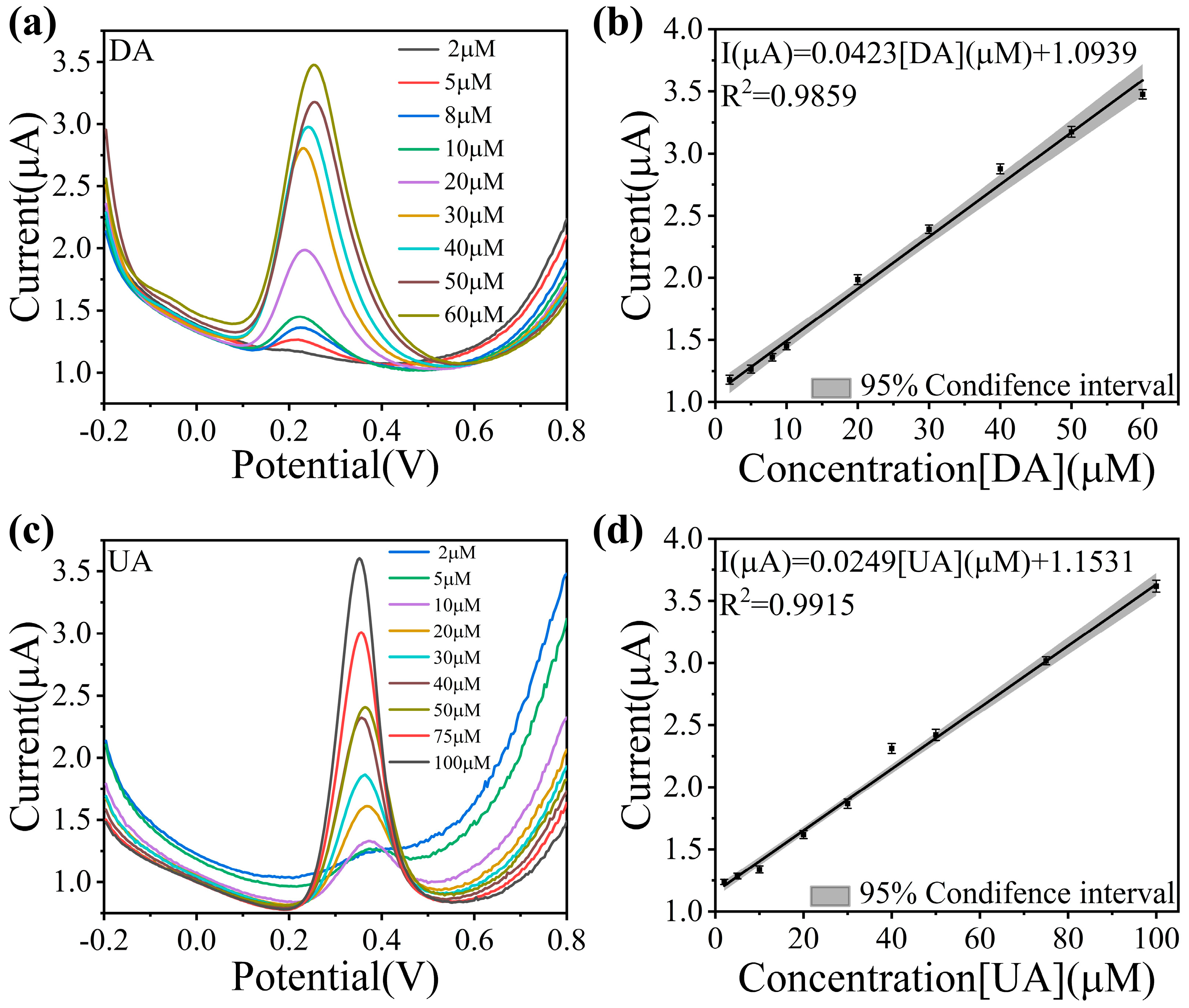
3.3. Analysis of Electrochemical Sensing Performance to DA and UA

Previous tests have demonstrated the excellent performance of Cu-N/Ti3C2Tx. To further investigate its sensing and detection ability for DA and UA, the response of Cu-N/Ti3C2Tx to different concentrations of DA and UA was investigated by CV and DPV. The results for DA were shown in Figure 4a and Figure S6a. The peak currents increased with the increase in DA concentration, with a good linear relationship between the two (Figure 4b). The gray area in Figure 4b represents the 95% confidence interval, indicating that there is a 95% probability that the true average peak response current falls within this range for a given concentration, supporting the reliability of the linear model. Based on the results of the DPV test, the linear correlation equation was plotted in the calibration graph:
I μ A = 0.0423 C D A μ M + 1.0939 R 2 = 0.9859
The same excellent sensing performance is present in the UA tests shown in Figure 4c and Figure S6b, with a good linearity of the peak currents in the range of 5–100 μM (Figure 4d) and a linear correlation equation of:
I μ A = 0.0249 C U A μ M + 1.1531 R 2 = 0.9915
Taking into account the above experimental results and data, the Cu-N/Ti3C2Tx modified electrode had good detection performance for DA and UA with highly sensitive response (1958.3 and 1152.7 μA·mM−1·cm−2 for DA and UA) and very low detection limit (0.0584 and 0.0993 μM for DA and UA). In comparison with other analog sensors, the Cu-N/Ti3C2Tx exhibits very low detection limits for detecting DA and UA (Table S1).
The fast response time is an important indicator of the sensor detection performance, which requires the sensor to have an extremely high sensitivity to the active substance. The I-t transient tests for DA and UA are shown in Figure 5a,b. The Cu-N/Ti3C2Tx reaches 95% of the steady-state current within 1.1 s and 1.4 s, respectively, indicating high transient responsiveness to DA and UA. Interference immunity is also a key capability for sensor performance testing. The strong adsorption and oxidizing activity of ascorbic acid (AA) are common sources of interference in electrochemical sensors. The high concentration of glucose in body fluids tends to lead to nonspecific adsorption. Na+ and K+ may alter the structure of the bilayer by electrostatic interaction, affecting the efficiency of electron transfer. Divalent Ca2+ and Zn2+ have a stronger charge shielding effect and may exacerbate charge rearrangement on the sensor surface. As shown in Figure 5c, a comparison of the DPV response of the Cu-N/Ti3C2Tx modified electrode in CaCl2, KCl, NaCl, ZnCl2, AA, and glucose verified that there was no response current to interfering substances. Figure 5d shows the DPV response currents of the sensor to DA and UA in the presence of different interfering substances, with RSDs of 2.31% (DA) and 1.96% (UA), which proves that the Cu-N/Ti3C2Tx has an extremely excellent anti-interference capability. Figure 5e shows the CV curves of the same Cu-N/Ti3C2Tx modified electrode cycled for 10 and 20 times, and the RSDs of the oxidation-reduction currents were 1.58% and 1.66%, respectively, confirming that Cu-N/Ti3C2Tx has good repeatability. Cu-N/Ti3C2Tx shows excellent reproducibility in Figure S7: the six modified electrodes were tested in 50 μM DA and 100 μM UA. In addition, stability is a key indicator of the sensor’s performance. To investigate the stability of Cu-N/Ti3C2Tx, the prepared electrodes were stored at room temperature for 15 days in total. During this period, the peak current under the same conditions was recorded every day. The results are shown in Figure 5f; the electrode still maintains about 94.4% of the sensing performance after 7 days, but the sensing performance decreases significantly after the 10th day, and the current attenuation reaches 14.3% by the 15th day of the test. The sensor maintains excellent stability over a period of 7 days. In addition, a cross-concentration control experiment was designed in order to test the simultaneous synchronization of the DA and UA detection. As shown in Figure S8, the oxidation peak current of DA showed a significant linear increase with its concentration when the UA concentration was kept static at 50 μM, while the fluctuation of the oxidation peak position response value of UA was small. Conversely, UA showed concentration-dependent enhancement when the DA concentration was kept static at 20 μM. The results showed that the oxidation processes of DA and UA on the electrode surface followed independent electron transfer paths when the two components coexisted, which proved that Cu-N/Ti3C2Tx modified electrode possessed excellent synchronous detection characteristics. Lastly, the performance of Cu-N/Ti3C2Tx was tested in real samples by DPV using the standard spiking recovery method. Human urine samples were spiked with 0.1 M PBS (pH = 7.4) diluted to 1000 times and spiked with known different concentrations (10, 30, and 50 μM) of DA and UA, respectively. The recovery rates for DA and UA calculated from the DPV responses of three independent experiments were 98.2–103.9% and 98.9–101.2%, respectively (Table 1), indicating good feasibility of the detection in real samples.

4. Conclusions

In this study, a high-efficiency electrochemical sensor based on Cu nanoclusters anchored on nitrogen-doped crumpled Ti3C2Tx MXene was successfully developed for the parallel detection of DA and UA. The synergistic integration of 3D crumpled architecture and N-doping effectively mitigated MXene restacking, exposed abundant active sites, and enhanced ion accessibility, while Cu nanoclusters further amplified electrocatalytic activity by accelerating electron transfer. Structural characterization confirmed the crumpled structure induced by surface strain and the uniform dispersion of Cu nanoclusters (3.0 wt%), which collectively optimized conductivity and surface reactivity. The Cu-N/Ti3C2Tx sensor demonstrated remarkable dual-analyte detection capabilities, achieving ultrahigh sensitivity (1958.3 and 1152.7 μA·mM⁻1·cm⁻2 for DA and UA, respectively), ultralow detection limits (0.058 and 0.099 μM), and rapid response times (<1.5 s). Crucially, the sensor exhibited exceptional selectivity against common interferents (e.g., ascorbic acid) and maintained 94.4% stability after 7 days, underscoring its robustness for practical applications. Differential pulse voltammetry validated independent linear detection ranges (2~60 μM for DA, 5~100 μM for UA), enabling accurate quantification in complex biofluids. This work advances the design of MXene-based nanocomposites for biomolecular sensing and provides a simple and convenient non-enzymatic platform for real-time monitoring of DA and UA.

Supplementary Materials

The following supporting information can be downloaded at https://www.mdpi.com/article/10.3390/s25082508/s1: Figure S1: XRD patterns of different materials, Figure S2: O 1s high-resolution XPS spectra, Figure S3: Relationship between scan rate and peak oxidation current for Cu-N/Ti3C2Tx in 0.1 M KCl and 5 mM [Fe(CN)6]3−/4−, Figure S4: Relationship between scan rate and peak current for Cu-N/Ti3C2Tx in DA and UA, Figure S5: The Nyquist plots of EIS in DA and UA, Figure S6: CV curves for Cu-N/Ti3C2Tx at different concentrations of DA and UA, Figure S7: Response currents of Cu-N/Ti3C2Tx modified six electrodes, Figure S8: Simultaneous detection performance of DA and UA by Cu-N/Ti3C2Tx, Table S1: Detection performance comparison of previously reported analogues and this work. References [59,60,61,62,63,64,65,66,67].

Author Contributions

H.Y. and B.Z. conceived the project. H.Y. and C.R. conducted the experiments. H.Y., C.R., and S.G. contributed to material characterization. H.Y., C.R., and B.Z. wrote the manuscript. T.W., B.Z., and F.-Z.X. supervised this project. All authors discussed and commented on the manuscript. All authors have read and agreed to the published version of the manuscript.

Funding

This work was supported by the National Natural Science Foundation of China (Grant No. 52422505, No. 12274124), the Shanghai Pilot Program for Basic Research (Grant No. 22TQ1400100-6), the Fundamental Research Funds for the Central Universities, and the Innovative Research Group Project of the National Natural Science Foundation of China (Grant No. 52321002), the National Natural Science Foundation of China (Grant No. 62301314).

Institutional Review Board Statement

Not applicable.

Informed Consent Statement

Not applicable.

Data Availability Statement

Data are contained within the article.

Conflicts of Interest

The authors declare no conflict of interest.

References

  1. Sriram, B.; Kogularasu, S.; Wang, S.-F.; Sheu, J.-K. Deep Eutectic Solvent-Mediated Synthesis of Spinel Zinc Chromite Nanoparticles: A Simple Label-Free Electrochemical Sensor for Dopamine and Ascorbic Acid. ACS Appl. Nano Mater. 2023, 6, 17593–17602. [Google Scholar] [CrossRef]
  2. Shukla, R.P.; Aroosh, M.; Matzafi, A.; Ben-Yoav, H. Partially Functional Electrode Modifications for Rapid Detection of Dopamine in Urine. Adv. Funct. Mater. 2021, 31, 2004146. [Google Scholar] [CrossRef]
  3. Shi, Z.; Wu, X.; Zou, Z.; Yu, L.; Hu, F.; Li, Y.; Guo, C.; Li, C.M. Screen-Printed Analytical Strip Constructed with Bacteria-Templated Porous N-Doped Carbon Nanorods/Au Nanoparticles for Sensitive Electrochemical Detection of Dopamine Molecules. Biosens. Bioelectron. 2021, 186, 113303. [Google Scholar] [CrossRef]
  4. Hu, F.X.; Hu, T.; Chen, S.; Wang, D.; Rao, Q.; Liu, Y.; Dai, F.; Guo, C.; Yang, H.B.; Li, C.M. Single-Atom Cobalt-Based Electrochemical Biomimetic Uric Acid Sensor with Wide Linear Range and Ultralow Detection Limit. Nano-Micro Lett. 2021, 13, 7. [Google Scholar] [CrossRef] [PubMed]
  5. Feng, J.; Li, Q.; Cai, J.; Yang, T.; Chen, J.; Hou, X. Electrochemical Detection Mechanism of Dopamine and Uric Acid on Titanium Nitride-Reduced Graphene Oxide Composite with and without Ascorbic Acid. Sens. Actuators B Chem. 2019, 298, 126872. [Google Scholar] [CrossRef]
  6. Sriram, B.; Shajahan, S.; Hsu, Y.; Wang, S.-F.; Haija, M.A. Electrochemical Behavior of a Copper Tungstate-Fabricated Disposable Strip for the Rapid and Real-Time Detection of Uric Acid. ACS Appl. Nano Mater. 2023, 6, 1–9. [Google Scholar] [CrossRef]
  7. Xie, X.; Wang, D.P.; Guo, C.; Liu, Y.; Rao, Q.; Lou, F.; Li, Q.; Dong, Y.; Li, Q.; Yang, H.B.; et al. Single-Atom Ruthenium Biomimetic Enzyme for Simultaneous Electrochemical Detection of Dopamine and Uric Acid. Anal. Chem. 2021, 93, 4916–4923. [Google Scholar] [CrossRef]
  8. Wu, Y.; Deng, P.; Tian, Y.; Feng, J.; Xiao, J.; Li, J.; Liu, J.; Li, G.; He, Q. Simultaneous and Sensitive Determination of Ascorbic Acid, Dopamine and Uric Acid via an Electrochemical Sensor Based on PVP-Graphene Composite. J. Nanobiotechnol. 2020, 18, 112. [Google Scholar] [CrossRef]
  9. Mei, H.; Peng, J.; Wang, T.; Zhou, T.; Zhao, H.; Zhang, T.; Yang, Z. Overcoming the Limits of Cross-Sensitivity: Pattern Recognition Methods for Chemiresistive Gas Sensor Array. Nano-Micro Lett. 2024, 16, 269. [Google Scholar] [CrossRef]
  10. Li, Q.; Qiu, Y.; Han, W.; Zheng, Y.; Wang, X.; Xiao, D.; Mao, M.; Li, Q. Determination of Uric Acid in Biological Samples by High Performance Liquid Chromatography-Electrospray Ionization-Tandem Mass Spectrometry and Study on Pathogenesis of Pulmonary Arterial Hypertension in Pulmonary Artery Endothelium Cells. RSC Adv. 2018, 8, 25808–25814. [Google Scholar] [CrossRef]
  11. Nie, Z.; Long, X.; Bouroubi, N.E.H.; Liu, H.; Cao, S.; Chen, Y.; Zheng, X.; Xia, J. Biosynthesis and Detection of Domoic Acid from Diatom Pseudo-Nitzschia: A Review. Curr. Pharm. Biotechnol. 2023, 24, 599–610. [Google Scholar] [CrossRef] [PubMed]
  12. Wang, T.; Zhang, H.; Wu, Y.; Jiang, W.; Chen, X.; Zeng, M.; Yang, J.; Su, Y.; Hu, N.; Yang, Z. Target Discrimination, Concentration Prediction, and Status Judgment of Electronic Nose System Based on Large-Scale Measurement and Multi-Task Deep Learning. Sens. Actuators B Chem. 2022, 351, 130915. [Google Scholar] [CrossRef]
  13. Ma, Z.; Xu, Y.; Li, P.; Cheng, D.; Zhu, X.; Liu, M.; Zhang, Y.; Liu, Y.; Yao, S. Self-Catalyzed Surface Reaction-Induced Fluorescence Resonance Energy Transfer on Cysteine-Stabilized MnO2 Quantum Dots for Selective Detection of Dopamine. Anal. Chem. 2021, 93, 3586–3593. [Google Scholar] [CrossRef]
  14. Kim, J.; Park, H.; Ryu, J.; Jeon, O.; Paeng, I.R. COMPETITIVE ENZYME-LINKED IMMUNOSORBENT ASSAY FOR A SELECTIVE AND SENSITIVE DETERMINATION OF DOPAMINE IN THE PRESENCE OF ASCORBIC ACID AND URIC ACID. J. Immunoass. Immunochem. 2009, 31, 33–44. [Google Scholar] [CrossRef]
  15. Balkourani, G.; Brouzgou, A.; Tsiakaras, P. A Review on Recent Advancements in Electrochemical Detection of Dopamine Using Carbonaceous Nanomaterials. Carbon 2023, 213, 118281. [Google Scholar] [CrossRef]
  16. Ni, W.; Wang, T.; Wu, Y.; Liu, X.; Li, Z.; Yang, R.; Zhang, K.; Yang, J.; Zeng, M.; Hu, N.; et al. Multi-Task Deep Learning Model for Quantitative Volatile Organic Compounds Analysis by Feature Fusion of Electronic Nose Sensing. Sens. Actuators B Chem. 2024, 417, 136206. [Google Scholar] [CrossRef]
  17. Qin, Z.; Tang, B.; Zhang, G.; Zhu, C.; Jiang, K.; Zhang, B.; Xuan, F.-Z. Single-Atom Ni-N4 for Enhanced Electrochemical Sensing. Nano Res. 2024, 17, 7658–7664. [Google Scholar] [CrossRef]
  18. Zhao, R.; Wang, T.; Li, J.; Shi, Y.; Hou, M.; Yang, Y.; Zhang, Z.; Lei, S. Two-Dimensional Covalent Organic Frameworks for Electrocatalysis: Achievements, Challenges, and Opportunities. Nano Res. 2023, 16, 8570–8595. [Google Scholar] [CrossRef]
  19. Matsoso, B.J.; Lerotholi, T.; Coville, N.J.; Jones, G. Designing Parameters of Surfactant-Free Electrochemical Sensors for Dopamine and Uric Acid on Nitrogen Doped Graphene Films: Evaluating the Correlation between the Content of N-Configurations and the Sensing for Electrochemically Active Molecules. Johns. Matthey Technol. Rev. 2019, 63, 76–88. [Google Scholar] [CrossRef]
  20. Azizpour Moallem, Q.; Beitollahi, H. Electrochemical Sensor for Simultaneous Detection of Dopamine and Uric Acid Based on a Carbon Paste Electrode Modified with Nanostructured Cu-Based Metal-Organic Frameworks. Microchem. J. 2022, 177, 107261. [Google Scholar] [CrossRef]
  21. Zhu, J.; Chen, L.; Ni, W.; Cheng, W.; Yang, Z.; Xu, S.; Wang, T.; Zhang, B.; Xuan, F. NiO/ZnO Nanocomposites for Multimodal Intelligent MEMS Gas Sensors. ACS Sens. 2025, acssensors.4c02789. (in press). [Google Scholar] [CrossRef]
  22. Yue, J.-Y.; Song, L.-P.; Wang, Y.-T.; Yang, P.; Ma, Y.; Tang, B. Fluorescence/Colorimetry/Smartphone Triple-Mode Sensing of Dopamine by a COF-Based Peroxidase-Mimic Platform. Anal. Chem. 2022, 94, 14419–14425. [Google Scholar] [CrossRef]
  23. Xin, M.; Li, J.; Ma, Z.; Pan, L.; Shi, Y. MXenes and Their Applications in Wearable Sensors. Front. Chem. 2020, 8, 297. [Google Scholar] [CrossRef] [PubMed]
  24. Shekhirev, M.; Shuck, C.E.; Sarycheva, A.; Gogotsi, Y. Characterization of MXenes at Every Step, from Their Precursors to Single Flakes and Assembled Films. Prog. Mater. Sci. 2021, 120, 100757. [Google Scholar] [CrossRef]
  25. Pei, Y.; Zhang, X.; Hui, Z.; Zhou, J.; Huang, X.; Sun, G.; Huang, W. Ti3C2TX MXene for Sensing Applications: Recent Progress, Design Principles, and Future Perspectives. ACS Nano 2021, 15, 3996–4017. [Google Scholar] [CrossRef] [PubMed]
  26. Chu, T.; Wang, G.; Zhang, X.; Jia, Y.; Dai, S.; Liu, X.; Zhang, L.; Yang, X.; Zhang, B.; Xuan, F.-Z. High-Density Dual-Structure Single-Atom Pt Electrocatalyst for Efficient Hydrogen Evolution and Multimodal Sensing. Nano Lett. 2024, 24, 9666–9674. [Google Scholar] [CrossRef]
  27. Rong, C.; Su, T.; Li, Z.; Chu, T.; Zhu, M.; Yan, Y.; Zhang, B.; Xuan, F.-Z. Elastic Properties and Tensile Strength of 2D Ti3C2Tx MXene Monolayers. Nat Commun 2024, 15, 1566. [Google Scholar] [CrossRef]
  28. Chen, Y.; Xie, X.; Xin, X.; Tang, Z.-R.; Xu, Y.-J. Ti3C2Tx-Based Three-Dimensional Hydrogel by a Graphene Oxide-Assisted Self-Convergence Process for Enhanced Photoredox Catalysis. ACS Nano 2019, 13, 295–304. [Google Scholar] [CrossRef]
  29. Mao, X.; Qin, Z.; Ge, S.; Rong, C.; Zhang, B.; Xuan, F. Strain Engineering of Electrocatalysts for Hydrogen Evolution Reaction. Mater. Horiz. 2023, 10, 340–360. [Google Scholar] [CrossRef]
  30. Zhang, X.; Miao, J.; Zhang, P.; Zhu, Q.; Jiang, M.; Xu, B. 3D Crumbled MXene for High-Performance Supercapacitors. Chin. Chem. Lett. 2020, 31, 2305–2308. [Google Scholar] [CrossRef]
  31. Jiang, Y.; Liang, P.; Tang, M.; Sun, S.; Min, H.; Han, J.; Shen, X.; Yang, H.; Chao, D.; Wang, J. A High-Throughput Screening Permeability Separator with High Catalytic Conversion Kinetics for Li–S Batteries. J. Mater. Chem. A 2022, 10, 22080–22092. [Google Scholar] [CrossRef]
  32. Bhamore, J.R.; Jha, S.; Mungara, A.K.; Singhal, R.K.; Sonkeshariya, D.; Kailasa, S.K. One-Step Green Synthetic Approach for the Preparation of Multicolor Emitting Copper Nanoclusters and Their Applications in Chemical Species Sensing and Bioimaging. Biosens. Bioelectron. 2016, 80, 243–248. [Google Scholar] [CrossRef] [PubMed]
  33. Mahmoudi, E.; Hajian, A.; Rezaei, M.; Afkhami, A.; Amine, A.; Bagheri, H. A Novel Platform Based on Graphene Nanoribbons/Protein Capped Au-Cu Bimetallic Nanoclusters: Application to the Sensitive Electrochemical Determination of Bisphenol A. Microchem. J. 2019, 145, 242–251. [Google Scholar] [CrossRef]
  34. Das, N.K.; Ghosh, S.; Priya, A.; Datta, S.; Mukherjee, S. Luminescent Copper Nanoclusters as a Specific Cell-Imaging Probe and a Selective Metal Ion Sensor. J. Phys. Chem. C 2015, 119, 24657–24664. [Google Scholar] [CrossRef]
  35. Wang, D.; Nie, Y.; Wang, P.; Ma, Q. In Situ Synthesis of Cu Nanoclusters/CeO2 Nanorod as Aggregated Induced ECL Probe for Triple-Negative Breast Cancer Detection. Talanta 2023, 258, 124400. [Google Scholar] [CrossRef]
  36. Li, Q.; Wang, X.; Chen, L.; Liu, X.; Ma, J.; Wang, L.; Wang, W. Cu/Cu2O Nanoparticles Modified Ti3C2 MXene with in-Situ Formed TiO2-X for Detection of Hydrogen Peroxide. Ceram. Int. 2023, 49, 9632–9641. [Google Scholar] [CrossRef]
  37. Li, Q.; Fu, S.; Wang, X.; Wang, L.; Liu, X.; Gao, Y.; Li, Q.; Wang, W. Electrochemical and Photoelectrochemical Detection of Hydrogen Peroxide Using Cu2O/Cu Nanowires Decorated with TiO2− x Deriving from MXenes. ACS Appl. Mater. Interfaces 2022, 14, 57471–57480. [Google Scholar] [CrossRef]
  38. Nasrollahpour, H.; Sánchez, B.J.; Sillanpää, M.; Moradi, R. Metal Nanoclusters in Point-of-Care Sensing and Biosensing Applications. ACS Appl. Nano Mater. 2023, 6, 12609–12672. [Google Scholar] [CrossRef]
  39. Han, Y.; Zhang, P.; Duan, X.; Gao, X.; Gao, L. Advances in Precise Synthesis of Metal Nanoclusters and Their Applications in Electrochemical Biosensing of Disease Biomarkers. Nanoscale 2025, 17, 3616–3634. [Google Scholar] [CrossRef]
  40. Chu, T.; Rong, C.; Zhou, L.; Mao, X.; Zhang, B.; Xuan, F. Progress and Perspectives of Single-Atom Catalysts for Gas Sensing. Adv. Mater. 2023, 35, 2206783. [Google Scholar] [CrossRef]
  41. Shahzad, F.; Alhabeb, M.; Hatter, C.B.; Anasori, B.; Man Hong, S.; Koo, C.M.; Gogotsi, Y. Electromagnetic Interference Shielding with 2D Transition Metal Carbides (MXenes). Science 2016, 353, 1137–1140. [Google Scholar] [CrossRef]
  42. Zhang, Q.; Wang, J.; Yu, Q.; Li, Q.; Fan, R.; Li, C.; Fan, Y.; Zhao, C.; Cheng, W.; Ji, P.; et al. Metal/MXene Composites via in Situ Reduction. Nat. Synth 2025, 4, 252–261. [Google Scholar] [CrossRef]
  43. Xie, S.; Shu, J.; Huang, H.; Li, J.; Yue, R.; Xu, J. Engineering the Highly Efficient Heterogeneous Catalyst Based on PdCu Nanoalloy and Nitrogen-Doped Ti3C2Tx MXene for Ethanol Electrooxidation. J. Colloid Interface Sci. 2023, 639, 314–323. [Google Scholar] [CrossRef] [PubMed]
  44. Jin, L.; You, S.; Yao, Y.; Chen, H.; Wang, Y.; Liu, Y. An Electroactive Single-Atom Copper Anchored MXene Nanohybrid Filter for Ultrafast Water Decontamination. J. Mater. Chem. A 2021, 9, 25964–25973. [Google Scholar] [CrossRef]
  45. Bao, W.; Liu, L.; Wang, C.; Choi, S.; Wang, D.; Wang, G. Facile Synthesis of Crumpled Nitrogen-Doped MXene Nanosheets as a New Sulfur Host for Lithium–Sulfur Batteries. Adv. Energy Mater. 2018, 8, 1702485. [Google Scholar] [CrossRef]
  46. Li, Z.; Yu, L.; Milligan, C.; Ma, T.; Zhou, L.; Cui, Y.; Qi, Z.; Libretto, N.; Xu, B.; Luo, J.; et al. Two-Dimensional Transition Metal Carbides as Supports for Tuning the Chemistry of Catalytic Nanoparticles. Nat. Commun. 2018, 9, 5258. [Google Scholar] [CrossRef]
  47. Fang, Y.; Yang, X.; Chen, T.; Xu, G.; Liu, M.; Liu, J.; Xu, Y. Two-Dimensional Titanium Carbide (MXene)-Based Solid-State Electrochemiluminescent Sensor for Label-Free Single-Nucleotide Mismatch Discrimination in Human Urine. Sens. Actuators B Chem. 2018, 263, 400–407. [Google Scholar] [CrossRef]
  48. Li, Z.; Wang, P.; Liang, Z.; Wang, D.; Ma, Q. Cu NCs@MXene-Luminescent Faraday Cage and Cu Nanocone/Bi NPs Array-Based Saliva Exosome Assay for Asthma Evaluation. Chem. Eng. J. 2024, 493, 152650. [Google Scholar] [CrossRef]
  49. Lu, C.; Yang, L.; Yan, B.; Sun, L.; Zhang, P.; Zhang, W.; Sun, Z. Nitrogen-Doped Ti3C2 MXene: Mechanism Investigation and Electrochemical Analysis. Adv. Funct Mater. 2020, 30, 2000852. [Google Scholar] [CrossRef]
  50. Gu, H.; Yue, W.; Hu, J.; Niu, X.; Tang, H.; Qin, F.; Li, Y.; Yan, Q.; Liu, X.; Xu, W.; et al. Asymmetrically Coordinated Cu–N1C2 Single-Atom Catalyst Immobilized on Ti3C2Tx MXene as Separator Coating for Lithium–Sulfur Batteries. Adv. Energy Mater. 2023, 13, 2204014. [Google Scholar] [CrossRef]
  51. Liu, H.; Hu, Z.; Liu, Q.; Sun, P.; Wang, Y.; Chou, S.; Hu, Z.; Zhang, Z. Single-Atom Ru Anchored in Nitrogen-Doped MXene (Ti3C2Tx) as an Efficient Catalyst for the Hydrogen Evolution Reaction at All pH Values. J. Mater. Chem. A 2020, 8, 24710–24717. [Google Scholar] [CrossRef]
  52. Ramalingam, V.; Varadhan, P.; Fu, H.; Kim, H.; Zhang, D.; Chen, S.; Song, L.; Ma, D.; Wang, Y.; Alshareef, H.N.; et al. Heteroatom-Mediated Interactions between Ruthenium Single Atoms and an MXene Support for Efficient Hydrogen Evolution. Adv. Mater. 2019, 31, 1903841. [Google Scholar] [CrossRef] [PubMed]
  53. Shi, Y.; Han, F.; Xie, L.; Zhang, C.; Li, T.; Wang, H.; Lai, W.-F.; Luo, S.; Wei, W.; Wang, Z.; et al. A MXene of Type Ti3C2Tx Functionalized with Copper Nanoclusters for the Fluorometric Determination of Glutathione. Microchim. Acta 2020, 187, 38. [Google Scholar] [CrossRef]
  54. Rajendrachari, S.; Basavegowda, N.; Vinaykumar, R.; Narsimhachary, D.; Somu, P.; Lee, M.-J. Electrocatalytic Determination of Methyl Orange Dye Using Mechanically Alloyed Novel Metallic Glass Modified Carbon Paste Electrode by Cyclic Voltammetry. Inorg. Chem. Commun. 2023, 155, 111010. [Google Scholar] [CrossRef]
  55. Yoon, Y.; Lee, M.; Kim, S.K.; Bae, G.; Song, W.; Myung, S.; Lim, J.; Lee, S.S.; Zyung, T.; An, K. A Strategy for Synthesis of Carbon Nitride Induced Chemically Doped 2D MXene for High-Performance Supercapacitor Electrodes. Adv. Energy Mater. 2018, 8, 1703173. [Google Scholar] [CrossRef]
  56. Jiang, D.; Wei, M.; Du, X.; Qin, M.; Shan, X.; Chen, Z. One-Pot Synthesis of ZnO Quantum Dots/N-Doped Ti3C2 MXene: Tunable Nitrogen-Doping Properties and Efficient Electrochemiluminescence Sensing. Chem. Eng. J. 2022, 430, 132771. [Google Scholar] [CrossRef]
  57. Zhang, X.; Yan, W.; Zhang, J.; Li, Y.; Tang, W.; Xu, Q. NiCo-Embedded in Hierarchically Structured N-Doped Carbon Nanoplates for the Efficient Electrochemical Determination of Ascorbic Acid, Dopamine, and Uric Acid. RSC Adv. 2015, 5, 65532–65539. [Google Scholar] [CrossRef]
  58. Ma, Y.; Wei, Z.; Wang, Y.; Ding, Y.; Jiang, L.; Fu, X.; Zhang, Y.; Sun, J.; Zhu, W.; Wang, J. Surface Oxygen Functionalization of Carbon Cloth toward Enhanced Electrochemical Dopamine Sensing. ACS Sustain. Chem. Eng. 2021, 9, 16063–16072. [Google Scholar] [CrossRef]
  59. Zhang, W.; Li, X.; Liu, X.; Song, K.; Wang, H.; Wang, J.; Li, R.; Liu, S.; Peng, Z. A Novel Electrochemical Sensor Based on Pd Confined Mesoporous Carbon Hollow Nanospheres for the Sensitive Detection of Ascorbic Acid, Dopamine, and Uric Acid. Molecules 2024, 29, 2427. [Google Scholar] [CrossRef]
  60. You, Q.; Guo, Z.; Zhang, R.; Chang, Z.; Ge, M.; Mei, Q.; Dong, W.-F. Simultaneous Recognition of Dopamine and Uric Acid in the Presence of Ascorbic Acid via an Intercalated MXene/PPy Nanocomposite. Sensors 2021, 21, 3069. [Google Scholar] [CrossRef]
  61. Yao, W.; Guo, H.; Liu, H.; Li, Q.; Wu, N.; Li, L.; Wang, M.; Fan, T.; Yang, W. Highly Electrochemical Performance of Ni-ZIF-8/ N S-CNTs/CS Composite for Simultaneous Determination of Dopamine, Uric Acid and L-Tryptophan. Microchem. J. 2020, 152, 104357. [Google Scholar] [CrossRef]
  62. Wang, Y.; Zhao, P.; Gao, B.; Yuan, M.; Yu, J.; Wang, Z.; Chen, X. Self-Reduction of Bimetallic Nanoparticles on Flexible MXene-Graphene Electrodes for Simultaneous Detection of Ascorbic Acid, Dopamine, and Uric Acid. Microchem. J. 2023, 185, 108177. [Google Scholar] [CrossRef]
  63. Shang, L.; Li, R.; Li, H.; Yu, S.; Sun, X.; Yu, Y.; Ren, Q. The Simultaneous Detection of Dopamine and Uric Acid In Vivo Based on a 3D Reduced Graphene Oxide–MXene Composite Electrode. Molecules 2024, 29, 1936. [Google Scholar] [CrossRef] [PubMed]
  64. Murugan, N.; Jerome, R.; Preethika, M.; Sundaramurthy, A.; Sundramoorthy, A.K. 2D-Titanium Carbide (MXene) Based Selective Electrochemical Sensor for Simultaneous Detection of Ascorbic Acid, Dopamine and Uric Acid. J. Mater. Sci. Technol. 2021, 72, 122–131. [Google Scholar] [CrossRef]
  65. Liu, K.; Chen, Y.; Dong, X.; Huang, H. Simultaneous Voltammetric Determination of Dopamine and Uric Acid Based on MOF-235 Nanocomposite. Inorg. Chem. Commun. 2022, 142, 109584. [Google Scholar] [CrossRef]
  66. Jia, D.; Yang, T.; Wang, K.; Wang, H.; Wang, E.; Chou, K.-C.; Hou, X. Ti3C2Tx Coated with TiO2 Nanosheets for the Simultaneous Detection of Ascorbic Acid, Dopamine and Uric Acid. Molecules 2024, 29, 2915. [Google Scholar] [CrossRef]
  67. Gong, J.; Tang, H.; Wang, M.; Lin, X.; Wang, K.; Liu, J. Novel Three-Dimensional Graphene Nanomesh Prepared by Facile Electro-Etching for Improved Electroanalytical Performance for Small Biomolecules. Mater. Des. 2022, 215, 110506. [Google Scholar] [CrossRef]
Figure 1. Schematic synthesis process and microstructure characterizations of Cu-N/Ti3C2Tx. (a) Schematic synthesis of Cu-N/Ti3C2Tx. (b) SEM image. (c) Atomic-resolution HAADF-STEM image (the red circles mark the Cu nanoclusters). (d) HAADF-STEM image and (e) EDS mapping images (Ti, purple; Cu, green; C, red; N, blue) of Cu-N/Ti3C2Tx.
Sensors 25 02508 g001
Figure 2. XPS analysis results of Cu-N/Ti3C2Tx, N/Ti3C2Tx, and Cu-Ti3C2Tx. (a) XPS survey spectra of Cu-N/Ti3C2Tx, N/Ti3C2Tx, and Cu/Ti3C2Tx. (b) N 1s high-resolution XPS spectra. (c) C 1s high-resolution XPS spectra. (d) Ti 2p high-resolution XPS spectra. (e) Cu 2p high-resolution XPS spectra.
Sensors 25 02508 g002
Figure 3. Electrochemical responses. CV curves of Cu-N/Ti3C2Tx, N/Ti3C2Tx, and Cu-Ti3C2Tx in 0.1 M KCl with (a) 5 mM [Fe(CN)6]3−/4−, and in PBS (pH = 7.4, 0.1 M) with (b) 0.5 mM DA, (c) 0.5 mM UA. (d) The Nyquist plots of EIS at Cu-N/Ti3C2Tx, N/Ti3C2Tx, and Cu-Ti3C2Tx in 0.1 M KCl with 5 mM [Fe(CN)6]3−/4− electrolyte.
Sensors 25 02508 g003
Figure 4. Electrochemical sensing performance. (a) DPV curves for Cu-N/Ti3C2Tx at different concentrations of DA. (b) The linear relationship between the peak current responses and the DA concentration. (c) DPV curves for Cu-N/Ti3C2Tx at different concentrations of UA. (d) The linear relationship between the peak current responses and the UA concentration.
Sensors 25 02508 g004
Figure 5. Response time, selectivity, repeatability, and stability of electrochemical sensing. Instantaneous response time of Cu-N/Ti3C2Tx to (a) DA and (b) UA. (c) DPV responses of Cu-N/Ti3C2Tx for DA, UA, and other interferences. (d) Influence of multiple interfering substances on the effects of simultaneous detection of 50 μM DA and UA. (e) Stability of Cu-N/Ti3C2Tx after 10 and 20 cycles of scanning in 0.1 mM DA and UA. (f) The oxidation peak current on the same Cu-N/Ti3C2Tx at 15 days of storage at room temperature.
Sensors 25 02508 g005
Table 1. Cu-N/Ti3C2Tx sensor results for spiked DA and UA in human urine samples.
AnalyteConcentration Added (µM)Found (µM) aRecovery (%)RSD (%)
DA1010.39103.93.27
3029.4598.22.41
5051.19102.43.68
UA1010.12101.21.92
3029.7199.14.01
5049.4698.91.88
a Mean of three independent experiments.
Disclaimer/Publisher’s Note: The statements, opinions and data contained in all publications are solely those of the individual author(s) and contributor(s) and not of MDPI and/or the editor(s). MDPI and/or the editor(s) disclaim responsibility for any injury to people or property resulting from any ideas, methods, instructions or products referred to in the content.

