Article

SERS-Based Immunochromatographic Assay for Sensitive Detection of Escherichia coli O157:H7 Using a Novel WS2-AuDTNB Nanotag

1
College of Chemistry and Life Sciences, Beijing University of Technology, No. 100 Pingleyuan, Beijing 100124, China
2
Chinese Academy of Inspection and Quarantine, No. A3, Gaobeidian Road, Beijing 100123, China
*
Authors to whom correspondence should be addressed.
Sensors 2025, 25(8), 2457; https://doi.org/10.3390/s25082457
Submission received: 2 March 2025 / Revised: 27 March 2025 / Accepted: 1 April 2025 / Published: 14 April 2025
(This article belongs to the Special Issue Molecular Opto-Electronic Sensing Devices and Techniques)

Abstract

:

Highlights

What are the main findings?
  • Developed a novel WS2-Au nanocomposite coupled with 5,5′-dithiobis-(2-nitrobenzoic acid) (DTNB) and antibodies as sensitive surface-enhanced Raman scattering (SERS) nanotags for E. coli O157:H7 detection.
  • Demonstrated a broad linear detection range (8 × 10²–8 × 10⁷ CFU/mL), a low detection limit (175 CFU/mL), high specificity and good accuracy in real food samples (milk, pork).
What are the implications of the main findings?
  • A rapid assay for qualitative and quantitative detection of E. coli O157:H7, overcoming limitations of traditional ICA methods.
  • A cost-effective, user-friendly surface-enhanced Raman scattering (SERS)-based immunochromatographic assay (SERS-ICA) platform with great potential for point-of-care testing and detection of other foodborne pathogens.

Abstract

E. coli O157:H7 contamination in food and the environment poses a serious threat to human health. Rapid and sensitive identification of foodborne pathogens remains challenging. Here, we prepared tungsten disulfide (WS2)–Au nanocomposites coupled with the Raman signal molecule 5,5′-dithio-bis-(2-nitrobenzoic acid) (DTNB) and antibodies to replace the conventional colloidal gold nanoparticles and applied SERS-active nanotags in the SERS-ICA method for highly sensitive detection of E. coli O157:H7. The large surface area and numerous effective SERS hotspots of WS2-Au nanotags provide superior SERS signals. Under optimized conditions, this ICA achieves the quantitative detection of E. coli O157:H7 in a broad linear range of 8 × 10²–8 × 10⁷ CFU/mL and at a low detection limit of 175 CFU/mL. In addition, the test strip indicates high specificity for E. coli O157:H7 identification, favorable reproducibility, and shows good accuracy in the detection of actual food samples, such as milk and pork. The proposed assay can be used for rapid qualitative and quantitative detection of E. coli O157:H7 and has great potential for field application.

1. Introduction

Escherichia coli O157:H7 (E. coli O157:H7), a significant Shiga toxin-producing E. coli serotype, is frequently associated with outbreaks. It is prone to contaminating everyday foodstuffs and is transmitted among humans via the fecal–oral transmission route [1,2]. Humans become infected through direct contact with animals harboring the pathogen, their feces, contaminated soil or water, or by consuming undercooked meat products, as well as dairy products, vegetables, and fruits that have been tainted. E. coli O157:H7 infections are highly pathogenic and lethal, capable of causing diarrhea and complications such as hemolytic uremic syndrome, severe hemorrhagic diarrhea, and thrombotic thrombocytopenic purpura, posing severe challenges to food hygiene and human health [3,4]. The infective dose of E. coli O157:H7 is very low, necessitating strict control in food products [5]. The gold-standard methods for identifying E. coli O157:H7 encompass culture isolation, selective enrichment, biochemical identification, and serological typing, which typically require 2–3 days for detection [6]. Modern bacterial detection methods, including polymerase chain reaction (PCR) [7], DNA sequencing [8], and enzyme-linked immunosorbent assay (ELISA) [9], can deliver accurate and sensitive results. However, these methods often involve complex sample preparation steps and rely on sophisticated laboratory equipment, specialized laboratory conditions, and skilled personnel, limiting their suitability for rapid, point-of-care testing [10,11]. Therefore, the development of simple, rapid, and sensitive detection methods for E. coli O157:H7 in food samples holds significant scientific and practical importance.
Immunochromatographic assay (ICA) is favored for notable advantages, such as its low cost, ease of operation, rapid detection, providing visible results to the naked eye, and the capability for on-site testing [12,13,14,15]. However, traditional ICA typically utilizes gold nanoparticles as colorimetric labels and relies on visual interpretation for result determination, which is limited by insufficient sensitivity and poor quantitative detection capabilities. To address these limitations, researchers have introduced surface-enhanced Raman scattering (SERS) label-based technology into ICA systems. This integration provides powerful and stable SERS signals, thereby significantly enhancing the accuracy, sensitivity, and quantitative capabilities of the assay. SERS-ICA offers several benefits, including a lower detection limit and the ability to perform quantitative and multiplex detection of various analytes [16,17,18]. In conventional SERS immunochromatography, SERS nanotags modified with specific antibodies bind to the target analytes through antigen–antibody-specific reactions. Highly sensitive quantitative detection of target analytes is achieved by analyzing the characteristic peak intensity of the Raman reporter molecules on the detection line. SERS nanotags primarily consist of three parts: SERS enhancement substrates, Raman reporter molecules, and biomolecular recognition molecules [19,20]. The analytical performance of the SERS-ICA method greatly depends on the SERS enhancement substrates used. The key to developing new SERS nanotags lies in creating stable SERS hotspots to enhance the SERS signals and improve detection sensitivity.
In SERS-ICA, traditional spherical nanomaterials have the disadvantages of easy agglomeration, a low loading capacity, and poor fluidity. In recent years, transition metal dichalcogenides (TMDCs), such as MoS2, WS2, and MoSe2, have been applied in the field of electrochemical, fluorescent, and colorimetric biosensors [21,22,23]. As a typical semiconducting TMDC, WS2 possesses many specific characteristics, including a layered structure, a large specific surface area, excellent electronic and optoelectronic properties, and a large direct bandgap, which holds great potential for SERS applications [24,25,26]. The SERS signal of TMDCs is principally attributed to charge transfer processes between the SERS materials and the adsorbed molecules, as well as interfacial dipole–dipole interactions. The enhancement of the SERS signal is dominated by chemical mechanisms [27]. Due to the resonance of near-field and far-field optical properties of TMDCs, the Raman spectral intensity can be enhanced by a factor of 102 through chemical enhancement. Heterostructures and TMDC–precious metal nanocomposites fully co-ordinate the electromagnetic and chemical enhancement mechanisms on the surface of noble metal nanostructures, making them highly promising for developing SERS substrates. Some TMDC–metal heterostructures, such as Ag@WS2, AuNPs/MoS2/graphene, and AuNPs@DA@MoS2, exhibit significant SERS enhancement effects [28,29,30,31].
In this study, we proposed WS2-Au SERS labels and incorporated them into SERS-ICA for the ultrasensitive and quantitative determination of the common foodborne bacteria E. coli O157:H7 in food samples. In the WS2-Au nanocomposites, WS2 nanosheets were utilized as a support material for AuNPs, providing abundant anchoring sites and thus efficiently capturing the target pathogens. The strong coupling between WS2 and Au nanostructures through surface plasmon excitation could be beneficial to further enhance the effectiveness of SERS [32,33,34]. Through conditional optimization, the established assay enabled the sensitive identification of E. coli O157:H7 using a single SERS-ICA strip within 20 min. The limit of detection (LOD) for E. coli O157:H7 was 175 CFU/mL. The proposed WS2-Au-based SERS-ICA demonstrated high reproducibility and reliability in real food samples, such as milk and pork. Hence, the WS2-Au-based SERS-ICA holds significant promise for the rapid and sensitive detection of bacteria in the POCT field.

2. Materials and Methods

2.1. Reagents and Equipment

Nitrocellulose (NC) membranes (Sartorius Stedim UniSart CN140, Sartorius, Göttingen, Germany), sample pads (Millipore GFCP20300, Millipore, Billerica, MA, USA), absorbent pads (Millipore CFSP223000, Millipore, Billerica, MA, USA), and polyvinyl chloride (PVC) base plates were purchased from Shanghai Jinbiao Biotechnology Co., Ltd., Shanghai, China. Tetrachloroaurate (III) trihydrate (HAuCl4·3H2O, 98%), trisodium citrate (Na3C6H5O7·2H2O, 99.8%), sodium dihydrogen phosphate (NaH2PO4, 99%), potassium dihydrogen phosphate (KH2PO4, 99%), potassium carbonate (K2CO3, 99.0%), sodium chloride (NaCl, 99%), potassium chloride (KCl, 99%), borax (Na2B4O7·10H2O, 99.5%), boric acid (H3BO3, AR), bovine serum albumin (BSA, 98%), and Proclin (ProclinTM300) were bought from Sigma Corporation, St. Louis, MO, USA. Monolayer WS2 nanosheets were bought from Beike 2D materials Co., Ltd., Beijing, China. E. coli O157:H7 monoclonal antibody (mAb) and goat anti-mouse IgG antibody were provided by our laboratory. 5,5′-dithiobis-(2-nitrobenzoic acid) (DTNB, 99%) and Tween-20 were obtained from Aladdin Reagent Co., Ltd., Shanghai, China. All other chemicals were of analytical grade. Food samples, including milk and pork, were purchased from the local supermarket.
A UV-Vis spectrophotometer (U-3310, Hitachi Ltd., Tokyo, Japan) was used to obtain UV–visible absorption spectra. A transmission electron microscope (JEM-2100F, 200 kV; JEOL Ltd., Tokyo, Japan) was used to acquire TEM images. Elemental mapping of the substrates was performed using an energy-dispersive X-ray analyzer (7593-H; Horiba Ltd., Northampton, UK). A three-dimensional spraying instrument (XYZ 3050; Biodot Ltd., Irvine, CA, USA) was used to spray the antibody onto the NC membrane, and a programmable cutting machine (CM 4000, Biodot Ltd., Irvine, CA, USA) was used to cut the test strips. The SERS intensity was measured by a laser confocal microscope Raman spectrometer (InVia Reflex; Renishaw, UK), recording laser excitation wavelengths under 785 nm, with a diffraction grating of 1200 grooves/mm, an exposure time of 4 s, a laser output power of 1 mW, and 50× objective lens focusing.

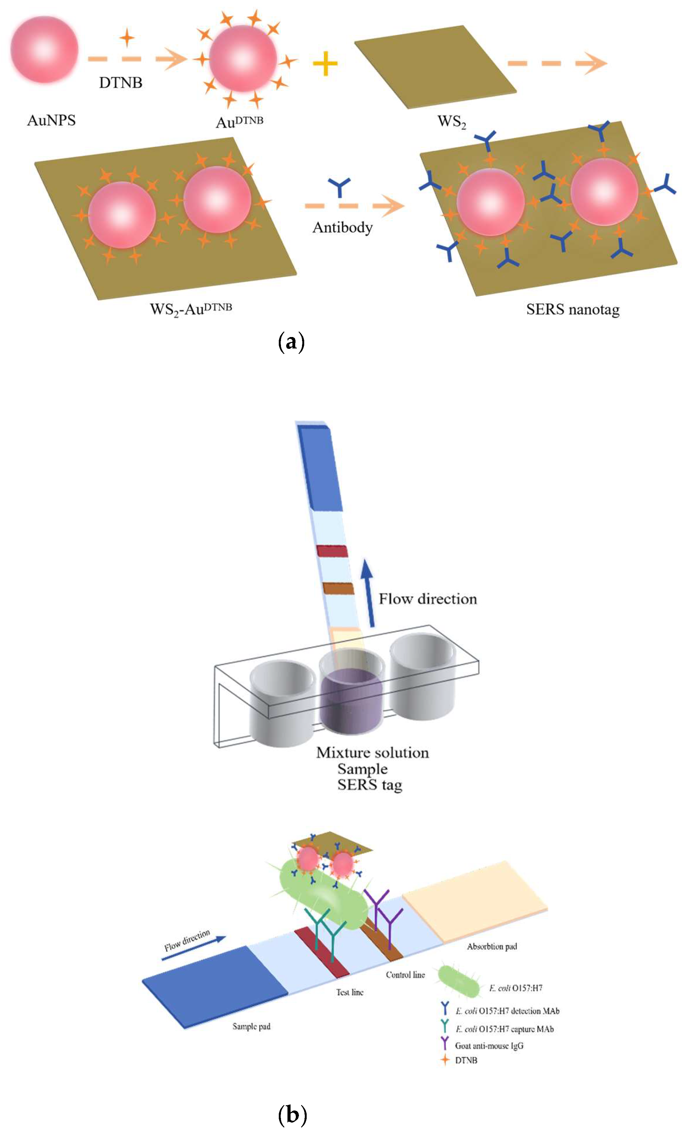
2.2. Synthesis of WS2-AuDTNB Nanotags

The preparation process of the SERS nanotags is illustrated in Figure 1a. AuNPs were synthesized through a modified trisodium citrate reduction method [35]. Briefly, 1 mL of 1% (w/v) HAuCl4·3H2O solution was added to 99 mL of deionized water and stirred in a microwave synthesizer. When the temperature rose to 98 °C, it was maintained stably for 2 min, after which 1.2 mL of 1% (w/v) trisodium citrate solution was rapidly added. The solution was then boiled for an additional 5 min. Once the solution turned wine-red, it was removed from the heat, cooled to room temperature, and then stored in a 4 °C refrigerator. Next, the Raman reporter molecule DTNB (20 μL, 10 mM) was added to 10 mL of the prepared AuNP solution and stirred continuously for 2 h at room temperature. Excess DTNB was removed by centrifugation (12,000 rpm, 10 min), and the precipitate was resuspended in 2 mL of ultrapure water. Subsequently, 2 mL of AuDTNB solution was mixed with 2 mL WS2 nanosheets (1 mg/mL), and the mixture was ultrasonicated for 30 min, followed by overnight stirring. The formed WS2-Au nanocomposites were centrifuged (12,000 rpm, 10 min) and resuspended in 2 mL of ultrapure water.

2.3. Preparation of Antibody-Modified WS2-AuDTNB Nanotags

A quantity of 10 µL of 0.05 M K2CO3 was added to the 1 mL prepared WS2-AuDTNB solution to adjust the pH, then incubated with the 0.2 mg/mL E. coli O157:H7 detection monoclonal antibody (mAb) for 30 min. The mixture was added with 100 μL of 10 % BSA (w/v) and vortexed for 30 min to block the unreacted carboxyl sites on the surface of the SERS nanotags. Finally, antibody-modified WS2-AuDTNB SERS nanotags were collected by centrifugation (12,000 rpm, 10 min) and resuspended in 100 μL of antibody preservation solution containing 0.1 M boric acid-buffered solution, 10% (w/v) BSA, and proclin.

2.4. SERS-ICA Test Strip Assembly

E. coli O157:H7 capture antibody and goat anti-mouse IgG antibody were sprayed onto the NC membrane to form the detection line (T-line) and the control line (C-line), respectively. The sprayed NC membrane was then dried overnight in an oven at 37 °C. The sample pad was treated with 0.01 M Tris solution (containing 0.5% Tween-20 (w/v), pH 8.0) and subsequently dried. To assemble the ICA strips, the treated NC membrane was affixed to the center of the PVC backing plate, with the sample pad and absorbent pad placed on either side of the NC membrane. The assembled card was cut into 4 mm wide strips using the CM400 programmable cutting machine and stored in a desiccant-sealed bag.

2.5. WS2-AuDTNB-Based ICA Strips for Analysis of E. coli O157:H7

Standard E. coli O157:H7 bacterial strains were provided by the Testing and Evaluation Center’s laboratory at the Chinese Academy of Inspection and Quarantine. Different concentrations of standard bacterial solutions, ranging from 8 × 10² to 8 × 10⁷ CFU/mL, were diluted in blocking solution. Then, 6 μL of immunoprobe (WS2-AuDTNB) was mixed with 100 μL of E. coli O157:H7 standard solution in a 96-well plate, and the strips were inserted for the reaction. As the reaction proceeded, the T-line and C-line began to show a dark-purple color visible to the naked eye, and the entire reaction process was completed within 20 min. The SERS signals arising from the DTNB embedded in the immunoprobe captured by the detection antibody on the T-line could be measured with the confocal microscopic Raman spectrometer.

2.6. Spiked Sample Preparation and Detection

Milk and pork were used to evaluate the practical application of this WS2-Au based SERS-ICA. E. coli O157:H7 standard solutions were spiked into pork and milk to prepare 8 × 10⁴ to 8 × 10⁶ CFU/mL spiked samples. For pork samples, 50 µL of E. coli O157:H7 solution was added to 4 g of minced meat, followed by the addition of 4 mL of PBS buffer. The mixture was vortexed for 10 min on a shaker and then centrifuged (10,000 rpm, 10 min) to obtain the supernatant. For milk samples, 10 µL of E. coli O157:H7 solution was mixed with 990 µL of milk and thoroughly mixed. These spiked samples were tested using the WS2-Au based ICA, adhering to the previously outlined analytical method.

3. Results

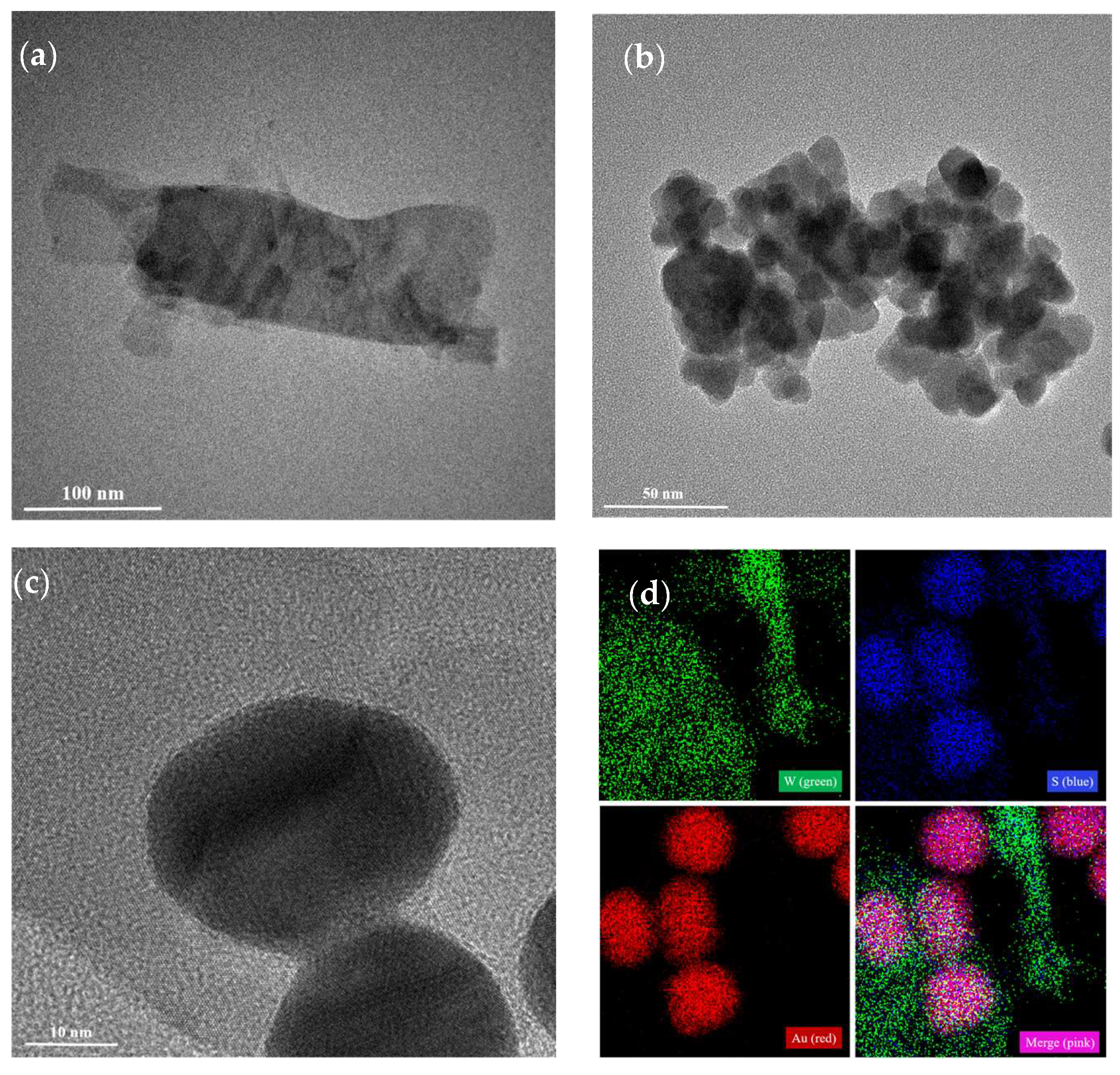
3.1. Characterization of Au, WS2-AuNPs, and SERS Nanotags

The formation of the WS2-Au nanocomposites was based on the Hard and Soft Acids and Bases (HSAB) theory, according to which the soft sulfur surface layer of WS2 binds with the soft acid Au nanoparticles [36,37]. The morphologies and structures of the WS2-Au nanocomposites were investigated by TEM. The AuNPs possessed a uniform spherical morphology, with an average size of 26.65 nm, as measured from TEM images using ImageJ 2025 software (Figure S1). As shown in Figure 2a, the monolayer WS2 nanosheet exhibited a thin layer of a wrinkled structure with a large surface area and good dispersion. In the case of the WS2-Au nanocomposites (Figure 2b,c), the Au nanoparticles were integrated onto the surface of the WS2 nanosheets. Figure 2d exhibits the element mapping results of the WS2-Au nanocomposites; the distribution of W (green), S (blue), and Au (red) on the nanosheet and their merging were demonstrated clearly. The UV-Vis absorbance spectra were measured to characterize the localized surface plasmon resonance (LSPR) properties of the AuNPs and WS2-AuDTNB, as shown in Figure S2. The ultraviolet absorption at 529 nm corresponds to the strong LSPR band of the Au nanoparticles. The absorption spectrum of WS2 shows broad absorption in the visible region, primarily due to exciton transitions in WS2. For WS2-Au, the absorption peak displayed a modest red shift and an expansion of the absorption peak [38]. The above results demonstrate the successful formation of the WS2-Au nanocomposites. Based on the Tauc plot of WS2 shown in Figure S3, the band gap of the WS2 nanosheet was estimated to be 2.09 eV.
The SERS activities of the Au and WS2-Au were investigated by DTNB. As revealed in Figure S4, many more fold-enhanced Raman signals of 10−6 M DTNB were detected on the WS2-Au SERS platform than those of Au nanoparticles. This enhancement was attributed to the synergistic effect of the chemical enhancement provided by the WS2 nanosheets and the electromagnetic enhancement between Au and WS2.

3.2. Construction of WS2-Au-Based ICA for Determination of E. coli O157:H7

The principle of the SERS-ICA test strips for detecting E. coli O157:H7, based on the double-antibody sandwich method, is schematically illustrated in Figure 1b. The WS2-AuDTNB nanotags were mixed and bound with the E. coli O157:H7 sample solution in the microwell to form a complex and then dipped into test strips. Under the action of capillary chromatography, the formed nanotag–E. coli O157:H7 antigen complex was captured by the immobilized antibody as it flowed to the T-line, while the remaining uncaptured complexes continued to flow to the C-line and were captured by the goat anti-mouse antibody. Ultimately, the targets could be qualitatively observed through color changes in the test zone and quantitatively analyzed by measuring the SERS signal intensity on the T-line.

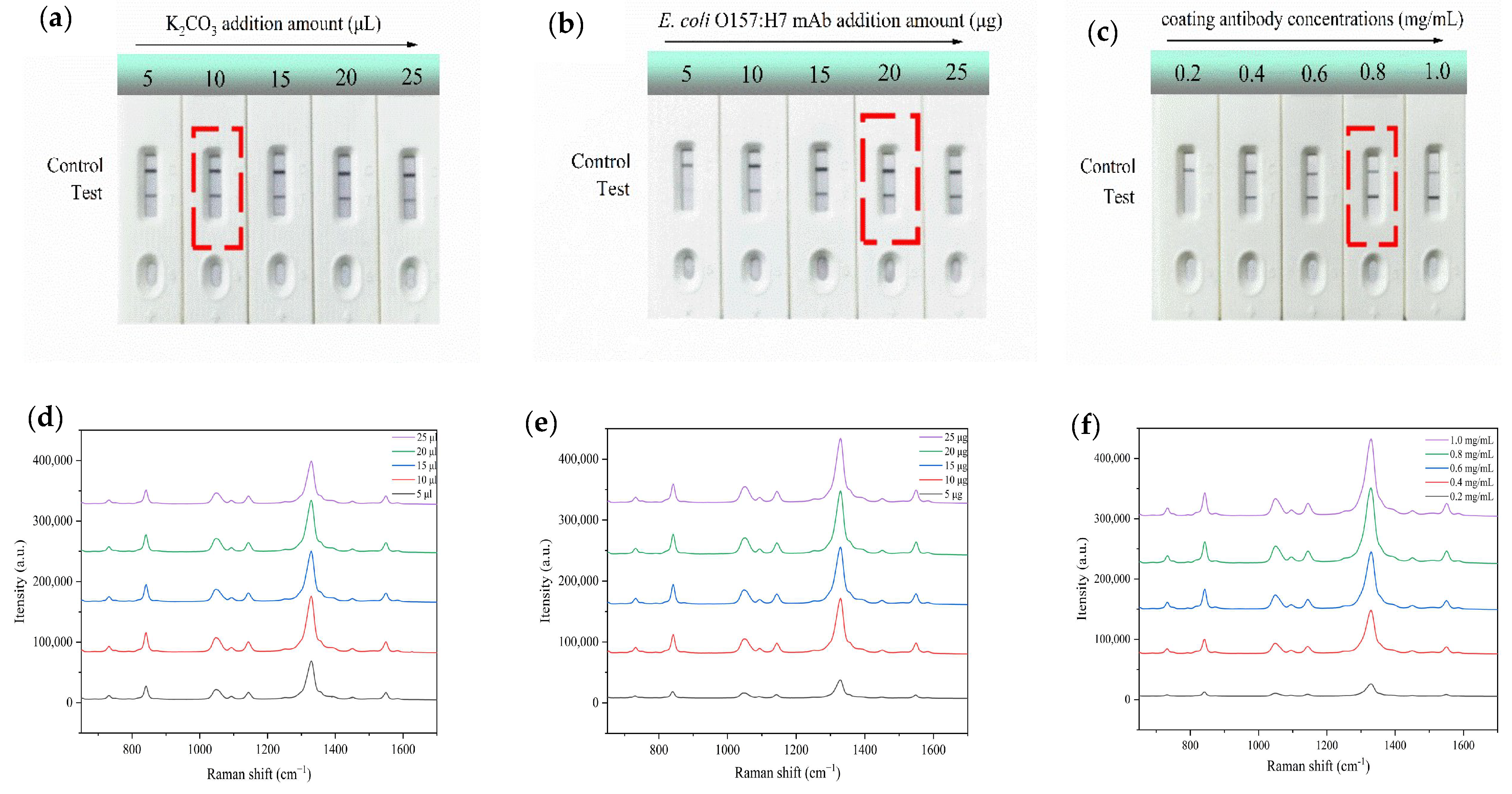
3.3. Optimization of the Assay Conditions

To establish a more sensitive and accurate SERS-ICA analysis method, the pH value of the solution and the amount of detection antibody during the preparation of the WS2-AuDTNB nanotags, as well as the coating antibody concentration, were optimized. The effects of different level parameters were assessed by measuring the Raman signal intensity on the T-line.
The pH value is a key factor influencing SERS nanotag fabrication, which affects the aggregation of nanotags. By adjusting the pH using 0.05 M K2CO3 solution, the addition amount was varied from 5 to 25 μL. Figure 3a shows that with the addition of K2CO3 solution, the color development of the T-lines on the test strip gradually intensifies. When the amount of K2CO3 added reaches 10 μL, the color of the T-lines on the test strip is the deepest. However, if the amount of K2CO3 solution is further increased, the color of the T-lines on the test strip gradually fades. Excessive K2CO3 solution destabilizes the electrical double layer of AuNPs, leading to their aggregation and preventing effective migration to the T-line. Additionally, the elevated pH may impair antibody activity, ultimately resulting in weakened color development on the T-line. Combined with the SERS measurement results in Figure 3d, the SERS intensity is highest when the amount of K2CO3 added is 10 μL. Herein, 10 μL of K2CO3 was applied for further experiments.
In addition, the amount of 0.2 mg/mL antibody for SERS nanotag preparation was optimized. As indicated in Figure 3b, with increasing amounts of E. coli O157:H7 mAb antibody, the T-lines’ color on the strips gradually became darker. The SERS intensity, as measured in Figure 3e, reached a maximum when the antibody amount was 20 μg. Therefore, 20 μg of antibody was chosen as the optimal amount for preparing WS2-AuDTNB-Ab.
Finally, the concentration of the coating antibody on the NC membrane was optimized. Figure 3c,f illustrate that as the antibody concentration increased from 0.2 mg/mL to 0.8 mg/mL, the color and intensity of the T-line increased rapidly and then showed a slight increase, indicating saturation of the antibody and bacterial immune response. Consequently, the optimal concentration for the coating antibody was identified as 0.8 mg/mL.

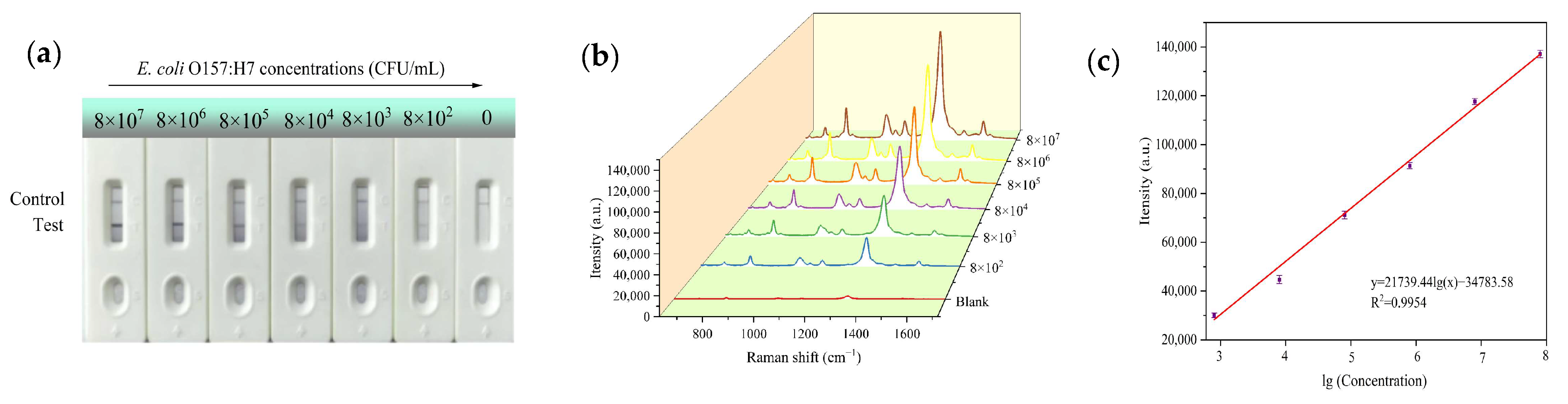
3.4. Sensitivity of the Assay

In order to evaluate the sensitivity of the SERS-ICA, a series of E. coli O157:H7 samples with a range of different concentrations (8 × 10²–8 × 10⁷ CFU/mL) were applied to SERS-ICA procedures under the above optimized conditions.
For qualitative analysis, Figure 4a displays the WS2-Au-based SERS-ICA strip for E. coli O157:H7 detection at various concentrations. Visual qualitative analyses were performed by three independent observers to identify a visible color signal distinct from blank negative controls (blanks). Under standardized white-light illumination, it was observed that the concentration of E. coli O157:H7 decreased and the dark-purple color of the T-line gradually faded due to fewer nanoprobes being captured at the T-line. The visual sensitivity, consistently observed by the naked eye, was estimated to be 8 × 10² CFU/mL. SERS spectra of different E. coli O157:H7 concentrations were measured for quantitative detection. As shown in Figure 4b, the SERS signal intensity gradually increased with the E.coli O157:H7 concentration. The characteristic peak intensities of DTNB at 1329 cm−1 could be employed as a basis for the standard curve of the SERS-based ICA. In Figure 4c, the curve demonstrates a good linear relationship within the concentration range of 8 × 10² CFU/mL to 8 × 10⁷ CFU/mL. The linear equation at 1329 cm−1 can be described as y = 21739.44 lg(x) − 34783.58, with a correlation coefficient of R2 = 0.9954, which can provide a reliable basis for determination of E.coli O157:H7 in quantitative analysis. On the basis of the formula LOD = yblank + 3 × SDblank, where yblank and SDblank are the average Raman intensity and standard deviation of the blank control, respectively [39], the LOD was calculated as 175  CFU/mL.

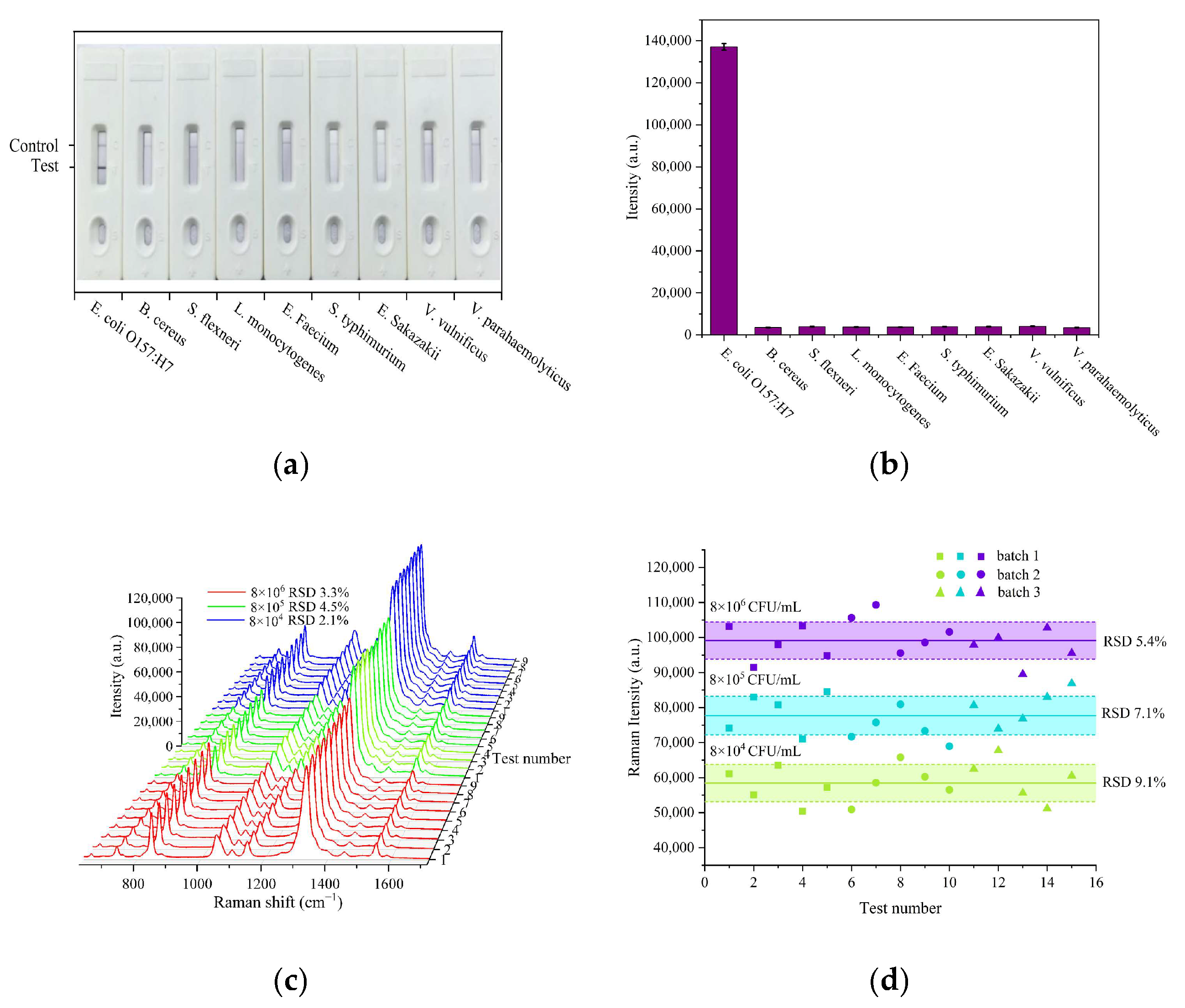
3.5. Specificity and Repeatability Evaluations

The specificity and repeatability of this ICA strip were evaluated using eight other pathogenic bacteria, including Bacillus cereus, Shigella flexneri, Listeria monocytogenes, Enterococcus faecium, Salmonella typhimurium, Enterobacter sakazakii, Vibrio vulnificus, and Vibrio parahaemolyticus, as interfering strains for SERS-ICA detection. As shown in Figure 5a,b, only the assay for E. coli O157:H7 generated a distinct color band and showed strong SERS signals in the corresponding T-lines, while no obvious color band or SERS intensity appeared on the T-lines for other non-target bacteria. This result is related to the specific affinity of the antibodies coated on the NC membranes for E. coli O157:H7. Compared to other reported methods, as shown in Table S1, the WS2-Au-based SERS-ICA strips significantly improve the detection sensitivity for the target bacteria. To assess the reproducibility, SERS intensities on the T-line of 8 × 10⁶, 8 × 10⁵, and 8 × 10⁴ CFU/mL of E. coli O157:H7 were tested at nine different points, with RSDs of 3.3%, 4.5%, and 2.1% (Figure 5c). Batch-to-batch reproducibility was evaluated using strips prepared in three different batches (Figure 5d). The RSDs for these tests were 5.4%, 7.1%, and 9.1% for 8 × 10⁶, 8 × 10⁵, and 8 × 10⁴ CFU/mL of E. coli O157:H7, respectively. These results confirmed the good specificity and reproducibility of the proposed assay.

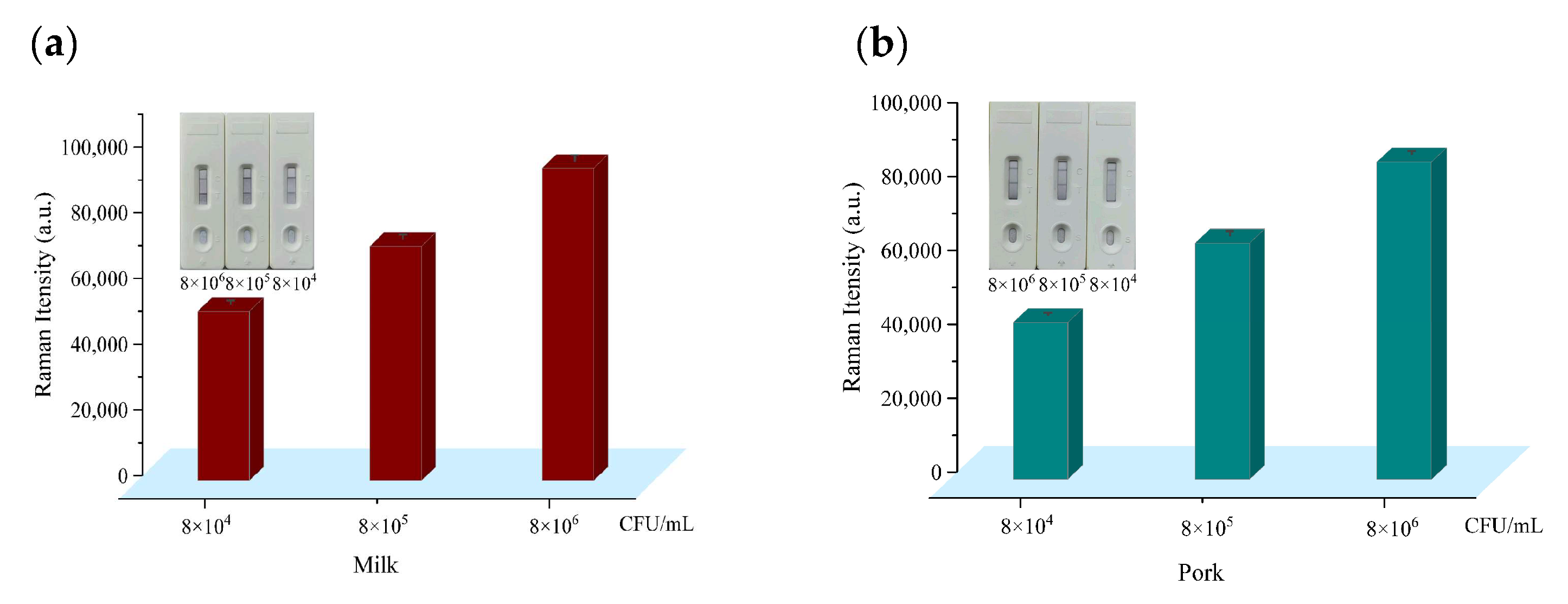
3.6. Detection of E. coli O157:H7 in Spiked Milk and Pork Samples

It is extremely essential to investigate the practicability of the proposed SERS-based ICA for sample analysis. Accordingly, pasteurized milk and pork samples obtained from a supermarket were spiked with E. coli O157:H7 standard solution and applied to the proposed assay under the optimized conditions, and the results are shown in Figure 6a,b. The contents of E. coli O157:H7 in the spiked samples were quantified based on the standard curve. The recoveries of E. coli O157:H7 from the spiked samples measured by SERS-based ICA are presented in Table S2. The average recoveries for E. coli O157:H7 pathogens in milk and pork were 95.3–117.6% and 86.8–101.5%, respectively, with RSDs ranging from 2.02% to 9.52%. These results indicate that this novel strategy is a feasible approach to effectively detect E. coli O157:H7 in real food samples.

4. Conclusions

Due to the high SERS enhancement, good biocompatibility, and high stability of the designed WS2-AuDTNB high-performance SERS nanotags, we have successfully developed a low-cost and user-friendly SERS-ICA platform for rapid and sensitive detection of E. coli O157:H7. The assay utilizes immune complex formation on the T-line, where the SERS signal intensity at 1329 cm−1 (DTNB) correlates with bacterial concentration. Under optimal experimental conditions, this method achieves a detection limit of 175 CFU/mL, with the entire assay completed within 20 min. The SERS-ICA possesses satisfactory sensitivity, specificity, and reproducibility, as well as an acceptable recovery rate (86.8–117.6%), in real food samples. This proposed method proves to have important prospects in point-of-care testing and can be used for the detection of other pathogens by coupling corresponding specific antibodies.
Future research can prioritize enhancing detection sensitivity through rational design of SERS-active substrates, where hierarchical nanostructures and strategic integration of semiconductor materials with metallic nanoparticles can generate high-density electromagnetic hotspots for amplified Raman signals. Concurrently, portable device integration will be advanced by coupling SERS readouts with smartphone-based spectral analyzers, while multiplexed pathogen identification in complex matrices necessitates further exploration. Collectively, these innovations aim to merge laboratory-grade analytical precision with field-operational simplicity, addressing critical gaps in foodborne pathogen monitoring.

Supplementary Materials

The following supporting information can be downloaded at https://www.mdpi.com/article/10.3390/s25082457/s1: Figure S1: The Au size distribution obtained by Image J 2025 software; Figure S2: UV-vis absorption spectra of AuNPs, WS2, and WS2-Au; Figure S3: Tauc plot of WS2; Figure S4: SERS spectra of 10−6 M DTNB molecules on Au and WS2-Au; Table S1: Comparison with other techniques for bacteria detection; Table S2: Recoveries of E. coli O157:H7 spiked in milk and pork. References [40,41,42,43,44,45] are cited in the supplementary materials.

Author Contributions

Conceptualization, D.W., Y.C. and Y.J.; methodology, D.W., Y.C., Q.Z. and J.C.; validation, D.W., Y.C., Q.Z., J.C., C.L. and Y.J.; formal analysis, D.W.; investigation, D.W. and Y.C.; resources, Y.C., Q.Z., J.C. and Y.J.; data curation, C.L.; writing—original draft preparation, D.W. and Y.J.; writing—review and editing, D.W., Y.C., Q.Z., J.C., C.L., Y.L., Y.J. and X.Q.; visualization, D.W. and C.L.; supervision, Y.L., Y.J. and X.Q.; project administration, Y.L. and X.Q.; funding acquisition, Y.L. and X.Q. All authors have read and agreed to the published version of the manuscript.

Funding

This research was funded by the National Key Research and Development Program of China, grant number 2022YFF0607900; the basic research expenses of CAIQ, grant number 2024JK020; the NMPA Key Laboratory for POCT Technology Transforming and Quality Control; and the Key Laboratory of Food Quality and Safety for State Market Regulation.

Institutional Review Board Statement

Not applicable.

Informed Consent Statement

Not applicable.

Data Availability Statement

The data presented in this study are available upon request from the corresponding author.

Acknowledgments

The authors thank the Testing and Evaluation Center’s laboratory at the Chinese Academy of Inspection and Quarantine (CAIQ; Beijing, China) for providing the bacterial strains used in the experimental detection.

Conflicts of Interest

The authors declare no conflicts of interest.

References

  1. Tack, D.M.; Kisselburgh, H.M.; Richardson, L.C.; Geissler, A.; Griffin, P.M.; Payne, D.C.; Gleason, B.L. Shiga Toxin-Producing Escherichia coli Outbreaks in the United States, 2010–2017. Microorganisms 2021, 9, 1529. [Google Scholar] [CrossRef] [PubMed]
  2. Koyun, O.Y.; Balta, I.; Corcionivoschi, N.; Callaway, T.R. Disease Occurrence in- and the Transferal of Zoonotic Agents by North American Feedlot Cattle. Foods 2023, 12, 904. [Google Scholar] [CrossRef] [PubMed]
  3. Ito, H.; Terai, A.; Kurazono, H.; Takeda, Y.; Nishibuchi, M. Cloning and nucleotide sequencing of Vero toxin 2 variant genes from Escherichia coli O91: H21 isolated from a patient with the hemolytic uremic syndrome. Microb. Pathog. 1990, 8, 47–60. [Google Scholar] [CrossRef] [PubMed]
  4. Shin, J.H.; Hong, J.; Go, H.; Park, J.; Kong, M.; Ryu, S.; Kim, K.P.; Roh, E.; Park, J.K. Multiplexed Detection of Foodborne Pathogens from Contaminated Lettuces Using a Handheld Multistep Lateral Flow Assay Device. J. Agric. Food Chem. 2018, 66, 290–297. [Google Scholar] [CrossRef]
  5. Oluwarinde, B.O.; Ajose, D.J.; Abolarinwa, T.O.; Montso, P.K.; Du Preez, I.; Njom, H.A.; Ateba, C.N. Safety Properties of Escherichia coli O157:H7 Specific Bacteriophages: Recent Advances for Food Safety. Foods 2023, 12, 3989. [Google Scholar] [CrossRef]
  6. Fu, J.; Zhou, Y.; Huang, X.; Zhang, W.; Wu, Y.; Fang, H.; Zhang, C.; Xiong, Y. Dramatically Enhanced Immunochromatographic Assay Using Cascade Signal Amplification for Ultrasensitive Detection of Escherichia coli O157:H7 in Milk. J. Agric. Food Chem. 2020, 68, 1118–1125. [Google Scholar]
  7. Ferone, M.; Gowen, A.; Fanning, S.; Scannell, A.G.M. Microbial detection and identification methods: Bench top assays to omics approaches. Compr. Rev. Food Sci. Food Saf. 2020, 19, 3106–3129. [Google Scholar]
  8. Kim, J.H.; Oh, S.W. Rapid detection for low numbers of Escherichia coli o157:h7 by real-time pcr in cabbage using a combination of filtration, short microbial enrichment, and dna concentration within 4 h. LWT-Food Sci. Technol. 2021, 139, 110520. [Google Scholar] [CrossRef]
  9. Kong, J.; Fan, C.; Liao, X.; Chen, A.; Yang, S.; Zhao, L.; Li, H. Accurate detection of Escherichia coli O157:H7 and Salmonella enterica serovar typhimurium based on the combination of next-generation sequencing and droplet digital PCR. LWT-Food Sci. Technol. 2022, 168, 113913. [Google Scholar] [CrossRef]
  10. Panchal, N.; Jain, V.; Elliott, R.; Flint, Z.; Worsley, P.; Duran, C.; Banerjee, T.; Santra, S. Plasmon-Enhanced Bimodal Nanosensors: An Enzyme-Free Signal Amplification Strategy for Ultrasensitive Detection of Pathogens. Anal. Chem. 2022, 94, 13968–13977. [Google Scholar] [CrossRef]
  11. Yu, Q.; Wu, T.; Tian, B.; Li, J.; Liu, Y.; Wu, Z.; Jin, X.; Wang, C.; Wang, C.; Gu, B. Recent advances in SERS-based immunochromatographic assay for pathogenic microorganism diagnosis: A review. Anal. Chim. Acta 2024, 1286, 341931. [Google Scholar] [CrossRef] [PubMed]
  12. Zeng, R.; Zhou, F.; Wang, Y.; Liao, Z.; Qian, S.; Luo, Q.; Zheng, J. Polydopamine modified colloidal gold nanotag-based lateral flow immunoassay platform for highly sensitive detection of pathogenic bacteria and fast evaluation of antibacterial agents. Talanta 2024, 278, 126525. [Google Scholar] [CrossRef] [PubMed]
  13. Shao, Y.; Xiong, S.; Huang, M.; Liu, B.; Huang, Z.; Xu, D.; Chen, X.; Xiong, Y. Recent advances in multifunctional nanomaterials enhanced immunochromatographic assays. TrAc Trend Anal. Chem. 2024, 181, 118013. [Google Scholar] [CrossRef]
  14. Wang, Q.; Long, M.; Lv, C.; Xin, S.; Han, X.; Jiang, W. Lanthanide-labeled fluorescent-nanoparticle immunochromatographic strips enable rapid and quantitative detection of Escherichia coli O157:H7 in food samples. Food Control 2020, 109, 106894. [Google Scholar] [CrossRef]
  15. Wang, R.; Kim, K.; Choi, N.; Wang, X.; Lee, J.; Jeon, J.H.; Rhie, G.; Choo, J. Highly sensitive detection of high-risk bacterial pathogens using sers-based lateral flow assay strips. Sens. Actuators B Chem. 2018, 270, 72–79. [Google Scholar] [CrossRef]
  16. Hong, X.; Mao, Y.; Yang, C.; Liu, Z.; Li, M.; Du, D. Contamination of Zearalenone from China in 2019 by a Visual and Digitized Immunochromatographic Assay. Toxins 2020, 12, 521. [Google Scholar] [CrossRef]
  17. Li, J.; Wu, T.; Wang, C.; Tu, J.; Song, X.; Shao, Y.; Wang, C.; Qi, K.; Xiao, R. Nanogapped Fe3O4-Au Surface-Enhanced Raman Scattering Tags for the Multiplex Detection of Bacteria on an Immunochromatographic Strip. ACS Appl. Nano Mater. 2022, 5, 12679–12689. [Google Scholar] [CrossRef]
  18. Shu, R.; Liu, S.; Huang, L.; Li, Y.; Sun, J.; Zhang, D.; Zhu, M.; Wang, J. Enzyme-Mimetic nano-immunosensors for amplified detection of food hazards: Recent advances and future trends. Biosens. Bioelectron. 2022, 217, 114577. [Google Scholar] [CrossRef]
  19. Zheng, S.; Wang, C.; Li, J.; Wang, W.; Yu, Q.; Wang, C.; Wang, S. Graphene oxide-based three-dimensional au nanofilm with high-density and controllable hotspots: A powerful film-type sers tag for immunochromatographic analysis of multiple mycotoxins in complex samples. Chem. Eng. J. 2022, 448, 137760. [Google Scholar] [CrossRef]
  20. Mahmoudi, T.; Pourhassan-Moghaddam, M.; Shirdel, B.; Baradaran, B.; Morales-Narváez, E.; Golmohammadi, H. (Nano)tag-antibody conjugates in rapid tests. J. Mater. Chem. B 2021, 9, 5414–5438. [Google Scholar] [CrossRef]
  21. Wang, L.; Xu, D.; Jiang, L.; Gao, J.; Tang, Z.; Xu, Y.; Chen, X.; Zhang, H. Transition Metal Dichalcogenides for Sensing and Oncotherapy: Status, Challenges, and Perspective. Adv. Funct. Mater. 2021, 31, 2004408. [Google Scholar]
  22. Shorie, M.; Kumar, V.; Kaur, H.; Singh, K.; Tomer, V.K.; Sabherwal, P. Plasmonic DNA hotspots made from tungsten disulfide nanosheets and gold nanoparticles for ultrasensitive aptamer-based SERS detection of myoglobin. Mikrochim. Acta 2018, 185, 158. [Google Scholar] [CrossRef] [PubMed]
  23. Nisar, S.; Dastgeer, G.; Shahzadi, M.; Shahzad, Z.M.; Elah, E.; Irfan, A.; Eom, J.; Kim, H.; Kim, D. Gate-assisted MoSe2 transistor to detect the streptavidin via supporter molecule engineering. Mater. Today Nano 2023, 24, 100405. [Google Scholar]
  24. Cong, C.; Shang, J.; Wang, Y.; Yu, T. Optical Properties of 2D Semiconductor WS2. Adv. Opt. Mater. 2018, 6, 1700767. [Google Scholar]
  25. Hu, Y.; Huang, Y.; Wang, Z.; Wang, Y.; Ye, X.; Wong, W.; Li, C.; Sun, D. Gold/WS2 nanocomposites fabricated by in-situ ultrasonication and assembling for photoelectrochemical immunosensing of carcinoembryonic antigen. Mikrochim. Acta 2018, 185, 570. [Google Scholar]
  26. Wang, J.; Lu, Y.; Quan, W.; Hu, J.; Yang, P.; Song, G.; Fu, J.; Peng, Y.; Tong, L.; Ji, Q.; et al. Epitaxial Growth of Monolayer WS2 Single Crystals on Au(111) Toward Direct Surface-Enhanced Raman Spectroscopy Detection. ACS Nano 2024, 18, 26359–26368. [Google Scholar]
  27. Tang, X.; Hao, Q.; Hou, X.; Lan, L.; Li, M.; Yao, L.; Zhao, X.; Ni, Z.; Fan, X.; Qiu, T. Exploring and Engineering 2D Transition Metal Dichalcogenides toward Ultimate SERS Performance. Adv. Mater. 2024, 36, 2312348. [Google Scholar]
  28. Anju, K.S.; Midhun, P.S.; Kumar, K.R.; Rajeev, M.K. Tailoring surface enhanced Raman scattering platform based on pulsed laser deposited MoS2-Ag hybrid nanostructure. Mater. Chem. Phys. 2022, 276, 125286. [Google Scholar]
  29. Alamri, M.; Sakidja, R.; Goul, R.; Ghopry, S.; Wu, J.Z. Plasmonic Au nanoparticles on 2D MoS2/graphene van der waals heterostructures for high-sensitivity surface-enhanced raman spectroscopy. ACS Appl. Nano Mater. 2019, 2, 1412–1420. [Google Scholar]
  30. Yuan, H.; Yu, S.; Kim, M.; Li, J.E.; Kang, H.; Jiang, D.; Ramasamy, M.S.; Kim, D.H. Dopamine-mediated self-assembled anisotropic Au nanoworms conjugated with MoS2 nanosheets for SERS-based sensing. Sens. Actuators B Chem. 2022, 371, 132453. [Google Scholar]
  31. Chen, M.; Liu, D.; Du, X.; Luo, K.H.; Wang, S.; Zhou, B.; Pan, H. 2D materials: Excellent substrates for surface-enhanced Raman scattering (SERS) in chemical sensing and biosensing. TrAC Trends Anal. Chem. 2020, 130, 115983. [Google Scholar] [CrossRef]
  32. Song, X.; You, X.; Ren, X.; Zhang, X.; Tang, D.; Li, X. Vertically aligned Ag-decorated MoS2 nanosheets supported on polyvinyl alcohol flexible substrate enable high-sensitivity and self-cleaning SERS devices. J. Environ. Chem. Eng. 2023, 11, 109437. [Google Scholar] [CrossRef]
  33. Chen, Y.; Liu, H.; Tian, Y.; Du, Y.; Ma, Y.; Zeng, S.; Gu, C.; Jiang, T.; Zhou, J. In Situ Recyclable Surface-Enhanced Raman Scattering-Based Detection of Multicomponent Pesticide Residues on Fruits and Vegetables by the Flower-like MoS2-Ag Hybrid Substrate. ACS Appl. Mater. Interfaces 2020, 12, 14386–14399. [Google Scholar] [CrossRef] [PubMed]
  34. Zhang, D.; Wu, Y.C.; Yang, M.; Liu, X.; Coileáin, C.Ó.; Abid, M.; Abid, M.; Wang, J.J.; Shvets, I.; Xu, H.; et al. Surface enhanced Raman scattering of monolayer MX2 with metallic nano particles. Sci. Rep. 2016, 6, 30320. [Google Scholar] [CrossRef]
  35. Frens, G. Controlled Nucleation for the Regulation of the Particle Size in Monodisperse Gold Suspensions. Nat. Phys. Sci. 1973, 241, 20–22. [Google Scholar] [CrossRef]
  36. Zhou, K.G.; Mao, N.N.; Wang, H.X.; Peng, Y.; Zhang, H.L. A Mixed-Solvent Strategy for Efficient Exfoliation of Inorganic Graphene Analogues. Angew. Chem. Int. Ed. Engl. 2011, 50, 10839–10842. [Google Scholar] [CrossRef]
  37. Hwang, D.Y.; Suh, D.H. Universal surface modification of transition metal dichalcogenide decorated with metal nanoparticles for surface enhanced Raman scattering. Mater. Res. Bull. 2017, 90, 73–80. [Google Scholar] [CrossRef]
  38. Pramanik, A.; Davis, D.; Patibandla, S.; Begum, S.; Ray, P.; Gates, K.; Gao, Y.; Chandra Ray, P. A WS2-gold nanoparticle heterostructure-based novel SERS platform for the rapid identification of antibiotic-resistant pathogens. Nanoscale Adv. 2020, 2, 2025–2033. [Google Scholar] [CrossRef]
  39. Putnin, T.; Ngamaroonchote, A.; Wiriyakun, N.; Ounnunkad, K.; Laocharoensuk, R. Dually functional polyethylenimine-coated gold nanoparticles: A versatile material for electrode modification and highly sensitive simultaneous determination of four tumor markers. Microchim. Acta 2019, 186, 305. [Google Scholar] [CrossRef]
  40. Pang, L.; Wang, L.; Liang, Y.; Wang, Z.; Zhang, W.; Zhao, Q.; Yang, X.; Jiang, Y. G-triplex/hemin DNAzyme mediated colorimetric aptasensor for Escherichia coli O157:H7 detection based on exonuclease III-assisted amplification and aptamers-functionalized magnetic beads. Talanta 2024, 269, 125457. [Google Scholar] [CrossRef]
  41. Liu, H.B.; Chen, C.Y.; Zhang, C.N.; Du, X.J.; Li, P.; Wang, S. Functionalized AuMBA@Ag Nanoparticles as an Optical and SERS Dual Probe in a Lateral Flow Strip for the Quantitative Detection of Escherichia coli O157:H7. J. Food Sci. 2019, 84, 2916–2924. [Google Scholar] [PubMed]
  42. He, Q.; Pan, J.; Xu, Z.; Hammock, B.D.; Li, D. Development of a nanobody-based immunoassay for the detection of Escherichia coli O157:H7 in food samples. Food Chem. 2025, 473, 142987. [Google Scholar] [PubMed]
  43. Zhang, G.; Huang, Z.; Hu, L.; Wang, Y.; Deng, S.; Liu, D.; Peng, J.; Lai, W. Molecular Engineering Powered Dual-Readout Point-of-Care Testing for Sensitive Detection of Escherichia coli O157:H7. ACS Nano. 2023, 17, 23723–23731. [Google Scholar] [PubMed]
  44. Kim, J.E.; Shin, J.H.; Park, J.P. An engineered antimicrobial peptide as an alternative bioreceptor for the detection of pathogenic Escherichia coli O157:H7. J. Electroanal. Chem. 2024, 953, 118003. [Google Scholar] [CrossRef]
  45. Zhu, M.; Liu, J.; Jiang, X.; Zhang, Y.; Zhang, J.; Wu, J. Bacteria-imprinted impedimetric sensor based on doping-induced nanostructured polypyrrole for determination of Escherichia coli. Mikrochim. Acta 2023, 190, 431. [Google Scholar] [CrossRef]
Figure 1. Schematic illustration of the fabrication procedure of SERS-ICA. (a) Preparation and antibody modification of WS2-AuDTNB SERS nanotags. (b) ICA strip procedure for E. coli O157:H7 detection.
Sensors 25 02457 g001
Figure 2. Characterization of the fabricated WS2-Au complexes. TEM images of (a) WS2 and (b) WS2-Au. (c) HRTEM images of AuNPs on the WS2. (d) EDX elemental (W, S, Au, and merging) mapping images of the WS2-Au.
Sensors 25 02457 g002
Figure 3. Images of optimized (a) pH value, (b) E. coli O157:H7 mAb, and (c) coated antibody concentrations. SERS intensities of different (d) K2CO3 volumes, (e) E. coli O157:H7 mAb addition amounts, (f) and coated antibody concentrations.
Sensors 25 02457 g003
Figure 4. (a) Images of SERS-ICA detection of E. coli O157:H7 standard bacterial solutions at different concentrations. (b) SERS spectra for different concentrations of E. coli O157:H7. (c) SERS signal intensities at 1329 cm−1 increasing linearly with logarithmic concentration of E. coli O157:H7.
Sensors 25 02457 g004
Figure 5. Selectivity and reproducibility of the developed ICA for E. coli O157:H7. (a) Digital photograph of 9 strips of different bacteria. (b) Corresponding Raman intensities at 1329 cm−1. (c) SERS signals of nine tests of E. coli O157:H7. (d) SERS intensities at 1329 cm−1 measured in three different batches of strips.
Sensors 25 02457 g005
Figure 6. Practicability test of the WS2-Au-based ICA for E. coli O157:H7 detection in (a) milk and (b) pork samples.
Sensors 25 02457 g006
Disclaimer/Publisher’s Note: The statements, opinions and data contained in all publications are solely those of the individual author(s) and contributor(s) and not of MDPI and/or the editor(s). MDPI and/or the editor(s) disclaim responsibility for any injury to people or property resulting from any ideas, methods, instructions or products referred to in the content.

