Abstract
An effective deposition of a cinchonine layer on a platinum metal surface can be easily achieved through the cathodic reduction of a cinchonine hydrochloride methanolic solution at a controlled potential of −220 mV vs. the silver standard electrode (SSE). A coated screen-printed platinum electrode has proven to be suitable for cinchonine determination in water, urine, and serum at µg L−1 concentration levels using differential pulse voltammetry in a phosphate buffer solution (pH 7.0). The limits of detection (LOD) and quantitation (LOQ) were 0.6 µg L−1 and 1.8 µg L−1, respectively.
1. Introduction
In recent years, screen-printed electrodes (SPEs) have undergone remarkable advancements, with numerous scientific papers published on sensors and biosensors. SPEs offer several advantages over conventional electrodes, as they can be efficiently used for microvolumes and as single-use sensors, eliminating the need for electrode maintenance. This has paved the way for the development of reproducible, cost-effective, precise, and highly sensitive sensors. Thus, SPEs are ideal for decentralized assays and the development of (bio)sensors in environmental [1,2], clinical [3], and agri-food applications, as well as for electrosynthesis and compound characterization [4,5]. On the other hand, to enhance selectivity and/or sensitivity in various applications, appropriate compounds can be used to modify the electrode surface [6,7,8,9,10,11], including that of SPEs [12,13]. However, weak adsorption, particularly of organic molecules onto the electrode surface, has traditionally posed a challenge, limiting the effectiveness of electrochemical techniques for organic system analysis [14,15,16,17]. In this study, electrochemical deposition was employed to create a cinchonine-based organic thin film on a platinum electrode surface via the cathodic reduction of a cinchonine hydrochloride methanolic solution. Cinchonine (CN) is an alkaloid isolated from Cinchona succirubra and, when combined with other Cinchona alkaloids such as quinine, quinidine, and cinchonidine, plays a crucial role in pharmacological activity [18]. It is widely used as an important antimalarial agent [19,20,21], exhibits antiarrhythmic properties [22], provides resistance against various types of tumors [23], and acts as a potent inhibitor of human platelet aggregation [24,25]. Notably, cinchonine exhibits lower toxicity and higher activity compared to other quinine-related compounds [26]. Its lethal dose (LD50) in humans is 456 mg kg−1, with an estimated median lethal dose of 152 mg kg−1 [27]. Numerous analytical studies have focused on the determination of cinchonine-type alkaloids, both due to their medicinal and commercial significance and because of their narrow therapeutic window between ineffective and toxic doses. Standard quantification methods for the four main Cinchona alkaloids and their formulations rely exclusively on spectrophotometry [28,29], which lacks sensitivity, or liquid chromatography [30,31], which is time-consuming. To develop more efficient, sensitive, selective, and, above all, faster methods, various alternative approaches have been explored, including electroanalytical techniques [32,33,34,35,36,37,38]. In this study, we present a novel electrochemical sensor for determining cinchonine concentrations in the microgram-per-liter range, offering faster and more sensitive analysis compared to conventional techniques. The proposed sensor leverages the strong adsorption affinity of platinum for cinchonine while benefiting from the advantages of screen-printed electrodes. The adsorption of various cinchona alkaloids onto platinum surfaces has been extensively studied [39,40,41], with some exhibiting irreversible adsorption, requiring relatively high potentials for desorption [42]. As illustrated in Scheme 1, the cathodic reduction of a methanolic cinchonine hydrochloride solution induces a hydrogen evolution reaction, leading to the stable adsorption of the insoluble alkaloid on the platinum cathode surface [43].

Scheme 1.
Proposed mechanism for the deposition of cinchonine on a platinum surface via the cathodic reduction of its hydrochloride salt in a methanolic solution.
This modified electrode was employed to quantify the preferentially adsorbed species, specifically cinchonine and quinidine, using conventional differential pulse voltammetry. Notably, an increase in sensitivity will be demonstrated compared to an unmodified screen-printed electrode. The results obtained with this sensor are presented and discussed in detail in this paper.
2. Materials and Methods
2.1. Reagents
All reagents used in this study were of analytical grade, and all solutions were prepared using ultrapure solvents purchased from Merck. Cinchonine, cinchonine monohydrochloride hydrate, quinine hydrochloride dihydrate, and quinidine hydrochloride monohydrate were all obtained from Merck, while cinchonidine hydrochloride was purchased from Alfa Chemistry. N,O-Bis(trimethylsilyl)trifluoroacetamide (BSTFA) for GC derivatization and CCl4, suitable for IR spectroscopy were acquired from Supelco-Merck. All human serum and urine samples were obtained from the University Affiliated Hospital.
2.2. Instrumentation and Apparatus
Differential pulse voltammetry (DPV) and chronoamperometry (CA) were performed using an electrochemical system comprising an Autolab PGSTAT302F potentiostat/galvanostat (EcoChemie, Utrecht, The Netherlands) with an IME663 interface, connected to the 663 VA Stand system (Metrohm, Basel, Switzerland), and controlled by Nova 1.10.5 software (EcoChemie, Utrecht, The Netherlands). The stand was equipped with a screen-printed electrode holder (Metrohm, 6.1241.090) for connecting the working electrode, along with an external Ag/AgCl reference electrode (saturated with 3 mol L−1 KCl, SSE) and an external platinum auxiliary electrode, both from Metrohm. These components were used for cathodic deposition at a controlled potential. The working electrodes were commercially available screen-printed platinum (SP-Pt) electrodes (Aux.: Pt; Ref.: Ag), purchased from Metrohm-Dropsens (DRP-550 from Metrohm, Oviedo, Spain). Both potentiostatic and galvanostatic modes were employed for electrodeposition on the Pt surface of the SP-Pt working electrode. Additionally, an analytical balance with a sensitivity of 0.01 mg (Mod. XS105DR from Mettler-Toledo, Milan, Italy) was used for mass measurements, and a Julabo circulator (Mod. F12 from Thermo Fisher, Monza, Italy) was used to maintain a constant temperature. Fourier-transform infrared (FTIR) spectra were analyzed using an Agilent Cary 630 FTIR spectrophotometer (Agilent, Cernusco sul Naviglio, Italy) equipped with a ZnSe multi-bounce ATR sampling module. GC–MS analyses were performed using an Agilent 7890A GC (Agilent Technologies, Cernusco sul Naviglio, Italy) system coupled with an Agilent 5975 MSD (Agilent Technologies, Cernusco sul Naviglio, Italy) system.
2.3. Procedures
2.3.1. Solutions
A 4.00 mg L−1 cinchonine standard solution was prepared by dissolving 4.50 ± 0.01 mg of cinchonine monohydrochloride hydrate (99%) in distilled water to a final volume of 10.0 mL, followed by vortexing for 5 min. The working standard solution was obtained through successive dilutions. As previously reported, a standard solution can also be prepared by dissolving cinchonine in an acidic solution [44]. Solutions of the other alkaloids were prepared in a similar manner. For DPV measurements, a solution containing 0.02 mol dm−3 phosphate buffer (pH 7.0) and 0.1 mol dm−3 KClO4 was used as a blank (electrolyte solution).
2.3.2. Electrode Preparation
Electrochemical deposition was performed using a computer-controlled potentiostat (Autolab PGSTAT302F, from EcoChemie, Utrecht, The Netherlands) with a three-electrode cell, consisting of a Pt bar as the counter electrode and an Ag/AgCl electrode as the silver standard reference electrode (SSE). The working electrode was a screen-printed platinum electrode (SP-Pt). During deposition, only the WE contact of the screen-printed electrode holder was connected to the Autolab station. A white organic layer was deposited on the SP-Pt electrode surface through the cathodic reduction of alkaloid-derived ammonium salt solutions, performed at a controlled deposition potential of −0.220 V vs. SSE for 60 s [45]. The deposition solution consisted of 3 mg mL−1 cinchona alkaloid hydrochloride in methanol. The process was carried out at a constant temperature of 10 °C. After deposition, the screen-printed platinum electrode coated with alkaloid (SP-Pt/CN for cinchonine) was thoroughly rinsed with methanol. To confirm the successful deposition of cinchona, the deposited layer was removed by immersing a coated electrode in a vial containing approximately 2 mL of carbon tetrachloride [46], which was then placed in an ultrasonic bath for 10 s. This process was repeated with additional SP-Pt/CN electrodes to further concentrate the solution. The resulting organic solution was then deposited drop by drop onto the ZnSe crystal of the ATR module in the spectrophotometer. The recorded FTIR-ATR spectrum matched that of cinchonine (Figure S1). A similarly prepared solution was treated with BSTFA/Et3N and injected into the GC-MS to verify the purity and composition of the deposit. GC-MS analysis confirmed that the cathode deposit consists exclusively of cinchonine (Figure S2).
2.3.3. Differential Pulse Voltammetry Measurements
For DPV measurements, 10.0 mL of the blank solution was placed in a glass voltammetric cell from Metrohm. The DPV experimental parameters were set as follows: scan range from +0.20 V to +1.10 V, pulse amplitude of 50 mV, pulse time of 50 ms, and scan rate of 10 mV s−1. For methodology validation, cinchonine quantification was performed in different samples. The standard addition method was used for calibration, with calibration plots obtained through successive additions of cinchonine standard solution into the blank solution. All experiments were conducted at a controlled temperature of 25.0 °C using a thermostatic cell. Prior to electrochemical measurements, solutions were degassed with pure nitrogen.
2.3.4. Data Treatment
All data analyses were conducted and interpreted using NOVA 1.10.5 software, while additional calculations were performed with the EXCEL® program. Sensitivities were expressed as the slope of the calibration plot. Limits of detection (LODs) and limits of quantification (LOQs) were calculated as three and ten times the standard deviation of the intercept of the calibration line, respectively, divided by the slope [47,48]. Repeatability was evaluated through ten consecutive measurements at a concentration within the middle of the linear range, while reproducibility was determined from the relative standard deviation (RSD) of the slopes of three independent calibration plots.
3. Results and Discussion
3.1. Preliminary Studies
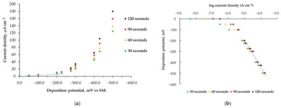
It is well known that cinchona alkaloids adsorb onto the Pt surface [40] and that the two asymmetric carbon atoms influence the chirality of hydrogenation products [49]. Preliminary experiments were conducted on platinum electrodes at different fixed current densities, ranging from 1.0 mA to 10 μA. These experiments enabled the deposition of alkaloid layers on the Pt electrode surface; however, the deposits were later found to be poorly adherent, likely due to the simultaneous and uncontrollable evolution of hydrogen. Similar deposits were observed even when deposition was carried out at low temperatures. Based on these preliminary results, further experiments were performed using an SP-Pt electrode at a controlled potential to mitigate hydrogen evolution [50,51] during deposition on the Pt electrode surface. Various deposition potentials were investigated, ranging from 0 to −500 mV vs. SSE. Figure 1a illustrates the relationship between the applied potentials and the current densities (μA cm−2) measured during cinchonine electrodeposition at controlled potential. Deposition times of 30, 60, 90, and 120 s were tested. As shown in Figure 1a, significant current consumption due to H2 evolution occurs when a more cathodic deposition potential is applied (Eapp < −300 mV) [52]. This suggests that cinchonine chemisorption can be effectively achieved under controlled potential conditions at moderately negative potentials, where hydrogen evolution is negligible. To further analyze this, we plotted the deposition potential against log current density (Figure 1b). The graph shows a linear trend above −220 mV, leading to the selection of this deposition potential for subsequent experiments [53]. Figure S3 shows the chronoamperometric curves recorded at different deposition potentials.

Figure 1.
Current density–potential relationship derived from the applied deposition potentials and the corresponding current densities (μA cm−2) (a) and the relationship between the logarithm of current densities (A cm−2) and the applied deposition potentials (b) obtained during electrodeposition at different controlled potentials. Deposition times: 30 s, 60 s, 90 s, 120 s. Deposition temperature: 10 °C.
To improve the homogeneity of the deposited layer, depositions were carried out at low temperatures, ranging from 20 °C to 5 °C [54]. To this end, a series of SP-Pt/CN electrodes was prepared under potential control at different temperatures (20 °C, 15 °C, 10 °C, and 5 °C) and evaluated for reproducibility and stability as probes for differential pulse voltammetric determinations. The stability of the modified SP-Pt/CN electrodes obtained at different temperatures was examined through multiscan studies to identify the optimal deposition conditions. The best electrode was determined based on the smallest relative standard deviation (RSD) in peak current [55]. In these studies, ten consecutive scans were performed in a 20.0 μg L−1 cinchonine solution. This test also helped assess whether the chemisorbed species would diffuse from the electrode surface into the bulk solution during continuous measurements under the same conditions. No significant decrease in peak currents, which could indicate the diffusion of some particles from the electrode interface into the solution, was observed during the multiscan test. Instead, only a variation in peak current values was detected, with a calculated RSD ranging from 1.1% to 6.4% (Table 1). These results confirm that the electroactive material remains confined to the electrode surface and does not detach into the bulk solution. Since the RSD value for electrochemical methods and stationary printed electrodes should be below 5%, the reproducibility of the developed method is considered acceptable under most of our experimental conditions [35].

Table 1.
Relative standard deviation (RSD, %) of the peak current for SP-Pt/CN electrodes prepared at a controlled potential of −220 mV vs. SSE for different deposition times and temperatures. The RSD of peak current values was measured in a phosphate buffer solution (pH 7.0) containing 4.0 μg L−1 cinchonine, using sixteen independently fabricated electrodes.
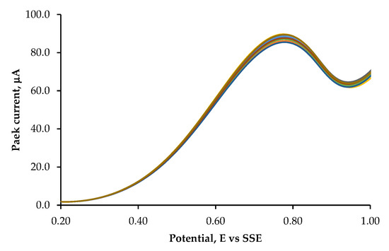
In particular, the deposition conditions achieved at 10 °C for 60 s, at a controlled potential of −220 mV vs. SSE, resulted in the lowest RSD value (1.1%), as shown in Figure 2. Therefore, the developed sensor demonstrates good stability and reproducibility for cinchonine detection and will be used to quantify cinchonine in real samples, such as urine and plasma. The advantage of this modified SPE/Pt is that it can be regenerated after approximately ten uses. Simply immersing it in methanol in an ultrasonic bath for 10 s and repeating the cathodic deposition process is sufficient for regeneration.

Figure 2.
Stability study using DPV measurements in a 20.0 μg L−1 cinchonine solution: DPV measurements show a 1.1% change in RSD peak current after 10 scans. The reported DP voltammograms correspond to 10 scans performed in a 0.02 mol dm−3 phosphate buffer (pH 7.0) with 0.1 mol dm−3 KClO4 as the supporting electrolyte. Measurements were recorded on a modified SP-Pt/CN electrode prepared at 10 °C for 60 s, at a controlled potential of −220 mV vs. SSE. The scan rate was set to 10 mV s−1.
3.2. Electrode Testing
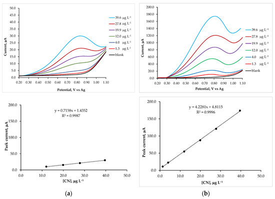
Pulse voltammetric techniques, such as DPV, are effective and rapid electroanalytical methods with well-established advantages, including excellent discrimination against background current and low detection limits [56]. To demonstrate the sensitivity of SP-Pt/CN electrodes for the electrochemical measurement of cinchona alkaloids, the effect of varying cinchonine concentration using DPV mode was investigated. The standard addition method was employed for calibration. Differential pulse voltammograms and the corresponding calibration plots, shown in Figure 3, were obtained through successive additions of a cinchonine standard solution (4.00 mg L−1) into a blank solution (10.0 mL). Considering the physiological conditions of the human body, a pH of 7.0 was selected, as this value prevents cinchonine protonation and ensures a stable sensor response with cinchonine adsorbed on its surface [33]. As illustrated in Figure 3a, the screen-printed platinum electrode produced a weaker and less intense signal compared to the SP-Pt/CN electrode. At cinchonine concentrations below 4 μg L−1, the DPV signal is nearly indistinguishable from the blank voltammogram, suggesting slow electrode kinetics for the faradaic processes generated by cinchonine oxidation, despite the well-documented strong affinity of platinum for cinchonine [43]. In contrast, the SP-Pt/CN electrode exhibited a well-defined and more intense characteristic oxidation peak, even at concentrations as low as 1.3 μg L−1 (Figure 3b). This clearly indicates that the cinchonine layer enhances the sensitivity of the working electrode by promoting electrochemical processes on a larger and more specific surface area due to the adsorbed layer [16]. From the electrochemical current–potential responses shown in Figure 3a,b, the peak current (Ip) was measured at various cinchonine concentrations. The resulting calibration plot showed a linear relationship with cinchonine concentration over a range of 1.3 μg L−1 to 39.6 μg L−1, following the regression equation: Ip (μA) = 4.228 c (μg L−1) + 4.812 with a correlation coefficient of 0.9996.

Figure 3.
DPV voltammograms recorded in a 0.02 mol dm−3 phosphate buffer solution at pH 7.0, with 0.1 mol dm−3 KClO4 as the supporting electrolyte, using an SP-Pt electrode (a) and an SP-Pt/CN electrode (b). Measurements were performed in solutions containing increasing concentrations of cinchonine. The insets below display the corresponding calibration plots, with lines drawn to emphasize the different slopes. The steeper slope observed for the SP-Pt/CN electrode confirms its higher sensitivity as a sensor.
The limits of detection (LOD) and quantitation (LOQ) were calculated from the oxidation peak currents using the following equations [47]:
where σ represents the standard deviation of the oxidation peak current (three runs), and m is the slope of the corresponding calibration curves. For the SP-Pt/CN electrode, the LOD and LOQ were found to be 0.6 μg L−1 and 1.8 μg L−1, respectively. In contrast, the calibration plots for the untreated SP-Pt electrode exhibited a linear relationship with cinchonine concentration only at concentrations above 12.0 μg L−1, with a regression equation: Ip (μA) = 0.716 c (μg L−1) + 1.435, and a correlation coefficient of 0.9987. Additionally, the untreated SP-Pt electrode had a lower slope than the SP-Pt/CN electrode. Since slope is a measure of sensitivity, a steeper line with a larger slope indicates a more sensitive measurement [57]. Indeed, for the untreated SP-Pt electrode, the LOD and LOQ decreased to 5.2 μg L−1 and 17.5 μg L−1, respectively. All data are summarized in Table 2, which compares the detection limits for cinchonine determination obtained using various sensors, including our modified electrode and conventional methods [29,32]. This study demonstrated a significantly lower detection limit compared to the untreated screen-printed Pt electrode, as well as to other modified sensors reported in the literature.
LOD = 3 σ/m
LOQ = 10 σ/m

Table 2.
Comparison of the cinchonine detection and quantification limits determined in this study with those reported in the literature. Spectrophotometry and HPLC-UV are the standard conventional methods.
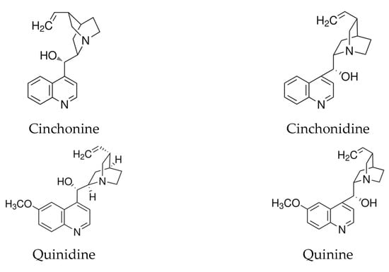
In this study, we also tested other cinchona alkaloids, including cinchonidine, quinine, and quinidine, considering that cinchonine and cinchonidine, as well as quinine and quinidine, are stereoisomers with the same molecular formulas and similar structures (Figure 4).

Figure 4.
Molecular structures of cinchonine, cinchonidine, quinidine, and quinine as cinchona alkaloids. Cinchonidine and cinchonine are stereoisomers; quinine and quinidine are structurally identical to cinchonidine and cinchonine, respectively, except for an additional methoxy group at the C6′ position of the quinoline ring.
We found that only SP-Pt electrodes with electrodeposited cinchonine were sufficiently stable for subsequent determinations. The balance between the adsorption strength of these molecules on the Pt surface and their varying solubilities can explain the behavior of the different cinchona alkaloids. This observation is consistent with findings reported by Zaera, who clarified certain apparent contradictions in the literature regarding the effectiveness of these four cinchona alkaloids as chiral catalysis promoters [43]. No favorable electrochemical conditions were identified for the deposition of quinine and cinchonidine, likely due to their significantly higher solubility in methanol compared to the other two alkaloids [46]. Additionally, the electrode with deposited quinidine was found to be insufficiently stable for measurements in neutral aqueous solutions due to its greater solubility in this medium.
3.3. Selectivity
Several expected organic species were selected to evaluate the selectivity of the cinchonine sensor (Table 3). The effect of potential interferents on the electrochemical determination of cinchonine was investigated by fixing the initial cinchonine concentration at 6.0 μg L−1 (10× LOD) and the interferent concentrations at 0.6 μg L−1 (1:0.1), 6.0 μg L−1 (1:1), or 60 μg L−1 (10×). Subsequently, varying amounts of cinchonine were added to the initial solution, and recovery studies conducted using the standard addition method showed satisfactory recovery rates ranging from 91.8% to 109.6% (Table 3). This indicates that all tested species caused no interference when present in concentrations up to 10 times higher than that of cinchonine, with the exception of quinidine. This behavior can be attributed to the structural similarity between quinidine and cinchonine, differing only by the presence of an additional methoxy group at the C6′ position of the quinoline ring (Figure 4). However, quinidine does not interfere when its concentration is less than 1/10 that of cinchonine. Additionally, the SP-Pt/CN electrode was successfully used for the determination of quinidine in the absence of cinchonine.

Table 3.
Recovery data related to the interference of different species in the determination of cinchonine using the proposed sensor. The cinchonine concentration was fixed at 6 μg L−1 in a 0.02 mol dm−3 phosphate buffer (pH 7.0) with 0.1 mol dm−3 KClO4 as the supporting electrolyte. The interferent concentrations were set at 0.6 μg L−1 (1:0.1), 6.0 μg L−1 (1:1), and 60 μg L−1 (10×).
3.4. Application of the Sensor: Determination of Cinchonine in Urine and Serum
The proposed SP-Pt/CN electrode was successfully applied for the determination of cinchonine in serum and urine samples obtained from the University Affiliated Hospital. The cinchonine concentration in human serum reaches its peak within 1–2 h after assimilation as a drug, which occurs rapidly and almost completely in the stomach and intestine. Approximately 3.6–6% of the total administered dose is excreted unchanged in the urine [58]. A series of sample solutions were prepared by adding an appropriate amount of cinchonine standard solution to 10.0 mL of serum or urine. Then, 1000 μL of each solution was transferred into the voltammetric cell and diluted to 10.0 mL with buffer solution. Subsequently, specific volumes of cinchonine standard solution were added using a micro-syringe, and differential pulse voltammograms (DPVs) were recorded after each standard addition. The results, summarized in Table 4, demonstrate that the SP-Pt/CN electrode enables the reliable determination of cinchonine, yielding satisfactory recovery values. Freshly prepared SP-Pt/CN electrodes can be stored before testing, as they remain stable.

Table 4.
Application of the SP-Pt/CN sensor for the determination of cinchonine concentration in urine and serum samples spiked with different amounts of cinchonine and diluted 10-fold with an electrolyte solution suitable for voltammetric measurements.
4. Conclusions
In this study, we demonstrate that cinchonine can be effectively deposited on a screen-printed platinum surface via the cathodic reduction of its hydrochloride salt dissolved in a methanolic solution. A stable cinchonine layer is formed when the deposition is performed for 60 s at a controlled potential of −220 mV vs. the silver standard electrode, with the temperature maintained at 10 °C. The repeatability and reproducibility of the modified electrode were assessed by calculating the relative standard deviation (RSD) of the peak current values from 10 consecutive DPV measurements (multiscan studies) conducted at a cinchonine concentration of 20.0 μg L−1, which lies in the middle of the linear range. The obtained RSD value confirms good reproducibility and indicates that the electrode surface remains stable during voltammetric measurements with the coated screen-printed electrode. Modified screen-printed platinum electrodes were successfully employed for cinchonine determination at physiological pH 7.0. The proposed sensor, which leverages the strong adsorption tendency of platinum for cinchonine along with the advantages of screen-printed electrodes, exhibited a very low detection limit and a wide linear dynamic range. The limits of detection (LOD) and quantification (LOQ) were 0.6 μg L−1 and 1.8 μg L−1, respectively, which are significantly lower than those reported for other conventional methods. Finally, the applicability of our sensor for clinical assays of cinchonine in human serum and urine was investigated, yielding promising results.
Supplementary Materials
The following supporting information can be downloaded at: https://www.mdpi.com/article/10.3390/s25072216/s1. Figure S1: ATR-FTIR spectra of pure cinchonine and deposited layers on Pt surfaces, dissolved in a CCl4 solution and concentrated drop by drop onto a ZnSe crystal, placed in the sample compartment of an ATR sampling module of the Cary 630 FTIR spectrophotometer. Figure S2: Mass spectrum of the deposited layers on Pt surfaces, dissolved in a CCl4 solution, derivatized with BSTFA, and injected into a GC-MS. Figure S3: Chronoamperometric curves recorded at various applied deposition potentials. Reference [59] is cited in the Supplementary Materials.
Author Contributions
Conceptualization, T.C. and L.P.; methodology, T.C.; software, T.C.; validation, T.C.; formal analysis, T.C.; investigation, T.C.; resources, T.C.; data curation, T.C.; writing—original draft preparation, T.C.; writing—review and editing, T.C.; visualization, T.C.; supervision, T.C.; funding acquisition, T.C. All authors have read and agreed to the published version of the manuscript.
Funding
This research was funded by i-Foria B.V., Amsterdam, The Netherlands, and the Icarus EU project (Funding Number 2023-1-IT02-KA220-ADU-000152409).
Institutional Review Board Statement
Not applicable.
Informed Consent Statement
Not applicable.
Data Availability Statement
Data are contained within the article and Supplementary Materials.
Conflicts of Interest
The authors declare no conflicts of interest. The funder had no role in the study design, data collection, analysis, or interpretation, nor in the writing of the manuscript or the decision to publish the results.
Abbreviations
The following abbreviations are used in this manuscript:
| SPE | Screen–printed electrode |
| SP-Pt/CN | Screen–printed platinum electrode coated with cinchonine |
| DPV | Differential pulse voltammetry |
| RSD | Relative standard deviation |
References
- Beitollahi, H.; Mohammadi, S.Z.; Safaei, M.; Tajik, S. Applications of electrochemical sensors and biosensors based on modified screen-printed electrodes: A review. Anal. Methods 2020, 12, 1547–1560. [Google Scholar] [CrossRef]
- Wang, X.; Zhang, Z.; Wu, G.; Xu, C.; Wu, J.; Zhanga, X.; Liu, J. Applications of electrochemical biosensors based on functional antibody-modified screen-printed electrodes: A review. Anal. Methods 2022, 14, 7–16. [Google Scholar] [CrossRef]
- Arduini, F.; Micheli, L.; Moscone, D.; Palleschi, G.; Piermarini, S.; Ricci, F.; Volpe, G. Electrochemical biosensors based on nanomodified screen-printed electrodes: Recent applications in clinical analysis. Trends Anal. Chem. 2016, 79, 114–126. [Google Scholar] [CrossRef]
- Crapnell, R.D.; Banks, C.E. Electroanalytical Overview: Screen-Printed Electrochemical Sensing Platforms. ChemElectroChem 2024, 11, e202400370. [Google Scholar] [CrossRef]
- Kelíšková, P.; Matvieiev, O.; Janíková, L.; Šelešovská, R. Recent advances in the use of screen-printed electrodes in drug analysis: A review. Curr. Opin. Electrochem. 2023, 42, 101408. [Google Scholar] [CrossRef]
- Bard, A.J. Chemical modification of electrodes. J. Chem. Educ. 1983, 60, 302–304. [Google Scholar] [CrossRef]
- Edwards, G.A.; Bergren, A.J.; Porter, M.D. Chemically Modified Electrodes. Handb. Electrochem. 2007, 8, 295–327. [Google Scholar] [CrossRef]
- Murray, R.W.; Ewing, A.G.; Durst, R.A. Chemically modified electrodes. Molecular design for electroanalysis. Anal. Chem. 1987, 59, 379A–390A. [Google Scholar] [CrossRef]
- Durst, R.A.; Baumner, A.J.; Murray, R.W.; Buck, R.P.; Andrieux, C.P. Chemically Modified Electrodes: Recommended Terminology and Definitions, IUPAC. Pure Appl. Chem. 1997, 69, 1317–1323. [Google Scholar] [CrossRef]
- White, H.S.; Leddy, J.; Bard, A.J. Polymer films on electrodes. 8. Investigation of charge-transport mechanisms in Nafion polymer modified electrodes. J. Am. Chem. Soc. 1982, 104, 4811–4817. [Google Scholar] [CrossRef]
- Zen, J.-M.; Kumar, A.S.; Tsai, D.-M. Recent Updates of Chemically Modified Electrodes in Analytical Chemistry. Electroanalysis 2003, 15, 1073–1087. [Google Scholar] [CrossRef]
- Perez-Ràfols, C.; Serrano, N.; Díaz-Cruz, J.M.; Arino, C.; Esteban, M. Mercury Films on Commercial Carbon Screen-Printed Devices for the Analysis of Heavy Metal Ions: A Critical Evaluation. Electroanalysis 2015, 27, 1345–1349. [Google Scholar] [CrossRef]
- Josypcuk, B.; Tvorynska, S. Screen-printed electrodes covered by mercury film or meniscus. Electrochim. Acta 2025, 513, 145565. [Google Scholar] [CrossRef]
- Siria, J.W.; Baldwin, R.P. Adsorption Pre-Concentration and Analysis of Dopamine at Platinum Electrode Surfaces. Anal. Lett. 1980, 13, 577–588. [Google Scholar] [CrossRef]
- Trasatti, S. Adsorption of organic substances at electrodes: Recent advances. Electrochim. Acta 1992, 37, 2137–2144. [Google Scholar] [CrossRef]
- Mittal, M.; Sardar, S.; Jana, A.E. Nanofabrication techniques for semiconductor chemical sensors. In Handbook of Nanomaterials for Sensing Applications; Micro and Nano Technologies; Elsevier: Amsterdam, The Netherlands, 2021; pp. 119–137. [Google Scholar] [CrossRef]
- Ayala, M.C.; López, L.L.; Jaramillo-Botero, A.; Valencia, D. Electrochemical modified electrode with bismuth film for ultrasensitive determination of aluminum (III). J. Electroanal. Chem. 2022, 919, 116552. [Google Scholar] [CrossRef]
- Parveen, S.; Maurya, N.; Meena, A.; Luqman, S. Cinchonine: A Versatile Pharmacological Agent Derived from Natural Cinchona Alkaloids. Curr. Top. Med. Chem. 2024, 24, 343–363. [Google Scholar] [CrossRef]
- Tracy, J.W.; Webster, J.L. Drugs used in the chemotherapy of protozoal infections. In The Pharmacological Basis of Therapeutics, 9th ed.; Hardman, J.G., Limbird, L.E., Molino, V.P.B., Ruddon, R.W., Gilman, A.G., Eds.; The McGraw Hill: New York, NY, USA, 1996; pp. 800–808. [Google Scholar]
- Sullivan, D.J. Cinchona Alkaloids: Quinine and Quinidine. In Treatment and Prevention of Malaria; Springer Basel AG.: Baltimore, MD, USA, 2012; Volume 41, pp. 45–68. [Google Scholar] [CrossRef]
- Rasoanaivo, P.; Wright, C.W.; Willcox, M.L.; Gilbert, B. Whole plant extracts versus single compounds for the treatment of malaria: Synergy and positive interactions. Malar. J. 2011, 10 (Suppl. S1), S4. [Google Scholar] [CrossRef]
- Levy, S.; Azoulay, S. Stories About the Origin of Quinquina and Quinidine. J. Cardiovasc. Electrophysiol. 1994, 5, 635–636. [Google Scholar] [CrossRef]
- Genne, P.; Duchamp, O.; Solary, E.; Pinard, D.; Belon, J.P.; Dimanche-Boitrel, M.T.; Chauffert, B. Comparative effects of quinine and cinchonine in reversing multidrug resistance on human leukemic cell line K562/ADM. Leukemia 1994, 8, 160–164. [Google Scholar] [PubMed]
- Shah, B.H.; Nawaz, Z.; Virani, S.S.; Ali, I.Q.; Saeed, S.A.; Gilani, A.H. The inhibitory effect of cinchonine on human platelet aggregation due to blockade of calcium influx. Biochem. Pharmacol. 1998, 56, 955–960. [Google Scholar] [CrossRef] [PubMed]
- Tang, D.; Wang, X.; Wu, J.; Li, Y.; Li, C.; Qiao, X.; Fan, L.; Chen, Y.; Zhu, H.; Zhang, Z.; et al. Cinchonine and cinchonidine alleviate cisplatin-induced ototoxicity by regulating PI3K-AKT signaling. CNS Neurosci Ther. 2024, 30, e14403. [Google Scholar] [CrossRef] [PubMed]
- Genne, P.; Ducham, O.; Solary, E.; Magnette, J.; Belon, J.P.; Chauffert, B. Cinchonine per os: Efficient circumvention of P-glycoprotein-mediated multidrug resistance. Anticancer Drug Des. 1995, 10, 103–118. [Google Scholar] [PubMed]
- Johnson, C.C.; Poe, C.F. Toxicity of Some Cinchona Alkaloids. Acta Pharmacol. 1948, 4, 265–274. [Google Scholar] [CrossRef]
- Council of Europe. European Pharmacopoeia, 8th ed.; Cinchona bark; Council of Europe: Strasbourg, France, 2016; pp. 1208–1209. [Google Scholar]
- Grant, H.S.; Jones, H.J. Spectrophotometric Determination of Cinchona Alkaloids. Anal. Chem. 1950, 22, 679–681. [Google Scholar] [CrossRef]
- Murauer, A.; Ganzera, M. Quantitative determination of major alkaloids in Cinchona bark by Supercritical Fluid Chromatography. J. Chromatogr. A 2018, 1554, 117–122. [Google Scholar] [CrossRef]
- Holmfred, E.; Cornett, C.; Maldonado, C.; Rønsted, N.; Honoré Hansen, S. An Optimised Method for Routine Separation and Quantification of Major Alkaloids in Cortex Cinchona by HPLC Coupled with UV and Fluorescence Detection. Phytochem. Anal. 2017, 28, 374–380. [Google Scholar] [CrossRef]
- Horie, M.; Oishi, M.; Ishikawa, F.; Shindo, T.; Yasui, A.; Ogino, S.; Ito, K. Liquid Chromatographic Analysis of Cinchona Alkaloids in Beverages. J. AOAC Int. 2006, 89, 1042–1047. [Google Scholar] [CrossRef]
- Yin, F.; Xu, X. Construction and Analytical Application of a Novel Ion-Selective Capacitive Sensor for Determination of Cinchonine. Anal. Lett. 2004, 37, 3129–3147. [Google Scholar] [CrossRef]
- Hu, W.; Wu, N.; Li, D.; Yang, Y.; Qie, S.; Su, S.; Xu, R.; Li, W.; Hu, M. The fluorescence distinction of chiral enantiomers: A Zn coordination polymer sensor for the detection of cinchonine and cinchonidine. J. Mater. Chem. C 2025, 13, 592–599. [Google Scholar] [CrossRef]
- Dushna, O.; Dubenska, L.; Marton, M.; Hatala, M.; Vojs, M. Sensitive and selective voltammetric method for determination of quinoline alkaloid, quinine in soft drinks and urine by applying a boron-doped diamond electrode. Microchem. J. 2023, 191, 108839. [Google Scholar] [CrossRef]
- Yang, Y.; Yang, X.; Yang, H.-F.; Liu, Z.-M.; Liu, Y.-L.; Shen, G.-L.; Yu, R.-Q. Electrochemical sensor for cinchonine based on a competitive host–guest complexation. Anal. Chim. Acta 2005, 528, 135–142. [Google Scholar] [CrossRef]
- Prideaux, E.B.R.; Winfield, F.T. The determination of quinine, cinchonine and cinchonidine with the quinhydrone electrode, and the choice of end-points in alkaloidal titrations. Analyst 1930, 55, 561–565. [Google Scholar] [CrossRef]
- Yuan, J.-B.; Tan, Y.-G.; Nie, L.-H.; Yao, S.-Z. Piezoelectric quartz crystal sensors based on ion-pair complexes for the determination of cinchonine in human serum and urine. Anal. Chim. Acta 2002, 454, 65–74. [Google Scholar] [CrossRef]
- Blaser, H.U.; Jalett, H.P.; Monti, D.M.; Reber, J.F.; Wehrli, J.T. Modified Heterogeneous Platinum Catalysts for the Enantioselective Hydrogenation of α-Ketoesters. Stud. Surf. Sci. Catal. 1998, 41, 153–163. [Google Scholar] [CrossRef]
- Ma, Z.; Zaera, F. Competitive Chemisorption between Pairs of Cinchona Alkaloids and Related Compounds from Solution onto Platinum Surfaces. J. Am. Chem. Soc. 2006, 128, 16414–16415. [Google Scholar] [CrossRef]
- Fietkau, N.; Bussar, R.; Baltruschat, H. The stability of adsorbed quinoline and cinchonine on poly- and monocrystalline platinum surfaces. Electrochim. Acta 2006, 51, 5626–5635. [Google Scholar] [CrossRef]
- Bakos, I.; Szabo, S.; Bartok, M.; Kalman, E. Adsorption of cinchonidine on platinum: An electrochemical study. J. Electroanal. Chem. 2002, 532, 113–119. [Google Scholar] [CrossRef]
- Ma, Z.; Lee, I.; Zaera, F. Factors Controlling Adsorption Equilibria from Solution onto Solid Surfaces: The Uptake of Cinchona Alkaloids on Platinum Surfaces. J. Am. Chem. Soc. 2007, 129, 16083–16090. [Google Scholar] [CrossRef]
- Hariyanti, H.; Kurniati, N.F.; Sumirtapura, Y.C.; Mauludin, R. Development and validation of an analytical method for the determination of nanostructured lipid carrier’s cinchonine used direct method modified by liquid-liquid extraction using high-performance liquid chromatography. J. Res Pharm. 2023, 27, 913–923. [Google Scholar] [CrossRef]
- Brankovic, S.R. Electrochemical Deposition as Surface Controlled Phenomenon: Fundamentals and Applications. J. Electrochem. Soc. 2016, 163, Y21. [Google Scholar] [CrossRef]
- Ma, Z.; Zaera, F. Role of the Solvent in the Adsorption-Desorption Equilibrium of Cinchona Alkaloids Between Solution and a Platinum Surface: Correlations among Solvent Polarity, Cinchona Solubility, and Catalytic Performance. J. Phys. Chem. B 2005, 109, 406–414. [Google Scholar] [CrossRef] [PubMed]
- Long, G.L.; Winefordner, J.D. Limit of detection: A closer look at the IUPAC detection. Anal. Chem. 1983, 55, 712A–724A. [Google Scholar] [CrossRef]
- Thompson, M.; Ellison, S.L.R.; Wood, R. Harmonized guidelines for single laboratory validation of methods of analysis (IUPAC Technical Report). Pure Appl. Chem. 2002, 74, 835–855. [Google Scholar] [CrossRef]
- Exner, C.; Pfaltz, A.; Studer, M.; Blaser, H.-U. Heterogeneous Enantioselective Hydrogenation of Activated Ketones Catalyzed by Modified Pt-Catalysts: A Systematic Structure-Selectivity Study. Adv. Synth. Catal. 2003, 345, 1253–1260. [Google Scholar] [CrossRef]
- Halli, P.; Heikkinen, J.J.; Elomaa, H.; Wilson, B.J.; Jokinen, V.; Yliniemi, K.; Franssila, S.; Lundström, M. Platinum Recovery from Industrial Process Solutions by Electrodeposition–Redox Replacement. ACS Sustain. Chem. Eng. 2018, 6, 14631–14640. [Google Scholar] [CrossRef]
- Posada, J.O.G.; Hall, P.J. Controlling hydrogen evolution on electrodes. Int. J. Hydrog. Energy 2016, 41, 20807–20817. [Google Scholar] [CrossRef]
- Ma, F.; Lennox, R.B. Potential-Assisted Deposition of Alkanethiols on Au: Controlled Preparation of Single- and Mixed-Component SAMs. Langmuir 2000, 16, 6188–6190. [Google Scholar] [CrossRef]
- Bockris, J.O.M.; Koch, D.F.A. Comparative rates of the electrolytic evolution of hydrogen on iron, tungsten, and platinum. J. Phys. Chem. 1961, 65, 1941–1948. [Google Scholar] [CrossRef]
- Chunga, C.-K.; Chang, W.-T. Handbook of Manufacturing Engineering and Technology; Springer: London, UK, 2013. [Google Scholar] [CrossRef]
- Bard, A.J.; Faulkner, L.R. Electrochemical Methods: Fundamentals and Applications, 2nd ed.; Wiley and Sons: New York, NY, USA, 2001. [Google Scholar]
- Kissinger, P.T.; Heineman, W.R. Laboratory Techniques in Electroanalytical Chemistry, 2nd ed.; Marcel Dekker, Inc.: New York, NY, USA, 1996; ISBN 0-8247-9445-1. [Google Scholar]
- Moosavi, S.M.; Ghassabian, S. Linearity of Calibration Curves for Analytical Methods: A Review of Criteria for Assessment of Method Reliability. In Calibration and Validation of Analytical Methods—A Sampling of Current Approaches; Mark, T., Ed.; IntechOpen Limited: London, UK, 2018; Chapter 6; pp. 109–127. [Google Scholar] [CrossRef]
- Brodie, B.B.; Baer, J.E.; Craig, L.C. Metabolic products of the cinchona alkaloids in human urine. J. Biol. Chem. 1951, 188, 567–581. [Google Scholar] [CrossRef]
- Isidorov, V.A. GC-MS of Biologically and Environmentally Significant Organic Compounds: TMS Derivatives; MS spectrum of Cinchonine, monoTMS; John Wiley & Sons: Hoboken, NJ, USA, 2020; p. 481. [Google Scholar]
Disclaimer/Publisher’s Note: The statements, opinions and data contained in all publications are solely those of the individual author(s) and contributor(s) and not of MDPI and/or the editor(s). MDPI and/or the editor(s) disclaim responsibility for any injury to people or property resulting from any ideas, methods, instructions or products referred to in the content. |
© 2025 by the authors. Licensee MDPI, Basel, Switzerland. This article is an open access article distributed under the terms and conditions of the Creative Commons Attribution (CC BY) license (https://creativecommons.org/licenses/by/4.0/).