Article

The Innovative XClinic Tool: A Pilot Study Validating Its Precision in Measuring Range of Motion in Healthy Individuals

1
Department of Human Neurosciences, Sapienza University of Rome, 00185 Rome, Italy
2
Department of Public Health and Infectious Diseases, Sapienza University of Rome, 00185 Rome, Italy
3
IRCCS Neuromed, Via Atinense, 18, 86077 Pozzilli, Italy
4
Euleria Health, Via delle Zigherane 4/A, 38068 Trento, Italy
5
Department of Life, Health and Environmental Sciences, University of L’Aquila, 67010 L’Aquila, Italy
6
Department of Anatomical, Histological, Forensic and Orthopaedical Sciences, Sapienza University of Rome, 00185 Rome, Italy
*
Author to whom correspondence should be addressed.
Sensors 2025, 25(5), 1331; https://doi.org/10.3390/s25051331
Submission received: 19 December 2024 / Revised: 16 February 2025 / Accepted: 18 February 2025 / Published: 21 February 2025
(This article belongs to the Section Wearables)

Abstract

:
Background: Kinematics experts and physical therapists have implemented the use of sensors for 3D motion analysis, both for static and dynamic movements. XClinic movement sensors are advanced devices designed to analyze movement patterns with high precision. The aim of this study was to validate wearable XClinic sensors for range of motion (ROM) in healthy subjects and obtain normative data. Participants were enrolled at the Sapienza University of Rome in 2024. All participants had to be healthy subjects aged between 18 and 65 years. Data on their demographics, employment and physical activity were collected. All the subjects were tested to assess the active ROM of their shoulder, hip, knee and ankle bilaterally. The same movements were tested using a goniometer to investigate validity, and SF-36 was administered. Fifty subjects were enrolled. The mean age was 28.2 (SD 10.8) years. For the left shoulder, construct validity showed statistically significant values for flexion, extension and extra-rotation, while for the right shoulder, construct validity showed statistically significant values for all movements except intra-rotation. The results concerning the right hip showed statistically significant values for flexion, extra-rotation, intra-rotation and adduction. The left hip showed statistically significant values for all movements except extension. Both the right and left knees showed statistically significant values for flexion. Both the right and left ankles showed statistically significant values for all movements. XClinic sensors offer a reliable and valid solution for the precise monitoring of the ROM of the shoulder and lower limb joints, making them an invaluable asset for clinicians and researchers.

1. Introduction

Rapid technological developments have occurred in recent years. Computer- and robot-assisted forms of therapy have proven to be an important component for optimizing rehabilitation [1,2]. The monitoring and analysis of bioelectrical signals and movements can aid in the diagnosis, prevention and examination of a wide range of issues. Remote monitoring and ambulatory monitoring are growing needs in the healthcare environment [3,4]. Objective and quantitative measurement is paramount in a detailed and reliable physical examination [5,6]. In recent years, biomedical engineers, kinematics experts and physical therapists have developed and implemented into research settings the use of quantitative sensors for 3D motion analysis, both for static and dynamic movements close to real life conditions described by patients and athletes [7,8]. To date, improvements in this field, including in the quality of data collection, a reduction in economic costs and user-friendly portable devices, have made these technologies increasingly widespread [9]. A real revolution started when kinematic sensors became wireless and wearable, allowing them to analyze ergonomics in the workplace or athletes’ performance on the field [10,11,12]. Among them, inertial sensors seem to be reliable, cost-effective and practical because they can integrate 3D data from a gyroscopic sensor, a magnetometer and an accelerometer to output a stable coordinate system expressed by Euler angles [13,14]. This new device should be tested to make the results equivalent to conventional measurement methods such as goniometry, dynamometry, optoelectronic kinematic analysis and ground contact force timing [15,16]. The possibility of using them as a certified alternative to common measurement methods in everyday clinical practice is interesting both for clinicians and patients, because with one device, a professional can collect more reliable data-saving time, money and space [17]. Furthermore, it is possible to have real-time feedback, which can be used for incorporating functional exercise prescriptions, telemedicine and objective outcome measures into rehabilitation programs and follow-ups [18]. XClinic movement sensors by Ferrox (Codognè, Italy) are advanced devices designed to monitor and analyze physical activities and movement patterns with high precision. In particular, they are able to measure an active range of motion of the shoulder, hip, knee and ankle and to assess balance in different positions. These sensors are particularly utilized in healthcare, sports and rehabilitation to provide detailed insights into a person’s motion. XClinic sensors are designed to provide precise and objective assessments of joint movement. These wearable and wireless devices are based on inertial sensors (accelerometer, gyroscope and magnetometer), allowing real-time monitoring of movement with high sensitivity. The key innovation of XClinic sensors lies in their ability to integrate and analyze data from multiple sensors, generating highly reliable measurements and reducing the margin of error typically associated with manual assessments, such as those performed with a traditional goniometer. Due to their compact and portable design, these sensors can be used not only in clinical and rehabilitation settings but also for sport performance monitoring and ergonomic applications, enabling a more detailed analysis of movement biomechanics. Additionally, the system’s digital interface allows for data storage and comparison over time, facilitating patient follow-ups and optimizing therapeutic strategies (https://www.ferrox.it/riabilitazione-digitale/, accessed on 20 February 2025). Overall, XClinic movement sensors by Ferrox provide an advanced solution for tracking and analyzing physical activity, delivering valuable insights that can significantly enhance health, performance and overall well-being [19].
For example, in post-surgical recovery (e.g., after knee or shoulder reconstruction), they can enable continuous tracking of ROM progress, ensuring that therapeutic interventions are optimized. In neurological rehabilitation, such as for patients with stroke-induced motor impairments, XClinic sensors can facilitate the precise monitoring of movement recovery and support tailored tele-rehabilitation programs. Moreover, in injury prevention, these sensors can detect subtle movement asymmetries that may predispose individuals to musculoskeletal injuries, allowing for early intervention strategies. XClinic sensors improve upon traditional methods like goniometry and optoelectronic motion capture by offering a portable, cost-effective and user-friendly solution. Unlike marker-based motion analysis, which requires specialized lab setups, XClinic sensors can be used in real-world settings—from rehabilitation clinics to sports training environments—without external calibration systems. Additionally, their real-time data processing and cloud-based integration enable automated movement analysis, potentially enhancing AI-driven rehabilitation protocols.
To further aid comprehension, incorporating conceptual diagrams illustrating sensor placement, data acquisition and clinical application scenarios would be beneficial. A comparison table between traditional ROM assessment methods and XClinic sensors could also highlight key advantages in terms of accuracy, portability and cost-effectiveness.

Motivation

Recent advancements in biomechanical assessment and rehabilitation have highlighted the need for more precise, objective and user-friendly tools for monitoring joint mobility. Traditional methods, such as manual goniometry, often rely on the expertise of the clinician, leading to variability in measurements and requiring time-consuming procedures that are difficult to standardize across different settings. The XClinic system addresses these limitations by integrating wearable inertial sensors (accelerometers, gyroscopes and magnetometers), allowing for real-time, wireless and highly accurate range of motion (ROM) assessments. Unlike optical motion capture systems, which require laboratory setups, XClinic provides a portable and cost-effective alternative that can be used in clinical rehabilitation, sports performance monitoring and workplace ergonomics. In rehabilitation, it enables the continuous tracking of motor recovery in patients recovering from injuries or neurological conditions, optimizing treatment strategies. In sports, it allows for detailed movement analysis, helping athletes prevent injuries and enhance performance. Moreover, in occupational health, it facilitates postural and movement assessments to prevent work-related musculoskeletal disorders. By offering automated, reproducible and user-friendly motion analysis, XClinic enhances traditional biomechanical evaluations, making precise ROM tracking more accessible across multiple fields.
The aim of this study is to validate XClinic wearable sensors for ROM assessment in a sample of healthy subjects, to investigate their psychometric properties and obtain normative data on the devices.

2. Materials and Methods

This study was conducted by a research group at the Sapienza University of Rome named “Riabilitazione Evidenze e Sviluppo (RES)”, who were involved in different studies on rehabilitation [20,21,22].

2.1. Participants

The study was performed in accordance with the ethical standards as laid down in the 1975 Declaration of Helsinki and its later amendments. The participants of this cross-sectional study were enrolled at the Sapienza University of Rome from September 2023 to February 2024. Informed consent was obtained from all subjects involved in the study [23,24]. All participants had to be healthy subjects, with a Short Form Health Survey-36 (SF-36) score at least of 40% in the domain “General Health”; a Mini Mental State Examination (MMSE) score of over 24; and aged between 18 and 65 years. Exclusion criteria were road accidents and/or surgery for other reasons in the last year; neurological or cardio-pulmonary diseases; being pregnant and psychiatric conditions.

2.2. Data Collection

For each subject the following data were collected: demographics, Body Mass Index (BMI), employment and physical activity. All the subjects were tested to assess their active Range of Motion (ROM) relative to the following joints: shoulder, hip, knee and ankle bilaterally. For each joint, specific movements were tested:
-
Shoulder: flexion, extension, abduction, internal rotation, external rotation.
-
Hip: flexion (with flexed knee), extension, abduction, adduction, internal rotation, external rotation.
-
Knee: flexion, extension.
-
Ankle: plantar flexion, dorsiflexion, inversion, eversion.
The same movements were tested using a goniometer, the gold standard for the assessment of ROM, to investigate construct validity; also, muscle strength was tested using the Medical Research Council (MRC) scale, considering the following muscles, respectively, in the upper and lower limbs:
Upper limb muscles: anterior and posterior deltoid, pectoralis major, supraspinatus, subscapularis, infraspinatus, teres minor, biceps brachii and triceps brachii.
Lower limb muscles: iliopsoas, gluteus maximus, hamstring muscles, gluteus medius, gluteus minor, tensor fasciae latae, quadriceps, adductors, tibialis anterior, gastrocnemius, soleus, tibialis posterior, peroneus longus and peroneus brevis.
In addition to ROM and strength assessment, a Short Form Health Survey 36 (SF-36) questionnaire was administered to subjects [25].
The ROM assessment with both the goniometer and Xclinic sensors, the strength test and the administration of questionnaires were performed by three physiotherapists. Goniometer evaluation was performed following Hazel and Clarkson guidelines [26].

2.3. Evaluation Using Sensors

The Xclinic kit (Version 10) was composed as follows: two wearable sensors, charging equipment, adjustable straps and bands to secure the sensors in place on the body, instructional material and a user interface device (a tablet in this case) with the Xclinic app installed.
To effectively use the Xclinic sensors by Ferrox, the following steps were carried out:
-
Unpacking and Initial Setup: sensors and related equipment were carefully unpacked and a check for any user manuals or quick start guides included in the package was performed.
-
Powering the Sensors: before each evaluation, the operators ensured that the sensors were charged or connected to a power source.
-
Connecting to a Network: specifically, to a local Wi-Fi network. The manufacturer’s instructions for connecting to the network were followed.
-
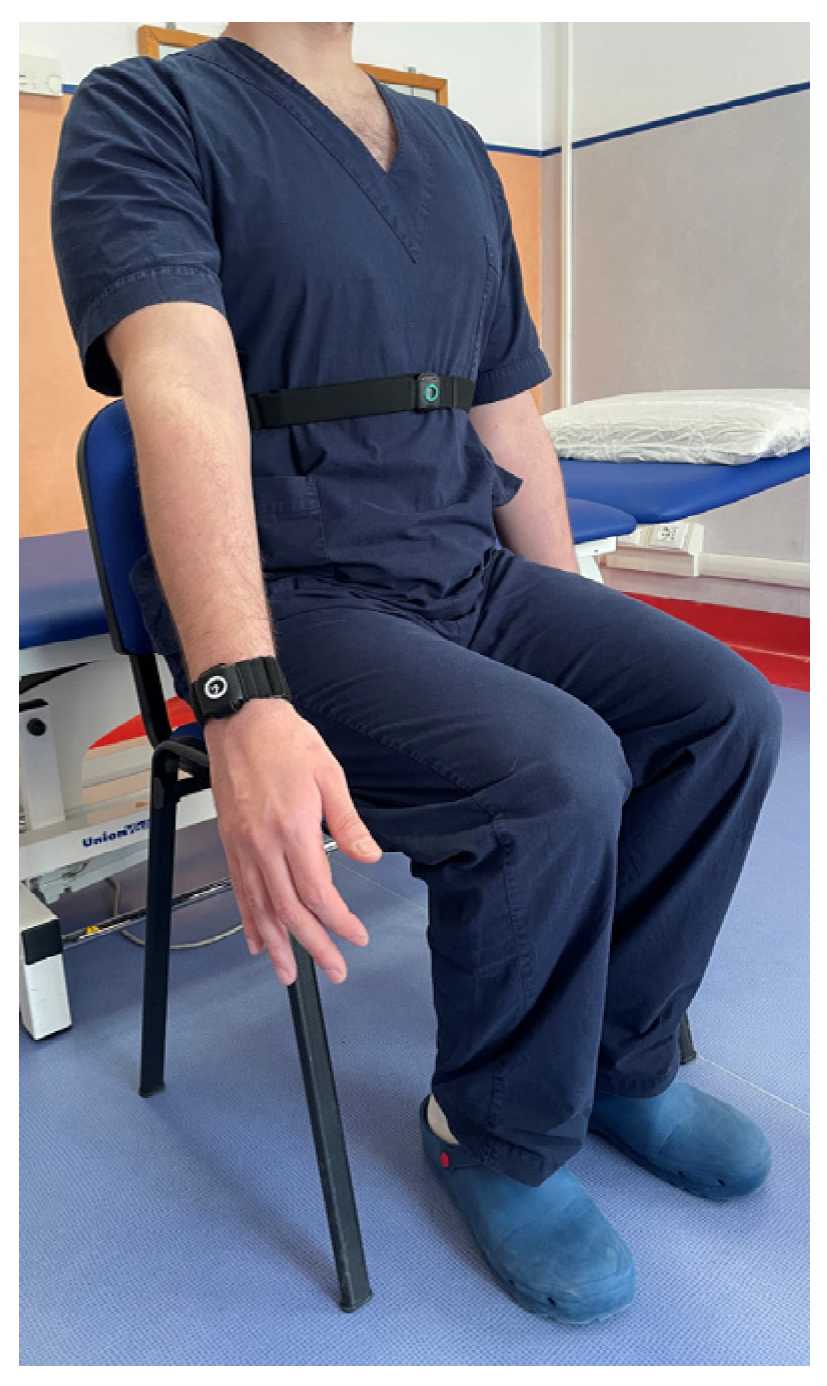
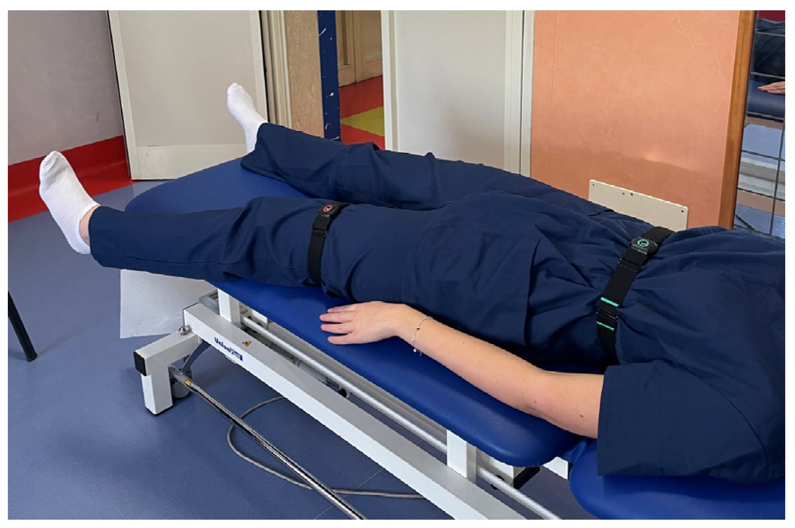
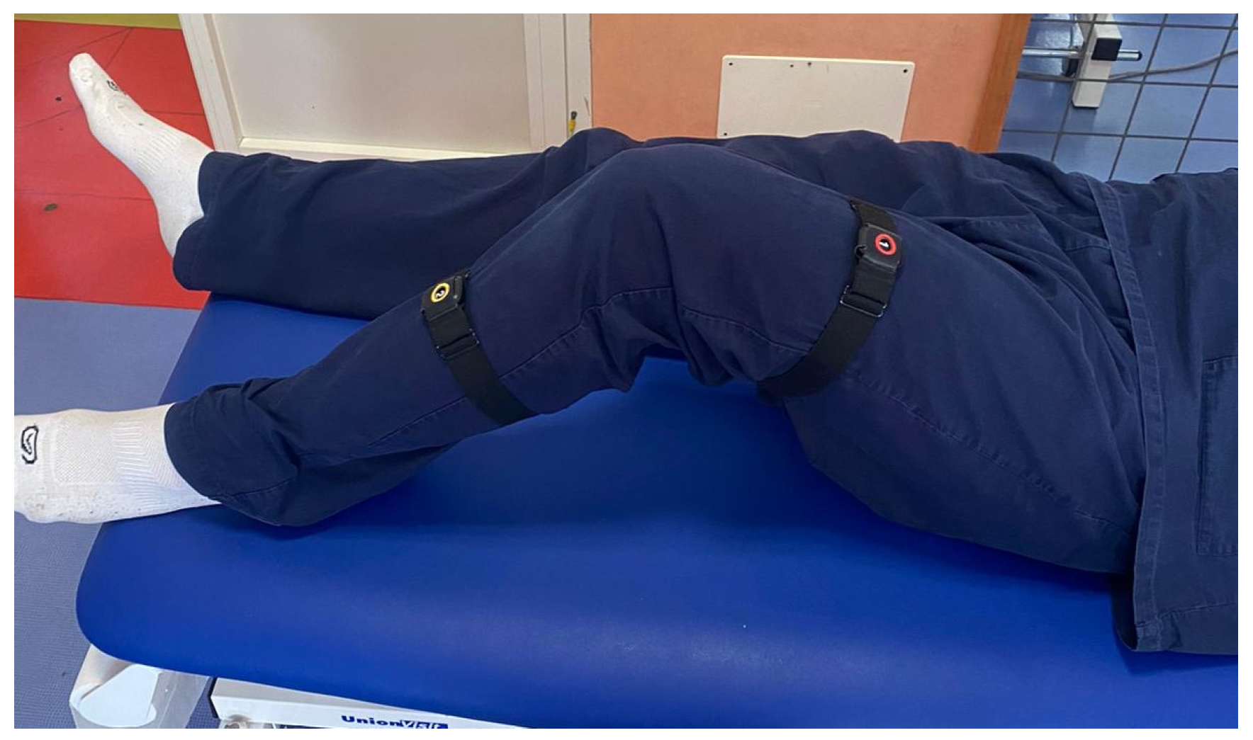
Sensor Placement: sensors were placed in the locations to be monitored, precisely in correspondence with the upper and lower segment of the considered joint, to register the angle of movement. The operators ensured that the placement followed the guidelines provided by Ferrox. For each joint, the sensors were placed as follows: on the thorax and wrist for the shoulder (Figure 1), on the thorax and thigh for the hip (Figure 2), on the thigh and lower leg for the knee (Figure 3) and on the lower leg and foot for the ankle (Figure 4). This placement allowed the sensors to capture the relative motion between the segments, ensuring an accurate assessment of joint angles. Participants wore the Xclinic sensors during all movements.
-
Configuration and Calibration: the software was used to configure and calibrate the sensors. The calibration process began by instructing the participant to remain immobile in a neutral anatomical position, allowing the software to establish baseline sensor offsets; a standardized setup and calibration process was followed.
Once a unique account was created for each subject in the Xclinic software (v. 1.0) and calibration was successfully completed, the assessment of range of motion (ROM) could begin. The software continuously recorded and stored movement data throughout the trials.
Participants followed a predefined movement protocol, which included specific instructions regarding range of motion and number of repetitions (3 for each movement). A trained researcher supervised each session to ensure correct execution and proper sensor function. Additionally, data collection was performed at a fixed sampling rate to ensure uniformity across participants. These procedures were implemented to guarantee the reliability and reproducibility of the measurements, minimizing any variability due to sensor misalignment or inconsistencies in movement execution.
The measurements using the Xclinic sensors and the goniometer were carried out separately, following a specific order: First, the range of motion (ROM) was assessed using the Xclinic sensors, and only afterward was the manual goniometer measurement performed. This sequence was chosen to ensure that the digital measurement remained unaffected by any prior manual positioning of the limb during the goniometric evaluation. To minimize variability between the two methods, several standardization procedures were followed. Participants were asked to perform the same predefined movements for both assessments. Additionally, the goniometric measurements were always taken by the same trained examiner to reduce any potential variability.

2.4. Statistical Analyses

The statistical analyses were performed using SPSS Statistics version 27. Demographic characteristics were reported as mean ± standard deviation (SD) or percentage, depending on the type of data. Internal consistency was assessed through item-by-item analyses for each movement, while construct validity was evaluated using Pearson’s correlation, comparing the ROM values obtained with the Xclinic sensors to those measured with a goniometer. Pearson’s correlation was also used to examine the relationship between ROM and SF-36 scores. The study followed the guidelines outlined in the “COnsensus-based Standards for the selection of health Measurement Instruments” (COSMIN) [27].
All data were initially entered into an Excel database, ensuring a structured and organized dataset before statistical analysis.
Once all measurements were collected, the raw data from Excel were imported into SPSS for processing. SPSS generated a new database where data procedures were applied to calculate psychometric properties. Descriptive statistics were then used to calculate the mean and standard deviation for each movement. A Pearson’s correlation analysis was performed to assess the relationship between the two measurement methods, as well as the association between ROM values and SF-36 scores. To evaluate reliability, internal consistency was analyzed through item-by-item correlations.
The final tables presented in Section 3 were derived from the SPSS database.

3. Results

3.1. Characteristics of the Sample

Fifty healthy subjects were enrolled. The mean age of the sample was 28.2, with a standard deviation of 10.8; 58% of the sample was composed by females. Among the included subjects 52% practiced sport, while 54% were workers and 76% were students. These results are shown in Table 1.

3.2. Reliability

Item-by-Item Analyses

The item-by-item evaluation of the left shoulder highlighted a statistically significant moderate direct correlation between flexion and extension, and intra and extra rotation. The results are shown in Table 2. The item-by-item evaluation of the right shoulder highlighted a moderate and statistically significant correlation between flexion and extension, extension and intra-rotation and intra and extra-rotation. The results for direct and inverse correlations are shown in Table 3.
As regards the item-by-item analyses of the left hip, the results show that there is a statistically significant inverse correlation between extension and extra rotation, adduction and abduction and extension and extra rotation. Right hip analyses show statistically significant direct correlations between intra and extra rotation, adduction and extra rotation, adduction and extra rotation, adduction and flexion, extension and extra rotation and flexion and extension. Data on direct and inverse correlations are shown in Table 4 and Table 5.
The item-by-item evaluation of the right knee highlighted a statistically significant direct correlation between flexion and extension ROM (Table 6 and Table 7).
The item-by-item evaluation of the right ankle showed a statistically significant correlation between the following movements: plantar and dorsal flexion, inversion and plantar flexion, eversion and dorsal flexion and eversion and inversion. Left ankle analyses highlighted a statistically significant correlation between dorsal and plantar flexion, eversion and dorsal flexion and inversion and dorsal flexion. The results for direct and inverse correlations are shown in Table 8 and Table 9.
Finally, item-by-item analyses was also performed with regards to shoulder ROM and SF 36 (Table 10). These highlighted a statistically significant correlation between left shoulder flexion and general health; shoulder extension and level of energy/fatigue; abduction and role limitations due to physical health; role limitations due to emotional problems, social functioning and pain and extra rotation and role limitations due to emotional problems and social functioning.

3.3. Construct Validity (Goniometric Evaluation)

Construct validity was carried out by comparing the value obtained through the sensors’ assessments with the goniometric values; the results concerning the left shoulder showed statistically significant values for flexion, extension and extra-rotation (moderate statistically significant correlations, p < 0.01). The results are reported in Table 11. The results concerning the right shoulder showed statistically significant values for all the movements except for intra-rotation (moderate statistically significant correlations, p < 0.01). The results are shown in Table 12 and Table 13.
The results concerning the right hip showed statistically significant values for flexion, extra-rotation, intra rotation and adduction. The results for the left hip showed statistically significant values for all the movements except for extension. Data are shown in Table 13 and Table 14.
The data regarding both the right and left knee showed statistically significant values for flexion ROM (Table 15 and Table 16).
The results regarding both the right and left ankle showed statistically significant values for all the assessed movements. Correlations are shown in Table 17 and Table 18.

3.4. Construct Validity (SF-36)

Construct validity was also performed by comparing the values obtained through sensors assessments with the scores obtained in several domains of SF-36 survey. Correlations are shown in Table 19, Table 20, Table 21 and Table 22.

4. Discussion

The objective of the study was to validate XClinic wearable sensors for ROM assessment in a sample of healthy subjects, and to investigate its psychometric properties. Item-by-item analyses were performed to evaluate the internal consistency of the Xclinic sensors. These analyses showed statistically significant correlations between specific movements; in particular, it is interesting to observe the inverse correlation found between flexion and extension in the shoulder joint. This result was also found between abduction and adduction in the hip joint, and in flexion and extension in the knee joint. An inverse correlation also exists between the following movements of the ankle: inversion and eversion, and plantar and dorsal flexion. These data suggest that Xclinic sensors are able to detect the difference between opposite movements in space; to date, no studies have investigated the internal consistency of measurements using inertial sensors.
This finding is consistent with the expected behavior of these joints, where an increase in one movement naturally corresponds to a decrease in the opposite movement. The fact that the Xclinic sensors detected these relationships suggests that they provide reliable and internally consistent measurements.
Secondly, the construct validity of the device was investigated through comparisons with goniometer measurements. The current literature is not extensive regarding studies focusing on the validation of inertial sensors vs. goniometers in measuring the ROM of joints. In our study, statistically significant values were found, in particular when comparing shoulder flexion and extension—in fact, Xclinic data relative to these movements were similar to those collected using a goniometer. This data is in line with the current literature, as stated in the study of Kaszyński et al. [28]. As regards both the hip and knee joint, the flexion ROM obtained the most significant values—that is to say, this represented the movement that most correlated with the values measured with the goniometer. In general, rotation movements show less agreement with those measured with the goniometer; this resulted in these movements being more complicated to measure, because the equipment required more accurate placing, and this suggests the necessity of training professionals to deal with Xclinic sensors. This data on rotation assessments is also concordant with the current literature [29]. The joint that showed more statistically significant correlations between goniometer and Xclinic evaluation was the ankle, with regards to all the studied movements (plantar and dorsal flexion, inversion, eversion); the reason for this could be that these are smaller movements, so they are easier to perform and to assess with both the device and goniometer. To date, as far as we know, no studies have investigated the validity of inertial sensors specifically in the ankle joint for healthy individuals.
However, the study presents some limitations: Firstly, the sample is composed of only 50 subjects; it would be recommended to increase the number to obtain more significant data. In addition, the goniometer is considered a standard tool for assessing the ROM of any joint in the physiotherapeutic or orthopedic field, but it has a significant limitation: the obtained results often depend on the professional’s skills and experience [30]. Recommendations for future studies should concern a more detailed investigation of the tool’s psychometric properties, such as its reliability, to prove its reproducibility over time, as well as its responsiveness, to demonstrate its capacity to detect changes in specific clinical conditions.

5. Medico-Legal Implications

From a medico-legal perspective, this technological aid—aimed at the analysis of joint ROM—can find different fields of application. First of all, according to Law 219/2017, it is always important to respect the patient’s health and to act in total safety, thus providing all the necessary information about the treatment and its purposes. In addition, the correct assessment of joint functional limitations and postural analysis is the fulcrum of patient assessment in the field of social security medicine. Given the great sensitivity to the issue of disability, an objective and scientifically valid assessment provides an excellent aid for patients who need social benefits. Finally, it should be remembered that, in terms of civil liability, victims of road accidents are entitled to compensation, paid on the basis of the biological damage caused, which will be quantified on the basis of the clinical and instrumental data. In this case too, a meticulous damage evaluation, supported by specific markers, makes it possible to provide an accurate estimate of the functional limitation—allowing on the one hand the formulation of the right diagnosis and the correct therapeutic process, and also allowing the patient to obtain adequate compensation.

6. Conclusions

The evaluation of the Xclinic sensors by Ferrox underscores their substantial validity and reliability in clinical and environmental monitoring applications. Through rigorous testing and comparison with the established gold standard, these sensors have demonstrated a good degree of accuracy and consistency in data collection. Their robust design, ease of use, and advanced features ensure that they meet the demands of modern healthcare and environmental monitoring, providing professionals with dependable and precise data.
In summary, Xclinic sensors by Ferrox offer a reliable and valid solution for precise monitoring of the ROM of shoulder and lower limb joints, making them an invaluable asset for clinicians and environmental scientists alike. Their accuracy and consistency proven here can now reinforce their status as a leading choice in sensor technology, promoting better decision making and improved outcomes in their respective fields. Finally, this approach can have a great impact at the medico-legal level, both in terms of ethics and in the context of correct biological damage assessment.

Author Contributions

Conceptualization, G.V. and P.F.; Data curation, I.R.; Formal analysis, G.G.; Investigation, I.R. and E.A.; Methodology, G.G., E.A. and G.V.; Project administration, P.F.; Resources, G.S. and R.L.R.; Software, E.D.S.; Validation, G.G., R.L.R. and G.V.; Writing—original draft, I.R. and G.S.; Writing—review and editing, I.R. All authors have read and agreed to the published version of the manuscript.

Funding

This research received no external funding.

Institutional Review Board Statement

The study was conducted in accordance with the Declaration of Helsinki, and was approved by the Ethics Committee of Lazio Area 1, Policlinico Umberto I (protocol code 0969/2024 and date of approval 7 November 2024).

Informed Consent Statement

Informed consent was obtained from all subjects involved in the study.

Data Availability Statement

The raw data supporting the conclusions of this article will be made available by the authors on request.

Acknowledgments

We would like to express our gratitude to “Associazione Nazionale fra le Imprese Assicuratrici” (ANIA) for their generous support and funding of this research. Their assistance has been crucial in enabling the successful execution and completion of this study. We would also like to extend our special thanks to Umberto Guidoni, for his valuable support and encouragement throughout the project.

Conflicts of Interest

Enrica Di Sipio is affiliated with the company, Euleria Health. The authors declare no conflicts of interest.

References

  1. Jakob, I.; Kollreider, A.; Germanotta, M.; Benetti, F.; Cruciani, A.; Padua, L.; Aprile, I. Robotic and Sensor Technology for Upper Limb Rehabilitation. PM&R 2018, 10, S189–S197. [Google Scholar]
  2. Payedimarri, A.B.; Ratti, M.; Rescinito, R.; Vanhaecht, K.; Panella, M. Effectiveness of Platform-Based Robot-Assisted Rehabilitation for Musculoskeletal or Neurologic Injuries: A Systematic Review. Bioengineering 2022, 9, 129. [Google Scholar] [CrossRef] [PubMed]
  3. Nascimento, L.M.S.D.; Bonfati, L.V.; Freitas, M.L.B.; Mendes Junior, J.J.A.; Siqueira, H.V.; Stevan, S.L. Sensors and Systems for Physical Rehabilitation and Health Monitoring—A Review. Sensors 2020, 20, 4063. [Google Scholar] [CrossRef] [PubMed]
  4. Tormene, P.; Bartolo, M.; De Nunzio, A.M.; Fecchio, F.; Quaglini, S.; Tassorelli, C.; Sandrini, G. Estimation of human trunk movements by wearable strain sensors and improvement of sensor’s placement on intelligent biomedical clothes. Biomed. Eng. OnLine 2012, 11, 95. [Google Scholar] [CrossRef]
  5. Mohajan, H. Two Criteria for Good Measurements in Research: Validity and Reliability. Ann. Spiru Haret Univ. Econ. Ser. 2017, 17, 59–82. [Google Scholar] [CrossRef]
  6. Htay, M.; Kamath, G.; Anand, K.M.; Sugathan, S.; Luyee, E.; Sahoo, S. Overview of Reliability and Validity of Assessments in Medical Education. Int. J. Transform. Health Prof. Educ. 2023, 1, 14–18. [Google Scholar]
  7. Villa, S.; Casillas-Perez, D.; Jiménez Martín, A.; García, J. Inertial Sensors for Human Motion Analysis: A Comprehensive Review. IEEE Trans. Instrum. Meas. 2023, 72, 4006439. [Google Scholar] [CrossRef]
  8. Yusuf, K.M.S.T.; Ahmad Nazri, A.; Mustapha, G.; Mahmud, J. Analysis of static and dynamic motion accuracy for kinect-virtual Sensei system. ARPN J. Eng. Appl. Sci. 2015, 10, 7328–7335. [Google Scholar]
  9. Keri, M.I.; Shehata, A.W.; Marasco, P.D.; Hebert, J.S.; Vette, A.H. A Cost-Effective Inertial Measurement System for Tracking Movement and Triggering Kinesthetic Feedback in Lower-Limb Prosthesis Users. Sensors 2021, 21, 1844. [Google Scholar] [CrossRef]
  10. Seçkin, A.Ç.; Ateş, B.; Seçkin, M. Review on Wearable Technology in Sports: Concepts, Challenges and Opportunities. Appl. Sci. 2023, 13, 10399. [Google Scholar] [CrossRef]
  11. Lind, C.; Diaz-Olivares, J.; Lindecrantz, K.; Eklund, J. A Wearable Sensor System for Physical Ergonomics Interventions Using Haptic Feedback. Sensors 2020, 20, 6010. [Google Scholar] [CrossRef] [PubMed]
  12. Cossich, V.R.A.; Carlgren, D.; Holash, R.J.; Katz, L. Technological Breakthroughs in Sport: Current Practice and Future Potential of Artificial Intelligence, Virtual Reality, Augmented Reality, and Modern Data Visualization in Performance Analysis. Appl. Sci. 2023, 13, 12965. [Google Scholar] [CrossRef]
  13. Bergamini, E.; Ligorio, G.; Summa, A.; Vannozzi, G.; Cappozzo, A.; Sabatini, A. Estimating Orientation Using Magnetic and Inertial Sensors and Different Sensor Fusion Approaches: Accuracy Assessment in Manual and Locomotion Tasks. Sensors 2014, 14, 18625–18649. [Google Scholar] [CrossRef] [PubMed]
  14. Sabatini, A.M. Estimating three-dimensional orientation of human body parts by inertial/magnetic sensing. Sensors 2011, 11, 1489–1525. [Google Scholar] [CrossRef] [PubMed]
  15. Fonseca, M.C. Quantification, Reduction and Management of Kinematic Variability in Clinical Gait Analysis. Ph.D. Thesis, Université Claude Bernard-Lyon, Lyon, France, 2022. [Google Scholar]
  16. Everett, T.; Kell, C. Human Movement: An Introductory Text; Elsevier Health Sciences: Amsterdam, The Netherlands, 2010; p. 281. [Google Scholar]
  17. Khanna, N.N.; Maindarkar, M.A.; Viswanathan, V.; Fernandes, J.F.; Paul, S.; Bhagawati, M.; Ahluwalia, P.; Ruzsa, Z.; Sharma, A.; Kolluri, R.; et al. Economics of Artificial Intelligence in Healthcare: Diagnosis vs. Treatment. Healthcare 2022, 10, 2493. [Google Scholar] [CrossRef]
  18. Muñoz-Tomás, M.T.; Burillo-Lafuente, M.; Vicente-Parra, A.; Sanz-Rubio, M.C.; Suarez-Serrano, C.; Marcén-Román, Y.; Franco-Sierra, M.Á. Telerehabilitation as a Therapeutic Exercise Tool versus Face-to-Face Physiotherapy: A Systematic Review. Int. J. Environ. Res. Public. Health 2023, 20, 4358. [Google Scholar] [CrossRef]
  19. Riabilitazione Digitale per Centri di Fisioterapia—Ferrox [Internet]. Available online: https://www.ferrox.it/riabilitazione-digitale/?_gl=1*1ayh52k*_up*MQ..&gclid=Cj0KCQjwkdO0BhDxARIsANkNcrcITSK3e1XeEHWd2hdDUaanHuuLtJxUtyHBZOSUrGqgQ7k6SXMzjq4aAtUcEALw_wcB (accessed on 15 July 2024).
  20. Sellitto, G.; Ruotolo, I.; Ianniello, A.; Felicetti, F.; D’Ambrosi, G.; Berardi, A.; Galeoto, G.; Conte, A.; Pozzilli, C. Clinical variables influencing the perception of fatigue in people with multiple sclerosis: A cross-sectional study using FSIQ-RMS. BMC Neurol. 2024, 24, 138. [Google Scholar] [CrossRef]
  21. Ruotolo, I.; Sellitto, G.; Berardi, A.; Simeon, R.; Panuccio, F.; Amadio, E.; Ugolini, A.; Fabbrini, G.; Galeoto, G. Psychometric properties of the Parkinson’s disease Questionnaire-39 and its short form Parkinson’s disease Questionnaire-8: A systematic review and meta-analysis. J. Clin. Neurosci. Off. J. Neurosurg. Soc. Australas 2024, 123, 100–117. [Google Scholar] [CrossRef]
  22. Ruotolo, I.; Sellitto, G.; Berardi, A.; Panuccio, F.; Simeon, R.; D’Agostino, F.; Galeoto, G. Validation of the Work-Related Quality of Life Scale in Rehabilitation Health Workers: A Cross-Sectional Study. Med. Lav. 2024, 115, e2024025. [Google Scholar]
  23. Galeoto, G.; Mollica, R.; Astorino, O.; Cecchi, R. Il consenso informato in fisioterapia: Proposta di una modulistica. G. Ital. Med. Lav. Ergon. 2015, 37, 245–254. [Google Scholar]
  24. Galeoto, G.; De Santis, R.; Marcolini, A.; Cinelli, A.; Cecchi, R. II consenso informato in Terapia Occupazionale: Proposta di una modulistica. G. Ital. Med. Lav. Ergon. 2016, 38, 107–115. [Google Scholar] [PubMed]
  25. Apolone, G.; Mosconi, P. The Italian SF-36 Health Survey: Translation, validation and norming. J. Clin. Epidemiol. 1998, 51, 1025–1036. [Google Scholar] [CrossRef] [PubMed]
  26. Clarkson, H.M. Musculoskeletal Assessment: Joint Motion and Muscle Testing; Wolters Kluwer/Lippincott Williams & Wilkins Health: Waltham, MA, USA, 2013; p. 532. [Google Scholar]
  27. Mokkink, L.B.; Terwee, C.B.; Patrick, D.L.; Alonso, J.; Stratford, P.W.; Knol, D.L.; Bouter, L.M.; De Vet, H.C. The COSMIN checklist for assessing the methodological quality of studies on measurement properties of health status measurement instruments: An international Delphi study. Qual. Life Res. 2010, 19, 539–549. [Google Scholar] [CrossRef] [PubMed]
  28. Kaszyński, J.; Baka, C.; Białecka, M.; Lubiatowski, P. Shoulder Range of Motion Measurement Using Inertial Measurement Unit–Concurrent Validity and Reliability. Sensors 2023, 23, 7499. [Google Scholar] [CrossRef]
  29. Marshall, C.J.; El-Ansary, D.; Pranata, A.; Ganderton, C.; O’Donnell, J.; Takla, A.; Tran, P.; Wickramasinghe, N.; Tirosh, O. Validity and Reliability of a Novel Smartphone Tele-Assessment Solution for Quantifying Hip Range of Motion. Sensors 2022, 22, 8154. [Google Scholar] [CrossRef]
  30. Chapleau, J.; Canet, F.; Petit, Y.; Laflamme, G.Y.; Rouleau, D.M. Validity of Goniometric Elbow Measurements: Comparative Study with a Radiographic Method. Clin. Orthop. 2011, 469, 3134–3140. [Google Scholar] [CrossRef]
Figure 1. Sensor Placement—Shoulder.
Sensors 25 01331 g001
Figure 2. Sensor Placement—Hip.
Sensors 25 01331 g002
Figure 3. Sensor Placement—Knee.
Sensors 25 01331 g003
Figure 4. Sensor Placement—Ankle.
Sensors 25 01331 g004
Table 1. Characteristics of the sample.
Average ± SDN
Age28.2 ± 10.850
(N%)
Age Ranges20–2422 (44)
25–2918 (36)
30–6410 (20)
GenderFemale29 (58)
Sport 26 (52)
Workers 27 (54)
Students 38 (76)
Table 2. Item-by-item correlation for left shoulder.
FlexionExtensionAbductionIntrarotationExtrarotation
Flexion1−0.405 **0.169−0.0210.301 *
Extension−0.405 **1−0.209−0.194−0.255
Abduction0.169−0.20910.2370.226
Intrarotation−0.021−0.1940.23710.299 *
Extrarotation0.301 *−0.2550.2260.299 *1
* p < 0.05; ** p < 0.01.
Table 3. Item-by-item correlation for right shoulder.
FlexionExtensionAbductionIntrarotationExtrarotation
Flexion 1−0.292 *0.1820.1860.175
Extension −0.292 *10.073−0.300 *−0.109
Abduction 0.1820.0731−0.0640.151
Intrarotation 0.186−0.300 *−0.06410.418 **
Extrarotation 0.175−0.1090.1510.418 **1
* p < 0.05; ** p < 0.01.
Table 4. Item-by-item correlation for left hip.
ExtrarotationIntrarotationAdductionAbduction FlexionExtension
Extrarotation10.2380.035−0.019−0.080−0.335 *
Intrarotation0.23810.054−0.2460.184−0.037
Adduction0.0350.0541−0.290 *0.045−0.016
Abduction−0.019−0.246−0.290 *10.1830.244
Flexion−0.0800.1840.0450.18310.266
Extension−0.335 *−0.037−0.0160.2440.2661
* p < 0.05; ** p < 0.01.
Table 5. Item-by-item correlation for right hip.
ExtrarotationIntrarotationAdductionAbduction FlexionExtension
Extrarotation10.663 **0.349 *0.0420.055−0.386 **
Intrarotation0.663 **10.0240.0480.191−0.254
Adduction0.349 *0.0241−0.2620.295 *−0.117
Abduction0.0420.048−0.2621−0.145−0.087
Flexion0.0550.1910.295 *−0.14510.286 *
Extension−0.386 **−0.254−0.117−0.0870.286 *1
* p < 0.05; ** p < 0.01.
Table 6. Item-by-item correlation for left knee.
FlexionExtension
Flexion10.152
Extension0.1521
* p < 0.05; ** p < 0.01.
Table 7. Item-by-item correlation for right knee.
FlexionExtension
Flexion10.821 **
Extension0.821 **1
* p < 0.05; ** p < 0.01.
Table 8. Item-by-item correlation for left ankle.
Plantar FlexionDorsal FlexionEversionInversion
Plantar flexion1−0.469 **−0.0280.371 **
Dorsal flexion−0.469 **10.542 **0.171
Eversion−0.0280.542 **10.328 *
Inversion0.371 **0.1710.328 *1
* p < 0.05; ** p < 0.01.
Table 9. Item-by-item correlation for right ankle.
Plantar FlexionDorsal FlexionEversionInversion
Plantar flexion1−0.526 **−0.0010.105
Dorsal flexion−0.526 **10.406 **0.353 *
Eversion−0.0010.406 **10.194
Inversion0.1050.353 *0.1941
* p < 0.05; ** p < 0.01.
Table 10. Item-by-item correlation between shoulder and SF-36.
Physical FunctioningRole Limitations Due to Physical HealthRole Limitations Due to Emotional ProblemsEnergy/
Fatigue
Emotional Well-BeingSocial
Functioning
PainGeneral Health
LeftFlexion 0.181−0.1120.0580.0620.2040.0370.0380.382 **
Extension 0.015−0.068−0.0610.384 **0.1810.037−0.112−0.058
Abduction 0.0840.472 **0.505 **−0.0370.1490.361 *0.359 *0.072
Intrarotation −0.1460.0250.094−0.176−0.080−0.003−0.079−0.062
Extrarotation 0.1540.1790.280 *0.1720.284 *0.323 *0.2170.234
RightFlexion 0.0210.0170.086−0.1110.0340.0190.026−0.080
Extension −0.193−0.175−0.2560.2110.116−0.042−0.277−0.030
Abduction 0.032−0.0110.004−0.048−0.0230.0660.0110.033
Intrarotation −0.0540.2110.186−0.1740.0670.1570.090−0.079
Extrarotation 0.0270.0840.2790.0550.1450.2690.073−0.027
* p < 0.05; ** p < 0.01.
Table 11. Left shoulder construct validity.
Goniometric Evaluation
FlexionExtensionAbductionIntrarotationExtrarotation
Xclinic
evaluation
Flexion0.532 **−0.382 **−0.1150.0440.338 *
Extension0.0450.554 **0.0830.163−0.132
Abduction0.061−0.1030.075−0.1050.162
Intrarotation−0.291 *−0.036−0.1540.244−0.007
Extrarotation0.002−0.0830.0970.292 *0.589 **
* p < 0.05; ** p < 0.01
Table 12. Right shoulder construct validity.
Goniometric Evaluation
FlexionExtensionAbductionIntrarotationExtrarotation
Xclinic
evaluation
Flexion0.575 **−0.339 *0.0040.0880.204
Extension−0.284 *0.559 **−0.2400.065−0.078
Abduction−0.055−0.299 *0.015−0.1570.218
Intrarotation0.133−0.060−0.2200.477 **0.241
Extrarotation−0.011−0.030−0.1930.2210.612 **
* p < 0.05; ** p < 0.01
Table 13. Right hip construct validity.
Goniometric Evaluation
ExtrarotationIntrarotationAdductionAbductionFlexionExtension
Xclinic
evaluation
Extrarotation0.559 **0.441 **0.200−0.0960.129−0.220
Intrarotation0.393 **0.744 **0.096−0.0870.291 *−0.098
Adduction0.2000.0650.537 **−0.0030.215−0.132
Abduction0.0780.1130.2060.170−0.130−0.026
Flexion0.1850.397 **0.303 *0.1640.771 **0.015
Extension0.1300.137−0.1520.440 **−0.0150.393 **
* p < 0.05; ** p < 0.01
Table 14. Left hip construct validity.
Goniometric Evaluation
ExtrarotationIntrarotationAdductionAbductionFlexionExtension
Xclinic
evaluation
Extrarotation0.344 *0.0280.223−0.1660.1510.167
Intrarotation0.284 *0.763 **0.151−0.1160.258−0.025
Adduction0.2030.0550.292 *−0.1150.010−0.085
Abduction0.153−0.0160.1540.530 **0.0750.135
Flexion0.300 *0.298 *0.1200.295 *0.677 **0.168
Extension0.2390.154−0.1710.385 **−0.0520.265
* p < 0.05; ** p < 0.01
Table 15. Right knee construct validity.
Goniometric Evaluation
FlexionExtension
Xclinic
evaluation
Flexion0.702 **0.027
Extension0.1360.213
* p < 0.05; ** p < 0.01.
Table 16. Left knee construct validity.
Goniometric Evaluation
FlexionExtension
Xclinic
evaluation
Flexion0.724 **−0.001
Extension0.0540.265
* p < 0.05; ** p < 0.01.
Table 17. Right ankle construct validity.
Goniometric Evaluation
Plantar FlexionDorsal FlexionEversionInversion
Xclinic
evaluation
Plantar flexion0.653 **−0.395 **0.1140.283 *
Dorsal flexion−0.1790.604 **0.300 *0.190
Eversion0.1040.1260.535 **0.179
Inversion0.293 *−0.0200.2080.722 **
* p < 0.05; ** p < 0.01.
Table 18. Left ankle construct validity.
Goniometric Evaluation
Plantar FlexionDorsal FlexionEversionInversion
Xclinic
evaluation
Plantar flexion0.741 **−0.537 **−0.0770.364 **
Dorsal flexion−0.323 *0.558 **0.417 **0.041
Eversion−0.0720.368 **0.778 **0.372 **
Inversion0.285 *−0.0170.1860.693 **
* p < 0.05; ** p < 0.01
Table 19. Shoulder construct validity (SF-36).
Physical FunctioningRole Limitations Due to Physical HealthRole Limitations Due to Emotional ProblemsEnergy/
Fatigue
Emotional Well-BeingSocial FunctioningPainGeneral Health
LeftFlexion0.181−0.1120.0580.0620.2040.0370.0380.382 **
Extension0.015−0.068−0.0610.384 **0.1810.037−0.112−0.058
Abduction0.0840.472 **0.505 **−0.0370.1490.361 *0.359 *0.072
Intrarotation−0.1460.0250.094−0.176−0.080−0.003−0.079−0.062
Extrarotation0.1540.1790.280 *0.1720.284 *0.323 *0.2170.234
RightFlexion0.0210.0170.086−0.1110.0340.0190.026−0.080
Extension−0.193−0.175−0.2560.2110.116−0.042−0.277−0.030
Abduction0.032−0.0110.004−0.048−0.0230.0660.0110.033
Intrarotation−0.0540.2110.186−0.1740.0670.1570.090−0.079
Extrarotation0.0270.0840.2790.0550.1450.2690.073−0.027
* p < 0.05; ** p < 0.01.
Table 20. Hip construct validity (SF-36).
Physical FunctioningRole Limitations Due to Physical HealthRole Limitations Due to Emotional ProblemsEnergy/
Fatigue
Emotional Well-BeingSocial FunctioningPainGeneral Health
LeftExtrarotation−0.357 *0.0190.086−0.129−0.1300.028−0.195−0.272
Intrarotation−0.099−0.045−0.0130.0060.034−0.0040.0710.077
Adduction−0.093−0.0270.0410.0560.2130.085−0.0280.156
Abduction0.011−0.059−0.1190.1510.0620.030−0.071−0.005
Flexion0.0250.029−0.0440.303 *0.372 **0.1350.1760.306 *
Extension0.2140.0190.1610.2080.1980.1650.1700.009
RightExtrarotation−0.2400.0900.0600.027−0.0070.055−0.039−0.161
Intrarotation−0.124−0.0050.0230.1480.049−0.0070.0340.013
Adduction−0.073−0.128−0.290 *−0.0320.130−0.073−0.1390.060
Abduction0.0290.1360.1240.2000.2140.1810.1610.335 *
Flexion0.0790.007−0.0090.2280.298 *0.0950.1160.301 *
Extension0.291 *0.1010.1850.0570.1990.1930.2160.017
* p < 0.05; ** p < 0.01.
Table 21. Knee construct validity (SF-36).
Physical FunctioningRole Limitations Due to Physical HealthRole Limitations Due to Emotional ProblemsEnergy/
Fatigue
Emotional Well-BeingSocial FunctioningPainGeneral Health
LeftFlexion0.1890.0520.1230.0560.1890.1230.2280.130
Extension0.0820.2140.287 *0.2390.351 *0.357 *0.2060.218
RightFlexion0.187−0.007−0.019−0.0040.1680.0410.0540.156
Extension0.1710.0370.1020.0760.2130.1570.1880.191
* p < 0.05; ** p < 0.01.
Table 22. Ankle construct validity (SF-36).
Physical FunctioningRole Limitations Due to Physical HealthRole Limitations
Due to Emotional Problems
Energy/
Fatigue
Emotional Well-BeingSocial FunctioningPainGeneral HealthLefs-I
LeftPlantar flexion−0.0090.046−0.113−0.0920.005−0.0390.0760.1610.000
Dorsal flexion0.1220.025−0.0210.0790.0550.0070.026−0.1190.222
Eversion0.023−0.097−0.190−0.0530.018−0.195−0.057−0.0840.233
Inversion0.2010.0910.013−0.1750.0150.0020.166−0.0850.226
RightPlantar flexion0.033−0.021−0.191−0.272−0.211−0.2550.057−0.2270.197
Dorsal flexion0.017−0.0140.0770.2180.1470.081−0.0060.0620.103
Eversion−0.239−0.033−0.125−0.202−0.091−0.153−0.142−0.1920.038
Inversion0.2250.0330.1330.0070.2170.0940.1290.1330.332 *
* p < 0.05; ** p < 0.01.
Disclaimer/Publisher’s Note: The statements, opinions and data contained in all publications are solely those of the individual author(s) and contributor(s) and not of MDPI and/or the editor(s). MDPI and/or the editor(s) disclaim responsibility for any injury to people or property resulting from any ideas, methods, instructions or products referred to in the content.

